The surface-exposed E2 protein of classical swine fever virus is required for its interaction with host cells. A short motif within this protein varies between strains of different virulence. The importance of two particular amino acid residues in determining the properties of a highly virulent strain of the virus has been analyzed. Each of the different viruses tested proved highly virulent, but one of them produced earlier, but not more severe, disease. By analyzing the virus genomes present within infected pigs, it was found that the viruses which replicated within inoculated animals were only a subset of those within the virus inoculum. Furthermore, following contact transmission, it was shown that a very restricted set of viruses had transferred between animals. There were no significant differences in the virus populations present in various tissues of the infected animals. These results indicate mechanisms of virus population change during transmission between animals.
KEYWORDS: SNP analysis, next-generation sequencing, pestiviruses, viremia, virus tropism
ABSTRACT
Classical swine fever virus (CSFV) contains a specific motif within the E2 glycoprotein that differs between strains of different virulence. In the highly virulent CSFV strain Koslov, this motif comprises residues S763/L764 in the polyprotein. However, L763/P764 represent the predominant alleles in published CSFV genomes. In this study, changes were introduced into the CSFV strain Koslov (here called vKos_SL) to generate modified CSFVs with substitutions at residues 763 and/or 764 (vKos_LL, vKos_SP, and vKos_LP). The properties of these mutant viruses, in comparison to those of vKos_SL, were determined in pigs. Each of the viruses was virulent and induced typical clinical signs of CSF, but the vKos_LP strain produced them significantly earlier. Full-length CSFV cDNA amplicons (12.3 kb) derived from sera of infected pigs were deep sequenced and cloned to reveal the individual haplotypes that contributed to the single-nucleotide polymorphism (SNP) profiles observed in the virus population. The SNP profiles for vKos_SL and vKos_LL displayed low-level heterogeneity across the entire genome, whereas vKos_SP and vKos_LP displayed limited diversity with a few high-frequency SNPs. This indicated that vKos_SL and vKos_LL exhibited a higher level of fitness in the host and more stability at the consensus level, whereas several consensus changes were observed in the vKos_SP and vKos_LP sequences, pointing to adaptation. For each virus, only a subset of the variants present within the virus inoculums were maintained in the infected pigs. No clear tissue-dependent quasispecies differentiation occurred within inoculated pigs; however, clear evidence for transmission bottlenecks to contact animals was observed, with subsequent loss of sequence diversity.
IMPORTANCE The surface-exposed E2 protein of classical swine fever virus is required for its interaction with host cells. A short motif within this protein varies between strains of different virulence. The importance of two particular amino acid residues in determining the properties of a highly virulent strain of the virus has been analyzed. Each of the different viruses tested proved highly virulent, but one of them produced earlier, but not more severe, disease. By analyzing the virus genomes present within infected pigs, it was found that the viruses which replicated within inoculated animals were only a subset of those within the virus inoculum. Furthermore, following contact transmission, it was shown that a very restricted set of viruses had transferred between animals. There were no significant differences in the virus populations present in various tissues of the infected animals. These results indicate mechanisms of virus population change during transmission between animals.
INTRODUCTION
Classical swine fever virus (CSFV) is the causative agent of an economically important and highly contagious disease of pigs termed classical swine fever (CSF). The virus belongs to the genus Pestivirus, within the family Flaviviridae (1). Pestiviruses are enveloped and contain a single-stranded, positive-sense RNA genome approximately 12.3 kb in length. This RNA contains a single, long open reading frame (ORF) encoding a large polyprotein flanked by 5′ and 3′ untranslated regions (UTRs) (1) that are critical for the autonomous replication of the genome via negative-strand RNA molecules (2, 3). The polyprotein is co- and posttranslationally processed by cellular and viral proteases to yield 12 mature cleavage products; these comprise 4 structural (C, Erns, E1, and E2) and 8 nonstructural (Npro, p7, NS2, NS3, NS4A, NS4B, NS5A, and NS5B) proteins (1).
Positive-strand RNA virus populations exist as quasispecies of distinct but closely related variants (4, 5). Such virus populations can contribute to the distinct tropisms of virus variants within the infected host (6, 7), e.g., as seen in animals persistently infected (PI) with bovine viral diarrhea virus (BVDV), another pathogenic pestivirus (8). The high rate of change in RNA virus populations can partly be explained by the relatively high error rate of the RNA-dependent RNA polymerase, which lacks proofreading activity, although additional factors, such as within-host dynamics or cell tropism, also affect the apparent rate of change (9). A large number of CSFV strains exist and have been classified into three genotypes, termed 1, 2, and 3, each having several subgroups (genotypes 1.1 to 1.4, 2.1 to 2.3, and 3.1 to 3.4) (10–13). Different strains differ considerably in their virulence and include high-, moderate-, and low-virulence variants (14). Most genotypes show a distinct geographical distribution pattern (12), but there is no clear correlation between genotype and virulence (15). Low-virulence strains result in no or mild disease, whereas highly virulent strains cause acute disease, including high fever, hemorrhages, central nervous system (CNS) disorders, and high mortality. The highly virulent CSFV strain Koslov (genotype 1.1) induces pronounced convulsions and seizures due to infection of the CNS; thus, it has a distinct phenotype (and tropism) compared to strains with lower virulence that do not affect the CNS (16, 17).
The E2 glycoprotein of pestiviruses is the major surface component of the virion; it is essential for the virus life cycle and is the main immunogen (18). It has been implicated, together with Erns and E1, in virus adsorption to host cells (18–21) and for determining tropism in cell culture (19, 20). The E2 glycoprotein shows a high variability among different CSFV strains (22). Modifications introduced into this glycoprotein have been reported to have an important effect on CSFV virulence (23–28). Residues 753 to 765 within the E2 protein comprise an antigenic region and residue L763 together with residues K761 and P764, are critical for reactivity with certain monoclonal antibodies (MAbs) (29). CSFV strains encoding L763 and P764 represent the predominant alleles within published full-length CSFV genomes in the CSF database of the European Community Reference Laboratory (30, 31), especially in genotype 2, which contains mostly moderately virulent strains (32). Vaccine strains, such as C strain and GPE−, both contain L763 and P764, and no residue other than L763 is observed in vaccine strains (30, 31). However, the highly virulent Koslov strain has S763 and L764 (and is therefore referred to here as vKos_SL). Interestingly, P764 has been observed as a minority variant (2%) in pigs inoculated with vKos_SL, and P764 appeared subsequently at very high frequency (>99%) in the rescued virus population from an infected contact pig (17).
In the present study, two separate infection studies in pigs were performed using vKos_SL (with the consensus sequence of the CSFV strain Koslov; GenBank accession number KF977607) and rescued mutant viruses containing nonsynonymous mutations in the codons for residues 763 and 764 within the E2 glycoprotein. These studies were designed to elucidate the role, if any, of these residues in the properties (including growth and tropism) of the highly virulent strain Koslov. Each of the virus variants caused severe disease in pigs following inoculation. The virus populations present within the virus inoculums and within the infected pigs were analyzed by deep sequencing across the entire genomes. Furthermore, the predominant virus haplotypes present within the infected pigs were determined.
RESULTS
Mutant virus rescue and characterization in cell culture.
The bacterial artificial chromosome (BAC) containing the full-length cDNA corresponding to the CSFV strain Koslov (here termed Kos_SL) (17) was modified by site-directed mutagenesis to change the codons for residues 763 and 764 within the E2 glycoprotein, producing the single-mutant BACs Kos_LL and Kos_SP, as well as the double-mutant BAC Kos_LP. RNA transcripts were produced from these BACs and introduced into porcine kidney (PK15) cells by electroporation to rescue viruses, which were passaged once prior to titer determination. The vKos_SL, rescued from the parental BAC, and each of the rescued mutant viruses grew to a similar level during serial passaging in PK15 cells (Fig. 1A). Furthermore, in low-multiplicity-of-infection (MOI) growth curves in PK15 cells, the vKos_SL and vKos_LL strains showed similar growth kinetics as the wild-type (wt) strain Koslov (Fig. 1B and C) indicating that the substitution at residue 763 had no major effect on growth rate in vitro; vKos_SP and vKos_LP were not tested in this assay.
FIG 1.
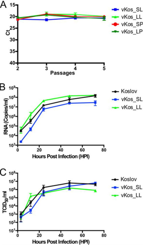
Virus yields during serial passaging and growth kinetics of vKos_SL and its variants in PK15 cells. (A) Assays for CSFV RNA, using reverse transcription-quantitative PCR (RT-qPCR), for the rescued vKos_SL, vKos_LL, vKos_SP, and vKos_LP variants following serial passages in PK15 cells (RNA levels are indicated as threshold cycle [CT] values). (B) Growth kinetics of CSFV strains Koslov, vKos_SL, and vKos_LL in PK15 cells measured by RT-qPCR assays (no. of viral RNA copies/ml) at 3, 12, 24, 48, and 72 h after infection. (C) Growth kinetics of CSFV strains Koslov, vKos_SL, and vKos_LL in PK15 cells measured by titration (TCID50/ml) of virus harvests prepared at 3, 12, 24, 48, and 72 h after infection.
Infection study I in pigs.
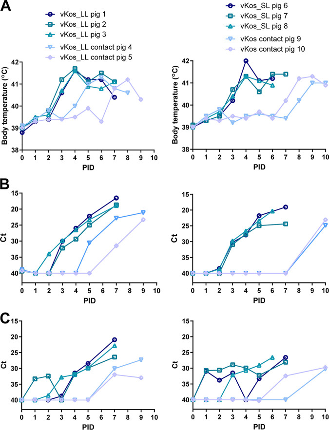
To elucidate the importance for the CSFV of residue S763 within pigs, virus challenge was performed in 2 groups of 3 pigs using intranasal inoculation with 106 50% tissue culture infective dose (TCID50) of vKos_LL or vKos_SL, respectively; in addition, each group included two noninoculated pigs, to assess contact transmission of the virus. The vKos_LL and vKos_SL each induced severe disease in the inoculated pigs and, due to pronounced clinical signs of CSF (including high fever, anorexia, dyspnea, ataxia, and convulsions), these pigs were euthanized, at the latest, by postinfection day 7 (PID7) (Fig. 2A). High fever (above 41°C) was observed as early as PID3 in pigs inoculated with vKos_LL and on PID4 in pigs inoculated with vKos_SL (Fig. 3A), coinciding with high viral RNA loads in both blood (Fig. 3B) and nasal swabs (Fig. 3C). The contact pigs in both groups displayed similar disease progression, but with a delayed onset of disease compared to that in the inoculated pigs (Fig. 3A, B, and C). The necropsies revealed gross morphological changes in the form of discoloration and/or necrosis within the tonsils of all infected pigs. Furthermore, several pigs (in both groups) had excess peritoneal fluid, petechial bleedings in the kidney, and hemorrhages in the spleen. Thus, the rescued viruses vKos_SL and vKos_LL are highly pathogenic in pigs, and changing residue 763 did not significantly affect either virus growth in the pigs or transmission to contact animals.
FIG 2.
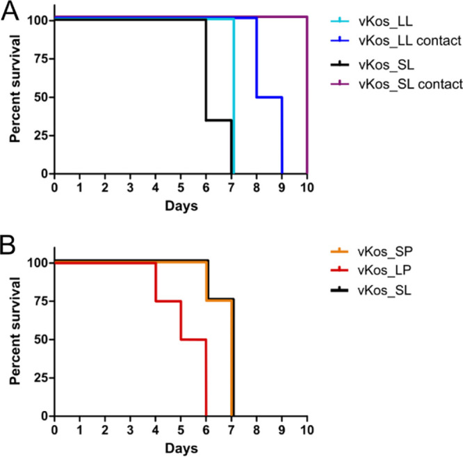
Mortality of pigs experimentally infected with rescued variants of the Koslov strain of CSFV. (A) Mortality of pigs inoculated with vKos_LL and vKos_SL and in “in-contact” animals in infection study I. (B) Mortality of pigs inoculated with vKos_SP, vKos_LP, and vKos_SL in infection study II. Note the significantly earlier mortality in the pigs infected with vKos_LP (see the main text). This earlier mortality was statistically significant (P < 0.0189) as assessed using the Mantel-Cox test.
FIG 3.
Time course of infection of pigs with vKos_LL and vKos_SL in inoculated and contact pigs. Pigs were inoculated with the indicated viruses on day 0, and contact pigs were kept with them throughout the experiment. (A) Body temperatures of pigs during the course of infection. (B) Levels of CSFV RNA in the blood of pigs as measured by RT-qPCR assays. Data are presented as CT values. (C) Levels of CSFV RNA in nasal swabs from pigs as measured by RT-qPCR assays. Data are presented as CT values.
Infection study II in pigs.
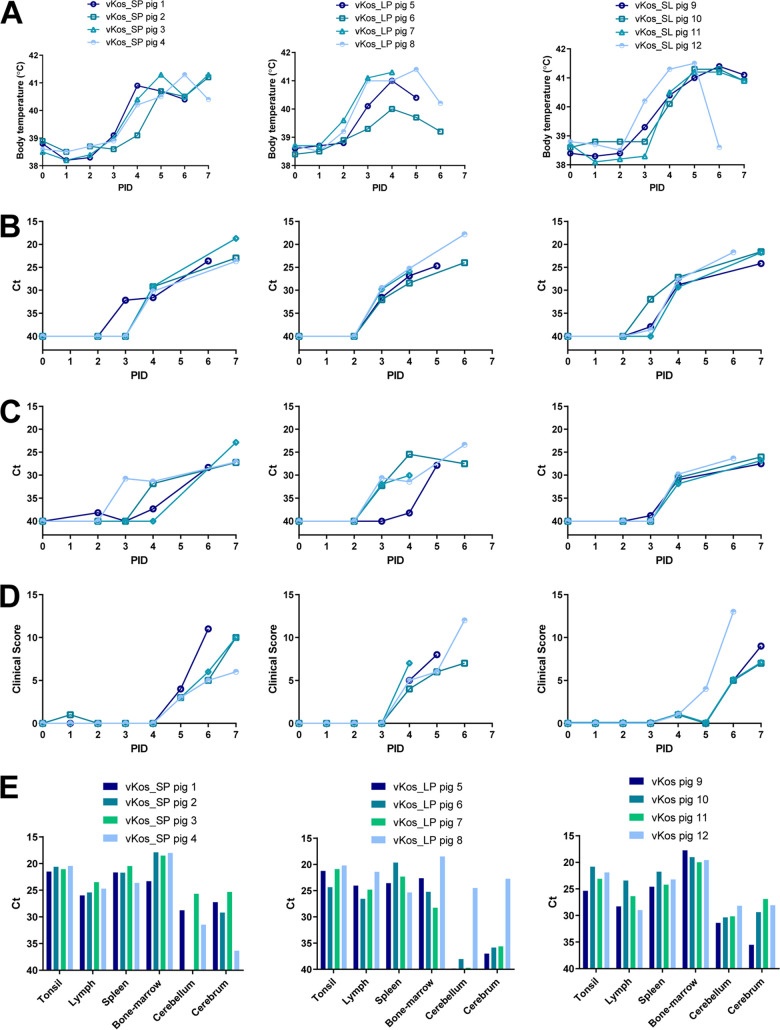
To elucidate the importance for the virus of residue L764 alone or in conjunction with residue S763, three groups of 4 pigs were challenged by intranasal inoculation, with 106 TCID50 of vKos_SP, vKos_LP, or vKos_SL, respectively. vKos_SP, vKos_LP, and vKos_SL each produced severe disease in the pigs and, due to pronounced clinical signs (as above), all inoculated pigs were euthanized at the latest by PID7 (Fig. 2B). The pigs inoculated with vKos_LP had to be euthanized 1 or 2 days earlier than pigs inoculated with vKos_SL and vKos_SP, as they developed earlier (but similar) symptoms of CSF (Fig. 4A), as also shown by the clinical scores (Fig. 4D). The reduced survival time of the pigs inoculated with the vKos_LP compared to those inoculated with vKos_SL was shown to be statistically significant (P < 0.0189) using the Mantel-Cox test. Consistent with the earlier mortality due to vKos_LP, high fever (above 41°C) was observed as early as PID3 in pigs inoculated with vKos_LP, while pigs inoculated with vKos_SL and vKos_SP had high fever from PID4 and PID5, respectively (Fig. 4A). In each case, the fever coincided the appearance of clinical signs (Fig. 4D) and with high viral RNA loads in both blood (Fig. 4B) and nasal swabs (Fig. 4C). As in infection study I, necropsies revealed various gross pathological changes, which often involved discoloration and/or necrotic lesions within the tonsils, excessive peritoneal fluid, and petechial bleedings in the kidney.
FIG 4.
Time course of infection of pigs with vKos_SP, vKos_LP, and vKos_SL. (A) Body temperatures of inoculated pigs. (B) Levels of CSFV RNA in the blood of pigs as measured by RT-qPCR assays; data are presented as CT values. (C) Levels of CSFV RNA in nasal swabs of pigs as measured by RT-qPCR assays; data are presented as CT values. (D) Clinical scores for pigs were assessed on a daily basis and scored as previously described (16). (E) Levels of CSFV RNA in tissues of pigs as measured by RT-qPCR assays; data are presented as CT values.
Reverse transcription-quantitative PCR (RT-qPCR) assays were performed on RNA extracted from tonsil, lymph nodes, spleen, bone marrow, cerebellum, and cerebrum from all pigs (Fig. 4E). Viral RNA was detected in nearly all tissues of all infected pigs, with high levels present in the tonsils, lymph nodes, spleen, and bone marrow. A lower level of viral RNA was detected in the cerebellum and cerebrum of most infected pigs compared to that in other tissues. This distribution is typical for highly virulent CSFVs that, in the acute form of the disease, produce a rapid-onset viremia with high viral loads in primary and secondary lymphatic tissues followed by infection of the CNS.
It is concluded that modifying residue 764 in conjunction with residue 763 can modify the properties of the virus in pigs. The vKos_LP variant produced symptoms more quickly, and the infected animals required earlier euthanasia.
Single-nucleotide polymorphism analyses (infection study I).
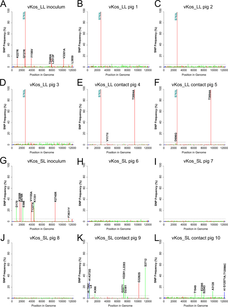
In order to elucidate the quasispecies composition of the virus in infected pigs, deep sequencing was performed on full-length cDNA amplicons derived from the vKos_LL and vKos_SL inocula used for infection of pigs, as well as from serum (collected on the day of euthanasia) of the inoculated and contact-infected pigs from each group (Fig. 5). In comparison to the consensus vKos_SL sequence, the vKos_LL inoculum had >99% presence of the L763 variant, as expected. In addition, this inoculum displayed seven other single-nucleotide polymorphisms (SNPs) at a frequency above 5% (but below 20%) in the coding region of the genome (Fig. 5A); these SNPs were detected at a frequency of <1%, or not at all, in the inoculated pigs, as well as in the contact pigs (Fig. 5B to F). The vKos_LL from serum samples displayed low-level sequence variation (up to 6% frequency) scattered across the genome as synonymous and nonsynonymous mutations in the inoculated pigs (Fig. 5B to D). The vKos_LL motif was maintained at >99% frequency in the virus within serum of infected pigs. The contact pigs both became infected with virus that contained a nonsynonymous amino acid substitution, T3004A (at >99% frequency), located in the NS5A coding region (Fig. 5E and F). This result appears indicative of a transmission bottleneck, as the substitution had been present at only low levels in the inoculated pigs (approximately 1%, 0.5%, and 4% in pigs 1, 2, and 3, respectively) and in the inoculum (<0.4%); it does not occur in sequences of any published CSFV strains (retrieved from GenBank [33]). This apparent bottleneck resulted in fewer SNPs scattered across the genome, and these occurred at rather lower frequencies (<2.5%) in the contact pigs compared to the inoculated pigs, except for SNP A2460G (amino acid D696G) which was unique for contact pig 5 (and occurred at around 4% frequency) and a unique SNP, G3884A (amino acid V1171I), in contact pig 4 (which was present at a level of around 3%).
FIG 5.
Single-nucleotide polymorphism (SNP) frequency plots across the entire genome of CSFV derived from inoculums and serum samples from infection study I. (A) vKos_LL inoculum. (B) vKos_LL pig 1 serum. (C) vKos_LL pig 2 serum. (D) vKos_LL pig 3 serum. (E) vKos_LL contact pig 4 serum. (F) vKos_LL contact pig 5 serum. (G) vKos_SL inoculum. (H) vKos_SL pig 6 serum. (I) vKos_SL pig 7 serum. (J) vKos_SL pig 8 serum. (K) vKos_SL contact pig 9 serum. (L) vKos_SL contact pig 10 serum. Note that the reference sequence in each case was the consensus bacterial artificial chromosome (BAC) clone Kos_SL sequence. Synonymous changes are indicated in green, nonsynonymous changes in red, and changes in the untranslated regions (UTRs) in blue. Dotted line indicates 5% frequency.
The vKos_SL inoculum for infection study I had a collection of fairly high-frequency SNPs (frequencies of up to 40%) scattered across the genome (Fig. 5G); strikingly, these were all present at a <5% frequency in the inoculated pigs (Fig. 5H to J) and thus seemed to be selected against in vivo. The virus in contact pig 9 contained one synonymous mutation (C11509T [amino acid S3712]; 56% frequency) at the consensus level (Fig. 5K). Additionally, 4 SNPs with frequencies between 8 and 9%, 1 SNP at 17%, and another 4 SNPs with frequencies between 27 and 34% were observed in this serum sample. Contact pig 10 contained a virus population with 6 SNPs occurring at frequencies of 7 to 13% (Fig. 5L), of which 4 are in the coding region. They occurred at <1%, or undetectably, in the other pigs. Interestingly, the L764P substitution was observed at 1 to 2% frequency in all vKos_SL-inoculated pigs and in contact pig 10 (as observed previously [17]). This substitution was also observed in the inoculum at a <1% frequency.
It is apparent that within infected pigs a less diverse population of viruses is present than can be observed in cell culture. During contact transmission from an infected animal to an uninfected pig, a clear “bottleneck” can result in the fixation of a previously minor variant in the population within the recipient.
SNP analysis (infection study II).
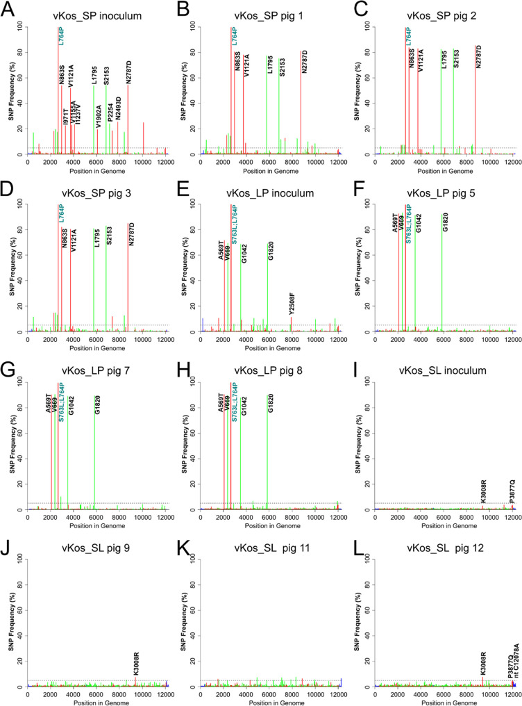
Deep sequencing was performed on the following full-length cDNA amplicons derived from the virus inoculums for study II (Fig. 6): vKos_SP, vKos_LP, and vKos_SL (note that the vKos_SL inoculum used in this experiment was derived from an independent rescue of the virus from RNA transcripts compared to that used in infection study I). The vKos_SL products displayed low-level variation (Fig. 6I), whereas vKos_SP and vKos_LP had multiple SNPs that included several with high frequencies (50 to 70%) in addition to the introduced changes encoding residues 763/764 (see Fig. 6A and E, respectively). As in infection study I, full-length cDNA amplicons were also derived from serum samples from three pigs in each group (vKos_SP pigs 1, 2, and 3; vKos_LP pigs 5, 7, and 8; and vKos_SL pigs 9, 11, and 12) and were then analyzed in the same way (see Fig. 6).
FIG 6.
SNP frequency plots across the entire genome of CSFV derived from inoculums and serum samples from infection study II. (A) vKos_SP inoculum. (B) vKos_SP pig 1 serum. (C) vKos_SP pig 2 serum. (D) vKos_SP pig 3 serum. (E) vKos_LP inoculum. (F) vKos_LP pig 5 serum. (G) vKos_LP pig 7 serum. (H) vKos_LP pig 8 serum. (I) vKos_SL inoculum. (J) vKos_SL pig 9 serum. (K) vKos_SL pig 11 serum. (L) vKos_SL pig 12 serum. Note that the reference sequence in each case is the consensus BAC clone Kos_SL sequence. Synonymous changes are indicated in green, nonsynonymous changes in red, and changes in the UTRs in blue. Dotted line indicates 5% frequency.
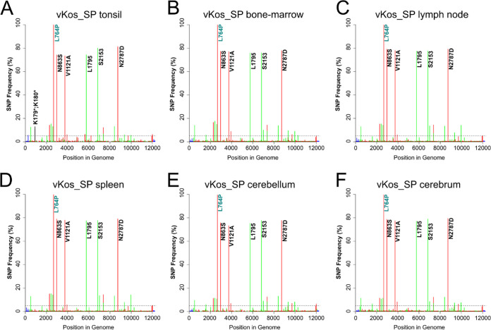
The vKos_SP in the inoculum and in serum of the infected pigs had multiple SNPs, in addition to the L764P change, scattered across the genome, with 5 SNPs being present at frequencies of >70%; these were (A2961G [amino acid N863S], T3735C [amino acid V1121A], T5756C [amino acid L1795], T6832C [amino acid S2153], and A8732G [amino acid N2787D]), which seemed to be associated with the L764P substitution (Fig. 6B to D). These SNPs were also present in the vKos_SP inoculum at above 50% frequency (Fig. 6A), together with a number of low-level SNPs that were not maintained in the pigs (Fig. 6B to D).
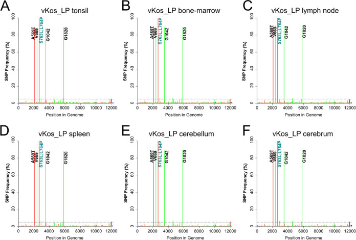
Similarly, vKos_LP, in addition to the S763L and L764P changes, also had low-frequency SNPs scattered across the entire genome in the serum of the inoculated pigs (Fig. 6F to H), and 4 SNPs were present at >80% frequency (G2078A [amino acid A569T], A2380G [amino acid V669], G3499A [amino acid G1042], and A5833G [amino acid G1820]). These changes appeared to be directly associated with the S763L and L764P (LP motif), since G2078A (amino acid A569T) and A2380G (amino acid V669) were linked in the sequencing reads. These SNPs were also present in the inoculum at >69% frequency and are frequent in published CSFV coding sequences (CDS).
As in infection study I, it is apparent that a less diverse population of viruses is present in the infected pigs than was observed in cell culture. Some SNPs can be maintained at high levels (>50%) but without reaching fixation. Particular clusters of major SNPs were present in association with the different substitutions at residue 764.
Quasispecies composition in different tissues of infected pigs.
To elucidate the quasispecies composition in different tissues, one pig from each group (vKos_SP pig 1, vKos_LP pig 8, and vKos_SL pig 12) was selected for in-depth characterization of SNPs in the viral RNA extracted from tonsils, lymph node, spleen, bone marrow, cerebellum, and cerebrum. In pig 12, no SNPs were present in the vKos_SL at a frequency greater than 10% in any of the different tissues; only 3 fairly low-frequency SNPS (about 5% frequency) were detected (data not shown) as in the serum (Fig. 6J to L).
In contrast, the vKos_LP in tissues of pig 8 (Fig. 7) had 4 high-frequency SNPs (about 80% frequency; one modified the codon for A569 [to make T569] and three were synonymous changes in the codons for residues V669, G1042, and G1820), in addition to the LP motif. Furthermore, there were a variety of additional SNPs at a frequency of about 5% in all tissues. The SNP A7896T (amino acid Y2508F) in vKos_LP occurred at approximately 11% frequency in the inoculum (Fig. 6E) but was not detectable in most tissues of the inoculated pigs (Fig. 7). Overall, it was apparent that, for each virus, the predominant virus populations in the different tissues were very similar and closely matched the population profile detected in serum (see Fig. 7 and 8); only the minor variants differed between the tissues. However, there were clear differences in the spectrum of SNPs present within the three different virus populations.
FIG 7.
SNP frequency plots of tissue samples from vKos_LP-inoculated pig 8 (infection study II). (A) vKos_LP tonsil. (B) vKos_LP bone marrow. (C) vKos_LP lymph node. (D) vKos_LP spleen. (E) vKos_LP cerebellum. (F) vKos_LP cerebrum. Note that the reference sequence in each case is the consensus BAC clone Kos_SL sequence. Synonymous changes are indicated in green, nonsynonymous changes in red, and changes in the UTRs in blue. Dotted line indicates 5% frequency.
FIG 8.
SNP frequency plots of tissue samples from vKos_SP-inoculated pig 1 (infection study II). (A) vKos_SP tonsil. (B) vKos_SP bone marrow. (C) vKos_SP lymph node. (D) vKos_SP spleen. (E) vKos_SP cerebellum. (F) vKos_SP cerebrum. Note the reference sequence in each case is the consensus BAC clone Kos_SL sequence. Synonymous changes are indicated in green, nonsynonymous changes in red, stop codons gained in black, and changes in the UTRs in blue. Dotted line indicates 5% frequency.
Surprisingly, the virus (vKos_SP) population in the tonsil of pig 1 contained stop codons in place of the codons encoding residues K179 and K180 (Fig. 8A), at frequencies of approximately 11 and 13%, respectively. The stop codon replacements for the codons for K179 and K180 (designated K179* and K180*, respectively) were only detected in the cerebellum and cerebrum at a frequency of approximately 1% each (Fig. 8E and F). The K179* and K180* variants were not detected in the vKos_SP inoculum (Fig. 6A). However, several other SNPs occurred at a level of approximately 20 to 25% in the vKos_SP inoculum but were only present at approximately 1% or less in all the pig tissues tested (Fig. 8), consistent with the pattern seen in serum of each inoculated pig (Fig. 6B, C, and D) and thus appeared to be selected against in vivo.
In samples from infection study II, vKos_SL had many low-level SNPs (all at <10% frequency) scattered across the entire genome in the inoculated pigs (Fig. 6J to L), with only 3 SNPs at a frequency above 5% (A9396G [amino acid K3000R], C12003A [amino acid P3877Q], and C12078A). The frequencies of these variants in the inoculum were approximately 3%, 4%, and undetectable, respectively (Fig. 6I). The virus population distribution within pig 12 was similar to that within pigs 9 and 11 (Fig. 6J to L), indicating that pig 12 was representative for the group. The absence of high-frequency SNPs detected in serum of each pig infected with the vKos_SL variant (Fig. 6J to L) and in tissues of pig 12 (data not shown) indicates that vKos_SL had a higher level of fitness in the hosts (i.e., it was well adapted to the host and hence able to replicate efficiently) compared to the other variants. Interestingly, the S763L substitution was present at a 1% frequency in the serum of inoculated pig 11. Furthermore, the L764P substitution was observed at a 0.4% frequency in the serum, tonsils, bone marrow, and lymph nodes of pig 12 and in the inoculum.
Virus haplotyping.
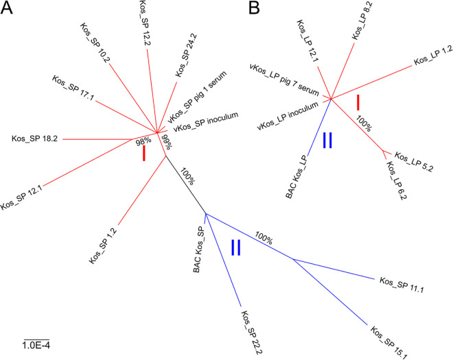
To gain further insight into the haplotypes which constitute the viral populations, unique full-length cDNA clones were generated from viral RNA present in the sera of vKos_SP pig 1 and vKos_LP pig 7. Sequencing of the individual vKos_SP-derived cDNA clones revealed the haplotypes present in the virus population and, when considered together, reflected the profile of the SNPs described above for the overall population. Out of the 10 cDNA clones examined, 6 contained the SNPs A2961G (amino acid N863S), T3735C (amino acid V1121A), T5756C (amino acid L1795), T6832C (amino acid S2153), and A8732G (amino acid N2787D), all together (Table 1). One other clone (Kos_SP1.2) contained these same SNPs with the exception of A8732G (Table 1). This shows that these SNPs are linked, as the majority of these individual SNPs were not observed in isolation in the cDNA clones. Thus, variants with these multiple SNPs represent the major haplotypes of the viral population. This is consistent with the SNP analysis of the uncloned fragments, which showed that all of these SNPs are present at a >75% frequency in the serum of pig 1 infected with vKos_SP (Table 1). Each of the full-length vKos_SP cDNA clone sequences was unique, with 3 to 12 mutations besides those seen in the major haplotypes of the vKos_SP viral population (data not shown). A total of 52 unique mutations were observed in the CDS of the 10 clones, with one causing a frameshift in the Erns encoding region. A phylogenetic tree for the vKos_SP cDNA clone sequences showed the population structure. The unrooted tree (see Fig. 9A) was mostly star-like, with a majority of cDNA sequences branching out from two nodes representing the vKos_SP inoculum/pig 1 serum sequences (haplotype I) and the BAC Kos_SP clone sequence (haplotype II), respectively. The majority of the cDNA clones were unique, forming two apparent clades representing the two major haplotypes (I and II) present in the population. The proportion of cDNA clones representing haplotype I (70%) was consistent with the frequency of the major SNPs observed in the SNP analysis (75 to 80%).
TABLE 1.
Identification of virus (vKos_SP) haplotypes present in serum
| cDNA clone | Nucleotide change (amino acid change)d
|
Haplotype | |||||
|---|---|---|---|---|---|---|---|
| T2664Ca (L764P) | A2961G (N863S) | T3735C (V1121A) | T5756C (L1795) | T6832C (S2153) | A8732G (N2787D) | ||
| Kos_SPb | + | − | − | − | − | − | II |
| Kos_SP 11.1c | + | − | − | − | − | − | II |
| Kos_SP 12.1c | + | + | + | + | + | + | I |
| Kos_SP 15.1c | + | − | − | − | − | − | II |
| Kos_SP 17.1c | + | + | + | + | + | + | I |
| Kos_SP 1.2c | + | + | + | + | + | − | I |
| Kos_SP 10.2c | + | + | + | + | + | + | I |
| Kos_SP 12.2c | + | + | + | + | + | + | I |
| Kos_SP 18.2c | + | + | + | + | + | + | I |
| Kos_SP 22.2c | + | − | − | − | − | − | II |
| Kos_SP 24.2c | + | + | + | + | + | + | I |
| vKos_SP inoculum | + (>99%) | + (54%) | + (52%) | + (54%) | + (54%) | + (54%) | |
| vKos_SP pig 1 serum | + (>99%) | + (80%) | + (78%) | + (77%) | + (75%) | + (81%) | |
Mutation introduced by site-directed mutagenesis.
BAC clone.
Individual serum-derived cDNA clone.
A minus sign (−) indicates that the variant was not detected in the cDNA and a plus sign (+) indicates that the variant was detected.
FIG 9.
Phylogenetic reconstruction of CSFV populations inferred from full-length cDNA clones by tree reconstruction using MrBayes (47). (A) Phylogenetic structure of the virus populations that were present in the vKos_SP-infected pig 1 serum inferred from the full-length cDNA clones, with distinct haplotypes I and II shown as colored clades (red and blue, respectively). Posterior probabilities of the branches between the nodes of >95% are shown. (B) Phylogenetic structure of the virus populations that were present in the vKos_LP-infected pig 7 serum inferred from the full-length cDNA clones, with the distinct haplotype I shown in red and the parental Kos_LP mutant sequence in blue. Posterior probabilities of the branches between the nodes of >95% are shown.
The 5 full-length cDNA clones obtained for vKos_LP reflected the major haplotype observed in the virus population, as they each contained the SNPs G2078A (amino acid A569T), A2380G (amino acid V669), G3499A (amino acid G1042), and A5833G (amino acid G1820) (Table 2). This, together with the linked reads, shows that there is a specific linkage between these SNPs, presumably indicating adaptation to accommodate the presence of the LP motif. This correlates with the SNP analysis where variants with these SNPs were present at >89% frequency in the serum of pig 7 infected with vKos_LP. Four out of the five vKos_LP cDNA clones were unique, with 2 to 6 mutations besides those seen in the major haplotypes of the vKos_LP viral population (data not shown). A total of 17 unique mutations were observed in these 5 clones. The unrooted phylogenetic tree for the vKos_LP cDNA clone sequences (Fig. 9B) was also star-like, with the cDNA sequences branching out from a single node representing the vKos_LP inoculum and pig 7 serum sequences (haplotype I). None of the serum-derived clones matched the BAC Kos_LP clone sequence (haplotype II). The majority of the cDNA clones formed no apparent clades and had a low degree of order, consistent with the high frequency of haplotype I (89 to 90%) present in the viral population and the small number of clones obtained to represent the population.
TABLE 2.
Identification of virus (vKos_LP) haplotypes present in serum
| cDNA clone | Nucleotide change (amino acid change)d
|
Haplotype | |||||
|---|---|---|---|---|---|---|---|
| G2078A (A569T) | A2380G (V669) | C2661Ta (S763L) | T2664Ca (L764P) | G3499A (G1042) | A5833G (G1820) | ||
| Kos_LPb | − | − | + | + | − | − | II |
| Kos_LP 12.1c | + | + | + | + | + | + | I |
| Kos_LP 1.2c | + | + | + | + | + | + | I |
| Kos_LP 5.2c | + | + | + | + | + | + | I |
| Kos_LP 6.2c | + | + | + | + | + | + | I |
| Kos_LP 8.2c | + | + | + | + | + | + | I |
| vKos_LP inoculum | + (71%) | + (69%) | + (>99%) | + (>99%) | + (69%) | + (69%) | |
| vKos_LP pig 7 serum | + (90%) | + (90%) | + (>99%) | + (>99%) | + (89%) | + (89%) | |
Mutation introduced by site-directed mutagenesis.
BAC clone.
Individual serum-derived cDNA clone.
A minus sign (−) indicates that the variant was not detected in the cDNA and a plus sign (+) indicates that the variant was detected.
These studies clearly show the value of preparing and sequencing long cDNA clones to identify the distribution of specific SNPs among particular haplotypes within the virus population.
DISCUSSION
Each of the variants of the highly virulent Koslov strain of CSFV was successfully rescued from BAC clones. The vKos_LL variant of the highly virulent Koslov strain of CSFV displayed similar growth kinetics in PK15 cells to those of the rescued consensus sequence virus (vKos_SL) and the wt (uncloned) Koslov virus (with S763), strongly indicating that the S763L substitution had no influence on virus growth in vitro (Fig. 1B). Detailed analyses of the growth kinetics of vKos_SP and vKos_LP were not performed; however, they grew to similar levels as vKos_SL during serial passaging in PK15 cells (Fig. 1A). Thus, these substitutions at residues 763 and 764 apparently have no major effect on growth rate in vitro.
All pigs experimentally infected with vKos_SL and the different variants with substitutions at residues 763 and/or 764 displayed similar clinical signs of disease. However, the vKos_LP variant induced significantly earlier (albeit no more severe) clinical signs of disease as judged by nearly all parameters. The importance of residues 763 and 764 in modulation of the speed of virus transmission to other animals warrants further study.
Deep sequencing analysis of the viruses present within infected pigs revealed that vKos_SL and vKos_LL had high diversity (albeit at a low level) spread across the entire ORF in inoculated pigs with no signs of virus adaptation. However, two contact pigs that became infected with vKos_LL had virus that contained a nonsynonymous mutation (resulting in substitution T3004A) at >99% frequency. This SNP likely rose to fixation in the virus population due to a bottleneck effect, as it was only present at a low frequency in the inoculated pigs. Furthermore, there was a difference in the virus population distribution between the inoculated pigs and the contact pigs for vKos_LL (see Fig. 5B to F) with a loss of diversity and adaptation in the NS protein coding regions. This is probably again due to the transmission to the contact pigs being a bottleneck, resulting in a founder effect with a reduced amount of genetic heterogeneity in the viral population.
Both the vKos_SP and vKos_LP variants displayed lower diversity across the entire ORF compared to that in the vKos_SL-inoculated pigs (Fig. 6). However, in contrast to the results with vKos_SL, the vKos_SP inoculum contained several nonsynonymous and synonymous changes at the consensus level (i.e., >50%). Three changes were nonsynonymous (resulting in substitutions N863S, V1121A, and N2787D) and located in the coding regions for E2, p7, and NS5A, respectively. Residues V1121 and N2787 are highly conserved in the genomes of CSFVs (>99% and 98%, respectively), while N863 is observed at 35% frequency (30, 31). These SNPs existed at a frequency of 52 to 54% in the inoculum and at a higher frequency (>70%) in the viruses collected from infected pigs. Interestingly, the S763/P764 variant, in conjunction with residue E761, is not observed in the CSF database (30, 31), and it seems to be a somewhat artificial combination, as only R761 is observed together with S763 and P764. This might explain the collection of mutations that were also found in this virus. Variants with low or neutral advantage can be maintained at a higher than expected frequency due to being linked to a high-fitness variant that is well represented in the virus population (4). The synonymous mutations in the vKos_SP variant may have a possible effect on the secondary RNA structure (34) or may just be neutral passengers carried along with the nonsynonymous mutations.
The rescued vKos_LP acquired 4 additional mutations at the consensus level, one of which was a nonsynonymous mutation G2078A (amino acid A569T), which appeared to be linked to A2380G (amino acid V669). Both the A569 and T569 variants are frequently observed (36% and 62%, respectively) in published CSFV strains. Residue 569 is located in E1, within a putative fusion peptide-like motif (residues 551 to 579) consisting of hydrophobic residues (35). A similar stretch of hydrophobic residues is found in the E1 protein of hepatitis C virus, another member of the Flaviviridae, and this is believed to play a role in membrane fusion (36, 37). A shift from a small hydrophobic residue to an uncharged residue with a hydroxyl group could have an impact on this motif and its potential role in fusion. The increase in frequency of these four SNPs in the infected animals indicates that they are either advantageous for the virus or are coupled to an advantageous variant. Interestingly, stop codons were observed in the virus population present in the tonsils in vKos_LP-infected pigs at a >10% frequency, indicating that some sort of complementation must take place at the quasispecies level to maintain such a high frequency of deleterious mutations. Members of the quasispecies should not be considered independently acting variants, as the internal interactions of the variants determine the overall behavior of the quasispecies population (5).
Only low-level SNPs (<7% SNP frequency) were found to be scattered across the genome of vKos_SL in the infected pigs in study II, consistent with the results from the infection study I, albeit using a different virus inoculum. Together, these data strongly indicate that vKos_SL is highly stable at the quasispecies level in pigs, displaying a high level of fitness, as is vKos_LL, which also seems to be a common variant of wt Koslov, as seen in a separate study (38). In that study, the S763L substitution was observed together with 9 silent SNPs present at an approximately 40% frequency in the inoculum derived from blood of pigs infected with the wt Koslov (CSFV/1.1/dp/CSF0382/XXXX/Koslov), and the substitution S763L was also present at frequencies of 59% and 19% in the tonsils and serum, respectively. Furthermore, several E2 sequences of wt Koslov in the CSF database also contain this version (LL) of the motif.
CSFV isolates can be expected to consist of a much more diverse quasispecies than the viruses rescued from unique cDNA clones, as used in the current study, as reported previously (39). Many of the SNPs detected in the inocula and different tissues of inoculated pigs were also present in the viral RNA extracted from the respective sera. This is expected since the virus in serum is likely a collection of all the viruses present in the different tissues. The virus spreads through the peripheral blood from its primary site of replication in the tonsils to the regional lymph nodes, then to the bone marrow, visceral lymph nodes, and lymphoid structures associated with the small intestine and spleen (40).
Interestingly, S763L was detected (at a low level) in the serum of pig 11 inoculated with vKos_SL, and this change has previously been suggested, together with P968H, to contribute to the difference in virulence between rescued viruses vKos_SL and the attenuated vKos_3aa (17). In addition, the L764P substitution was observed at a low frequency (<2%) in the serum of most pigs infected with vKos_SL from infection study I and also at a lower frequency (<0.4%) in several tissues of pig 12 infected with vKos_SL from infection study II. In our previous study with vKos_SL, L764P was detected at a 2% frequency in inoculated pigs and fixed in one contact pig. Several studies indicate that residue 764 is under positive selection (41, 42). This appears to be consistent with the greater speed of infection observed for the vKos_LP variant in pigs (Fig. 2B and Fig. 4A to D).
The SNP analyses indicated that the virus variants studied here are each able to enter, replicate, and spread within different tissues, and that the quasispecies composition remains nearly the same across all tissues, with some compartmentalization observed only in the tonsils. A similar tendency was observed for wt Koslov in a previous study, where one specific virus variant was present at a much higher level in the tonsils of naive pigs compared to those of vaccinated pigs (38). The lack of distinct differences in SNP profiling across the tissues is in contrast to that of BVDV in PI animals, in which the viral population exhibits tissue specific compartmentalization (8). However, in the PI host, the virus replicates in the absence of selective pressure from the adaptive immune system (43), and given the nature of PI animals, the virus has had more time to establish itself and evolve. This is consistent with observations from PI animals with Hobi-like atypical bovine pestivirus that display an increase in variability of the viral quasispecies over time (44). As all of the vKos variants tested here are highly virulent, with infected pigs being euthanized no later than PID7, the virus populations do not have much time to evolve separately in the different tissues, and the results seen in this study could also be influenced by the presence of blood in each tissue. The blood should contain the same pool of viruses seen in serum as discussed above. In a previous study, the virus population in pigs chronically infected with CSFV appeared to be stable at the quasispecies level (39); however, these results were based purely on blood and leukocyte samples.
To gain deeper insight into the haplotypes that make up the virus population, full-length cDNA clones were generated and sequenced. The spectrum of sequences from these individual cDNA clones derived from the sera of pigs infected with vKos_SP and vKos_LP described the major haplotypes present in the viral population and, when considered together, matched the virus population structure. Both phylogenetic reconstructions displayed low degrees of order, most likely due to the use of clone-derived virus inocula for the infection experiments. This result is in agreement with those of our earlier studies, in which a low degree of order was seen in the phylogenetic reconstruction of cDNA clones from vKos_SL-infected serum (45), whereas, in contrast, a high degree of order was seen with cDNA clones obtained from a field isolate of the moderately virulent CSFV strain Roesrath (46).
In summary, we have analyzed the characteristics within infected pigs of CSFVs that express mutant E2 proteins and determined the virus population dynamics during the infection. The variants exhibited similar disease progression, but with significantly earlier signs of CSFV infection observed for the vKos_LP variant. A clear bottleneck effect in the transmission of viruses to contact pigs was observed. This resulted in loss of virus diversity and fixation of mutations in the populations that were present in the infected pigs. No apparent compartmentalization of different virus populations was observed across infected tissues in inoculated pigs. The production and sequencing of individual full-length cDNA clones derived from infected serum revealed the major haplotypes present in the viral populations and, when considered together, reflected the profile of SNPs detected in the whole population. These studies indicate the nature of virus population change during infection within individual animals and resulting from virus transmission between animals.
MATERIALS AND METHODS
Primers.
Oligonucleotide primers used are listed in Table 3.
TABLE 3.
List of oligonucleotide primers used in this study
| Primer | Sequence (5′→3′) | Reference or source |
|---|---|---|
| CSF-cDNA-1 | GGG CCG TTA GGA AAT TAC CTT AG | 45 |
| CSF-Kos_NotI-T7-1-59 | TCT ATA TGC GGC CGC TAA TAC GAC TCA CTA TAG TAT ACG AGG TTA GTT CAT TCT CGT ATG CAT GAT TGG ACA AAT CAA AAT TTC AAT TTG G | 48 |
| CSF-Kos_1-59 | GTA TAC GAG GTT AGT TCA TTC TCG TAT GCA TGA TTG GAC AAA TCA AAA TTT CAA TTT GG | Modified from reference 48 |
| CSF-Kos_12313aR | GGG CCG TTA GGA AAT TAC CTT AGT CCA ACT GT | 48 |
| CSF-Kos-6240-RT | TCT ATA GGG TGT TTC TGC CC | 38 |
| CSF-Kos-6176-R | CTG GTG TTG CGG TCA TGG CTA CTA C | 38 |
| CSF-Kos-5981-F | GGG GAG ATG AAA GAA GGG GAC ATG | 38 |
| CSFV-Kos-ELL-F | GGC ATC ACT GCA TAA GGA GGC TTT ACT CAC TTC CGT GAC | This study |
| CSFV-Kos-ESP-F | GGC ATC ACT GCA TAA GGA GGC TTC ACC CAC TTC CGT GAC | This study |
| CSFV-Kos-ELP-F | GGC ATC ACT GCA TAA GGA GGC TTT ACC CAC TTC CGT GAC | This study |
| Kos-2720R | CCG AAC CCG AAG TCA TCT CCC AT | This study |
Cells.
Porcine kidney (PK15) cells (obtained from the Cell Culture Collection at the Friedrich-Loeffler-Institut, Germany) were propagated in Dulbecco’s modified Eagle’s medium (DMEM) containing 5% fetal calf serum (FCS).
Generation of BACs containing nonsynonymous mutations.
The BACs Kos_LL (C2661T;S763L), Kos_SP (T2664C;L764P), and Kos_LP (C2661T;S763P/T2664C;L764P) were produced by site-directed mutagenesis using a megaprimer approach (47). Briefly, the BAC clone Kos (GenBank accession number KF977607; a full-length cDNA clone of the CSFV strain Koslov [CSFV/1.1/dp/CSF0382/XXXX/Koslov; GenBank accession number HM237795.1 (48)], here called Kos_SL) (17), was used as the template for the megaprimer PCRs with forward primers Kos-ELL-F, CSFV-Kos-ESP-F, and CSFV-Kos-ELP-F and reverse primer Kos-2720R (see Table 3). The megaprimers (105 bp), after gel purification with a GeneJet Gel extraction kit (Thermo Scientific), were used for the site-directed mutagenesis with Kos_SL as the template, and the products were transformed into Escherichia coli DH10B (Invitrogen, Carlsbad, CA). BAC DNAs were purified from 4-ml overnight cultures using the Zymo BAC DNA miniprep kit (Zymo Research). Full-length amplicons were obtained by long PCR, as previously described (17, 49), using the BAC DNAs as the template and primers CSF-Kos_Not1-T7-1-59 and CSF-Kos_12313aR (Table 3). PCR products were purified using a GeneJet PCR purification kit (Thermo Scientific) and in vitro transcribed using the MEGAscript T7 kit (Invitrogen).
Rescue of viruses.
Viruses were rescued from RNA transcripts following electroporation of PK15 cells, as described previously (50), and passaged once in PK15 cells. Titrations were performed in triplicate, using polyclonal porcine antiserum for staining of infected cells.
Infection study I.
In total, 10 crossbred pigs (14 weeks of age) obtained from a high health status swine herd (51) were used for the infection study. For both viruses, three pigs were inoculated with vKos_SL or vKos_LL via the intranasal route with a defined virus dose (106 TCID50), and two in-contact pigs in each group were mock inoculated with cell culture medium. Rectal body temperature and clinical signs were monitored on a daily basis and scored as previously described (16). At predefined days (PID0, 1, 2, 3, 4, 5, 7, 10, and 14), EDTA-blood and serum samples were collected for virological examination. Furthermore, nasal swabs were obtained on the same sampling days, and the viral RNA from these and from EDTA-blood was purified using a MagNA Pure LC total nucleic acid isolation kit (Roche). RT-qPCR was used to determine the level of viral RNA as described previously (52). At the end of the experiment, or before for animal welfare reasons if the preset humane endpoint (HEP) criteria were reached, pigs were euthanized with intravascular injection of pentobarbital. All pigs were subjected to necropsy and gross pathological examination after euthanasia.
Infection study II.
In total, 12 weaner pigs (6 weeks of age) obtained from a conventional Danish swine herd with specific pathogen-free status were used for the infection study. In each group, four pigs were inoculated with vKos_SL, vKos_SP, or vKos_LP, respectively, via the intranasal route with a defined virus dose (106 TCID50). Rectal body temperature and clinical signs were monitored on a daily basis and scored as previously described (16). At predefined days (PID0, 2, 3, 4, 7 and 10), EDTA-blood and serum samples were collected for virological examination as above. Nasal swabs were obtained on the same days, and tissue samples from tonsils, lymph nodes, spleen, bone marrow, cerebellum, and cerebrum were collected during postmortem examination after euthanasia (as above). RNA from collected samples was extracted, and the level of viral RNA was determined using RT-qPCR as described previously (52). Pigs 4, 6, and 10 (one from each group) were not included in the sequencing data set, as they were euthanized together with the second-to-last pig in their group for animal welfare reasons and not due to HEP criteria.
The survival curves of the pigs were analyzed using the Mantel-Cox test within GraphPad Prism v7.0 software (La Jolla, CA).
For both infection studies I and II, all experimental procedures, euthanasia, animal care, and maintenance were conducted in accordance with Danish and European Union (EU) legislation (Consolidation Act 474 15/05/2014 and EU Directive 2010/63/EU) and with approval from the Danish Animal Experimentation Inspectorate (license number 2012-15-2934-00182).
Preparation of cDNA from viral RNA.
RNA was extracted using a combined TRIzol/RNeasy protocol (53) from tonsils, bone marrow, serum, lymph nodes, spleen, cerebellum, and cerebrum of pigs on the day of euthanasia. This extracted RNA was used to generate full-length or two overlapping half-length cDNA amplicons, using a modified version of the long RT-PCR method described previously (17, 49). Briefly, the total RNA was reverse transcribed using Maxima H Minus reverse transcriptase (Thermo Scientific) and the specific cDNA primers CSF-cDNA-1 and CSF-Kos-6240-RT (Table 3). The cDNA was then amplified by long PCR using AccuPrime high-fidelity DNA polymerase (Thermo Scientific) with primers CSF-Kos_NotI-T7-1-59 and CSF-Kos_12313aR for full-length amplicons, CSF-Kos_NotI-T7-1-59 and CSF-Kos-6176R for the 5′ end, and CSF-Kos-5981-F and CSF-Kos_12313aR for the 3′ end of the half-length cDNA amplicons.
Cloning using the pCR XL-2-TOPO vector.
Full-length cDNA amplicons, representing vKos_SP and vKos_LP, were obtained from extracted RNA from the serum of infected animals (vKos_SP pig 1 and vKos_LP pig 7). Briefly, cDNA was generated as above, and full-length viral cDNA was produced by long PCR, using the Q5 high-fidelity DNA polymerase kit (New England BioLabs) and primers CSF-Kos_1-59 and CSF-Kos_12313aR. The products were gel purified with a GeneJet gel extraction kit (Thermo Scientific) and quantified using a Qubit fluorometric quantitation system (Thermo Scientific). They were inserted into the pCR-XL-2-TOPO vector using the TOPO XL-2 kit (Thermo Scientific) as previously described (45) and transformed into E. coli. Plasmid DNA was prepared from individual colonies and sequenced as described below.
Sequencing.
Full-length and half-length amplified cDNA amplicons and cDNA clones were sequenced at the DTU Multi-Assay Core (DMAC, Kongens Lyngby, Denmark) using Nextera XT DNA library preparation and the MiSeq system (Illumina, San Diego, CA). Consensus sequences were obtained by mapping the reads to the vKos_SL reference sequence (GenBank accession number KF977607.1) using CLC Genomics Workbench v.9.5.2 (CLC bio, Aarhus, Denmark). Consensus sequences were aligned using MAFFT in Geneious (Biomatters, Auckland, New Zealand). Low-frequency SNPs (>0.1%) were identified for cDNA amplicons using a combination of BWA, SAMtools, Lo-Freq-snp-caller (default minimum coverage of 10), and SnpEff, as described previously (17, 46). Phylogeny was constructed using MrBayes v3.2.1 (54, 55) on full-length cDNA sequence alignments (general time-reversible [GTR] model, number of substitution types [nst] = 6). The Markov chain Monte Carlo algorithm was run for 10,000,000 iterations, with a sampling frequency of 7,000, using two independent runs with three chains each, in order to check for convergence. Burn-in was set at 25% of samples. The consensus tree was visualized in FigTree v1.4.3.
Data availability.
All relevant data are included in this published article. The data sets used and/or analyzed during the current study are available from the corresponding author on request.
ACKNOWLEDGMENTS
We thank the laboratory technicians and animal caretakers at Lindholm for their invaluable work during the infection studies. We thank Jens Nielsen for management of infection study I. We are grateful for excellent assistance from Marlene D. Dalgaard (DMAC, DTU).
This study was funded by internal resources from the National Veterinary Institute within the Technical University of Denmark and the University of Copenhagen. U.F. was supported by the Weimann Foundation and by the Independent Research Fund Denmark (grant 4004-00598 to J.B.) and the Danish Cancer Society (grant R204-A12639 to J.B.).
T.B.R., U.F., L.L., G.J.B., and C.M.J. conceived the study and developed the approach. T.B.R., U.F., and L.L. carried out the infection studies. C.M.J. carried out the haplotype cloning experiments. T.B.R., U.F., and C.M.J. carried out sequence analyses. All authors contributed to result interpretation. All authors contributed to the drafting and revision of the manuscript. All authors read and approved the final manuscript.
REFERENCES
- 1.Lindenbach BD, Thiel HJ, Rice CM, Murray CL, Thiel HJ, Rice CM. 2013. Flaviviridae: the viruses and their replication, p 712–746. In Fields virology, 6th ed. Lippincott Williams & Wilkins, New York, NY. [Google Scholar]
- 2.Collett MS, Anderson DK, Retzel E. 1988. Comparisons of the pestivirus bovine viral diarrhoea virus with members of the Flaviviridae. J Gen Virol 69:2637–2643. doi: 10.1099/0022-1317-69-10-2637. [DOI] [PubMed] [Google Scholar]
- 3.Meyers G, Rümenapf T, Thiel HJ. 1989. Molecular cloning and nucleotide sequence of the genome of hog cholera virus. Virology 171:555–567. doi: 10.1016/0042-6822(89)90625-9. [DOI] [PubMed] [Google Scholar]
- 4.Lauring AS, Andino R. 2010. Quasispecies theory and the behavior of RNA viruses. PLoS Pathog 6:e1001005. doi: 10.1371/journal.ppat.1001005. [DOI] [PMC free article] [PubMed] [Google Scholar]
- 5.Domingo E, Sheldon J, Perales C. 2012. Viral quasispecies evolution. Microbiol Mol Biol Rev 76:159–216. doi: 10.1128/MMBR.05023-11. [DOI] [PMC free article] [PubMed] [Google Scholar]
- 6.Vignuzzi M, Stone JK, Arnold JJ, Cameron CE, Andino R. 2006. Quasispecies diversity determines pathogenesis through cooperative interactions in a viral population. Nature 439:344–348. doi: 10.1038/nature04388. [DOI] [PMC free article] [PubMed] [Google Scholar]
- 7.Bordería AV, Isakov O, Moratorio G, Henningsson R, Agüera-González S, Organtini L, Gnädig NF, Blanc H, Alcover A, Hafenstein S, Fontes M, Shomron N, Vignuzzi M. 2015. Group selection and contribution of minority variants during virus adaptation determines virus fitness and phenotype. PLoS Pathog 11:e1004838. doi: 10.1371/journal.ppat.1004838. [DOI] [PMC free article] [PubMed] [Google Scholar]
- 8.Dow N, Chernick A, Orsel K, van Marle G, van der Meer F. 2015. Genetic variability of bovine viral diarrhea virus and evidence for a possible genetic bottleneck during vertical transmission in persistently infected cattle. PLoS One 10:e0131972. doi: 10.1371/journal.pone.0131972. [DOI] [PMC free article] [PubMed] [Google Scholar]
- 9.Peck KM, Lauring AS. 2018. The complexities of viral mutation rates. J Virol 92:e01031-17. doi: 10.1128/JVI.01031-17. [DOI] [PMC free article] [PubMed] [Google Scholar]
- 10.Lowings P, Ibata G, Needham J, Paton D. 1996. Classical swine fever virus diversity and evolution. J Gen Virol 77:1311–1321. doi: 10.1099/0022-1317-77-6-1311. [DOI] [PubMed] [Google Scholar]
- 11.Deng M-C, Huang C-C, Huang T-S, Chang CY, Lin Y-J, Chien M-S, Jong M-H. 2005. Phylogenetic analysis of classical swine fever virus isolated from Taiwan. Vet Microbiol 106:187–193. doi: 10.1016/j.vetmic.2004.12.014. [DOI] [PubMed] [Google Scholar]
- 12.Paton DJ, McGoldrick A, Greiser-Wilke I, Parchariyanon S, Song JY, Liou PP, Stadejek T, Lowings JP, Björklund H, Belák S. 2000. Genetic typing of classical swine fever virus. Vet Microbiol 73:137–157. doi: 10.1016/s0378-1135(00)00141-3. [DOI] [PubMed] [Google Scholar]
- 13.Postel A, Schmeiser S, Perera CL, Rodríguez LJP, Frias-Lepoureau MT, Becher P. 2013. Classical swine fever virus isolates from Cuba form a new subgenotype 1.4. Vet Microbiol 161:334–338. doi: 10.1016/j.vetmic.2012.07.045. [DOI] [PubMed] [Google Scholar]
- 14.Floegel-Niesmann G, Blome S, Gerss-Dülmer H, Bunzenthal C, Moennig V. 2009. Virulence of classical swine fever virus isolates from Europe and other areas during 1996 until 2007. Vet Microbiol 139:165–169. doi: 10.1016/j.vetmic.2009.05.008. [DOI] [PubMed] [Google Scholar]
- 15.Leifer I, Ruggli N, Blome S. 2013. Approaches to define the viral genetic basis of classical swine fever virus virulence. Virology 438:51–55. doi: 10.1016/j.virol.2013.01.013. [DOI] [PubMed] [Google Scholar]
- 16.Mittelholzer C, Moser C, Tratschin JD, Hofmann MA. 2000. Analysis of classical swine fever virus replication kinetics allows differentiation of highly virulent from avirulent strains. Vet Microbiol 74:293–308. doi: 10.1016/S0378-1135(00)00195-4. [DOI] [PubMed] [Google Scholar]
- 17.Fahnøe U, Pedersen AG, Risager PC, Nielsen J, Belsham GJ, Höper D, Beer M, Rasmussen TB. 2014. Rescue of the highly virulent classical swine fever virus strain “Koslov” from cloned cDNA and first insights into genome variations relevant for virulence. Virology 468–470:379–387. doi: 10.1016/j.virol.2014.08.021. [DOI] [PubMed] [Google Scholar]
- 18.Hulst MM, Moormann R. 1997. Inhibition of pestivirus infection in cell culture by envelope proteins Erns and E2 of classical swine fever virus: Erns and E2 interact with different receptors. J Gen Virol 78:2779–2787. doi: 10.1099/0022-1317-78-11-2779. [DOI] [PubMed] [Google Scholar]
- 19.van Gennip HGP, van Rijn PA, Widjojoatmodjo MN, de Smit AJ, Moormann R. 2000. Chimeric classical swine fever viruses containing envelope protein Erns or E2 of bovine viral diarrhoea virus protect pigs against challenge with CSFV and induce a distinguishable antibody response. Vaccine 19:447–459. doi: 10.1016/S0264-410X(00)00198-5. [DOI] [PubMed] [Google Scholar]
- 20.Liang D, Fernandez-Sainz I, Ansari IH, Gil L, Vassilev V, Donis RO. 2003. The envelope glycoprotein E2 is a determinant of cell culture tropism in ruminant pestiviruses. J Gen Virol 84:1269–1274. doi: 10.1099/vir.0.18557-0. [DOI] [PubMed] [Google Scholar]
- 21.Wang Z, Nie Y, Wang P, Ding M, Deng H. 2004. Characterization of classical swine fever virus entry by using pseudotyped viruses: E1 and E2 are sufficient to mediate viral entry. Virology 330:332–341. doi: 10.1016/j.virol.2004.09.023. [DOI] [PubMed] [Google Scholar]
- 22.Postel A, Schmeiser S, Bernau J, Meindl-Boehmer A, Pridotkas G, Dirbakova Z, Mojzis M, Becher P. 2012. Improved strategy for phylogenetic analysis of classical swine fever virus based on full length E2 encoding sequences. Vet Res 43:50. doi: 10.1186/1297-9716-43-50. [DOI] [PMC free article] [PubMed] [Google Scholar]
- 23.Risatti GR, Borca MV, Kutish GF, Lu Z, Holinka LG, French RA, Tulman ER, Rock DL. 2005. The E2 glycoprotein of classical swine fever virus is a virulence determinant in swine. J Virol 79:3787–3796. doi: 10.1128/JVI.79.6.3787-3796.2005. [DOI] [PMC free article] [PubMed] [Google Scholar]
- 24.Risatti GR, Holinka LG, Carrillo C, Kutish GF, Lu Z, Tulman ER, Sainz IF, Borca MV. 2006. Identification of a novel virulence determinant within the E2 structural glycoprotein of classical swine fever virus. Virology 355:94–101. doi: 10.1016/j.virol.2006.07.005. [DOI] [PubMed] [Google Scholar]
- 25.Risatti GR, Holinka LG, Fernandez-Sainz I, Carrillo C, Kutish GF, Lu Z, Zhu J, Rock DL, Borca MV. 2007. Mutations in the carboxyl terminal region of E2 glycoprotein of classical swine fever virus are responsible for viral attenuation in swine. Virology 364:371–382. doi: 10.1016/j.virol.2007.02.025. [DOI] [PubMed] [Google Scholar]
- 26.Risatti GR, Holinka LG, Fernandez Sainz I, Carrillo C, Lu Z, Borca MV. 2007. N-linked glycosylation status of classical swine fever virus strain Brescia E2 glycoprotein influences virulence in swine. J Virol 81:924–933. doi: 10.1128/JVI.01824-06. [DOI] [PMC free article] [PubMed] [Google Scholar]
- 27.van Gennip HGP, Vlot AC, Hulst MM, De Smit AJ, Moormann R. 2004. Determinants of virulence of classical swine fever virus strain Brescia. J Virol 78:8812–8823. doi: 10.1128/JVI.78.16.8812-8823.2004. [DOI] [PMC free article] [PubMed] [Google Scholar]
- 28.Tamura T, Sakoda Y, Yoshino F, Nomura T, Yamamoto N, Sato Y, Okamatsu M, Ruggli N, Kida H. 2012. Selection of classical swine fever virus with enhanced pathogenicity reveals synergistic virulence determinants in E2 and NS4B. J Virol 86:8602–8613. doi: 10.1128/JVI.00551-12. [DOI] [PMC free article] [PubMed] [Google Scholar]
- 29.Chang CY, Huang CC, Deng MC, Huang YL, Lin YJ, Liu HM, Lin YL, Wang FI. 2012. Antigenic mimicking with cysteine-based cyclized peptides reveals a previously unknown antigenic determinant on E2 glycoprotein of classical swine fever virus. Virus Res 163:190–196. doi: 10.1016/j.virusres.2011.09.019. [DOI] [PubMed] [Google Scholar]
- 30.Greiser-Wilke I, Zimmermann B. 2015. The CSF database of the European Community Reference Laboratory. http://viro60.tiho-hannover.de/eg/csf/.
- 31.Postel A, Schmeiser S, Zimmermann B, Becher P. 2016. The European Classical Swine Fever Virus Database: blueprint for a pathogen-specific sequence database with integrated sequence analysis tools. Viruses 8:302. doi: 10.3390/v8110302. [DOI] [PMC free article] [PubMed] [Google Scholar]
- 32.Leifer I, Hoeper D, Blome S, Beer M, Ruggli N. 2011. Clustering of classical swine fever virus isolates by codon pair bias. BMC Res Notes 4:521. doi: 10.1186/1756-0500-4-521. [DOI] [PMC free article] [PubMed] [Google Scholar]
- 33.Benson DA, Cavanaugh M, Clark K, Karsch-Mizrachi I, Lipman DJ, Ostell J, Sayers EW. 2013. GenBank. Nucleic Acids Res 41:D36–D42. doi: 10.1093/nar/gks1195. [DOI] [PMC free article] [PubMed] [Google Scholar]
- 34.Belalov IS, Lukashev AN. 2013. Causes and implications of codon usage bias in RNA viruses. PLoS One 8:e56642. doi: 10.1371/journal.pone.0056642. [DOI] [PMC free article] [PubMed] [Google Scholar]
- 35.El Omari K, Iourin O, Harlos K, Grimes JM, Stuart DI. 2013. Structure of a pestivirus envelope glycoprotein E2 clarifies its role in cell entry. Cell Rep 3:30–35. doi: 10.1016/j.celrep.2012.12.001. [DOI] [PMC free article] [PubMed] [Google Scholar]
- 36.Drummer HE, Boo I, Poumbourios P. 2007. Mutagenesis of a conserved fusion peptide-like motif and membrane-proximal heptad-repeat region of hepatitis C virus glycoprotein E1. J Gen Virol 88:1144–1148. doi: 10.1099/vir.0.82567-0. [DOI] [PubMed] [Google Scholar]
- 37.Li H-F, Huang C-H, Ai L-S, Chuang C-K, Chen S. 2009. Mutagenesis of the fusion peptide-like domain of hepatitis C virus E1 glycoprotein: involvement in cell fusion and virus entry. J Biomed Sci 16:89. doi: 10.1186/1423-0127-16-89. [DOI] [PMC free article] [PubMed] [Google Scholar]
- 38.Fahnøe U, Pedersen AG, Johnston CM, Orton RJ, Höper D, Beer M, Bukh J, Belsham GJ, Rasmussen TB. 2019. Virus adaptation and selection following challenge of animals vaccinated against classical swine fever virus. Viruses 11:932. doi: 10.3390/v11100932. [DOI] [PMC free article] [PubMed] [Google Scholar]
- 39.Jenckel M, Blome S, Beer M, Höper D. 2017. Quasispecies composition and diversity do not reveal any predictors for chronic classical swine fever virus infection. Arch Virol 162:775–786. doi: 10.1007/s00705-016-3161-8. [DOI] [PubMed] [Google Scholar]
- 40.Mmerman JJ, Karriker LA, Ramirez A, Schwartz KJ, Stevenson G. 2012. Diseases of swine, 10th ed John Wiley & Sons, Oxford, UK. [Google Scholar]
- 41.Tang F, Pan Z, Zhang C. 2008. The selection pressure analysis of classical swine fever virus envelope protein genes Erns and E2. Virus Res 131:132–135. doi: 10.1016/j.virusres.2007.08.015. [DOI] [PubMed] [Google Scholar]
- 42.Wu Z, Wang Q, Feng Q, Liu Y, Teng J, Yu AC, Chen J. 2010. Correlation of the virulence of CSFV with evolutionary patterns of E2 glycoprotein. Front Biosci (Elite Ed) 2:204–220. doi: 10.2741/e83. [DOI] [PubMed] [Google Scholar]
- 43.Chase C. 2013. The impact of BVDV infection on adaptive immunity. Biologicals 41:52–60. doi: 10.1016/j.biologicals.2012.09.009. [DOI] [PubMed] [Google Scholar]
- 44.Weber MN, Bauermann FV, Canal CW, Bayles DO, Neill JD, Ridpath JF. 2017. Temporal dynamics of ‘HoBi’-like pestivirus quasispecies in persistently infected calves generated under experimental conditions. Virus Res 227:23–33. doi: 10.1016/j.virusres.2016.09.018. [DOI] [PubMed] [Google Scholar]
- 45.Johnston CM, Fahnøe U, Belsham GJ, Rasmussen TB. 2018. Strategy for efficient generation of numerous full-length cDNA clones of classical swine fever virus for haplotyping. BMC Genomics 19:600. doi: 10.1186/s12864-018-4971-8. [DOI] [PMC free article] [PubMed] [Google Scholar]
- 46.Fahnøe U, Pedersen AG, Dräger C, Orton RJ, Blome S, Höper D, Beer M, Rasmussen TB. 2015. Creation of functional viruses from non-functional cDNA clones obtained from an RNA virus population by the use of ancestral reconstruction. PLoS One 10:e0140912. doi: 10.1371/journal.pone.0140912. [DOI] [PMC free article] [PubMed] [Google Scholar]
- 47.Risager PC, Fahnøe U, Gullberg M, Rasmussen TB, Belsham GJ, Fahnoe U, Gullberg M, Rasmussen TB, Belsham GJ. 2013. Analysis of classical swine fever virus RNA replication determinants using replicons. J Gen Virol 94:1739–1748. doi: 10.1099/vir.0.052688-0. [DOI] [PubMed] [Google Scholar]
- 48.Leifer I, Hoffmann B, Hoper D, Rasmussen TB, Blome S, Strebelow G, Horeth-Bontgen D, Staubach C, Beer M. 2010. Molecular epidemiology of current classical swine fever virus isolates of wild boar in Germany. J Gen Virol 91:2687–2697. doi: 10.1099/vir.0.023200-0. [DOI] [PubMed] [Google Scholar]
- 49.Rasmussen TB, Reimann I, Uttenthal Å, Leifer I, Depner K, Schirrmeier H, Beer M. 2010. Generation of recombinant pestiviruses using a full-genome amplification strategy. Vet Microbiol 142:13–17. doi: 10.1016/j.vetmic.2009.09.037. [DOI] [PubMed] [Google Scholar]
- 50.Friis MB, Rasmussen TB, Belsham GJ. 2012. Modulation of translation initiation efficiency in classical swine fever virus. J Virol 86:8681–8692. doi: 10.1128/JVI.00346-12. [DOI] [PMC free article] [PubMed] [Google Scholar]
- 51.Ladekjaer-Mikkelsen A-S, Nielsen J, Stadejek T, Storgaard T, Krakowka S, Ellis J, McNeilly F, Allan G, Bøtner A. 2002. Reproduction of postweaning multisystemic wasting syndrome (PMWS) in immunostimulated and non-immunostimulated 3-week-old piglets experimentally infected with porcine circovirus type 2 (PCV2). Vet Microbiol 89:97–114. doi: 10.1016/s0378-1135(02)00174-8. [DOI] [PMC free article] [PubMed] [Google Scholar]
- 52.Hoffmann B, Depner K, Schirrmeier H, Beer M. 2006. A universal heterologous internal control system for duplex real-time RT-PCR assays used in a detection system for pestiviruses. J Virol Methods 136:200–209. doi: 10.1016/j.jviromet.2006.05.020. [DOI] [PubMed] [Google Scholar]
- 53.Rasmussen TB, Reimann I, Hoffmann B, Depner K, Uttenthal Å, Beer M. 2008. Direct recovery of infectious pestivirus from a full-length RT-PCR amplicon. J Virol Methods 149:330–333. doi: 10.1016/j.jviromet.2008.01.029. [DOI] [PubMed] [Google Scholar]
- 54.Ronquist F, Huelsenbeck JP. 2003. MrBayes 3: Bayesian phylogenetic inference under mixed models. Bioinformatics 19:1572–1574. doi: 10.1093/bioinformatics/btg180. [DOI] [PubMed] [Google Scholar]
- 55.Huelsenbeck JP, Ronquist F. 2001. MRBAYES: Bayesian inference of phylogenetic trees. Bioinformatics 17:754–755. doi: 10.1093/bioinformatics/17.8.754. [DOI] [PubMed] [Google Scholar]
Associated Data
This section collects any data citations, data availability statements, or supplementary materials included in this article.
Data Availability Statement
All relevant data are included in this published article. The data sets used and/or analyzed during the current study are available from the corresponding author on request.


