Abstract
We examine anharmonic contributions to the optical phonon modes in bulk Td-MoTe2 through temperature-dependent Raman spectroscopy. At temperatures ranging from 100 K to 200 K, we find that all modes redshift linearly with temperature in agreement with the Grüneisen model. However, below 100 K we observe nonlinear temperature dependent frequency shifts in some modes. We demonstrate that this anharmonic behavior is consistent with the decay of an optical phonon into multiple acoustic phonons. Furthermore, the highest frequency Raman modes show large changes in intensity and linewidth near T ≈ 250 K that correlate well with the Td→1T ′ structural phase transition. These results suggest that phonon-phonon interactions can dominate anharmonic contributions at low temperatures in bulk Td-MoTe2, an experimental regime that is currently receiving attention in efforts to understand Weyl semimetals.
I. INTRODUCTION
Transition metal dichalcogenides (TMDs) exhibit a wide range of layer-dependent phenomena depending on the choice of the transition metal and chalcogen atoms. Beginning with the discovery of photoluminescence from monolayer MoS2,1,2 most studies of TMDs have focused on the semiconducting 2H crystal phase in MoS2, MoSe2, WS2, WSe2, and MoTe2.3–8. However, TMDs can exist in multiple structural phases that exhibit unique electronic properties, including the 2H (α, hexagonal, space group P63/mmc), 1T′ (β, monoclinic, space group P21/m), and Td (γ, orthorhombic, space group Pnm21) crystal phases.9–11 The 2H structure is similar to the honeycomb lattice of graphene, but has broken sublattice symmetry and therefore a large bandgap ranging from 1–2 eV. For most TMDs, 2H is the dominant structural phase due to large ligand-field stabilization energies.11 However, for MoTe2 the ground-state energy difference between the 2H and the 1T′ phases is minimal,11,12 allowing both to be realized experimentally under ambient conditions. 1T′-MoTe2 has a distorted octahedral (monoclinic) structure and has been synthesized by a modified growth method13–16 as well as a low-temperature solution phase synthesis procedure,17 and has recently been found to be a low-bandgap semiconductor when in few-layer form.13 In addition, laser heating can drive MoTe2 out of the 2H phase into a metallic phase with a Raman spectrum distinct from 1T′-MoTe2,18 a discovery that potentially enables MoTe2 metal-semiconductor homojunctions as well as phase change memories.19
1T′-MoTe2 transitions into Td-MoTe2, an orthorhombic phase with broken inversion symmetry, when cooled below T ≈ 250 K. This 1T′→Td transition in MoTe2 has been observed in temperature-dependent electrical measurements as hysteresis after a warming/cooling cycle,15,20–22 low-temperature Raman spectra through the activation of an inversion-symmetry forbidden shear mode,22,23 X-ray diffraction (XRD) measurements,24,25 and electron diffraction measurements.26 The lattice constants in Td-MoTe2 and 1T′-MoTe2 are only slightly different10,15,25 but nevertheless lead to striking modifications of the electronic structure, the most notable of which is the prediction of a type II Weyl semimetal phase.27 This exciting electronic state has also been predicted in WTe228 and the alloy MoxW1–xTe2,29 and recent experimental results have confirmed the presence of the Weyl semimetal state in all three systems.30–38
The clear importance of Td-MoTe2 demands a systematic investigation of its properties and how they evolve under external stimuli. For example, high pressure has been shown to increase the superconducting transition temperature in Td-MoTe2 substantially.15 Thus far there has been little work on understanding how the vibrational properties evolve with temperature22,23 and the role of electron-phonon or phonon-phonon interactions in 1T′ or Td-MoTe2, both of which play important roles in the electronic properties of materials. Understanding the origin of anharmonic effects in MoTe2 is therefore important for futur explorations of low-temperature phenomena.
Here, we use temperature-dependent Raman spectroscopy to characterize anharmonic contributions to four prominent optical phonon modes in bulk Td-MoTe2. Our measurements extend down to 5 K, allowing us to observe departures from the commonly-observed Grüneisen behavior. We find that two phonon modes exhibit modest changes in frequency with temperature, while the remaining two modes exhibit large, nonlinear changes in linewidth, frequency, and intensity. All four modes exhibit a change in frequency and slope around T ≈ 250 K, which correlates well with the Td→1T′ structural phase transition. These observations are consistent with a regime where anharmonic contributions arising from optical phonon decay into multiple acoustic phonons are substantial.39 Our results are the first studies of phonon anharmonicity in Td-MoTe2 and provide crucial information for understanding the low-temperature electronic and vibrational properties of this highly-relevant material.
II. EXPERIMENTAL METHODS
MoTe2 single crystals were produced by the chemical vapor transport (CVT) method with iodine as the transport agent. First, MoTe2 powder was synthesized by annealing a stoichiometric mixture of molybdenum (99.999%) and tellurium (99.9%) powders at 750 °C for 72 h in an evacuated and sealed quartz ampoule. To obtain MoTe2 single crystals, approximately 2 g of polycrystalline MoTe2 powder and a small amount of iodine (99.8%, 4 mg/cm3) were sealed in an evacuated quartz ampoule of 170 mm in length and 13 mm in diameter. The ampoule was placed in a horizontal furnace with a temperature gradient so that the end containing the MoTe2 charge maintained a temperature of 1000 °C, while the opposite end was kept at about 950 °C. After 168 hours in the furnace, the ampoule was quenched in ice-water and the MoTe2 single crystalline platelets were extracted. According to θ − 2θ XRD scans, the platelets crystallized in the 1T′ form with the lattice parameters a = 6.339(3) Å, b = 3.466(4) Å, c = 13.844(3) Å, and β = 93.84(5)°.
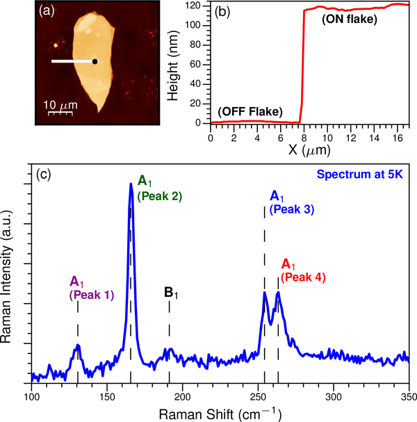
Following structural characterization, the bulk 1T′-MoTe2 crystal was mechanically exfoliated using tape and deposited onto Si/SiO2 substrates with an oxide thickness of 285 nm. Atomic force microscopy (AFM) images (Fig. 1a) indicated that the flake we studied is ≈ 120 nm thick (Fig. 1b) and therefore lies well within the bulk regime, implying that interactions with the substrate can be neglected. It is notable that both the starting crystal and bulk flake we studied were exposed to atmosphere for prolonged periods of time, which is known to oxidize defects in 2H-MoTe2.40 A recent study demonstrated a similar effect in 1T ′-MoTe2 where the Raman signal degraded during measurement, presumably due to photooxidation.16 In order to avoid these effects, we performed all Raman experiments in vacuum and verified that our signal does not degrade after numerous cooldowns and measurements.
FIG. 1.
(a) AFM image of the MoTe2 flake on a Si/SiO2 substrate. The black circle on the flake marks the location where Raman spectra were acquired. (b) Height profile of the MoTe2 flake extracted from (a) along the white line. The flake is over 100 nm thick, which corresponds to the bulk regime. (c) Raman spectra of Td-MoTe2 acquired at 5 K. The four peaks examined in this study are labeled along with the symmetry assignments.
Raman measurements were performed from 5 K to 320 K on a home-built, confocal microscope integrated with a closed-cycle cryostat. The excitation source was a 532 nm laser focused through a 0.5 NA long working distance objective with a 50X magnification. Since the sample was excited in a backscattering geometry, we could detect Raman modes with Ag and Bg symmetry for 1T′ and A1 and B1 symmetry modes for Td. The laser spot diameter was ≈ 1.5 μm and the laser power was ≈1.4 mW at the sample for all measurements. Raman scattering from the sample was directed to a 500 mm focal length spectrometer with a LN2 cooled CCD. The spectrometer and camera were calibrated using a Hg-Ar atomic line source and the spectrometer grating was positioned so that each Raman spectrum included the filtered laser line, allowing us to easily account for any drift in the laser wavelength. The instrumental response function (IRF) for these measurements was ≈ 3.9 cm−1.
III. RESULTS AND DISCUSSION
We present an example Raman spectrum acquired at 5 K in Fig. 1c, at which point MoTe2 is in the Td phase. We observe four prominent peaks at ≈ 130 cm−1, 165 cm−1, 254 cm−1, and 264 cm−1, all with A1 symmetry, which we refer to as Peaks 1–4, respectively. There is also a weak feature at 190 cm−1 that has B1 symmetry, but its signal is not sufficient for temperature-dependent analysis. These assignments are based on polarization-dependent Raman measurements of the 1T′ phase at 300 K, which will be reported elsewhere, and density functional theory calculations of the phonon eigenspectrum.17 The Raman tensors for the Ag(Bg) modes and the A1(B1) modes are identical in structure for the backscattering configuration used here, which implies that they will evolve into an A1(B1) symmetry at low temperature (Supporting Information).
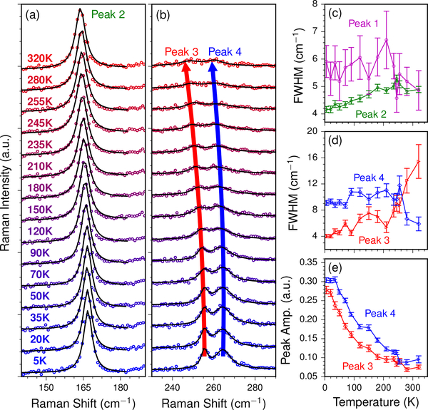
As we increase the sample temperature from 5 K to 320 K, all peaks soften although the magnitude of the redshift is different for each. In Fig. 2a we present temperature-dependent Raman spectra for Peak 2 along with Lorentzian fits (black curves). Fig. 2b shows similar data for Peaks 3 and 4, and all spectra in Fig. 2 have been normalized by the intensity of Peak 2. A similar analysis of Peak 1 is presented in the Supporting Information.
FIG. 2.
Temperature-dependent Raman spectra for (a) Peak 2 and (b) Peaks 3 and 4. Spectra are fit to either a single or double Lorentzian function with a linear background. The sample temperature increases from 5 K (bottom) to 320 K (top). In both panels, the spectra have been normalized by the intensity of Peak 2. Similar data for Peak 1 is presented in the Supporting Information. (c) FWHM of Peaks 1 and 2 extracted from the fits versus temperature. (d) FWHM of Peaks 3 and 4 extracted from the fits versus temperature. (e) Amplitudes of Peaks 3 and 4 extracted from the fits versus temperature. 1σ error bars are included in panels (c) - (e).
We find that the full width at half maximum (FWHM) linewidth of Peak 1 does not change substantially over the entire temperature range (Fig. 2c). The FWHM of Peak 2 is limited by the IRF and also exhibits modest temperature dependence, which suggests that phonon-phonon interactions are weak for these modes. Peaks 3 and 4 exhibit markedly different behavior. As the sample temperature increases, these modes both redshift substantially and the FWHM linewidth of Peak 3 broadens (Fig. 2d). We quantify this behavior by fitting Peaks 3 and 4 to a double Lorentzian function plus a linear background, which allows us to extract the center frequency, amplitude, and linewidth of each mode. At 5 K, the IRF limits the FWHM of Peak 3, which is narrower than Peak 4 (≈ 4 cm−1 versus ≈ 9 cm−1). However, Peak 3 steadily broadens with increasing temperature from 5 K to 250 K, at which point the two modes have similar linewidths. Over the same temperature range, the amplitude of Peak 4 decreases, becoming comparable to that of Peak 3 by 250 K (Fig. 2e). It is notable that observed changes in peak intensity and linewidth correlate well with the Td→1T′ transition temperature,15,20–23,25 suggesting that the relative intensity and linewidth of Peaks 3 and 4 are sensitive to the change in structural phase.
The frequency of all examined Raman peaks softens with increasing temperature (Fig. 3). Temperature-induced shifts in Raman frequencies are typically fit using the linear Grüneisen model:41–44
| (1) |
ω(T) is the temperature-dependent phonon frequency, ωo is the harmonic phonon frequency at 0 K, and χ is the first order temperature coefficient. The Grüneisen model combines the effects of thermal expansion and the phonon self-energy into χ and is sufficient when where kB is the Boltzmann constant. At lower temperatures, ω(T) can become nonlinear and Eq. 1 is not necessarily valid. We illustrate this explicitly in Fig. 3, where the dashed line is a fit of Eq. 1 to the data from 100 K to 200 K. While there is good agreement at low T < 100 K for Peaks 1 and 2 (Figs. 3a and 3b), large deviations exist in this range for Peaks 3 and 4 (Figs. 3c and 3d). The fact that the linewidths of Peaks 1 and 2 exhibit comparably little variation suggests that thermal expansion and/or electron-phonon coupling, rather than phonon-phonon interactions, dominate anharmonic effects in these modes. For Peaks 3 and 4, we suggest that thermal expansion of the lattice makes a negligible contribution to the phonon frequency, and instead changes in the phonon self-energy from anharmonic coupling between different phonon branches are the dominant effect.45 Theoretical work by Balkanski et al. demonstrated that the temperature dependence of optical phonons can be accounted for by a decay pathway resulting in multiple acoustic phonons.39 ω(T) in this formalism is given by
| (2) |
Where and ωB is the 0 K harmonic phonon frequency. In the treatment by Ref. 39, the constant A represents an anharmonic contribution to the frequency involving the decay of an optical phonon into two acoustic phonons. At T = 0, ω(0) = ωB + A, which implies that A represents a third order correction to the phonon self-energy.39 This model has been successfully applied to a variety of nanomaterials to explain nonlinear temperature dependence in optical phonon frequencies.42,45–48
FIG. 3.
Raman frequency versus temperature for (a) Peak 1, (b) Peak 2, (c) Peak 3, and (d) Peak 4. In each panel, we fit the data to the Grüneisen model (dashed lines) in Eq. 1 from 100 – 200 K and to Eq. 2 (solid lines) from 0 – 250 K. The best fits are extrapolated over the entire temperature range and 1σ error bars are included.
We fit the frequencies of Peaks 1–4 (Figs. 3a - 3d) to Eq. 2 up to 250 K in order to isolate the Td crystal phase. The extracted values of A, ωB, χ, and ωo are summarized in Table I and compared to bulk 2H-MoS2.45 We find that Peaks 3 and 4 have larger values of A which, when combined with the significant changes in FWHM, suggest that phonon interactions dominate the anharmonic coupling. Note that an additional term in Eq. 2 corresponding to fourth order processes has been omitted as it was not required to fit our data.39 Furthermore, we observe that all four modes deviate from both Eqs. 1 and 2 above T ≈ 250 K, which we interpret as the onset of the 1T′ phase with distinct anharmonic behaviors. The unique behavior of Peaks 3 and 4 is attributed to the apparent sensitivity of their atomic displacements to interlayer coupling. As Td transitions to 1T′, the a and b lattice parameters increase by 0.0198 Å and 0.0375 Å, respectively, while the c lattice parameter decreases by 0.0447 Å.38 This implies that the dominant changes are in the directions of the b lattice vector and the out-of-plane c lattice vector. Furthermore, the angle between the b and c lattice vectors changes from 90° in Td to 93.84° in 1T′.38 These combined structural changes imply that phonon modes sensitive to interlayer coupling will be most affected by the Td→1T′ transition. Peak 2 depends only weakly on the number of layers,13 and therefore it is expected to respond minimally to changes in layer alignment and separation. In contrast, Peaks 3 and 4 are sensitive to flake thickness13 and are therefore likely to respond to changes in the spacing and alignment of the layers during the Td→1T′ transition.
TABLE I.
Measured values of A, ωB, χ, and ωo for Peaks 1–4 along with literature values for bulk 2H-MoS2.
| Mode | Material | Ref. | A (cm−1) | ωB (cm−1) | χ (cm−1/K)a | ωo (cm−1)a |
|---|---|---|---|---|---|---|
| Peak 1 | Bulk Td-MoTe2 | this work | −0.390 ± 0.027 | 131.25 ± 0.091 | −0.0089 ± 0.004 | 131.24 ± 0.537 |
| Peak 2 | ” | ” | −0.717 ± 0.009 | 167.39 ± 0.022 | −0.0089 ± 0.001 | 166.77 ± 0.122 |
| Peak 3 | ” | ” | −3.019 ± 0.094 | 258.51 ± 0.116 | −0.0312 ± 0.002 | 257.91 ± 0.303 |
| Peak 4 | ” | ” | −2.052 ± 0.105 | 266.08 ± 0.140 | −0.0227 ± 0.003 | 265.74 ± 0.438 |
| A1g | Bulk 2H-MoS2 | 45 | −5.687 | - | −0.0197 | - |
| ” | ” | −3.058 | - | −0.0221 | - |
Calculated from fits to data from 100 K to 200 K.
IV. CONCLUSION
In summary, we have investigated anharmonicity of the optical phonon modes in bulk Td-MoTe2. Changes in phonon frequency, linewidth, and amplitude are determined for four modes and the results correlate well with the Td→1T′ structural phase transition. ω(T) is nonlinear for two modes, which is consistent with an anharmonic contribution arising from optical phonon decay into multiple acoustic phonons. The large changes in frequency and linewidth for these two modes as Td→1T′ indicates they are highly sensitive to interlayer separation and alignment, an observation that is consistent with prior characterizations of their dependence on layer number. All four modes exhibit a change in slope and an increase in frequency at T ≈ 250 K which we attribute to the Td→1T′ structural phase transition. These results highlight the important, and in some cases dominant, role of phonon-phonon interactions in Td-MoTe2. Further studies connecting the atomic displacement of these modes to their anharmonicity are desirable and additional investigation of suspended, few-layer MoTe2 will illuminate the impact of interlayer coupling on anharmonicity in this exciting material.
Supplementary Material
ACKNOWLEDGMENTS
J.J., I.S., and P.M.V. acknowledge support from the Office of Naval Research through grant N-00014-15-1-2357 and from the GMU OSCAR Program. R.B. thanks the National Research Council Research Associateship Programs for its support.
Footnotes
SUPPORTING INFORMATION
See supporting information for the analysis of Peak 1 and a discussion of the Raman tensors of 1T′ and Td MoTe2.
REFERENCES
- 1.Mak KF, Lee C, Hone J, Shan J, and Heinz TF, Physical Review Letters 105, 136805 (2010). [DOI] [PubMed] [Google Scholar]
- 2.Splendiani A, Sun L, Zhang Y, Li T, Kim J, Chim C-Y, Galli G, and Wang F, Nano Letters 10, 1271 (2010). [DOI] [PubMed] [Google Scholar]
- 3.Yang J, Lü T, Myint YW, Pei J, Macdonald D, Zheng J-C, and Lu Y, ACS Nano 9, 6603 (2015). [DOI] [PubMed] [Google Scholar]
- 4.Gutiérrez HR, Perea-Ló N, Elías AL, Berkdemir A, Wang B, Lv R, López-Urías F, Crespi VH, Terrones H, and Terrones M, Nano Letters 13, 3447 (2013). [DOI] [PubMed] [Google Scholar]
- 5.Chhowalla M, Shin HS, Eda G, Li L-J, Loh KP, and Zhang H, Nature Chemistry 5, 263 (2013). [DOI] [PubMed] [Google Scholar]
- 6.Wang QH, Kalantar-Zadeh K, Kis A, Coleman JN, and Strano MS, Nature Nanotechnology 7, 699 (2012). [DOI] [PubMed] [Google Scholar]
- 7.Geim AK and Grigorieva IV, Nature 499, 419 (2013). [DOI] [PubMed] [Google Scholar]
- 8.Mak KF and Shan J, Nature Photonics 10, 216 (2016). [Google Scholar]
- 9.Wilson J and Yoffe A, Advances in Physics 18, 193 (1969). [Google Scholar]
- 10.Dawson WG and Bullett DW, Journal of Physics C: Solid State Physics 20, 6159 (1987). [Google Scholar]
- 11.K.C. S, Zhang C, Hong S, Wallace RM, and Cho K, 2D Materials 2, 035019 (2015). [Google Scholar]
- 12.Li Y, Duerloo K-AN, Wauson K, and Reed EJ, Nature Communications 7, 10671 (2016). [DOI] [PMC free article] [PubMed] [Google Scholar]
- 13.Keum DH, Cho S, Kim JH, Choe D-H, Sung H-J, Kan M, Kang H, Hwang J-Y, Kim SW, Yang H, Chang KJ, and Lee YH, Nature Physics 11, 482 (2015). [Google Scholar]
- 14.Park JC, Yun SJ, Kim H, Park J-H, Chae SH, An S-J, Kim J-G, Kim SM, Kim KK, and Lee YH, ACS Nano 9, 6548 (2015). [DOI] [PubMed] [Google Scholar]
- 15.Qi Y, Naumov PG, Ali MN, Rajamathi CR, Schnelle W, Barkalov O, Hanfland M, Wu S-C, Shekhar C, Sun Y, Süβ V, Schmidt M, Schwarz U, Pippel E, Werner P, Hillebrand R, Förster T, Kampert E, Parkin S, Cava RJ, Felser C, Yan B, and Medvedev SA, Nature Communications 7, 11038 (2016). [DOI] [PMC free article] [PubMed] [Google Scholar]
- 16.Naylor CH, Parkin WM, Ping J, Gao Z, Zhou YR, Kim Y, Streller F, Carpick RW, Rappe AM, Drndić M, Kikkawa JM, and Johnson ATC, Nano Letters, acs.nanolett.6b01342 (2016). [DOI] [PMC free article] [PubMed] [Google Scholar]
- 17.Sun Y, Wang Y, Sun D, Carvalho BR, Read CG, Lee C.-h., Lin Z, Fujisawa K, Robinson JA, Crespi VH, Terrones M, and Schaak RE, Angewandte Chemie International Edition 55, 2830 (2016). [DOI] [PubMed] [Google Scholar]
- 18.Cho S, Kim S, Kim JH, Zhao J, Seok J, Keum DH, Baik J, Choe D-H, Chang KJ, Suenaga K, Kim SW, Lee YH, and Yang H, Science 349, 625 (2015). [DOI] [PubMed] [Google Scholar]
- 19.Duerloo K-AN, Li Y, and Reed EJ, Nature Communications 5, 4214 (2014). [DOI] [PubMed] [Google Scholar]
- 20.Hughes HP and Friend RH, Journal of Physics C: Solid State Physics 11, L103 (1978). [Google Scholar]
- 21.Zandt T, Dwelk H, Janowitz C, and Manzke R, Journal of Alloys and Compounds 442, 216 (2007). [Google Scholar]
- 22.Zhang K, Bao C, Gu Q, Ren X, Zhang H, Deng K, Wu Y, Li Y, Feng J, and Zhou S, (2016), arXiv:1606.05071. [DOI] [PMC free article] [PubMed] [Google Scholar]
- 23.Chen S-Y, Goldstein T, Ramasubramaniam A, and Yan J, (2016), arXiv:1602.03566. [Google Scholar]
- 24.Clarke R, Marseglia E, and Hughes HP, Philosophical Magazine Part B 38, 121 (1978). [Google Scholar]
- 25.Wang YY, Li AZ, Wang YH, Liang Y, Jiang J, Nan HY, Ni ZH, Wang D, Zhong B, and Wen GW, Materials Research Express 3, 025007 (2016). [Google Scholar]
- 26.Manolikas C, van Landuyt J, and Amelinckx S, Physica Status Solidi (a) 53, 327 (1979). [Google Scholar]
- 27.Sun Y, Wu S-C, Ali MN, Felser C, and Yan B, Physical Review B 92, 161107 (2015). [Google Scholar]
- 28.Soluyanov AA, Gresch D, Wang Z, Wu Q, Troyer M, Dai X, and Bernevig BA, Nature 527, 495 (2015). [DOI] [PubMed] [Google Scholar]
- 29.Chang T-R, Xu S-Y, Chang G, Lee C-C, Huang S-M, Wang B, Bian G, Zheng H, Sanchez DS, Belopolski I, Alidoust N, Neupane M, Bansil A, Jeng H-T, Lin H, and Zahid Hasan M, Nature Communications 7, 10639 (2016). [DOI] [PMC free article] [PubMed] [Google Scholar]
- 30.Xu N, Wang ZJ, Weber AP, Magrez A, Bugnon P, Berger H, Matt CE, Ma JZ, Fu BB, Lv BQ, Plumb NC, Radovic M, Pomjakushina E, Conder K, Qian T, Dil JH, Mesot J, Ding H, and Shi M, (2016), arXiv:1604.02116. [Google Scholar]
- 31.Belopolski I, Xu S-Y, Ishida Y, Pan X, Yu P, Sanchez DS, Neupane M, Alidoust N, Chang G, Chang T-R, Wu Y, Bian G, Zheng H, Huang S-M, Lee C-C, Mou D, Huang L, Song Y, Wang B, Wang G, Yeh Y-W, Yao N, Rault JE, Fèvre PL,Bertran F, Jeng H-T, Kondo T, Kaminski A, Lin H, Liu Z, Song F, Shin S, and Hasan MZ, (2016), arXiv:1604.07079. [Google Scholar]
- 32.Wu Y, Jo NH, Mou D, Huang L, Bud’ko SL, Canfield PC, and Kaminski A, (2016), arXiv:1604.05176. [Google Scholar]
- 33.Wang C, Zhang Y, Huang J, Nie S, Liu G, Liang A, Zhang Y, Shen B, Liu J, Hu C, Ding Y, Liu D, Hu Y, He S, Zhao L, Yu L, Hu J, Wei J, Mao Z, Shi Y, Jia X,Zhang F, Zhang S, Yang F, Wang Z, Peng Q, Weng H, Dai X, Fang Z, Xu Z, Chen C, and Zhou XJ, (2016), arXiv:1604.04218. [Google Scholar]
- 34.Deng K, Wan G, Deng P, Zhang K, Ding S, Wang E, Yan M, Huang H, Zhang H, Xu Z, Denlinger J, Fedorov A, Yang H, Duan W, Yao H, Wu Y, Fan y. S., Zhang H, Chen X, and Zhou S, (2016), arXiv:1603.08508. [Google Scholar]
- 35.Liang A, Huang J, Nie S, Ding Y, Gao Q, Hu C, He S, Zhang YY, Wang C, Shen B, Liu J, Ai P, Yu L, Sun X, Zhao W, Lv S, Liu D, Li C, Zhang YY, Hu Y, Xu Y, Zhao L, Liu G, Mao Z, Jia X, Zhang F, Zhang S, Yang F, Wang Z, Peng Q, Weng H, Dai X, Fang Z, Xu Z, Chen C, and Zhou XJ, (2016), arXiv:1604.01706. [Google Scholar]
- 36.Jiang J, Liu ZK, Sun Y, Yang HF, Rajamathi R, Qi YP, Yang LX, Chen C, Peng H, Hwang CC, Sun SZ, Mo SK, Vobornik I, Fujii J, Parkin SSP, Felser C, Yan BH, and Chen YL, (2016), arXiv:1604.00139. [Google Scholar]
- 37.Huang L, McCormick TM, Ochi M, Zhao Z, Suzuki M.-t., Arita R, Wu Y, Mou D, Cao H, Yan J, Trivedi N, and Kaminski A, (2016), arXiv:1603.06482. [DOI] [PubMed] [Google Scholar]
- 38.Tamai A, Wu QS, Cucchi I, Bruno FY, Ricco S, Kim TK, Hoesch M, Barreteau C, Giannini E, Bernard C, Soluyanov AA, and Baumberger F, (2016), arXiv:1604.08228. [Google Scholar]
- 39.Balkanski M, Wallis R, and Haro E, Physical Review B 28, 1928 (1983). [DOI] [PubMed] [Google Scholar]
- 40.Chen B, Sahin H, Suslu A, Ding L, Bertoni MI, Peeters FM, and Tongay S, ACS Nano 9, 5326 (2015). [DOI] [PubMed] [Google Scholar]
- 41.Jana MK, Singh A, Late DJ, Rajamathi CR, Biswas K, Felser C, Waghmare UV, and Rao CNR, Journal of Physics: Condensed Matter 27, 285401 (2015). [DOI] [PubMed] [Google Scholar]
- 42.Łapińska A, Taube A, Judek J, and Zdrojek M, The Journal of Physical Chemistry C 120, 5265 (2016). [Google Scholar]
- 43.Pawbake A, Pawar M, Jadkar SR, and Late DJ, Nanoscale, 3008 (2016). [DOI] [PubMed] [Google Scholar]
- 44.Late DJ, Shirodkar SN, Waghmare UV, Dravid VP, and Rao CNR, ChemPhysChem 15, 1592 (2014). [DOI] [PubMed] [Google Scholar]
- 45.Su L, Zhang Y, Yu Y, and Cao L, Nanoscale 6, 4920 (2014). [DOI] [PubMed] [Google Scholar]
- 46.Taube A, L apińska A, Judek J, and Zdrojek M, Applied Physics Letters 107, 013105 (2015). [Google Scholar]
- 47.Duzynska A, Judek J, and Zdrojek M, Applied Physics Letters 105, 213105 (2014). [Google Scholar]
- 48.Taube A, Judek J, Jastrzbski C, Duzynska A, Świtkowski K, and Zdrojek M, ACS Applied Materials and Interfaces 6, 8959 (2014). [DOI] [PubMed] [Google Scholar]
Associated Data
This section collects any data citations, data availability statements, or supplementary materials included in this article.


