Abstract
β-Catenin is an important component of the Wnt signalling pathway. As dysregulation or mutation of this pathway causes many diseases, including cancer, the β-Catenin level is carefully regulated by the destruction complex in the Wnt signalling pathway. However, the mechanisms underlying the regulation of β-Catenin ubiquitination and degradation remain unclear. Here, we find that WNK (With No Lysine [K]) kinase is a potential regulator of the Wnt signalling pathway. We show that WNK protects the interaction between β-Catenin and the Glucose-Induced degradation Deficient (GID) complex, which includes an E3 ubiquitin ligase targeting β-Catenin, and that WNK regulates the β-Catenin level. Furthermore, we show that WNK inhibitors induced β-Catenin degradation and that one of these inhibitors suppressed xenograft tumour development in mice. These results suggest that WNK is a previously unrecognized regulator of β-Catenin and a therapeutic target of cancer.
Subject terms: Cancer, Growth factor signalling, Ubiquitin ligases
Sato et al. find that WNK (With No Lysine [K]) acts as a positive regulator of the Wnt signaling pathway by attenuating the interaction between β-Catenin and the Glucose Induce degradation Deficient (GID) complex, and show that a WNK inhibitor also functions as a Wnt inhibitor, suppressing xenograft tumor development in mice. These findings suggest that WNK is a regulator of β-Catenin and a potential therapeutic target
Introduction
The Wnt signalling pathway is an essential pathway in multicellular organisms that has already been identified to function in cell proliferation, differentiation and homeostasis1–3. Dysregulation or mutations of this pathway are related to many diseases, such as type II diabetes4, autosomal dominant polycystic kidney5 and many cancer types6. The Wnt signalling pathway is divided into three classes: the canonical pathway, planar cell polarity pathway and Ca pathway. In the canonical Wnt pathway, β-Catenin is a core component whose protein regulation is a key process in the Wnt signalling pathway. In the absence of Wnt, the destruction complex formed by Axin1, Adenomatous Polyposis Coli (APC) and Glycogen Synthase Kinase 3β (GSK3β) phosphorylates the N-terminus of β-Catenin, which is in turn recognized and ubiquitinated by βTrCP E3 ubiquitin ligases6,7. Ubiquitinated β-Catenin is degraded by the proteasome, which regulates the basal β-Catenin levels. The Wnt signalling pathway is activated by the binding of Wnt ligand to a receptor complex8,9, which leads to Dishevelled (DVL) recruitment, where DVL polymerizes with Axin and the remaining components of the destruction complex. This polymerization inactivates the destruction complex, inducing β-Catenin accumulation in the cytoplasm. Accumulated β-Catenin translocates to the nucleus and regulates the transcription of target genes10. However, because several ubiquitination enzymes of β-Catenin have been identified and shown to be Wnt-independent11–17, the mechanisms underlying the regulation of β-Catenin ubiquitination remain unclear.
The WNK (With No Lysine [K]) kinases are atypical serine/threonine kinase family members that are conserved among many species18–20 and comprise four members. WNK1 and WNK4 have been identified as the causative genes of pseudohypoaldosteronism type II21, and WNK1 is also a causative gene of hereditary sensory and autonomic neuropathy type 2A22. WNK kinases are required for epidermal growth factor-mediated extracellular signal-regulated kinase 5 activation, and WNK family members are also involved in proliferation, migration and differentiation23–25. Previously, we found that WNK1 and WNK4 induce the expression of Lhx8 and are important for neural specification through GSK3β26,27. WNK was also identified as a positive regulator of the Wnt signalling pathway; however, the detailed mechanism is unknown28. Thus, WNK has several functions associated with many developmental processes.
Here, we demonstrated that WNK is a positive regulator of the Wnt signalling pathway. WNK attenuates the interaction between β-Catenin and the glucose-induced degradation deficient (GID) complex, which is an E3 ubiquitin ligase of β-Catenin, suggesting that WNK might regulate β-Catenin levels. Furthermore, we showed that the WNK inhibitor also functions as a Wnt inhibitor, suppressing xenograft tumour development in mice. These findings suggest that WNK is a regulator of β-Catenin and might be a therapeutic cancer target.
Results
WNK functions as a positive regulator of the Wnt signalling pathway
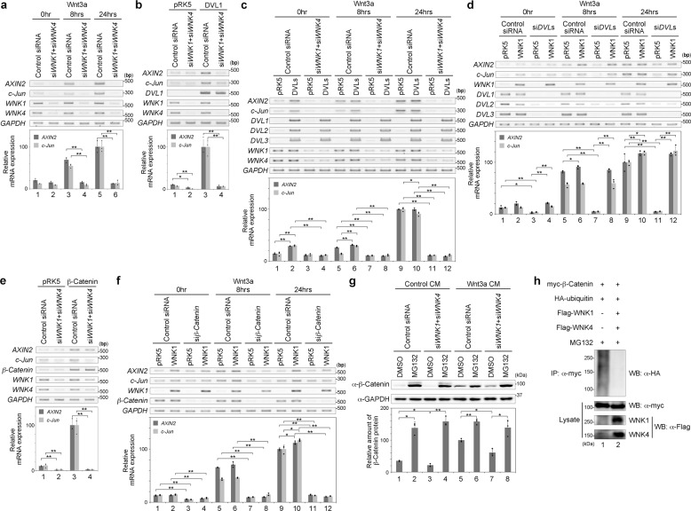
We previously showed that GSK3β, a component of the Wnt signalling pathway, functions downstream of the WNK signalling pathway27. GSK3β acts as a negative regulator of the Wnt signalling pathway6,7. Additionally, WNK was shown to be involved in the phosphorylation of Dsh (fly homologue of DVL) under Wnt stimulation in Drosophila and to work as a positive regulator of the Wnt signalling pathway in HEK293T cells28. Thus, we considered whether WNK interacts with the Wnt signalling pathway. First, we examined whether the knockdown of WNK affects the Wnt signalling pathway. We performed RT-PCR to examine the expression of the Wnt target genes AXIN2 (early response gene) and c-Jun (late response gene) under the knockdown of WNK1 and WNK4 in HEK293T cells. We found that the expression levels of AXIN2 and c-Jun were activated by Wnt stimulation (Fig. 1a). The knockdown of both WNK1 and WNK4 using siRNA significantly reduced the induction of both AXIN2 and c-Jun (Fig. 1a). These results suggest that WNK is a positive regulator in the Wnt signalling pathway.
Fig. 1. WNK is a positive regulator of the Wnt signalling pathway.
a Gene expression was examined by RT-PCR or quantitative RT-PCR in HEK293T cells following Wnt stimulation or the knockdown of both WNK1 and WNK4. n = 3 biologically independent experiments. Dots indicate individual data. b Gene expression was examined by RT-PCR or quantitative RT-PCR in HEK293T cells following the expression of DVL1 or knockdown of both WNK1 and WNK4. n = 3 biologically independent experiments. Dots indicate individual data. c Gene expression was examined by RT-PCR or quantitative RT-PCR in HEK293T cells following the treatment with Wnt3a stimulation, the expression of DVL1, DVL2 and DVL3, or knockdown of both WNK1 and WNK4. n = 3 biologically independent experiments. Dots indicate individual data. d Gene expression was examined by RT-PCR or quantitative RT-PCR in HEK293T cells following Wnt stimulation of the expression of WNK1 or knockdown of DVL1, DVL2 and DVL3. n = 3 biologically independent experiments. Dots indicate individual data. e Gene expression was examined by RT-PCR or quantitative RT-PCR in HEK293T cells following the expression of β-Catenin or knockdown of both WNK1 and WNK4. n = 3 biologically independent experiments. Dots indicate individual data. f Gene expression was examined by RT-PCR or quantitative RT-PCR in HEK293T cells following Wnt stimulation, the expression of WNK1 or knockdown of β-Catenin. n = 3 biologically independent experiments. Dots indicate individual data. g Western blot analysis of endogenous protein expression following Wnt stimulation, the knockdown of both WNK1 and WNK4, or MG132 (10 μM, 6 h) treatment in HEK293T cells. n = 3 biologically independent experiments. Dots indicate individual data. h Western blot analysis of ubiquitinated β-Catenin under the expression of WNK1 and WNK4 in HEK293T cells. Values and error bars express mean ± standard deviation (SD). * indicates p < 0.05 and ** indicates p < 0.005. p value was calculated by Bonferroni test.
WNK is thought to be a cytoplasmic protein20, and therefore we next analysed the relationship between WNK and DVL, the most upstream cytoplasmic component of the Wnt signalling pathway. We found that DVL1 expression activates the Wnt signalling pathway6,7 (Fig. 1b). The knockdown of both WNK1 and WNK4 suppressed the expression of AXIN2 and c-Jun activated by DVL1 expression (Fig. 1b). Exogenous expression of all DVLs (DVL1, DVL2 and DVL3) did not rescue the suppression of Wnt3a-activated gene expression of AXIN2 and c-Jun by the knockdown of both WNK1 and WNK4 (Fig. 1c). Conversely, the knockdown of all DVL genes using siRNA suppressed the expression of AXIN2 and c-Jun activated by Wnt3a stimulation (Fig. 1d). Exogenous expression of WNK1 rescued this suppression by DVL knockdown (Fig. 1d). These results indicate that WNK1 acts as a downstream element of Dvl in the Wnt signalling pathway.
WNK regulates the Wnt signalling pathway by controlling the β-Catenin level
We next analysed the relationship between WNK and β-Catenin in the Wnt signalling pathway. The expression of β-Catenin activated the target genes of the Wnt signalling pathway6,7. The knockdown of both WNK1 and WNK4 suppressed the expression of AXIN2 and c-Jun activated by β-Catenin expression (Fig. 1e). The knockdown of β-Catenin using siRNA suppressed the expression of AXIN2 and c-Jun activated by Wnt3a stimulation, while the exogenous expression of WNK1 could not rescue this suppression by β-Catenin knockdown (Fig. 1f). These results suggest that the function of β-Catenin in Wnt signalling is required, but not sufficient, for WNK expression. Therefore, we examined whether WNK regulates the β-Catenin levels. β-Catenin is degraded via the proteasome under normal conditions, and after Wnt stimulation, β-Catenin accumulates in the cytoplasm6,7. The knockdown of both WNK1 and WNK4 reduced the β-Catenin level under both normal conditions and Wnt-stimulated conditions (Fig. 1g). MG132, a proteasome inhibitor, blocked the degradation of β-Catenin under both conditions29 (Fig. 1g). Moreover, MG132 still blocked the degradation of β-Catenin following the knockdown of both WNK1 and WNK4 (Fig. 1g). We also checked the ubiquitination level of β-Catenin. As shown in Fig. 1h, the expression of both WNK1 and WNK4 reduced the ubiquitination level of β-Catenin under MG132 treatment. These results suggest that WNK regulates the β-Catenin level by regulating the ubiquitination of β-Catenin.
WNK is involved in the ubiquitination of β-Catenin via the GID complex
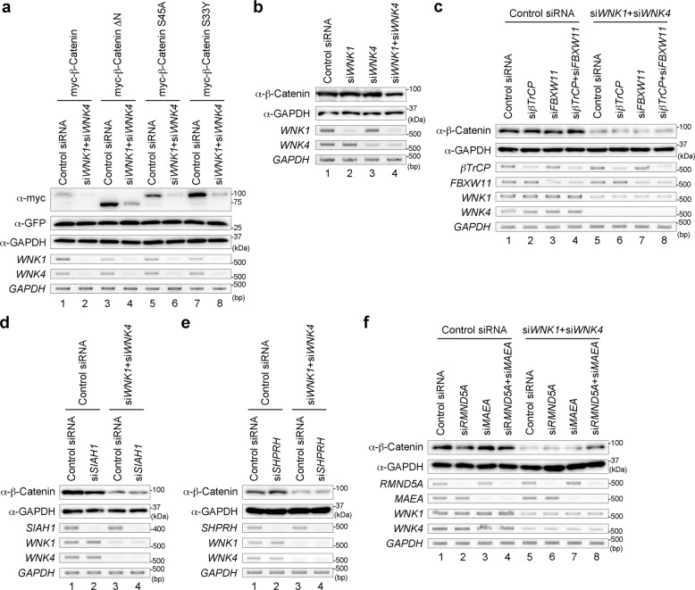
β-Catenin is phosphorylated by GSK3β in the destruction complex and is ubiquitinated by βTrCP E3 ubiquitin ligase11,30. Because the N-terminal deletion of β-Catenin (β-Catenin ΔN) deletes the phosphorylation and ubiquitination sites, β-Catenin ΔN is no longer degraded and functions as a constitutively active form31. Because WNK is related to β-Catenin ubiquitination, we next examined the β-Catenin ΔN level under conditions with the knockdown of both WNK1 and WNK4. Surprisingly, similar to the normal β-Catenin level, the β-Catenin ΔN level was also reduced by the knockdown of both WNK1 and WNK4 (Fig. 2a). The levels of other constitutively active mutants of β-Catenin, such as β-Catenin S33Y and S45A, were also reduced (Fig. 2a). As shown above, the expression of WNK1 and WNK4 suppressed the ubiquitination of wild-type β-Catenin (Fig. 1g). Therefore, we checked the ubiquitination level of β-Catenin ΔN. Similar to wild-type β-Catenin, the ubiquitination level of β-Catenin ΔN was reduced by the expression of both WNK1 and WNK4 (Supplementary Fig. 1a). In the human colorectal cancer cell line SW480, which has a deletion in APC, β-Catenin is not degraded by the destruction complex32. In SW480 cells, the knockdown of both WNK1 and WNK4 led to a reduced β-Catenin level (Fig. 2b). Furthermore, the reduced β-Catenin level caused by the knockdown of both WNK1 and WNK4 could not be rescued by the knockdown of both βTrCP and FBXW11 (also known as βTrCP2) in SW480 cells (Fig. 2c). These results suggest that WNK is not related to the ubiquitination of β-Catenin by the destruction complex containing βTrCP.
Fig. 2. WNK regulates the protein level of β-Catenin through the GID complex.
a Western blot analysis of β-Catenin following the knockdown of both WNK1 and WNK4 in HEK293T cells. GFP is co-transfected for checking the transfection efficiency. b Western blot analysis of endogenous β-Catenin following the knockdown of WNK1 and/or WNK4 in SW480 cells. c–f Western blot analysis of endogenous β-Catenin following the knockdown of both WNK1 and WNK4, or the knockdown of βTrCP and/or βTrCP2 (c), SIAH1 (d), SHPRH (e), or RMND5A, and/or MAEA (f) in SW480 cells.
Previous reports identified several E3 ubiquitin ligases for β-Catenin other than βTrCP, such as SIAH1, Jade1, EDD, Mule, SHPRH and the GID complex (also known as the CTLH complex)11–17. Among these ligases, Jade-1 interacts with the N-terminus of β-Catenin like βTrCP12, and Mule targets β-Catenin only under activated Wnt conditions17, suggesting that Jade-1 and Mule are unlikely to be related to WNK function. Furthermore, because EDD targets intact β-Catenin, EDD might not be involved in WNK function. Therefore, we examined whether the knockdown of the genes encoding SIAH1, SHPRH and GID complex could rescue the reduced β-Catenin level by the knockdown of WNK. We found that the reduction of β-Catenin by the knockdown of both WNK1 and WNK4 could not be rescued by the knockdown of SIAH1 or SHPRH using siRNA in SW480 cells (Fig. 2d, e). These findings also suggest that SIAH1 or SHPRH is not involved in WNK function.
Our previous report suggests that the GID complex is involved in the ubiquitination of β-Catenin and is independent of the Wnt signalling pathway16. Among the components of the GID complex, RMND5A and MAEA (mammalian homologues of yeast GID2 and GID9, respectively) encode E3 ubiquitin ligases33–35. We confirmed that RMND5A and MAEA ubiquitinated β-Catenin directly (Supplementary Fig. 1b). The reduction of β-Catenin by the knockdown of both WNK1 and WNK4 could be rescued by the knockdown of both RMND5A and MAEA (Fig. 2f). Next, we constructed deletion mutants of the RING finger domain of RMND5A and MAEA (RMND5A ΔR and MAEA ΔR, respectively) as dominant-negative forms. The reduction of β-Catenin by the knockdown of both WNK1 and WNK4 could also be rescued by the expression of both RMND5A ΔR and MAEA ΔR (Supplementary Fig. 1c). These results suggest that the GID complex is involved in the ubiquitination of β-Catenin associated with WNK function.
WNK attenuates the interaction between β-Catenin and MAEA
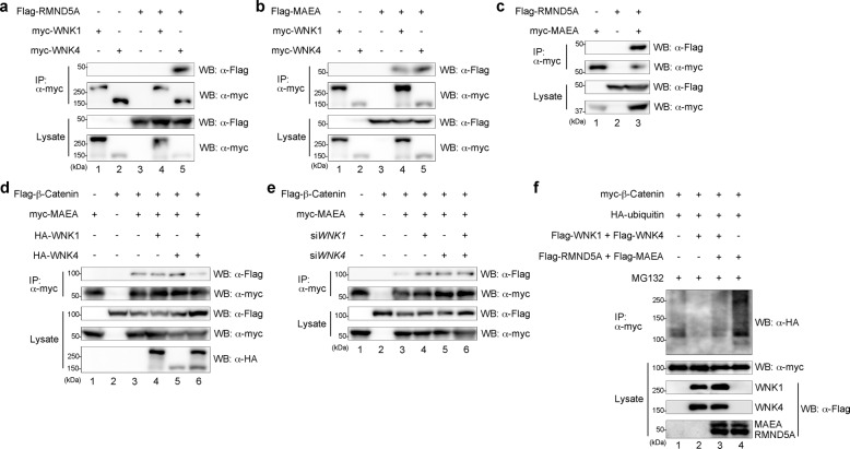
As shown above, because RMND5A and MAEA of the GID complex are related to WNK, we examined the interaction between WNK and RMND5A or MAEA by immunoprecipitation. We transiently expressed Flag-tagged RMND5A or MAEA together with myc-tagged WNK1 or WNK4. When the cell extracts were subjected to immunoprecipitation with anti-myc antibodies, followed by immunoblotting, we found that RMND5A interacted with WNK4 (Fig. 3a) and that MAEA interacted with WNK1 and WNK4 (Fig. 3b). Because RMND5A and MAEA interacted with each other, like yeast GID2 and GID936 (Fig. 3c), it was suggested that WNK forms a complex with the GID complex.
Fig. 3. WNK attenuates the interaction between β-Catenin and MAEA.
a, b The interaction between WNK1 or WNK4, and RMND5A (a) or MAEA (b) was examined in HEK293T cells by co-immunoprecipitation. c The interaction between RMND5A and MAEA was examined in HEK293T cells by co-immunoprecipitation. d, e The interaction between β-Catenin and MAEA following the expression of WNK1 and/or WNK4 (d) or the knockdown of WNK1 and/or WNK4 (e) was examined in HEK293T cells by co-immunoprecipitation. f Western blot analysis of ubiquitinated β-Catenin following the expression of WNK1 and WNK4, and/or RMND5A and MAEA in HEK293T cells.
MAEA interacted with β-Catenin, but RMND5A was not detected (Fig. 3d, e). Therefore, we examined whether WNK affected the interaction between β-Catenin and MAEA. The interaction between β-Catenin and MAEA was reduced by the expression of both WNK1 and WNK4, but was not reduced by the expression of only WNK1 or WNK4 (Fig. 3d). By contrast, the knockdown of WNK1 and/or WNK4 increased the interaction of β-Catenin and MAEA (Fig. 3e). We next examined whether WNK or the GID complex has the opposite effect on the ubiquitination of β-Catenin. As shown in Fig. 3f, the expression of both RMND5A and MAEA increased the ubiquitination of β-Catenin and rescued the reduction of β-Catenin ubiquitination by WNK expression. These results suggest that the presence of WNK attenuates the interaction between β-Catenin and MAEA and inhibits the ubiquitination of β-Catenin by the GID complex.
The WNK inhibitor functions as an inhibitor of the WNK–GID complex interaction
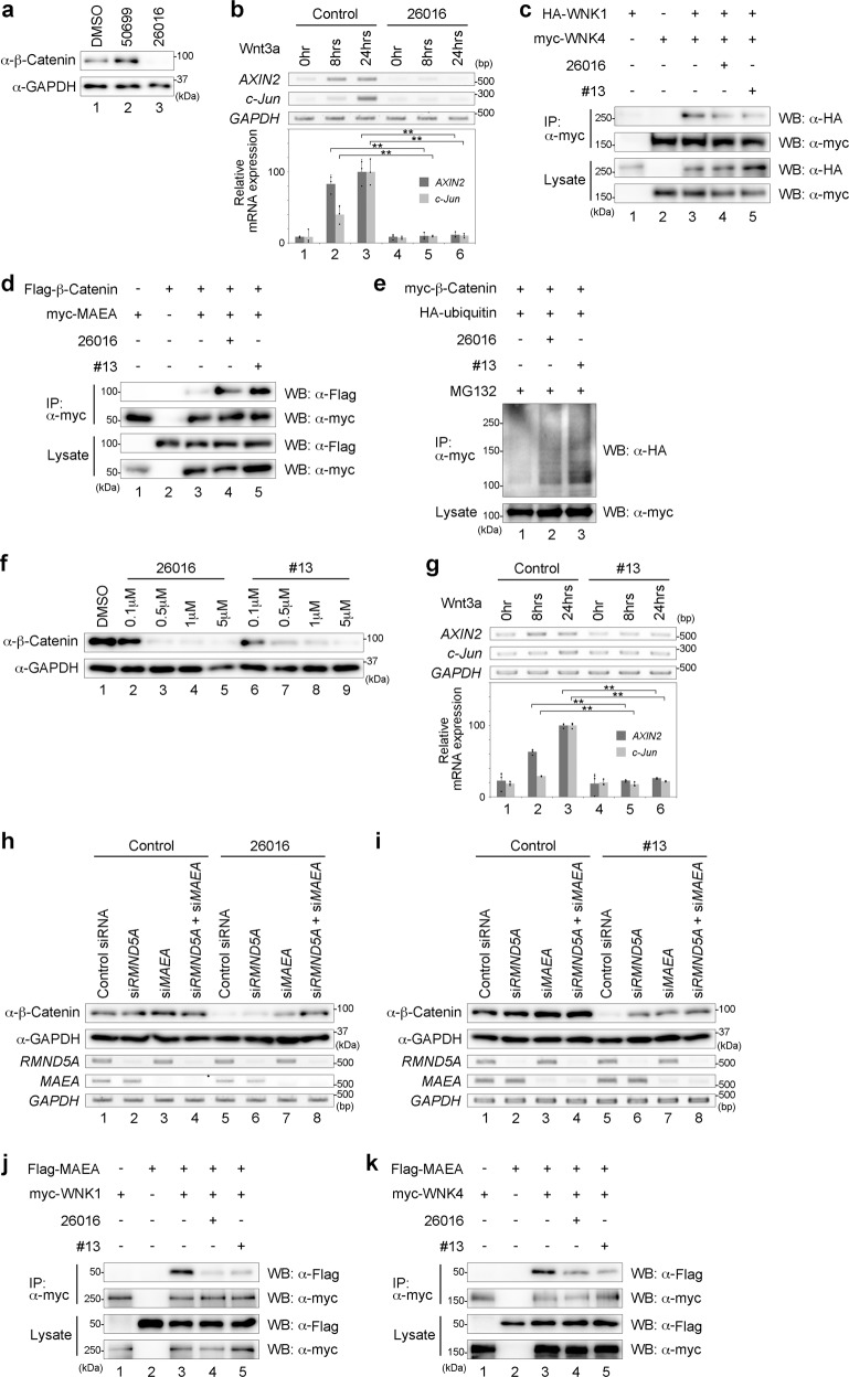
In WNK signalling, SPAK/OSR1 are cofactors and substrates of WNK. Previous research identified two compounds as inhibitors of the WNK signalling pathway, which inhibit the WNK–OSR interaction37. Thus, we checked whether these inhibitors also reduced the β-Catenin level. In SW480 cells, we found that STOCKS2S-26016 (hereafter referred to as 26016) reduced the β-Catenin level, but STOCK1S-50699 (hereafter referred to as 50699) did not (Fig. 4a). The expression levels of AXIN2 and c-Jun activated by Wnt3a stimulation in HEK293T cells were also inhibited in the presence of 26016 (Fig. 4b). We next examined the function of 26016 in WNK signalling regarding β-Catenin degradation. We found that 26016 inhibited the interaction of WNK1 and WNK4 (Fig. 4c) and that the interaction between β-Catenin and MAEA increased under 26016 treatment (Fig. 4d), similar to the results of WNK knockdown (Fig. 3e). Furthermore, treatment with 26016 increased the ubiquitination level of β-Catenin (Fig. 4e). These results suggest that the WNK inhibitor 26016 functions as an inhibitor of the Wnt signalling pathway, similar to the effect of WNK knockdown.
Fig. 4. WNK inhibitors function as Wnt inhibitors.
a Western blot analysis of endogenous β-Catenin following treatment with 26016 in SW480 cells. b Gene expression was examined by RT-PCR or quantitative RT-PCR in HEK293T cells following Wnt stimulation or treatment with 26016. n = 3 biologically independent experiments. Dots indicate individual data. c The interaction between WNK1 and WNK4 following treatment with 26016 or #13 was examined in HEK293T cells by co-immunoprecipitation. d The interaction between β-Catenin and MAEA following treatment with 26016 or #13 was examined in HEK293T cells by co-immunoprecipitation. e Western blot analysis of ubiquitinated β-Catenin following treatment with 26016 or #13 in HEK293T cells. f Western blot analysis of endogenous β-Catenin following treatment with 26016 or #13. g Gene expression by RT-PCR or quantitative RT-PCR analysis was examined in HEK293T cells following Wnt stimulation or treatment with #13. n = 3 biologically independent experiments. Dots indicate individual data. h, i Western blot analysis of endogenous β-Catenin following the knockdown of RMND5A and/or MAEA, or treatment with 26016 (h) or #13 (i) in SW480 cells. j The interaction between WNK1 and MAEA following treatment with 26016 or #13 was examined in HEK293T cells by co-immunoprecipitation. k The interaction between WNK4 and MAEA following treatment with 26016 or #13 was examined in HEK293T cells by co-immunoprecipitation. Values and error bars express mean ± standard deviation (SD). ** indicates p < 0.005. p value was calculated by Bonferroni test.
Derivatives of 26016 were developed in a previous study38. Thus, we checked whether derivatives of 26016 also affected the β-Catenin level in SW480 cells and found that derivative #13 (hereafter referred to as #13) also reduced the β-Catenin level (Fig. 4f). #13 also inhibited the interaction between WNK1 and WNK4 (Fig. 4c), increased the interaction between β-Catenin and MAEA (Fig. 4d), and suppressed the expression levels of AXIN2 and c-Jun activated by Wnt3a stimulation (Fig. 4g). Moreover, #13 increased the ubiquitination level of β-Catenin (Fig. 4e), suggesting that 26016 and #13 share similar activities for WNK signalling and β-Catenin degradation. Furthermore, we found that the knockdown of RMND5A and MAEA could rescue the reduced β-Catenin level by the effect of 26016 or #13 (Fig. 4h, i, respectively). The expression of the dominant-negative forms of RMND5A and MAEA (RMND5A ΔR and MAEA ΔR) could also rescue the reduced β-Catenin level (Supplementary Fig. 2a, b, respectively). We next examined the effect of WNK inhibitors on WNK and GID complex. We found that WNK inhibitors suppressed the binding between WNK1 and MAEA, or WNK4 and MAEA (Fig. 4j, k, respectively), but not the binding between WNK4 and RMND5A (Supplementary Fig. 2c). These results suggest that WNK inhibitors 26016 and #13 work as inhibitors of the WNK–GID complex interaction.
WNK can regulate the β-Catenin level in colorectal cancer cells
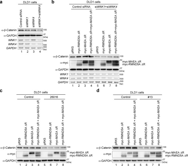
In colorectal cancer cells, missense mutations are frequently found in exon 3 of CTNNB1, which encodes the N-terminal region of β-Catenin, including serine–threonine phosphorylation sites for GSK3β that induce β-Catenin degradation39. Mutations of the N-terminal region such as S33, S37 and S41 cause stabilization of β-Catenin and activation of target gene expression driving tumour formation40. As shown above, we identified that WNK regulates the β-Catenin level through GID E3 ligase and showed that the N-terminal region of β-Catenin is dispensable for WNK-dependent degradation (Figs. 2 and 3), suggesting that the mechanism of WNK-dependent β-Catenin degradation is a candidate therapeutic target for colorectal cancer. To explore this possibility, we checked the effect of WNK on the β-Catenin level in several colorectal cancer cell lines (DLD1 and HCT116 cells) in addition to SW480 cells. Consistent with the data from SW480 cells, knockdown of both WNK1 and WNK4 reduced the β-Catenin levels in DLD1 and HCT116 cells (Fig. 5a and Supplementary Fig. S3a, respectively). Next, we checked the effect of the RMND5A ΔR and MAEA ΔR mutants on the β-Catenin level in these cells. The reduction in β-Catenin level by the knockdown of both WNK1 and WNK4 was rescued by the expression of the dominant-negative forms of RNMD5A and MAEA (Fig. 5b and Supplementary Fig. S3b, respectively). In addition, the reduction of β-Catenin by WNK inhibitors 26016 and #13 was also restored by RMND5A ΔR and MAEA ΔR in these cells (Fig. 5c, d and Supplementary Fig. S3c, d, respectively). These results suggest that WNK regulates the β-Catenin level in various colorectal cancer cells.
Fig. 5. WNK regulates the protein level of β-Catenin in DLD1 colorectal cancer cells.
a Western blot analysis of endogenous β-Catenin following the knockdown of WNK1 and/or WNK4 in DLD1 cells. b–d Western blot analysis of endogenous β-Catenin following the knockdown of both WNK1 and WNK4 (b), treatment with the WNK inhibitor 26016 (c) or #13 (d), or the expression of RMND5A ΔR and/or MAEA ΔR in DLD1 colorectal cancer cells.
WNK inhibitors induce xenograft tumour growth regression
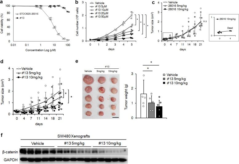
Previous studies reported that the induction of β-Catenin degradation by small molecules is effective to suppress colorectal cancer development41, suggesting that β-Catenin degradation mediated by the WNK inhibitors 26016 and #13 would repress colorectal cancer development. Therefore, we evaluated the anti-tumour effect of WNK inhibitors. SW480 cells were treated with 26016 and #13, and cell viability or growth was measured. 26016 effectively induced cell death after 24 h, while #13 only slightly affected cell death, even at a high dose (Fig. 6a). However, #13 caused significant suppression of cell growth (Fig. 6b). We next analysed the effects of 26016 and #13 on xenograft tumour formation in vivo. SW480 cells were subcutaneously injected into immunodeficient mice, 26016 or #13 was injected intraperitoneally twice per week, and tumour growth was monitored. We observed little or no effect of 26016 on xenograft progression at low doses, but at a high dose of 26016, four of five mice died after only two injections (Fig. 6c). This suggests that 26016 has more severe toxicity than efficacy in vivo. By contrast, treatment of #13 resulted in reduced tumour size and weight in a dose-dependent manner compared with the findings for tumours from vehicle injection (Fig. 6d, e). Moreover, the amount of β-Catenin in the tumours treated with #13 was reduced in a manner dependent on the concentration of #13 and tumour size (Fig. 6f). These findings suggest that #13 has low toxicity and prevents colorectal cancer development through β-Catenin degradation.
Fig. 6. WNK inhibitor suppresses the xenograft development of colorectal cancer cells.
a Cell viability assays of SW480 cells treated with 26016 or #13 for 24 h. n = 3 biologically independent experiments. Circles indicate individual data. b Growth curves of SW480 cells treated with #13 for the indicated days. n = 3 biologically independent experiments. Circles indicate individual data. c, d Immunodeficient mice were subcutaneously injected with SW480 cells (1 × 10⁶ cells). The dose of 5 or 10 mg/kg of 26016 (c) and #13 (d) was intraperitoneally injected twice a week for 3 weeks, and the tumour size was measured before the injection of inhibitors. The inset shows high magnification of the rectangle in (c). n = 5 biologically independent animals. Circles indicate individual data, n.s. indicates not significant. e The tumours and tumour weights after injection of #13 for 3 weeks in each mouse are indicated in (d). Circles indicate individual data. Scale bar, 1 cm. f Western blot analysis of β-Catenin expression level in SW480 xenografts after injection of #13. Values and error bars express mean ± standard deviation (SD). * indicates p < 0.05. p value was calculated by Student’s t test versus vehicle.
Discussion
In the Wnt signalling pathway, to regulate β-Catenin expression, the destruction complex phosphorylates β-Catenin and then ubiquitinates it through βTrCP7. The ubiquitinated β-Catenin is degraded by the proteasome. However, several E3 ubiquitin ligases other than βTrCP have been identified, including the GID complex11–17. In this study, we showed that WNK affected the protein and ubiquitination levels of not only wild-type β-Catenin (Fig. 1) but also β-Catenin ΔN (Fig. 2). We also found that WNK can control the β-Catenin level through the GID complex. Knockdown of RMND5A and MAEA of the GID complex could rescue the effect of WNK knockdown (Fig. 2), and the presence of both WNK1 and WNK4 could change the interaction between β-Catenin and MAEA (Fig. 3), suggesting that the simultaneous stabilization of WNK1 and WNK4 would be important in the destruction complex of the canonical Wnt signalling pathway. The levels of both WNK1 and WNK4 were increased by Wnt stimulation (Supplementary Fig. S4), as well as the β-Catenin level, although the transcription level of neither WNK1 nor WNK4 changed. Moreover, we previously reported that GSK3β is bound to WNK127, suggesting that WNK proteins might be located in the destruction complex of Wnt signalling. Taken together, our data showed that the presence of both WNK1 and WNK4 might provide fine-tuning to attenuate the interaction between β-Catenin and the GID complex and inhibit the ubiquitination of β-Catenin in the Wnt signalling pathway.
A previous study reported that WNK is involved in the hyper-phosphorylation of Dsh in Drosophila S2 cells and that WNK functions as a positive regulator of the Wnt signalling pathway28. However, we found that the knockdown of WNK1 and WNK4 had no effect on DVL1 phosphorylation induced by Wnt stimulation (Supplementary Fig. 5), and that WNK acts downstream of DVL (Fig. 1). Furthermore, WNK affected the β-Catenin ΔN level (Fig. 2) and was not associated with βTrCP E3 ubiquitin ligase (Fig. 3). Therefore, our results suggest that DVL is not related to WNK function in the Wnt signalling pathway. Further study will be required concerning the interaction between WNK and DVL.
β-Catenin degradation under normal conditions is likely regulated by phosphorylation through the destruction complex of the Wnt signalling pathway and ubiquitination by βTrCP. In addition, several E3 ubiquitin ligases other than βTrCP, such as SIAH1, SHPRH and GID complex11,13,15,16, are involved in β-Catenin degradation, indicating that β-Catenin is degraded through multiple mechanisms. We found that the β-Catenin degradation mediated through the knockdown of WNK could not be rescued by knockdown of βTrCPs, SIAH1 or SHPRH (Fig. 2). These results indicate that WNK controls β-Catenin degradation independently of βTrCPs, SIAH1 or SHPRH. However, WNK might be located near the destruction complex as we discussed above, suggesting that the GID complex localizes near the destruction complex with βTrCP. Furthermore, we found that the protein levels of WNK1 and WNK4 were upregulated by Wnt stimulation (Supplementary Fig. S4). This suggests the possibility that Wnt signalling also regulates the activity of the GID complex for fine-tuning the level of β-Catenin protein with βTrCP-mediated degradation.
In this study, we found that the knockdown of WNK caused the reduction of not only the wild type but also the constitutively active type of β-Catenin through the GID complex (Fig. 2). This suggests that WNK ordinarily prevents the interaction between the E3-ligases RMND5A and MAEA, and β-Catenin. A previous paper showed that SHPRH is a negative regulator of Wnt signalling and is involved in the degradation of both the wild type and active forms of nuclear β-Catenin15. However, we did not detect the involvement of SHPRH in the regulation of β-Catenin degradation by WNK (Fig. 2). Furthermore, the lysine demethylase KDM2a/b was recently identified as a regulator of β-Catenin methylation42. KDM2a/b in the nucleus demethylates β-Catenin, which in turn enhances the ubiquitination level of β-Catenin and degradation of β-Catenin, even β-Catenin ΔN. Because WNK in the destruction complex protects the GID complex activity in the cytoplasm, it appears that WNK functions independently of SHPRH and KDM2a/b.
As stated above, we identified that WNK controls the β-Catenin level via the GID complex (Figs. 2 and 3). Moreover, we found that the WNK inhibitors 26016 and #13 function as inhibitors of the Wnt signalling pathway. As shown in Figs. 4 and 5 and Supplementary Fig. 3, WNK inhibitors reduced the β-Catenin level in several colorectal cancer cell lines. However, the WNK inhibitor 26016 caused death in mice, and the other inhibitor, #13, clearly repressed the growth of xenograft tumour cells (Fig. 5). The structural difference between 26016 and #13 is a substitution of the side chain from 2-furanylmethylamino to 3-cyanobenzamido38. We still do not know how this structural difference affects the viability of mice, but our results indicate that the WNK inhibitor #13 likely works as an antitumour drug in vivo. Because the regulation of β-Catenin degradation by WNK is so unique, the WNK inhibitor is a potential anti-tumour drug. β-Catenin is involved not only in colorectal cancer but also in other cancers, such as breast cancer, lung cancer and leukaemia6. Furthermore, abnormal β-Catenin accumulation causes other diseases, such as autosomal dominant polycystic kidney disease5. These findings indicate that WNK is a potential target for treating cancers or other diseases via the dysregulation of β-Catenin.
Methods
Ethics statement
All animal experiments were performed in accordance with the ethical guidelines of Tokyo Medical and Dental University, Nippon Medical School and the Law and Notification of the Government of Japan. Animal protocols were reviewed and approved by the animal welfare committee of Tokyo Medical and Dental University and the Ethics Committee on Animal Experiments of Nippon Medical School.
Cultured cell lines
The cell lines used in this study were HEK293T, SW480, HCT116 and DLD1. Dulbecco’s Modified Eagle Medium (DMEM) supplemented with 10% foetal bovine serum (FBS) was used to culture HEK293 and SW480 cells, McCoy’s 5A supplemented with 10% FBS was used to culture HCT116 cells and RPMI 1640 supplemented with 10% FBS was used to culture DLD1 cells. For the co-transfection of short interfering RNA (siRNA) and plasmids, we used the TransIT-X2 Dynamic Delivery System (Mirus Bio) and polyethyleneimine (Polysciences), respectively. The siRNA target sequences are summarized in Supplementary Table 1.
Antibodies and chemicals
The antibodies used in this study were as follows: rabbit anti-β-Catenin (Abcam), rabbit anti-Flag, rabbit anti-HA, rabbit anti-myc (Medical & Biological Laboratory), rabbit anti-WNK1, rabbit anti-WNK4, mouse anti-myc (Cell Signaling Technology) and anti-rabbit HRP-conjugated (GE) antibodies.
STOCKS2S-26016 was purchased from Tocris and #13 was synthesized at the Kagechika laboratory. MG132 was purchased from Merck.
Reverse-transcription polymerase chain reaction (RT-PCR) analysis
Total RNA was isolated using TRIzol reagent (Invitrogen). cDNA synthesis was carried out using Moloney murine leukaemia virus reverse transcriptase (Invitrogen). GAPDH was used to normalize the cDNA samples. The sequences of the PCR primer pairs are summarized in Supplementary Table 2.
Quantification
Quantitative PCR was performed using the Applied Biosystems 7300 Real-Time PCR Cycler (ABI) and THUNDERBIRD SYBR qPCR Mix (TOYOBO). GAPDH was used to normalize the cDNA samples.
Plasmids
We made several WNK1 or WNK4 constructs with different molecular tags (myc, HA) from WNK1 or WNK4 constructs with Flag-tag26. DVL1, DVL2, DVL3, β-Catenin, RMND5A and MAEA cDNA were obtained by RT-PCR and were constructed in the pRK5 vector. To construct the constitutively active form of β-Catenin, as well as dominant-negative forms of RMND5A and MAEA, we performed site-directed mutagenesis.
Immunoprecipitation
HEK293T cells were transfected with the indicated expression vector. Lysates were prepared from transfected cells and were immunoprecipitated with the indicated antibodies and Protein A/G PLUS-agarose (Santa Cruz Biotechnology). Immunoprecipitates were subjected to sodium dodecyl sulphate–polyacrylamide gel electrophoresis and western blotting, and were then detected using the image analyser LAS-4000 Mini (GE).
Cell viability and growth
Cell viability was analysed using WST1 assays (Takara). SW480 cells (2 × 10⁴ cells) in 96-well plates were treated with the indicated chemicals for 24 h, and WST1 was added to each well. The absorbance (450 or 630 nm) was measured using a microplate reader, and the viability of WNK inhibitor-treated cells was determined. For cell growth, SW480 cells (1 × 10⁵) were seeded in six-well plates and were treated with WNK inhibitor #13. The number of #13-treated cells was counted by Vi-CELL (Beckman) on the indicated days, and cell growth curves were created.
Xenograft mouse model
Five-week-old BALB/cAJ1-nu/nu male mice were obtained from CLEA Japan. All of the mice used in the experiments were assigned randomly. SW480 cells (1 × 10⁶) were subcutaneously injected into the mice. When the tumour size reached approximately 200 mm3, all of the mice were injected intraperitoneally with the indicated chemicals twice weekly for 3 weeks at a dose of 5 or 10 mg/kg. The tumour sizes were measured before each injection. At the end of the experiments after injection for 3 weeks, the animals were sacrificed and the tumour weights were measured. Collected tumours were homogenized and applied for immunoblotting assays to analyse β-Catenin expression.
Statistics and reproducibility
All experiments were conducted at least three times independently. The data were analysed using Microsoft Excel (Microsoft) and StatPlus (AnalystSoft). Values and error bars express mean ± standard deviation (SD) and are representative of at least three independent experiments.
Reporting summary
Further information on the research design is available in the Nature Research Reporting Summary linked to this article.
Supplementary information
Description of Additional Supplementary Files
Acknowledgements
We thank Yoko Mitsutomo for technical assistance. This study was supported by Grants-in-Aid for Scientific Research from the Ministry of Education, Culture, Sports, Science and Technology of Japan (A.S., M.S. and H.S.) and Nanken-Kyoten, Tokyo Medical and Dental University (H.S.). We thank Edanz Group (https://en-author-services.edanzgroup.com/ac) for editing a draft of this manuscript.
Author contributions
A.S. and H.S. designed the study. A.S., M.S. and T.G. performed experiments and analysed the data. M.S. and N.T. performed xenograft experiments. H.M. and H.K. synthesized chemical compounds. All authors discussed the data. A.S., M.S. and H.S. wrote the manuscript.
Data availability
The material used in this study as #13 is available from H.K. upon reasonable request. The other data are available from the corresponding author upon request. Source data behind the graphs are available in Supplementary Data 1. All full immunoblot and gel images are shown in Supplementary Fig. 6.
Competing interests
The authors declare no competing interests.
Footnotes
Publisher’s note Springer Nature remains neutral with regard to jurisdictional claims in published maps and institutional affiliations.
These authors contributed equally: Atsushi Sato, Masahiro Shimizu.
Supplementary information
Supplementary information is available for this paper at 10.1038/s42003-020-01386-2.
References
- 1.Steinhart Z, Angers S. Wnt signaling in development and tissue homeostasis. Development. 2018;145:dev146589. doi: 10.1242/dev.146589. [DOI] [PubMed] [Google Scholar]
- 2.Goldstein B, Takeshita H, Mizumoto K, Sawa H. Wnt signals can function as positional cues in establishing cell polarity. Dev. Cell. 2006;10:391–396. doi: 10.1016/j.devcel.2005.12.016. [DOI] [PMC free article] [PubMed] [Google Scholar]
- 3.Loh KM, van Amerongen R, Nusse R. Generating cellular diversity and spatial form: Wnt signaling and the evolution of multicellular animals. Dev. Cell. 2016;38:643–655. doi: 10.1016/j.devcel.2016.08.011. [DOI] [PubMed] [Google Scholar]
- 4.Welters HJ, Kulkarni RN. Wnt signaling: relevance to beta-cell biology and diabetes. Trends Endocrinol. Metab. 2008;19:349–355. doi: 10.1016/j.tem.2008.08.004. [DOI] [PubMed] [Google Scholar]
- 5.Benzing T, Simons M, Walz G. Wnt signaling in polycystic kidney disease. J. Am. Soc. Nephrol. 2007;18:1389–1398. doi: 10.1681/ASN.2006121355. [DOI] [PubMed] [Google Scholar]
- 6.Zhan T, Rindtorff N, Boutros M. Wnt signaling in cancer. Oncogene. 2017;36:1461–1473. doi: 10.1038/onc.2016.304. [DOI] [PMC free article] [PubMed] [Google Scholar]
- 7.Nusse R, Clevers H. Wnt/β-Catenin signaling, disease, and emerging therapeutic modalities. Cell. 2017;169:985–999. doi: 10.1016/j.cell.2017.05.016. [DOI] [PubMed] [Google Scholar]
- 8.Bhanot P, et al. A new member of the frizzled family from Drosophila functions as a Wingless receptor. Nature. 1996;382:225–230. doi: 10.1038/382225a0. [DOI] [PubMed] [Google Scholar]
- 9.Tamai K, et al. LDL-receptor-related proteins in Wnt signal transduction. Nature. 2000;407:530–535. doi: 10.1038/35035117. [DOI] [PubMed] [Google Scholar]
- 10.Lien WH, Fuchs E. Wnt some lose some: transcriptional governance of stem cells by Wnt/β-catenin signaling. Genes Dev. 2014;28:1517–1532. doi: 10.1101/gad.244772.114. [DOI] [PMC free article] [PubMed] [Google Scholar]
- 11.Winston JT, et al. The SCFbeta-TRCP-ubiquitin ligase complex associates specifically with phosphorylated destruction motifs in IkappaBalpha and beta-catenin and stimulates IkappaBalpha ubiquitination in vitro. Genes Dev. 1999;13:270–283. doi: 10.1101/gad.13.3.270. [DOI] [PMC free article] [PubMed] [Google Scholar]
- 12.Chitalia VC, et al. Jade-1 inhibits Wnt signalling by ubiquitylating beta-catenin and mediates Wnt pathway inhibition by pVHL. Nat. Cell Biol. 2008;10:1208–1216. doi: 10.1038/ncb1781. [DOI] [PMC free article] [PubMed] [Google Scholar]
- 13.Dimitrova YN, et al. Direct ubiquitination of beta-catenin by Siah-1 and regulation by the exchange factor TBL1. J. Biol. Chem. 2010;285:13507–13516. doi: 10.1074/jbc.M109.049411. [DOI] [PMC free article] [PubMed] [Google Scholar]
- 14.Hay-Koren A, Caspi M, Zilberberg A, Rosin-Arbesfeld R. The EDD E3 ubiquitin ligase ubiquitinates and up-regulates beta-catenin. Mol. Biol. Cell. 2011;22:399–411. doi: 10.1091/mbc.e10-05-0440. [DOI] [PMC free article] [PubMed] [Google Scholar]
- 15.Qu Y, et al. Axitinib blocks Wnt/β-catenin signaling and directs asymmetric cell division in cancer. Proc. Natl Acad. Sci. USA. 2016;113:9339–9344. doi: 10.1073/pnas.1604520113. [DOI] [PMC free article] [PubMed] [Google Scholar]
- 16.Goto T, Matsuzawa J, Iemura S, Natsume T, Shibuya H. WDR26 is a new partner of Axin1 in the canonical Wnt signaling pathway. FEBS Lett. 2016;590:1291–1303. doi: 10.1002/1873-3468.12180. [DOI] [PMC free article] [PubMed] [Google Scholar]
- 17.Dominguez-Brauer C, et al. E3 ubiquitin ligase Mule targets β-catenin under conditions of hyperactive Wnt signaling. Proc. Natl Acad. Sci. USA. 2017;114:E1148–E1157. doi: 10.1073/pnas.1621355114. [DOI] [PMC free article] [PubMed] [Google Scholar]
- 18.Moniz S, Jordan P. Emerging roles for WNK kinases in cancer. Cell Mol. Life Sci. 2010;67:1265–1276. doi: 10.1007/s00018-010-0261-6. [DOI] [PMC free article] [PubMed] [Google Scholar]
- 19.Veríssimo F, Jordan P. WNK kinases, a novel protein kinase subfamily in multi-cellular organisms. Oncogene. 2001;20:5562–5569. doi: 10.1038/sj.onc.1204726. [DOI] [PubMed] [Google Scholar]
- 20.Xu B, et al. WNK1, a novel mammalian serine/threonine protein kinase lacking the catalytic lysine in subdomain II. J. Biol. Chem. 2000;275:16795–16801. doi: 10.1074/jbc.275.22.16795. [DOI] [PubMed] [Google Scholar]
- 21.Wilson FH, et al. Human hypertension caused by mutations in WNK kinases. Science. 2001;293:1107–1112. doi: 10.1126/science.1062844. [DOI] [PubMed] [Google Scholar]
- 22.Shekarabi M, et al. Mutations in the nervous system-specific HSN2 exon of WNK1 cause hereditary sensory neuropathy type II. J. Clin. Invest. 2008;118:2496–2505. doi: 10.1172/JCI34088. [DOI] [PMC free article] [PubMed] [Google Scholar]
- 23.Sun X, Gao L, Yu RK, Zeng G. Down-regulation of WNK1 protein kinase in neural progenitor cells suppresses cell proliferation and migration. J. Neurochem. 2006;99:1114–1121. doi: 10.1111/j.1471-4159.2006.04159.x. [DOI] [PubMed] [Google Scholar]
- 24.Tu SW, Bugde A, Luby-Phelps K, Cobb MH. WNK1 is required for mitosis and abscission. Proc. Natl Acad. Sci. USA. 2011;108:1385–1390. doi: 10.1073/pnas.1018567108. [DOI] [PMC free article] [PubMed] [Google Scholar]
- 25.Björklund M, et al. Identification of pathways regulating cell size and cell-cycle progression by RNAi. Nature. 2006;439:1009–1013. doi: 10.1038/nature04469. [DOI] [PubMed] [Google Scholar]
- 26.Sato A, Shibuya H. WNK signaling is involved in neural development via Lhx8/Awh expression. PLoS ONE. 2013;8:e55301. doi: 10.1371/journal.pone.0055301. [DOI] [PMC free article] [PubMed] [Google Scholar]
- 27.Sato A, Shibuya H. Glycogen synthase kinase 3ß functions as a positive effector in the WNK signaling pathway. PLoS ONE. 2018;13:e0193204. doi: 10.1371/journal.pone.0193204. [DOI] [PMC free article] [PubMed] [Google Scholar]
- 28.Serysheva E, et al. Wnk kinases are positive regulators of canonical Wnt/β-catenin signalling. EMBO Rep. 2013;14:718–725. doi: 10.1038/embor.2013.88. [DOI] [PMC free article] [PubMed] [Google Scholar]
- 29.Aberle H, Bauer A, Stappert J, Kispert A, Kemler R. beta-catenin is a target for the ubiquitin-proteasome pathway. EMBO J. 1997;16:3797–3804. doi: 10.1093/emboj/16.13.3797. [DOI] [PMC free article] [PubMed] [Google Scholar]
- 30.Yost C, et al. The axis-inducing activity, stability, and subcellular distribution of beta-catenin is regulated in Xenopus embryos by glycogen synthase kinase 3. Genes Dev. 1996;10:1443–1454. doi: 10.1101/gad.10.12.1443. [DOI] [PubMed] [Google Scholar]
- 31.Munemitsu S, Albert I, Rubinfeld B, Polakis P. Deletion of an amino-terminal sequence beta-catenin in vivo and promotes hyperphosporylation of the adenomatous polyposis coli tumor suppressor protein. Mol. Cell Biol. 1996;16:4088–4094. doi: 10.1128/MCB.16.8.4088. [DOI] [PMC free article] [PubMed] [Google Scholar]
- 32.Munemitsu S, Albert I, Souza B, Rubinfeld B, Polakis P. Regulation of intracellular beta-catenin levels by the adenomatous polyposis coli (APC) tumor-suppressor protein. Proc. Natl Acad. Sci. USA. 1995;92:3046–3050. doi: 10.1073/pnas.92.7.3046. [DOI] [PMC free article] [PubMed] [Google Scholar]
- 33.Pfirrmann T, et al. RMND5 from Xenopus laevis is an E3 ubiquitin-ligase and functions in early embryonic forebrain development. PLoS ONE. 2015;10:e0120342. doi: 10.1371/journal.pone.0120342. [DOI] [PMC free article] [PubMed] [Google Scholar]
- 34.Braun B, et al. Gid9, a second RING finger protein contributes to the ubiquitin ligase activity of the Gid complex required for catabolite degradation. FEBS Lett. 2011;585:3856–3861. doi: 10.1016/j.febslet.2011.10.038. [DOI] [PubMed] [Google Scholar]
- 35.Maitland MER, et al. The mammalian CTLH complex is an E3 ubiquitin ligase that targets its subunit muskelin for degradation. Sci. Rep. 2019;9:9864. doi: 10.1038/s41598-019-46279-5. [DOI] [PMC free article] [PubMed] [Google Scholar]
- 36.Santt O, et al. The yeast GID complex, a novel ubiquitin ligase (E3) involved in the regulation of carbohydrate metabolism. Mol. Biol. Cell. 2008;19:3323–3333. doi: 10.1091/mbc.e08-03-0328. [DOI] [PMC free article] [PubMed] [Google Scholar]
- 37.Mori T, et al. Chemical library screening for WNK signalling inhibitors using fluorescence correlation spectroscopy. Biochem. J. 2013;455:339–345. doi: 10.1042/BJ20130597. [DOI] [PubMed] [Google Scholar]
- 38.Ishigami-Yuasa M, et al. Development of WNK signaling inhibitors as a new class of antihypertensive drugs. Bioorg. Med. Chem. 2017;25:3845–3852. doi: 10.1016/j.bmc.2017.05.034. [DOI] [PubMed] [Google Scholar]
- 39.Gao C, et al. Exon 3 mutations of CTNNB1 drive tumorigenesis: a review. Oncotarget. 2018;9:5492–5508. doi: 10.18632/oncotarget.23695. [DOI] [PMC free article] [PubMed] [Google Scholar]
- 40.Kim S, Jeong S. Mutation hotspots in the β-catenin gene: lessons from the Human Cancer Genome Databases. Mol. Cell. 2019;42:8–16. doi: 10.14348/molcells.2018.0436. [DOI] [PMC free article] [PubMed] [Google Scholar]
- 41.Hwang SY, et al. Direct targeting of β-catenin by a small molecule stimulates proteasomal degradation and suppresses oncogenic Wnt/β-catenin signaling. Cell Rep. 2016;16:28–36. doi: 10.1016/j.celrep.2016.05.071. [DOI] [PMC free article] [PubMed] [Google Scholar]
- 42.Lu L, et al. Kdm2a/b lysine demethylases regulate canonical Wnt signaling by modulating the stability of nuclear β-catenin. Dev. Cell. 2015;33:660–674. doi: 10.1016/j.devcel.2015.04.006. [DOI] [PubMed] [Google Scholar]
Associated Data
This section collects any data citations, data availability statements, or supplementary materials included in this article.
Supplementary Materials
Description of Additional Supplementary Files
Data Availability Statement
The material used in this study as #13 is available from H.K. upon reasonable request. The other data are available from the corresponding author upon request. Source data behind the graphs are available in Supplementary Data 1. All full immunoblot and gel images are shown in Supplementary Fig. 6.


