Naidoo J, Cottrell TR, Lipson EJ, et al. Chronic immune checkpoint inhibitor pneumonitis. J Immunother Cancer 2020;8:e000840. doi: 10.1136/jitc-2020-000840.
Since the online publication of this article, the authors have noticed the following errors:
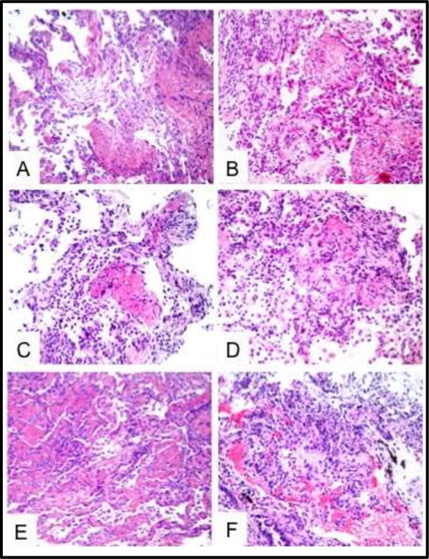
Figure 2 is incorrect. Please see corrected figure 2 shown below as figure 1:
Figure 1.

Pathologic Features of Patients with Chronic PD-1 Pneumonitis. H&Estaining of lung biopsy samples from patients with chronic pneumonitis. (A)Patient 1: Bronchiolitis obliterans Organizing Pneumonia (BOOP), described inmanuscript as organizing pneumonia, with an intra-alveolar fibroblast plug andorganizing alveolar fibrin; (B) Patient 2: Acute lung injury with acutefibrinous organizing pneumonia and organizing diffuse alveolar damage; (C) Patient 3: Organizing alveolar fibrin consistent with exudative phase of BOOP;(D) Patient 4: Organizing pneumonia (BOOP) with organizing alveolar fibrin; (E)Patient 5: Organizing alveolar fibrin consistent with exudative phase of BOOP;(F): Patient 6: Organizing pneumonia (BOOP).
The legend for figure 3 was incorrectly linked to online supplemental figure 2 and vice versa. The correct figure 3 legend is ‘Chronic pneumonitis is associated with brisk lymphocytic inflammation, including many proliferating PD-1 +CD8+T cells. A representative case of chronic pneumonitis (Patient 5) with abundant lymphocytic inflammation is shown (H&E, top left, see also figure 2). Profiling of the inflammatory microenvironment with multiplex immunofluorescence (top center) reveals a dramatic recruitment of PD-1 +lymphocytes (green, top right) and numerous CD8 +cytotoxic T cells (white, bottom left). Many of the PD-1 +and CD8+lymphocytes are positive for the proliferation marker Ki67 (red, bottom center), including numerous PD1 +CD8+T cells (bottom right, arrows highlight PD-1 +CD8+Ki67+cells). Original magnification 200 x.’
jitc-2020-000840corr1supp001.pdf (794.6KB, pdf)
The correct online supplemental figure 2 legend is ‘Highly proliferative PD-1 +cell accumulation characterizes chronic pneumonitis. Low power H&E and mIF images highlight the relative abundance of Ki67 +PD-1+lymphocytes in chronic pneumonitis (top row) relative to sparse inflammatory cells seen in histologically unremarkable lung tissue (bottom row). Original magnification 200 x.’
The updated supplementary files are linked to this correction article.
jitc-2020-000840corr1supp002.pdf (228.5KB, pdf)
Associated Data
This section collects any data citations, data availability statements, or supplementary materials included in this article.
Supplementary Materials
jitc-2020-000840corr1supp001.pdf (794.6KB, pdf)
jitc-2020-000840corr1supp002.pdf (228.5KB, pdf)


