Abstract
Hepatocyte nuclear factor 1 homeobox alpha (HNF1α) is a transcription factor involved in endodermal organogenesis and pancreatic precursor cell differentiation and development. Earlier studies have reported a role for HNF1α in pancreatic ductal adenocarcinoma (PDAC) but it is controversial. The mechanism by which it impacts PDAC is yet to be explored in depth. In this study, using the online databases we observed that HNF1α is upregulated in PDAC, which was also confirmed by our immunohistochemical analysis of PDAC tissue microarray. Silencing HNF1α reduced the proliferative, migratory, invasive and colony forming capabilities of pancreatic cancer cells. Key markers involved in these processes (pPI3K, pAKT, pERK, Bcl2, Zeb, Snail, Slug) were significantly changed in response to alterations in HNF1α expression. On the other hand, overexpression of HNF1α did not induce any significant change in the aggressiveness of pancreatic cancer cells. Our results demonstrate that reduced expression of HNF1α leads to inhibition of pancreatic cancer growth and progression, which indicates that it could be a potential oncogene and target for PDAC.
Subject terms: Cancer, Biomarkers
Introduction
Advances in cancer research, diagnosis, and treatment have led to an overall increase in the 5-year survival rate for all cancers combined. However, minimal progress has been made with respect to pancreatic ductal adenocarcinoma (PDAC)—a highly lethal disease with a 5-year survival rate of only 8%1. While PDAC is not among the most common forms of cancer, it is predicted to become the second leading cause of cancer-related death by 20302. Given that less than 20% of PDAC patients present as suitable candidates for surgical resection, chemotherapy remains the main mode of treatment1. However, chemotherapy is often ineffective, due to the highly metastatic nature and complexity of the disease3. While identifying potential biomarkers for development of targeted drug therapy remains critical, there is also a strong need to reassess the role of specific biomarkers that may provide insight into the intricate nature of this disease.
One such biomarker is hepatocyte nuclear factor 1 alpha (HNF1α), a transcription factor first identified in hepatocytes that is expressed in the yolk sac endoderm, and in the developing kidney, liver, and pancreas in a spatio-temporal manner4. In adult tissues, HNF-1 regulates epithelia-specific gene expression of the gut, lung, liver, pancreas, and urogenital tract5–7. Aside from its role in liver development, HNF1α has also been shown to be involved in pancreas development as one of the central regulators of pancreatic precursor cell differentiation8,9. In some previous studies, PDAC tissue was found to express lower levels of HNF1α compared to normal pancreatic tissue, suggesting that its dysregulation may be a key step in PDAC development9. As such, HNF1α has been suggested to play a tumor suppressing role, specifically in both liver and pancreatic cancers. However, recent studies have shown an oncogenic role for HNF1α10,11. The current knowledge on the exact role of HNF-1α in pancreatic carcinogenesis is contradictory, and the significance of targeting this protein for therapeutic purposes remains unidentified. Its mechanism of action within PDAC has yet to be elucidated.
With this previous knowledge in mind, we set out to study the effects of HNF1α in PDAC. We focused specifically on the role of HNF1α in the development and progression of PDAC in hopes to apply this knowledge in the development of more targeted and effective treatments for this disease. We found that silencing HNF1α reduced the proliferative, migratory, invasive and colony forming capabilities of pancreatic cancer cells, consistent with previous research that has identified it as a novel oncogene in pancreatic cancer10. Thus, continued research into the role of HNF1α is worthy of further exploration.
Results
HNF1α expression in PDAC cells
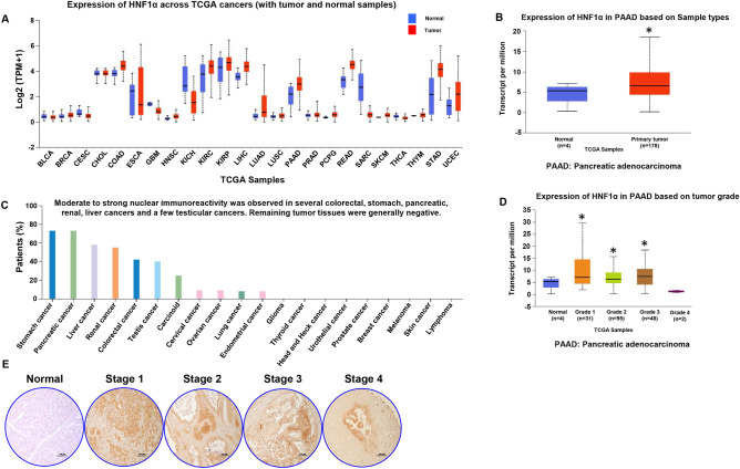
The expression levels of HNF1α in all cancers (found by using TCGA database through the UALCAN portal) revealed higher HNF1α expression in PDAC pancreata compared to normal pancreata (Fig. 1A,B). Next, we analyzed the immunoreactivity of HNF1α among all cancers in the TCGA database and found that PDAC had the second highest immunoreactivity next only to stomach cancer (Fig. 1C). Analysis of expression of HNF1α in PDAC based on tumor grade indicates that early stage disease has higher expression and as the disease progresses the expression decreases (Fig. 1D). Immunohistochemical analyses of PDAC TMA revealed increased expression of HNF1α in early stage PDAC compared to the normal control, with expression decreasing as the stage of PDAC advanced (Fig. 1E). The difference in staining could be due to the occurrence of huge desmoplasia in advanced pancreatic cancers.
Figure 1.
Expression levels of HNF1α in cancers. (A) Expression of HNF1α across TCGA cancers with tumor and normal samples. (B) Expression of HNF1α in pancreatic adenocarcinoma (n = 178) and normal pancreas (n = 4). (C) Immunoreactivity of HNF1α across TCGA cancers. (D) Expression of HNF1α in pancreatic adenocarcinoma based on tumor grade; normal (n = 4), grade 1 (n = 31) grade 2 (n = 95), grade 3 (n = 48) and grade 4 (n = 2). (E) Immunohistochemistry analysis of normal pancreas tissue and tumor stages of human pancreatic ductal adenocarcinoma tissues, HNF1α-positive cells indicated by brown staining at × 100 magnification. *p < 0.05.
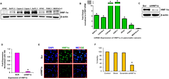
The expression of HNF1α was analyzed in several PDAC cell lines (BxPC-3, Capan-2, Capan-1, AsPC-1, HPAC, PANC-1, and MIAPaCa-2) and normal pancreas cell line (hTERT HPNE). Western blot analysis showed that there was a range of HNF1α expression in this panel of pancreatic cell lines, with AsPC-1 having very high HNF1α expression and HPAC having very low HNF1α expression (Fig. 2A). Data from qRT-PCR analysis was similar to western blot analysis; we observed that the PDAC cells express higher levels of HNF1α compared to normal pancreatic cells (Fig. 2B). Immunofluorescence analysis also verified the increased expression levels of HNF1α in AsPC-1 cells compared to hTERT HPNE normal pancreas cells, showing HNF1α is mainly localized in the nucleus of the PDAC cells (Supplementary Fig. S1A).
Figure 2.
Expression of HNF1α in pancreatic cancer lines and its suppression using siRNAs. (A) Western blot analysis of HNF1α expression in a panel of pancreatic cancer cell lines (HPAC, Capan-2, Capan-1, AsPC-1, PANC-1, Mia PaCa-2, and BxPC-3). (B) Real-time PCR analysis of mRNA expression of HNF1α in pancreatic cancer lines compared to normal pancreas cell line hTERT HPNE. The fold expression of HNF1α in hTERT HPNE was equated to one. Each bar represents the mean of three independent experiments (p < 0.05). Expression of HNF1α in hTERT HPNE cells was considered as unit value = 1. (C) Western blot analysis of HNF1α protein expression in HNF1α silenced AsPC-1 pancreatic cancer cells. (D) Relative mRNA expression of HNF1α was assessed by real-time PCR in siHNF1α AsPC-1 cells. Data shown as mean ± SEM. Experiments (n = 3) were repeated three times in triplicates. *p < 0.05. (E) Representative photographs of immunofluorescence analysis of HNF1α expression in siHNF1α AsPC-1 cells at × 100 magnification. (F) MTS assay was used to measure cell viability in response to HNF1α silencing in AsPC-1 cells.
To investigate its role in PDAC, HNF1α was silenced in AsPC-1 and overexpressed in HPAC cells. Three distinct HNF1α-specific siRNAs (subtypes A, B, and C) were used to observe the efficacy of silencing HNF1α. Western blot analysis revealed the 30 nM siRNA subtype “A” as most effective in silencing HNF1α in AsPC-1 cells (Fig. 2C and Supplementary Fig. S1B), which was further validated by qRTPCR (Fig. 2D) and immunoflurorescence (Fig. 2E). Three different colonies transfected with 1 µg concentration of HNF1α pcDNA were used to survey the efficacy of HNF1α overexpression. Colony 3, which was transfected with 1 µg of HNF1α pcDNA, showed the highest expression of HNF1α according to western blot analysis (Supplementary Fig. S2A). qRT-PCR analysis performed confirmed the efficacy of overexpression of HNF1α in HPAC cells (Supplementary Fig. S2B). The HNF1α-silenced AsPC-1 and HNF1α-overexpressing HPAC cells generated were used for all further experiments.
HNF1α influences cell proliferation and survival in PDAC cells
Cancer cells retain the ability to proliferate rapidly and evade death12. In order to assess whether HNF1α expression influences these abilities in pancreatic cancer cells, cell viability was measured in HNF1α-silenced AsPC-1 cells and HNF1α-overexpressing HPAC cells. Silencing HNF1α decreased the viability of AsPC-1 cells significantly (by nearly 70%) when compared to scramble control AsPC-1 cells (Fig. 2F). Overexpression of HNF1α, on the other hand, had a very minimal effect on the viability of HPAC cells when compared to pCMV control HPAC cells (Supplementary Fig. S3A).
Targeting HNF1α impacts proliferation of PDAC cells through PI3K-AKT-mTOR signaling
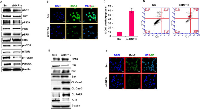
Silencing HNF1α in AsPC-1 cells significantly reduced expression levels of the active forms of AKT, PI3K, ERK, mTOR and p70s6k. Accumulation of total forms of ERK, mTOR and p70S6K was observed in HNF1α silenced AsPC-1 cells (Fig. 3A). Overexpression of HNF1α in HPAC did not significantly reduce the active forms of these proteins (Supplementary Fig. S3B). Immunofluorescence imaging also verified the reduced expression of proliferative marker pAKT in response to suppression of HNF1α (Fig. 3B). These data reveal that silencing HNF1α alters the proliferative and survival potential of PDAC cells through the PI3K/AKT/mTOR signaling pathway.
Figure 3.
Silencing HNF1α gene expression inhibits proliferation and induces apoptosis in PDAC. (A) Expression levels of pAKT, AKT, PP13K, PI3K, pERK, ERK, pmTOR, mTOR, pP70S6K, and P70S6K were determined by western blot in silenced HNF1α AsPC-1 cells. (B) Immunofluorescence analysis showed cell proliferation markers for pAKT in siHNF1α AsPC-1 cells at × 100 magnification. (C,D) Apoptosis analysis of siHNF1α AsPC-1 cells measured by flow cytometry. (E) Western blot analysis of apoptotic markers in siHNF1α AsPC-1 cells. (F) Immunofluorescence analysis of BCl2 in siHNF1α AsPC-1 cells at × 100 magnification. Data shown as mean ± SEM. Experiments (n = 3) were repeated three times in triplicates. *p < 0.05.
Suppressing HNF1α gene expression induces apoptosis
The role of HNF1α in apoptosis was examined using Annexin V/PI staining. Silencing HNF1α drastically increased apoptosis by 48.3% (Fig. 3C,D) while over expression of HNF1α slightly reduced apoptosis by 3.2% (Supplementary Fig. S4A & S4B). To further confirm the role of HNF1α in apoptosis, the key molecular markers involved in this process were studied. Silencing HNF1α in AsPC-1 cells resulted in increased levels of pro-apoptotic proteins p53, Bim, Bak, cleaved caspases 3 and 8, and cleaved PARP, while the expression of anti-apoptotic protein Bcl-2 was reduced (Fig. 3E). Over expression of HNF1α in HPAC cells did not change these apoptotic markers (Supplementary Fig. S4C). Immunofluorescence data further confirmed the role of HNF1α in apoptosis by detecting the reduced expression of anti-apoptotic marker Bcl-2 in response to suppression of HNF1α (Fig. 3F). Our results indicate that the significance of HNF1α expression is crucial in apoptosis.
HNF1α influences migration, invasion, and anchorage-independent growth of PDAC cells
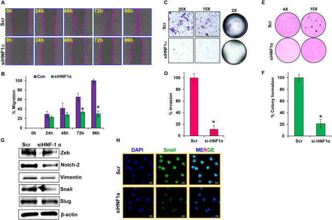
Silencing HNF1α in AsPC-1 cells significantly decreased the migratory abilities of these cells when compared to the control cells (Fig. 4A). Control cells migrated nearly 100% and closed the scratch within 96 h, while HNF1α-silenced AsPC-1 cells only migrated about 30% (Fig. 4B). Overexpression of HNF1α in HPAC cells had no observable effect on the migratory abilities of these cells compared to control cells (Supplementary Fig. S5A,B). Silencing HNF1α reduced invasion by nearly 90% in AsPC-1 cells compared to the control (Fig. 4C,D). Overexpression of HNF1α, however, had very little effect (< 20%) on the invasive capabilities of HPAC cells compared to the control (Supplementary Fig. S5C,D).
Figure 4.
HNF1α regulates metastatic characteristics of PDAC. (A,B) Wound-healing assay was performed in siHNF1α AsPC-1 cells; migration was analyzed using Nikon Biostation CT at 2 h intervals for up to 96 h at × 4 magnification. (C,D) Invasiveness of siHNF1α AsPC-1 cells and overexpression HNF1α HPAC cells were observed using a Matrigel invasion assay, and captured using Nikon Eclipse TS 100 microscope at × 20 and × 100 magnification. (E,F) Colony formation assay was performed in siHNF1α AsPC-1 cells. (G) EMT markers analyzed by western blot in siHNF1α AsPC-1 cells. (H) Immunofluorescence analysis of Snail in siHNF1α AsPC-1 cells, captured using Nikon SMZ 1500 microscope at × 40 magnification. Data shown as mean ± SEM. Experiments (n = 3) were repeated three times in triplicates. *p < 0.05.
Silencing HNF1α greatly reduced colony formation (~ 80%) in AsPC-1 cells compared to the control cells (Fig. 4E,F). There was no significant effect on colony formation when HNF1α was overexpressed in HPAC cells compared to the control cells (Supplementary Fig. S6A,B). Overall, the data collected suggest that silencing HNF1α greatly reduces migration, invasion, and colony formation in PDAC cells.
To confirm HNF1α’s contribution in metastatic colony formation, we studied a variety of key genes that have been shown to be involved in epithelial to mesenchymal transition (EMT) using western blotting analysis. Suppressing HNF1α expression in AsPC-1 cells, resulted in an observable reduction in Zeb, Notch-2, Vimentin, Snail, and Slug expression (Fig. 4G), while overexpressing HNF1α in HPAC was not able to effect any change in the expression of these EMT markers (Supplementary Fig. S6C). Immunofluorescence imaging also verified the reduced expression levels of Snail in response to suppression of HNF1α in PDAC cells (Fig. 4H). These data indicates that HNF1α influences metastatic processes by altering the expression of key signaling molecules involved in EMT.
Impact of HNF1α expression in PDAC preclinical models
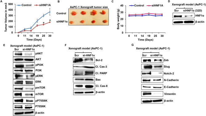
To confirm our in vitro outcomes, we also conducted in vivo studies using athymic nude mice. In these mice, either HNF1α silenced AsPC-1 cells or parental AsPC-1 cells were transplanted into the flanks of the nude mice. By silencing HNF1α, tumor growth was reduced in xenografts when compared to control tumors (Fig. 5A,B). There was not significant change in the body weights among the two groups (Fig. 5C). The xenograft tumors from both the groups were surgically excised and used for molecular analysis. First, we looked for the levels of expression of HNF1α and found that HNF1α silenced xenografts still had lower levels of HNF1α expression compared to control xenografts (Fig. 5D).
Figure 5.
Silencing HNF1α inhibits PDAC tumorigenesis. (A) Tumor growth curve of siHNF1α AsPC-1 cells. (B) AsPC-1 Xenograft tumors from control and siHNF1α silenced groups (n = 6). (C) Body weights of AsPC-1 control and siHNF1α silenced experimental nude mice (n = 6). (D) Western blot analysis of HNF1α protein expression in siHNF1α AsPC-1 cells. (E) Western blot analysis of cell proliferation markers in siHNF1α AsPC-1 xenografts. (F) Western blot analysis of apoptotic marker in siHNF1α AsPC-1 cells and overexpression HNF1α in HPAC xenograft. (G) Western blot analysis of EMT markers in siHNF1α AsPC-1 cells. Data shown as mean ± SEM. Experiments (n = 3) were repeated three times in triplicates. *p < 0.05.
Further, we examined effect of HNF1α on proliferation, EMT, and apoptosis markers using xenograft tumor tissues from HNF1α silenced AsPC-1 and control xenograft tumor tissues. Suppression of HNF1α led to downregulation of proliferative markers: AKT, PI3K, ERK, mTOR and P70S6K (Fig. 5E). Apoptotic markers were analyzed in the HNF1α-silenced group and showed decreased anti-apoptotic marker Bcl-2, increased cleaved caspase 3, and cleaved PARP. Bax and caspase 8 were decreased significantly compared to control group (Fig. 5F). Immunoblot data showed a decrease in EMT markers in HNF1α-silenced tissue (Zeb, Slug, Notch-2, N-Cadherin, and Vimentin), while E-cadherin levels stayed constant. EMT markers Snail and N-Cadherin were both downregulated in the HNF1α-silenced AsPC-1 xenograft tumor tissues compared to the control experimental group (Fig. 5G).
Discussion
Initially HNF1α was described as a glucose metabolism regulator, with a possibly important role in diabetes13–15. HNF1α is also potentially enriched in the exocrine-like/ADEX group of tumors, and the HNF1α positive subtype of PDAC is resistant to certain tyrosine kinase inhibitors and paclitaxel16. HNF1α -positive PDAC patients are expected to respond better to intensive chemotherapy according to the FOLFIRINOX protocol17.
The data from TCGA database analyzed showed that the expression and immunoreactivity of HNF1α was upregulated in PDAC tissues compared to normal tissues. Further, the dataset also showed the differential expression of HNF1α at different PDAC tumor grades. Immunohistochemical analysis of the PDAC tissue microarray also showed that the expression of HNF1α protein was higher in PDAC tissues compared to normal noncancerous tissues. Further, it also confirmed the differential expression of HNF1α at different tumor grades.
We found high expression of HNF1α in PDAC cell lines compared to the normal pancreas cell line. In particular, the expression of HNF1α is high in AsPC-1 and Capan-1 compared to other panels of PDAC cell lines screened. This could be due to the fact that these two cell lines were derived from metastatic sites, while the other cell lines were derived from primary cancers. In addition, it has been demonstrated that the doubling time of AsPC -1 cells is shorter than the other PDAC cell lines18. This finding provides compelling insight that HNF1α may play a role in PDAC. When examining the role of HNF1α on PDAC cells after altering its expression, the present study found that HNF1α is an oncogene. For a gene to be considered as an oncogene, it has to have the ability to initiate and promote cancer development19. In this process, it is well established that epithelial to mesenchymal transition plays a vital role in inducing and increasing cancer cell invasion, migration and colony forming capabilities20,21. Decreased expression of HNF1α resulted in significantly reduced levels of proliferation, migration, invasion, and colony formation of PDAC cells, compared to the control. On the other hand, when overexpressed, HNF1α had little to no effect on these same PDAC cell abilities. Thus, these findings suggest HNF1α plays a role as an oncogene and not as a tumor suppressor, as previously thought.
Earlier it has been demonstrated that tumor suppressor miR-484 modulates the ZEB1 and SMAD222 and WNT/ MAPK pathway by directly targeting HNF1α in cervical cancer cells11. Further, it also been shown that HNF1α significantly promoted pancreatic cancers by influencing fibroblast growth factor receptor 423. Recently, it has been shown that HNF1α is a putative regulator of PDAC stem cell gene signature10. Further, it has also been demonstrated that HNF1α is required for tumor growth, tumorsphere formation, invasion and migration. In addition, the mechanism by which HNF1α promotes PDAC stemness is by regulating the pluripotency factor POU5F1/OCT4. Expression of HNF1α upregulated genes has been associated with poor survival outcomes in PDAC patients10.
Our data demonstrates that overexpression of HNF1α did not significantly alter any of the key processes and key markers involved in PDAC growth and progression. The reason we did not see any significant effect of HNF1α overexpression on PDAC growth and progression could be due to the fact that HPAC cells are highly aggressive to begin with, and increasing the expression of HNF1α might not have been able to increase their aggressiveness any further.
Overall, our data demonstrate that when silenced, HNF1α significantly reduces PDAC cell proliferation, migration, invasion, and colony formation, demonstrating the oncogenic role it plays in PDAC. While the exact role of HNF1α remains somewhat elusive, it provides insight into the complexity of PDAC and demonstrates a need for further studies that may lead to the identification of suitable targets for successful PDAC treatment. Considering the highly deadly and intricate nature of this disease, it is critical that targets of interest be evaluated completely in order to establish successful PDAC treatments that may change the dire outlook for patients with this disease.
Materials and methods
Cell lines
HPAC, Capan-2, Capan-1, AsPC-1, PANC-1, MIA PaCa-2 and BxPC-3 (pancreatic cancer cell lines) and hTERT HPNE (normal pancreas cell line) were all obtained from the American Type Culture Collection. All cell lines were tested for mycoplasma contamination. HPAC, PANC-1, BxPC-3 and AsPC-1 cell lines were cultured in RPMI-1640 media containing 10% FBS, 100 Units/mL of penicillin, and 100 μg/mL of streptomycin. All cells were cultured as described in our earlier study24.
UALCAN database analysis
UALCAN (https://ualcan.path.uab.edu) is a publically available database, which is a comprehensive and interactive online resource that can be used to analyze cancer omics data. This web resource is built on PERL-CGI platform. This web resource provides access to TCGA datasets and also provides additional information about genes by linking to the human protein atlas, Pubmed, Human Protein Resource Database, GeneCards, etc.25. We used the UALCAN database to identify the significance of HNF1α expression in pancreatic cancer. We analyzed the expression and immunoreactivity of HNF1α across TCGA cancers including pancreatic cancers. We further also analyzed the expression of HNF1α in pancreatic cancer based on tumor grade.
Silencing of HNF1α in AsPC-1 Cell Line
AsPC-1 cells were seeded in a 6-well plate at a density of 2.5 × 10⁵ cells/well. Twenty-four hours after seeding, cells were transfected with various concentrations (10 nM, 30 nM and 50 nM) of HNF1α different siRNAs (subtype A, B and C) for 48 h (Origene, Catalog # SR304741). Scrambled siRNA was used as a control. MIrus bio TransIT siQUEST transfection reagent (Mirus Bio) was used to perform all the siRNA transfection studies as described earlier24,26.
Sequences for HNF1α siRNA subtypes “A”, “B”, and “C”
SR304741A-rGrGrArGrUrGrCrArArUrArGrGrGrCrGrGrArArUrGrCrATC.
SR304741B-rGrGrArCrArGrGrArCrUrArArCrArCrUrCrArGrArArGrCCT.
SR304741C-rCrGrGrUrGrUrGrCrGrCrUrArUrGrGrArCrArGrCrCrUrGCG.
Overexpression of HNF1α in HPAC Cells
DH5α competent cells (Invitrogen) were transformed with 2 µg of plasmid DNA for human HNF1α (Origene, Catalog # SC300093) or PCMV-XL6 cloning vector alone (Origene, Catalog # PCMV6XL6). The transformed cultures were spread on a pre-warmed 4% agar plate with 100 µg/mL ampicillin (Life Technologies). The plate was incubated at 37 °C overnight. Single colonies were selected and inoculated in LB broth with 25 µg/mL ampicillin overnight. DNA was isolated and purified from HNF1α plasmid-transfected E.coli cells using Invitrogen Pure Link HiPure Plasmid Filter Maxiprep Kit (Life Technologies). HPAC cells were seeded in 6-well plates at a density of 2.5 × 10⁵ cells/well. Twenty-four hours after seeding, cells were transfected with 1 μg of HNF1α pDNA from four different colonies. The transfection was carried out using MIrus bio TransIT 2020 transfection reagent (Mirus Bio) as described earlier24. In order to maintain efficient overexpression and avoid toxicity, the ratio of transfection reagent to plasmid DNA was conserved at 5:1 according to the manufacturer’s protocol. HNF1α overexpression was verified with western blot and immunofluorescence analysis. The third colony transfected with 1 μg HNF1α pDNA was the most effective in overexpressing HNF1α. This was used for all HNF1α overexpression experiments, including cell proliferation, migration, invasion, colony formation assay, immunofluorescence, xenograft studies, mRNA, and protein analysis.
Pancreatic adenocarcinoma tissue array
The pancreatic adenocarcinoma tissue microarray (TMA) from US Biomax, Inc. were subjected to immunohistochemistry (IHC) to measure HNF1α expression levels. This microarray consist of formalin-fixed paraffin embedded samples (n = 24) of pancreatic adenocarcinoma at different stages along with normal pancreatic tissues.
Immunohistochemical analysis (IHC)
IHC was performed as described earlier27. In brief, TMA sections were deparaffinized and hydrated using decreasing concentrations of ethanol baths. Following antigen retrieval with trilogy, the tissue samples were blocked and then incubated at 4 °C with a primary antibody against HNF1α followed by Ultra Marque polyscan HRP labeled secondary antibody (Cell Marque). Sections were stained with 3,3′diaminobenzidine and counterstained with hematoxylin and dehydrated. Finally, the slides were sealed with mounting media (Surgipath Medical Industries), and images were captured using a Nikon Microscope-ECLIPSE 50i. IHC staining for different proteins were quantitated at 5 random areas per section with at least 200 cells per field.
Cell viability assay
The MTS (3-(4,5-dimethylthiazol-2-yl)-5-(3-carboxymethoxyphenyl)-2-(4-sulfophenyl)-2H-tetrazolium) assay was performed as described earlier28. MTS reagent was added for 4 h to HNF1α-silenced AsPC-1 cells and HNF1α-overexpressing HPAC cells t and cell viability was measured using a microplate reader (CLARIOstar, BMG LABTECH).
Wound healing assay
HNF1α-silenced AsPC-1 cells and HNF1α-overexpressing HPAC cells, were seeded in 6-well plates at a density of 2.5 × 10⁵ cells/well and maintained at 37ºC in a 5% CO2 environment for 48 h. A scratch was created with a sterile pipette tip once the cells reached monolayer confluency. The cells detached due to the scratch were removed and the respective complete growth media was added to the plates. Then, the cells were provided their respective complete growth media. The distance migrated by the cells were capturedand calculated at 2 h intervals for 96 h using the Nikon Biostation CT. NIS-Element AR software26.
Matrigel invasion assay
HNF1α-silenced AsPC-1 cells and HNF1α-overexpressing HPAC cells were plated in the upper chamber of a transwell polycarbonate insert coated with matrigel (1 mg/ml) at a density of 8 × 10⁴ cells/well. The complete growth media for each cell line was added as a chemoattractant in the lower chamber of the transwell. The invaded cells were fixed and stained with 0.2% crystal violet in 5% formalin. Excess stain was removed by washing with PBS. Nikon Eclipse TS 100 microscope was used to capture five arbitrarily selected fields to calculate the number of invaded cells26.
Colony formation assay
A clonogenic assay was also performed with siHNF1α AsPC-1 cells and OVHNF1α HPAC cells, each plated separately on 60-mm dishes with a top layer of 0.7% agarose at a density of 2 × 10⁴ cells and a bottom layer of 1% agar. Cells were maintained by changing the complete growth media three times a week. Then, colonies were fixed and stained with 0.2% crystal violet in 5% formalin solution. Colonies were counted manually and images were obtained using a Nikon SMZ 1500 microscope as described earlier27.
Immunofluorescence analysis
AsPC-1 and HPAC cells were each plated at a density of 10 × 103 cells/well in an 8-well chamber slide. Twenty-four hours after seeding, the AsPC-1 and HPAC cells were transfected with HNF1α siRNA or HNF1α overexpression plasmid and incubated for 48 h. The cells were fixed with 100% methanol for 10 min, followed by 100% acetone fixation for another 10 min. Cells were permeabilized using 0.2% Triton X-100 in PBS for 20 min and blocked with 5% BSA for 1 h. Slides were incubated overnight with the primary antibody. Subsequently, Alexa fluor 488-conjugated secondary antibody (Life Technologies) was added and incubated for 1 h. The cells were then washed and counterstained with DAPI. Slides were observed using a Nikon laser scanning confocal microscope.
Quantitative reverse transcriptase real time PCR (qRT-PCR)
Total RNA was extracted from hTERT HPNE and PDAC cell lines using the Trizol reagent (Invitrogen, Carlsbad, CA, USA). RNA concentration and quality were verified using a NanoDrop 2000 spectrophotometer (ThermoFisher Scientific). cDNA was prepared using the RT2 first strand kit (Qiagen). Human specific primers for HNF1α (QT00085428) and RRN18S (QT00199367) uwere purchased from Qiagen. The qRT-PCR was performed in triplicates using the Quantitech SYBR green kit (Qiagen) in a StepOne Plus Real Time PCR system (Applied Biosystems). The data from qRT-PCR was analyzed using comparative Ct method as described earlier24.
Western blot analysis
Mammalian protein extraction reagent (M-PER) was used to extract proteins from cultured cells and xenograft tumor tissues. BCA method was used to quantify the extracted proteins at 490 nm using withCLARIOstar, BMG LABTECH. Proteins were resolved using SDS-PAGE and then transferred onto PVDF membranes. The membranes were blocked with 5% BSA (Sigma-Aldrich Corporation), for 45 min. The membranes were incubated overnight at 4ºC with primary antibodies. The membranes were washed 3 times with TBS-T, followed by a 2-h incubation with the respective secondary horseradish peroxide-coupled antibodies. The expression levels of proteins from various treated groups were measured using enhanced chemiluminescence (GE Las-4000) as described earlier29.
All methods described above were in accordance to the institutional guidelines and approved by Texas Tech University Health Sciences Center El Paso Institutional Biosafety Committee.
Xenograft studies
All the animal experiments performed in this study followed the institutional guidelines and was approved by the Texas Tech University Health Sciences Center El Paso Institutional Animal Care and Use Committee. HNF1α silenced AsPC-1 cells and their respective parental cells, were implanted subcutaneously in both the flanks (1.0 × 10⁶ cells/flank) of six week old male athymic nude mice (Harlan Laboratories) (n = 6). Tumor volume and body weight were measured twice a week. Thirty days post transplantation, the mice were euthanized and the xenograft tumors were surgically excised. Excised tissues were fixed in formalin or snap frozen for further histological and molecular analysis respectively24.
Statistical analysis
Statistical analysis was performed using GraphPrism version 6.01 software (GraphPad Software, Inc.). Power analysis was used to calculate sample size and to study the effect difference between groups. Statistical significance was calculated by two-tailed unpaired t-test on two groups. A value of p < 0.05 was considered statistically significant. All data are expressed as mean ± SEM from at least three independent experiments. All animal experiments had at least 6 animals per group. Animals were randomly assigned to the different groups.
Supplementary information
Acknowledgements
We thank Texas Tech University Health Sciences Center El Paso for supporting this project.
Author contributions
R.S. co-designed, performed most of the experiments also did data analysis and wrote the manuscript. J.M. performed some cell culture, immunoblot, IHC and migration assay. K.F. performed some invasion assays and RTPCR. C.P. performed some cell culture and immunoblots. A.G. performed some Immunofluorescence experiments. MS performed some immunoblots. S.R. and D.P. performed some cell culture assays and immunoblots. E.P. performed cell culture and flow cytometry. M.C. performed cell culture and cell proliferation assays. R.L. co-designed experiments, performed in vivo experiments, data analysis and edited the manuscript and provided overall supervision.
Competing interests
The authors declare no competing interests.
Footnotes
Publisher's note
Springer Nature remains neutral with regard to jurisdictional claims in published maps and institutional affiliations.
Contributor Information
Ramadevi Subramani, Email: ramadevi.subramani@ttuhsc.edu.
Rajkumar Lakshmanaswamy, Email: rajkumar.lakshmanaswamy@ttuhsc.edu.
Supplementary information
is available for this paper at 10.1038/s41598-020-77287-5.
References
- 1.Siegel RL, Miller KD, Jemal A. Cancer statistics, 2018. CA Cancer. J. Clin. 2018;68:7–30. doi: 10.3322/caac.21442. [DOI] [PubMed] [Google Scholar]
- 2.Rahib L, et al. Projecting cancer incidence and deaths to 2030: the unexpected burden of thyroid, liver, and pancreas cancers in the United States. Cancer Res. 2014;74:2913–2921. doi: 10.1158/0008-5472.CAN-14-0155. [DOI] [PubMed] [Google Scholar]
- 3.Mangge H, et al. New diagnostic and therapeutic aspects of pancreatic ductal adenocarcinoma. Curr. Med. Chem. 2017;24:3012–3024. doi: 10.2174/0929867324666170510150124. [DOI] [PubMed] [Google Scholar]
- 4.Cereghini S, Ott MO, Power S, Maury M. Expression patterns of vHNF1 and HNF1 homeoproteins in early postimplantation embryos suggest distinct and sequential developmental roles. Development. 1992;116:783–797. doi: 10.1242/dev.116.3.783. [DOI] [PubMed] [Google Scholar]
- 5.Harries LW, Brown JE, Gloyn AL. Species-specific differences in the expression of the HNF1A, HNF1B and HNF4A genes. PLoS ONE. 2009;4:e7855. doi: 10.1371/journal.pone.0007855. [DOI] [PMC free article] [PubMed] [Google Scholar]
- 6.Haumaitre C, Reber M, Cereghini S. Functions of HNF1 family members in differentiation of the visceral endoderm cell lineage. J. Biol. Chem. 2003;278:40933–40942. doi: 10.1074/jbc.M304372200. [DOI] [PubMed] [Google Scholar]
- 7.Hajarnis SS, et al. Transcription factor hepatocyte nuclear factor-1beta (HNF-1beta) regulates MicroRNA-200 expression through a long noncoding RNA. J. Biol. Chem. 2015;290:24793–24805. doi: 10.1074/jbc.M115.670646. [DOI] [PMC free article] [PubMed] [Google Scholar]
- 8.Luo Z, et al. Hepatocyte nuclear factor 1A (HNF1A) as a possible tumor suppressor in pancreatic cancer. PLoS ONE. 2015;10:e0121082. doi: 10.1371/journal.pone.0121082. [DOI] [PMC free article] [PubMed] [Google Scholar]
- 9.Hoskins JW, et al. Transcriptome analysis of pancreatic cancer reveals a tumor suppressor function for HNF1A. Carcinogenesis. 2014;35:2670–2678. doi: 10.1093/carcin/bgu193. [DOI] [PMC free article] [PubMed] [Google Scholar]
- 10.Abel EV, et al. HNF1A is a novel oncogene that regulates human pancreatic cancer stem cell properties. Elife. 2018 doi: 10.7554/eLife.33947. [DOI] [PMC free article] [PubMed] [Google Scholar]
- 11.Hu Y, Wu F, Liu Y, Zhao Q, Tang H. DNMT1 recruited by EZH2-mediated silencing of miR-484 contributes to the malignancy of cervical cancer cells through MMP14 and HNF1A. Clin. Epigenet. 2019;11:186. doi: 10.1186/s13148-019-0786-y. [DOI] [PMC free article] [PubMed] [Google Scholar]
- 12.Shen L, Shi Q, Wang W. Double agents: genes with both oncogenic and tumor-suppressor functions. Oncogenesis. 2018;7:25. doi: 10.1038/s41389-018-0034-x. [DOI] [PMC free article] [PubMed] [Google Scholar]
- 13.Bonner C, et al. Bone morphogenetic protein 3 controls insulin gene expression and is down-regulated in INS-1 cells inducibly expressing a hepatocyte nuclear factor 1A-maturity-onset diabetes of the young mutation. J. Biol. Chem. 2011;286:25719–25728. doi: 10.1074/jbc.M110.215525. [DOI] [PMC free article] [PubMed] [Google Scholar]
- 14.Kawasaki E, et al. Identification and functional analysis of mutations in the hepatocyte nuclear factor-1alpha gene in anti-islet autoantibody-negative Japanese patients with type 1 diabetes. J. Clin. Endocrinol. Metab. 2000;85:331–335. doi: 10.1210/jcem.85.1.6304. [DOI] [PubMed] [Google Scholar]
- 15.Lin B, Morris DW, Chou JY. Hepatocyte nuclear factor 1alpha is an accessory factor required for activation of glucose-6-phosphatase gene transcription by glucocorticoids. DNA Cell Biol. 1998;17:967–974. doi: 10.1089/dna.1998.17.967. [DOI] [PubMed] [Google Scholar]
- 16.Noll EM, et al. CYP3A5 mediates basal and acquired therapy resistance in different subtypes of pancreatic ductal adenocarcinoma. Nat. Med. 2016;22:278–287. doi: 10.1038/nm.4038. [DOI] [PMC free article] [PubMed] [Google Scholar]
- 17.Muckenhuber A, et al. Pancreatic ductal adenocarcinoma subtyping using the biomarkers hepatocyte nuclear factor-1Aa and cytokeratin-81 correlates with outcome and treatment response. Clin. Cancer Res. 2018;24:351–359. doi: 10.1158/1078-0432.CCR-17-2180. [DOI] [PubMed] [Google Scholar]
- 18.Deer EL, et al. Phenotype and genotype of pancreatic cancer cell lines. Pancreas. 2010;39:425–435. doi: 10.1097/MPA.0b013e3181c15963. [DOI] [PMC free article] [PubMed] [Google Scholar]
- 19.Storz P, Crawford HC. Carcinogenesis of pancreatic ductal adenocarcinoma. Gastroenterology. 2020;158:2072–2081. doi: 10.1053/j.gastro.2020.02.059. [DOI] [PMC free article] [PubMed] [Google Scholar]
- 20.Lamouille S, Xu J, Derynck R. Molecular mechanisms of epithelial-mesenchymal transition. Nat. Rev. Mol. Cell Biol. 2014;15:178–196. doi: 10.1038/nrm3758. [DOI] [PMC free article] [PubMed] [Google Scholar]
- 21.Su D, et al. Tumor-neuroglia interaction promotes pancreatic cancer metastasis. Theranostics. 2020;10:5029–5047. doi: 10.7150/thno.42440. [DOI] [PMC free article] [PubMed] [Google Scholar]
- 22.Hu Y, et al. miR-484 suppresses proliferation and epithelial-mesenchymal transition by targeting ZEB1 and SMAD2 in cervical cancer cells. Cancer. Cell. Int. 2017;17:36. doi: 10.1186/s12935-017-0407-9. [DOI] [PMC free article] [PubMed] [Google Scholar] [Retracted]
- 23.Shah RN, Ibbitt JC, Alitalo K, Hurst HC. FGFR4 overexpression in pancreatic cancer is mediated by an intronic enhancer activated by HNF1alpha. Oncogene. 2002;21:8251–8261. doi: 10.1038/sj.onc.1206020. [DOI] [PubMed] [Google Scholar]
- 24.Subramani R, et al. FOXC1 plays a crucial role in the growth of pancreatic cancer. Oncogenesis. 2018;7:1–11. doi: 10.1038/s41389-018-0061-7. [DOI] [PMC free article] [PubMed] [Google Scholar]
- 25.Chandrashekar DS, et al. UALCAN: a portal for facilitating tumor subgroup gene expression and survival analyses. Neoplasia. 2017;19:649–658. doi: 10.1016/j.neo.2017.05.002. [DOI] [PMC free article] [PubMed] [Google Scholar]
- 26.Subramani R, et al. Targeting insulin-like growth factor 1 receptor inhibits pancreatic cancer growth and metastasis. PLoS ONE. 2014;9:e97016. doi: 10.1371/journal.pone.0097016. [DOI] [PMC free article] [PubMed] [Google Scholar]
- 27.Subramani R, et al. Gedunin inhibits pancreatic cancer by altering sonic hedgehog signaling pathway. Oncotarget. 2017;8:10891–10904. doi: 10.18632/oncotarget.8055. [DOI] [PMC free article] [PubMed] [Google Scholar]
- 28.Subramani R, et al. Growth hormone receptor inhibition decreases the growth and metastasis of pancreatic ductal adenocarcinoma. Exp. Mol. Med. 2014;46:e117. doi: 10.1038/emm.2014.61. [DOI] [PMC free article] [PubMed] [Google Scholar]
- 29.Arumugam A, et al. Silencing growth hormone receptor inhibits estrogen receptor negative breast cancer through ATP-binding cassette sub-family G member 2. Exp. Mol. Med. 2019;51:1–3. doi: 10.1038/s12276-018-0197-8. [DOI] [PMC free article] [PubMed] [Google Scholar]
Associated Data
This section collects any data citations, data availability statements, or supplementary materials included in this article.


