Guinea pigs (Cavia porcellus) are mammals belonging to the suborder Hystricomorph, 1 of the 2 main suborders of rodents. This species is native to South America and was domesticated thousands of years ago as a source of meat. Currently guinea pigs are raised mainly as pets or laboratory animals. Dermatological conditions are the most commonly diagnosed pathologies in guinea pigs (1,2), with alopecia being the most prevalent clinical dermatological sign, followed by pruritus, scaling, and crusty lesions (3). Alopecia is defined as the absence of hair in an area where it is normally present. This clinical sign can be purely esthetic, or it may be a manifestation of a disease with important health consequences. As alopecia is of multifactorial etiology, it is important to obtain an accurate diagnosis in guinea pigs with this clinical sign. Treatment based on signs alone could lead to failure or only temporary improvement in the patient, causing frustration for both the owner and the treating veterinarian.
Clinical history
As in other species, data collection is of utmost importance as part of an appropriate diagnostic approach. Most dermatoses in guinea pigs have a direct relationship with nutrition and management of the environment; therefore, problem-oriented veterinary records are of great importance for this species (4).
Important information to be collected (4–6):
Origin of the animal (adoption, pet shop, etc.)
Breed or type, sex, and age (juvenile, adult, geriatric, or known date of birth)
Behavioral changes
Weight gain or weight loss
Environmental factors (area, humidity, and temperature)
Feeding and management (diet, bedding, cleaning, cage disinfection)
Other animals owned by the household (same or different species)
Contagion to other animals or humans
Other clinical signs
Age at onset of alopecia
Manifestation of itching, before or after the onset of alopecia
Response to previous treatment
Clinical examination
Guinea pigs are docile, which makes their inspection easy. The initial approach consists of examining the animal from its cage to obtain information on the mental state, locomotion, and respiratory rate. Subsequently, it is necessary to weigh the animal, measure rectal temperature and heart rate, examine mucous membranes, and palpate peripheral lymph nodes (4,7). After the general physical examination has been completed, a careful examination of the coat should be done to look for excessive hair loss and to determine if ectoparasites are present. Alopecia may appear as focal lesions, multifocal lesions, and as large areas of total or partial hair loss. In addition, dermatological lesions such as desquamation, erythema, scabs, excoriations and their distribution on the skin should be evaluated.
It should be remembered that guinea pigs have minimal or absent hair density between the nose and lips, around the lips, on the external pinna, and behind the ears (7).
Useful diagnostic tests
Multiple tests can be performed when presented with a guinea pig with alopecia (Table 1). The use and order of these tests should be based on patient history, clinical examination, and established differential diagnoses.
Table 1.
Diagnostic tests for diagnosis of alopecia in guinea pigs.
| Diagnostic testsa | Comments |
|---|---|
| Hair plucks and trichograms | Identification of self-induced alopecia, observation of ectoparasites (lice, mites), postpartum effluvium (telogen hairs), observation of dermatophyte spores |
| Tape strips | Observation of ectoparasites (lice, mites) |
| Cytology | Morphological identification of infectious microorganisms, observation of inflammatory cells, approach to neoplasms |
| Skin scrapes | Identification of ectoparasites |
| Fungal culture | Identification of dermatophytes; the toothbrush technique is useful |
| Bacterial cultures and antibiogram | Bacterial identification and antibiotic sensitivity |
| Biopsy | Dermatophytosis, neoplasms |
| Imaging (ultrasound, X-ray) | Detection of ovarian cysts, neoplasms, hypovitaminosis C |
Common causes of alopecia in guinea pigs (Table 2)
Table 2.
Causes of alopecia in guinea pigs.
| Causes of non-self-induced alopecia | Causes of self-induced alopecia |
|---|---|
| Barbering (caused by other guinea pigs) | Acariasis (Chirodiscoides caviae, Trixacarus caviae, Ornithonyssus bacoti, Demodex caviae, Acarus farris) |
| Dermatophytosis | Dermatophytosis Barbering (by patient itself) |
| Effluvium postpartum | Pediculosis (Gliricola porcelli, Gyropus ovalis) |
| Pregnancy-associated alopecia | Cutaneous lymphoma |
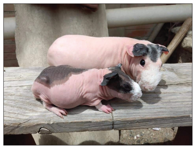
| Hereditary predisposition (Skinny pig) (Figure 4) | |
| Hyperadrenocorticism | |
| Hypovitaminosis C | |
| Hyperthyroidism | |
| Ovarian cysts |
Vitamin C deficiency
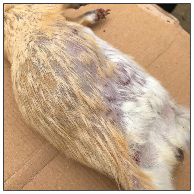
Guinea pigs, like primates, do not synthesize vitamin C because they lack the enzyme L-gulonolactone oxidase, which is necessary for the biosynthesis of vitamin C from glucose (10). Guinea pigs deficient in vitamin C negatively regulate the expression of type IV collagen and elastin, resulting in defects in the integrity of the blood vessels, leading to bleeding or bruising of the skin. Vitamin C deficiency can also affect other organs and reduce immune function (10–12). Guinea pigs need vitamin C in their diet, with an absolute requirement of 10 mg/kg body weight (BW) per day, increasing to 30 mg/kg BW per day during pregnancy (5). Hypovitaminosis C should be considered as a potential underlying factor in bacterial, fungal, or parasitic skin diseases. Cutaneous clinical signs such as alopecia (Figure 1), rough hair, peeling of the ears, petechiae, ecchymosis, and bruising may be present, as well as multiple nonspecific signs, such as weight loss, anorexia, reduced growth rate, stiff or crawling gait, lameness, bleeding gums, diarrhea, pain (manifested by teeth grinding or excessive vocalization), poor wound healing, or even death (5,7,10). The diagnosis is made based on the clinical history including systemic and dermatological signs, diagnostic tests and previous therapeutic results. Radiographs are useful in revealing alterations in the costochondral junctions and widening of the epiphyses of the long bones. Serum Vitamin C levels can be used to confirm the diagnosis (3,10,12). Treatment consists of correction of clinical signs, diet, and vitamin C supplementation of 50 mg/kg BW to 100 mg/kg BW daily parenterally until signs improve. The same dose is subsequently administered orally. Once the animal has returned to normal, dietary supplementation is continued (5,10,12).
Figure 1.
Hypovitaminosis C. Note the presence of alopecia and petechiae.
Dermatophytosis (ringworm)
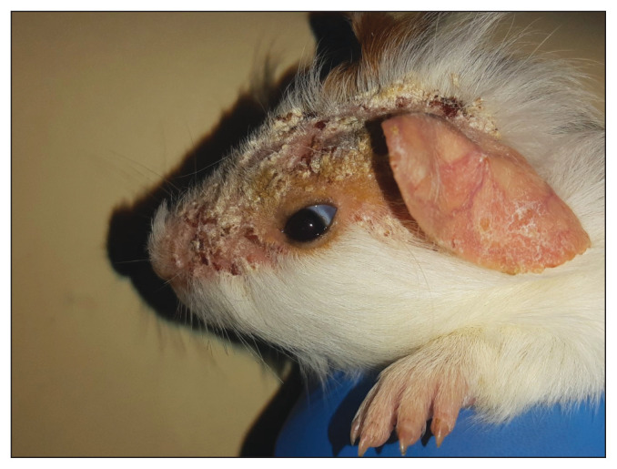
Dermatophytes are filamentous keratinophilic fungi, which generally affect the hair, nails, and keratinized layers of the skin. These fungi are a common cause of skin disease in guinea pigs (13). The incidence of this zoonosis has now increased in guinea pig owners (14). Dermatophyte species belonging to the Trichophyton mentagrophytes complex are mainly reported (7). Very young, old, or immunosuppressed animals are susceptible to infection; however, in healthy animals, spontaneous resolution is common (13). Alopecia is the most frequent clinical sign of this pathology, followed by scabs and scaling, with a more prevalent distribution on the head and face (Figure 2). However, it can affect other areas of the body less frequently, including legs, and flanks of the abdomen. Moderate to severe itching may be present or absent. Without pruritus, the animal may not exhibit dermatological signs and may remain asymptomatic (4,9,14). Since T. mentagrophytes complex dermatophytes do not emit fluorescence under ultraviolet light, the diagnostic value of Wood’s lamp is limited in guinea pigs. Direct examination of hairs, fungal cultures, and histopathology (including periodic acid Schiff, Grocott’s Methenamine Silver staining), however, are useful for the diagnostic approach to dermatophytosis in this species (2,5,7,13). As treatment, topical products containing chlorhexidine, miconazole, enilconazole or systemic pharmacotherapy such as terbinafine 20 mg/kg BW, PO, q24h, and itraconazole 5 mg/kg BW, PO, q24h are effective in guinea pigs affected with dermatophytosis (5,7).
Figure 2.
Dermatophytosis.
Acariasis
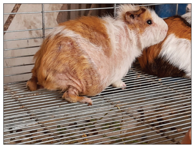
Trixacarus caviae is the most common mite found in guinea pigs, being reported mainly in males and juveniles (1,3). It is a sarcoptiform parasite that generates severe clinical signs and zoonotic potential (2,7). These mites burrow into the skin, creating epidermal tunnels and eliciting a cell-mediated immune response that causes itching (8). Clinical signs are often severe itching (giving the appearance of seizures or spastic movements), generalized alopecia (Figure 3), hyperkeratosis, erythema, and secondary pyoderma (2,3,8). Acariasis is primarily diagnosed in animals from pet stores (4). Diagnosis can be made by skin scraping or acetate tape impressions (3). Treatments include: ivermectin 0.4 mg/kg BW, SC, every 10 to 14 d for 4 treatments; selamectin 15 mg/kg BW, single topical dose; or aerosol products such as fipronil or permethrin (7,8).
Figure 3.
Trixacarus caviae infestation.
Chirodiscoides caviae is another species-specific mite very common in guinea pigs (7). Physical contact and the tendency to gather in the presence of any potential predator facilitates its transmission, with many cases of animals from pet stores being reported (15). The life cycle of C. caviae is approximately 14 d. As clinical signs, guinea pigs present erythema, rough hair coat, pruritus, alopecia, scaling, and ulcers, although some patients may remain asymptomatic for long periods of time (16). Chirodiscoides caviae is found mainly in the flank and trunk area, and the diagnostic techniques that have been used are: trichogram, acetate tape impression, brushing, and scraping (2,15). Effective treatments include subcutaneous ivermectin at a dose of 0.2 to 0.4 mg/kg BW, q7 to 10d for 3 treatments; topical selamectin at a dose of 12 to 15 mg/kg BW, q2wk; and, single topical application of 0.05 mL of moxidectin 1% and imidacloprid 10% (15).
Pediculosis
Gliricola porcelli, the most prevalent louse in guinea pigs, is grayish-yellow in color, and measures between 1 and 2 μm in length. These lice feed on skin debris, being found mainly on the neck, ears, and ventral abdomen. Its life cycle is about 14 to 21 d. Clinical signs of infestation are variable, with pruritus and rough fur being common symptoms. The animal may remain asymptomatic without significant pruritus. Diagnosis can be made by direct visualization of eggs or lice using acetate tape and/or flea combing (3,11,17). Treatment options include subcutaneous ivermectin 0.2 to 0.4 mg/kg BW, q10d for 3 treatments; or, single topical application of 0.05 mL of moxidectin 1% and imidacloprid 10% (5,15,17).
Ovarian cysts
Ovarian cysts are a common finding in middle-aged to older guinea pigs. In most cases, both ovaries are affected, although unilateral cases are also seen with the right ovary being more commonly affected (18). This disorder has a reported prevalence of 76% in female guinea pigs aged 1.5 to 5 y (12). The most common clinical sign, often observed by owners, is progressive hair loss in the flank region and abdomen, without itching or abnormal appearance of the skin. Hair loss is presumed to be the result of increased levels of estrogen produced by neoplastic ovarian cysts or steroidogenic follicular cysts. In addition, crusting of the skin around the nipples, changes in behavior (gathering in groups, aggression, sexual behaviors) may be present along with non-specific systemic clinical signs of loss of appetite, lethargy, or vocalization when manipulated. Some animals may be asymptomatic and cysts may be found incidentally at necropsy (5,6,12). Diagnosis is based on the clinical history, abdominal palpation, imaging techniques such as radiography and ultrasound. The cysts can be up to 10 cm in size and tender on palpation. In some cases, they are associated with concurrent cystic endometrial hyperplasia, mucometra, endometritis, and fibroleiomyoma (5). Surgical treatment is by ovariohysterectomy and is the treatment of choice. Oophorectomy is not recommended because ovarian cysts are associated with multiple uterine diseases (6).
Figure 4.
Skinny pigs. The skinny pig owes its origin to the crossing of hairy guinea pigs and hairless guinea pigs, the latter comes from a strain related to a genetic mutation identified in 1978 in Canada.
Footnotes
The Veterinary Dermatology column is a collaboration of The Canadian Veterinary Journal with the Canadian Academy of Veterinary Dermatology (CAVD). Established in 1986, the CAVD is a not-for-profit organization for veterinarians, veterinary technicians and technologists, and students with an interest in veterinary dermatology. Being under the umbrella of the World Association of Veterinary Dermatology, the mission of the CAVD is to advance the science and practice of veterinary dermatology in Canada by providing education and resources for veterinary teams, supporting research, and promoting excellence in care for animals affected with skin and ear disease.
The CAVD invites everyone with a professional interest in dermatology to join (www.cavd.ca) and share our passion for dermatology, including continuing education in this dynamic field. Annual membership fees are: $50 for regular members and free for students.
Use of this article is limited to a single copy for personal study. Anyone interested in obtaining reprints should contact the CVMA office (hbroughton@cvma-acmv.org) for additional copies or permission to use this material elsewhere.
References
- 1.Minarikova A, Hauptman K, Jeklova E, Knotek Z, Jekl V. Diseases in pet guinea pigs: A retrospective study in 1000 animals. Vet Rec. 2015;177:200. doi: 10.1136/vr.103053. [DOI] [PubMed] [Google Scholar]
- 2.Palmeiro BS, Roberts H. Clinical approach to dermatologic disease in exotic animals. Vet Clin Exot Anim. 2013;16:523–577. doi: 10.1016/j.cvex.2013.05.003. [DOI] [PMC free article] [PubMed] [Google Scholar]
- 3.White SD, Sanchez-Migallon D, Paul-Murphy J, Hawkins MG. Skin diseases in companion guinea pigs (Cavia porcellus): A retrospective study of 293 cases seen at the Veterinary Medical Teaching Hospital, University of California at Davis (1990–2015) Vet Dermatol. 2016;27:1–8. doi: 10.1111/vde.12348. [DOI] [PubMed] [Google Scholar]
- 4.Mitchell MA, Tully TN. Current Therapy in Exotic Pet Practice. 1st ed. St. Louis, Missouri: Elsevier; 2016. pp. 1–47. [Google Scholar]
- 5.Paterson S. Skin diseases and treatment of guinea pigs. In: Paterson S, editor. Skin Diseases of Exotic Pets. 1st ed. Oxford, UK: Blackwell Science; 2006. pp. 232–250. [Google Scholar]
- 6.Bean AD. Ovarian cysts in the guinea pig (Cavia porcellus) Vet Clin Exot Anim. 2013;16:757–776. doi: 10.1016/j.cvex.2013.05.008. [DOI] [PubMed] [Google Scholar]
- 7.Pignon C, Mayer J. Guinea pig. In: Quesenberry KE, Orcutt CJ, Mans C, Carpenter JW, editors. Ferrets, Rabbits, and Rodents Clinical Medicine and Surgery. 4th ed. St. Louis, Missouri: Elsevier; 2020. pp. 270–297. [Google Scholar]
- 8.Donnelly TM. Pruritus and alopecia in guinea pigs. Trixacarus caviae infestation. Lab Animal. 2004;33:21–23. doi: 10.1038/laban0704-21. [DOI] [PubMed] [Google Scholar]
- 9.Bartosch T, Frank A, Günther C, et al. Trichophyton benhamiae and T. mentagrophytes target guinea pigs in a mixed small animal stock. Vet Mycol. 2019;23:37–42. doi: 10.1016/j.mmcr.2018.11.005. [DOI] [PMC free article] [PubMed] [Google Scholar]
- 10.Meredith A. Hypovitaminosis C in the Guinea pig (Cavia Porcellus) Companion Animal. 2006;11:81–82. [Google Scholar]
- 11.Ellis C, Mori M. Skin diseases of rodents and small exotic mammals. Vet Clin North Am Exot Anim Pract. 2001;4:493–542. doi: 10.1016/s1094-9194(17)30041-5. [DOI] [PubMed] [Google Scholar]
- 12.Riggs SM. Guinea pigs. In: Mitchell MA, Tully TN, editors. Manual of Exotic Pet Practice. 1st ed. St Louis, Missouri: Saunders Elsevier; 2009. pp. 456–473. [Google Scholar]
- 13.Donnelly TM, Rush EM, Lackner PA. Ringworm in small exotic pets. Semin Avian Exot Pet Med. 2000;9:82–93. [Google Scholar]
- 14.Kraemer A, Hein J, Heusinger A, Mueller RS. Clinical signs, therapy and zoonotic risk of pet guinea pigs with dermatophytosis. Mycoses. 2013;56:168–172. doi: 10.1111/j.1439-0507.2012.02228.x. [DOI] [PubMed] [Google Scholar]
- 15.D’Ovidio D, Santoro D. Prevalence of fur mites (Chirodiscoides caviae) in pet guinea pigs (Cavia porcellus) in southern Italy. Vet Dermatol. 2014;25:135–137. doi: 10.1111/vde.12110. [DOI] [PubMed] [Google Scholar]
- 16.Schönfelder J, Henneveld K, Schönfelder A, Hein J, Müller R. Concurrent infestation of Demodex caviae and Chirodiscoides caviae in a guinea pig. A case report. Tierärzt Prax. 2010;38:28–30. [PubMed] [Google Scholar]
- 17.Kim SH, Jun HK, Yoo MJ, Kim DH. Use of a formulation containing imidacloprid and moxidectin in the treatment of lice infestation in guinea pigs. Vet Dermatol. 2008;19:187–188. doi: 10.1111/j.1365-3164.2008.00665.x. [DOI] [PubMed] [Google Scholar]
- 18.Pilny A. Ovarian cystic disease in guinea pigs. Vet Clin Exot Anim. 2014;17:69–75. doi: 10.1016/j.cvex.2013.09.003. [DOI] [PubMed] [Google Scholar]


