Abstract
Understanding inorganic nanocrystal (NC) growth dynamic pathways under their native fabrication environment remains a central goal of science, as it is crucial for rationalizing novel nanoformulations with desired architectures and functionalities. We here present an in-situ method for quantifying, in real time, NCs’ size evolution at sub-nm resolution, their concentration, and reactants consumption rate for studying NC growth mechanisms. Analyzing sequential high-resolution liquid-state 19F-NMR spectra obtained in-situ and validating by ex-situ cryoTEM, we explore the growth evolution of fluoride-based NCs (CaF2 and SrF2) in water, without disturbing the synthesis conditions. We find that the same nanomaterial (CaF2) can grow by either a particle-coalescence or classical-growth mechanism, as regulated by the capping ligand, resulting in different crystallographic properties and functional features of the fabricated NC. The ability to reveal, in real time, mechanistic pathways at which NCs grow open unique opportunities for tunning the properties of functional materials.
Subject terms: Materials chemistry, Nanoscale materials, Nanoparticles, NMR spectroscopy
Understanding nanocrystal growth pathways under their native fabrication environment remains a central goal of science. By synthesizing nanofluorides under in-situ NMR conditions, the authors are able to probe their sub-nm growth evolution, elucidating their formation by coalescence or monomer-attachment.
Introduction
Colloidal inorganic nanocrystals (NCs) have revolutionized science thanks to their provision of a controllable synthetic platform for tackling fundamental challenges in materials science and their utility in a variety of applications, from catalysis and renewable energy to nanomedicine1–6. Their exceptional features (e.g., optical, magnetic, chemical, catalytic, and electrical) often stem from their composition, size, and morphology, emphasizing the essentialness of mechanistic knowledge on NC formation from solution for strategizing novel fabrications. The relationships between NC growth pathways and resultant architectures and functions were revealed thanks to the development of a variety of ex situ and in situ methodologies7. Both spectroscopy- and microscopy-based techniques allow us to monitor NCs’ size evolution, alternations in chemical composition, crystallography properties, and change in shape throughout the course of their formation8–19. However, they often require temporal reaction sampling, destructive-energy radiation applications, the use of out-of-equilibrium conditions (e.g., drying, freezing, heating, vacuum) and custom-made apparatuses. Thus, there is a need to implement accessible, alternative in situ approaches that can be performed under authentic nanofabrication settings.
Fluoride-based NCs (i.e., nanofluorides) have garnered much attention recently due to their potential use in optics, catalysis, optoelectronics, bioimaging, superconductor devices, batteries, and lubricants20. Amongst their advantages over other inorganic NCs, their low phonon energies and high chemical stability stand out, making nanofluorides one of the preferable host-materials for upconversion fluorescence21–26. These unique characteristics are governed by their crystallinity, size, shape, and introduced impurities, which are strictly regulated by their formation pathways27–29. Interestingly, the rich fluoride content and relatively small-size of colloidal nanofluorides result in a characteristically high-resolution 19F-NMR spectrum while in solution30. This property has been exploited to design water-dispersed CaF2 NCs as nanotracers for in vivo 19F-MRI applications, but also provides unprecedented opportunity to use nanofluorides as a platform for tracking NC formation in real time with a high-resolution (HR) NMR setup.
The unique capabilities of NMR-based approaches, which allow the monitoring of changes in the chemical environment of a variety of NMR active nuclei, offer a versatile tool for studying growth pathways and kinetics, surface chemistry, and the physical properties of both organic and inorganic materials31–36. In particular, the spectral resolution of NMR shows promise for the characterization of nanomaterials by way of detecting both the organic ligands37 and inorganic core elements38 as a means to explore their formation in solution. Moreover, the ability to spectrally resolve surface from bulk atoms via NMR39–43 makes this method an attractive way to characterize inorganic NC architecture.
Inspired by these studies, we herein present a high-resolution liquid-state NMR-based approach for studying the dynamics of NCs’ size evolution in a quantitative manner without disturbing the reaction conditions (e.g., avoiding sampling, destructive radiation, unwanted heating, undesired freezing, drying, vacuum, etc.). By synthesizing nanofluorides under in situ NMR conditions, and based on the ability to detect the 19F-NMR signals of MF2 (M = Ca2+ or Sr2+) in water, we are able to probe their sub-nm growth over the entire course of their formation, highlighting their controllable growth mechanisms (coalescence vs. classical simple grow), which result in different morphological and functional features.
Results
HR-NMR accurately determines the diameter of NCs in solution
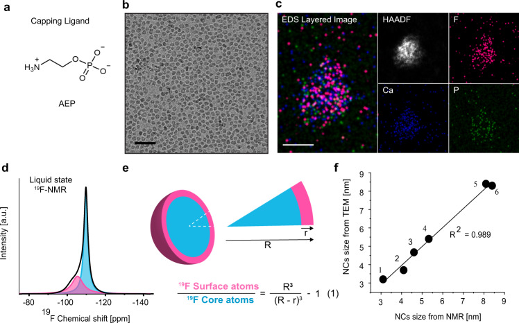
Prior to conducting HR-NMR studies on water-dispersed, small-sized CaF2 NCs capped with 2-aminoethyl phosphate (AEP, at pH7, molecular structure, Fig. 1a), they were synthesized and fully characterized with complementary techniques. Cryo-TEM images revealed monodispersed NCs with an average size of 3.7 ± 0.6 nm (Fig. 1b and Supplementary Fig. 1a) with the confirmed composition of calcium, fluoride, and phosphate (from the ligand), as evaluated by high-angle annular dark-field (HAADF) STEM imaging and energy dispersive X-ray spectroscopy (EDS) (Fig. 1c). The fluorite, faced cubic centered (fcc), crystal structure of the obtained AEP-CaF2 NCs was confirmed by powder X-ray diffraction (XRD, Supplementary Fig. 1b) and their relatively small size as well as their monodispersity were preserved in water, as revealed by dynamic light scattering (DLS, Supplementary Fig. 1c).
Fig. 1. Characterization of AEP-CaF2 NCs.
a Molecular structure of an AEP ligand at pH 7. b Cryo-TEM AEP-CaF2 NCs (scale bar 20 nm). c STEM-EDS elemental maps of an agglomerate of dried AEP-CaF2 NCs (scale bar 10 nm). Shown are a combined map of the NCs elements, a high-angle annular dark-field (HAADF) image (white) and elemental maps of each element, i.e., F (pink), Ca (blue), and P (green). (d) Liquid-state, high-resolution 19F-NMR spectrum of water-dispersed AEP-CaF2 NCs and its deconvoluted signals, attributed to 19F atoms at the surface of the NCs (-105 ppm, pink) and in the NC core (−109 ppm, light blue). e Schematic illustration of the CaF2 NC with its surface (pink) and core atoms (light blue) and the equation (1) used to evaluate the size of NCs from the 19F-NMR spectrum (Eq. (1) and Supplementary Note 1). f Correlation between the sizes measured with NMR and TEM of various CaF2 and SrF2 NCs bearing different ligands (AEP, Citrate (Cit), Oleic Acid (OA) and Polyethylene Glycol (PEG)); (1) Cit-CaF2 (Fig. 5b) (2) AEP-CaF2 (3) AEP-SrF2 (Fig. 3) (4) OA-SrF2 (Supplementary Fig. 3b) (5) PEG-CaF2 (Supplementary Fig. 3a) (6) OA-CaF2 (Supplementary Fig. 3c).
The high-resolution 19F-NMR studies of the synthesized AEP-CaF2 NCs in water resulted in a 19F-NMR spectrum, from which two populations of fluorides could be detected, at −105 and −109 ppm, assigned to fluorides at the surface and core of the NCs, respectively44,45 (Fig. 1d). This assignment was further supported by the significant paramagnetic effect on the 19F-NMR peak attributed to the fluorides on the surface of the formulation (Supplementary Fig. 2) emphasizing one of the key features of NMR to provide detailed information that reflects the chemical environment of constituent nuclei. Indeed, NMR studies of nuclei within the NCs (rather than nuclei of the coating ligands) can distinguish between the nuclei at the NC’s solid core and those located at the outer surface, due to differences in chemical environments, relaxation properties, and crystallinity characteristics39–43.
We next deconvolved the obtained 19F-NMR spectrum for AEP-CaF2 NCs, using computational line fitting to quantify the ratio between the 19F-atoms at the surface of CaF2 NCs (Fig. 1d, pink) and the 19F-atoms at the core of CaF2 NCs (Fig. 1d, light-blue). From this ratio and by using Eq. (1) (Fig. 1e, Supplementary Note 1), we could calculate the average diameter of the AEP-CaF2 NCs in water, which was found to be 4.1 nm (Fig. 1d). This ability to quantify the average diameter of water-dispersed nanofluorides from their HR-19F-NMR spectrum by classification and quantification of surface and core atoms provides a unique approach for evaluating the average size of NCs while in solution.
To demonstrate the robustness and reproducibility of this approach, we applied it to evaluate the sizes of various nanofluoride (CaF2 or SrF2) fabrications (Fig. 1f and Supplementary Fig. 3) from their HR 19F-NMR spectra and found them to be in good correlation (r2 = 0.989) with the sizes obtained from HR-TEM images. This ability to accurately determine the average size of a variety of small-sized NCs demonstrates not only the uniqueness but also the potential variability of the proposed HR-NMR-based method. Importantly, magic-angle spinning (MAS) solid-state 19F-NMR of the same synthetic AEP-CaF2 colloids’ powder showed a similar 19F-NMR spectrum (Supplementary Fig. 4a) to that obtained using high-resolution 19F-NMR (Supplementary Fig. 4b), from which similar NC diameters were calculated. This observation confirms that water-dispersed AEP-CaF2 NCs tumble fast enough in solution so as to average out the intrinsic homonuclear dipolar interactions, allowing to directly detect their fluoride content with HR liquid-state 19F-NMR setups.
Observing NC growth in real time with HR-NMR
The ability to evaluate NC sizes at nanometer scale directly from solution without disturbing the system, using the HR 1D-NMR spectrum, offers a platform to monitor, in real time, NC growth dynamics. We validated this possibility by synthesizing nanofluorides in an NMR tube under ambient conditions while acquiring consecutive 19F-NMR spectra from reaction initiation to final formation, monitoring simultaneously the size of the NCs, their concentration at each timepoint of the reaction and the consumption of the F- anions. Controlling the reaction conditions allowed to shed light on the dynamics of NC formation, highlighting their growth mechanism.
To synthesize the NCs, we started by adding a solution of Ca2+ cations to an aqueous reaction solution containing both F- anions and AEP capping ligands.
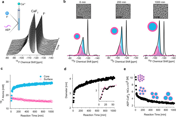
The NMR tube was then placed in a 9.4T NMR spectrometer and sequential 19F-NMR spectra were acquired to follow the temporal formation of CaF2 colloids, resulting in a 19F-NMR stack plot (Fig. 2a). Our results showed that each 19F-NMR spectrum consisted of two main distinct peaks that could be, simultaneously, identified and assigned. One peak resonated at −120 ppm, assigned to the free F- anion, indicating its consumption with time (Supplementary Fig. 5). The second downfield 19F-NMR peak was attributed to the fluoride content of the CaF2 NC, from which the NC’s diameter could be determined using the approach outlined in Fig. 1. Representative line fittings of the experimental 19F-NMR spectra obtained at three different time points after reaction initiation (i.e., 6, 200, and 1000 min) are presented in Fig. 2b, depicting the varying ratio between the two 19F-spin populations of NCs, which reflects their size evolution. Representative cryo-TEM images of the reaction solution at the same reaction time points (i.e., 6, 200, and 1000 min) are shown above each spectrum in Fig. 2b. The amounts of 19F-atoms on the NC surface (pink) and in its core (light blue) were quantified and plotted as a function of the reaction time (Fig. 2c, 3 min temporal resolution). The diameter of the CaF2 NCs at each timepoint was then derived from each individual 19F-NMR spectrum (using Eq. (1) and Supplementary Note 1) and plotted as a function of reaction time (Fig. 2d). From that plot, a notably rapid formation (3 min after reaction initiation) of ultrasmall CaF2 NCs (2.3 nm) is observed, followed by a period of relatively fast growth (up to 3.2 nm within the first 20 min), which then gives way to much slower growth dynamics. In the regime of rapid NCs growth, the 19F-NMR resonance of the surface atoms was significantly affected, as expected for smaller NCs that display a larger curvature (Supplementary Fig. 6).
Fig. 2. Real-Time, high-resolution 19F-NMR of CaF2 NC evolution in water.
a Stack plot of real-time 19F NMR spectra after the addition of Ca+2 to the aqueous solution of AEP and F- anions. Shown spectra were taken every 30 min (experimental temporal resolution, 3 min). b Deconvoluted 19F-NMR spectra from three different time points during the reaction (6, 200, and 1000 min), showing the signals of 19F atoms on the surface (pink) and in the core (blue). Upper images are the correspondent cryo-TEM images of AEP-CaF2 NCs. c Quantitative plot of the 19F-NMR signals of the fluoride atoms in the core and on the surface of the formed NCs, calculated from the convoluted peaks’ areas at −109 ppm and −105 ppm, respectively. d Evolution of the AEP-CaF2 NC diameter in water throughout the reaction process. Diameters were derived from Eq. (1). Inset shows the first 50 min of the reaction; dashed lines are a visual guide for the two respective slopes of the NC evolution stages. e The concentration of AEP-CaF2 NCs (multiply by 105 in the graph) in the reaction solution as a function of reaction time, calculated from Supplementary Equation 2.
Intriguingly, a mild but pronounced decrease in NC diameter was observed between 20 and 30 min (Fig. 2d inset) that is followed by a continuous growth, a phenomenon that could indicate coalescence events during NC growth10,46,47. In parallel to the fast growth rate of the NCs (first 20 min, Fig. 2d), a significant reduction in their concentration was noted (first 20 min, Fig. 2e and Supplementary Equation 2), which was followed by a milder decrease in their total concentration. This observation that numerous small NCs transform into larger (but fewer) NCs, together with the detected intermediate stage (Fig. 2d inset), supports the notion that AEP-CaF2 NCs predominantly grow through coalescence7,48, a conclusion that is in good agreement with the “coalescence point” that can clearly be seen in the inset of Fig. 2d and found to be reproducible (N = 4, Supplementary Fig. 7). Such coalescence events were also captured by both cryo-TEM (Supplementary Fig. 8) and high-resolution TEM (Supplementary Fig. 9) images sampled 25 min from the reaction initiation. Such a mechanistic pathway is promoted by the unfavorable thermodynamic state of initially formed ultrasmall NCs10, together with a relatively high number of NCs in solution, which may increase their collision probabilities and, essentially, lead to their coalescence to form larger, more stable NCs. Note here that Ostwald ripening mechanistic pathway may also occur but it is probably much less prominent than coalescence for AEP-CaF2 NCs growth, as reflected in both in situ NMR spectroscopy (Fig. 2 and Supplementary Fig. 7) and TEM imaging (Supplementary Fig. 8 and Supplementary Fig. 9). Importantly, we further validated the temporal change in the size of the NCs over time with ex situ cryo-TEM. To that end, the reaction mixture was sampled at three different stages (6 min, 200 min, and 1000 min; Fig. 2b and Supplementary Fig. 10). The NC diameters obtained from cryo-TEM images (Supplementary Fig. 10e) were in a very good correlation (Supplementary Fig. 10f, R2 = 0.98) with those calculated from in situ 19F-NMR at the same reaction stages.
The generality of the proposed in-situ 19F-NMR for studying NC formation and growth was demonstrated also for SrF2 NCs (Fig. 3 and Supplementary Fig. 11). Compared to CaF2 NCs, the SrF2 NCs were larger, as determined by cryo-TEM (Fig. 3a, b) and DLS (Fig. 3c). The difference in size in the sub-nm range could be clearly depicted by the HR-19F-NMR approach (Fig. 3d, e), and the method was applied to monitor, in situ and in real time, the formation of AEP-SrF2 NCs. Notably, at three minutes after reaction initiation, the SrF2 NCs were much larger than the CaF2 NCs (2.9 nm vs. 2.3 nm, respectively), a size difference that was preserved throughout the reaction. Similar to the case of CaF2, the temporal evolution of SrF2 NCs is characterized by an initial fast growth followed by a moderate leveling-off of the size with time. Importantly, for both investigated nanofluorides, the concentration of the NCs in the solution decreased in parallel to their growth (Figs. 2d, e, 3e, and Supplementary Fig. 11b). To further elucidate this formation mechanism, we monitored the ligands during NC synthesis by way of in situ liquid-state 31P-NMR (Supplementary Fig. 12). The 31P-NMR peak of free AEP in solution is a clear narrow triplet. Upon the initial formation of NCs, this triplet broadens probably due to the restricted mobility of the bound ligands to the numerous very small NCs that had formed. As the reaction progresses, when the number of NCs in the solution drops (Fig. 2e and Supplementary Fig. 11b), free AEP-ligands are re-released to the solution and their 31P-NMR peak narrows again due to their faster tumbling rates.
Fig. 3. AEP-SrF2 NC formation.
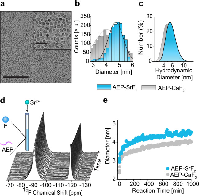
a Cryo-TEM of AEP-SrF2 in water (1000 min after reaction initiation). b, c Size distributions of AEP-SrF2 (light blue) and AEP-CaF2 NCs (gray) as obtained from (b) cryo-TEM and (c) DLS. d Stack plot of the real-time 19F-NMR spectra of AEP-SrF2 synthesis. e Size evolution of the diameter of NCs in water throughout the reaction process for AEP-SrF2 (light blue) and for AEP-CaF2 (gray), as derived from Eq. (1).
The role of ligands in the NC growth pathways
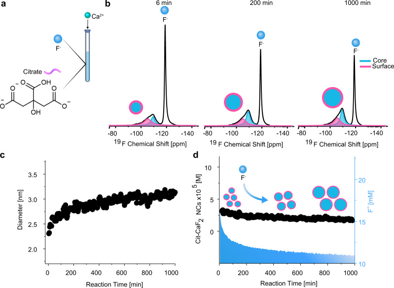
It is well known that ligands regulate NC formation mechanisms and dynamics, as they were found to be key elements in stabilizing both the reaction intermediates8,49 and formed colloids50,51. Specifically, differences in NC growth mechanisms have been attributed to the different affinities of the ligands to the cations52,53 and to their role in stabilizing the formed colloids54. Therefore, we decided to study the growth dynamics of CaF2 NCs in water using a different capping ligand. In contrast to AEP (Fig. 1a), negatively charged ligands may hinder NC interactions at the NC’s initial growth stage, due to electrostatic repulsion, which could lead to a different growth mechanism55. Interestingly, applying in situ 19F-NMR to the synthesis of CaF2 NCs in the presence of the negatively charged citric acid (citrate at pH = 7, Fig. 4a) as the capping ligand revealed a different growth mechanism than that observed for AEP-CaF2 (compare Fig. 2 and Fig. 4). We detected in the representative 19F-NMR spectra obtained at different reaction time points (i.e., 6 min, 200 min, and 1000 min, Fig. 4b) a notable consumption of free fluoride (peak at −120 ppm). This observation implies a growth mechanism that differs from particle-coalescence found for AEP-CaF2. Assigning the 19F-NMR peaks at −105 and −109 ppm to the fluorides of the surface and of the core of Cit-CaF2NCs, respectively, allows to determine their size evolution (using Eq. (1) and Supplementary Note 1), revealing their continuous growth (Fig. 4c), while their final size was further verified by TEM (Supplementary Fig. 13).
Fig. 4. Ligand effect on CaF2 NC’s growth mechanism.
a Schematic illustration of the real-time synthesis of Cit-CaF2 NCs in water and of the molecular structure of citrate. b 19F-NMR spectra from three different time points during the reaction (6 min, 200 min, and 1000 min), showing the deconvoluted signals of 19F atoms on the surface (pink) and in the core (blue) in addition to the peak at −120 ppm assigned to free F- ions. c Size evolution of Cit-CaF2 NCs in water from a real-time, high-resolution 19F-NMR data set. d Concentration of Cit-CaF2 NCs (multiply by 105 in the graph) in water (left axis, black) or F- ion consumption (right axis, light blue) as a function of the reaction time.
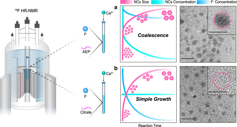
Importantly, contrary to that observed for AEP-CaF2 NCs (Fig. 2e), the concentration of the growing Cit-CaF2 NCs was near-constant throughout the synthesis (Fig. 4d, black). This observation, in addition to the significant depletion of the F- anion throughout Cit-CaF2’s growth (which is negligible for AEP-CaF2, Supplementary Fig. 5), suggests a different growth mechanism. While AEP-CaF2 NCs grow mostly by coalescence, as reflected by the pronounced reduction in their concentration (Fig. 2), Cit-CaF2 NCs primarily grow by the classical simple-growth mechanism10. This result emphasizes another advantage of our proposed approach. Besides its capability to determine NCs’ size evolution, it can also differentiate between two different growth mechanisms as schematically depicted in Fig. 5 (coalescence vs. simple classical growth, Fig. 5a, b). Nevertheless, it is important to mention here that although the proposed in situ NMR approach allowed to investigate NC growth pathways, it failed to pick-up early and rapid nucleation phases.
Fig. 5. Ligand effect on CaF2 NC’s growth mechanisms and crystallographic properties.
Schematic illustration of CaF2 NC formation with different ligands; (a) AEP and (b) Citrate. The NCs’ size (in pink), concentration (in turquoise) and F- ion concentration in solution (in blue). HR-TEM images of AEP-CaF2 NCs presenting twinning defects (inset, in a) and of Cit-CaF2 NCs presenting a crystalline morphology (inset, in b).
Finally, we studied the crystallographic features and spin-lattice relaxation properties of AEP-CaF2 and Cit-CaF2 NCs, as it was shown that the growth mechanism as well as the properties of the capping ligands impact the obtained architectures and characteristics of as-synthesized NCs56,57. Interestingly, HR TEM images of the coalesced AEP-CaF2 NCs showed crystallographic twinning defects for majority of the NCs (Fig. 5a and Supplementary Fig. 14), which were not observed in the crystalline Cit-CaF2 NCs (Fig. 5b and Supplementary Fig. 14). In addition, the longitudinal relaxation time T1 of the NCs’ fluoride content was shorter in AEP-CaF2 NCs as compared to that of Cit-CaF2 NCs (Supplementary Fig. 15). Such shorter T1 values of the defected NCs correlate with previous reports where mechanical stress-mediated lattice distortions of CaF2 powders resulted in significant reduced T1 values58, and should play a critical role in their future design as MRI tracers59. Our presented method for elucidating the contribution of the capping ligands to the nanofluoride’s characteristics could be used for tuning them as functional nanomaterials for a variety of uses, as both their crystallinity60 and relaxation times59 were found to be key features in their performances.
In summary, we show here an approach for the real-time monitoring of NC evolution in solution based on the ability to detect in situ NMR signals of the elements of as-synthesized nanoformulations. The demonstrated ability to accurately determine the average NC diameter at each timepoint during the synthesis has provided us with valuable insight into NC growth mechanisms in solution. We found that in the presence of a phosphate-based ligand, NCs grow predominantly via particle coalescence, while a carboxylate-based ligand leads to a simple-growth mechanism of the same material. Each pathway yields different crystallographic features, and, consequently, distinct relaxation properties, highlighting the link between crystal growth dynamics, structure, and function. These findings should have numerous implications in the growing field of nanofluorides23–25,27,59,61 but also for other types of nanomaterials62–66 where the crystal properties determine the NC functionality. The generality of our NMR-based approach for revealing in-depth mechanistic insights of fluoride-NCs, is highlighted by the similar pattern of the NC evolution profiles found for other types of NCs studied by other in situ methods10,46,47. Therefore, we envision that the fundamental principles in NCs growth underlined here have the potential to serve as a foundation for future studies of other types of materials. Finally, the accessibility of high-resolution NMR spectrometers, in addition to the versatility of NMR acquisition schemes and the ability to study a variety of nuclei (beyond 1H and 19F), could be implemented to elucidate the growth mechanism from a perspective different from those provided by existing in situ techniques.
Supplementary information
Acknowledgements
This project has received funding from the European Research Council (ERC) under the European Union’s Horizon 2020 research and innovation program (grant agreement No 677715).
Author contributions
R.M. and A.B.-S. designed the study. R.M. and A.L. designed the CaF2 synthesis and R.M. carried out the real-time synthesis of the nanofluorides. R.M. and L.A. designed and performed the high-resolution NMR experiments. R.M., V.A., and M.L. performed the computational line fitting of the NMR data. V.A. and M.L. performed the solid-state NMR experiments. H.W. and B.R. performed the cryo-TEM experiments. L.H. and O.B performed the EM-related studies (TEM and EDS). R.M. analyzed all the data shown and R.M. and A.B.-S. wrote the manuscript.
Data availability
The data that support the findings of this study are available from the corresponding author upon reasonable request.
Competing interests
The authors declare no competing interests.
Footnotes
Peer review information Nature Communications thanks Jeffrey Reimer, Gilles Bruylants and the other, anonymous, reviewer(s) for their contribution to the peer review of this work.
Publisher’s note Springer Nature remains neutral with regard to jurisdictional claims in published maps and institutional affiliations.
Supplementary information
Supplementary information is available for this paper at 10.1038/s41467-020-20512-6.
References
- 1.Park J, et al. Ultra-large-scale syntheses of monodisperse nanocrystals. Nat. Mater. 2004;3:891–895. doi: 10.1038/nmat1251. [DOI] [PubMed] [Google Scholar]
- 2.Alivisatos P. The use of nanocrystals in biological detection. Nat. Biotechnol. 2004;22:47–52. doi: 10.1038/nbt927. [DOI] [PubMed] [Google Scholar]
- 3.Yu JH, et al. High-resolution three-photon biomedical imaging using doped ZnS nanocrystals. Nat. Mater. 2013;12:359–366. doi: 10.1038/nmat3565. [DOI] [PubMed] [Google Scholar]
- 4.Lee J-H, et al. Artificially engineered magnetic nanoparticles for ultra-sensitive molecular imaging. Nat. Med. 2006;13:95. doi: 10.1038/nm1467. [DOI] [PubMed] [Google Scholar]
- 5.Wang D, Li Y. Bimetallic nanocrystals: liquid‐phase synthesis and catalytic applications. Adv. Mater. 2011;23:1044–1060. doi: 10.1002/adma.201003695. [DOI] [PubMed] [Google Scholar]
- 6.Schaller RD, Klimov VI. High efficiency carrier multiplication in PbSe nanocrystals: implications for solar energy conversion. Phys. Rev. Lett. 2004;92:186601. doi: 10.1103/PhysRevLett.92.186601. [DOI] [PubMed] [Google Scholar]
- 7.Thanh NT, Maclean N, Mahiddine S. Mechanisms of nucleation and growth of nanoparticles in solution. Chem. Rev. 2014;114:7610–7630. doi: 10.1021/cr400544s. [DOI] [PubMed] [Google Scholar]
- 8.Chang H, et al. Molecular-level understanding of continuous growth from iron-oxo clusters to iron oxide nanoparticles. J. Am. Chem. Soc. 2019;141:7037–7045. doi: 10.1021/jacs.9b01670. [DOI] [PubMed] [Google Scholar]
- 9.Li J, Wang H, Lin L, Fang Q, Peng X. Quantitative identification of basic growth channels for formation of monodisperse nanocrystals. J. Am. Chem. Soc. 2018;140:5474–5484. doi: 10.1021/jacs.8b01296. [DOI] [PubMed] [Google Scholar]
- 10.Zheng H, et al. Observation of single colloidal platinum nanocrystal growth trajectories. Science. 2009;324:1309–1312. doi: 10.1126/science.1172104. [DOI] [PubMed] [Google Scholar]
- 11.Cheong S, Watt J, Ingham B, Toney MF, Tilley RD. In situ and ex situ studies of platinum nanocrystals: growth and evolution in solution. J. Am. Chem. Soc. 2009;131:14590–14595. doi: 10.1021/ja9065688. [DOI] [PubMed] [Google Scholar]
- 12.Williamson MJ, Tromp RM, Vereecken PM, Hull R, Ross FM. Dynamic microscopy of nanoscale cluster growth at the solid–liquid interface. Nat. Mater. 2003;2:532–536. doi: 10.1038/nmat944. [DOI] [PubMed] [Google Scholar]
- 13.Peng ZA, Peng X. Mechanisms of the shape evolution of CdSe nanocrystals. J. Am. Chem. Soc. 2001;123:1389–1395. doi: 10.1021/ja0027766. [DOI] [Google Scholar]
- 14.Polte J, et al. Mechanism of gold nanoparticle formation in the classical citrate synthesis method derived from coupled in situ XANES and SAXS evaluation. J. Am. Chem. Soc. 2010;132:1296–1301. doi: 10.1021/ja906506j. [DOI] [PubMed] [Google Scholar]
- 15.Kwon SG, et al. Kinetics of monodisperse iron oxide nanocrystal formation by “heating-up” process. J. Am. Chem. Soc. 2007;129:12571–12584. doi: 10.1021/ja074633q. [DOI] [PubMed] [Google Scholar]
- 16.Yuk JM, et al. High-resolution EM of colloidal nanocrystal growth using graphene liquid cells. Science. 2012;336:61–64. doi: 10.1126/science.1217654. [DOI] [PubMed] [Google Scholar]
- 17.Kwon O, et al. Exsolution trends and co-segregation aspects of self-grown catalyst nanoparticles in perovskites. Nat. Commun. 2017;8:15967. doi: 10.1038/ncomms15967. [DOI] [PMC free article] [PubMed] [Google Scholar]
- 18.Shen X, et al. Deconvolution of octahedral Pt3Ni nanoparticle growth pathway from in situ characterizations. Nat. Commun. 2018;9:4485. doi: 10.1038/s41467-018-06900-z. [DOI] [PMC free article] [PubMed] [Google Scholar]
- 19.Mikhlin Y, et al. Submicrometer intermediates in the citrate synthesis of gold nanoparticles: new insights into the nucleation and crystal growth mechanisms. J. Colloid Interface Sci. 2011;362:330–336. doi: 10.1016/j.jcis.2011.06.077. [DOI] [PubMed] [Google Scholar]
- 20.Fedorov P, Luginina A, Kuznetsov S, Osiko V. Nanofluorides. J. Fluor. Chem. 2011;132:1012–1039. doi: 10.1016/j.jfluchem.2011.06.025. [DOI] [Google Scholar]
- 21.Stouwdam J, Veggel F. Near-infrared emission of redispersible Er3+, Nd3+, and Ho3+ doped LaF3 Nanoparticles. Nano Lett. 2002;2:733–737. doi: 10.1021/nl025562q. [DOI] [Google Scholar]
- 22.Heer S, Kömpe K, Güdel HU, Haase M. Highly efficient multicolor upconversion emission in transparent colloids of lanthanide-doped NaYF4 nanocrystals. Adv. Mat. 2004;16:2102–2105. doi: 10.1002/adma.200400772. [DOI] [Google Scholar]
- 23.Wang F, Liu X. Upconversion multicolor fine-tuning: visible to near-infrared emission from lanthanide-doped NaYF4 nanoparticles. J. Am. Chem. Soc. 2008;130:5642–5643. doi: 10.1021/ja800868a. [DOI] [PubMed] [Google Scholar]
- 24.Zhao J, et al. Single-nanocrystal sensitivity achieved by enhanced upconversion luminescence. Nat. Nanotechnol. 2013;8:729–734. doi: 10.1038/nnano.2013.171. [DOI] [PubMed] [Google Scholar]
- 25.Liu Y, et al. Amplified stimulated emission in upconversion nanoparticles for super-resolution nanoscopy. Nature. 2017;543:229–233. doi: 10.1038/nature21366. [DOI] [PubMed] [Google Scholar]
- 26.Deng R, et al. Temporal full-colour tuning through non-steady-state upconversion. Nat. Nanotechnol. 2015;10:237–242. doi: 10.1038/nnano.2014.317. [DOI] [PubMed] [Google Scholar]
- 27.Wang F, et al. Simultaneous phase and size control of upconversion nanocrystals through lanthanide doping. Nature. 2010;463:1061–1065. doi: 10.1038/nature08777. [DOI] [PubMed] [Google Scholar]
- 28.Mai HX, et al. High-quality sodium rare-earth fluoride nanocrystals: controlled synthesis and optical properties. J. Am. Chem. Soc. 2006;128:6426–6436. doi: 10.1021/ja060212h. [DOI] [PubMed] [Google Scholar]
- 29.Chen D, et al. Modifying the size and shape of monodisperse bifunctional alkaline-earth fluoride nanocrystals through lanthanide doping. J. Am. Chem. Soc. 2010;132:9976–9978. doi: 10.1021/ja1036429. [DOI] [PubMed] [Google Scholar]
- 30.Ashur I, Allouche‐Arnon H, Bar‐Shir A. Calcium fluoride nanocrystals: tracers for in vivo 19F magnetic resonance imaging. Angew. Chem. Int. Ed. Engl. 2018;57:7478–7482. doi: 10.1002/anie.201800838. [DOI] [PubMed] [Google Scholar]
- 31.Hughes CE, Williams PA, Harris KD. “CLASSIC NMR”: an in-situ NMR strategy for mapping the time-evolution of crystallization processes by combined liquid-state and solid-state measurements. Angew. Chem. Int. Ed. Engl. 2014;53:8939–8943. doi: 10.1002/anie.201404266. [DOI] [PMC free article] [PubMed] [Google Scholar]
- 32.Goulet PJ, Lennox RB. New insights into Brust−Schiffrin metal nanoparticle synthesis. J. Am. Chem. Soc. 2010;132:9582–9584. doi: 10.1021/ja104011b. [DOI] [PubMed] [Google Scholar]
- 33.Li Y, et al. Mechanistic insights into the Brust-Schiffrin two-phase synthesis of organo-chalcogenate-protected metal nanoparticles. J. Am. Chem. Soc. 2011;133:2092–2095. doi: 10.1021/ja1105078. [DOI] [PubMed] [Google Scholar]
- 34.Doyen M, Bartik K, Bruylants G. UV–Vis and NMR study of the formation of gold nanoparticles by citrate reduction: Observation of gold–citrate aggregates. J. Colloid Interface Sci. 2013;399:1–5. doi: 10.1016/j.jcis.2013.02.040. [DOI] [PubMed] [Google Scholar]
- 35.Marbella LE, Millstone JE. NMR techniques for noble metal nanoparticles. Chem. Mater. 2015;27:2721–2739. doi: 10.1021/cm504809c. [DOI] [Google Scholar]
- 36.Vioglio PC, et al. A Strategy for probing the evolution of crystallization processes by low-temperature solid-state NMR and dynamic nuclear polarization. J. Phys. Chem. Lett. 2019;10:1505–1510. doi: 10.1021/acs.jpclett.9b00306. [DOI] [PubMed] [Google Scholar]
- 37.Liu X, Yu M, Kim H, Mameli M, Stellacci F. Determination of monolayer-protected gold nanoparticle ligand-shell morphology using NMR. Nat. Commun. 2012;3:1182. doi: 10.1038/ncomms2155. [DOI] [PMC free article] [PubMed] [Google Scholar]
- 38.Straney PJ, Marbella LE, Andolina CM, Nuhfer NT, Millstone JE. Decoupling mechanisms of platinum deposition on colloidal gold nanoparticle substrates. J. Am. Chem. Soc. 2014;136:7873–7876. doi: 10.1021/ja504294p. [DOI] [PubMed] [Google Scholar]
- 39.Ladizhansky V, Hodes G, Vega S. Surface properties of precipitated CdS nanoparticles studied by NMR. J. Phys. Chem. B. 1998;102:8505–8509. doi: 10.1021/jp981711a. [DOI] [Google Scholar]
- 40.Rees GJ, et al. Characterisation of platinum-based fuel cell catalyst materials using 195Pt wideline solid state NMR. Phys. Chem. Chem. Phys. 2013;15:17195–17207. doi: 10.1039/c3cp52268g. [DOI] [PubMed] [Google Scholar]
- 41.Thayer AM, Steigerwald ML, Duncan TM, Douglass DC. NMR study of semiconductor molecular clusters. Phys. Rev. Lett. 1988;60:2673–2676. doi: 10.1103/PhysRevLett.60.2673. [DOI] [PubMed] [Google Scholar]
- 42.de Queiroz TB, et al. Probing surface effects on α-NaYF4 nanoparticles by nuclear magnetic resonance. J. Phys. Chem. C. 2020;124:9523–9535. doi: 10.1021/acs.jpcc.0c00776. [DOI] [Google Scholar]
- 43.Lovingood DD, Achey R, Paravastu AK, Strouse GF. Size- and site-dependent reconstruction in CdSe QDs evidenced by 77Se{1H} CP-MAS NMR spectroscopy. J. Am. Chem. Soc. 2010;132:3344–3354. doi: 10.1021/ja907511r. [DOI] [PubMed] [Google Scholar]
- 44.Sadoc A, et al. NMR parameters in alkali, alkaline earth and rare earth fluorides from first principle calculations. Phys. Chem. Chem. Phys. 2011;13:18539–18550. doi: 10.1039/c1cp21253b. [DOI] [PubMed] [Google Scholar]
- 45.Witter R, Roming M, Feldmann C, Ulrich AS. Multilayered core–shell structure of polyol-stabilized calcium fluoride nanoparticles characterized by NMR. J. Colloid Interface Sci. 2013;390:250–257. doi: 10.1016/j.jcis.2012.09.001. [DOI] [PubMed] [Google Scholar]
- 46.Niu K-Y, Liao H-G, Zheng H. Visualization of the coalescence of bismuth nanoparticles. Microsc. Microanal. 2014;20:416–424. doi: 10.1017/S1431927614000282. [DOI] [PubMed] [Google Scholar]
- 47.Polte J, et al. Nucleation and growth of gold nanoparticles studied via in situ small angle X-ray scattering at millisecond time resolution. ACS Nano. 2010;4:1076–1082. doi: 10.1021/nn901499c. [DOI] [PubMed] [Google Scholar]
- 48.Polte J. Fundamental growth principles of colloidal metal nanoparticles—a new perspective. CrystEngComm. 2015;17:6809–6830. doi: 10.1039/C5CE01014D. [DOI] [Google Scholar]
- 49.Boles MA, Ling D, Hyeon T, Talapin DV. The surface science of nanocrystals. Nat. Mater. 2016;15:141. doi: 10.1038/nmat4526. [DOI] [PubMed] [Google Scholar]
- 50.Nenon DP, et al. Design principles for trap-free CsPbX3 nanocrystals: enumerating and eliminating surface halide vacancies with softer Lewis bases. J. Am. Chem. Soc. 2018;140:17760–17772. doi: 10.1021/jacs.8b11035. [DOI] [PubMed] [Google Scholar]
- 51.Lazzari S, et al. Ligand-mediated nanocrystal growth. Langmuir. 2018;34:3307–3315. doi: 10.1021/acs.langmuir.8b00076. [DOI] [PubMed] [Google Scholar]
- 52.van Embden J, Mulvaney P. Nucleation and growth of CdSe nanocrystals in a binary ligand system. Langmuir. 2005;21:10226–10233. doi: 10.1021/la051081l. [DOI] [PubMed] [Google Scholar]
- 53.Owen JS, Chan EM, Liu H, Alivisatos AP. Precursor conversion kinetics and the nucleation of cadmium selenide nanocrystals. J. Am. Chem. Soc. 2010;132:18206–18213. doi: 10.1021/ja106777j. [DOI] [PubMed] [Google Scholar]
- 54.Kovalenko MV, Scheele M, Talapin DV. Colloidal nanocrystals with molecular metal chalcogenide surface ligands. Science. 2009;324:1417–1420. doi: 10.1126/science.1170524. [DOI] [PubMed] [Google Scholar]
- 55.Heuer-Jungemann A, et al. The role of ligands in the chemical synthesis and applications of inorganic nanoparticles. Chem. Rev. 2019;119:4819–4880. doi: 10.1021/acs.chemrev.8b00733. [DOI] [PubMed] [Google Scholar]
- 56.Leite E, et al. Crystal growth in colloidal tin oxide nanocrystals induced by coalescence at room temperature. Appl. Phys. Lett. 2003;83:1566–1568. doi: 10.1063/1.1605241. [DOI] [Google Scholar]
- 57.Penn RL, Banfield JFJS. Imperfect oriented attachment: dislocation generation in defect-free nanocrystals. Science. 1998;281:969–971. doi: 10.1126/science.281.5379.969. [DOI] [PubMed] [Google Scholar]
- 58.Abdellatief M, Abele M, Leoni M, Scardi P. Combined X-ray diffraction and solid-state 19F magic angle spinning NMR analysis of lattice defects in nanocrystalline CaF2. J. Appl. Crystalogr. 2013;46:1049–1057. doi: 10.1107/S0021889813016944. [DOI] [Google Scholar]
- 59.Mashiach R, et al. Inducing defects in 19F-nanocrystals provides paramagnetic-free relaxation enhancement for improved in vivo hotspot MRI. Nano Lett. 2020;20:7207–7212. doi: 10.1021/acs.nanolett.0c02549. [DOI] [PMC free article] [PubMed] [Google Scholar]
- 60.Dyck NC, van Veggel FC, Demopoulos GP. Size-dependent maximization of upconversion efficiency of citrate-stabilized beta-phase NaYF4:Yb3+,Er3+ crystals via annealing. ACS Appl. Mater. Interfaces. 2013;5:11661–11667. doi: 10.1021/am403100t. [DOI] [PubMed] [Google Scholar]
- 61.Fan X, et al. Defect-enriched iron fluoride-oxide nanoporous thin films bifunctional catalyst for water splitting. Nat. Commun. 2018;9:1809. doi: 10.1038/s41467-018-04248-y. [DOI] [PMC free article] [PubMed] [Google Scholar]
- 62.Bekenstein Y, et al. The making and breaking of lead-free double perovskite nanocrystals of cesium silver-bismuth halide compositions. Nano Lett. 2018;18:3502–3508. doi: 10.1021/acs.nanolett.8b00560. [DOI] [PubMed] [Google Scholar]
- 63.Kong M, et al. Tuning the relative concentration ratio of bulk defects to surface defects in TiO2 nanocrystals leads to high photocatalytic efficiency. J. Am. Chem. Soc. 2011;133:16414–16417. doi: 10.1021/ja207826q. [DOI] [PubMed] [Google Scholar]
- 64.Hardy DA, Tigaa RA, McBride JR, Ortega RE, Strouse GF. Structure-fFunction correlation: engineering high quantum yields in down-shifting nNanophosphors. J. Am. Chem. Soc. 2019;141:20416–20423. doi: 10.1021/jacs.9b11045. [DOI] [PubMed] [Google Scholar]
- 65.Zhao Z, et al. Octapod iron oxide nanoparticles as high-performance T2 contrast agents for magnetic resonance imaging. Nat. Commun. 2013;4:2266. doi: 10.1038/ncomms3266. [DOI] [PubMed] [Google Scholar]
- 66.Mann AKP, Wu Z, Calaza FC, Overbury SH. Adsorption and reaction of acetaldehyde on shape-controlled CeO2 nanocrystals: elucidation of structure–function relationships. ACS Catal. 2014;4:2437–2448. doi: 10.1021/cs500611g. [DOI] [Google Scholar]
Associated Data
This section collects any data citations, data availability statements, or supplementary materials included in this article.
Supplementary Materials
Data Availability Statement
The data that support the findings of this study are available from the corresponding author upon reasonable request.


