Abstract
Background
Outcomes are poorer in metastatic colorectal cancer (mCRC) patients with BRAF V600E mutations than those without it, but the effect of these mutations on treatment response is unclear. This real-world study assessed the effects of antiangiogenic-based treatment and systemic inflammatory factors on outcomes in patients with BRAF V600-mutated mCRC.
Methods
This real-world, multicenter, retrospective, observational study included patients with BRAF V600-mutated mCRC treated in eight hospitals in Spain. The primary endpoints were overall survival (OS) and progression-free survival (PFS); overall response rate (ORR) and disease control rate (DCR) were also assessed. The effect of first- and second-line treatment type on OS, PFS, ORR, and DCR were evaluated, plus the impact of systemic inflammatory markers on these outcomes. A systemic inflammation score (SIS) of 1–3 was assigned based on one point each for platelet-lymphocyte ratio (PLR) ≥200, neutrophil-lymphocyte ratio (NLR) ≥3, and serum albumin < 3.6 g/dL.
Results
Of 72 patients, data from 64 were analyzed. After a median of 69.1 months, median OS was 11.9 months and median first-line PFS was 4.4 months. First-line treatment was triplet chemotherapy-antiangiogenic (12.5%), doublet chemotherapy-antiangiogenic (47.2%), doublet chemotherapy-anti-EGFR (11.1%), or doublet chemotherapy (18.1%). Although first-line treatment showed no significant effect on OS, antiangiogenic-based regimens were associated with prolonged median PFS versus non-antiangiogenic regimens. Negative predictors of survival with antiangiogenic-based treatment were NLR, serum albumin, and SIS 1–3, but not PLR. Patients with SIS 1–3 showed significantly prolonged PFS with antiangiogenic-based treatment versus non-antiangiogenic-based treatment, while those with SIS=0 showed no PFS benefit.
Conclusions
Antiangiogenic-based regimens, SIS, NLR, and albumin were predictors of survival in patients with mCRC, while SIS, NLR and serum albumin may predict response to antiangiogenic-based chemotherapy.
Trial registration
GIT-BRAF-2017-01.
Keywords: Antiangiogenic-based chemotherapy, BRAF V600E mutations, Metastatic colorectal cancer, Systemic inflammation score
Background
Worldwide, colorectal cancer (CRC) is the third most common cancer in men and the second most common cancer in women, with the age-standardized rate per 100,000 ranging from 31.8 to 51.2 in the countries with the highest incidence [1].
The presence of BRAF mutations, which are present in 5–10% of patients with mCRC, is a known adverse prognostic factor, especially in the metastatic setting. Based on retrospective analyses of mCRC patients, those with BRAF V600E mutations share common clinical characteristics, such as female gender, older age at diagnosis, and primary tumor location in right-side colon [2]. Molecular classifications of colorectal cancer show that patients with BRAF mutations cluster together on the CMS1 subgroup, which is also characterized by high immune activation and infiltration and the co-occurrence of microsatellite instability (MSI) [3].
Systemic inflammation is also known to be prognostic of poor clinical outcomes in patients with CRC [4]. In particular, abnormal acute inflammatory phase proteins (e.g. elevated C-reactive protein [CRP] and decreased serum albumin) and increased counts of neutrophils and platelets relative to lymphocytes have been associated with adverse outcomes [4].
Although we recognize that BRAF V600E-mutated CRC is a distinct clinical and biological subgroup, there are no published series on treating these patients in routine clinical practice, and our understanding is based on limited data from clinical trials. Therefore, it is important to further evaluate the prognostic factors that may influence treatment outcomes in patients with BRAF-mutated mCRC in a representative cohort. For example, it is unclear how the presence of BRAF V600E mutations could predict a poorer response to treatment [5–8].
Controversy remains regarding the choice of first-line therapy in patients with BRAF-mutated mCRC. The European Society for Medical Oncology (ESMO) recommends treatment with either doublet or triplet chemotherapy (CT), with the addition of the vascular endothelial growth factor (VEGF) antibody bevacizumab in patients with BRAF mutations and epidermal growth factor receptor (EGFR) antibodies in patients without RAS mutations [9]. There is also evidence that BRAF mutations are predictive of a negative response to EGFR treatment, as indicated in two meta-analyses that showed a lack of benefit from the addition of anti-EGFR treatment to doublet CT in patients with RAS-wild type/BRAF-mutated mCRC [10, 11].
The future of BRAF V600E-mutated CRC treatment is likely to be influenced by the results of ongoing clinical trials of BRAF-targeted treatment strategies. A previous study in patients with BRAF V600-mutated non-melanoma cancer showed that monotherapy with the BRAF inhibitor vemurafenib had limited efficacy in CRC patients [12]. Preclinical data has demonstrated the importance of coupling direct BRAF inhibition with EGFR inhibition in order to block signaling pathway feedback loops [13]. Based on these findings, preliminary clinical trial data have demonstrated promising antitumor activity with combined targeted treatment, including vemurafenib coupled with the EGFR inhibitor cetuximab and irinotecan as second- or third-line therapy [14], and ongoing clinical trials are exploring the role of combination BRAF and EGFR inhibition in first-line therapy.
This study, which was initiated before the introduction of BRAF-targeted treatment for mCRC, aimed to elucidate which factors were associated with poorer outcomes in patients with BRAF V600-mutated mCRC and whether the ESMO recommendation for doublet CT plus an antiangiogenic agent was associated with improved outcomes by performing a retrospective analysis of clinical outcomes in more than 70 patients with BRAF V600-mutated mCRC from eight hospitals in Spain.
Methods
This was a multicenter, retrospective, observational study in patients with BRAF V600-mutated mCRC who treated with the standard of care between 2011 and 2018 at eight hospitals in the Galician Research Group on Digestive Tumors (GITuD) network in the Galicia Autonomous Community, Spain.
The study was conducted in accordance with the Good Clinical Practice Guidelines and the Declaration of Helsinki and obtained the approval of the Ethical Committee of Sanitary Area Santiago-Lugo, Spain (registration code: 2017/453). All patients provided written or oral informed consent before an independent witness of the research team prior to inclusion in the study.
Eligibility criteria
Patients were eligible for inclusion if they were over 18 years of age and had a confirmed diagnosis of mCRC with the BRAF V600 mutation. Exclusion criteria were a history of neoplasm within the previous 5 years (except cervical carcinoma in situ or basal cell carcinoma of the skin); an inability to understand the study procedures or provide informed consent; and incomplete clinical or follow-up data.
Study endpoints
The primary study endpoints were overall survival (OS) and progression-free survival (PFS). OS was defined as the time from the start of treatment until death by any cause. PFS was defined as the time from treatment start to confirmed radiologic progression or death by any cause.
Secondary endpoints were overall response rate (ORR; defined as the proportion of patients who achieved complete response [CR] or partial response [PR]) and disease control rate (DCR; defined as the proportion of patients who achieved CR, PR, or stable disease [SD] lasting ≥ 6 weeks after the start of treatment).
Exploratory outcomes included the impact of first- and second-line treatment on PFS and OS, and the prognostic impact of clinical factors and systemic inflammation.
Systemic inflammation was assessed using (i) the neutrophil-to-lymphocyte ratio (NLR), with NLR subgroups categorized as ≥ 3 vs < 3; (ii) the platelet-to-lymphocyte ratio (PLR), with PLR subgroups categorized as ≥ 200 vs < 200; and (iii) serum albumin levels (≥ 3.6 g/dL vs < 3.6 g/dL). For each patient, a systemic inflammation score (SIS) was calculated by adding up the number of risk factors, defined as serum albumin < 3.6 g/dL, NLR ≥3 and PLR ≥ 200, with one point assigned to each risk factor.
Statistical analysis
The primary analysis was conducted using the Kaplan–Meier method to estimate median PFS and OS and 95% confidence intervals (CIs). Differences between survival curves were compared using the log-rank test with a two-sided significance level of 0.05. The chi-squared test or Fisher’s exact test (depending on the sample size) was used to compare clinical and demographic variables. Statistical analyses were carried out using IBM SPPS Statistics V25.0.
Results
Patient population
The study included 72 patients who received treatment for mCRC between November 2010 and November 2018. The exploratory efficacy analysis included 64 patients (88.9%); eight patients were excluded from this analysis because they only received palliative care.
Patient baseline characteristics are summarized in Table 1. Median (range) age was 62.4 (30–83) years and 54.2% of patients were female. The median (range) number of metastatic locations was 2 (1–5); 25% of patients had metastases in ≥ 3 locations. Regarding prognostic factors, 27.8% of patients had ECOG PS 2–3, 48.6% had tumors in the right colon, 31.9% had high-grade tumors, 6.9% had deficiency of mismatch repair proteins, and 59.4% had synchronous tumors.
Table 1.
Patient characteristics and their prognostic impact on overall survival
| Characteristics | N = 72 | OS (months) | HR (95% CI) | p-valuea |
|---|---|---|---|---|
| Age, years | ||||
| Median | 62.4 | – | – | – |
| Range | 30–83 | |||
| Sex, n (%) | ||||
| Male | 33 (45.8) | 9.4 | 1.317 (0.8–2.2) | 0.308 |
| Female | 39 (54.2) | 12.8 | ||
| ECOG PS, n (%) | ||||
| 0–1 | 52 (72.2) | 11.9 | 0.632 (0.3–1.2) | 0.161 |
| 2–3 | 20 (27.8) | 7.1 | ||
| Tumor location, n (%) | ||||
| Right-sided | 35 (48.6) | 10.6 | – | 0.296 |
| Left-sided | 22 (30.6) | 11.3 | ||
| Rectum | 15 (20.8) | 13.1 | ||
| Histological grade, n (%) | ||||
| Low grade (G1–2) | 37 (51.4) | 11.9 | 0.895 (0.5–1.7) | 0.723 |
| High grade (G3) | 23 (31.9) | 11.1 | ||
| Unknown | 12 (16.7) | |||
| Mismatch repair proteins, n (%) | n=33 | |||
| Conserved | 31 (93.1) | 12.8 | 0.146 (0.1–0.8) | 0.010 |
| Deficiency | 2 (6.9) | 4.9 | ||
| Tumor presentation, n (%) | ||||
| Synchronous | 50 (69.4) | 12.4 | 0.903 (0.5–1.6) | 0.730 |
| Metachronous | 22 (30.6) | 5.9 | ||
| Primary tumor surgery, n (%) | ||||
| No | 25 (34.7) | 9.6 | 1.932 (1.1–3.4) | 0.023 |
| Yes | 47 (65.3) | 11.9 | ||
| Metastasectomy, n (%) | ||||
| No | 57 (79.2) | 9.6 | 2.307 (1.2–4.6) | 0.011 |
| Yes | 15 (20.8) | 19.6 | ||
CI Confidence interval, ECOG PS Eastern Cooperative Oncology Group performance status, G Grade, HR Hazard ratio, OS Overall survival
aSignificant values are indicated in bold
Efficacy
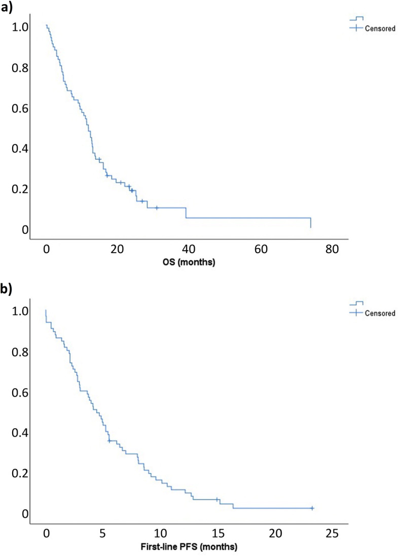
In the 64 patients who were evaluated for efficacy, median OS was 11.9 months (95% CI, 9.7–14.0 months) (Fig. 1a) and median first-line PFS was 4.4 months (95% CI, 3.2–5.7 months), after a median follow-up of 69.1 months (Fig. 1b). The best responses achieved were CR in five patients (7.8%), PR in 16 (25.0%), SD in 18 (28.1%), and progressive disease (PD) in 20 (31.3%); response was unevaluable due to clinical decline in five patients (7.8%). When the five unevaluable patients were excluded, 8.5% of patients had a CR, 27.1% had a PR, 30.5% had SD, and 33.9% had PD. The ORR was 35.6% and the DCR was 66.1%.
Fig. 1.

First-line a overall survival (OS) and b progression-free survival (PFS) in the overall population
First-line treatment
First-line treatment consisted of triplet CT plus antiangiogenic therapy in nine patients (12.5%), doublet CT plus antiangiogenic therapy in 34 (47.2%), doublet CT plus anti-EGFR therapy in eight (11.1%), and doublet CT in 13 (18.1%).
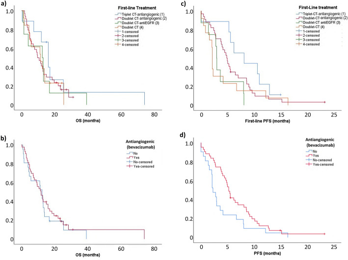
There were no statistical differences in OS according to type of treatment, with OS being 16.0 months with triplet CT plus antiangiogenic therapy, 9.6 months with doublet CT plus antiangiogenic therapy, 12.4 months with doublet CT plus anti-EGFR therapy, and 11.3 months with doublet CT (p = 0.628) (Fig. 2a). Median OS was 11.8 months in patients who received antiangiogenic-based CT compared with 12.4 months in those who did not (hazard ratio [HR], 1.196; 95% CI, 0.7–2.1; p = 0.529) (Fig. 2b).
Fig. 2.
First-line overall survival (OS) a across all treatment regimens and b comparing first-line regimens with vs without antiangiogenic therapy (bevacizumab), and first-line progression-free survival (PFS) c across all treatment regimens and d comparing first-line regimens with vs without antiangiogenic therapy (bevacizumab)
First-line PFS varied across treatments, being 8.1 months with triplet CT plus antiangiogenic therapy, 4.8 months with doublet CT plus antiangiogenic therapy, 2.9 months with doublet CT plus anti-EGFR therapy, and 2.1 months with doublet CT (p = 0.091) (Fig. 2c).
Median first-line PFS was significantly higher in patients treated with antiangiogenic-based CT, being 5.2 months with triplet or doublet CT plus antiangiogenics and 2.3 months with doublet CT with or without anti-EGFR therapy (HR, 0.848; 95% CI, 0.3–0.9; p = 0.033) (Fig. 2d). No statistical differences in first-line PFS were found between antiangiogenic-based regimens (triplet CT vs doublet CT HR, 0.543; 95% CI, 0.2–1.2; p = 0.123) or between doublet CT with versus without anti-EGFR therapy (HR, 0.989; 95% CI, 0.4–2.4; p = 0.929).
First-line ORR based on type of treatment was 75% with triplet CT plus antiangiogenic therapy, 34.4% with doublet CT plus antiangiogenic, 12.5% with doublet CT plus anti-EGFR, and 27.3% with doublet CT (p = 0.063; Fisher’s test). ORR was 45.2% with triplet or doublet CT plus antiangiogenic therapy and 21.1% with doublet CT with or without anti-EGFR therapy (p = 0.149).
The first-line DCR was 100% with triplet CT plus antiangiogenic therapy, 71.9% with doublet CT plus antiangiogenic, 50.0% with doublet CT plus anti-EGFR, and 36.4% with doublet CT (p = 0.015; Fisher’s test). DCR was significantly higher with triplet or doublet CT plus antiangiogenic than with doublet CT with or without anti-EGFR (77.5% vs 42.1%; p = 0.017).
Second-line treatment
Second-line treatment was required in 61 patients. Second-line treatment consisted of doublet CT plus antiangiogenic therapy in 20 patients (32.8%), doublet CT plus anti-EGFR therapy in three (4.9%), and doublet CT in six (9.8%). This analysis excluded 32 patients; 27 (44.3%) received best supportive care, four (6.6%) received single-agent CT and one (1.6%) enrolled in a clinical trial.
In the 29 patients who were evaluated for second-line efficacy, median PFS was 2.8 months (95% CI, 2.4–3.2 months) and median OS was 5.9 months (95% CI, 3.9–7.2 months). Best responses consisted of PR in five patients (17.2%), SD in eight (27.6%), and PD in 13 (44.8%); response was unevaluable in three patients (10.3%). Excluding the three unevaluable patients, the ORR was 19.2% and the DCR was 50%.
There were no statistical differences in OS according to treatment type, with median second-line OS being 7.1 months with doublet CT plus antiangiogenic therapy, 5.9 months with doublet CT plus anti-EGFR therapy, and 3.4 months with doublet CT (p = 0.335). Median second-line OS was 7.1 months in patients who received antiangiogenic-based CT compared with 5.3 months in those who received doublet CT with or without anti-EGFR therapy (HR, 0.532; 95% CI, 0.2–1.2; p = 0.140).
Second-line PFS varied numerically across treatments, being 3.5 months with doublet CT plus antiangiogenic therapy, 2.8 months with doublet CT plus anti-EGFR therapy, and 0.1 months with doublet CT (p = 0.079). PFS was significantly prolonged in patients treated with second-line antiangiogenic-based CT, being 3.5 months with doublet CT plus antiangiogenic therapy and 2.3 months with doublet CT with or without anti-EGFR therapy (HR, 0.402; 95% CI, 0.2–0.9; p = 0.032).
Second-line ORR according treatment type was 26.3% with doublet CT plus antiangiogenic therapy, 0% with doublet CT with or without anti-EGFR therapy (p = 0.190). Second-line DCR according to treatment type was 63.2% with doublet CT plus antiangiogenic therapy, 0% with doublet CT plus anti-EGFR therapy, and 25% with doublet CT (p = 0.096). The second-line ORR and DCR were significantly higher among patients treated with antiangiogenic-based CT versus doublet CT with or without anti-EGFR therapy (26.3% vs 0%; p = 0.153 and 63.2% vs 14.3%; p = 0.020, respectively).
Prognostic factors
Of the patient characteristics that were analyzed as potential prognostic factors, the characteristics that were associated with significantly improved OS were conserved mismatch repair (p = 0.010), primary tumor resection (p = 0.023), and metastasis resection (p = 0.011; Table 1).
Systemic inflammation score
Regarding the SIS parameters, 50.7% of patients had NLR ≥ 3, 43.7% had PLR ≥ 200 and 19.4% had serum albumin < 3.6 g/dL (Table 2). NLR ≥ 3 was positively associated with PLR ≥ 200 (p < 0.001), but neither NLR (p = 0.543) nor PLR (p = 0.375) was associated with low serum albumin levels.
Table 2.
Systemic inflammation parameters and their prognostic impact on overall survival
| Parameter | n (%) | Median OS, months | HR (95% CI) | p-valuea |
|---|---|---|---|---|
| NLR | ||||
| < 3 | 35 (49.3%) | 7.8 | 1.934 (1.1–3.3) | 0.014 |
| ≥ 3 | 36 (50.7%) | 13.7 | ||
| PLR | ||||
| < 200 | 40 (56.3%) | 5.4 | 1.567 (0.9–2.7) | 0.096 |
| ≥ 200 | 31 (43.7%) | 13.0 | ||
| Serum albumin levels | ||||
| < 3.6 g/dL | 14 (19.4%) | 4.9 | 2.142 (1.1–4.5) | 0.040 |
| ≥ 3.6 g/dL | 47 (65.3%) | 12.4 | ||
| Unknown | 11 (15.3%) | – | – | – |
| SIS | ||||
| 0 | 25 (35.2%) | 16.0 | – | 0.004 |
| 1 | 18 (25.4%) | 7.8 | ||
| 2 | 21 (29.6%) | 5.7 | ||
| 3 | 7 (9.9) | 4.0 | ||
| SIS groups | ||||
| 0 | 25 (35.2%) | 16.0 | 0.475 (0.3–0.8) | 0.008 |
| 1–3 | 46 (64.8%) | 7.3 | ||
CI Confidence interval, HR Hazard ratio, NLR Neutrophil-to-lymphocyte ratio, PLR Platelet-to-lymphocyte ratio, SIS Systemic inflammation score
aSignificant values are indicated in bold
Both increased NLR (p = 0.014) and reduced serum albumin levels (p = 0.040) had a significant negative prognostic impact on OS. Increased PLR showed a non-significant trend towards worse survival (p = 0.096; Table 2).
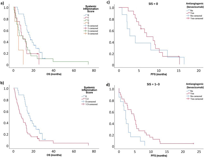
The SIS score was predictive of survival; median OS was 16.0 months in patients with no factors, 7.8 months with 1 factor, 5.7 months with 2 factors, and 4.0 months with 3 factors (p = 0.004) (Fig. 3a). Patients with an SIS score of 0 also had a significantly longer median OS than those with an SIS score of 1–3 (16.0 vs 7.3 months; HR, 0.475, 95% CI, 0.3–0.8; p = 0.008) (Fig. 3b).
Fig. 3.
Overall survival (OS) according to systemic inflammation scores (SIS) of a 0, 1, 2 or 3 and b 1–3 vs 0, and progression-free survival (PFS) with vs without antiangiogenic therapy (bevacizumab) in patients with SIS of c 0 and d 1–3
There was a significant benefit with antiangiogenic-based CT in patients with an SIS score of 1–3, but not in those with a score of 0. In patients with an SIS score of 1–3, median PFS was 4.8 months for antiangiogenic-based regimens versus 2.1 months for non-antiangiogenic-based CT (HR, 2.340; 95% CI, 1.1–4.8; p = 0.016) (Fig. 3c). Conversely, in patients with SIS score 0, median PFS was 7.0 versus 2.8 months, respectively (HR, 1.266; 95% CI, 0.5–3.2; p = 0.616) (Fig. 3d).
Discussion
This retrospective study of 72 patients with BRAF V600-mutated mCRC is the largest published series to represent the wide spectrum of patients that are seen in routine clinical practice. Overall, the relatively short median OS (11.9 months) and first-line median PFS (4.4 months), combined with the fact that eight patients could not receive any active treatment, confirms previous observations that BRAF mutations confer a worse prognosis and poorer response to treatment than those with wild-type BRAF tumors when associated with advanced disease at diagnosis. For example, in a study of mCRC patients who were treated with first-line doublet CT plus anti-EGFR therapy (panitumumab), median OS was 9.2–10.4 months and a median PFS was 5.4–6.0 months in patients with BRAF-mutated tumors compared with 15.1–40.0 months and 7.8–12.6 months, respectively, in those with wild-type BRAF mCRC [15].
Interestingly, although left-side tumors were present in almost 50% of the patients in our study, only 6.9% had alterations in mismatch repair genes. A Swedish study previously suggested that the percentage of MSI alteration in early stage BRAF-mutated CRC might reach 50%, but in this case the presence of MSI as a positive prognostic factor overcame the negative prognosis conferred by BRAF mutation [16], meaning that most patients with left-sided BRAF-mutated tumors and MSI may not develop metastases. The Swedish study also found that rectal BRAF-mutated tumors had an adverse prognosis and were not associated with MSI [16].
The ESMO clinical guidelines recommend first-line treatment with a combination of doublet or triplet CT plus an antiangiogenic therapy in patients with BRAF-mutated mCRC to increase the first-line response rate and PFS [9]. Because the start of our real-world study predates the publication of this ESMO recommendation, our study aimed to show that this approach led to better outcomes in patients with BRAF-mutated mCRC. In fact, almost 60% of patients received first-line treatment with doublet or triplet CT plus antiangiogenic therapy (i.e. bevacizumab), and antiangiogenic-based treatment was associated with notably longer PFS and greater DCR than non-antiangiogenic-based treatment. Although there was no difference in OS when stratified according to presence or absence of antiangiogenic therapy, the small number of patients in each treatment group means that this analysis may have lacked the statistical power to detect a difference in OS.
The potential benefit of adding antiangiogenic therapy in the general CRC patient population is unclear. For example, a phase III, multicenter, randomized trial found no clinical benefit of adding bevacizumab to FOLFOX4 or FOLFIRI in first-line treatment of mCRC patients [17], while a meta-analysis showed that bevacizumab significantly reduced mortality risk in primary tumor-resected mCRC patients compared with patients without primary tumor resection [18]. In another meta-analysis, this one in patients with mutated BRAF V600E who received second-line treatment with FOLFIRI ± an antiangiogenic within the TRIBE, TRIBE-2, VELOUR and RAISE studies, anti-angiogenics were found to have a significant advantage over placebo in terms of OS (HR, 0.50, 95% CI, 0.29–0.85; p = 0.01) [19], suggesting that patients with BRAF-mutated tumors might benefit specially from antiangiogenic therapy. The reason why our study found a potential beneficial effect of antiangiogenic therapy might reside in the particular importance of inflammation in this molecular subgroup.
Due to the timeframe of data accrual, patients in our study received BRAF inhibitors, such as encorafenib. Single-agent BRAF inhibitor treatment has demonstrated limited effectiveness against BRAF V600-mutated mCRC [20], because mCRC tumors harboring BRAF mutations rapidly develop adaptive resistance to RAS [21]. Combining a BRAF inhibitor with EGFR or MEK inhibitors may result in improved outcomes [21]. Indeed, in the BEACON trial in patients with BRAF V600-mutated mCRC, second-line treatment with a BRAF inhibitor (encorafenib), an anti-EGFR antibody (cetuximab), and a MEK inhibitor (binimetinib) resulted in significantly prolonged median OS compared with control (9.0 vs 5.4 months; p < 0.001) [22]. Therefore, although first-line treatment with CT plus anti-EGFR showed no survival benefit, the combination of anti-EGFR therapy plus BRAF inhibitors may be a new standard for second-line treatment of patients with BRAF-mutated mCRC.
In our study, we evaluated the prognostic and predictive value of a set systemic inflammation markers that have been previously described elsewhere [4], and used them to create a prognostic score tool. PLR and NLR have been studied as prognostic factors in most tumor types [23], and hypoalbuminemia has been identified as a prognostic indicator in CRC [24]. The majority of patients (64.8%) with BRAF-mutated CRC in our study displayed at least one inflammation marker, confirming the significance of inflammation in this subgroup. Both increased NLR and reduced serum albumin had a significant negative prognostic impact on OS in our study, while PLR showed a non-significant trend. The SIS was also correlated with OS, with median OS being highest in patients with a score of 0 and lowest in those with a score of 3. There was a treatment benefit of antiangiogenic therapy in patients with any systemic inflammation (SIS 1–3) but not in those with a score of 0, although PFS was numerically prolonged in this subgroup. This most likely reflects a lack of statistical power, rather than there being no benefit in patients with no inflammatory factors. In a study in patients with mCRC receiving bevacizumab, PFS was longer in patients with low NLR (< 3.44) than those with high NLR (≥ 3.44), and in patients who had received anti-EGFR therapy, PFS was longer in patients with low PLR (< 160.66) versus high PLR (≥ 160.66) [23]. Although this study did not show a significant impact of PLR or NLR on PFS, this suggests that inflammatory markers may predict benefit of bevacizumab in CRC. While our study found hypoalbuminemia to be a negative prognostic indicator on its own, previous studies, including that of McMillan and colleagues, found only elevated CRP levels, not hypoalbuminemia, to be negative predictors of survival [25]. However, hypoalbuminemia combined with CRP is part of the modified Glasgow Prognostic Scale (mGPS) [25]. The risk of death has been found to be significantly increased in patients with a mGPS score of 2 versus 0 (p = 0.017) [26]. Although our study found that none of the SIS parameters were predictive of response to anti-EGFR therapy, this may have been an effect of the small number of patients treated with anti-EGFR therapy.
The limitations of this study include the retrospective nature of its study design, the lack of a control arm, and the relatively small numbers of patients in some treatment categories.
Conclusions
This study confirms that the BRAF V600E mutation is a prognostic indicator of poor outcomes in patients with mCRC. Antiangiogenic-based regimens and systemic inflammatory markers, including elevated PLR and NLR and decreased serum albumin, are prognostic in this population, while SIS, elevated NLR, and decreased serum albumin may also predict response to first-line antiangiogenic-based treatment.
Acknowledgements
We would like to thank Marion Barnett on behalf of Springer Healthcare Communications who prepared the first draft of the manuscript.
Abbreviations
- CIs
Confidence intervals
- CR
Complete response
- CRC
Colorectal cancer
- CRP
C-reactive protein
- CT
Chemotherapy
- DCR
Disease control rate
- EGFR
Epidermal growth factor receptor
- ESMO
European Society for Medical Oncology
- GITuD
Galician Research Group on Digestive Tumors
- HR
Hazard ratio
- mCRC
Metastatic colorectal cancer
- mGPS
Modified Glasgow Prognostic Scale
- MSI
Microsatellite instability
- NRL
Neutrophil-lymphocyte ratio
- ORR
Overall response rate
- OS
Overall survival
- PD
Progressive disease
- PLR
Platelet-lymphocyte ratio
- PFS
Progression-free survival
- PR
Partial response
- SD
Stable disease
- SIS
Systemic inflammation score
- VEGF
Vascular endothelial growth factor
Authors’ contributions
NML, AFM, MCR, EMBV, JCCG, JCMM, MJF, ACC, CRR, DAR, MCC, YVI, MLPA, PGV, BGS, MSF, ACM, FVR, SCF, MRL had contributed to the study design, patient enrollment, data analyses, and preparation and review of the manuscript. All have read and approved the manuscript.
Funding
The authors have not declared a specific grant for this research from any funding agency in the public, commercial or not-for-profit sectors.
Availability of data and materials
Data are available upon reasonable request.
Ethics approval and consent to participate
The study was conducted in accordance with the Good Clinical Practice Guidelines and the Declaration of Helsinki. All patients provided written informed consent before an independent witness of the research team prior to inclusion in the study. This study was approved by Local Clinical Research Ethics Committee of Galicia (CAEI). All patients provided written informed consent before an independent witness of the research team prior to inclusion in the study.
Consent for publication
Not applicable – this article does not contain identifying information for any patients.
Competing interests
NML has received personal fees from Amgen, Sanofi, Roche, Merck, Leopharma, Celgene-BMS, Lilly, Ipsen, and Pfizer.
AFM has received personal fees from Amgen, Merck, Roche, Sanofi, Lilly, Bayer, Servier, BMS, and EISAI.
EMBV has received personal fees from Leopharma and Rovi.
JCCG has received honoraria for advisory role from Merck, Amgen, Roche, and Sanofi.
MLPA has received personal fees from Roche and Merck, and grants from Merck and Ipsen.
PGV has received honoraria for consulting or advisory roles from Amgen, has participated in a speakers’ bureau for Amgen and Lilly, and has received fees for travel, accommodations, or other expenses from Amgen, Lilly, and Merck.
BGS has received personal fees from Amgen, Merck, Roche, Sanofi, Servier, EISAI, Celgene, and Incyte, and non-financial support from Amgen, Merck, Roche, Sanofi, Servier, EISAI, Celgene, and Incyte.
MSF has received honoraria for advisory role from Amgen, Servier, and Merck, fees as speaker’s bureau from BMS, Amgen, and Merck, and travel and educational support from Roche, Servier, and Sanofi.
MCR, JCMM, MJF, ACC, CRR, DAR, MCC, YVI, ACM, FVR, SCF, and MRL declare that they have no competing interests.
Footnotes
Publisher’s Note
Springer Nature remains neutral with regard to jurisdictional claims in published maps and institutional affiliations.
References
- 1.World Cancer Research Fund, American Institute for Cancer Research . Colorectal cancer statistics. 2018. [Google Scholar]
- 2.Lee HM, Morris V, Napolitano S, Kopetz S. Evolving strategies for the management of BRAF-mutant metastatic colorectal cancer. Oncology (Williston Park) 2019;33(6):206–211. [PubMed] [Google Scholar]
- 3.Guinney J, Dienstmann R, Wang X, de Reyniès A, Schlicker A, Soneson C, et al. The consensus molecular subtypes of colorectal cancer. Nat Med. 2015;21(11):1350–1356. doi: 10.1038/nm.3967. [DOI] [PMC free article] [PubMed] [Google Scholar]
- 4.Tuomisto AE, Mäkinen MJ, Väyrynen JP. Systemic inflammation in colorectal cancer: underlying factors, effects, and prognostic significance. World J Gastroenterol. 2019;25(31):4383–4404. doi: 10.3748/wjg.v25.i31.4383. [DOI] [PMC free article] [PubMed] [Google Scholar]
- 5.Fariña-Sarasqueta A, van Lijnschoten G, Moerland E, Creemers GJ, Lemmens VE, Rutten HJ, et al. The BRAF V600E mutation is an independent prognostic factor for survival in stage II and stage III colon cancer patients. Ann Oncol. 2010;21(12):2396–2402. doi: 10.1093/annonc/mdq258. [DOI] [PubMed] [Google Scholar]
- 6.Van Cutsem E, Köhne C-H, Láng I, Folprecht G, Nowacki MP, Cascinu S, et al. Cetuximab plus irinotecan, fluorouracil, and leucovorin as first-line treatment for metastatic colorectal cancer: updated analysis of overall survival according to tumor KRAS and BRAF mutation status. J Clin Oncol. 2011;29(15):2011–2019. doi: 10.1200/JCO.2010.33.5091. [DOI] [PubMed] [Google Scholar]
- 7.Bokemeyer C, Van Cutsem E, Rougier P, Ciardiello F, Heeger S, Schlichting M, et al. Addition of cetuximab to chemotherapy as first-line treatment for KRAS wild-type metastatic colorectal cancer: pooled analysis of the CRYSTAL and OPUS randomised clinical trials. Eur J Cancer. 2012;48(10):1466–1475. doi: 10.1016/j.ejca.2012.02.057. [DOI] [PubMed] [Google Scholar]
- 8.Phipps AI, Buchanan DD, Makar KW, Burnett-Hartman AN, Coghill AE, Passarelli MN, et al. BRAF mutation status and survival after colorectal cancer diagnosis according to patient and tumor characteristics. Cancer Epidemiol Biomark Prev. 2012;21(10):1792–1798. doi: 10.1158/1055-9965.EPI-12-0674. [DOI] [PMC free article] [PubMed] [Google Scholar]
- 9.Van Cutsem E, Cervantes A, Adam R, Sobrero A, Van Krieken JH, Aderka D, et al. ESMO consensus guidelines for the management of patients with metastatic colorectal cancer. Ann Oncol. 2016;27(8):1386–1422. doi: 10.1093/annonc/mdw235. [DOI] [PubMed] [Google Scholar]
- 10.Pietrantonio F, Petrelli F, Coinu A, Di Bartolomeo M, Borgonovo K, Maggi C, et al. Predictive role of BRAF mutations in patients with advanced colorectal cancer receiving cetuximab and panitumumab: a meta-analysis. Eur J Cancer. 2015;51(5):587–594. doi: 10.1016/j.ejca.2015.01.054. [DOI] [PubMed] [Google Scholar]
- 11.Rowland A, Dias MM, Wiese MD, Kichenadasse G, McKinnon RA, Karapetis CS, et al. Meta-analysis of BRAF mutation as a predictive biomarker of benefit from anti-EGFR monoclonal antibody therapy for RAS wild-type metastatic colorectal cancer. Br J Cancer. 2015;112(12):1888–1894. doi: 10.1038/bjc.2015.173. [DOI] [PMC free article] [PubMed] [Google Scholar]
- 12.Hyman DM, Puzanov I, Subbiah V, Faris JE, Chau I, Blay JY, et al. Vemurafenib in multiple nonmelanoma cancers with BRAF V600 mutations. N Engl J Med. 2015;373(8):726–736. doi: 10.1056/NEJMoa1502309. [DOI] [PMC free article] [PubMed] [Google Scholar]
- 13.Corcoran RB, Ebi H, Turke AB, Coffee EM, Nishino M, Cogdill AP, et al. EGFR-mediated re-activation of MAPK signaling contributes to insensitivity of BRAF mutant colorectal cancers to RAF inhibition with vemurafenib. Cancer Discov. 2012;2(3):227–235. doi: 10.1158/2159-8290.CD-11-0341. [DOI] [PMC free article] [PubMed] [Google Scholar]
- 14.Kopetz S, McDonough SL, Morris VK, Lenz HJ, Magliocco AM, Atreya CE, et al. Randomized trial of irinotecan and cetuximab with or without vemurafenib in BRAF-mutant metastatic colorectal cancer (SWOG 1406) J Clin Oncol. 2017;35(4 suppl):520. doi: 10.1200/JCO.2017.35.4_suppl.520. [DOI] [PMC free article] [PubMed] [Google Scholar]
- 15.Siena S, Rivera F, Taieb J, Peeters M, Prenen H, Koukakis R, et al. Survival outcomes in patients with RAS wild type metastatic colorectal cancer classified according to Köhne prognostic category and BRAF mutation status. Clin Colorectal Cancer. 2018;17(1):50–7.e8. doi: 10.1016/j.clcc.2017.09.006. [DOI] [PubMed] [Google Scholar]
- 16.Seppälä TT, Böhm JP, Friman M, Lahtinen L, Väyrynen VM, Liipo TK, et al. Combination of microsatellite instability and BRAF mutation status for subtyping colorectal cancer. Br J Cancer. 2015;112(12):1966–1975. doi: 10.1038/bjc.2015.160. [DOI] [PMC free article] [PubMed] [Google Scholar]
- 17.Passardi A, Nanni O, Tassinari D, Turci D, Cavanna L, Fontana A, et al. Effectiveness of bevacizumab added to standard chemotherapy in metastatic colorectal cancer: final results for first-line treatment from the ITACa randomized clinical trial. Ann Oncol. 2015;26(6):1201–1207. doi: 10.1093/annonc/mdv130. [DOI] [PubMed] [Google Scholar]
- 18.Cao D, Zheng Y, Xu H, Ge W, Xu X. Bevacizumab improves survival in metastatic colorectal cancer patients with primary tumor resection: a meta-analysis. Sci Rep. 2019;9(1):20326. doi: 10.1038/s41598-019-56528-2. [DOI] [PMC free article] [PubMed] [Google Scholar]
- 19.Gelsomino F, Casadei-Gardini A, Rossini D, Boccaccino A, Masi G, Cremolini C, et al. The role of anti-angiogenics in pre-treated metastatic BRAF-mutant colorectal cancer: a pooled analysis. Cancers (Basel). 2020;12(4). 10.3390/cancers12041022. [DOI] [PMC free article] [PubMed]
- 20.Kopetz S, Desai J, Chan E, Hecht JR, O'Dwyer PJ, Maru D, et al. Phase II pilot study of vemurafenib in patients with metastatic BRAF-mutated colorectal cancer. J Clin Oncol. 2015;33(34):4032–4038. doi: 10.1200/JCO.2015.63.2497. [DOI] [PMC free article] [PubMed] [Google Scholar]
- 21.Yaeger R, Cercek A, O'Reilly EM, Reidy DL, Kemeny N, Wolinsky T, et al. Pilot trial of combined BRAF and EGFR inhibition in BRAF-mutant metastatic colorectal cancer patients. Clin Cancer Res. 2015;21(6):1313–1320. doi: 10.1158/1078-0432.CCR-14-2779. [DOI] [PMC free article] [PubMed] [Google Scholar]
- 22.Kopetz S, Grothey A, Yaeger R, Van Cutsem E, Desai J, Yoshino T, et al. Encorafenib, binimetinib, and cetuximab in BRAF V600E-mutated colorectal cancer. N Engl J Med. 2019;381(17):1632–1643. doi: 10.1056/NEJMoa1908075. [DOI] [PubMed] [Google Scholar]
- 23.Dogan E, Bozkurt O, Sakalar T, Derin S, Inanc M, Ozkan M. Impact of neutrophil-lymphocyte and platelet-lymphocyte ratio on antiEGFR and bevacizumab efficacy in metastatic colorectal cancer. J BUON. 2019;24(5):1861–1869. [PubMed] [Google Scholar]
- 24.Nazha B, Moussaly E, Zaarour M, Weerasinghe C, Azab B. Hypoalbuminemia in colorectal cancer prognosis: nutritional marker or inflammatory surrogate? World J Gastrointest Surg. 2015;7(12):370–377. doi: 10.4240/wjgs.v7.i12.370. [DOI] [PMC free article] [PubMed] [Google Scholar]
- 25.McMillan DC, Crozier JE, Canna K, Angerson WJ, McArdle CS. Evaluation of an inflammation-based prognostic score (GPS) in patients undergoing resection for colon and rectal cancer. Int J Color Dis. 2007;22(8):881–886. doi: 10.1007/s00384-006-0259-6. [DOI] [PubMed] [Google Scholar]
- 26.Song A, Eo W, Lee S. Comparison of selected inflammation-based prognostic markers in relapsed or refractory metastatic colorectal cancer patients. World J Gastroenterol. 2015;21(43):12410–12420. doi: 10.3748/wjg.v21.i43.12410. [DOI] [PMC free article] [PubMed] [Google Scholar]
Associated Data
This section collects any data citations, data availability statements, or supplementary materials included in this article.
Data Availability Statement
Data are available upon reasonable request.


