Abstract
The aim of this study is to investigate the nationwide incidence and treatment pattern of retinopathy of prematurity (ROP) in South Korea. Using the population-based National Health Insurance database (2007–2018), the nationwide incidence of ROP among premature infants with a gestational age (GA) < 37 weeks (GA < 28 weeks, GA28; 28 weeks ≤ GA < 37 weeks; GA28-37) and the percentage of ROP infants who underwent treatment [surgery (vitrectomy, encircling/buckling); retinal ablation (laser photocoagulation, cryotherapy)] were evaluated. We identified 141,964 premature infants, 42,300 of whom had ROP, with a nationwide incidence of 29.8%. The incidence of ROP in GA28 group was 4.3 times higher than in GA28-37 group (63.6% [2240/3522] vs 28.9% [40,060/138,442], p < 0.001). As for the 12-year trends, the incidence of ROP decreased from 39.5% (3308/8366) in 2007 to 23.5% (2943/12,539) in 2018. 3.0% of ROP infants underwent treatment (25.0% in GA28; 1.7% in GA28-37); 0.2% (84/42,300) and 2.9% (1214/42,300) underwent surgery and retinal ablation, respectively. The overall percentage of ROP infants who underwent treatment has decreased from 4.7% in 2007 to 1.8% in 2018. This first Korean nationwide epidemiological study of ROP revealed a decreased incidence of ROP and a decreased percentage of ROP infants undergoing conventional treatment during a 12-year period.
Subject terms: Eye diseases, Epidemiology, Paediatric research
Introduction
Retinopathy of prematurity (ROP) is a vision-threatening disease1,2 which affects the blood vessels of the developing retina and occurs primarily in premature infants3. It is the most widely recognized cause of visual impairment after preterm birth2. A meta‐analysis of 13 population‐based studies reported an annual incidence of 20,000 infants blind from ROP2. The proportion of blindness in children attributable to ROP ranges from 10 to 37.4% worldwide4,5.
There have been population-based studies of ROP in several countries, with different definitions used and different outcomes reported6–10. In the United States, the incidence of ROP among newborns with length of hospital stay (LOS) of more than 28 days was 12.8–19.9% in the studies using the publicly available pediatric inpatient care databases6,7. A recent study in Taiwan reported an ROP incidence of 36.6% among premature infants with LOS of more than 28 days using the National Health Insurance Research Database (NHIRD)8. In a Swedish national registry data, ROP was found in 31.9% of infants with a gestational age (GA) of < 31 weeks9, whereas in England, a dataset derived from the National Health Service (NHS) database revealed that 12.6% of babies with birth weight (BW) less than 1500 g had ROP in 201110. In a Turkish neonatal intensive care units (NICUs) network, any stage of ROP was seen in 27–30% of infants with BW ≤ 1500 g, GA ≤ 32 weeks or with an unstable clinical course11,12. In South Korea, there was no population-based epidemiological study until a survey of the Korean Neonatal Network (KNN) database, a national registry for very-low-birth-weight infants (VLBWI; less than 1500 g BW), reported the total incidence of ROP to be 34.1% among VLBWIs13. However, the need for studies of the nationwide epidemiology of ROP in South Korea has remained as the KNN data covered about 70% of the overall admissions of VLBWIs born in the nation at that time14.
Treatment of ROP, to date, is largely by retinal ablation including cryotherapy or laser photocoagulation15, and also includes surgical procedures including vitrectomy and scleral buckling/encircling16–19. Recently, the efficacy of intravitreal anti-vascular endothelial growth factor (VEGF) in ROP has been demonstrated in the form of mono or combination therapy20,21. The safety of the anti-VEGF drugs is not yet completely established in infants though there has been widespread use of anti-VEGF injections since the 2010s, which has influenced treatment patterns of ROP worldwide21,22. There have been several studies regarding to the impact of anti-VEGF treatment in ROP in South Korea23,24, however, none demonstrated the treatment trend in ROP in this anti-VEGF era.
The National Health Insurance (NHI) database in South Korea allows researchers to obtain population-based, epidemiological data. It includes information on the total population and provides big data, including information on disease occurrence and treatment in the entire population. Although the use of intravitreal anti-VEGF in ROP was not covered by NHI service and was not registered to NHI database during the study period, the use of conventional treatment could be analyzed. Therefore, in this current study, we used the NHI database to investigate (1) the nationwide incidence of ROP among preterm infants, and (2) the percentage of patients with ROP who have undergone treatment (retinal ablation or surgery) from 2007 to 2018 in South Korea.
Methods
This study was approved by the Institutional Review Board (IRB) of Hanyang University Guri Hospital, Gyunggi-do, South Korea. The requirement for written informed consent was waived because of the retrospective design (IRB no. 2018-04-001). The research was conducted according to the tenets of the Declaration of Helsinki.
Database
We used health claims data recorded between 2007 and 2018 in the Korean National Health Insurance Service (KNHIS) database. In South Korea, the health security system provides healthcare coverage to all citizens, which has two components: National Health Insurance (NHI) and Medical Aid. > 97% of the population is covered by NHI, which is the single national insurance provider in South Korea and a compulsory health insurance, and the remaining 3% of the population is covered by the Medical Aid program, a public assistance program providing healthcare for the poor25. The KNHIS database covers all NHI beneficiaries and Medical Aid recipients in South Korea, and includes data regarding diagnoses, procedures, prescription records, medical treatment records, sociodemographic characteristics, and direct medical costs for claims made. Patients in the KNHIS database are identified by a unique identification number (Korean Resident Registration Number) assigned to each Korean resident at birth; therefore, health care records can be used without any duplications or omissions. In addition to the KNHIS database, annual data for the number of total newborns during the study period was downloaded from the Korean Statistical Information Service (KOSIS) website (http://kosis.kr/eng/).
Cohort and case definition
In this study, cases were identified by the International Classification of Diseases, 10th edition (ICD-10). The KNHIS database manages claims using the Korean Classification of Disease (KCD), sixth edition, a modified version of the ICD-10, adapted for the Korean healthcare system. Premature infants were identified using the diagnostic code of “Extreme immaturity (P07.2, GA < 28 weeks)” or “Other preterm infants (P07.3, 28 weeks ≤ GA < 37 weeks)”, referred to as the GA28 group and the GA28-37 group, respectively.
Cases with congenital anomalies or perinatal injury which might affect the normal development were excluded as follows: encephaly (Q00.0–2), encephalocele (Q01.0–2,8), microcephaly (Q02), congenital hydrocephalus (Q03), other congenital malformations of brain (Q04.0–9), other disturbances of cerebral status of newborn (P91), disorders of muscle tone of newborns (P94), birth trauma (P10–15), intracranial non-traumatic hemorrhage of fetus and newborn (P52), intrauterine hypoxia (P20), and birth asphyxia (P21).
The definition of ROP was based on the diagnostic code of ROP (H351), within 180 days of the diagnosis of premature infant (P07.2–3).
Incidence and treatment pattern in patients with ROP
The incidence of ROP among premature infants in this nationwide population was calculated by dividing the number of patients who were diagnosed with ROP by the total number of premature infants. The annual cumulative incidence (%) was calculated, starting on 1 January of each year between 2007 and 2018. The incidence rates of ROP by sex, GA (GA28 vs GA28-37), and year were also calculated.
Patients who underwent treatment were identified using procedure codes for pars plana vitrectomy (S5121-2), retinal detachment surgery (S5130), retinal photocoagulation (S5160), or cryopexy (S5140). To exclude treatment for other diseases, we included only cases with procedure codes of S5121-2, S5130, S5160, and S5140 within a year after the diagnosis of ROP. ROP infants who underwent treatment, referred to as the total treatment group, were defined as cases with at least one of all these procedure codes. ROP cases with (1) at least one of the procedure codes for pars plana vitrectomy or retinal detachment surgery (scleral buckling or encircling) were categorized as surgery subgroup; (2) at least one of the procedure codes of retinal photocoagulation or cryopexy were categorized as retinal ablation subgroup. The percentage of ROP infants who underwent each treatment subgroup were compared within each GA group (GA28 and GA-28-37 groups).
Statistical analysis
The cumulative incidence (%), with 95% confidence intervals (CIs), were estimated using Poisson distribution. Odds ratios (ORs) with CIs were estimated using logistic regression analysis adjusted for sex, age, year of diagnosis, income level (grouped based on income quintiles), and area of residence (metropolitan cities and others). All two-sided p-values < 0.05 were considered statistically significant. All analyses were conducted using SAS version 9.4 (SAS Inc, Cary, NC).
Results
Patient demographics
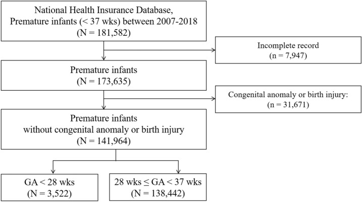
In total, 181,582 premature infants with a GA < 37 weeks were identified in South Korea during the 12-year period analyzed (2007 through 2018). Among them, 7947 cases with incomplete records and 31,671 cases with congenital anomaly or perinatal injury were excluded. The remaining 141,964 premature infants comprised the present study group. Figure 1 shows the flowchart of the enrollment of the study subjects. The overall and annual number of total newborns and premature infants is presented in Supplementary Table S1 online. The number of total newborns has decreased from 496,822 in 2007 to 326,822 in 2018, whereas the number of premature infants has increased from 8366 in 2007 to 12,539 in 2018.
Figure 1.
Flow chart illustrating the process of creating the cohort. Premature infants with a gestational age (GA) < 37 weeks who were born between 2007 and 2018 were enrolled. GA gestational age, ROP retinopathy of prematurity.
Incidence of ROP among premature infants
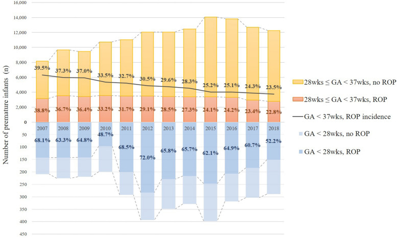
ROP was found in 42,300 newborns during the study period (total incidence, 29.8%). Table 1 shows the annual and overall cumulative incidences of ROP in premature infants according to age group and sex. The cumulative incidence of ROP among premature infants has decreased from 39.5% in 2007 to 23.5% in 2018; from 68.1 to 52.2% in the GA 28 group, from 38.8 to 22.8% in the GA28-37 group (Fig. 2). The overall incidence of ROP during the 12-year study period was 4.29 times higher in the GA28 group than in the GA28-37 group (adjusted OR 4.29, p < 0.001) and 0.97 times lower in males than females (adjusted OR 0.97, p < 0.001; Table 2).
Table 1.
The cumulative incidence of retinopathy of prematurity in premature infants from 2007 to 2018 according to gestational age and sex.
| Year | Total | Male | Female | ||||||
|---|---|---|---|---|---|---|---|---|---|
| Person-year | ROP (n) | Cumulative incidence (%) | Person-year | ROP (n) | Cumulative incidence (%) | Person-year | ROP (n) | Cumulative incidence (%) | |
| Total (GA < 37 weeks) | |||||||||
| 2007 | 8366 | 3308 | 39.5 | 4520 | 1799 | 39.8 | 3846 | 1509 | 39.2 |
| 2008 | 9890 | 3687 | 37.3 | 5456 | 1986 | 36.4 | 4434 | 1701 | 38.4 |
| 2009 | 9667 | 3579 | 37.0 | 5167 | 1922 | 37.2 | 4500 | 1657 | 36.8 |
| 2010 | 10,926 | 3656 | 33.5 | 5901 | 1958 | 33.2 | 5025 | 1698 | 33.8 |
| 2011 | 11,330 | 3702 | 32.7 | 6240 | 2005 | 32.1 | 5090 | 1697 | 33.3 |
| 2012 | 12,451 | 3794 | 30.5 | 6770 | 2027 | 29.9 | 5681 | 1767 | 31.1 |
| 2013 | 12,414 | 3673 | 29.6 | 6830 | 2021 | 29.6 | 5584 | 1652 | 29.6 |
| 2014 | 12,790 | 3623 | 28.3 | 6949 | 1949 | 28.0 | 5841 | 1674 | 28.7 |
| 2015 | 14,449 | 3635 | 25.2 | 7917 | 1970 | 24.9 | 6532 | 1665 | 25.5 |
| 2016 | 14,139 | 3546 | 25.1 | 7715 | 1927 | 25.0 | 6424 | 1619 | 25.2 |
| 2017 | 13,003 | 3154 | 24.3 | 7141 | 1722 | 24.1 | 5862 | 1432 | 24.4 |
| 2018 | 12,539 | 2943 | 23.5 | 6927 | 1619 | 23.4 | 5612 | 1324 | 23.6 |
| Overall | 141,964 | 42,300 | 29.8 | 77,533 | 22,905 | 29.5 | 64,431 | 19,395 | 30.1 |
| GA < 28 weeks | |||||||||
| 2007 | 207 | 141 | 68.1 | 92 | 61 | 66.3 | 115 | 80 | 69.6 |
| 2008 | 226 | 143 | 63.3 | 123 | 87 | 70.7 | 103 | 56 | 54.4 |
| 2009 | 219 | 142 | 64.8 | 110 | 71 | 64.5 | 109 | 71 | 65.1 |
| 2010 | 199 | 97 | 48.7 | 85 | 38 | 44.7 | 114 | 59 | 51.8 |
| 2011 | 292 | 200 | 68.5 | 131 | 82 | 62.6 | 161 | 118 | 73.3 |
| 2012 | 393 | 283 | 72.0 | 208 | 138 | 66.3 | 185 | 145 | 78.4 |
| 2013 | 348 | 229 | 65.8 | 171 | 115 | 67.3 | 177 | 114 | 64.4 |
| 2014 | 329 | 216 | 65.7 | 163 | 109 | 66.9 | 166 | 107 | 64.5 |
| 2015 | 398 | 247 | 62.1 | 216 | 130 | 60.2 | 182 | 117 | 64.3 |
| 2016 | 319 | 207 | 64.9 | 170 | 106 | 62.4 | 149 | 101 | 67.8 |
| 2017 | 303 | 184 | 60.7 | 153 | 93 | 60.8 | 150 | 91 | 60.7 |
| 2018 | 289 | 151 | 52.2 | 128 | 68 | 53.1 | 161 | 83 | 51.6 |
| Overall | 3522 | 2240 | 63.6 | 1750 | 1098 | 62.7 | 1772 | 1142 | 64.4 |
| 28 weeks ≤ GA < 37 weeks | |||||||||
| 2007 | 8159 | 3167 | 38.8 | 4428 | 1738 | 39.3 | 3731 | 1429 | 38.3 |
| 2008 | 9664 | 3544 | 36.7 | 5333 | 1899 | 35.6 | 4331 | 1645 | 38.0 |
| 2009 | 9448 | 3437 | 36.4 | 5057 | 1851 | 36.6 | 4391 | 1586 | 36.1 |
| 2010 | 10,727 | 3559 | 33.2 | 5816 | 1920 | 33.0 | 4911 | 1639 | 33.4 |
| 2011 | 11,038 | 3502 | 31.7 | 6109 | 1923 | 31.5 | 4929 | 1579 | 32.0 |
| 2012 | 12,058 | 3511 | 29.1 | 6562 | 1889 | 28.8 | 5496 | 1622 | 29.5 |
| 2013 | 12,066 | 3444 | 28.5 | 6659 | 1906 | 28.6 | 5407 | 1538 | 28.4 |
| 2014 | 12,461 | 3407 | 27.3 | 6786 | 1840 | 27.1 | 5675 | 1567 | 27.6 |
| 2015 | 14,051 | 3388 | 24.1 | 7701 | 1840 | 23.9 | 6350 | 1548 | 24.4 |
| 2016 | 13,820 | 3339 | 24.2 | 7545 | 1821 | 24.1 | 6275 | 1518 | 24.2 |
| 2017 | 12,700 | 2970 | 23.4 | 6988 | 1629 | 23.3 | 5712 | 1341 | 23.5 |
| 2018 | 12,250 | 2792 | 22.8 | 6799 | 1551 | 22.8 | 5451 | 1241 | 22.8 |
| Overall | 138,442 | 40,060 | 28.9 | 75,783 | 21,807 | 28.8 | 62,659 | 18,253 | 29.1 |
GA gestational age, ROP retinopathy of prematurity.
Figure 2.
Twelve-year trend of incidence of retinopathy of prematurity (ROP) among premature infants according to gestational age (GA) groups. The stacked bar graph above the horizontal axis indicates the annual number of premature infants of 28 weeks ≤ GA < 37 weeks, with and without ROP, and incidence of ROP (%). The stacked bar graph below the horizontal axis indicates the annual number of premature infants of GA < 28 weeks, with and without ROP, and incidence of ROP (%). The line graph indicates the annual incidence of ROP among premature infants with a GA < 37 weeks. GA gestational age, ROP retinopathy of prematurity.
Table 2.
The odds ratios (OR) of incidence of retinopathy of prematurity according to gestational age and sex during the 12-year study period.
| Person-year | ROP (n) | Cumulative incidence (%) | Crude | Adjusted | |||||
|---|---|---|---|---|---|---|---|---|---|
| OR | 95% CI | P-value | OR | 95% CI | P-value | ||||
| GA < 28 weeks | 3522 | 2240 | 63.6% | 4.29 | 4.00–4.60 | < .0001 | 4.29a | 4.00–4.59 | < .0001 |
| 28 weeks ≤ GA < 37 weeks | 138,442 | 40,060 | 28.9% | Reference | |||||
| Total (GA < 37 weeks) | |||||||||
| Male | 77,533 | 22,905 | 29.5% | 0.97 | 0.95–1.00 | 0.022 | 0.97b | 0.95–1.00 | < .0001 |
| Female | 64,431 | 19,395 | 30.1% | Reference | |||||
| GA < 28 weeks | |||||||||
| Male | 1750 | 1098 | 62.7% | 0.93 | 0.81–1.07 | 0.293 | 0.93b | 0.81–1.06 | 0.040 |
| Female | 1772 | 1142 | 64.4% | Reference | |||||
| 28 weeks ≤ GA < 37 weeks | |||||||||
| Male | 75,783 | 21,807 | 28.8% | 0.98 | 0.96–1.01 | 0.147 | 0.98b | 0.96–1.01 | < .0001 |
| Female | 62,659 | 18,253 | 29.1% | Reference | |||||
OR was estimated using logistic regression analysis.
GA gestational age, ROP retinopathy of prematurity.
aOR adjusted for sex, year of diagnosis, income level, area of residence.
bOR adjusted for year of diagnosis, income level, area of residence.
Treatment of ROP
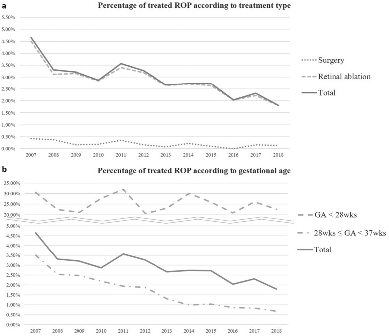
Among ROP patients, 3.0% (1246/42,300) underwent treatment during the 12-year of study period. Surgery and retinal ablation subgroups represented 0.2% (84/42,300) and 2.9% (1214/42,300) of the ROP infants, respectively. The total treatment group accounted for 25.0% (561/2240) of ROP in the GA28 group and 1.7% (685/40,060) in the GA28-37 group (Table 3). Figure 3 shows the 12-year trend in the treatment pattern for ROP. The overall percentage of ROP infants who underwent treatment decreased during study period from 4.7% in 2007 to 1.8% in 2018, with the sharpest decline between 2007 (4.7%) and 2008 (3.3%). The proportion of each treatment subgroup in total and for each gestational group also has decreased (Supplementary Table S2 online).
Table 3.
The percentage of patients with retinopathy of prematurity who underwent treatment during the 12-year of study period according to gestational age and sex.
| Treatment category | Total | Male | Female | ||||||
|---|---|---|---|---|---|---|---|---|---|
| ROP (n) | Treatment | ROP (n) | Treatment | ROP (n) | Treatment | ||||
| (n) | (%) | (n) | (%) | (n) | (%) | ||||
| Total (GA < 37 weeks) | |||||||||
| Surgery | 84 | 0.2 | 41 | 0.2 | 43 | 0.2 | |||
| Retinal ablation | 1214 | 2.9 | 606 | 2.7 | 608 | 3.1 | |||
| Total | 42,300 | 1246 | 3.0 | 22,905 | 621 | 2.7 | 19,395 | 625 | 3.2 |
| GA < 28 weeks | |||||||||
| Surgery | 31 | 1.4 | 15 | 1.4 | 16 | 1.4 | |||
| Retinal ablation | 550 | 24.6 | 266 | 24.2 | 284 | 24.9 | |||
| Total | 2240 | 561 | 25.0 | 1098 | 271 | 24.7 | 1142 | 290 | 25.4 |
| 28 weeks ≤ GA < 37 weeks | |||||||||
| Surgery | 53 | 0.1 | 26 | 0.1 | 27 | 0.2 | |||
| Retinal ablation | 664 | 1.7 | 340 | 1.6 | 324 | 1.8 | |||
| Total | 40,060 | 685 | 1.7 | 21,807 | 350 | 1.6 | 18,253 | 335 | 1.8 |
GA gestational age, ROP retinopathy of prematurity.
Figure 3.
Twelve-year trend of treatment in retinopathy of prematurity (ROP) according to the treatment type and gestational age (GA) groups. The annual percentage of ROP infants who underwent treatment according to the treatment type (A) and the GA groups (B) are presented. GA = Gestational age; ROP = retinopathy of prematurity.
Discussion
This first Korean nationwide study of ROP encompassing 12 years of premature infants in South Korea provides informative results regarding the incidence and treatment pattern for ROP among premature infants with a GA < 37 weeks. The national incidence of ROP has decreased in South Korea over a 12-year period with an overall incidence of 29.8%. The percentage of infants treated with retinal ablation or surgery for ROP has also decreased, with an overall percentage of 3.0%.
The national incidence of ROP of 29.8% in South Korea is hard to compare with other population-based studies in other countries because the definition of included premature infants in each study is not the same; GA was used in the current study whereas different definitions of GA, BW or LOS were used in other studies. A recent study in Taiwan, using the NHIRD which is similar to NHI database in South Korea, reported annual incidence of between 31 and 41% from 2002 to 2011 among all premature infants with LOS of more than 28 days, which might lead to underestimation of the number of premature infants8. The Turkish studies based on the NICUs network reported similar incidences of 30% and 27% in 2011–2013 and 2016–2017, respectively; between 2011–2013 and 2016–2017 though the included premature infants were only those with BW ≤ 1500 g, GA ≤ 32 weeks or with an unstable clinical course11,12. In European countries, an ROP incidence of 31.9% was found among infants with a GA of < 31 weeks between 2008 and 2015 in Sweden using national registry data9, and an annual incidence of 11–14% was reported among infants with BW < 1500 g between 2007 and 2011 (total incidence of 12.6% in 1990–2011) in England using the dataset from NHS10. In the United States, a study using a 20% representative sample of all US hospital discharges reported an ROP incidence of 15.6% (58,722/376,961) from 1997 to 2005 among newborns with LOS > 28 days6, and another study using the largest publicly available pediatric inpatient care database in the US reported incidences of 18.4% (9630/52,451) in 2009 and 19.9% (10,483/52,720) in 2012 among newborns with LOS > 28 days7. In South Korea, there have been several single-institution-based incidence studies. Cho and Koo reported an ROP rate of 21% (41/194) in infants with GA < 37 weeks or BW < 2000 g and who received supplemental oxygen therapy in 1991–199226, and another study reported an ROP rate of 14.1% (96/666) among premature infants with GA < 37 weeks or BW < 2500 g in 1990–199227. A more recent study showed an ROP incidence of 11.9% (24/201) in Korean infants with BW between 1500 and 2000 g or GA < 34 weeks in 2009–201328. The first population-based epidemiological study used the KNN database and reported a total incidence of ROP of 34.1% among VLBWIs between 2013 and 201413.
Several studies have reported an increase in ROP incidence. The Swedish study, with the most similar study period, reported an increase in incidence from 26.8% (187/698) in 2008 to 36.8% (283/2015) in 2015. They suggested that the continuous increase in the survival of the immature infants might contribute to the increasing incidence of ROP (and of the frequency of treatment)9. The increase of ROP incidence in the US studies from 14.7% (6201/42,178) in 2000 to 19.9% (10,483/52,720) in 2012 was explained by the widespread implementation of ROP screening guidelines after 2006, and recent advances in life-preserving technologies which have led to increased survival of premature infants, and also increased population of babies at risk for ROP7. Meanwhile, the Taiwanese study from 2002 to 2011 showed stationary incidence during the 10-year period, with a gradual decrease in the number of both premature infants and ROP patients8. In the present study, the incidence of ROP has decreased from 39.5% in 2007 to 23.5% in 2018, with a decrease in both the GA 28 (from 68.1 to 52.2%) and GA28-37 (38.8–22.8%) groups. The most likely explanation herein is the substantial increase in premature infants. It was noted that the number of newborns has decreased by 34.2% (from 496,822 to 326,822) whereas the number of premature infants enrolled in this study has markedly increased almost 1.5 times in 2018 compared to 2007 (149.9%, from 8366 to 12,539). As the analysis based on the NHI database has the limitation that cases registered with a proper diagnostic code could be enrolled, there should be a discrepancy between the number included in the present study and demographic-based statistical data. Moreover, since the current study was designed to enroll premature infants without congenital anomaly or birth injury, the real number of premature infants is estimated to be more than the number identified in this study. Nonetheless, the reduction in the annual number of newborns and the rapid increase in the annual number of premature infants before 37 weeks of GA, "the high-risk newborns”, themselves have been identified as a crucial health issue in recent decades in South Korea29,30. Although the number of any stage of ROP has only slightly decreased (3308–2943), the incidence of ROP among preterm infants has largely decreased by 40.5% (from 39.5 to 23.5%). The progressive advances in the guidelines for management of premature newborns to prevent the development of ROP may contributed to this decrease15,31, as the KNN in South Korea has reported an overall improvement in neonatal outcomes including ROP treatment from 2013 to 201632.
The overall incidence of ROP among premature infants was 4.3 times higher in the GA28 group (63.6%) than in the GA28-37 group (28.9%). Low gestational age is a well-known major risk factor for ROP15,33. The incidence of ROP among infants with GA ≤ 28 weeks in Turkey was reported to 52.8% in 2011–2013 compared to 27.6% in infants with GA 29–32 weeks, and 62.9% in 2016–2017 compared to 19.4% in infants with GA 29–32 weeks11,12. The Postnatal Growth and Retinopathy of Prematurity (G-ROP) study in the US and Canada showed an ROP incidence of 74.7% (2369/3173) in 2006–2011. In our analysis, the overall incidence of ROP was slightly (0.97 times), but significantly, lower in males than females. This is consistent with the US studies6,7, but differs from other studies reporting no difference in sex34, or the male preference35. Further studies are needed to conclusively determine the relationship between sex and ROP.
The frequency of treatment among ROP infants in South Korea was 3.0%, with a decrease from 4.7% in 2007 to 1.8% in 2018. Although there have been several nationwide studies reporting the treatment frequency in ROP, the included treatment modalities were different (Table 4). In addition to these nationwide studies, in the Turkish studies based on the NICUs network, infants with BW ≤ 1500 g, GA ≤ 32 weeks or with an unstable clinical course had a treatment frequency of 17.1% (810/4729, laser photocoaguoation and vitreoretinal surgery) in 2011–2013 and 24.4% (414/1695, laser photocoaguoation, intravitreal bevacizumab, and vitreoretinal surgery) in 2016–201711,12. The low treatment percentage in the present study could be mostly accounted for by the study population which includes more infants with large GA compared to other studies. Among ROP infants with GA < 28 weeks, the treatment percentage of 25.0% is comparable to other studies. Another important factor, which also explains the decreased trend of conventional treatment, is the emergence of anti-VEGF treatment. As anti-VEGF treatment for ROP has become widespread, it has replaced conventional treatment in many cases12,36. In South Korea, a study using KNN database between January 2013 and June 2014 examined the percentage of ROP infants who received conventional treatment (cryotherapy or laser photocoagulation and/or vitrectomy) and anti-VEGF treatment13. Among ROP in VLBWIs, 33.7% (231/686) underwent treatment: 63.6% (192) underwent conventional treatment only; 16.9% (84) underwent anti-VEGF treatment only; 19.5% (45) underwent both conventional and anti-VEGF treatment. The trend of using intravitreal anti-VEGF for ROP in South Korea has not been studied other than this KNN report though it has been reported to have been used in clinics since the late 2000s23,37–39. A recent retrospective study conducted in a Korean tertiary hospital reported the use of anti-VEGF for the treatment of ROP since 200637. They included 314 eyes between January 2006 to December 2016 to compare the outcome of primary intravitreal anti-VEGF treatment and laser photocoagulation for ROP; among those, 161 eyes were treated with laser treatment primarily and 153 eyes were treated with intravitreal anti-VEGF injection primarily. There were also other reports in which the combination of laser photocoagulation and intravitreal anti-VEGF were used for ROP23,38,39, one of which reported the use of anti-VEGF since 200638. Overall, this implies that the decrease in use of conventional treatments, especially retinal ablation, since 2007 shown in the present study may reflect the increase in use of anti-VEGF injection in this anti-VEGF era in South Korea. The overall lower treatment percentage throughout the study period may also have been partly attributed to the use of anti-VEGF injections that could not be included using the health claim database.
Table 4.
Nationwide studies of treatment in retinopathy of prematurity including retinal ablation and surgery in the last 10 years.
| Sweden9 | Taiwan8 | United States7 | England10 | Present Study | |
|---|---|---|---|---|---|
| Study period | 2008–2015 | 2002–2011 | 2006, 2009, 2012 | 1990–2011 | 2007–2018 |
| Population | GA < 31 weeks | Premature infants with LOS > 28 days | Premature infants with LOS > 28 days | Infants with BW < 1500 g | GA < 37 weeks |
| ROP (n) | 1,829 | 4,096 | 27,481 | NA | 42,300 |
| Treatment modality | NA | Cryotherapy, laser photocoagulation, intravitreal injection, scleral buckle, vitrectomy | Laser photocoagulation, scleral buckle, vitrectomy | Cryotherapy, laser photocoagulation | Cryotherapy, laser photocoagulation, scleral buckle, vitrectomy |
| Treatment | 18.0% (329/1829) | 6.5% | 8.31% | 13.3% in 1990; 11.8% in 2011 | 2.95% (GA < 28 weeks: 25.0%) |
| Trend | No significant change | Increased from 2.0% in 2002 to 9.3% in 2011 | NA | Decreased until 2006, Increased from 2005 | Decrease from 4.66% in 2007 to 1.80% in 2018 |
GA gestational age, LOS length of hospital stay, ROP retinopathy of prematurity.
The strength of the present study is that it was a large population-based study using a nationwide database, which allowed us to investigate national trends over a 12-year period. In addition, the possibility of missing data is expected to be very low for prematurity and ROP because all of premature infants need admission to hospitals. Therefore, our findings provide a valuable understanding of national epidemiology of ROP in South Korea. There were several limitations in the current study. First, the study subjects were identified by ICD-10 diagnostic and procedure codes. The precise definition of the study subjects is important, so we excluded cases with congenital anomalies or birth injuries to avoid overestimation. However, this could have led to an underestimation of the number of premature infants. Second, there are some limitations related to code registration that we figured out while conducting this analysis. The registered diagnostic codes were mostly integrated rather than sub-divided (i.e., P07.30 [28 weeks ≤ GA < 32 weeks] and P07.31 [32 weeks ≤ GA < 37 weeks] under the integrated code of P07.3 [28 weeks ≤ GA < 37 weeks, GA28-37 group]), and only a small portion of the subjects were registered using the sub-divided codes. Therefore, we could not divide the GA28-37 group into smaller GA groups. Additionally, as it is not compulsory for clinicians to register both, the diagnostic codes of GA and BW, it seemed that the diagnostic code of BW was likely to be missing once that of GA was registered. Among our study population, the number of those with diagnostic codes with low BW (P07.0 [“Extremely low birth weight”, BW < 1000 g] and P07.1 [“Other low birth weight”, 1000 g ≤ BW < 2500 g]) were much smaller compared to the total number of premature infants, so the incidence of ROP according to BW among our study population could not be analyzed in the present study. Third, detailed information regarding the prematurity and additional disease details such as the severity of ROP were not available from NHI database. Therefore, we defined subgroups of premature infants according to GA, but could not correlate this with BW or severity of ROP. Last, we investigated the treatment patterns for ROP in South Korea, but the use of anti-VEGF treatment was not provided by NHI database. As the popularity of anti-VEGF treatment in ROP is increasing, standardized guidelines are needed. In addition, a further nationwide study of treatment trends in ROP including anti-VEGF treatment would give further insights into adoption of this newer treatment.
In conclusion, this is the first nationwide incidence and treatment trend study of ROP in South Korea using a population-based database over 12 years. A decrease in the incidence of ROP among premature infants was found, and the incidence was 0.97 times lower in males than in females, and 4.29 times higher in the GA28 group than in the GA28-37 group. The percentage of ROP infants who underwent conventional treatment also showed a decreasing trend during the study period. The decrease in conventional treatment may suggest the increasing use of anti-VEGF treatment, and the impact of anti-VEGF treatment should be investigated in an additional study. Finally, the findings of the present study may throw light on our understanding of the epidemiology of ROP in South Korea and help establish the national strategies in managing premature infants and ROP.
Supplementary Information
Acknowledgements
This research was supported by the National Research Foundation of Korea (NRF) grants funded by the Korea government (NRF-2020R1A2C1010229 to HC and NRF- 2020R1F1A1048475 to YUS). The authors would like to thank Biostatistical Consulting and the Research Lab of Hanyang University for assistance with statistical analysis.
Author contributions
H.C., I.K., and Y.U.S. planned and designed the study. E.H.H. and Y.U.S. wrote the main manuscript text, and prepared the figures. G.H.B. and I.K. provided the data mining and statistical assistance. G.H.B., E.H.H., Y.U.S., R.H., S.J.A., and Y.J.C. acquired, analyzed and interpreted the data. L.S., I.K., and H.C. did critical revision of the manuscript for important intellectual content. Y.U.S. and H.C. obtained funding. H.C. and I.K. supervised the study. All authors reviewed the manuscript.
Data availability
The data that support the findings of this study are available from the corresponding author upon reasonable request.
Competing interests
The authors declare no competing interests.
Footnotes
Publisher's note
Springer Nature remains neutral with regard to jurisdictional claims in published maps and institutional affiliations.
These authors contributed equally: Eun Hee Hong and Yong Un Shin.
Contributor Information
Inah Kim, Email: inahkim@hanyang.ac.kr.
Heeyoon Cho, Email: hycho@hanyang.ac.kr.
Supplementary Information
The online version contains supplementary material available at 10.1038/s41598-021-80989-z.
References
- 1.Gibson DL, Sheps SB, Hong S, Schechter MT, McCormick AQ. Retinopathy of prematurity-induced blindness: Birth weight-specific survival and the new epidemic. Pediatrics. 1990;86:405–412. [PubMed] [Google Scholar]
- 2.Blencowe H, Lawn JE, Vazquez T, Fielder A, Gilbert C. Preterm-associated visual impairment and estimates of retinopathy of prematurity at regional and global levels for 2010. Pediatr. Res. 2013;74:35. doi: 10.1038/pr.2013.205. [DOI] [PMC free article] [PubMed] [Google Scholar]
- 3.Flynn JT, et al. A cohort study of transcutaneous oxygen tension and the incidence and severity of retinopathy of prematurity. N. Engl. J. Med. 1992;326:1050–1054. doi: 10.1056/NEJM199204163261603. [DOI] [PubMed] [Google Scholar]
- 4.Gilbert C, Muhit M. Twenty years of childhood blindness: What have we learnt? Community Eye Health. 2008;21:46. [PMC free article] [PubMed] [Google Scholar]
- 5.Darlow BA. Retinopathy of prematurity: New developments bring concern and hope. J. Paediatr. Child Health. 2015;51:765–770. doi: 10.1111/jpc.12860. [DOI] [PubMed] [Google Scholar]
- 6.Lad, E. M., Hernandez-Boussard T., Morton J. M. & Moshfeghi D. M. Incidence of retinopathy of prematurity in the United States: 1997 through 2005. Am. J. Ophthalmol.148, 451–458 (2009). [DOI] [PubMed]
- 7.Ludwig CA, Chen TA, Hernandez-Boussard T, Moshfeghi AA, Moshfeghi DM. The epidemiology of retinopathy of prematurity in the United States. Ophthal. Surg. Lasers Imaging Retina. 2017;48:553–562. doi: 10.3928/23258160-20170630-06. [DOI] [PubMed] [Google Scholar]
- 8.Kang EY, et al. Retinopathy of prematurity trends in Taiwan: A 10-year nationwide population study. Invest. Ophthalmol. Vis. Sci. 2018;59:3599–3607. doi: 10.1167/iovs.18-24020. [DOI] [PubMed] [Google Scholar]
- 9.Holmström G, et al. Increased frequency of retinopathy of prematurity over the last decade and significant regional differences. Acta Ophthalmol. (Copenh). 2018;96:142–148. doi: 10.1111/aos.13549. [DOI] [PubMed] [Google Scholar]
- 10.Painter SL, Wilkinson AR, Desai P, Goldacre MJ, Patel CK. Incidence and treatment of retinopathy of prematurity in England between 1990 and 2011: Database study. Br. J. Ophthalmol. 2015;99:807–811. doi: 10.1136/bjophthalmol-2014-305561. [DOI] [PubMed] [Google Scholar]
- 11.Bas AY, Koc E, Dilmen U. Incidence and severity of retinopathy of prematurity in turkey. Br. J. Ophthalmol. 2015;99:1311–1314. doi: 10.1136/bjophthalmol-2014-306286. [DOI] [PubMed] [Google Scholar]
- 12.Bas AY, et al. Incidence, risk factors and severity of retinopathy of prematurity in Turkey (tr-rop study): A prospective, multicentre study in 69 neonatal intensive care units. Br. J. Ophthalmol. 2018;102:1711–1716. doi: 10.1136/bjophthalmol-2017-311789. [DOI] [PMC free article] [PubMed] [Google Scholar]
- 13.Hwang JH, Lee EH, Kim EA. Retinopathy of prematurity among very-low-birth-weight infants in Korea: Incidence, treatment, and risk factors. J. Kor. Med. Sci. 2015;30(Suppl 1):S88–94. doi: 10.3346/jkms.2015.30.S1.S88. [DOI] [PMC free article] [PubMed] [Google Scholar]
- 14.Chang YS, Park H-Y, Park WS. The Korean neonatal network: An overview. J. Korean Med. Sci. 2015;30(Suppl 1):S3–S11. doi: 10.3346/jkms.2015.30.S1.S3. [DOI] [PMC free article] [PubMed] [Google Scholar]
- 15.Hellström A, Smith LE, Dammann O. Retinopathy of prematurity. Lancet. 2013;382:1445–1457. doi: 10.1016/S0140-6736(13)60178-6. [DOI] [PMC free article] [PubMed] [Google Scholar]
- 16.Hinz BJ, de Juan E, Repka MX. Scleral buckling surgery for active stage 4a retinopathy of prematurity. Ophthalmology. 1998;105:1827–1830. doi: 10.1016/S0161-6420(98)91023-5. [DOI] [PubMed] [Google Scholar]
- 17.Repka MX, Tung B, Good WV, Capone A, Shapiro MJ. Outcome of eyes developing retinal detachment during the early treatment for retinopathy of prematurity study. Arch. Ophthalmol. 2011;129:1175–1179. doi: 10.1001/archophthalmol.2011.229. [DOI] [PMC free article] [PubMed] [Google Scholar]
- 18.Singh R, et al. Long-term visual outcomes following lens-sparing vitrectomy for retinopathy of prematurity. Br. J. Ophthalmol. 2012;96:1395–1398. doi: 10.1136/bjophthalmol-2011-301353. [DOI] [PubMed] [Google Scholar]
- 19.Repka MX, et al. Outcome of eyes developing retinal detachment during the early treatment for retinopathy of prematurity study (etrop) Arch. Ophthalmol. 2006;124:24–30. doi: 10.1001/archopht.124.1.24. [DOI] [PubMed] [Google Scholar]
- 20.Mintz-Hittner HA, Kennedy KA, Chuang AZ. Efficacy of intravitreal bevacizumab for stage 3+ retinopathy of prematurity. N. Engl. J. Med. 2011;364:603–615. doi: 10.1056/NEJMoa1007374. [DOI] [PMC free article] [PubMed] [Google Scholar]
- 21.Sankar, M. J., Sankar J. & Chandra P. Anti-vascular endothelial growth factor (vegf) drugs for treatment of retinopathy of prematurity. Cochrane Database Syst. Rev.1, Cd009734 (2018). [DOI] [PMC free article] [PubMed]
- 22.Cayabyab R, Ramanathan R. Retinopathy of prematurity: Therapeutic strategies based on pathophysiology. Neonatology. 2016;109:369–376. doi: 10.1159/000444901. [DOI] [PubMed] [Google Scholar]
- 23.Chung EJ, Kim JH, Ahn HS, Koh HJ. Combination of laser photocoagulation and intravitreal bevacizumab (avastin®) for aggressive zone i retinopathy of prematurity. Graefes Arch. Clin. Exp. Ophthalmol. 2007;245:1727. doi: 10.1007/s00417-007-0661-y. [DOI] [PubMed] [Google Scholar]
- 24.Han J, Kim SE, Lee SC, Lee CS. Low dose versus conventional dose of intravitreal bevacizumab injection for retinopathy of prematurity: A case series with paired-eye comparison. Acta Ophthalmol. (Copenh). 2018;96:e475–e478. doi: 10.1111/aos.13004. [DOI] [PubMed] [Google Scholar]
- 25.Kim, S. T. & Seong S. C. 2016 national health insurance statistical yearbook. 622 (Health Insurance Review and Assessment Service, National Health Insurance service, 2017).
- 26.Cho YU, Koo BS. Incidence and risk factors in retinopathy of prematurity. J. Korean Ophthalmol. Soc. 1993;34:851–859. [Google Scholar]
- 27.Lee JW, Jung JK, Kang JH, Jung GY, Kim MU. A clinical study of retinopathy of prematurity. J. Korean Pediatr. Soc. 1994;37:636–641. [Google Scholar]
- 28.Park SH, Yum HR, Kim S, Lee YC. Retinopathy of prematurity in Korean infants with birthweight greater than 1500 g. Br. J. Ophthalmol. 2016;100:834–838. doi: 10.1136/bjophthalmol-2015-306960. [DOI] [PubMed] [Google Scholar]
- 29.Lee JH, Youn Y, Chang YS. Short-term and long-term outcomes of very-low-birth-weight infants in Korea: Korean neonatal network update in 2019. Clin. Exp. Pediatr. 2020;63:284–290. doi: 10.3345/cep.2019.00822. [DOI] [PMC free article] [PubMed] [Google Scholar]
- 30.Moon JY, Hahn WH, Shim KS, Chang JY, Bae CW. Changes of maternal age distribution in live births and incidence of low birth weight infants in advanced maternal age group in Korea. Kor. J. Perinatol. 2011;22:30–36. [Google Scholar]
- 31.Castillo A, Deulofeut R, Critz A, Sola A. Prevention of retinopathy of prematurity in preterm infants through changes in clinical practice and spo2 technology. Acta Paediatr. 2011;100:188–192. doi: 10.1111/j.1651-2227.2010.02001.x. [DOI] [PMC free article] [PubMed] [Google Scholar]
- 32.Lee JH, Noh OK, Chang YS. Neonatal outcomes of very low birth weight infants in Korean neonatal network from 2013 to 2016. J. Korean Med. Sci. 2019;34:1. doi: 10.3904/kjim.2018.381. [DOI] [PMC free article] [PubMed] [Google Scholar]
- 33.Darlow BA, et al. Prenatal risk factors for severe retinopathy of prematurity among very preterm infants of the Australian and New Zealand neonatal network. Pediatrics. 2005;115:990–996. doi: 10.1542/peds.2004-1309. [DOI] [PubMed] [Google Scholar]
- 34.Cryotherapy for Retinopathy of Prematurity Cooperative Group. The natural ocular outcome of premature birth and retinopathy. Status at 1 year. Arch Ophthalmol.112, 903–912 (1994). [DOI] [PubMed]
- 35.Binet ME, Bujold E, Lefebvre F, Tremblay Y, Piedboeuf B. Role of gender in morbidity and mortality of extremely premature neonates. Am. J. Perinatol. 2012;29:159–166. doi: 10.1055/s-0031-1284225. [DOI] [PubMed] [Google Scholar]
- 36.Adams GGW, et al. Treatment trends for retinopathy of prematurity in the UK: Active surveillance study of infants at risk. BMJ Open. 2017;7:e013366. doi: 10.1136/bmjopen-2016-013366. [DOI] [PMC free article] [PubMed] [Google Scholar]
- 37.Kang HG, et al. Intravitreal ranibizumab versus laser photocoagulation for retinopathy of prematurity: Efficacy, anatomical outcomes and safety. Br. J. Ophthalmol. 2019;103:1332–1336. doi: 10.1136/bjophthalmol-2018-312272. [DOI] [PubMed] [Google Scholar]
- 38.Kim R, Kim YC. Posterior pole sparing laser photocoagulation combined with intravitreal bevacizumab injection in posterior retinopathy of prematurity. J. Ophthalmol. 2014;1:1. doi: 10.1155/2014/257286. [DOI] [PMC free article] [PubMed] [Google Scholar]
- 39.Lee JY, Chae JB, Yang SJ, Yoon YH, Kim J-G. Effects of intravitreal bevacizumab and laser in retinopathy of prematurity therapy on the development of peripheral retinal vessels. Graefes. Arch. Clin. Exp. Ophthalmol. 2010;248:1257–1262. doi: 10.1007/s00417-010-1375-0. [DOI] [PubMed] [Google Scholar]
Associated Data
This section collects any data citations, data availability statements, or supplementary materials included in this article.
Supplementary Materials
Data Availability Statement
The data that support the findings of this study are available from the corresponding author upon reasonable request.


