Abstract
Diaporthe species have often been reported as plant pathogens, endophytes and saprophytes, commonly isolated from a wide range of infected plant hosts. In the present study, twenty strains obtained from leaf spots of twelve host plants in Yunnan Province of China were isolated. Based on a combination of morphology, culture characteristics and multilocus sequence analysis of the rDNA internal transcribed spacer region (ITS), translation elongation factor 1-α (TEF), β-tubulin (TUB), calmodulin (CAL), and histone (HIS) genes, these strains were identified as eight new species: Diaporthe camelliae-sinensis, D. grandiflori, D. heliconiae, D. heterostemmatis, D. litchii, D. lutescens, D. melastomatis, D. pungensis and two previously described species, D. subclavata and D. tectonendophytica. This study showed high species diversity of Diaporthe in tropical rain forests and its hosts in south-western China.
Keywords: Diaporthaceae , Diaporthales , phylogeny, taxonomy, 8 new taxa
Introduction
Diaporthe is a genus in the Diaporthaceae family (Diaporthales), with the asexual morph previously known as Phomopsis and type species Diaporthe eres Nitschke collected from Ulmus sp. in Germany (Nitschke 1870). Nevertheless, with the implementation of “one fungus one name” nomenclature, the generic names Diaporthe and Phomopsis are no longer used for both morphs of this genus, and Rossman et al. (2015) gave priority to the older name Diaporthe Nitschke over Phomopsis (Sacc.) Bubák because it was published first, encountered commonly in literatures and represents the majority of species. The sexual morph of Diaporthe is characterized by: immersed perithecial ascomata and an erumpent pseudostroma with more or less elongated perithecial necks; unitunicate clavate to cylindrical asci; fusoid, ellipsoid to cylindrical, septate or aseptate, hyaline ascospores, biseriately to uniseriately arranged in the ascus, sometimes having appendages (Udayanga et al. 2011; Senanayake et al. 2017, 2018). The asexual morph is characterized by ostiolate conidiomata, with cylindrical phialides producing three types of hyaline, aseptate conidia (Udayanga et al. 2011; Gomes et al. 2013): type I: α-conidia, hyaline, fusiform, straight, guttulate or eguttulate, aseptate, smooth-walled; type II: β-conidia, hyaline, filiform, straight or hamate, aseptate, smooth-walled, eguttulate; type III: γ-conidia, rarely produced, hyaline, multiguttulate, fusiform to subcylindrical with an acute or rounded apex, while the base is sometimes truncate. The gamma conidia rarely produced and observed, those species described, having a third type of spores are D. ampelina (Berk. & M.A. Curtis) R.R. Gomes, Glienke & Crous, D. cinerascens Sacc., D. eres Nitschke, D. hongkongensis R.R. Gomes, C. Glienke & Crous, D. limonicola Guarnaccia & Crous, D. oncostoma (Duby) Fuckel, D. perseae (Zerova) R.R. Gomes, C. Glienke & Crous, D. raonikayaporum R.R. Gomes, C. Glienke & Crous (Gomes et al. 2013; Guarnaccia and Crous 2017; Guo et al. 2020).
Currently, more than 1100 epithets of Diaporthe are listed in Index Fungorum (http://www.indexfungorum.org/; accessed 1 June 2020), but only one-fifth of these taxa have been studied with molecular data (Guo et al. 2020; Yang et al. 2020; Zapata et al. 2020). They are widely distributed and have a broad range of hosts from economically significant agricultural crops to ornamental plants including Camellia, Castanea, Citrus, Glycine, Helianthus, Juglans, Persea, Pyrus, Vaccinium and Vitis (van Rensburg et al. 2006; Santos and Phillips 2009; Crous et al. 2011a, b, 2016; Santos et al. 2011; Thompson et al. 2011; Grasso et al. 2012; Huang et al. 2013; Lombard et al. 2014; Gao et al. 2015, 2016, 2017; Udayanga et al. 2012, 2015; Guarnaccia et al. 2016; Dissanayake et al. 2017; Guarnaccia and Crous 2017; Fan et al. 2018; Senanayake et al. 2018; Guo et al. 2020). Many Diaporthe species have been reported as destructive plant pathogens, innocuous endophytes and saprobes (Murali et al. 2006; Udayanga et al. 2012; Gomes et al. 2013; Ménard et al. 2014; Guarnaccia et al. 2016; Torres et al. 2016; Senanayake et al. 2018). However, the biology and lifestyle of some of them remain unclear (Vilka and Volkova 2015).
From previous studies, the methods of species identification and classification in genus Diaporthe were based on criteria such as morphological characters like the size and shape of ascomata (Udayanga et al. 2011) and conidiomata (Rehner and Uecker 1994). However, in recent studies, determining species boundaries only by morphological characters was demonstrated to be not always informative due to their variability under changing environmental conditions (Gomes et al. 2013). As for phylogenetic analysis for Diaporthe species, the use of a five-locus dataset (ITS-TUB-TEF-CAL-HIS) is the optimal combination for species delimitation as revealed by Santos et al. (2017). Thus, in recent years, many Diaporthe species have been described based on a polyphasic approach combined with morphological characterization and their host associations (Guarnaccia and Crous 2017; Gao et al. 2017; Yang et al. 2018, 2020; Crous et al. 2020; Dayarathne et al. 2020; Guo et al. 2020; Hyde et al. 2020; Li et al. 2020; Zapata et al. 2020).
In this study, we propose eight novel species and two previously described species of Diaporthe, collected in Yunnan Province of China on twelve plant host genera, based on their morphological characters in culture, and molecular phylogenetic analysis.
Materials and methods
Isolation and morphological studies
The leaves of samples were collected from Yunnan Province, China. Isolations from surface sterilized leaf tissues were conducted following the protocol of Gao et al. (2014). Tissue fragments (5 × 5 mm) were taken from the margin of leaf lesions and surface-sterilized by consecutively immersing in 75% ethanol solution for 1 min, 5% sodium hypochlorite solution for 30 s, and finally rinsed in sterile distilled water for 1 min. The pieces were dried with sterilized paper towels and transferred on potato dextrose agar (PDA) in petri plates (Cai et al. 2009). All the PDA plates were incubated at biochemical incubator at 25 °C for 2–4 days, and hyphae were picked out of the periphery of the colonies and inoculated onto new PDA plates.
Following 2–3 weeks of incubation, photographs of the fungal colonies were taken at 7 days and 15 days using a Powershot G7X mark II digital camera. Micromorphological characters were observed and documented in distilled water from microscope slides under Olympus SZX10 stereomicroscope and Olympus BX53 microscope, both supplied with Olympus DP80 HD color digital cameras to photo-document fungal structures. All fungal strains were stored in 10% sterilized glycerin at 4 °C for further studies. Voucher specimens were deposited in the Herbarium of Plant Pathology, Shandong Agricultural University (HSAUP). Living strain cultures were deposited in the Shandong Agricultural University Culture Collection (SAUCC). Taxonomic information on the new taxa was submitted to MycoBank (http://www.mycobank.org).
DNA extraction and amplification
Genomic DNA was extracted from fungal mycelia on PDA, using a modified cetyltrimethylammonium bromide (CTAB) protocol as described in Guo et al. (2000). The internal transcribed spacer regions with intervening 5.8S nrRNA gene (ITS), part of the beta-tubulin gene region (TUB), partial translation elongation factor 1-alpha (TEF), histone H3 (HIS) and calmodulin (CAL) genes were amplified and sequenced by using primers pairs ITS4/ITS5 (White et al. 1990), Bt2a/Bt2b (Glass and Donaldson 1995), EF1-728F/EF1-986R (Carbone and Kohn 1999), CAL-228F/CAL-737R (Carbone and Kohn 1999) and CYLH3F/H3-1b (Glass and Donaldson 1995; Crous et al. 2004), respectively.
PCR was performed using an Eppendorf Master Thermocycler (Hamburg, Germany). Amplification reactions were performed in a 25 μL reaction volume which contained 12.5 μL Green Taq Mix (Vazyme, Nanjing, China), 1 μL of each forward and reverse primer (10 μM) (Biosune, Shanghai, China), and 1 μL template genomic DNA in amplifier, and were adjusted with distilled deionized water to a total volume of 25 μL.
PCR parameters were as follows: 95 °C for 5 min, followed by 35 cycles of denaturation at 95 °C for 30 s, annealing at a suitable temperature for 30 s, extension at 72 °C for 1 min and a final elongation step at 72 °C for 10 min. Annealing temperature for each gene was 55 °C for ITS, 60 °C for TUB, 52 °C for TEF, 54 °C for CAL and 57 °C for HIS. The PCR products were visualized on 1% agarose electrophoresis gel. Sequencing was done bi-directionally, conducted by the Biosune Company Limited (Shanghai, China). Consensus sequences were obtained using MEGA 7.0 (Kumar et al. 2016). All sequences generated in this study were deposited in GenBank (Table 1).
Table 1.
Species and GenBank accession numbers of DNA sequences used in this study with new sequences in bold.
| Species | Strain/Isolate | Host/Substrate | GenBank accession number | ||||
|---|---|---|---|---|---|---|---|
| ITS | TUB | TEF | CAL | HIS | |||
| Diaporthe alnea | CBS 146.46* | Alnus sp. | KC343008 | KC343976 | KC343734 | KC343250 | KC343492 |
| D. anacardii | CBS 720.97* | Anacardium occidentale | KC343024 | KC343992 | KC343750 | KC343266 | KC343508 |
| D. baccae | CBS 136972* | Vaccinium corymbosum | KJ160565 | – | KJ160597 | – | – |
| D. batatas | CBS 122.21 | Ipomoea batatas | KC343040 | KC344008 | KC343766 | KC343282 | KC343524 |
| D. camelliae-sinensis | SAUCC194.92* | Camellia sinensis | MT822620 | MT855817 | MT855932 | MT855699 | MT855588 |
| SAUCC194.103 | Castanea mollissima | MT822631 | MT855828 | MT855943 | MT855710 | MT855599 | |
| SAUCC194.104 | Castanea mollissima | MT822632 | MT855829 | MT855944 | MT855711 | MT855600 | |
| SAUCC194.108 | Machilus pingii | MT822636 | MT855833 | MT855948 | MT855715 | MT855603 | |
| D. canthii | CBS 132533* | Canthium inerme | JX069864 | KC843230 | KC843120 | KC843174 | – |
| D. chamaeropis | CBS 753.70 | Spartium junceum | KC343049 | KC344017 | KC343775 | KC343291 | KC343533 |
| D. cinerascens | CBS 719.96 | Ficus carica | KC343050 | KC344018 | KC343776 | KC343292 | KC343534 |
| D. cissampeli | CPC 27302 | Cissampelos capensis | KX228273 | KX228384 | – | – | KX228366 |
| D. citri | CBS 230.52 | Citrus sinensis | KC343052 | KC344020 | KC343778 | KC343294 | KC343536 |
| D. collariana | MFLUCC 17-2636* | Magnolia champaca | MG806115 | MG783041 | MG783040 | MG783042 | – |
| D. convolvuli | CBS 124654 | Convolvulus arvensis | KC343054 | KC344022 | KC343780 | KC343296 | KC343538 |
| D. cytosporella | AR 5149 | Citrus sinensis | KC843309 | KC843223 | KC843118 | KC843143 | – |
| D. destruens | SPPD-1 | Solanum tuberosum | JN848791 | JX421691 | – | – | – |
| D. dorycnii | MFLU 17-1015* | Dorycnium hirsutum | KY964215 | KY964099 | KY964171 | – | – |
| D. elaeagni | CBS 504.72 | Elaeagnus sp. | KC343064 | KC344032 | KC343790 | KC343306 | KC343548 |
| D. elaeagni-glabrae | LC4802* | Elaeagnus glabra | KX986779 | KX999212 | KX999171 | KX999281 | KX999251 |
| D. endophytica | CBS 133811* | Schinus terebinthifolius | KC343065 | KC344033 | KC343791 | KC343307 | KC343549 |
| D. eres | AR5193* | Ulmus laevis | KJ210529 | KJ420799 | KJ210550 | KJ434999 | KJ420850 |
| D. foeniculina | CBS 123208 | Foeniculum vulgare | KC343104 | KC344072 | KC343830 | KC343346 | KC343588 |
| D. fructicola | MAFF 246408 | Passiflora edulis | LC342734 | LC342736 | LC342735 | LC342738 | LC342737 |
| D. grandiflori | SAUCC194.84* | Heterostemma grandiflorum | MT822612 | MT855809 | MT855924 | MT855691 | MT855580 |
| D. heliconiae | SAUCC194.75 | Heliconia metallica | MT822603 | MT855800 | MT855915 | MT855682 | MT855571 |
| SAUCC194.77* | Heliconia metallica | MT822605 | MT855802 | MT855917 | MT855684 | MT855573 | |
| D. heterophyllae | CPC 26215 | Acacia heterophylla | MG600222 | MG600226 | MG600224 | MG600218 | MG600220 |
| D. heterostemmatis | SAUCC194.85* | Heterostemma grandiflorum | MT822613 | MT855810 | MT855925 | MT855692 | MT855581 |
| SAUCC194.102 | Camellia sinensis | MT822630 | MT855827 | MT855942 | MT855709 | MT855598 | |
| D. hickoriae | CBS 145.26* | Carya glabra | KC343118 | KC344086 | KC343844 | KC343360 | KC343602 |
| D. inconspicua | CBS 133813* | Maytenus ilicifolia | KC343123 | KC344091 | KC343849 | KC343365 | KC343607 |
| D. kongii | T12509H* | Helianthus annuus | JF431301 | KJ197272 | JN645797 | – | – |
| D. litchii | SAUCC194.12 | Elaeagnus conferta | MT822540 | MT855737 | MT855854 | MT855625 | MT855509 |
| SAUCC194.22* | Litchi chinensis | MT822550 | MT855747 | MT855863 | MT855635 | MT855519 | |
| D. longicolla | FAU599 | Glycine max | KJ590728 | KJ610883 | KJ590767 | KJ612124 | KJ659188 |
| D. lutescens | SAUCC194.36* | Chrysalidocarpus lutescens | MT822564 | MT855761 | MT855877 | MT855647 | MT855533 |
| D. macintoshii | BRIP 55064a* | Rapistrum rugostrum | KJ197289 | KJ197269 | KJ197251 | – | – |
| D. masirevicii | BRIP 57330 | Chrysanthemoides monilifera subsp. rotundata | KJ197275 | KJ197255 | KJ197237 | – | – |
| BRIP 57892a* | Helianthus annuus | KJ197276 | KJ197257 | KJ197239 | – | – | |
| D. melastomatis | SAUCC194.55* | Melastoma malabathricum | MT822583 | MT855780 | MT855896 | MT855664 | MT855551 |
| SAUCC194.80 | Millettia reticulata | MT822608 | MT855805 | MT855920 | MT855687 | MT855576 | |
| SAUCC194.88 | Camellia sinensis | MT822616 | MT855813 | MT855928 | MT855695 | MT855584 | |
| D. melonis | CBS 507.78* | Cucumis melo | KC343142 | KC344110 | KC343868 | KC343384 | KC343626 |
| D. miriciae | BRIP 54736j* | Helianthus annuus | KJ197282 | KJ197262 | KJ197244 | – | – |
| D. neilliae | CBS 144.27 | Spiraea sp. | KC343144 | KC344112 | KC343870 | KC343386 | KC343628 |
| D. nigra | JZBH320170 | Ballota nigra | MN653009 | MN887113 | MN892277 | – | – |
| D. nomurai | CBS 157.29 | Morus sp. | KC343154 | KC344122 | KC343880 | KC343396 | KC343638 |
| D. oncostoma | CBS 100454 | Robinia pseudoacacia | KC343160 | KC344128 | KC343886 | KC343402 | KC343644 |
| CBS 109741 | Robinia pseudoacacia | KC343161 | KC344129 | KC343887 | KC343403 | KC343645 | |
| D. ovalispora | ZJUD93* | Citrus limon | KJ490628 | KJ490449 | KJ490507 | – | KJ490570 |
| D. parapterocarpi | CPC 22729 | Pterocarpus brenanii | KJ869138 | KJ869248 | – | – | – |
| D. parvae | PSCG 034* | Pyrus bretschneideri | MK626919 | MK691248 | MK654858 | – | MK726210 |
| D. passifloricola | CPC 27480* | Passiflora foetida | KX228292 | KX228387 | – | – | KX228367 |
| D. penetriteum | LC3353* | Camellia sinensis | KP714505 | KP714529 | KP714517 | – | KP714493 |
| LC3394 | Camellia sinensis | KP267893 | KP293473 | KP267967 | – | KP293544 | |
| D. phaseolorum | CBS 116019 | Caperonia palustris | KC343175 | KC344143 | KC343901 | KC343417 | KC343659 |
| CBS 116020 | Aster exilis | KC343176 | KC344144 | KC343902 | KC343418 | KC343660 | |
| D. phillipsii | CAA 817* | Dead twig | MK792305 | MN000351 | MK828076 | MK883831 | MK871445 |
| D. poincianellae | URM 7932 | Poincianella pyramidalis | MH989509 | MH989537 | MH989538 | MH989540 | MH989539 |
| D. pseudoinconspicua | G26 | Poincianella pyramidalis | MH122538 | MH122524 | MH122533 | MH122528 | MH122517 |
| D. psoraleae | CPC 21634 | Psoralea pinnata | KF777158 | KF777251 | KF777245 | – | – |
| D. pterocarpi | MFLUCC 10-0571 | Pterocarous indicus | JQ619899 | JX275460 | JX275416 | JX197451 | – |
| MFLUCC 10-0575 | Pterocarous indicus | JQ619901 | JX275462 | JX275418 | JX197453 | – | |
| D. pungensis | SAUCC194.89 | Camellia sinensis | MT822617 | MT855814 | MT855929 | MT855696 | MT855585 |
| SAUCC194.112* | Elaeagnus pungens | MT822640 | MT855837 | MT855952 | MT855719 | MT855607 | |
| D. ravennica | MFLUCC 17-1029 | Tamarix sp. | KY964191 | KY964075 | KY964147 | – | – |
| D. rosae | MFLUCC 17-2658 | Rosa sp. | MG828894 | MG843878 | – | MG829273 | – |
| D. rumicicola | MFLUCC18-0739 | Rumex sp. | MH846233 | MK049555 | MK049554 | – | – |
| D. saccarata | CBS 116311* | Protea repens | KC343190 | KC344158 | KC343916 | KC343432 | KC343674 |
| D. shennongjiaensis | CNUCC 201905 | Juglans regia | MN216229 | MN227012 | MN224672 | MN224551 | MN224560 |
| D. sojae | CBS 100.87* | Glycine soja | KC343196 | KC344164 | KC343922 | KC343438 | KC343680 |
| D. stictica | CBS 370.54 | Buxus sampervirens | KC343212 | KC344180 | KC343938 | KC343454 | KC343696 |
| D. subclavata | ZJUD95* | Citrus unshiu | KJ490630 | KJ490451 | KJ490509 | – | KJ490572 |
| SAUCC194.66 | Pometia pinnata | MT822594 | MT855791 | MT855906 | MT855674 | MT855562 | |
| D. subellipicola | KUMCC 17-0153 | on dead wood | MG746632 | MG746634 | MG746633 | – | – |
| D. tectonendophytica | MFLUCC 13-0471* | Tectona grandis | KU712439 | KU743986 | KU749367 | KU749354 | – |
| SAUCC194.11 | Elaeagnus conferta | MT822539 | MT855736 | MT855853 | MT855624 | MT855508 | |
| SAUCC194.63 | Pometia pinnata | MT822591 | MT855788 | MT855903 | MT855672 | MT855559 | |
| D. ueckerae | FAU656* | Cucumis melo | KJ590726 | KJ610881 | KJ590747 | KJ612122 | KJ659215 |
| D. unshiuensis | CFCC 52595 | Carya illinoensis | MH121530 | MH121607 | MH121572 | MH121448 | MH121488 |
| D. vangueriae | CPC 22703 | Vangueria infausta | KJ869137 | KJ869247 | – | – | – |
| D. velutina | LC4419* | Neolitsea sp. | KX986789 | KX999222 | KX999181 | KX999286 | KX999260 |
| D. virgiliae | CMW40755* | Virgilia oroboides | KP247573 | KP247582 | – | – | – |
| CMW40748 | Virgilia oroboides | KP247566 | KP247575 | – | – | – | |
| D. zaobaisu | PSCG 031* | Pyrus bretschneideri | MK626922 | MK691245 | MK654855 | – | MK726207 |
| Diaporthella corylina | CBS 121124 | Corylus sp. | KC343004 | KC343972 | KC343730 | KC343246 | KC343488 |
Isolates marked with “*” are ex-type or ex-epitype strains.
Phylogenetic analyses
Novel sequences generated from twenty strains in this study, and all reference available sequences of Diaporthe species downloaded from GenBank were used for phylogenetic analyses. Alignments of the individual locus were determined using MAFFT v. 7.110 by default settings (Katoh et al. 2017) and manually corrected where necessary. To establish the identity of the isolates at species level, phylogenetic analyses were conducted first individually for each locus and then as combined analyses of five loci (ITS, TUB, TEF, CAL and HIS regions). Phylogenetic analyses were based on maximum likelihood (ML) and Bayesian inference (BI) for the multi-locus analyses. For BI, the best evolutionary model for each partition was determined using MrModeltest v. 2.3 (Nylander 2004) and incorporated into the analyses. ML and BI were run on the CIPRES Science Gateway portal (https://www.phylo.org/) (Miller et al. 2012) using RaxML-HPC2 on XSEDE (8.2.12) (Stamatakis 2014) and MrBayes on XSEDE (3.2.7a) (Huelsenbeck and Ronquist 2001; Ronquist and Huelsenbeck 2003; Ronquist et al. 2012), respectively. For ML analyses the default parameters were used and BI was carried out using the rapid bootstrapping algorithm with the automatic halt option. Bayesian analyses included five parallel runs of 5,000,000 generations, with the stop rule option and a sampling frequency of 500 generations. The burn-in fraction was set to 0.25 and posterior probabilities (PP) were determined from the remaining trees. The resulting trees were plotted using FigTree v. 1.4.2 (http://tree.bio.ed.ac.uk/software/figtree) and edited with Adobe Illustrator CS5.1. New sequences generated in this study were deposited at GenBank (https://www.ncbi.nlm.nih.gov; Table 1), the alignments and trees were deposited in TreeBASE (http://treebase.org/treebase-web/home.html).
Results
Phylogenetic analyses
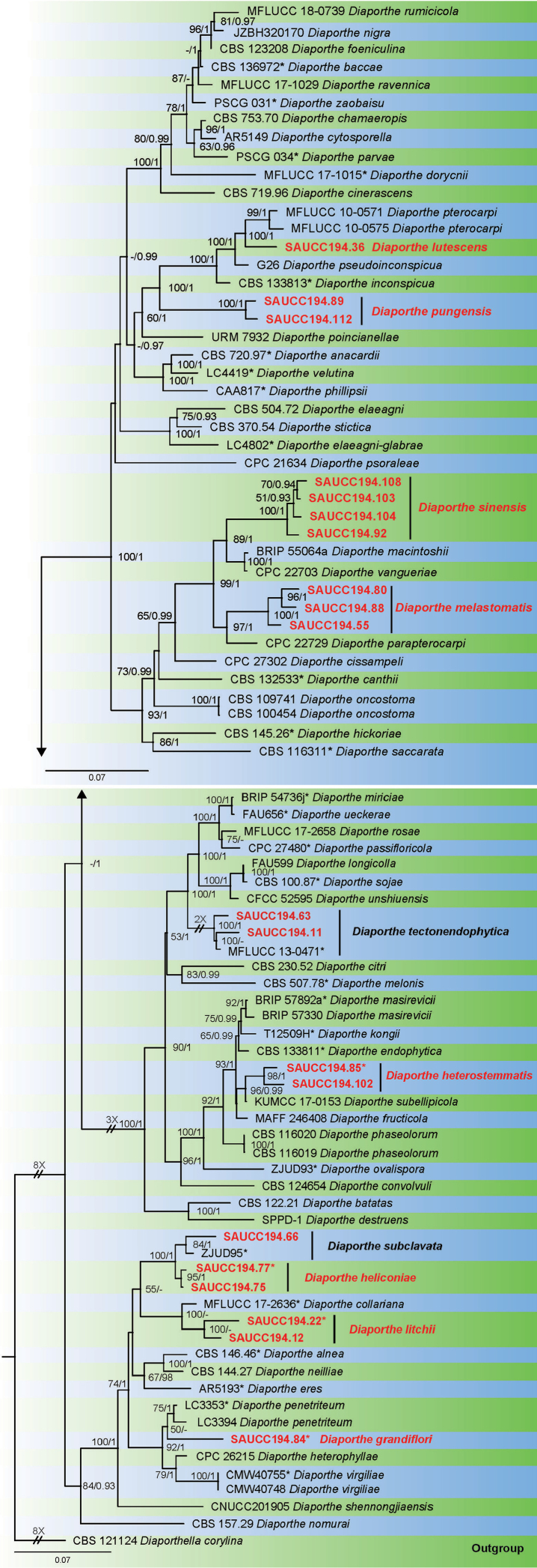
Twenty fungal strains of Diaporthe isolates from 15 plant hosts were sequenced (Table 1). These were analyzed by using multilocus data (ITS, TUB, TEF, CAL and HIS) composed of 87 isolates of Diaporthe, with Diaporthella corylina (CBS 121124) as an outgroup taxon. A total of 2856 characters including gaps were obtained in the phylogenetic analysis, viz. ITS: 1–650, TUB: 651–1263, TEF: 1264–1705, CAL: 1706–2279, HIS: 2280–2856. Of these characters, 1395 were constant, 475 were variable and parsimony-uninformative, and 986 were parsimony-informative. For the BI and ML analyses, the substitution model GTR+I+G for ITS, HKY+I+G for TUB, TEF and CAL, GTR+G for HIS were selected and incorporated into the analyses. The ML tree topology confirmed the tree topologies obtained from the BI analyses, and therefore, only the ML tree is presented (Fig. 1).
Figure 1.
Phylogram of Diaporthe based on combined ITS, TUB, TEF, CAL and HIS genes. The ML and BI bootstrap support values above 50% and 0.90 BYPP are shown at the first and second position, respectively. Strains marked with “*” are ex-type or ex-epitype. Strains from this study are shown in red. Three branches were shortened to fit the page size – these are indicated by symbol (//) with indication number showing how many times they are shortened.
ML bootstrap support values (≥ 50%) and Bayesian posterior probability (≥ 0.90) are shown as first and second position above nodes, respectively. Based on the five-locus phylogeny and morphology, 20 strains isolated in this study were assigned to 10 species, 8 of them are proposed and described here as new species (Fig. 1). Strains (SAUCC194.92, SAUCC194.103, SAUCC194.104 and SAUCC194.108) are D. camelliae-sinensis, strain (SAUCC194.84) – Diaporthe grandiflori, strains (SAUCC194.75 and SAUCC194.77) – D. heliconiae, strains (SAUCC194.85 and SAUCC194.102) – D. heterostemmatis, strains (SAUCC194.12 and SAUCC194.22) – D. litchii, strain (SAUCC194.36) – D. lutescens, strains (SAUCC194.55, SAUCC194.80 and SAUCC194.88) – D. melastomatis, strains (SAUCC194.89 and SAUCC194.112) – D. pungensis. One strain (SAUCC194.66) is of a previously described D. subclavata, and strains (SAUCC194.11 and SAUCC194.63) – of previously described D. tectonendophytica.
Taxonomy
Diaporthe camelliae-sinensis
S.T. Huang, J.W. Xia, X.G. Zhang & Z. Li sp. nov.
0DE548B0-B15D-5939-BDFE-852AC226BCB8
837600
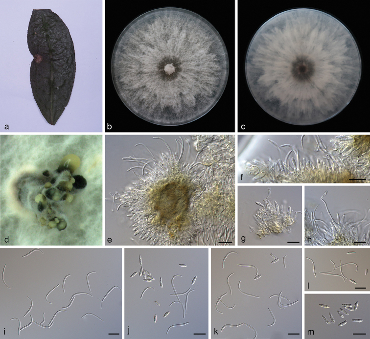
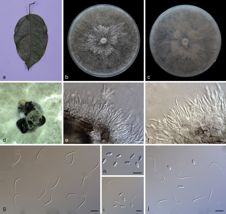
Figure 2.
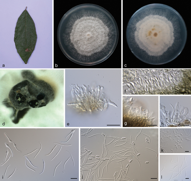
Diaporthe camelliae-sinensis (SAUCC194.92) a leaf of host plant b, c surface (b) and reverse (c) sides of colony after incubation for 15 days on PDAd conidiomata e–h conidiophores and conidiogenous cells i beta conidia j–l alpha conidia and beta conidia m alpha conidia. Scale bars: 10 μm (e–m).
Etymology.
Named after the host Camellia sinensis on which it was collected.
Diagnosis.
Diaporthe camelliae-sinensis can be distinguished from the closely related species D. macintoshii R.G. Shivas et al. and D. vangueriae Crous based on ITS, TUB and TEF loci. Diaporthe camelliae-sinensis differs from D. macintoshii in smaller α-conidia and from D. vangueriae in shorter β-conidia.
Type.
China, Yunnan Province: Xishuangbanna Tropical Botanical Garden, Chinese Academy of Sciences, on infected leaves of Camellia sinensis. 19 April 2019, S.T. Huang, HSAUP194.92, holotype, ex-holotype living culture SAUCC194.92.
Description.
Asexual morph: Conidiomata pycnidial, multi-pycnidia grouped together, globose, black, erumpent, coated with white hyphae, thick-walled, exuding creamy to yellowish conidial droplets from central ostioles. Conidiophores hyaline, smooth, septate, branched, densely aggregated, cylindrical, straight to sinuous, swelling at the base, tapering towards the apex, 10–15 × 1.5–2 μm. Conidiogenous cells 8.5–12 × 2–2.8 μm, phialidic, cylindrical, terminal, slightly tapering towards the apex. Alpha conidia, hyaline, smooth, aseptate, ellipsoidal to fusoid, 2–4 guttulate, apex subobtuse, base subtruncate, 7.5–10 × 1.8–2.5 µm (mean = 8.5 × 2.2 μm, n = 20). Beta conidia hyaline, aseptate, filiform, sigmoid to lunate, mostly curved through 90–180°, tapering towards the apex, base truncate, 20–30 × 1.2–1.6 µm (mean = 25.6 × 1.3 μm, n = 20). Gamma conidia and sexual morph not observed.
Culture characteristics.
Pure culture was isolated by subbing hyphal tips growing from surface sterilized diseased material. Colonies on PDA cover the Petri dish diameter after incubation for 15 days in dark conditions at 25 °C, cottony and radially with abundant aerial mycelium, sparse in the margin. With a tanned concentric ring of dense hyphae, white on surface side, white to pale yellow on reverse side.
Additional specimens examined.
China, Yunnan Province: Xishuangbanna Tropical Botanical Garden, Chinese Academy of Sciences, 19 April 2019, S.T. Huang. On infected leaves of Castanea mollissima, HSAUP194.103 and HSAUP194.104 paratype, living culture SAUCC194.103 and SAUCC194.104; on diseased leaves of Machilus pingii, HSAUP194.108 paratype, living culture SAUCC194.108.
Notes.
Four isolates are clustered in a clade distinct from its closest phylogenetic neighbor, D. macintoshii and D. vangueriae. Diaporthe camelliae-sinensis can be distinguished from D. macintoshii in ITS, TUB and TEF loci (23/558 in ITS, 2/463 in TUB and 20/328 in TEF); from D. vangueriae in ITS and TUB loci (23/558 in ITS and 1/423 in TUB). Morphologically, Diaporthe camelliae-sinensis differs from D. macintoshii in having guttulate alpha conidia and smaller alpha conidia (7.5–10 × 1.8–2.5 vs. 8.0–11.0 × 2.0–3.0 μm) (Thompson et al. 2015). Furthermore, Diaporthe camelliae-sinensis differs from D. vangueriae in shorter beta conidia (20–30 × 1.2–1.6 vs. 28–35 × 1.5–2.0 μm) and D. camelliae-sinensis can produce alpha conidia, but D. vangueriae could not (Crous et al. 2014).
Diaporthe grandiflori
S.T. Huang, J.W. Xia, X.G. Zhang & Z. Li sp. nov.
1A2EBCE2-3B37-5E62-8686-51F5666ADB69
837591
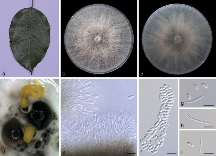
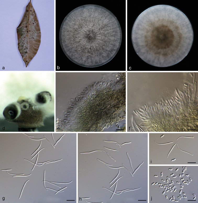
Figure 3.
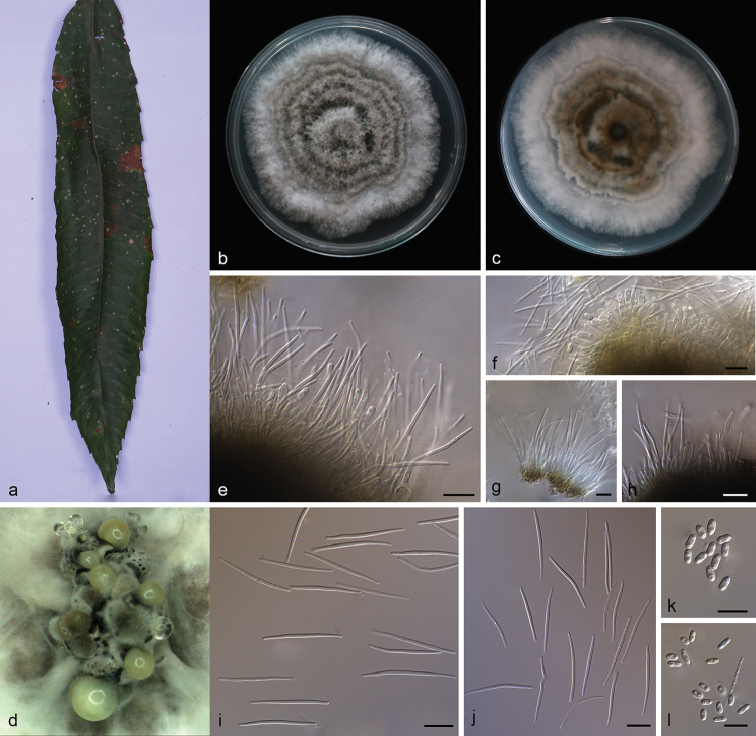
Diaporthe grandiflori (SAUCC194.84) a leaf of Heterostemma grandiflorumb, c surface (b) and reverse (c) sides of colony after incubation for 15 days on PDAd conidiomata e conidiophores and conidiogenous cells f alpha conidia g, i alpha conidia and beta conidia h beta conidia. Scale bars: 10 μm (e–i).
Etymology.
Named after the host Heterostemma grandiflorum on which it was collected.
Diagnosis.
Diaporthe grandiflori can be distinguished from the phylogenetically closely related species D. penetriteum Y.H. Gao & L. Cai in larger α-conidia and β-conidia.
Type.
China, Yunnan Province: Xishuangbanna Tropical Botanical Garden, Chinese Academy of Sciences, on infected leaves of Heterostemma grandiflorum. 19 April 2019, S.T. Huang, HSAUP194.84, holotype, ex-holotype living culture SAUCC194.84.
Description.
Asexual morph: Conidiomata pycnidial, subglobose to globose, solitary or aggregated in groups, black, erumpent, coated with white hyphae, thick-walled, exuding golden yellow spiral conidial cirrus from ostiole. Conidiophores hyaline, smooth, septate, branched, densely aggregated, cylindrical, straight to slightly sinuous, 9.5–16.5 × 1.9–2.8 μm. Conidiogenous cells 19.0–22.8 × 1.4–2.4 μm, cylindrical, multi-guttulate, terminal, tapering towards the apex. Alpha conidia abundant in culture, biguttulate, hyaline, smooth, aseptate, ellipsoidal, apex subobtuse, base subtruncate, 6.3–8.3 × 2.8–3.3 µm (mean = 7.5 × 2.9 μm, n = 20). Beta conidia, not numerous, hyaline, aseptate, filiform, slightly curved, tapering towards the apex, 21.5–30.5 × 1.5–2.1 µm (mean = 24.0 × 1.7 μm, n = 20). Gamma conidia not observed. Sexual morph not observed.
Culture characteristics.
Pure culture was isolated by subbing hyphal tips growing from surface sterilized plant material. Colonies on PDA cover the Petri dish after 15 days kept in dark conditions at 25 °C, cottony with abundant aerial mycelium, white on surface side, white to grayish on reverse.
Notes.
Phylogenetic analysis of a combined five loci showed that D. grandiflori (strain SAUCC194.84) formed an independent clade (Fig. 1) and is phylogenetically distinct from D. penetriteum. This species can be easily distinguished from D. penetriteum by 87 nucleotides difference concatenated alignment (24 in the ITS region, 1 TUB, 41 CAL and 21 HIS). Morphologically, D. grandiflori differs from D. penetriteum in larger α-conidia (6.3–8.3 × 2.8–3.3 vs. 4.5–5.5 × 1.5–2.5 μm) and longer β-conidia (21.5–30.5 × 1.5–2.1 vs. 16.5–27.5 × 1.0–2.0 μm) (Gao et al. 2016).
Diaporthe heliconiae
S.T. Huang, J.W. Xia, X.G. Zhang & Z. Li sp. nov.
222ED481-5804-5F9B-A431-9E9326612CFE
837592
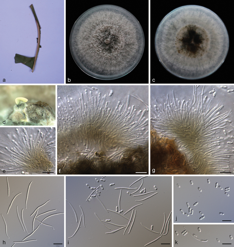
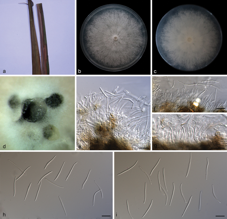
Figure 4.
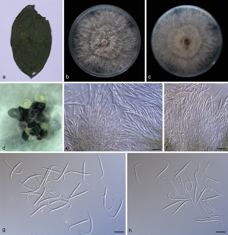
Diaporthe heliconiae (SAUCC194.77) a petiole of Heliconia metallicab, c surface (b) and reverse (c) sides of colony after incubation for 15 days on PDAd conidiomata on PDAe–g conidiophores and conidiogenous cells h beta conidia i alpha conidia and beta conidia j alpha conidia k alpha conidia and germinating conidia. All in water. Scale bars: 10 μm (e–k).
Etymology.
Named after the host Heliconia metallica on which it was collected.
Diagnosis.
Diaporthe heliconiae can be distinguished from the phylogenetically closely related species D. subclavata F. Huang, K.D. Hyde & Hong Y. Li in smaller α-conidia.
Type.
China, Yunnan Province: Xishuangbanna Tropical Botanical Garden, Chinese Academy of Sciences, on the symptomatic petiole of Heliconia metallica. 19 April 2019, S.T. Huang, HSAUP194.77, holotype, ex-holotype living culture SAUCC194.77.
Description.
Asexual morph: Conidiomata pycnidial, solitary or aggregated in groups, erumpent, thin-walled, superficial to embedded on PDA, dark brown to black, globose or subglobose, exuding creamy yellowish spiral conidial cirrus from the ostioles. Conidiophores hyaline, aseptate, cylindrical, straight to sinuous, branched, 16.5–25.0 × 1.3–1.8 µm. Alpha conidiogenous cells, cylindric-clavate, terminal, few guttulate, 11.5–18.0 × 1.0–1.5 µm. Beta conidiogenous cells, prismatic, terminal, few guttulate, 10.0–14.1 × 1.0–1.2 µm. Alpha conidia, hyaline, smooth, aseptate, ellipsoidal, 2–4 guttulate, apex subobtuse, base subtruncate, 5.0–6.5 × 2.0–2.5 µm (mean = 6.1 × 2.3 μm, n = 20). Beta conidia hyaline, aseptate, filiform, slightly curved, tapering towards the apex, 25.0–33.5 × 1.0–1.5 µm (mean = 29.4 × 1.3 μm, n = 20). Gamma conidia and sexual morph not observed.
Culture characteristics.
Pure culture was isolated by subbing hyphal tips growing from surface sterilized infected plant material. Colonies on PDA cover the Petri dish diameter after incubation for 15 days in dark conditions at 25 °C. Aerial mycelium abundant, cottony, white, dense in the center, sparse near the margin. White on surface side, white to tanned on reverse side.
Additional specimen examined.
China, Yunnan Province: Xishuangbanna Tropical Botanical Garden, Chinese Academy of Sciences, on the symptomatic petiole of Heliconia metallica. 19 April 2019, S.T. Huang, HSAUP194.75 paratype; living culture SAUCC194.75.
Notes.
Diaporthe heliconiae clade comprises strains SAUCC194.75 and SAUCC194.77, closely related to D. subclavata in the combined phylogenetic tree (Fig. 1). Diaporthe heliconiae can be distinguished based on ITS, TUB and HIS loci from D. subclavata (16/489 in ITS, 8/357 in TUB and 3/470 in HIS). Morphologically, Diaporthe heliconiae differs from D. subclavata in its smaller α-conidia (5.0–6.5 × 2.0–2.5 vs. 5.5–7.2 × 2.2–2.9 μm). Furthermore, in Diaporthe heliconiae β-conidia were obtained size 25.0–33.5 × 1.0–1.5 µm, while in D. subclavata β-conidia were not obtained (Huang et al. 2015).
Diaporthe heterostemmatis
S.T. Huang, J.W. Xia, X.G. Zhang & Z. Li sp. nov.
DF618B9F-FD4E-5BD1-A075-81F27B2272B0
837593
Figure 5.
Diaporthe heterostemmatis (SAUCC194.85) a leaf of host plant b, c surface (b) and reverse (c) sides of colony, after incubation for 15 days on PDAd conidiomata on PDAe, f conidiophores and conidiogenous cells g beta conidia h Alpha conidia i, j alpha conidia and beta conidia. Scale bars: 10 μm (e–j).
Etymology.
Named after the host Heterostemma grandiflorum on which it was collected.
Diagnosis.
Diaporthe heterostemmatis differs from its closest phylogenetic species D. subellipicola S.K. Huang & K.D. Hyde in ITS, TUB and TEF loci based on the alignments deposited in Tree-BASE.
Type.
China, Yunnan Province: Xishuangbanna Tropical Botanical Garden, Chinese Academy of Sciences, on infected leaves of Heterostemma grandiflorum. 19 April 2019, S.T. Huang, HSAUP194.85, holotype, ex-holotype living culture SAUCC194.85.
Description.
Asexual morph: Conidiomata pycnidial, 3–5 pycnidia grouped together, globose, black, erumpent, exuding creamy to yellowish conidial droplets from ostioles. Conidiophores hyaline, septate, branched, elliptical or cylindrical, straight to sinuous, 6.5–10.5 × 2.5–4.5 μm. Conidiogenous cells 5.3–11.8 × 1.5–3.2 μm, phialidic, cylindrical, enlarged towards the base, tapering towards the apex, slightly curved, neck up to 5.5 μm long, 2.0 μm wide. Alpha conidia, hyaline, smooth, aseptate, ellipsoidal, biguttulate, apex subobtuse, base subtruncate, 5.8–7.5 × 2.5–3.3 µm (mean = 6.5 × 3.0 μm, n = 20). Beta conidia hyaline, aseptate, filiform, few guttulate, hooked and mostly curved through 90–180°, tapering towards both ends, 16.0–22.7 × 1.0–1.5 µm (mean = 20.4 × 1.2 μm, n = 20). Gamma conidia and sexual morph not observed.
Culture characteristics.
Pure culture was isolated by subbing hyphal tips growing from surface sterilized plant material. Colonies on PDA cover the Petri dish diameter after incubation for 15 days in dark conditions at 25 °C. Aerial mycelium white, cottony, feathery, with concentric zonation, white on surface side, pale brown to black on reverse side.
Additional specimen examined.
China, Yunnan Province: Xishuangbanna Tropical Botanical Garden, Chinese Academy of Sciences, on infected leaves of Camellia sinensis. 19 April 2019, S.T. Huang, HSAUP194.102 paratype; living culture SAUCC194.102.
Notes.
This new species is proposed as the molecular data showed it forms a distinct clade with high support (ML/BI=98/1) and it appears most closely related to D. subellipicola. Diaporthe heterostemmatis can be distinguished from D. subellipicola by 57 nucleotides in concatenated alignment, in which 8 were distinct in the ITS region, 28 in the TUB region and 21 in the TEF region. Morphologically, D. subellipicola was observed only on the basis of the sexual morph and culture characteristics (Hyde et al. 2018).
Diaporthe litchii
S.T. Huang, J.W. Xia, X.G. Zhang, Z. Li sp. nov.
41E024A0-C3FA-5810-A1A6-9A424BDD1756
837595
Figure 6.
Diaporthe litchii (SAUCC194.22) a leaf of host plant b, c surface (b) and reverse (c) sides of colony after incubation for 15 days on PDAd conidiomata e, f conidiophores and conidiogenous cells g, h beta conidia i alpha conidia and beta conidia j alpha conidia. Scale bars: 10 μm (e–j).
Etymology.
Named after the host Litchi chinensis on which it was collected.
Diagnosis.
Diaporthe litchii differs from D. collariana R.H. Perrera & K.D. Hyde in smaller alpha conidia and shorter conidiophores.
Type.
China, Yunnan Province: Xishuangbanna Tropical Botanical Garden, Chinese Academy of Sciences, on infected leaves of Litchi chinensis. 19 April 2019, S.T. Huang, HSAUP194.22, holotype, ex-holotype living culture SAUCC194.22.
Description.
Asexual morph: Conidiomata pycnidial, 3–5 pycnidia grouped together, globose, black, erumpent, coated with white hyphae, creamy to yellowish conidial droplets exuded from central ostioles. Conidiophores hyaline, branched, densely aggregated, cylindrical, 10.5–15.0 × 1.8–2.5 μm. Conidiogenous cells 7.5–9.5 × 1.5–2.0 μm, cylindrical, terminal, straight to sinuous. Alpha conidia, hyaline, smooth, aseptate, ellipsoidal to fusiform, biguttulate, 3.8–5.0 × 1.5–2.3 µm (mean = 4.7 × 2.0 μm, n = 20). Beta conidia hyaline, aseptate, filiform, few guttulate, slightly curved, tapering towards both ends, 20.0–28.0 × 1.2–1.8 µm (mean = 23.2 × 1.2 μm, n = 20). Gamma conidia and sexual morph not observed.
Culture characteristics.
Pure culture was isolated by subbing hyphal tips growing from surface sterilized plant material. Colonies on PDA cover the Petri dish diameter after incubation for 15 days in dark conditions at 25 °C. Aerial mycelium abundant, white, cottony on surface, reverse white to pale brown with two concentric zonation.
Additional specimen examined.
China, Yunnan Province: Xishuangbanna Tropical Botanical Garden, Chinese Academy of Sciences, on diseased leaves of Elaeagnus conferta. 19 April 2019, S.T. Huang, HSAUP194.12 paratype; living culture SAUCC194.12.
Notes.
Diaporthe litchii comprises strains SAUCC194.12 and SAUCC194.22 can be distinguished from the closely related species D. collariana by 63 nucleotides difference in the concatenated alignment (9 in the ITS region, 34 TUB, 5 TEF and 15 CAL). Diaporthe litchii differs from D. collariana in smaller alpha conidia (3.8–5.0 × 1.5–2.3 vs. 4.7–5.6 × 1.7–2.2 μm) and shorter conidiophores (10.5–15.0 × 1.8–2.5 vs. 12–20 × 2.4–3.2 μm) (Perera et al. 2018).
Diaporthe lutescens
S.T. Huang, J.W. Xia, X.G. Zhang & Z. Li sp. nov.
4498E3D7-93FF-51B4-8A51-F81894354C95
837597
Figure 7.
Diaporthe lutescens (SAUCC194.36) a leaves of host plant b, c surface (b) and reverse (c) sides of colony after incubation for 15 days on PDAd conidiomata e–g conidiophores and conidiogenous cells h, i beta conidia. Scale bars: 10 μm (e–i).
Etymology.
Named after the host Chrysalidocarpus lutescens on which it was collected.
Diagnosis.
Diaporthe lutescens differs from D. pterocarpi (S. Hughes) D. Udayanga et al. and D. pseudoinconspicua T.G.L. Oliveira et al. in longer beta conidia and the types of conidia.
Type.
China, Yunnan Province: Xishuangbanna Tropical Botanical Garden, Chinese Academy of Sciences, on leaves of Chrysalidocarpus lutescens. 19 April 2019, S.T. Huang, HSAUP194.36, holotype, ex-holotype living culture SAUCC194.36.
Description.
Asexual morph: Conidiomata pycnidial, scattered or aggregated, black, erumpent, slightly raised above the surface of the culture medium, subglobose, exuding white creamy conidial droplets from central ostioles after 30 days incubation in light condition at 25 °C on PDA; pycnidial wall consists of black to dark brown, thin-walled cells. Conidiophores 10.2–17.0 × 1.8–3.0 μm, hyaline, unbranched, subcylindrical, septate, smooth, straight or slightly curved, obtuse at the apex, widened at base. Conidiogenous cells 5.7–9.1 × 1.4–2.6 μm, phialidic, cylindrical, terminal, straight to sinuous, tapering towards the apex. Beta conidia 20.8–28.8 × 1.2–2.0 μm (mean = 25.3 × 1.4 μm, n = 20), filiform, hyaline, straight or slightly curved, aseptate, base subtruncate, enlarged towards the apex. Alpha conidia and gamma conidia not observed.
Culture characteristics.
Pure culture was isolated by subbing hyphal tips growing from surface sterilized infected plant material. Colonies on PDA cover the petri plate diameter after incubation for 15 days in dark conditions at 25 °C, initially white, becoming grayish, reverse pale brown, with concentric rings of dense and sparse hyphae, irregular margin, fluffy aerial mycelium. Pycnidia formed in 15 days.
Notes.
From the phylotree, seen on Fig. 1, Diaporthe lutescens forms an independent clade and is phylogenetically distinct from D. pterocarpi and D. pseudoinconspicua. Diaporthe lutescens can be distinguished from D. pterocarpi in ITS, TUB, TEF and CAL loci by 77 nucleotide differences in concatenated alignment (43 in ITS, 2 in TUB, 29 in TEF and 17 in CAL), and from D. pseudoinconspicua in ITS, TUB, TEF, CAL and HIS loci by 65 nucleotide differences (18 in ITS, 3 in TUB, 23 in TEF, 8 in CAL and 13 in HIS). Moreover, D. lutescens differs from D. pterocarpi and D. pseudoinconspicua in having longer beta conidia (20.8–28.8 × 1.2–2.0 vs. 16.0–23.4 × 1.0–1.4 μm, and 20.8–28.8 × 1.2–2.0 vs. 18.0–21.0 × 1.0–1.5 μm). Furthermore, Diaporthe pterocarpi and D. pseudoinconspicua can produce α-conidia, but D. lutescens cannot (Crous et al. 2018a; Broge et al. 2020).
Diaporthe melastomatis
S.T. Huang, J.W. Xia, X.G. Zhang & Z. Li sp. nov.
B3D78854-073C-5B4E-BEDA-51AE211D1FC1
837598
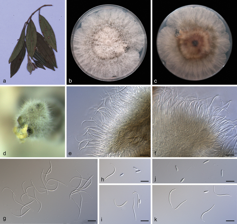
Figure 8.
Diaporthe melastomatis (SAUCC194.55) a branch with leaves of host plant b, c surface (b) and reverse (c) sides of colony after incubation for 15 days on PDAd conidiomata e, f conidiophores and conidiogenous cells g beta conidia h, i, k alpha conidia and beta conidia j alpha conidia. Scale bars: 10 μm (e–k).
Etymology.
Named after the host Melastoma malabathricum on which it was collected.
Diagnosis.
Diaporthe melastomatis differs from D. parapterocarpi Crous in smaller α-conidia and the types of conidia.
Type.
China, Yunnan Province: Xishuangbanna Tropical Botanical Garden, Chinese Academy of Sciences, on diseased leaves of Melastoma malabathricum. 19 April 2019, S.T. Huang, HSAUP194.55, holotype, ex-holotype living culture, SAUCC194.55.
Description.
Asexual morph: Conidiomata pycnidial, subglobose to globose, black, erumpent, coated with white hyphae, thick-walled, yellowish spiral conidial cirrus exuded from ostioles. Conidiophores hyaline, smooth, septate, branched, densely aggregated, cylindric-clavate, straight to slightly sinuous, tapering towards the apex, 14.5–21.0 × 2.0–3.2 μm. Conidiogenous cells 9.5–13.0 × 1.5–2.5 μm, cylindrical, guttulate, terminal, tapering towards the base. Alpha conidia, hyaline, smooth, aseptate, oblong ellipsoidal, 2–4 guttulate, apex subobtuse, base subtruncate, 5.5–8.5 × 1.7–2.5 µm (mean = 6.8 × 2.1 μm, n = 20). Beta conidia abundant in the culture, hyaline, aseptate, filiform, multi-guttulate, sigmoid to lunate, mostly curved through 90–180°, tapering towards both ends, 25.0–33.5 × 1.1–2.0 µm (mean = 27.6 × 1.4 μm, n = 20). Gamma conidia and sexual morph not observed.
Culture characteristics.
Pure culture was isolated by subbing hyphal tips growing from surface sterilized diseased material. Colonies on PDA cover the Petri diameter after incubation for 15 days in dark conditions at 25 °C, cottony and lobate with abundant aerial mycelium, hyphae white in the margin on surface side, with pale brown concentric ring of dense hyphae on reverse side.
Additional specimens examined.
China, Yunnan Province: Xishuangbanna Tropical Botanical Garden, Chinese Academy of Sciences, 19 April 2019, S.T. Huang. On diseased leaves of Millettia reticulata, HSAUP194.80 paratype, living culture SAUCC194.80; on infected leaves of Camellia sinensis, HSAUP194.88 paratype, living culture SAUCC194.88.
Notes.
Diaporthe melastomatis is introduced based on the multi-locus phylogenetic analysis, with three isolates clustering separately in a well-supported clade (ML/BI = 100/1). Diaporthe melastomatis is most closely related to D. parapterocarpi, but distinguished based on ITS and TUB loci from D. parapterocarpi by 32 nucleotides difference in the concatenated alignment, in which 20 are distinct in the ITS region, 12 in the TUB region. Morphologically, Diaporthe melastomatis differs from D. parapterocarpi in its smaller alpha conidia (5.5–8.5 × 1.7–2.5 vs. 8.0–10.0 × 2.5–3.0 μm). Furthermore, Diaporthe melastomatis can produce beta conidia, but D. parapterocarpi cannot (Crous et al. 2014).
Diaporthe pungensis
S.T. Huang, J.W. Xia, X.G. Zhang, Z. Li sp. nov.
8658054E-C61B-5A39-9B67-DB151DC5ECE2
837599
Figure 9.
Diaporthe pungensis (SAUCC194.112) a leaf of host plant b, c surface (b) and reverse (c) sides of colony after incubation for 15 days on PDAd conidiomata on PDAe–h conidiophores and conidiogenous cells i, l beta conidia j, k alpha conidia and beta conidia. Scale bars: 10 μm (e–l).
Etymology.
Named after the host Elaeagnus pungens on which it was collected.
Diagnosis.
Diaporthe pungensis differs from its closest phylogenetic species D. inconspicua R.R. Gomes et al. and D. poincianellae T.G.L. Oloveira et al. in ITS, TUB, TEF, CAL and HIS loci based on the alignments deposited in Tree-BASE.
Type.
China, Yunnan Province: Xishuangbanna Tropical Botanical Garden, Chinese Academy of Sciences, on diseased leaves of Elaeagnus pungens. 19 April 2019, S.T. Huang, HSAUP194.112, holotype, ex-holotype living culture SAUCC194.112.
Description.
Asexual morph: Conidiomata pycnidial, 3–5 pycnidia grouped together, superficial to embedded on PDA, erumpent, thin-walled, dark brown to black, globose or subglobose, exuding white creamy conidial mass from the ostioles. Conidiophores hyaline, aseptate, cylindrical, smooth, straight to sinuous, unbranched, 11.0–14.5 × 1.5–2.3 µm. Conidiogenous cells phialidic, cylindrical, terminal, 8.0–9.5 × 1.0–2.5 µm. Alpha conidia, hyaline, smooth, aseptate, ellipsoidal to fusoid, 2–3 guttulate, apex subobtuse, base subtruncate, 6.0–8.5 × 2.0–3.3 µm (mean = 6.6 × 2.5 μm, n = 20). Beta conidia hyaline, aseptate, eguttulate, filiform, slightly curved, tapering towards the apex, base truncate, some conidia are in the immature stage swollen in the middle, 24.0–28.9 × 1.0–2.0 µm (mean = 26.9 × 1.4 μm, n = 20). Gamma conidia not observed, sexual morph not observed.
Culture characteristics.
Pure culture was isolated by subbing hyphal tips growing from surface sterilized plant material. Colonies on PDA cover the 3/4 of Petri dish diameter after incubation for 15 days in dark conditions at 25 °C, flat, cottony in the center with medium developed aerial mycelium, sparse in the outer region. With several concentric rings of dense and sparse hyphae, irregular margin, white on surface side, white to pale yellow and cinnamon speckle on reverse side.
Additional specimen examined.
China, Yunnan Province: Xishuangbanna Tropical Botanical Garden, Chinese Academy of Sciences, on infected leaves of Camellia sinensis. 19 April 2019, S.T. Huang, HSAUP194.89 paratype, living culture SAUCC194.89.
Notes.
Diaporthe pungensis forms a distinct clade with high support (ML/BI = 100/1), and differed with the closely related species (D. inconspicua and D. poincianellae) on ITS, TUB, CAL and HIS loci (94% in ITS, 92% in TUB, 70% in TEF, 92% in CAL and 92% in HIS; and 95% in ITS, 94% in TUB, 80% in TEF, 94% in CAL and 89% in HIS, respectively). Moreover, Diaporthe pungensis differs from D. inconspicua, in having guttulate of alpha conidia, and having larger alpha conidia (6.0–8.5 × 2.0–3.3 vs. 5.5–6.5 × 1.5–2 μm) (Bezerra et al. 2018). Furthermore, Diaporthe pungensis can produce two types of conidia (α-conidia and β-conidia), but D. poincianellae only produce a α-conidia(Crous et al. 2018b).
Diaporthe subclavata
F. Huang, K.D. Hyde & H.Y. Li, Fung. Biol. 119: 343, 2015
450781D4-6E99-5B16-882D-25C1993AA7AD
Figure 10.
Diaporthe subclavata (SAUCC194.66) a leaf of Pometia pinnatab, c surface (b) and reverse (c) sides of colony after incubation for 15 days on PDAd conidiomata e–h conidiophores and conidiogenous cells i, j Beta conidia k, l Alpha conidia. Scale bars: 10 μm (e–l).
Description.
Asexual morph: Conidiomata pycnidial, multi-pycnidia grouped together, globose, black, erumpent, coated with white hyphae, creamy to yellowish conidial droplets exuded from central ostioles. Conidiophores hyaline, densely aggregated, cylindrical, straight to sinuous, tapering towards the apex, 13.5–23.0 × 2.0–3.0 μm. Alpha conidiogenous cells 7.0–10 × 1.8–2.5 μm, cylindrical, terminal, slightly curved. Beta conidiogenous cells 10.5–13.5 × 0.9–1.5 μm, cylindrical, hyaline, tapering towards the apex. Alpha conidia, hyaline, smooth, aseptate, ellipsoidal, multi-guttulate, apex subobtuse, base subtruncate, 4.7–5.8 × 2.4–2.9 µm (mean = 5.3 × 2.6 μm, n = 20). Beta conidia hyaline, aseptate, filiform, few guttulate, slightly curved, tapering towards the both ends, 25.5–32.0 × 1.0–1.6 µm (mean = 27.5 × 1.3 μm, n = 20). Gamma conidia and sexual morph not observed.
Culture characteristics.
Pure culture was isolated by subbing hyphal tips growing from surface sterilized diseased material. Colonies on PDA cover the Petri dish diameter after incubation for 15 days in dark conditions at 25 °C. Aerial mycelium white, cottony, feathery, with concentric zonation, white on surface side, pale brown to black on reverse side.
Specimen examined.
China, Yunnan Province: Xishuangbanna Tropical Botanical Garden, Chinese Academy of Sciences, on infected leaves of Pometia pinnata. 19 April 2019, S.T. Huang, HSAUP194.66, living culture SAUCC194.66.
Notes.
Diaporthe subclavata was originally described from the leaf with citrus scab of Citrus unshiu in Fujian Province, China (Huang et al. 2015). In the present study, isolated strain SAUCC194.66 from symptomatic leaves of Pometia pinnata was congruent with D. subclavata based on morphology and DNA sequences data (Fig. 1). We therefore present a description and illustration of D. subclavata as a known species for this clade, found on new host.
Diaporthe tectonendophytica
M. Doilom, A. J. Dissanayake & K.D. Hyde, Fung. Div., 82: 163, 2016
10FD94BE-E177-5477-9071-1000FAC80F69
Figure 11.
Diaporthe tectonendophytica (SAUCC194.11) a leaf of host plant b, c surface (b) and reverse (c) side of colony after incubation for 15 days on PDAd conidiomata on PDAe, f conidiophores and conidiogenous cells g, h beta conidia. Scale bars: 10 μm (e–h).
Description.
Asexual morph: Conidiomata pycnidial, aggregated, brownish to black, erumpent, subglobose, exuding white creamy conidial droplets from central ostioles after being kept for 30 days in light at 25 °C. Conidiophores 17.4–35.0 × 2.2–3.5 μm, hyaline, branched, subcylindrical, septate, straight or slightly curved, guttulate. Conidiogenous cells 11.3–15.0 × 1.7–2.5 μm (mean = 12.3 × 2.1 μm, n = 20), cylindric-clavate, hyaline, straight to slightly sinuous, tapering towards the apex. Beta conidia 25.0–31.8 × 0.9–1.8 μm (mean = 28.2 × 1.2 μm, n = 20), filiform, hyaline, guttulate, aseptate, hooked and mostly curved through 90–180°, swollen in the middle. Alpha conidia and Gamma conidia not observed.
Culture characteristics.
Pure culture was isolated by subbing hyphal tips growing from surface sterilized diseased material. Colonies on PDA cover the Petri dish diameter after incubation for 15 days in dark conditions at 25 °C, aerial mycelium abundant, white to grayish on surface side, pale yellow on reverse with concentric zonation. Pycnidia are formed on 15th day or later.
Specimens examined.
China, Yunnan Province: Xishuangbanna Tropical Botanical Garden, Chinese Academy of Sciences, 19 April 2019, S.T. Huang. On diseased leaves of Elaeagnus conferta HSAUP194.11, living culture SAUCC194.11; on diseased leaves of Pometia pinnata HSAUP194.63, living culture SAUCC194.63.
Notes.
Diaporthe tectonendophytica was originally described from the asymptomatic branches of Tectona grandis in Thailand (Doilom et al. 2017). In the present study, two strains (SAUCC194.11 and SAUCC194.63) from symptomatic leaves of Elaeagnus conferta and Pometia pinnata were congruent with D. tectonendophytica based on morphology and DNA sequences data (Fig. 1). We therefore describe D. tectonendophytica as a known species for this clade.
Discussion
In the current study, 87 reference sequences (including an outgroup taxon) were selected based on BLAST searches of NCBIs GenBank nucleotide database and were included in the phylogenetic analyses (Table 1). Phylogenetic analyses based on five combined loci (ITS, TUB, TEF, CAL and HIS), as well as morphological characters of the non-sexual morph obtained in culture, contributed to knowledge of the diversity of Diaporthe species in Yunnan Province. Based on a large set of freshly collected specimens from Yunnan province, China, 20 strains of Diaporthe species were isolated from 12 host genera (Table 1). As a result, eight new species are proposed: Diaporthe camelliae-sinensis, D. grandiflori, D. heliconiae, D. heterostemmatis, D. litchii, D. lutescens, D. melastomatis, D. pungensis and two previously described species were described and illustrated, D. subclavata and D. tectonendophytica.
Previously, species identification of Diaporthe was largely referred to the assumption of host-specificity, leading to the proliferation of names (Gomes et al. 2013). However, based on a polyphasic approach and known morphology, more than one species of Diaporthe can colonize a single host, while one species can be associated with different hosts (Gomes et al. 2013; Gao et al. 2017; Guarnaccia and Crous 2017; Guarnaccia et al. 2018; Guo et al. 2020). Our study can well support this phenomenon. On the one hand, Diaporthe grandiflori (SAUCC194.84) and D. heterostemmatis (SAUCC194.85) were collected from Heterostemma grandiflorum; D. camelliae-sinensis (SAUCC194.92), D. heterostemmatis (SAUCC194.102), D. melastomatis (SAUCC194.88), and D. pungensis (SAUCC194.89) and were isolated from Camellia sinensis; D. litchii (SAUCC194.12) and D. tectonendophytica (SAUCC194.11) were known on Elaeagnus conferta. On the other hand, the species of D. camelliae-sinensis collected from three hosts (Camellia sinensis, Castanea mollissima, Machilus pingii) D. melastomatis sampled from three hosts (Camellia sinensis, Melastoma malabathricum, Millettia reticulata) and D. litchii sampled from two hosts (Elaeagnus conferta, Litchi chinensis). These studies revealed a high diversity of Diaporthe species from different hosts. The descriptions and molecular data of Diaporthe represent an important resource for plant pathologists, plant quarantine officials and taxonomists.
Supplementary Material
Acknowledgements
This work was jointly supported by the National Natural Science Foundation of China (no. 31770016, 31750001, and 31900014) and the China Postdoctoral Science Foundation (no. 2018M632699).
Citation
Sun W, Huang S, Xia J, Zhang X, Li Z (2021) Morphological and molecular identification of Diaporthe species in south-western China, with description of eight new species. MycoKeys 77: 65–95. https://doi.org/10.3897/mycokeys.77.59852
References
- Bezerra JDP, Machado AR, Firmino AL, Rosado AWC, de Souza CAF, de Souza-Motta CM, de Sousa Freire KTL, Paiva LM, Magalhães OMC, Pereira OL. (2018) Mycological diversity description I. Acta Botanica Brasilica 32(4): 656–666. 10.1590/0102-33062018abb0154 [DOI] [Google Scholar]
- Broge M, Howard A, Biles CL, Udayanga D, Taff H, Dudley L, Bruton BD. (2020) First report of Diaporthe fruit rot of melons caused by D. Pterocarpi in Costa Rica. Plant Disease 104(5): 1550–1550. 10.1094/PDIS-08-19-1655-PDN [DOI] [Google Scholar]
- Cai L, Hyde KD, Taylor PWJ, Weir B, Waller J, Abang MM, Zhang ZJ, Yang YL, Phoulivong S, Liu ZY, Prihastuti H, Shivas RG, McKenzie EHC, Johnston PR. (2009) A polyphasic approach for studying Colletotrichum. Fungal Diversity 39: 183–204. [Google Scholar]
- Carbone I, Kohn LM. (1999) A method for designing primer sets for speciation studies in filamentous Ascomycetes. Mycologia 91(3): 553–556. 10.1080/00275514.1999.12061051 [DOI] [Google Scholar]
- Crous PW, Groenewald JZ, Risède JM, Simoneau P, Hywel-Jones NL. (2004) Calonectria species and their Cylindrocladium anamorphs: species with sphaeropedunculate vesicles. Studies in Mycology 50: 415–430. [DOI] [PMC free article] [PubMed] [Google Scholar]
- Crous PW, Groenewald JZ, Shivas RG, Edwards J, Seifert KA, Alfenas AC, Alfenas RF, Burgess TI, Carnegie AJ, Hardy GEStJ. (2011a) Fungal planet description sheets: 69–91. Persoonia 26(1): 108–156. 10.3767/003158511X581723 [DOI] [PMC free article] [PubMed] [Google Scholar]
- Crous PW, Summerell BA, Swart L, Denman S, Taylor JE, Bezuidenhout CM, Palm ME, Marincowitz S, Groenewald JZ. (2011b) Fungal pathogens of Proteaceae. Persoonia 27(1): 20–45. 10.3767/003158511X606239 [DOI] [PMC free article] [PubMed] [Google Scholar]
- Crous PW, Shivas RG, Quaedvlieg W, van der Bank M, Zhang Y, Summerell BA, Guarro J, Wingfield MJ, Wood AR, Alfenas AC. (2014) Fungal planet description sheets: 214–280. Persoonia 32(1): 184–306. 10.3767/003158514X682395 [DOI] [PMC free article] [PubMed] [Google Scholar]
- Crous PW, Wingfield MJ, Richardson DM, Roux JJL, Strasberg D, Edwards J, Roets F, Hubka V, Taylor PWJ, Heykoop M. (2016) Fungal planet description sheets: 400–468. Persoonia 36(1): 316–458. 10.3767/003158516X692185 [DOI] [PMC free article] [PubMed] [Google Scholar]
- Crous PW, Wingfield MJ, Burgess TI, Hardy GEStJ, Gene J, Guarro J, Baseia IG, Garcia D, Gusmao LFP, Souza-Motta CM. (2018a) Fungal planet description sheets: 716–784. Persoonia 40(1): 240–393. [DOI] [PMC free article] [PubMed] [Google Scholar]
- Crous PW, Luangsa-ard JJ, Wingfield MJ, Carnegie AJ, Hernández-Restrepo M, Lombard L, Roux J, Barreto RW, Baseia IG, Cano-Lira JF. (2018b) Fungal planet description sheets: 785–867. Persoonia 41(1): 238–417. [DOI] [PMC free article] [PubMed] [Google Scholar]
- Crous PW, Wingfield MJ, Schumacher RK, Akulov A, Bulgakov TS, Carnegie AJ, Jurjević Ž, Decock C, Denman S, Lombard L. (2020) New and interesting fungi. 3. Fungal Systematics and Evolution 6: 157–231. 10.3114/fuse.2020.06.09 [DOI] [PMC free article] [PubMed] [Google Scholar]
- Dayarathne MC, Jones EBG, Maharachchikumbura SSN, Devadatha B, Sarma VV. (2020) Morpho-molecular characterization of microfungi associated with marine based habitats. Mycosphere 11(1): 1–188. 10.5943/mycosphere/11/1/1 [DOI] [Google Scholar]
- Dissanayake AJ, Phillips AJL, Hyde KD, Yan JY, Li XH. (2017) The current status of species in Diaporthe. Mycosphere 8: 1106–1156. 10.5943/mycosphere/8/5/5 [DOI] [Google Scholar]
- Doilom M, Dissanayake AJ, Wanasinghe DN, Boonmee S, Liu JK, Bhat DJ, Taylor JE, Bahkali AH, McKenzie EHC, Hyde KD. (2017) Microfungi on Tectona Grandis (Teak) in Northern Thailand. Fungal Diversity 82(1): 107–182. 10.1007/s13225-016-0368-7 [DOI] [Google Scholar]
- Fan XL, Bezerra JDP, Tian CM, Crous PW. (2018) Families and genera of diaporthalean fungi associated with canker and dieback of tree hosts. Persoonia 40: 119–134. 10.3767/persoonia.2018.40.05 [DOI] [PMC free article] [PubMed] [Google Scholar]
- Gao YH, Sun W, Su YY, Cai L. (2014) Three new species of Phomopsis in Gutianshan Nature Reserve in China. Mycological Progress 13(1): 111–121. 10.1007/s11557-013-0898-2 [DOI] [Google Scholar]
- Gao YH, Su YY, Sun W, Cai L. (2015) Diaporthe species occurring on Lithocarpus glabra in China, with descriptions of five new species. Fungal Biology 119(5): 295–309. 10.1016/j.funbio.2014.06.006 [DOI] [PubMed] [Google Scholar]
- Gao YH, Liu F, Cai L. (2016) Unravelling Diaporthe species associated with Camellia. Systematics and Biodiversity 14(1): 102–117. 10.1080/14772000.2015.1101027 [DOI] [Google Scholar]
- Gao YH, Liu F, Duan W, Crous PW, Cai L. (2017) Diaporthe is paraphyletic. IMA fungus 8: 153–187. 10.5598/imafungus.2017.08.01.11 [DOI] [PMC free article] [PubMed] [Google Scholar]
- Glass NL, Donaldson GC. (1995) Development of primer sets designed for use with the PCR to amplify conserved genes from filamentous ascomycetes. Applied and Environmental Microbiology 61(4): 1323–1330. 10.1128/AEM.61.4.1323-1330.1995 [DOI] [PMC free article] [PubMed] [Google Scholar]
- Gomes RR, Glienke C, Videira SIR, Lombard L, Groenewald JZ, Crous PW. (2013) Diaporthe: a genus of endophytic, saprobic and plant pathogenic fungi. Persoonia: Molecular Phylogeny and Evolution of Fungi 31(1): 1–41. 10.3767/003158513X666844 [DOI] [PMC free article] [PubMed] [Google Scholar]
- Grasso FM, Marini M, Vitale A, Firrao G, Granata G. (2012) Canker and dieback on Platanus × acerifolia caused by Diaporthe scabra. Forest Pathology 42(6): 510–513. 10.1111/j.1439-0329.2012.00785.x [DOI] [Google Scholar]
- Guarnaccia V, Vitale A, Cirvilleri G, Aiello D, Susca A, Epifani F, Perrone G, Polizzi G. (2016) Characterisation and pathogenicity of fungal species associated with branch cankers and stem-end rot of avocado in Italy. European Journal of Plant Pathology 146(4): 963–976. 10.1007/s10658-016-0973-z [DOI] [Google Scholar]
- Guarnaccia V, Crous PW. (2017) Emerging citrus diseases in Europe caused by Diaporthe spp. IMA Fungus 8: 317–334. 10.5598/imafungus.2017.08.02.07 [DOI] [PMC free article] [PubMed] [Google Scholar]
- Guarnaccia V, Groenewald JZ, Woodhall J, Armengol J, Cinelli T, Eichmeier A, Ezra D, Fontaine F, Gramaje D, Gutierrez-Aguirregabiria A. (2018) Diaporthe diversity and pathogenicity revealed from a broad survey of grapevine diseases in europe. Persoonia 40(6): 135–153. 10.3767/persoonia.2018.40.06 [DOI] [PMC free article] [PubMed] [Google Scholar]
- Guo LD, Hyde KD, Liew ECY. (2000) Identification of endophytic fungi from Livistona chinensis based on morphology and rDNA sequences. New Phytologist 147(3): 617–630. 10.1046/j.1469-8137.2000.00716.x [DOI] [PubMed] [Google Scholar]
- Guo YS, Crous PW, Bai Q, Fu M, Yang MM, Wang XH, Du YM, Hong N, Xu WX, Wang GP. (2020) High diversity of Diaporthe species associated with pear shoot canker in China. Persoonia 45: 132–162. 10.3767/persoonia.2020.45.05 [DOI] [PMC free article] [PubMed] [Google Scholar]
- Huang F, Hou X, Dewdney MM, Fu Y, Chen GQ, Hyde KD, Li HY. (2013) Diaporthe species occurring on citrus in China. Fungal Diversity 61(1): 237–250. 10.1007/s13225-013-0245-6 [DOI] [Google Scholar]
- Huang F, Udayanga D, Wang XH, Hou X, Mei XF, Fu YS, Hyde KD, Li HY. (2015) Endophytic Diaporthe associated with Citrus: A phylogenetic reassessment with seven new species from China. Fungal Biology 119(5): 331–347. 10.1016/j.funbio.2015.02.006 [DOI] [PubMed] [Google Scholar]
- Huelsenbeck JP, Ronquist F. (2001) MRBAYES: bayesian inference of phylogeny. Bioinformatics 17(17): 754–755. 10.1093/bioinformatics/17.8.754 [DOI] [PubMed] [Google Scholar]
- Hyde KD, Chaiwan N, Norphanphoun C, Boonmee S, Camporesi E, Chethana KWT, Dayarathne MC, de Silva IN, Dissanayake AJ, Ekanayaka AH. (2018) Mycosphere notes 169–224. Mycosphere 9(2): 271–430. 10.5943/mycosphere/9/2/8 [DOI] [Google Scholar]
- Hyde KD, Dong Y, Phookamsak R, Jeewon R, Bhat DJ, Gareth Jones EB, Liu NG, Abeywickrama PD, Mapook A, Wei D. (2020) Fungal diversity notes 1151–1276: Taxonomic and phylogenetic contributions on genera and species of fungal taxa. Fungal Diversity 100(1): 1–273. 10.1007/s13225-020-00439-5 [DOI] [Google Scholar]
- Katoh K, Rozewicki J, Yamada KD. (2017) MAFFT online service: multiple sequence alignment, interactive sequence choice and visualization. Briefings in Bioinformatics 20(4): 1160–1166. 10.1093/bib/bbx108 [DOI] [PMC free article] [PubMed] [Google Scholar]
- Kumar S, Stecher G, Tamura K. (2016) MEGA7: Molecular evolutionary genetics analysis version 7.0 for bigger datasets. Molecular Biology and Evolution 33(7): 1870–1874. 10.1093/molbev/msw054 [DOI] [PMC free article] [PubMed] [Google Scholar]
- Li WJ, McKenzie EHC, Liu JK, Bhat DJ, Dai DQ, Camporesi E, Tian Q, Maharachchikumbura SSN, Luo ZL, Shang QJ. (2020) Taxonomy and phylogeny of hyaline-spored coelomycetes. Fungal Diversity 100(1): 279–801. 10.1007/s13225-020-00440-y [DOI] [Google Scholar]
- Lombard L, van Leeuwen GCM, Guarnaccia V, Polizzi G, van Rijswick PCJ, Rosendahl KCHM, Gabler J, Crous PW. (2014) Diaporthe species associated with Vaccinium, with specific reference to Europe. Phytopathologia Mediterranea 53(2): 287–299. [Google Scholar]
- Ménard L, Brandeis PE, Simoneau P, Poupard P, Sérandat I, Detoc J, Robbes L, Bastide F, Laurent E, Gombert J, Morel E. (2014) First report of umbel browning and stem necrosis caused by Diaporthe angelicae on carrot in France. Plant Disease 98(3): 421–422. 10.1094/PDIS-06-13-0673-PDN [DOI] [PubMed] [Google Scholar]
- Miller MA, Pfeiffer W, Schwartz T. (2012) The CIPRES science gateway: enabling high-impact science for phylogenetics researchers with limited resources. Proceedings of the 1st Conference of the Extreme Science and Engineering Discovery Environment. Bridging from the extreme to the campus and beyond. Association for Computing Machinery 39: 1–8. 10.1145/2335755.2335836 [DOI] [Google Scholar]
- Murali TS, Suryanarayanan TS, Geeta R. (2006) Endophytic Phomopsis species: host range and implications for diversity estimates. Canadian Journal of Microbiology 52(7): 673–680. 10.1139/w06-020 [DOI] [PubMed] [Google Scholar]
- Nitschke T. (1870) Pyrenomycetes Germanici (2nd ed.). Eduard Trewendt, Germany, Breslau, 161–320.
- Nylander JAA. (2004) MrModeltest v. 2. Program distributed by the author. Evolutionary Biology Centre, Uppsala University.
- Perera RH, Hyde KD, Dissanayake AJ, Jones EBG, Liu JK, Wei D, Liu ZY. (2018) Diaporthe collariana sp. nov., with prominent collarettes associated with Magnolia champaca fruits in Thailand. Studies in Fungi 3(1): 141–151. 10.5943/sif/3/1/16 [DOI] [Google Scholar]
- Rehner SA, Uecker FA. (1994) Nuclear ribosomal internal transcribed spacer phylogeny and host diversity in the coelomycete Phomopsis. Botany 72(11): 1666–1674. 10.1139/b94-204 [DOI] [Google Scholar]
- Ronquist F, Huelsenbeck JP. (2003) MrBayes 3: bayesian phylogenetic inference under mixed models. Bioinformatics 19(12): 1572–1574. 10.1093/bioinformatics/btg180 [DOI] [PubMed] [Google Scholar]
- Ronquist F, Teslenko M, van der Mark P, Ayres DL, Darling A, Höhna S, Larget B, Liu L, Suchard MA, Huelsenbeck JP. (2012) MrBayes 3.2: Efficient Bayesian phylogenetic inference and model choice across a large model space. Systematic Biology 61(3): 539–542. 10.1093/sysbio/sys029 [DOI] [PMC free article] [PubMed] [Google Scholar]
- Rossman AY, Adams GC, Cannon PF, Castlebury LA, Crous PW, Gryzenhout M, Jaklitsch WM, Mejia LC, Stoykov D, Udayanga D. (2015) Recommendations of generic names in Diaporthales competing for protection or use. IMA Fungus 6(1): 145–154. 10.5598/imafungus.2015.06.01.09 [DOI] [PMC free article] [PubMed] [Google Scholar]
- Santos JM, Phillips AJL. (2009) Resolving the complex of Diaporthe (Phomopsis) species occurring on Foeniculum vulgare in Portugal. Fungal Diversity 34: 111–125. [Google Scholar]
- Santos JM, Vrandečić K, Ćosić J, Duvnjak T, Phillips AJL. (2011) Resolving the Diaporthe species occurring on soybean in Croatia. Persoonia 27(1): 9–19. 10.3767/003158511X603719 [DOI] [PMC free article] [PubMed] [Google Scholar]
- Santos L, Alves A, Alves R. (2017) Evaluating multi-locus phylogenies for species boundaries determination in the genus Diaporthe PeerJ 5: e3120. 10.7717/peerj.3120 [DOI] [PMC free article] [PubMed]
- Senanayake IC, Crous PW, Groenewald JZ, Maharachchikumbura SSN, Jeewon R, Phillips AJL, Bhat DJ, Perera RH, Li QR, Li WJ. (2017) Families of Diaporthales based on morphological and phylogenetic evidence. Studies in Mycology 86: 217–296. 10.1016/j.simyco.2017.07.003 [DOI] [PMC free article] [PubMed] [Google Scholar]
- Senanayake IC, Jeewon R, Chomnunti P, Wanasinghe DN, Norphanphoun C, Karunarathna A, Pem D, Perera RH, Camporesi E, McKenzie EHC. (2018) Taxonomic circumscription of Diaporthales based on multigene phylogeny and morphology. Fungal Diversity 93(1): 241–443. 10.1007/s13225-018-0410-z [DOI] [Google Scholar]
- Stamatakis A. (2014) RAxML Version 8: A tool for phylogenetic analysis and post-analysis of large phylogenies. Bioinformatics 30(9): 1312–1313. 10.1093/bioinformatics/btu033 [DOI] [PMC free article] [PubMed] [Google Scholar]
- Thompson SM, Tan YP, Young AJ, Neate SM, Aitken EAB, Shivas RG. (2011) Stem cankers on sunflower (Helianthus annuus) in Australia reveal a complex of pathogenic Diaporthe (Phomopsis) species. Persoonia 27(1): 80–89. 10.3767/003158511X617110 [DOI] [PMC free article] [PubMed] [Google Scholar]
- Thompson SM, Tan YP, Shivas RG, Neate SM, Morin L, Bissett A, Aitken EAB. (2015) Green and brown bridges between weeds and crops reveal novel Diaporthe species in Australia. Persoonia 35(1): 39–49. 10.3767/003158515X687506 [DOI] [PMC free article] [PubMed] [Google Scholar]
- Torres C, Camps R, Aguirre R, Besoain XA. (2016) First report of Diaporthe rudis in Chile causing stem-end rot on hass avocado fruit imported from California, USA. Plant Disease 100(9): 1951–1951. 10.1094/PDIS-12-15-1495-PDN [DOI] [Google Scholar]
- Udayanga D, Liu X, McKenzie EH, Chukeatirote E, Bahkali AH, Hyde KD. (2011) The genus Phomopsis: biology, applications, species concepts and names of common phytopathogens. Fungal Diversity 50(1): 189–225. 10.1007/s13225-011-0126-9 [DOI] [Google Scholar]
- Udayanga D, Liu XZ, Crous PW, McKenzie EHC, Chukeatirote E, Hyde KD. (2012) A multi-locus phylogenetic evaluation of Diaporthe (Phomopsis). Fungal Diversity 56(1): 157–171. 10.1007/s13225-012-0190-9 [DOI] [Google Scholar]
- Udayanga D, Castlebury LA, Rossman AY, Chukeatirote E, Hyde KD. (2015) The Diaporthe sojae species complex: Phylogenetic re-assessment of pathogens associated with soybean, cucurbits and other field crops. Fungal Biology 119(5): 383–407. 10.1016/j.funbio.2014.10.009 [DOI] [PubMed] [Google Scholar]
- van Rensburg JCJ, Lamprecht SC, Groenewald JZ, Castlebury LA, Crous PW. (2006) Characterization of Phomopsis spp. associated with die-back of rooibos (Aspalathus linearis) in South Africa. Studies in Mycology 55: 65–74. 10.3114/sim.55.1.65 [DOI] [PMC free article] [PubMed] [Google Scholar]
- Vilka L, Volkova J. (2015) Morphological diversity of Phomopsis vaccinii isolates from cranberry (Vaccinium macrocarpon Ait.) in Latvia. Proceedings of the Latvia University of Agriculture 33: 8–18. 10.1515/plua-2015-0002 [DOI] [Google Scholar]
- White T, Bruns T, Lee S, Taylor FJRM, White TJ, Lee SH, Taylor L, Shawe-Taylor J. (1990) Amplification and direct sequencing of fungal ribosomal RNA genes for phylogenetics. PCR Protocols: A guide to methods and applications. Academic Press 18: 315–322. 10.1016/B978-0-12-372180-8.50042-1 [DOI] [Google Scholar]
- Yang Q, Du Z, Tian CM. (2018) Phylogeny and morphology reveal two new species of Diaporthe from Traditional Chinese Medicine in Northeast China. Phytotaxa 336(2): 159–170. 10.11646/phytotaxa.336.2.3 [DOI] [Google Scholar]
- Yang Q, Jiang N, Tian CM. (2020) Three new Diaporthe species from Shaanxi Province, China. MycoKeys 67: 1–18. 10.3897/mycokeys.67.49483 [DOI] [PMC free article] [PubMed] [Google Scholar]
- Zapata M, Palma MA, Aninat MJ, Piontelli E. (2020) Polyphasic studies of new species of Diaporthe from native forest in Chile, with descriptions of Diaporthe araucanorum sp. nov., Diaporthe foikelawen sp. nov. and Diaporthe patagonica sp. nov. International Journal of Systematic and Evolutionary Microbiology 70(5): 3379–3390. 10.1099/ijsem.0.004183 [DOI] [PubMed] [Google Scholar]
Associated Data
This section collects any data citations, data availability statements, or supplementary materials included in this article.


