Key Points
Question
Is the poly–(adenosine diphosphate–ribose) polymerase inhibitor olaparib effective against advanced pancreatic cancer with DNA damage repair other than germline BRCA variants (termed “BRCAness” in the literature)?
Findings
Two parallel nonrandomized phase 2 clinical trials of olaparib included 46 pretreated patients with advanced pancreatic cancer with BRCAness and found a partial response rate of 2%, a stable disease rate of 72%, a median progression-free survival of 3.7 months, and a median overall survival of 9.9 months without unexpected toxic effects.
Meaning
Administration of olaparib is apparently safe and may be therapeutically efficacious for pretreated patients with pancreatic cancer with BRCAness.
Abstract
Importance
The subtype of pancreatic ductal adenocarcinoma cancer (PDAC) with DNA damage repair (DDR) deficiency from BRCA1/2 variants has a favorable prognosis and is sensitive to platinum analogues and poly–(adenosine diphosphate–ripose) polymerase (PARP) inhibition with olaparib. Approximately 10% to 20% of patients with PDAC have DDR genetic alterations other than germline BRCA variants. This population has been termed as having BRCAness. An opportunity exists to define the clinical phenotype, molecular underpinnings, and effectiveness of PARP inhibitors for this population.
Objective
To examine the therapeutic effectiveness of the PARP inhibitor olaparib for patients with pancreatic cancer with BRCAness.
Design, Setting, and Participants
Two parallel phase 2 nonrandomized clinical trials were conducted from November 11, 2016, to October 2, 2018, among 46 patients in Israel and Texas to determine the effectiveness of olaparib as monotherapy in advanced, previously treated PDAC with BRCAness. Inclusion criteria were treatment with 1 or more prior systemic therapies for advanced PDAC, Eastern Cooperative Oncology Group performance status of 0 to 1, and lack of the germline BRCA1/2 variant. BRCAness in these studies was defined as previously known DDR genetic alterations (DDR-GAs), personal or family history of BRCA-associated cancers (without DDR-GAs), or ATM protein loss as determined by immunohistochemistry.
Main Outcomes and Measures
The primary study end point was the objective response rate, and the secondary end points were progression-free survival and overall survival (OS).
Results
Forty-eight patients were enrolled, and 46 (26 women [57%]; mean [SD] age, 65.5 [11.1] years) were evaluable. The median treatment duration with olaparib was 3.0 months (interquartile range, 1.8-6.4 months). A total of 24 patients had the DDR phenotype (DDR-GAs), 17 had a family history of BRCA-associated cancers without DDR-GAs, and 5 had ATM loss as determined by immunohistochemistry. The DDR-GAs included ATM (n = 14), PALB2 (n = 2), ARID1A (n = 3), BRCA somatic (n = 1), PTEN (n = 1), RAD51 (n = 1), CCNE (n = 1), and FANCB (n = 2). Common toxic effects were grade 1 to 2 anemia, fatigue, anorexia, and nausea. One patient had a confirmed partial response (2%), 33 patients experienced stable disease (72%), of whom 11 (24%) experienced disease stability longer than 4 months and 12 patients had progressive disease (26%). The response duration for the patient with confirmed partial response was 3.9 months. Median progression-free survival was 3.7 months (95% CI, 2.9-5.7) and was significantly higher for patients with DDR-GAs (5.7 months; 95% CI, 3.6-8.8 months; P = .008) and platinum-sensitive PDAC (4.1 months; 95% CI, 3.6-7.8 months; P = .01). The estimated median OS was 9.9 months (95% CI, 7.6-16.1 months) in the study and 13.6 months (95% CI, 9.69 to not reached) in the prespecified DDR-GA cohort.
Conclusions and Relevance
The definition of the BRCAness phenotype in PDAC may be limited to patients harboring DDR-GAs. In these 2 phase 2 nonrandomized clinical trials, olaparib was well tolerated and showed limited antitumor activity in patients with advanced, platinum-sensitive PDAC with DDR-GAs. These conclusions suggest a potential therapeutic opportunity for a subset of patients with PDAC.
Two parallel phase 2 nonrandomized clinical trials examine the therapeutic effectiveness of the poly–(adenosine diphosphate–ripose) polymerase inhibitor olaparib for patients with pancreatic cancer with DNA damage repair other than germline BRCA variants.
Introduction
During the past decade, extensive molecular profiling has been conducted on pancreatic ductal adenocarcinoma (PDAC) for actionable genetic alterations or pathways. Despite these efforts, targeted therapy offers limited clinical benefit for patients with this disease. One exception is variation in DNA damage repair (DDR) genes, including BRCA1 (OMIM 113705) or BRCA2 (OMIM 600185). Germline variants in these genes may lead to homologous recombination repair deficiency; BRCA-associated tumors, including breast, prostate, and ovarian tumors, respond to poly–(adenosine diphosphate–ribose) polymerase (PARP) inhibitors and platinum-based therapy.1,2 A recent study demonstrated that the PARP inhibitor olaparib improves progression-free survival (PFS) when administered in the maintenance setting after systemic therapy with platinum analogues to patients with PDAC with BRCA variants.3
The concept of “BRCAness” was introduced by Turner and colleagues4 to identify phenotypic changes in sporadic cancers that may lead to PARP inhibitor susceptibility. However, the definition of BRCAness in PDAC remains elusive. Comprehensive genomic profiling of PDAC indicates that 14% to 16% of patients have DDR genetic alterations (DDR-GAs), including BRCA, ATM (OMIM 607585), PALB2 (OMIM 610355), CHEK1 (OMIM 603078), FANCA (OMIM 607139), BARD1 (OMIM 601593), RAD50 (OMIM 604040), and ARID1A (OMIM 603024).5 To our knowledge, the role of PARP inhibitors in most of these variants other than BRCA is unknown. It is well known that several familial cancer syndromes are enriched with DDR-GAs. However, there may be a cohort of patients with familial cancers without detectable DDR-GAs. A previous retrospective study of 549 patients with PDAC demonstrated that family history of breast, ovarian, or pancreatic cancer correlated significantly with an improved survival with platinum-based therapy compared with sporadic cases, thereby resembling the DDR-GA phenotype, even in the absence of germline BRCA variants.6 In that study, as the number of relatives with familial cancers increased, their overall survival (OS) also improved with first-line platinum therapy. We therefore hypothesized that the BRCAness phenotype may include those with a family history of BRCA-associated cancers without detectable DDR-GAs using the current genetic sequencing platforms, and we included these cases as having “clinical BRCAness.” An additional method to identify BRCAness in this population is to explore other potential biomarkers of DDR-GAs, such as loss of ATM protein expression. Loss of ATM expression as detected by immunohistochemistry has been noted in familial PDAC and is associated with an adverse prognosis.7
The aim of the present studies was to explore the clinical effectiveness of the PARP inhibitor olaparib for patients with PDAC with a broad definition of BRCAness. Specifically, we investigated the effectiveness of PARP inhibitors in the following 3 subgroups: (1) patients with PDAC with genomic DDR-GAs (other than germline BRCA1/2), (2) patients with PDAC with a personal or family history of BRCA-associated cancers in the absence of detectable DDR-GAs, and (3) those with loss of ATM protein expression by immunohistochemistry.
Methods
Patients
Two parallel nonrandomized clinical trials were conducted from November 11, 2016, to October 2, 2018, for the same patient population at the MD Anderson Cancer Center, Houston, Texas, and the Sheba Medical Center, Tel Hashomer, Israel. Patients were older than 18 years with metastatic PDAC and lack of germline BRCA1/2 (as determined by results of Myriad myRisk test [Myriad Genetics Inc] or Color test [Color]). In addition, eligible patients were required to have either (1) a personal or family history (≥1 first-degree relative) of ovarian carcinoma or breast cancer at 50 years of age or younger or 2 relatives with breast, pancreatic, or prostate cancer (Gleason score, ≥7) at any age or (2) previously identified DDR-GAs (including but not limited to a somatic BRCA variant, somatic or germline: ATM, PALB2, CHEK1, FANCA, BARD1, RAD50, and ARID1A). The DDR-GAs had to be considered actionable (based on existing literature) for inclusion. At the Sheba Medical Center, ATM loss as assessed by immunohistochemistry was included using methods previously described8; ATM loss was defined as less than 10% neoplastic nuclear staining at any intensity in the presence of positive lymphocyte staining. Patients had received at least 1 prior therapy for metastatic disease to be eligible. After a protocol amendment at the MD Anderson Cancer Center, patients were required to be naive to platinum-based treatment or to not have experienced disease progression while receiving platinum-based treatment (trial protocol in Supplement 1). “Platinum-resistant PDAC” was defined as disease progression during or within 6 months of discontinuation of platinum-based therapy. A similar protocol amendment was not performed at the Sheba Medical Center because accrual was almost complete at the Sheba Medical Center at this time (trial protocol in Supplement 2). Patients were required to have measurable disease, as determined by modified Response Evaluation Criteria in Solid Tumors, version 1.1 (RECIST v1.1) criteria.9 Patients were required to have adequate organ function, acceptable bone marrow function, and an Eastern Cooperative Oncology Group performance status of 0 to 1. Patients were required to have progressed disease after non–platinum-based therapy or completed chemotherapy or to have developed intolerable toxic effects from chemotherapy to qualify for the trial. All patients provided written informed consent. Institutional review board approval was obtained from both of the participating sites.
Trial Design and Treatments
Two parallel phase 2, open-label, nonrandomized clinical trials of olaparib monotherapy for patients with advanced PDAC were conducted at both institutions. Eligible patients received 300-mg olaparib tablets twice daily. Treatment was continued until investigator-assessed objective radiologic disease progression (modified RECIST v1.1 criteria), toxic effects, or patient withdrawal.
End Points and Assessments
The primary end point was the objective response rate, assessed by investigators using modified RECIST v1.1 criteria. Secondary end points included PFS, defined as the time from enrollment until radiologic disease progression (assessed by investigator review using modified RECIST v1.1 criteria) or death by any cause, and OS, defined as the time from the date of enrollment to the date of death or last follow-up. Data for survival analysis were censored on May 5, 2019. Computed tomography or magnetic resonance imaging scans of the chest, abdomen, and pelvis were taken at baseline, every 8 weeks for 40 weeks, then every 12 weeks until trial discontinuation. Assessments for survival were carried out after a specified date (data cutoff date) to capture survival status at that point for each survival analysis.
Statistical Analysis
The primary end point was the objective response (ie, complete response or partial response [PR]) achieved during the treatment. The optimal 2-stage design of Thall et al10 was implemented for the study at both sites. With a type I error rate α of .10 and a β error rate of 0.2, and assuming an objective response rate of standard drug treatment of 5% and an objective response rate of new study treatment of 20%, 24 patients were to be enrolled at each site. Nine patients were enrolled in the first stage, and 15 patients in the second stage. The response outcome in the first 24 weeks was used for primary analysis. Data were pooled from both sites for combined analysis. In the first step, if a complete response or PR was observed among the first 9 patients treated at either site, the studies would continue. However, if no responses occurred, then the studies would be stopped for futility. The objective response rate and its corresponding exact 95% CI were estimated. Study updates were provided at both sites with a monthly communication. The Kaplan-Meier method was used to estimate the probability of OS and PFS. The log-rank test and Cox proportional hazards regression models were used to determine the association of OS or PFS with patient characteristics. In addition, a stratified log-rank test by site and Cox proportional hazards regression models including site as a covariate were used to evaluate the association of OS and PFS with the patients’ characteristics, adjusting for the effect of study site. Statistical analysis for all other demographic and clinical parameters was conducted as follows. Categorical patient characteristics were tabulated with frequencies and percentages. Continuous variables were summarized using descriptive statistics, such as median values and ranges. The Fisher exact test and the Wilcoxon rank sum test were applied to compare categorical and continuous patient characteristics between the 2 study sites, respectively. All P values were from 2-sided tests, and results were deemed statistically significant at P < .05.
Safety Analysis
All patients who received at least 1 dose of olaparib were included in the safety analysis. Safety and tolerability were assessed in terms of adverse events (Common Terminology Criteria for Adverse Events, version 4.0),11 deaths, laboratory test data, vital signs, and electrocardiogram results. Adverse events, their severity, and other categorical events were tabulated using frequencies and percentages. Continuous safety outcomes were summarized using descriptive statistics, including mean (SD) values, median values, and ranges. Unacceptable toxic effects were monitored for the phase 2 portion of the study using the method of Thall et al.10
Statistical Analysis for Exploratory Correlative Studies
The Fisher exact test and the 2-sample t test were used to compare the incidences of ATM loss and the variant profiles between responders and nonresponders. The Kaplan-Meier method was used to compare survival between the ATM high and loss groups, and Cox proportional hazards regression was used to measure PFS and OS in the study subgroups.
Results
Patients
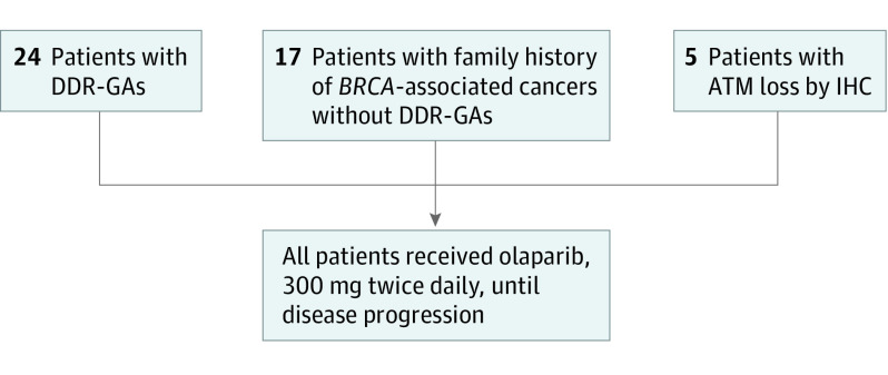
Forty-eight patients were enrolled into the 2 studies between November 2016 and October 2018. Of these, 46 patients (26 women [57%]; mean [SD] age, 65.5 [11.1] years) were evaluable for response (1 was found ineligible, and another withdrew consent; these patients received standard second-line chemotherapy). Patient demographic and baseline characteristics are reported in eTable 1 in Supplement 3. None of the patients had germline BRCA1/2. Included patients had a family history of specific cancers but without germline or somatic alterations (n = 17), DDR-GAs (n = 24), and ATM protein loss by immunohistochemistry (n = 5) (Figure 1). Only 1 patient with ATM loss was investigated with next-generation sequencing, and no DDR-GAs were identified. Testing indicated that DDR-GAs were present in the following genes (9 were germline variants and the rest were somatic): ATM (n = 14; 7 germline variants and 7 somatic), PALB2 (n = 2 germline variants), ARID1A (n = 3), BRCA somatic (n = 1), PTEN (n = 1), RAD51 (n = 1), CCNE (n = 1), and FANCB (2). Distribution in the 2 sites is as per eTable 2 in Supplement 3. Details of DDR-GAs and variant status are provided in eTable 1A in Supplement 3.
Figure 1. Flow Diagram.
Inclusion criteria: stage 4 pancreatic ductal adenocarcinoma cancer (PDAC), 1 or more prior systemic therapy for PDAC, Eastern Cooperative Oncology Group status 0 to 1, and negative for germline BRCA1/2 variant. BRCAness is defined by previously known DNA damage repair genetic alterations (DDR-GAs), personal or family history of BRCA-associated cancers (without DDR-GAs), or ATM protein loss as determined by immunohistochemistry (IHC).
Effectiveness
The median treatment duration with olaparib was 3.0 months (interquartile range, 1.8-6.4 months). The objective response rate was evaluated for 46 treated patients. Two patients had a PR (4%; exact 95% CI, 0.53%-14.8%), 1 of which was not confirmed (2%); these PRs occurred in 1 patient with PALB2 and 1 with somatic ATM variant. The response duration for the patient with confirmed PR was 3.9 months. Thirty-three patients experienced stable disease (72%; 95% CI, 59%-85%), of whom 11 (24%) experienced disease stability longer than 4 months, and 12 patients had progressive disease (26%; 95% CI, 13%-39%). The estimated median disease control duration was 2.9 months (95% CI, 2.0-6.0 months). The median follow-up time for censored patients was 7.4 months (interquartile range, 4.4-16.8 months). The percentage of patients with PR or stable disease vs progressive disease was similar between the sites, supporting the validity of the results.
Progression-Free Survival
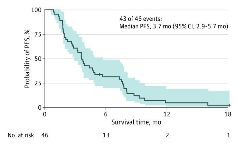
Median PFS was 3.7 months (95% CI, 2.9-5.7 months) (Figure 2). No significant PFS difference was noted between the 2 participating institutions. Progression-free survival was significantly longer among the patients with DDR-GAs (median PFS, 5.7 months; 95% CI, 3.6-8.8 months) vs patients enrolled based on family history alone (median PFS, 1.9 months; 95% CI, 1.8-4.7 months; P = .008) (eTable 2A and 2B and eFigure 1 in Supplement 3). There was also a significantly longer median PFS among patients with platinum-sensitive disease (4.1 months; 95% CI, 3.6-7.8 months) than among those with platinum-resistant disease (2.2 months; 95% CI, 1.8 months to not reached; P = .01) (eFigure 2 in Supplement 3). Variation in the ATM protein was the commonest DDR-GA noted in the trial (n = 14), while ATM protein loss as determined by immunohistochemistry was noted in 5 patients. There was no significant difference in PFS between patients with ATM variation and patients with ATM protein loss, although this analysis is limited by the small sample size of patients (5.0 months vs 3.3 months; P = .60). Furthermore, there was no significant PFS difference between patients with ATM variation and patients with ATM wild-type (5.0 months vs 3.6 months; P = .75) (Table; log-rank test and stratified log-rank test or Cox proportional hazards regression model adjusting for site effect to compare PFS between subgroups of patients).
Figure 2. Kaplan-Meier Survival Curve for Progression-Free Survival (PFS).
Table. Log-Rank Test and Stratified Log-Rank Test or Cox Proportional Hazards Regression Model Adjusting for Site Effect to Compare PFS Between Subgroups of Patients.
| Variable, level | No. | Event | Median PFS, mo (95% CI) | 1-y PFS rate (95% CI) | P value for log-rank test | Stratified log-rank or Cox model adjusting for site effect |
|---|---|---|---|---|---|---|
| All patients | 46 | 43 | 3.7 (2.9-5.7) | 0.05 (0.01-0.19) | NA | NA |
| Platinum naive | 2 | 1 | 2.3 (1.8-NR) | NA | .04 | 0.6109 for naive vs resistant; 0.0672 for sensitive vs resistanta |
| Platinum resistant | 10 | 10 | 2.2 (1.8-NR) | NA | NA | NA |
| Platinum sensitive | 34 | 32 | 4.1 (3.6-7.8) | 0.06 (0.02-0.24) | NA | NA |
| ATM loss | 5 | 5 | 3.3 (2.3-NR) | NA | .008 | 0.021b |
| DDR-GA | 24 | 22 | 5.7 (3.6-8.8) | 0.09 (0.02-0.33) | NA | NA |
| FH | 15 | 14 | 1.9 (1.8-4.7) | NA | NA | NA |
| PH | 2 | 2 | 2.5 (2.1-NR) | NA | NA | NA |
| FH, PH, or ATM loss | 22 | 21 | 2.6 (1.9-3.9) | NA | NA | NA |
Abbreviations: DDR-GA, DNA damage repair genetic alterations; FH, family history; NA, not applicable; NR, not reached; PFS, progression-free survival; PH, personal history; Platin, oxaliplatin or cisplatin.
From Cox proportional hazards regression model adjusting for site effect, all patients from MD Anderson Cancer Center were platinum sensitive, stratified log-rank test was not applicable.
From stratified log-rank test.
Overall Survival
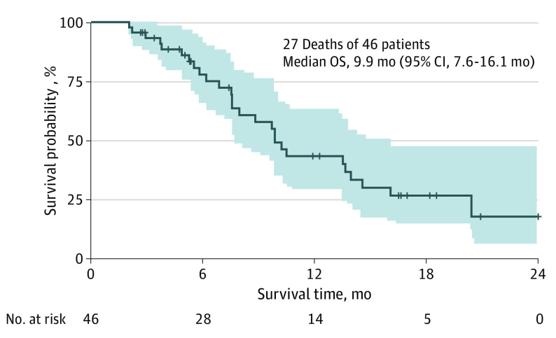
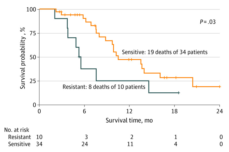
The median OS was 9.9 months (95% CI, 7.6-16.1 months) (Figure 3). Patients with DDR-GAs had numerically greater median OS (13.6 months [95% CI, 9.7 months to not reached]); however, there was no statistically significant difference between patients with PDAC with DDR-GAs and those with ATM protein loss or family history (eTable 3B in Supplement 3). There was a significant survival difference between patients with platinum-sensitive and patients with platinum-resistant disease (10.5 vs 5.4 months; P = .03) (Figure 4; eTable 3A in Supplement 3).
Figure 3. Kaplan-Meier Survival Curve of Overall Survival (OS).
Figure 4. Kaplan-Meier Survival Curve for Overall Survival (OS) Based on Platinum Sensitivity.
Safety
Olaparib at the study dosage was well tolerated, with most adverse events being grade 1 or 2. Fatigue, anorexia, nausea, anemia, and leukopenia were the most common toxic effects (eTable 4 in Supplement 3). Drug treatment discontinuation for toxic effects was required for only 1 patient.
Discussion
Recent molecular profiling studies offer novel therapeutic possibilities for PDAC; prominent among those is DNA repair inhibitors for the DDR subgroup. The POLO (Pancreas Cancer Olaparib Ongoing) study demonstrated that olaparib significantly improved PFS compared with placebo among patients with PDAC with BRCA1/2 variants.3 Tumors with molecular features that resemble BRCA variant cancers have been described as having BRCAness and have been hypothesized to respond to similar therapeutic approaches. The definition of BRCAness at a molecular level has been elusive, and our studies included the DDR-GAs commonly recognized in the literature after first excluding germline BRCA1/2 variants. However, these genetic alterations clearly represent a fraction of BRCAness, and therefore we expanded our inclusion criteria to further explore this phenotype. We included ATM protein loss detected by immunohistochemistry, based on prior preclinical studies and a cohort of patients with clinical BRCAness (ie, patients without DDR-GAs but with a strong personal history or family history).
The overall PFS with olaparib in the present studies was 3.7 months; this PFS is comparable to second-line chemotherapy regimens, including fluorouracil and liposomal irinotecan, and is reasonable given that the patients had received a median of 2 prior chemotherapy regimens for PDAC.12 However, this PFS appears to be inferior to that noted among patients with BRCA variants who received olaparib in the POLO trial.3 The toxicity profile of olaparib is favorable. The PFS and OS were significantly longer for patients with platinum-sensitive tumors and the presence of DDR-GAs. These results are consistent with recent reports targeting platinum-sensitive ovarian cancer wherein PARP inhibitors improved survival compared with placebo.13 In comparison, olaparib appears to have limited benefit for patients with clinical BRCAness without DDR-GAs in our trial. When these studies were originally planned, cross-resistance between platinum analogues and PARP inhibitors was not recognized. During the course of the study, this cross-resistance was recognized, and therefore the protocol was amended to exclude platinum-resistant PDAC. Our studies combine the results of 2 parallel trials with a similar study design conducted at 2 sister institutions, one in the US and other in Israel. The consistency of the results in these 2 centers adds to the strength of our findings.
The most common DDR variant noted was in the ATM gene. Deleterious ATM variants have been recognized in the germline with familial PDAC, suggesting that ATM is a susceptibility gene in hereditary PDAC.8 Somatic ATM variations occur in 2% to 18% of PDACs.14 In our studies, although confirmed PRs did not occur in the case of ATM variants, stable disease was common and was associated with prolonged PFS. Low ATM protein expression and p53 depletion were associated with olaparib sensitivity in lymphoma and gastric cancer preclinical models.15 The ATM protein plays a complex role as a cell-cycle checkpoint kinase regulator of a wide array of downstream proteins and a responder to DNA damage for genome stability. Activation signals of ATM through ATR (OMIM 107410) and CHEK1 (OMIM 603078) and inhibition of the latter may lead to greater clinical benefit than with PARP inhibitors. No conclusive statement can be made regarding the effectiveness of olaparib for patients with PDAC with ATM protein loss owing to the small sample size of patients included in the trial.
Limitations
Our studies have several limitations. The objective response rate was selected as the primary end point in this single-group study, and our results did not meet the prespecified success threshold. Most patients experienced stable disease as their best response to olaparib; therefore, PFS and OS may represent better surrogates for therapeutic benefit in this setting. The duration of PARP inhibitor treatment in the study was also limited and was lower than noted in the POLO trial.3 Preselection for DDR-GAs may improve this duration. We included both germline and somatic DDR-GAs in these studies, based on data on ovarian cancer.16 However, it is unknown if the effectiveness of PARP inhibitors in the case of PDAC will vary depending on the nature of these variants. Although we sought clarity regarding the definition of BRCAness in PDAC, we did not demonstrate a clear definition of this phenotype beyond the DDR-GA subgroup. The limited numbers of patients may have accounted for this deficiency. Future trials should explore these 3 populations (patients with DDR-GAs, patients with ATM protein loss, and patients with clinical BRCAness) separately. Whole-genome sequencing studies have demonstrated that “unstable” subtypes have structural variations and genomic instability, and this subtype is enriched with inactivation of DNA maintenance genes and a variant BRCA-like signature and that these subtypes express sensitivity to platinum and DNA repair inhibitors.17 Efforts to develop molecular signatures to capture BRCAness in breast and ovarian cancer and PDAC are ongoing, and it remains to be determined whether these signatures may be implemented in clinical trials for PDAC.18,19,20
Conclusions
In these prospective nonrandomized clinical trials of PARP inhibitors for patients with PDAC with BRCAness, the findings suggest that the PARP inhibitor olaparib may have therapeutic value for patients with platinum-sensitive PDAC with specific DDR-GAs other than germline BRCA variants. Future randomized clinical trials will be required to confirm the benefit of PARP inhibitors in this population.
Trial Protocol at MD Anderson Cancer Center
Trial Protocol at Sheba Medical Center
eFigure 1. Kaplan Meier Survival Curve for PFS Based on DDR-GA
eFigure 2. Kaplan Meier Survival Curve for PFS Based on Platinum Sensitivity
eTable 1A. Patient Demographics
eTable 1B. Gene List
eTable 2A. Distribution of Cases at Sheba Medical Center and MD Anderson Cancer Center
eTable 2B. Univariate Cox Model to Evaluate Risk Effect of DDR-GA vs ATM loss, FH or PH on PFS
eTable 3A. Log Rank Test to Compare OS Between Subgroups of Patients
eTable 3B. Univariate Cox Model to Evaluate Risk Effect of DDR-GA vs ATM loss, HF or PH on PFS
eTable 4. Summary of Adverse Events and Grade
Data Sharing Statement
References
- 1.Kaufman B, Shapira-Frommer R, Schmutzler RK, et al. Olaparib monotherapy in patients with advanced cancer and a germline BRCA1/2 mutation. J Clin Oncol. 2015;33(3):244-250. doi: 10.1200/JCO.2014.56.2728 [DOI] [PMC free article] [PubMed] [Google Scholar]
- 2.Fogelman DR, Wolff RA, Kopetz S, et al. Evidence for the efficacy of Iniparib, a PARP-1 inhibitor, in BRCA2-associated pancreatic cancer. Anticancer Res. 2011;31(4):1417-1420. [PubMed] [Google Scholar]
- 3.Golan T, Hammel P, Reni M, et al. Maintenance olaparib for germline BRCA-mutated metastatic pancreatic cancer. N Engl J Med. 2019;381(4):317-327. doi: 10.1056/NEJMoa1903387 [DOI] [PMC free article] [PubMed] [Google Scholar]
- 4.Turner N, Tutt A, Ashworth A. Hallmarks of “BRCAness” in sporadic cancers. Nat Rev Cancer. 2004;4(10):814-819. doi: 10.1038/nrc1457 [DOI] [PubMed] [Google Scholar]
- 5.Pishvaian MJ, Bender RJ, Halverson D, et al. Molecular profiling of patients with pancreatic cancer: initial results from the Know Your Tumor Initiative. Clin Cancer Res. 2018;24(20):5018-5027. doi: 10.1158/1078-0432.CCR-18-0531 [DOI] [PubMed] [Google Scholar]
- 6.Fogelman D, Sugar EA, Oliver G, et al. Family history as a marker of platinum sensitivity in pancreatic adenocarcinoma. Cancer Chemother Pharmacol. 2015;76(3):489-498. doi: 10.1007/s00280-015-2788-6 [DOI] [PMC free article] [PubMed] [Google Scholar]
- 7.Kim H, Saka B, Knight S, et al. Having pancreatic cancer with tumoral loss of ATM and normal TP53 protein expression is associated with a poorer prognosis. Clin Cancer Res. 2014;20(7):1865-1872. doi: 10.1158/1078-0432.CCR-13-1239 [DOI] [PMC free article] [PubMed] [Google Scholar]
- 8.Kim HS, Kim MA, Hodgson D, et al. Concordance of ATM (ataxia telangiectasia mutated) immunohistochemistry between biopsy or metastatic tumor samples and primary tumors in gastric cancer patients. Pathobiology. 2013;80(3):127-137. doi: 10.1159/000346034 [DOI] [PubMed] [Google Scholar]
- 9.Eisenhauer EA, Therasse P, Bogaerts J, et al. New response evaluation criteria in solid tumours: revised RECIST guideline (version 1.1). Eur J Cancer. 2009;45(2):228-247. doi: 10.1016/j.ejca.2008.10.026 [DOI] [PubMed] [Google Scholar]
- 10.Thall PF, Simon R, Ellenberg SS. A two-stage design for choosing among several experimental treatments and a control in clinical trials. Biometrics. 1989;45(2):537-547. doi: 10.2307/2531495 [DOI] [PubMed] [Google Scholar]
- 11.National Cancer Institute. Common Terminology Criteria for Adverse Events (CTCAE) v4.0. Accessed January 25, 2021. https://ctep.cancer.gov/protocoldevelopment/electronic_applications/ctc.htm#ctc_40
- 12.Wang-Gillam A, Hubner RA, Siveke JT, et al. NAPOLI-1 phase 3 study of liposomal irinotecan in metastatic pancreatic cancer: final overall survival analysis and characteristics of long-term survivors. Eur J Cancer. 2019;108:78-87. doi: 10.1016/j.ejca.2018.12.007 [DOI] [PubMed] [Google Scholar]
- 13.Pujade-Lauraine E, Ledermann JA, Selle F, et al. ; SOLO2/ENGOT-Ov21 investigators . Olaparib tablets as maintenance therapy in patients with platinum-sensitive, relapsed ovarian cancer and a BRCA1/2 mutation (SOLO2/ENGOT-Ov21): a double-blind, randomised, placebo-controlled, phase 3 trial. Lancet Oncol. 2017;18(9):1274-1284. doi: 10.1016/S1470-2045(17)30469-2 [DOI] [PubMed] [Google Scholar]
- 14.Armstrong SA, Schultz CW, Azimi-Sadjadi A, Brody JR, Pishvaian MJ. ATM dysfunction in pancreatic adenocarcinoma and associated therapeutic implications. Mol Cancer Ther. 2019;18(11):1899-1908. doi: 10.1158/1535-7163.MCT-19-0208 [DOI] [PMC free article] [PubMed] [Google Scholar]
- 15.Kubota E, Williamson CT, Ye R, et al. Low ATM protein expression and depletion of p53 correlates with olaparib sensitivity in gastric cancer cell lines. Cell Cycle. 2014;13(13):2129-2137. doi: 10.4161/cc.29212 [DOI] [PMC free article] [PubMed] [Google Scholar]
- 16.Tew WP, Lacchetti C, Ellis A, et al. PARP inhibitors in the management of ovarian cancer: ASCO guideline. J Clin Oncol. 2020;38(30):3468-3493. doi: 10.1200/JCO.20.01924 [DOI] [PMC free article] [PubMed] [Google Scholar]
- 17.Waddell N, Pajic M, Patch AM, et al. ; Australian Pancreatic Cancer Genome Initiative . Whole genomes redefine the mutational landscape of pancreatic cancer. Nature. 2015;518(7540):495-501. doi: 10.1038/nature14169 [DOI] [PMC free article] [PubMed] [Google Scholar]
- 18.Davies H, Glodzik D, Morganella S, et al. HRDetect is a predictor of BRCA1 and BRCA2 deficiency based on mutational signatures. Nat Med. 2017;23(4):517-525. doi: 10.1038/nm.4292 [DOI] [PMC free article] [PubMed] [Google Scholar]
- 19.Hillman RT, Chisholm GB, Lu KH, Futreal PA. Genomic rearrangement signatures and clinical outcomes in high-grade serous ovarian cancer. J Natl Cancer Inst. 2018;110(3). doi: 10.1093/jnci/djx176 [DOI] [PMC free article] [PubMed] [Google Scholar]
- 20.Connor AA, Denroche RE, Jang GH, et al. Integration of Genomic and Transcriptional Features in Pancreatic Cancer Reveals Increased Cell Cycle Progression in Metastases. Cancer Cell. 2019;35(2):267-282.e7. doi: 10.1016/j.ccell.2018.12.010 [DOI] [PMC free article] [PubMed] [Google Scholar]
Associated Data
This section collects any data citations, data availability statements, or supplementary materials included in this article.
Supplementary Materials
Trial Protocol at MD Anderson Cancer Center
Trial Protocol at Sheba Medical Center
eFigure 1. Kaplan Meier Survival Curve for PFS Based on DDR-GA
eFigure 2. Kaplan Meier Survival Curve for PFS Based on Platinum Sensitivity
eTable 1A. Patient Demographics
eTable 1B. Gene List
eTable 2A. Distribution of Cases at Sheba Medical Center and MD Anderson Cancer Center
eTable 2B. Univariate Cox Model to Evaluate Risk Effect of DDR-GA vs ATM loss, FH or PH on PFS
eTable 3A. Log Rank Test to Compare OS Between Subgroups of Patients
eTable 3B. Univariate Cox Model to Evaluate Risk Effect of DDR-GA vs ATM loss, HF or PH on PFS
eTable 4. Summary of Adverse Events and Grade
Data Sharing Statement


