Abstract
The study determined the effects of Lactobacillus salivarius (LS) administered early in the life of suckling piglets on their growth performance, gut morphology, and gut microbiota. Thirty litters of 3-day-old crossbreed piglets were randomly assigned to one of the three treatments, and treatments were commenced on day 3 after birth. During the whole period of the experiment, the piglets were kept with their mothers and left to suckle ad libitum while being supplemented with a milk formula with or without the bacterial probiotic supplemented. The control group (CON) was not treated with probiotics, the HLS group was treated with LS144 (HLS) screened from feces of fast-growing pigs with high body mass index (BMI) while the NLS group was supplemented with LS160 (NLS) screened from feces obtained from pigs of normal BMI. At the weaning time, a higher abundance of Actinobacteria, Lentisphaerae, and Elusimicrobia phyla were observed in NLS piglets, whereas the abundance of Fibrobacteres phylum was significantly reduced in NLS and HLS piglets compared with the CON. A greater abundance of Lactobacillus was detected in the HLS treatment compared with the CON. The abundance of Bacteroides and Fibrobacter was higher in the CON piglets compared with the HLS and NLS piglets. Compared with the CON group, the oral administration of LS significantly increased the number of Lactobacillus and villus height in the duodenum, jejunum, and ileum. Moreover, the villus height of the duodenum was significantly improved in the HLS treatment compared with the NLS treatment. Based on the findings in the neonatal piglet model, we suggest that oral supplementation of LS, particularly LS isolated from high BMI pigs, could be beneficial by improving the intestinal villus height.
Subject terms: Biotechnology, Microbiology
Introduction
In swine, weaning and suckling are by far the most stressful periods that imposes the highest rate of loss and mortality. The adverse effect of diarrhea is more critical in suckling and weanling pigs than mature pigs due to the immature immune system1,2. A serious pathogenic challenge or stress during this critical neonatal period impacts negatively on the piglets whole process of development2–4. Therefore, the management of gut microbiota of suckling pigs by controlling Clostridium and Escherichia coli colonization may efficiently reduce the economic loss2,3. The microbiota in the small intestine is a dynamic ecosystem with a diverse commensal bacterial population, which affects the immune development and health of piglets5–8. Piglets are born with basically a sterile gut and the colonization begins immediately after birth3,9. In addition, the intestinal tract of neonatal piglets is under influence of undefined factors such as mother's feces and environmental microbes2,3, particularly that suckling piglets eat about 20 g feces per day due to their suckling habit10. Therefore, regarding the immature and unstable gut microbiota, any environmental stressors or pathogenic challenges may quickly compromise the microbiota equilibrium and compromise suckling pig health conditions.
After the ban on antibiotic growth promoters (AGP), probiotics have been found to be one of the most suitable alternatives to replace the AGPs in the animal industry as growth promoters. Many strains of bacteria have been tested for use as probiotics including L. salivarius SGL19, Bacillus subtilis B2A, Lactobacillus acidophilus K31, and Enterococcus M7411–14. During the suckling period, milk as the main feed source is regarded to be the most effective factor in shaping the intestinal microbiota of neonatal piglets. Among the beneficial genus, Lactobacillus spp. can be considered as one of the best candidates due to their high proliferation rate when milk or milk products are used as substrates8. It has been shown that L. salivarius is able to trigger the growth of the population of Lactobacillus spp. bacteria and decrease the colonization of pathogens due to their great ability to adhere to intestinal epithelial cells and produce bacteriocins15–17. L. salivarius is a Gram-positive bacteria and one of the major inhabitant of pigs’ intestine that is tolerant of acidic conditions with an optimal pH range of 5.5–6.517,18. Moreover, in a recent study, L. salivarius exhibited activity against pathogenic bacteria such as Clostridia, Campylobacter, and Salmonella in both in vivo and in vitro18–21. Consequently, dietary supplementation of L. salivarius appears to be beneficial to the pig gut health by influencing intestinal gut microbial colonization.
In recent years, high-throughput sequencing platforms such as 16S rRNA gene amplicon sequencing is extensively being applied to reveal the community structures of the microbiota. It is reported that there is an interaction between the intestinal microbiota and body weight in pigs15,22,23, particularly in young animals due to the immature intestinal microbial community. In the current study, after the screening process of potential Lactobacillus sp. with high bile and acid tolerance, antimicrobial activity, and adhesion capacity, the L. salivarius (LS144) from the feces of fast-growing pigs was detected to be used for further analysis. In addition, as a control treatment, L. salivarius (LS160) from normal weight pigs was isolated through the same procedure. Regarding our in vitro tests, we hypothesized that the two targeted strains of L. salivarius have diverse influences on the microbial proportion of Firmicutes to Bacteroidetes. This in vivo study was undertaken to investigate the effects of L. salivarius (LS144 and LS160) on weight gain, intestinal microorganism composition, and intestinal histomorphology of suckling pigs.
Results
Microbial community structure
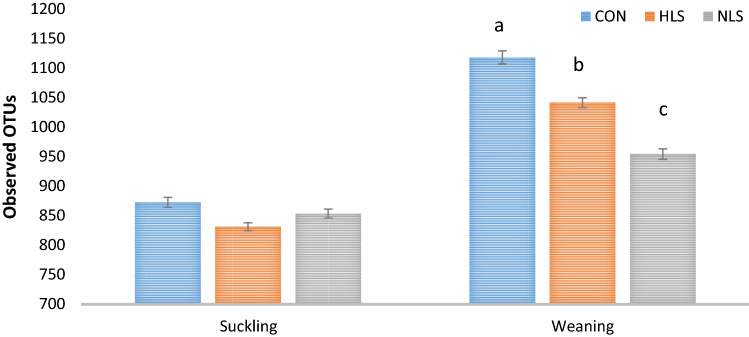
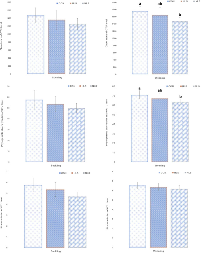
An average of 40,000 16S rRNA gene sequence reads was generated (Fig. 1). The number of observed OTUs (± SE) was 872.4 (± 19.3) for the CON group, 831.4 (± 18.5) for the HLS group (LS isolated from the feces of fast-growing pigs), and 853.6 (± 10.8) for the NLS (LS isolated from the feces of normal weight pigs) group at suckling period (Fig. 2). At weaning, the OTU value was 1117.3 (± 9.0) for the CON group, 1040.8 (± 11.5) for the HLS group, and 953.9 (± 7.9) for the NLS group. During the sucking phase, there was no difference in microbiota diversity (Fig. 3). However, a significant (p = 0.002) decrease in the Chao index (Fig. 3), which reflects the species evenness and richness, was observed in the NLS treatment compared with the CON at weaning. At weaning time, a higher (p = 0.005) phylogenetic diversity index was observed in pigs in the CON treatment compared with the NLS treatment. No difference in the Shannon index was detected between the treatments. The Adonis test for the analysis of similarities of unweighted UniFrac distances (Fig. 4a) indicated no difference between the treatments, however, there was a significant difference (R2 = 0.14, P < 0.01) between suckling and weaning time, showing that the microbiota of piglets was significantly changed over the time. There was a similar analysis of similarities between weighted UniFrac distances and unweighted UniFrac distances (Fig. 4b), which showed no difference among the treatments but a distinct clustering (R2 = 0.25, P < 0.01) between sucking and weaning times.
Figure 1.
Diversity of intestinal microbiota of piglets at different stages. Alpha diversity indices including Chao1 (a), PD whole tree (b), and Shannon (c) were observed at each number of sequencing reads.
Figure 2.
OTUs gain at the beginning (Suckling) and the end (weaning) of the experiment. CON, Control without probiotic; HLS, L. salivarius 144 isolated from fast-growing pig feces; NLS, L. salivarius160 isolated from normal weight pig feces and different superscript letters indicate significant differences (P < 0.05).
Figure 3.
Differences in the fecal microbial species richness and diversity indices (Chao 1, Shannon, OTU; ≥ 97% sequence similarity threshold) per treatment. Different superscript letters indicate significant differences (P < 0.05).
Figure 4.
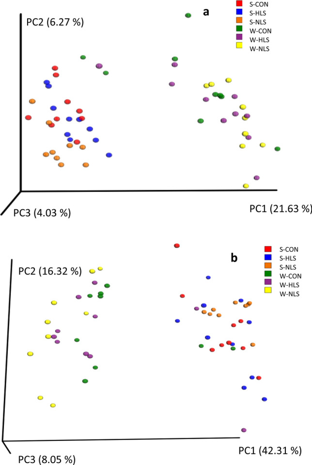
Beta diversity patterns of fecal microbial diversity in at the beginning (d 4, suckling) and end of the experiment (d 21, weaning) as assessed by principal coordinate analysis (PCoA) of unweighted and Weighted Unifrac. CON, Control without probiotic; HLS, L. salivarius 144 isolated from fast-growing pig feces; NLS, L. salivarius160 isolated from normal weight pig feces.
Taxa difference at the phylum level
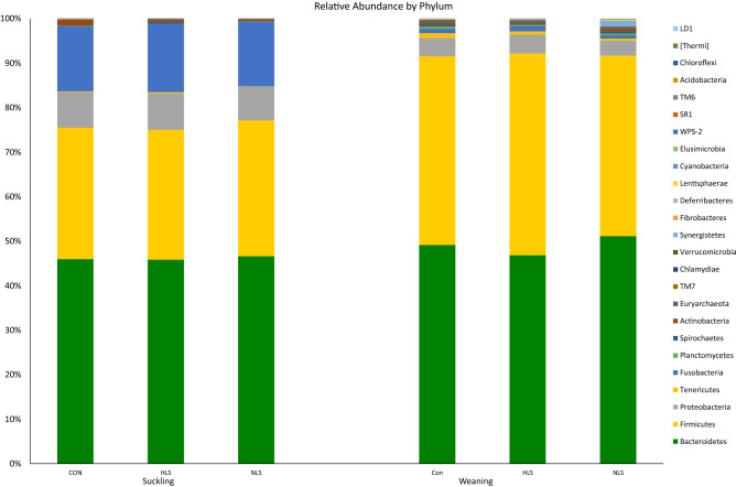
At the 97% similarity level, in total 25 phyla (Fig. 5) were detected. At the suckling period, the two dominant phyla detected in the three groups were Bacteroidetes (45.9%) and Firmicutes (29.8%). The analysis of microbiota in piglets showed a higher abundance of Firmicutes, Tenericutes, Lentisphaerae, Deferribacteres, Elusimicrobia, and Fibrobacteres phyla, and a lower abundance of Fusobacteria, Proteobacteria, and Actinobacteria in the weaning period compared with the suckling period (Table 1). At the weaning period, again Bacteroidetes (49.0%) and Firmicutes (42.8%) were the dominant phyla. A higher abundance of Actinobacteria, Lentisphaerae, and Elusimicrobia phyla were observed in NLS piglets, whereas the abundance of Fibrobacteres phylum was significantly reduced in NLS and HLS piglets compared with the CON.
Figure 5.
16S rRNA gene analysis revealed the relative abundance of fecal bacterial community structure at the phylum level in piglets orally treated with probiotics Lactobacillus salivarius 144 (HLS), L. salivarius 160 (NLS), or without probiotic (CON) at suckling (d 4) and weaning (d 21) periods.
Table 1.
Relative abundance of the fecal microbiota phyla of three groups at suckling (d 4) and weaning (d 21) periods.
| Suckling | Weaning | P-value | |||||||
|---|---|---|---|---|---|---|---|---|---|
| CON | HLS | NLS | CON | HLS | NLS | Suckling | Weaning | Time | |
| Bacteroidetes | 45.98 ± 7.9 | 44.61 ± 10.1 | 46.33 ± 11.3 | 49.15 ± 9.6 | 46.83 ± 9.3 | 51.14 ± 3.1 | 0.61 | 0.12 | 0.69 |
| Firmicutes | 29.69 ± 6.0 | 29.23 ± 14.2 | 30.42 ± 4.4 | 42.42 ± 9.2 | 45.35 ± 9.5 | 40.59 ± 5.1 | 0.48 | 0.23 | 0.00 |
| Fusobacteria | 14.63 ± 7.4 | 15.35 ± 9.7 | 14.60 ± 12.3 | 0.99 ± 0.61 | 1.20 ± 0.95 | 0.74 ± 0.61 | 0.78 | 0.21 | 0.00 |
| Proteobacteria | 9.033 ± 5.3 | 9.169 ± 4.8 | 7.724 ± 4.5 | 4.101 ± 0.7 | 4.229 ± 1.8 | 3.341 ± 0.8 | 0.52 | 0.12 | 0.00 |
| Actinobacteria | 1.276 ± 1.0 | 0.489 ± 0.28 | 0.654 ± 0.27 | 0.31 ± 0.15b | 0.40 ± 0.31ab | 0.67 ± 0.37a | 0.13 | 0.02 | 0.15 |
| Tenericutes | 0.143 ± 0.21 | 0.269 ± 0.71 | 0.048 ± 0.02 | 1.080 ± 0.93 | 0.739 ± 0.63 | 0.469 ± 0.23 | 0.33 | 0.06 | 0.00 |
| Planctomycetes | 0.120 ± 0.22 | 0.012 ± 0.02 | 0.063 ± 0.003 | 0.517 ± 0.41 | 0.276 ± 0.21 | 0.406 ± 0.36 | 0.11 | 0.13 | 0.00 |
| Lentisphaerae | 0.042 ± 0.08 | 0.006 ± 0.01 | 0.007 ± 0.009 | 0.032 ± 0.02b | 0.086 ± 0.11ab | 0.216 ± 0.30a | 0.11 | 0.05 | 0.01 |
| Deferribacteres | 0.002 ± 0.004 | 0.0001 ± .0004 | 0.0002 ± .0001 | 0.072 ± 0.13 | 0.017 ± 0.02 | 0.032 ± 0.03 | 0.17 | 0.14 | 0.01 |
| Elusimicrobia | 0.004 ± 0.003 | 0.001 ± 0.001 | 0.007 ± 0.006 | 0.017 ± 0.01b | 0.009 ± 0.007b | 0.114 ± 0.176a | 0.14 | 0.03 | 0.03 |
| Fibrobacteres | 0.004 ± 0.004 | 0.002 ± 0.002 | 0.015 ± 0.0038 | 0.073 ± 0.069a | 0.016 ± 0.022b | 0.017 ± 0.023b | 0.22 | 0.01 | 0.00 |
CON, Control; HLS, Lactobacilus salivarius 144 isolated from fast-growing pig feces; NLS, L. salivarius160 isolated from normal weight pig feces.
a-bMeans with different superscripts within rows are significantly different at P < 0.05.
Taxa difference at the genus level
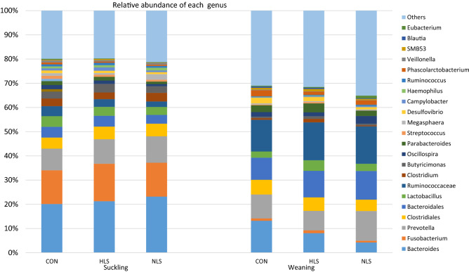
At the 97% similarity level, in total 450 genera (Fig. 6) were detected. At the genus level, three dominant genera, Bacteroides (21.69%), Fusobacterium (14.91%), and Prevotella (10.01%) were detected in the fecal microbiota of piglets at the suckling period, whereas microbiota of weaned piglets was dominated by Prevotella (10.07%), Bacteroides (8.47%), and Lactobacillus (3.31%). At weaning time, although the differences in the abundance of Lactobacillus did not differ between the HLS and NLS piglets, a significantly greater Lactobacillus population was recorded in the HLS treatment compared with the CON (Table 2). The abundance of Bacteroides and Fibrobacter was higher in the CON piglets compared with the HLS and NLS piglets (Table 2). Compared to the CON, the abundance of Phascolarctobacterium was lower in the HSL, and the abundance of Desulfovibrio, Clostridium, and Weissella was lower in the NLS treatment. The highest abundance of Christensenella and Limnohabitans genera were observed in the HLS treatment, whereas the highest abundance of Helicobacter and Methanosphaera was detected in the NLS treatment. The piglets fed HLS probiotic showed a lower abundance of Oscillospira, and a greater abundance of Bacteroides, Sarcina, Limnohabitans, and Christensenella compared with the NLS treatment.
Figure 6.
16S rRNA gene analysis revealed the relative abundance of fecal bacterial community structure at the genus level in piglets orally treated with probiotics Lactobacillus salivarius 144 (HLS), L. salivarius 160 (NLS), or without probiotic (CON) at suckling (d 4) and weaning (d 21) periods.
Table 2.
Relative abundance of the fecal microbiota genera of three groups at weaning (d 21).
| Item | Treatment | P-value | ||
|---|---|---|---|---|
| CON | HLS | NLS | ||
| Bacteroides | 13.19 ± 4.7a | 8.04 ± 3.3b | 4.20 ± 3.1c | 0.041 |
| Prevotella | 9.86 ± 6.9 | 8.06 ± 7.7 | 12.30 ± 6.1 | 0.428 |
| Ruminococcus | 1.57 ± 1.1 | 1.80 ± 1.1 | 1.17 ± 1.1 | 0.494 |
| Lactobacillus | 2.58 ± 1.4b | 4.40 ± 2.3a | 2.97 ± 2.4ab | 0.046 |
| Parabacteroides | 2.81 ± 1.6 | 3.72 ± 1.6 | 2.22 ± 1.6 | 0.139 |
| Oscillospira | 1.90 ± 1.3ab | 1.61 ± 0.65b | 3.21 ± 1.7a | 0.028 |
| Phascolarctobacterium | 2.50 ± 1.5a | 1.32 ± 1.1b | 1.81 ± 1.4ab | 0.010 |
| Desulfovibrio | 2.29 ± 0.86a | 1.69 ± 0.86ab | 0.96 ± 0.53b | < 0.01 |
| Campylobacter | 0.241 ± 0.21 | 0.359 ± 0.50 | 0.444 ± 0.45 | 0.589 |
| Streptococcus | 0.249 ± 0.18 | 0.552 ± 0.13 | 0.060 ± 0.025 | 0.308 |
| Clostridium | 1.437 ± 1.38a | 0.616 ± 0.38ab | 0.330 ± 0.30b | 0.028 |
| Fusobacterium | 0.983 ± 0.61 | 1.192 ± 0.95 | 0.741 ± 0.61 | 0.438 |
| Fibrobacter | 0.074 ± 0.07a | 0.016 ± 0.0b | 0.018 ± 0.0b | 0.010 |
| Helicobacter | 0.029 ± 0.02b | 0.043 ± 0.02b | 0.265 ± 0.18a | < 0.01 |
| Bifidobacterium | 0.014 ± 0.01 | 0.013 ± 0.02 | 0.026 ± 0.02 | 0.244 |
| Christensenella | 0.007 ± 0.001b | 0.021 ± 0.01a | 0.005 ± 0.003b | 0.020 |
| Sarcina | 0.004 ± 0.004ab | 0.015 ± 0.01a | 0.003 ± 0.002b | 0.027 |
| Weissella | 0.003 ± 0.003a | 0.001 ± 0.001ab | 0.0003 ± 0.0003b | 0.014 |
| Limnohabitans | 0.002 ± 0.002b | 0.024 ± 0.019a | 0.001 ± 0.001b | 0.011 |
| Methanosphaera | 0.001 ± 0.001b | 0.0001 ± 0.00003b | 0.004 ± 0.003a | < 0.01 |
CON, Control; HLS, Lactobacilus salivarius 144 isolated from fast-growing pig feces; NLS, L. salivarius160 isolated from normal weight pig feces.
a-bMeans with different superscripts within rows are significantly different at P < 0.05.
Intestinal digesta microbial population
Intestinal digesta analysis revealed a significant increase of Lactobacillus in the duodenum, jejunum, ileum, and cecum of pigs fed HLS and NLS Lactobacillus (P < 0.01). The total number of coliforms was significantly reduced in the duodenum of pigs in the HLS and NLS treatments compared with the CON treatment (P < 0.05), however, there were no differences in the population of coliforms in the jejunum, ileum, and cecum. There was no significant difference in the colonization of Clostridia among the treatments in all the segments of the intestine (Fig. 7).
Figure 7.
The population of lactobacilli, clostridia, and coliform at different sections of the intestine at the end of study (d21, weaning). CON, Control without probiotic; HLS, L. salivarius 144 isolated from fast-growing pig feces; NLS, L. salivarius160 isolated from normal weight pig feces.
Intestinal morphology
Both HLS and NLS treatment groups had significantly increased (P < 0.01) villi height throughout the 3 segments of the intestinal tract (duodenum, jejunum, and ileum) as compared with the CON group (Table 3). The crypt depth did not differ between the groups in all the 3 intestinal sections. However, the villus height and crypt depth ratio (VH:CD) differed significantly (P < 0.01) among the groups in the ileum with the greatest value in the HLS treatment group and the lowest value in the CON group.
Table 3.
Effects of dietary supplementation of Lactobacillus salivarius on intestinal gut morphology of piglets at weaning.
| Item | Treatment | SEM | P-value | ||
|---|---|---|---|---|---|
| CON | HLS | NLS | |||
| Villus height (VH) | |||||
| Duodenum | 257.07c | 283.46a | 269.03b | 3.71 | 0.001 |
| Jejunum | 246.87b | 278.16a | 270.76a | 4.67 | 0.002 |
| Ileum | 150.93b | 176.73a | 168.73a | 3.70 | 0.001 |
| Crypt depth (CD) | |||||
| Duodenum | 139.99 | 147.96 | 148.41 | 2.22 | 0.238 |
| Jejunum | 138.73 | 147.1 | 145.69 | 1.98 | 0.19 |
| Ileum | 121.8 | 117.5 | 123.22 | 1.32 | 0.191 |
| VH:CD | |||||
| Duodenum | 1.84 | 1.92 | 1.82 | 0.03 | 0.486 |
| Jejunum | 1.78 | 1.89 | 1.86 | 0.02 | 0.243 |
| Ileum | 1.24c | 1.51a | 1.37b | 0.03 | 0.001 |
CON, Control; HLS, L. salivarius 144 isolated from fast-growing pig feces; NLS, L. salivarius160 isolated from normal weight pig feces.
a-b Means with different superscripts within rows are significantly different at P < 0.05.
Weight gain
The effect of the HLS and NLS supplementation on piglet growth performance was shown in Table 4. Weight gain and ADG were not affected by the LS-treatment groups compared to the CON group.
Table 4.
Effect of dietary supplementation of Lactobacillus salivarius on piglet growth performance.
| Item | Treatment | SEM | P-value | ||
|---|---|---|---|---|---|
| CON | HLS | NLS | |||
| Initial BW, kg (3 days) | 1.55 | 1.54 | 1.54 | 0.01 | 0.274 |
| Finishing BW, kg (21 days) | 6.19 | 6.23 | 6.24 | 0.08 | 0.974 |
| ADG, g | 221.05 | 223.07 | 223.45 | 3.85 | 0.969 |
CON, Control; HLS, L. salivarius 144 isolated from fast-growing pig feces; NLS, L. salivarius160 isolated from normal weight pig feces. BW, body weight; ADG, average daily gain.
Discussion
Infancy is a critical period due to unstable gut microbiome structure2,3. Dietary supplementation of probiotic lactobacilli may modulate the microbial community in the gastrointestinal tract preventing diarrhea and stimulate growth14,24,25. This study was conducted to evaluate the effects of oral dietary supplementation of live L. salivarius suspension in suckling piglets concerning growth performance, intestinal bacterial diversity, and intestinal morphology. In the current study, we evaluated the fecal microbiota composition in piglets fed two different LS during the suckling period. The present study has revealed that the supplementation of NLS significantly decreased the observed OTUs than those for the CON group. The lower Phylogenetic diversity index and Chao index of feces bacteria in the NLS treatment suggested that probiotics may inhibit the growth of bacteria, which was consistent with Wang et al.26 who reported a lower bacterial diversity along with a promoted intestinal health when using L. casei ZX633. Several studies have reported that intestinal microbial richness is an index with a positive relation to body weight23. It has been known that the increased microbiota diversity is associated with a stable ecology and overall health of animals27. Despite the benefits, the abundance of microbiota may adversely affect the host in several ways such as immune system stimulation, nutrient competition, and the generation of toxic catabolites9.
In our study, the relative abundance of phyla in both LS-treated treatments were significantly changed compared with the CON. The microbiota of the NLS group showed a higher abundance of Actinobacteria, Lentisphaerae, and Elusimicrobia compared with that of the CON group, whereas a significantly higher Fibrobacteres level was observed in the CON group. The Fibrobacteres phylum is related to cellulolytic bacteria energy metabolism28, and the reasons for these significant differences are not clear. The diversity of major phyla including Bacteroidetes, Firmicutes, Fusobacteria, and Proteobacteria were unaffected by the HLS or NLS treatments. This is in agreement with the results reported in a previous study with no difference in the levels of the main phyla such as Firmicutes and Bacteroidetes when using probiotics18. Bacteroidetes and Firmicutes are the most abundant phyla in piglet intestinal microbiota, regardless of dietary probiotic and age9,29. In this study, the most abundant phyla were Bacteroidetes and Firmicutes accounted for less than 80% of the phyla at the suckling period, however, at weaning time both Bacteroidetes and Firmicutes showed to be the most abundant phyla (more than 90% of the phyla) in feces, which is in agreement with some earlier studies29,30. The abundance of Proteobacteria and Fusobacteria was dramatically reduced from the suckling period to the weaning period. Fusobacteria has the potential to be pathogenic and be related to cancer and some other diseases in animals31. Moreover, within the Proteobacteria phylum, there are some pathogenic genera such as Salmonella, Escherichia, and Helicobacter32. This change in microbial composition is related to age, physiological, and dietary factors16,29. As shown in our study, it can be suggested that pathobiont species are a part of normal microbiota in infants and any stressors such as changing the diet formulation or form (at weaning period) may trigger these potential pathogens to proliferate.
At the genus level, within Bacteroides, this study identified a higher relative abundance of Bacteroides in the CON pigs. Surprisingly, the population of Bacteroides in NLS pigs was lower than in HLS pigs. The genera Bacteroides and Prevotella were reported to be the normal inhabitant of the intestine in pigs and sows29,30,32 although their population in the fecal microbiota of suckling pigs was at a much lower abundance than a previous study33. Bacteroides are naturally mutualistic species in the intestine, however, some of them are opportunistic pathogens34. Bacteroides fragilis is a good example of a pathogenic Bacteroides with the potential of causing malignancy, inflammation, and diarrhea35. In this study, Bacteroides were the most abundant, and Fusobacterium and Prevotella remained less abundant microbiota, in contrast to a previous study32. The administered HLS probiotic increased the population of Lactobacillus, Limnohabitans, Sarcina, and Rhodoferax, which belong to the phyla Firmicutes and Proteobacteria. Moreover, Fusocaterium was the most prominent genus of the phylum Fusobacteria indigenous to the fecal microbiota of piglets in this study. A plethora of factors such as diet8,36–38 affects intestinal microbiota communities of the host. Interestingly, the microbiota analysis of feces indicates that there is a negative relationship between the abundance of Lactobacillus and Clostridium, as the supplementation of HLS decreased the population of Clostridium and increased the population of Lactobacillus in the feces. Clostridia species are normally known to be pathogenic2,4,12. As there was a significant increase in the abundance of fecal Lactobacillus in HSL piglets, it is not surprising that a higher population of Lactobacillus was detected in the jejunum, ileum, and cecum of piglets fed LS. Lactobacillus species are considered to be among the beneficial members of a normal microbiota5,7,22. In this study, the most evident response of using L. salivarius seems to be the stimulating role in the growth of intestinal Lactobacillus, irrespective of its strain. Despite the increase in the population of Lactobacillus in the jejunum, ileum, and cecum of piglets in NLS treatment, the total abundance of Lactobacillus in the microbiota of feces was not affected. Whereas piglets in the HLS group not only showed an increased population of Lactobacillus in the jejunum, ileum, cecum but also revealed a greater abundance of fecal Lactobacillus. An increase in the abundance of Lactobacillus during the suckling period is essential to encounter the weaning time when piglets are highly vulnerable to opportunistic pathogens. The decrease in the proportion of fecal clostridia in piglets administered HLS or NLS when compared to the CON may highlight the antipathogenic effects on Gram-negative bacterium. Probiotics aid the host animal in defense against pathogens through competitive exclusion and the production of antimicrobials5,12,39. A similar study also reported that L. salivarius greatly increased the integrity of epithelial cells in pigs by protecting the small intestine cells from the adherence of enterotoxigenic E. coli K88, resulting in a higher survival rate40. However, our study does not show a straightforward confirmation of anti-pathogenic factors due to insignificant differences in the colonization of Clostridium and E. coli in the jejunum and ileum.
Improved villus height is a marker for better digestive and absorptive intestinal capacities4,25. In the current study, L. salivarius (LS144 and LS160) supplementation had a positive effect on villus height in all the intestinal segments, the increase was more pronounced in the HLS group compared to the NLS group. Our result is in agreement with a similar earlier study that recorded a significant increase in the villus height with L. Plantarum CGMCC supplemented a group of piglets and enhanced VH:CD41. The VH:CD in the ileum section of the intestinal gut was significantly increased in the treatment groups, which is an indicator of the increased superficial absorptive area with a thinner lamina propria in this vital part of the gut where most of the absorption of nutrients occurs.
Overall effects revealed that L. salivarius supplemented piglets (HLS and NLS) had no effects on ADG compared to the CON group. The average body weight of pigs in this experiment was 6.22 kg at weaning, which was in a similar range as previous reports4,7,8,12,22,36. This result is consistent with a previous study supplementing multi-strain probiotic including L. acidophilus that equally revealed a marginal difference in growth performance between the treatment group and the control group12. However, in a similar study, the addition of L. casei into the diet increased the ADG of piglets7. The insignificant ADG but significantly improved villus height may be explained by the physiological status and age of piglets, as we used the probiotics in suckling piglets with milk as the main feed source, however, most of the significant studies used probiotics for weaned piglets with a solid meal as the main feed source7. A greater villus height may result in a better performance after weaning when the diet changes from liquid milk to solid feed. The short period of the experiment can also be another reason for the insignificant results.
Conclusion
In conclusion, based on the microbiota information, our study demonstrated that the population of beneficial bacteria such as Lactobacillus was significantly increased in the HLS-treated piglets. Moreover, the abundance of clostridia was decreased in the feces, which may emphasize the antimicrobial activity of HSL probiotic. These normal alterations in the gut microbiota at the suckling period decrease the susceptibility of weaned piglets to pathogenic infections at weaning time. The greater villus height of the duodenum, jejunum, and ileum can be considered as the indicators of the integrated intestine that may provide the potential for higher growth performance after weaning. This achievement may provide greater insight into the importance of intestinal microbiota manipulation during suckling, and future work focusing on the growth performance of weaned piglets seems necessary to confirm the improved growth potential in the suckling period.
Materials and methods
Animal care
This research was conducted according to the protocol approved by Kangwon National University institutional animal care and use committee (IACUC No.: KW-140509–1). All experiments were performed in accordance with relevant guidelines and regulations.
Animals, experimental designs, and diets
The experiment was conducted at a commercial pig farm in Gangneung in the Republic of Korea. Standard farm management and husbandry practices were routinely carried out by the farm staff. Thirty cross-bred three-day-old piglets (1.54 ± 0.44 kg; Duroc × Yorkshire × Landrace) were randomly divided into three groups (n = 10, for each treatment). Cross fostering was done before starting the experiment. Each experiment litter was housed individually with the dam in individual stainless steel pens with reinforced plastic floors. Piglets had ad libitum access to sow milk and water. Sows were fed on a common corn-soybean meal-based diet. The treatments included the CON (basal diet; milk formula without probiotic), CON plus 20 ml/day of probiotic L. salivarius144 isolated from fast-growing pigs (HLS; 1 × 10⁸ cfu/ ml), CON plus 20 ml/day of probiotic L. salivarius160 isolated from normal weight pigs (NLS; 1 × 10⁸ cfu/ml). The basal dry milk formula was designed as a sow milk supplement. The mentioned L. salivarius were selected after passing the screening tests such as antimicrobial activity. The screened L. salivarius (LS144 and LS160) probiotics were acquired from Kangwon National University, Laboratory of Microbial Genomics and Big Data, and stored at 4 °C in individualized centrifugal tubes.
Lactobacillus salivarius isolation and identification
Lactobacillus salivarus were isolated from the fecal samples of the fast-growing and normal weight of nine-week-old weaned pigs. The body weights of pigs (Landrace × Yorkshire × Duroc) were 15.35 ± 1.62 kg (mean ± SD) and 23.47 ± 2.11 kg (mean ± SD) for normal body weight and fast-growing pigs, respectively. Both groups fed with the same diet. To test the anti-pathogenic features, the isolated lactobacilli and L. salivarius KCTC 3600 as control were tested against Salmonella spp. as the most common pathogenic bacteria, which cause intestinal disease in swine42. After the screening process among the L. salivarus strains, two strains were isolated and identified as L. salivarus 160 (from normal weight pigs) and L. salivarius 144 (from fast-growing pigs). L. salivarius 144 and L. salivarius 160 species identification was based on species-primer sets targeting the genes43 and 16 s rRNA sequencing (L. salivarius 144, accession no. PRJNA669977; L. salivarius 160, accession no. PRJNA669979).
Animal feeding and management
The fresh formula was provided two times daily (0800 h. and 1400 h.). The diets were reconstituted at 200 g dry milk formula diet in 800 ml of warm water at 40 °C. Then 10 ml (1 × 10⁸ cfu/ ml) of probiotic cultures (LS144 and LS160) was added to the HLS and NLS treatments and offered to the piglets by 10 nipples. Viable probiotic cultures as confirmed by the manufacturer, containers of the lyophilized probiotic were stored at 4 °C. Prior to the beginning of the experiment (day1) and at the end of the experiment (day 18), individual piglet weight was recorded for calculation of weight gain, and ADG. At the end of the experiment, piglets were euthanized by the approved anesthetic, and exsanguination and digesta and tissue samples were harvested immediately.
Sample collection and analyses of intestinal digesta bacterial population
Digesta samples were obtained by stomach, duodenum, jejunum, ileum, and cecum puncturing for microbial population analysis as described by Hosseindoust et al.4. In short, one gram of digesta sample from each section of intestine including the duodenum, jejunum, ileum, and cecum was thoroughly mixed with 9 mL of sterile peptone PBS (0.1%). To determine the Lactobacillus spp. (using MRS agar + 0.200 g/l NaN3 + 0.500 g/l L‐cystine hydrochloride monohydrate, 48 h incubation at 37 °C; Difco Laboratories, Detroit), Clostridium spp. (TSC agar; 48 h incubation at 37 °C; Oxoid, Hampshire, UK) and coliforms (violet red bile agar, 24 h incubation at 37 °C; Merck Co., Ltd, Germany) were used. The bacterial concentration was calculated by the average of duplicate plates and expressed as (log, CFU/mL) before statistical analysis.
Fecal bacterial population determination through
Using a NucleoSpin Soil kit (Macherey–Nagel, Duren, Germany), genomic DNA was extracted from 300 mg of each fecal sample as per the manufacturer’s recommendation then stored at − 72° C awaiting analysis. The 16S ribosomal (rRNA) V4 region from the total extracted genomic DNA was amplified using Takara Ex-Taq DNA polymerase (Takara Bio, Shiga, Japan) and primer sets (forward: 5′-GGACTACHVGGGTWTCTAAT-3′ and reverse: 5′-GTGCCAGCMGCCGCGGTAA-3′). The amplification was performed in one cycle for 180 s (94 °C), following by 30 cycles for 45 s (94 °C), 60 s (55 °C), 90 s (72 °C), and one cycle for 10 min (72 °C). The separation and purification of amplicons were performed by using agarose gel electrophoresis, and QIAquick gel extraction kit (Qiagen, Valencia, CA, USA), respectively23. DNA library was sequenced on an Illumina MiSeq platform and paired-end sequence reads were generated which were then quality-trimmed and de-multiplexed using in-house Perl scripts. Filtered reads were processed and analyzed for microbial community diversity and richness indices using Quantitative Insights Into Microbial Ecology (QIIME 1.9.1)23,44. Each read was nominated as Operating Taxonomic Units (OTUs) when they showed a 97% sequencing identity with the Greengenes 13_8 database45. The OTUs were then normalized to 40,000 reads per sample by single rarefaction. Principal Coordinate Analysis (PCoA) was consequently drawn based on UniFrac distances as visualized with EMPeror Software23.
Intestinal histomorphology
Mucosal and histological tissue samples were collected from the duodenum, jejunum, and ileum for intestinal histomorphology analysis and the remained samples were frozen in liquid nitrogen and stored at − 80 °C. The duodenal, jejunal, and ileal samples were cut approximately 5 cm, fixed in neutral buffered 10% formalin for 24 h, then transferred into a 70% ethanol solution and embedded in wax, sectioned, and stained with hematoxylin and eosin. Finally, the slices were each mounted on slides for analysis as previously described46. To measure the intestinal morphology, five well-defined villi and crypts from each section were identified. The villus height, measured from the villi tips up to villi-crypt junction were recorded along with the crypt depth, measured from the villi base at the lowest point of the crypt. The evaluation of intestinal sample slides was performed by using Vanox-S Microscope (Olympus Corporation, Lake Success, NY) then the images were analyzed using SPOT basic imaging software (Diagnostic Instruments, Sterling Heights, MI).
Piglet performance
All the experimental animals were weighed individually on day one and the last day (day 18) of the experiment. This was used to calculate weight gain and ADG.
Statistical analyses
Statistical analyses for parametric variables were done using the Mixed procedure of SAS package (version 9.4, SAS Inst., Cary, NC, USA) in a randomized complete block design for growth performance, culture-based intestinal digesta, and intestinal morphology. For nonparametric variables including taxonomic comparisons from 16S rDNA sequencing analysis, the significances among the groups were tested by nonparametric Kruskal–Wallis test. Bonferroni correction test was used as a correction for multiple comparisons. The alpha diversity indices were calculated by QIIME pipeline (alpha_diversity.py) through rarefaction with 10 iterations using OTUs. One-way analysis of variance (ANOVA) with Tukey’s post-hoc test and Kruskal–Wallis test were conducted using R software (version 4.0.2). Differences of P < 0.01 and or P < 0.05 were considered as statistically significant. PCoA was analyzed based on unweighted and weighted UniFrac distances, and the influences on the microbial community at different sampling stages were calculated using Adonis statistical tests using QIIME, with 999 permutations.
Supplementary Information
Acknowledgements
This study was supported by 2021 the RDA Fellowship Program of National Institue of Animal Science, Rural Development Administration, Republic of Korea.
Author contributions
Conceptualization: J.M., J.B.P. Data curation: J.M., J.B.P. Formal Analysis: J.M., A.H., B.X., J.S.K. Funding acquisition: E.B.K., B.J.C. Investigation: K.Y.K., J.H.L. Methodology: A.H., J.B.P. Project administration: J.M., B.J.C. Resources: K.Y.K., J.S.K., J.B.P. Software: A.H., K.Y.K., B.X. Supervision: E.K., B.J.C. Validation: J.M., K.Y.K., J.B.P. Visualization: A.H., J.B.P., J.S.K. Writing: J.M., A.H., J.H.L., J.S.K. All the authors confirm that they reviewed the manuscript.
Competing of interests
The authors declare no compting of interests.
Footnotes
Publisher's note
Springer Nature remains neutral with regard to jurisdictional claims in published maps and institutional affiliations.
These authors contributed equally: Joseph Moturi and Kwang Yeol Kim.
Change history
4/21/2021
A Correction to this paper has been published: 10.1038/s41598-021-88639-0
Contributor Information
Jin Soo Kim, Email: kjs896@kangwon.ac.kr.
Byung Jo Chae, Email: bjchae@kangwon.ac.kr.
Supplementary Information
The online version contains supplementary material available at 10.1038/s41598-021-85630-7.
References
- 1.Ding X, Lan W, Liu G, Ni H, Gu JD. Exploring possible associations of the intestine bacterial microbiome with the pre-weaned weight gaining performance of piglets in intensive pig production. Sci. Rep. 2019;9:1–10. doi: 10.1038/s41598-019-52045-4. [DOI] [PMC free article] [PubMed] [Google Scholar]
- 2.Lee SH, Hosseindoust AR, Kim JS, Choi YH, Lee JH, Kwon IK, Chae BJ. Bacteriophages as a promising anti-pathogenic option in creep-feed for suckling piglets: targeted to control Clostridium spp. and coliforms faecal shedding. Livest. Sci. 2016;191:161–164. doi: 10.1016/j.livsci.2016.08.003. [DOI] [Google Scholar]
- 3.Mackie RI, Sghir A, Gaskins HR. Developmental microbial ecology of the neonatal gastrointestinal tract. Am. J. Clin. Nutr. 1999;69:1035–1045. doi: 10.1093/ajcn/69.5.1035s. [DOI] [PubMed] [Google Scholar]
- 4.Hosseindoust AR, Lee SH, Kim JS, Choi YH, Kwon IK, Chae BJ. Productive performance of weanling piglets was improved by administration of a mixture of bacteriophages, targeted to control Coliforms and Clostridium spp. shedding in a challenging environment. J. Anim. Physiol. An. N. 2017;101:98–107. doi: 10.1111/jpn.12567. [DOI] [PubMed] [Google Scholar]
- 5.Crespo-Piazuelo D, Estellé J, Revilla M, Criado-Mesas L, Ramayo-Caldas Y, Óvilo C, Fernández AI, Ballester M, Folch JM. Characterization of bacterial microbiota compositions along the intestinal tract in pigs and their interactions and functions. Sci. Rep. 2018;8:12727. doi: 10.1038/s41598-018-30932-6. [DOI] [PMC free article] [PubMed] [Google Scholar]
- 6.Yang J, Wang C, Liu L, Zhang M. Lactobacillus reuteri KT260178 supplementation reduced morbidity of piglets through its targeted colonization, improvement of cecal microbiota profile, and immune functions. Probiot. Antimicro. 2019 doi: 10.1007/s12602-019-9514-3. [DOI] [PubMed] [Google Scholar]
- 7.Jeong YD, Ko HS, Hosseindoust A, Choi YH, Chae BJ, Yu DJ, Cho ES, Cho KH, Shim SM, Ra CS, Choi JW. Lactobacillus-based fermentation product and lactose level in the feed for weanling pigs: effects on intestinal morphology, microbiota, gas emission, and targeted intestinal coliforms. Livest. Sci. 2019;227:90–96. doi: 10.1016/j.livsci.2019.06.018. [DOI] [Google Scholar]
- 8.Hosseindoust AR, Lee SH, Kim JS, Choi YH, Noh HS, Lee JH, Jha PK, Kwon IK, Chae BJ. Dietary bacteriophages as an alternative for zinc oxide or organic acids to control diarrhoea and improve the performance of weanling piglets. Vet. Med. 2017;62:53–61. doi: 10.17221/7/2016-VETMED. [DOI] [Google Scholar]
- 9.Liu Y, Zheng Z, Yu L, Wu S, Sun L, Wu S, Xu Q, Cai S, Qin N, Bao W. Examination of the temporal and spatial dynamics of the gut microbiome in newborn piglets reveals distinct microbial communities in six intestinal segments. Sci. Rep. 2019;9:3453. doi: 10.1038/s41598-019-40235-z. [DOI] [PMC free article] [PubMed] [Google Scholar]
- 10.Sansom BF, Gleed PT. The ingestion of sow's faeces by suckling piglets. Br. J. Nutr. 1981;46:451–456. doi: 10.1079/BJN19810053. [DOI] [PubMed] [Google Scholar]
- 11.Brandi J, Cheri S, Manfredi M, Di Carlo C, Vanella VV, Federici F, Bombiero E, Bazaj A, Rizzi E, Manna L, Cornaglia G. Exploring the wound healing, anti-inflammatory, anti-pathogenic and proteomic effects of lactic acid bacteria on keratinocytes. Sci. Rep. 2020;10:1–14. doi: 10.1038/s41598-020-68483-4. [DOI] [PMC free article] [PubMed] [Google Scholar]
- 12.Kim JS, Hosseindoust A, Lee SH, Choi YH, Kim MJ, Lee JH, Kwon IK, Chae BJ. Bacteriophage cocktail and multi-strain probiotics in the feed for weanling pigs: effects on intestine morphology and targeted intestinal coliforms and Clostridium. Animal. 2017;11:45–53. doi: 10.1017/S1751731116001166. [DOI] [PubMed] [Google Scholar]
- 13.Hosseindoust A, Mohammadi M, Yao ZP, Jung M, Kim IH. Dietary Bacillus subtilis B2A strain in laying hens challenged with Salmonella gallinarum: effects on egg production, egg quality, blood haptoglobin and targeted intestinal Salmonella shedding. J. Appl. Anim. 2018;46:512–517. doi: 10.1080/09712119.2017.1351369. [DOI] [Google Scholar]
- 14.Dlamini ZC, Langa RL, Aiyegoro OA, Okoh AI. Safety evaluation and colonisation abilities of four lactic acid bacteria as future probiotics. Probiot. Antimicro. 2019;11:397–402. doi: 10.1007/s12602-018-9430-y. [DOI] [PubMed] [Google Scholar]
- 15.Verso LL, Lessard M, Talbot G, Fernandez B, Fliss I. Isolation and selection of potential probiotic bacteria from the pig gastrointestinal tract. Probiot. Antimicro. 2018;10:299–312. doi: 10.1007/s12602-017-9309-3. [DOI] [PubMed] [Google Scholar]
- 16.Al Kassaa I, Hober D, Hamze M, Chihib NE, Drider D. Antiviral potential of lactic acid bacteria and their bacteriocins. Probiot. Antimicro. 2014;6:177–185. doi: 10.1007/s12602-014-9162-6. [DOI] [PubMed] [Google Scholar]
- 17.Tomás MSJ, Bru E, Wiese B, Nader-Macías MEF. Optimization of low-cost culture media for the production of biomass and bacteriocin by a Urogenital Lactobacillus salivarius strain. Probiot. Antimicro. 2010;2:2–11. doi: 10.1007/s12602-010-9037-4. [DOI] [PubMed] [Google Scholar]
- 18.Riboulet-Bisson E, Sturme MH, Jeffery IB, O'Donnell MM, Neville BA, Forde BM, Claesson MJ, Harris H, Gardiner GE, Casey PG, Lawlor PG. Effect of Lactobacillus salivarius bacteriocin Abp118 on the mouse and pig intestinal microbiota. PLoS ONE. 2012;7:31113. doi: 10.1371/journal.pone.0031113. [DOI] [PMC free article] [PubMed] [Google Scholar]
- 19.Gancarčíková S, Nemcová R, Popper M, Hrčková G, Sciranková Ľ, Maďar M, Mudroňová D, Vilček Š, Žitňan R. The influence of feed-supplementation with probiotic strain Lactobacillus reuteri CCM 8617 and alginite on intestinal microenvironment of SPF mice infected with Salmonella typhimurium CCM 7205. Probiotics Antimicro. 2019;11:493–508. doi: 10.1007/s12602-018-9413-z. [DOI] [PMC free article] [PubMed] [Google Scholar]
- 20.Abhisingha M, Dumnil J, Pitaksutheepong C. Selection of potential probiotic Lactobacillus with inhibitory activity against Salmonella and fecal coliform bacteria. Probiot. Antimicro. 2018;10:218–227. doi: 10.1007/s12602-017-9304-8. [DOI] [PubMed] [Google Scholar]
- 21.Kang CH, Han SH, Kim Y, Paek NS, So JS. In vitro probiotic properties of Lactobacillus salivarius MG242 isolated from human vagina. Probiot. Antimicro. 2018;10:343–349. doi: 10.1007/s12602-017-9323-5. [DOI] [PubMed] [Google Scholar]
- 22.Lee S, Hosseindoust A, Goel A, Choi Y, Kwon IK, Chae B. Effects of dietary supplementation of bacteriophage with or without zinc oxide on the performance and gut development of weanling pigs. Ital. J. Anim. Sci. 2016;15:412–418. doi: 10.1080/1828051X.2016.1188676. [DOI] [Google Scholar]
- 23.Han GG, Lee JY, Jin GD, Park J, Choi YH, Chae BJ, Kim EB, Choi YJ. Evaluating the association between body weight and the intestinal microbiota of weaned piglets via 16S rRNA sequencing. Appl. Microbiol. Biotechnol. 2017;101:5903–5911. doi: 10.1007/s00253-017-8304-7. [DOI] [PubMed] [Google Scholar]
- 24.Laskowska E, Jarosz ŁS, Grądzki Z. Effect of the EM Bokashi multimicrobial probiotic preparation on the non-specific immune response in pigs. Probiot. Antimicro. 2019;11:1264–1277. doi: 10.1007/s12602-018-9460-5. [DOI] [PMC free article] [PubMed] [Google Scholar]
- 25.Zommiti M, Chikindas ML, Ferchichi M. Probiotics-live biotherapeutics: a story of success, limitations, and future prospects—Not only for humans. Probiot. Antimicro. 2019 doi: 10.1007/s12602-019-09570-5. [DOI] [PubMed] [Google Scholar]
- 26.Wang X, Zhang M, Wang W, Lv H, Zhang H, Liu Y, Tan Z. The in vitro effects of the probiotic strain, Lactobacillus casei ZX633 on gut microbiota composition in infants with diarrhea. Front. Cell. Infect. Microbiol. 2020;10:576185. doi: 10.3389/fcimb.2020.576185. [DOI] [PMC free article] [PubMed] [Google Scholar]
- 27.Hooper L, Macpherson A. Immune adaptations that maintain homeostasis with the intestinal microbiota. Nat Rev Immunol. 2010;10:159–169. doi: 10.1038/nri2710. [DOI] [PubMed] [Google Scholar]
- 28.Jewell KA, Scott JJ, Adams SM, Suen GA. phylogenetic analysis of the phylum Fibrobacteres. Syst. Appl. Microbiol. 2013;36:376–382. doi: 10.1016/j.syapm.2013.04.002. [DOI] [PubMed] [Google Scholar]
- 29.Han GG, Lee JY, Jin GD, Park J, Choi YH, Kang SK, Chae BJ, Kim EB, Choi YJ. Tracing of the fecal microbiota of commercial pigs at five growth stages from birth to shipment. Sci. Rep. 2018;8:1–9. doi: 10.1038/s41598-018-24508-7. [DOI] [PMC free article] [PubMed] [Google Scholar]
- 30.Han GG, Lee JY, Jin GD, Park J, Choi YH, Kang SK, Chae BJ, Kim EB, Choi YJ. Tracing of the fecal microbiota of commercial pigs at five growth stages from birth to shipment. Sci. Rep. 2018;8:6012. doi: 10.1038/s41598-018-24508-7. [DOI] [PMC free article] [PubMed] [Google Scholar]
- 31.De Witte C, Flahou B, Ducatelle R, Smet A, De Bruyne E, Cnockaert M, Taminiau B, Daube G, Vandamme P, Haesebrouck F. Detection, isolation and characterization of Fusobacterium gastrosuis sp. Nov. colonizing the stomach of pigs. Syst. Appl. Microbiol. 2017;40:42–50. doi: 10.1016/j.syapm.2016.10.001. [DOI] [PubMed] [Google Scholar]
- 32.Chen L, Xu Y, Chen X, Fang C, Zhao L, Chen F. The maturing development of gut microbiota in commercial piglets during the weaning transition. Front. Microbiol. 2017;8:1688. doi: 10.3389/fmicb.2017.01688. [DOI] [PMC free article] [PubMed] [Google Scholar]
- 33.Kubasova T, Davidova-Gerzova L, Merlot E, Medvecky M, Polansky O, Gardan-Salmon D, Quesnel H, Rychlik I. Housing systems influence gut microbiota composition of sows but not of their piglets. PLoS ONE. 2017;12:170051. doi: 10.1371/journal.pone.0170051. [DOI] [PMC free article] [PubMed] [Google Scholar]
- 34.Johnson, D.I. & Beck. Bacterial pathogens and their virulence factors. Springer. 363–379 (2018).
- 35.Fathi P, Wu S. Suppl-1, M3: isolation, detection, and characterization of enterotoxigenic bacteroides fragilis in clinical samples. Open Microbiol J. 2016;10:57. doi: 10.2174/1874285801610010057. [DOI] [PMC free article] [PubMed] [Google Scholar]
- 36.Choi Y, Goel A, Hosseindoust A, Lee S, Kim K, Jeon S, Noh H, Kyong KI, Chae B. Effects of dietary supplementation of Ecklonia cava with or without probiotics on the growth performance, nutrient digestibility, immunity and intestinal health in weanling pigs. Ital. J. Anim. Sci. 2016;15:62–68. doi: 10.1080/1828051X.2015.1128685. [DOI] [Google Scholar]
- 37.Choi Y, Hosseindoust A, Goel A, Lee S, Jha PK, Kwon IK, Chae BJ. Effects of Ecklonia cava as fucoidan-rich algae on growth performance, nutrient digestibility, intestinal morphology and caecal microflora in weanling pigs. Asian-Austral. J. Anim. Sci. 2017;30:64. doi: 10.5713/ajas.16.0102. [DOI] [PMC free article] [PubMed] [Google Scholar]
- 38.Spring S, Premathilake H, DeSilva U, Shili C, Carter S, Pezeshki A. Low protein-high carbohydrate diets alter energy balance, gut microbiota composition and blood metabolomics profile in young pigs. Sci. Rep. 2020;10:1–15. doi: 10.1038/s41598-019-56847-4. [DOI] [PMC free article] [PubMed] [Google Scholar]
- 39.Piyadeatsoontorn S, Taharnklaew R, Upathanpreecha T, Sornplang P. Encapsulating viability of multi-strain Lactobacilli as 0. Probiot. Antimicro. 2019;11:438–446. doi: 10.1007/s12602-018-9418-7. [DOI] [PubMed] [Google Scholar]
- 40.Yeo S, Lee S, Park H, Shin H, Holzapfel W, Huh CS. Development of putative probiotics as feed additives: validation in a porcine-specific gastrointestinal tract model. Appl. Microbiol. Biotechnol. 2016;100:10043–10054. doi: 10.1007/s00253-016-7812-1. [DOI] [PMC free article] [PubMed] [Google Scholar]
- 41.Yu HF, Wang AN, Li XJ, Qiao SY. Effect of viable Lactobacillus fermentum on the growth performance, nutrient digestibility and immunity of weaned pigs. J. Anim. Feed. Sci. 2008;17:61–69. doi: 10.22358/jafs/66470/2008. [DOI] [Google Scholar]
- 42.Argüello H, Estellé J, Zaldívar-López S, Jiménez-Marín Á, Carvajal A, López-Bascón MA, Crispie F, O’Sullivan O, Cotter PD, Priego-Capote F, Morera L. Early Salmonella typhimurium infection in pigs disrupts microbiome composition and functionality principally at the ileum mucosa. Sci. Rep. 2018;8:1–12. doi: 10.1038/s41598-018-26083-3. [DOI] [PMC free article] [PubMed] [Google Scholar]
- 43.Song YL, Kato N, Liu CX, Matsumiya Y, Kato H, Watanabe K. Rapid identification of 11 human intestinal Lactobacillus species by multiplex PCR assays using group-and species-specific primers derived from the 16S–23S rRNA intergenic spacer region and its flanking 23S rRNA. FEMS Microbiol. Lett. 2000;187:167–173. doi: 10.1111/j.1574-6968.2000.tb09155.x. [DOI] [PubMed] [Google Scholar]
- 44.Caporaso JG, Kuczynski J, Stombaugh J, Bittinger K, Bushman FD, Costello EK, Fierer N, Pena AG, Goodrich JK, Gordon JI, Huttley GA. QIIME allows analysis of high-throughput community sequencing data. Nat Methods. 2010;7:335–336. doi: 10.1038/nmeth.f.303. [DOI] [PMC free article] [PubMed] [Google Scholar]
- 45.DeSaint TZ, Hugenholtz P, Larsen N, Rojas M, Brodie EL, Keller K, Huber T, Dalevi D, Hu P, Andersen GL. Greengenes, a chinese-checked 16S rRNA genes data base and workbench compatible with ARB. Appl Environ Microbiol. 2006;72:5069–5072. doi: 10.1128/AEM.03006-05. [DOI] [PMC free article] [PubMed] [Google Scholar]
- 46.Hosseindoust A, Lee S, Gook Nho W, Song YH, Shin JS, Laxman Ingale S, Rathi PC, Choi J, Chae B, Kim J. A dose–response study to evaluate the effects of pH-stable β-mannanase derived from Trichoderma citrinoviride on growth performance, nutrient retention, and intestine morphology in broiler chickens. Ital. J. Anim. Sci. 2019;18:147–154. doi: 10.1080/1828051X.2018.1500872. [DOI] [Google Scholar]
Associated Data
This section collects any data citations, data availability statements, or supplementary materials included in this article.


