Abstract
Blastomycosis is a fungal infection of immunocompetent hosts. We present a case of cutaneous blastomycosis acquired in New Brunswick, which provides evidence that this disease is endemic in Atlantic Canada. This case also demonstrates that the diagnosis of blastomycosis may be elusive. Perseverance, a high index of clinical suspicion and close cooperation with the microbiology laboratory may be required to diagnose this uncommon condition.
Case
A 63-year-old man with a painless skin lesion on the right thigh was referred to a dermatologist (D.N.K.) in June 1998. The lesion had started “like a boil,” according to the patient, and had enlarged slowly over a period of 1 year; purulent drainage occurred intermittently. He had been treated by other physicians with a succession of oral antibiotics, without improvement.
The patient's past medical history was noncontributory. He was otherwise well and denied fever, sweats, anorexia, fatigue and respiratory symptoms. He was a forestry worker and recreational boatsman but could not recall a penetrating injury to the thigh during either activity. He had not travelled outside central New Brunswick since a visit to Ontario in 1993.
Skin biopsy in June 1998 revealed necrotizing granulomas. Periodic acid – Schiff and Gram stains did not reveal fungi or bacteria. Specimens sent for bacterial and mycobacterial culture were negative. Fungal cultures, on inhibitory mould agar and brain–heart infusion agar with blood, grew a mould identified as Trichophyton sp., as did cultures of nail clippings, from a toenail with clinical onychomycosis. The patient completed a 12-week course of terbinafine in October 1998, by which time the onychomycosis had resolved; however, the thigh lesion worsened over the course of treatment.
The diagnosis of pyoderma gangrenosum was also considered. Workup for underlying occult inflammatory bowel disease yielded negative results. As a therapeutic trial for pyoderma gangrenosum, a small segment of the skin plaque was injected with steroid in November 1998, and worsening of the condition occurred in this area.
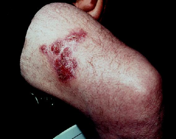
The patient's situation was re-evaluated by D.N.K. in March 1999. Physical examination was notable for a nontender, purplish, multifocal, boggy 6.5 х3.5 cm dermal plaque with overlying crusted scale on the right thigh (Fig. 1), without associated lymphadenopathy. The results of a complete blood count, determination of serum electrolytes, blood urea nitrogen, creatinine and liver enzymes and chest radiography were normal. An infectious disease specialist (J.J.R.) suggested a 4-week trial of doxycycline 100 mg twice daily by mouth for possible Mycobacterium marinum infection; however, the thigh lesion enlarged further over this period.

Fig. 1: Violaceous, crusted dermal plaque of cutaneous blastomycosis, in March 1999, 9 months after initial presentation to the dermatologist.
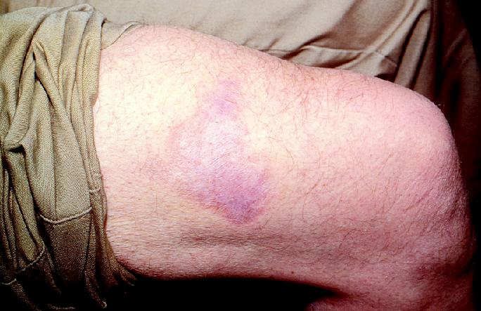
Skin biopsy was repeated at this time and material was sent for pathology and culture. Fungal cultures on Sabouraud dextrose agar at room temperature grew a mould with septate hyphae bearing pear-shaped conidia, whereas those incubated at 37°C grew yeast cells, with broad-based buds, consistent with Blastomyces dermatitidis. Confirmatory testing, for conversion of the mycelial form to the yeast form on special media, was performed by the Mycology Section of the Toronto Public Health Laboratory. The patient was treated with a single daily dose of itraconazole 200 mg, given orally, for 8 weeks. The lesion resolved and did not recur over a 6-month follow-up period (Fig. 2).

Fig. 2: Resolution of cutaneous blastomycosis after completion of 2-month course of itraconazole; atrophic scarring remains.
Comments
Blastomycosis is a chronic pyogranulomatous infection with the dimorphic fungus B. dermatitidis. This organism, along with Histoplasma capsulatum1 and Sporothrix schenkii,2 is one of the few fungi occurring naturally in Canada that can cause invasive disease in immunologically normal hosts.
In North America the region of endemicity for blastomycosis includes the Great Lakes region and the Mississippi and Ohio river valleys of the United States. In Canada most cases have been reported from Ontario and Quebec,3,4,5 with additional cases reported from Manitoba6,7 and Alberta.3 Outbreaks of blastomycosis have been reported in dogs in Saskatchewan,8 which indicates that B. dermatitidis is present there, at least in microfoci. There has been 1 report of blastomycosis in British Columbia, possibly acquired during prior residence in Toronto.9 Two cases of blastomycosis have been reported from Nova Scotia7,10 and 3 previous cases, with limited clinical information, have been reported from New Brunswick.7,11
In the natural environment, B. dermatitidis grows in the filamentous form, as a mould, whereas in the human host, it grows in the pathogenic form, as a unicellular yeast. Although most cases are associated with contact with soil,12 it is somewhat difficult to isolate Blastomyces from soil.9 Conidiation, the process by which potentially infectious conidia are released into the environment, apparently occurs only under specific conditions. Soil with high moisture content, high content of humus (decaying organic matter) and low pH, together with soil disturbance, are required.13,14 These conditions are often met along woodland rivers and would explain the association of both sporadic cases and outbreaks of blastomycosis with forestry, recreational activities along waterways and damp environments in general.3,4,5,6,9,12,13,14
The causative organism of blastomycosis may not survive well in clinical specimens. Hence, if blastomycosis is suspected, the microbiology laboratory should be asked to arrange for immediate culture on appropriate media.15 In addition to the possibility of processing delays or contamination, 2 other explanations may account for the failure to isolate Blastomyces from the initial fungal cultures in the case we have reported. Most strains of Blastomyces grow within 14 days, but growth may be delayed as long as 8 weeks.15 The first set of fungal cultures was discarded after only 14 days, which may have been inadequate for recovery of Blastomyces. Second, the mould may have been mistaken for the common dermatophyte Trichophyton, particularly in the absence of clinical information to suggest a more exotic diagnosis such as blastomycosis.
In most cases of blastomycosis the portal of entry is thought to be the respiratory tract. Infection may result in acute pneumonia but is more likely to be asymptomatic or to result in the insidious development of chronic pneumonia. A few patients experience disseminated disease, skin being the most common site of extrapulmonary involvement, followed by subcutaneous tissue, bones and joints, the genitourinary tract and the central nervous system.12,16
Cutaneous blastomycosis presents as a papulopustular lesion, which may enlarge to form a heaped-up, violaceous nodule with peripheral microabscesses or which may ulcerate to form a friable, plaque-like lesion resembling pyoderma gangrenosum. Like the patient we have described, approximately 10% of patients have isolated cutaneous disease.12 It is generally believed that the initiating event in these cases is asymptomatic pulmonary infection, with subsequent dissemination.16
Primary cutaneous blastomycosis (also known as inoculation blastomycosis) has been described, as a result of either injuries sustained in the pathology or microbiology laboratory or an animal bite. Inoculation blastomycosis is associated with painful lymphadenopathy and lymphangitis, induration and chancre formation, and frequent spontaneous resolution, all features that can be used to clinically differentiate this condition from cutaneous blastomycosis resulting from asymptomatic dissemination.17,18,19,20 The lack of lymphadenopathy, the prolonged, progressive course of the lesion, and the absence of a history of penetrating injury suggest that in this case, the lesion arose from dissemination of an asymptomatic pulmonary infection. However, we cannot definitively exclude the possibility of inoculation blastomycosis from a trivial injury.
The region of blastomycosis endemicity in Canada may be much larger than previously believed and currently reported in the standard reference texts of infectious disease.21,22 Clinicians in most regions of Canada, including Atlantic Canada, should include blastomycosis in the differential diagnosis of unexplained granulomatous pulmonary or cutaneous disease, particularly in patients with a history of woodland, waterway or soil exposure.
Footnotes
This article has been peer reviewed.
Acknowledgement: We thank Michael G. Worthington for reviewing the manuscript.
Competing interests: None declared.
Reprint requests to: Dr. Douglas Keeling, 640 Manawagonish Rd., Saint John NB E2M 3W4; dkeeling@health.nb.ca
References
- 1.Leznoff A, Frank H, Telner P, Rosensweig J, Brandt JL. Histoplasmosis in Montreal during the fall of 1963, with observations on erythema multiforme. CMAJ 1964;91:1154-60. [PMC free article] [PubMed]
- 2.Carr MM, Fielding JC, Sibbald G, Freiberg A. Sporotrichosis of the hand: an urban experience. J Hand Surg [Am] 1995;20:66-70. [DOI] [PubMed]
- 3.Sekhon AS, Bogorus MS, Sims HV. Blastomycosis: report of three cases from Alberta with a review of Canadian cases. Mycopathologia 1979;68:53-63. [DOI] [PubMed]
- 4.Kane J, Righter J, Krajden S, Lester RS. Blastomycosis: a new endemic focus in Canada. CMAJ 1983;129:728-31. [PMC free article] [PubMed]
- 5.Bakerspigel A, Kane J, Schaus D. Isolation of Blastomyces dermatitidis from an earthen floor in southwestern Ontario, Canada. J Clin Microbiol 1986;24:890-1. [DOI] [PMC free article] [PubMed]
- 6.Kepron MW, Schoemperlen CB, Hershfield ES, Zylak CJ, Cherniak RM. North American blastomycosis in central Canada: a review of 36 cases. CMAJ 1972;106:243-6. [PMC free article] [PubMed]
- 7.Nicolle LE, Rotstein C, Bourgault AM, St-Germain G, Garber G, and Canadian Infectious Diseases Society Invasive Fungal Registry. Invasive fungal infections in Canada from 1992 to 1994. Can J Infect Dis 1998;9:347-52. [DOI] [PMC free article] [PubMed]
- 8.Harasen GLG, Randall JW. Canine blastomycosis in southern Saskatchewan. Can Vet J 1986;30:375-8. [PMC free article] [PubMed]
- 9.DiSalvo AF. The ecology of Blastomyces dermatitidis. In: Al-Doory Y, DiSalvo AF, editors. Blastomycosis. New York: Plenum Medical Books; 1992. p. 43-73.
- 10.Gordon CA, Stewart WB. Treatment of North American blastomycosis with amphotericin B. CMAJ 1960;82:471-3. [PMC free article] [PubMed]
- 11.Grandbois J. La blastomycose nord-américaine au Canada. Laval Med 1963;34:714-31. [PubMed]
- 12.Witorsch P, Utz JP. North American blastomycosis: a study of 40 patients. Medicine (Baltimore) 1968;47:169-200. [DOI] [PubMed]
- 13.Klein BS, Vergeront JM, Weeks RJ, Kumar UN, Mathai G, Varkey B, et al. Isolation of Blastomyces dermatitidis in soil associated with a large outbreak of blastomycosis in Wisconsin. N Engl J Med 1986;314:529-34. [DOI] [PubMed]
- 14.Klein BS, Vergeront JM, DiSalvo AF, Kaufman L, Davis JP. Two outbreaks of blastomycosis along rivers in Wisconsin: isolation of Blastomyces dermatitidis from riverbank soil and evidence of its transmission along waterways. Am Rev Respir Dis 1987;136:1333-8. [DOI] [PubMed]
- 15.Larone DH. Thermally dimorphic fungi. In: Medically important fungi: a guide to identification. 3rd ed. Washington: ASM Press; 1995. p. 91-102.
- 16.Sarosi GA, Davies SF. Blastomycosis. Am Rev Respir Dis 1979;120:911-38. [DOI] [PubMed]
- 17.Larson DM, Eckman MR, Alber RL, Goldschmidt VG. Primary cutaneous (inoculation) blastomycosis: an occupational hazard to pathologists. Am J Clin Pathol 1983;79:253-5. [DOI] [PubMed]
- 18.Gnann JW, Bressler GS, Bodet CA, Avent CK. Human blastomycosis after a dog bite. Ann Intern Med 1983;98:48-9. [DOI] [PubMed]
- 19.Shadomy HJ, Utz JP. Deep fungal infections. In: Fitzpatrick TB, Eisen AZ, Wolff K, Freedberg IM, Ansten KF, editors. Dermatology in general medicine. 4th ed. New York: McGraw-Hill; 1993. p. 2479-82.
- 20.Larch WL, Schwartz MD. Accidental inoculation blastomycosis. Cutis 1977;19:334-7. [PubMed]
- 21.Chapman SW. Blastomyces dermatitidis. In: Mandell GL, Bennett JE, Dolin R, editors. Principles and practice of infectious diseases. 5th ed. New York: Churchill Livingston; 2000. p. 2733-46.
- 22.Deepe GS, Klein BS. Blastomyces. In: Gorbach SL, Bartlett JG, Blacklow NR, editors. Infectious diseases. 2nd ed. Philadelphia: WB Saunders; 1998. p. 2365-9.


