

Abstract
Background
On the American continent, cutaneous and mucocutaneous leishmaniasis (CL and MCL) are diseases associated with infection by several species of Leishmania parasites. Pentavalent antimonials remain the first‐choice treatment. There are alternative interventions, but reviewing their effectiveness and safety is important as availability is limited. This is an update of a Cochrane Review first published in 2009.
Objectives
To assess the effects of interventions for all immuno‐competent people who have American cutaneous and mucocutaneous leishmaniasis (ACML).
Search methods
We updated our database searches of the Cochrane Skin Group Specialised Register, CENTRAL, MEDLINE, Embase, LILACS and CINAHL to August 2019. We searched five trials registers.
Selection criteria
Randomised controlled trials (RCTs) assessing either single or combination treatments for ACML in immuno‐competent people, diagnosed by clinical presentation and Leishmania infection confirmed by smear, culture, histology, or polymerase chain reaction on a biopsy specimen. The comparators were either no treatment, placebo only, or another active compound.
Data collection and analysis
We used standard methodological procedures expected by Cochrane. Our key outcomes were the percentage of participants 'cured' at least three months after the end of treatment, adverse effects, and recurrence. We used GRADE to assess evidence certainty for each outcome.
Main results
We included 75 studies (37 were new), totalling 6533 randomised participants with ATL. The studies were mainly conducted in Central and South America at regional hospitals, local healthcare clinics, and research centres. More male participants were included (mean age: roughly 28.9 years (SD: 7.0)). The most common confirmed species were L. braziliensis, L. panamensis, and L. mexicana. The most assessed interventions and comparators were non‐antimonial systemics (particularly oral miltefosine) and antimonials (particularly meglumine antimoniate (MA), which was also a common intervention), respectively.
Three studies included moderate‐to‐severe cases of mucosal leishmaniasis but none included cases with diffuse cutaneous or disseminated CL, considered the severe cutaneous form. Lesions were mainly ulcerative and located in the extremities and limbs. The follow‐up (FU) period ranged from 28 days to 7 years. All studies had high or unclear risk of bias in at least one domain (especially performance bias). None of the studies reported the degree of functional or aesthetic impairment, scarring, or quality of life.
Compared to placebo, at one‐year FU, intramuscular (IM) MA given for 20 days to treat L. braziliensis and L. panamensis infections in ACML may increase the likelihood of complete cure (risk ratio (RR) 4.23, 95% confidence interval (CI) 0.84 to 21.38; 2 RCTs, 157 participants; moderate‐certainty evidence), but may also make little to no difference, since the 95% CI includes the possibility of both increased and reduced healing (cure rates), and IMMA probably increases severe adverse effects such as myalgias and arthralgias (RR 1.51, 95% CI 1.17 to 1.96; 1 RCT, 134 participants; moderate‐certainty evidence). IMMA may make little to no difference to the recurrence risk, but the 95% CI includes the possibility of both increased and reduced risk (RR 1.79, 95% CI 0.17 to 19.26; 1 RCT, 127 participants; low‐certainty evidence).
Compared to placebo, at six‐month FU, oral miltefosine given for 28 days to treat L. mexicana, L. panamensis and L. braziliensis infections in American cutaneous leishmaniasis (ACL) probably improves the likelihood of complete cure (RR 2.25, 95% CI 1.42 to 3.38), and probably increases nausea rates (RR 3.96, 95% CI 1.49 to 10.48) and vomiting (RR 6.92, 95% CI 2.68 to 17.86) (moderate‐certainty evidence). Oral miltefosine may make little to no difference to the recurrence risk (RR 2.97, 95% CI 0.37 to 23.89; low‐certainty evidence), but the 95% CI includes the possibility of both increased and reduced risk (all based on 1 RCT, 133 participants).
Compared to IMMA, at 6 to 12 months FU, oral miltefosine given for 28 days to treat L. braziliensis, L. panamensis, L. guyanensis and L. amazonensis infections in ACML may make little to no difference to the likelihood of complete cure (RR 1.05, 95% CI 0.90 to 1.23; 7 RCTs, 676 participants; low‐certainty evidence). Based on moderate‐certainty evidence (3 RCTs, 464 participants), miltefosine probably increases nausea rates (RR 2.45, 95% CI 1.72 to 3.49) and vomiting (RR 4.76, 95% CI 1.82 to 12.46) compared to IMMA. Recurrence risk was not reported.
For the rest of the key comparisons, recurrence risk was not reported, and risk of adverse events could not be estimated.
Compared to IMMA, at 6 to 12 months FU, oral azithromycin given for 20 to 28 days to treat L. braziliensis infections in ACML probably reduces the likelihood of complete cure (RR 0.51, 95% CI 0.34 to 0.76; 2 RCTs, 93 participants; moderate‐certainty evidence).
Compared to intravenous MA (IVMA) and placebo, at 12 month FU, adding topical imiquimod to IVMA, given for 20 days to treat L. braziliensis, L. guyanensis and L. peruviana infections in ACL probably makes little to no difference to the likelihood of complete cure (RR 1.30, 95% CI 0.95 to 1.80; 1 RCT, 80 participants; moderate‐certainty evidence).
Compared to MA, at 6 months FU, one session of local thermotherapy to treat L. panamensis and L. braziliensis infections in ACL reduces the likelihood of complete cure (RR 0.80, 95% CI 0.68 to 0.95; 1 RCT, 292 participants; high‐certainty evidence).
Compared to IMMA and placebo, at 26 weeks FU, adding oral pentoxifylline to IMMA to treat CL (species not stated) probably makes little to no difference to the likelihood of complete cure (RR 0.86, 95% CI 0.63 to 1.18; 1 RCT, 70 participants; moderate‐certainty evidence).
Authors' conclusions
Evidence certainty was mostly moderate or low, due to methodological shortcomings, which precluded conclusive results. Overall, both IMMA and oral miltefosine probably result in an increase in cure rates, and nausea and vomiting are probably more common with miltefosine than with IMMA.
Future trials should investigate interventions for mucosal leishmaniasis and evaluate recurrence rates of cutaneous leishmaniasis and its progression to mucosal disease.
Plain language summary
What are the benefits and risks of different treatments for American (muco)cutaneous leishmaniasis (a parasitic disease of the skin and mucous membranes)?
Why this question is important
American (muco)cutaneous leishmaniasis (ACML) is a disfiguring disease that affects people in Central and South America. It is caused by parasites that are transmitted to humans by sandflies. Different forms of ACML have different symptoms. People with the cutaneous form develop skin sores that often heal within a few months without treatment, but can leave scars. In people with mucosal or mucocutaneous leishmaniasis, destructive sores develop in the protective lining (mucous membranes) of the nose, mouth and throat.
To compare the effectiveness and risks of the many treatments for ACML, we reviewed evidence from research studies (randomised controlled trials). We looked for information on the proportion of people whose sores had healed three months or more after treatment, unwanted effects, quality of life, re‐appearance of sores, damage associated with the disease and prevention of scarring.
How we identified and assessed the evidence
First, we searched for all relevant studies. We then compared the results, summarised the evidence, and assessed the certainty of the evidence.
What we found
We found 75 studies on 6533 people (approximately 75% male; average age: 29 years).
‐ One study investigated children (2 to 12 years). ‐ Most studies (67) involved people with cutaneous leishmaniasis. ‐ Eight studies investigated people with mucosal or mucocutaneous leishmaniasis. ‐ The parasite Leishmania braziliensis caused the disease in 52 studies. ‐ Studies were conducted at regional hospitals, local clinics, and research centres. ‐ Studies lasted between 28 days and seven years. ‐ Most studies reported their funding source: the US army funded eight studies, industry funded 10, and institutional grants funded 33 (five of these also reported industry funding).
Treatments were mainly compared to a placebo (fake treatment) or meglumine antimoniate (an antimonial).
Here we report this review's main results. We were only able to report the risk of recurrence and side effects for the comparisons of meglumine antimoniate (MA) or miltefosine versus placebo and miltefosine versus MA.
Main results
Antimonials
Compared to placebo, MA may increase chances of complete healing of ACML, but treatment effects vary, so it is possible that it may make little to no difference. MA probably increases the likelihood of pain in the muscles or joints. There may be little to no difference in the risk of developing the disease again, but there is also a possibility of increased or reduced risk due to the wide range of effects seen.
Non‐antimonials
Miltefosine probably improves chances of complete healing of American cutaneous leishmaniasis (ACL) compared to placebo, but there may be little to no difference compared to treatment with MA in ACML. Miltefosine may make little to no difference to the risk of developing ACL again when compared to placebo, but treatment effects on recurrence varied, so it may also increase or decrease the risk. Miltefosine probably increases the likelihood of vomiting or nausea when compared to either placebo or MA in ACML. We do not know the effect on recurrence of miltefosine compared to MA.
Azithromycin probably reduces chances of complete healing of ACML compared to MA.
Imiquimod in combination with MA probably makes little to no difference to the chance of complete healing of ACL compared to MA in combination with placebo.
Physical therapies
Thermotherapy lowers the chance of complete healing of ACL compared to MA.
Immuno‐chemotherapy
Pentoxifylline plus MA probably makes little to no difference to chances of complete healing of ACML compared with MA plus placebo.
No study reported information about damage, prevention of scarring, or quality of life.
What this means
The main findings of this review suggest that:
‐ MA and miltefosine probably increase chances of complete healing; and ‐ vomiting or nausea are probably more common with miltefosine, and joint or muscle ache is probably more common with MA.
The evidence was mostly of moderate certainty, so the true results are likely close to what we found. Evidence was limited by the inclusion of very few people in some studies, and participants or investigators knowing which treatments they were receiving.
How‐up‐to date is this review?
The evidence in this Cochrane Review is current to August 2019.
Summary of findings
Background
Unfamiliar terms are described in the glossary in Table 8.
1. Glossary.
| Term | Definition |
| Aminoglycoside | antibiotic composed of an amino sugar structure with antimicrobial effect through the inhibition of protein synthesis |
| Anfotericin B | polyene antibiotic used for treatment of severe fungal infections and some protozoal infections such as leishmaniasis |
| Antigenic | a molecule that is capable of binding to an antibody or to an antigen receptor on a cell of the immune system, especially one that induces an immune response |
| Anthropic interventions | environmental modifications due to human activities such as deforestation, etc |
| Antimonials | medications composed of antimony salts for intravenous, intramuscular or subcutaneous application |
| Azalide | antibiotic composed of a macrolide ring containing nitrogen that inhibits microorganism growth through the inhibition of protein synthesis |
| Cryotherapy | local use of low temperature (freezing) for treating cutaneous lesions |
| Larynx | the hollow muscular organ forming an air passage from the pharynx to the trachea and holding the vocal cords in humans and other mammals |
| Lymph node | a small organ of the lymphatic system characterised by lymphoid tissue surrounded by a capsule of connective tissue with the function of antigen processing and presenting to organise the adaptive immune response |
| Miltefosine | alkyl‐phosphocholine compound medication used as oral treatment for leishmaniasis |
| Parenteral | route of administering medications other than the digestive tract |
| Paromomycin | an aminoglycoside with antiprotozoal activity |
| Pentamidine | a synthetic amidine derivative medication with antiprotozoal and antifungal agent used as intravenous or intramuscular treatment for leishmaniasis |
| Pentavalent antimony | the pentavalent form of antimony salts used for intravenous, intramuscular or subcutaneous application |
| Pentoxifylline | a methylxanthine derivative compound medication use for treatment of vascular disorders and as an adjuvant for treatment of leishmaniasis due to its capacity of modulate the immune response |
| Pharynx | the membrane‐lined cavity behind the nose and mouth, connecting them to the oesophagus |
| Photodynamic therapies | is a treatment that uses a drug, called a photosensitiser or photosensitising agent, and a particular type of light. When photosensitisers are exposed to a specific wavelength of light, they produce a form of oxygen that kills nearby cells |
| Purine analogue | medication that mimics purine bases essential for DNA synthesis |
| Reservoir | an organism (a vertebrate in the case of leishmaniasis) where an infectious agent lives and multiplies |
| Sympatric circulation | used to refer to the concomitant transmission of more than one parasite species in the same geographical area |
| Ulceration | the process of ulcer formation |
| Vector | is an organism (an arthropod – Diptera, Psycodidade – in the case of leishmaniasis) that does not cause disease itself but which spreads infection by conveying pathogens from one host to another |
| Zoonotic | a disease that can spread from animals to humans |
Description of the condition
Definition
On the American continent, human cutaneous and mucocutaneous leishmaniasis are zoonotic diseases associated with infection by several species of Leishmania parasites (Cupolillo 1994; Cupolillo 2001; Schonian 2010). The parasites are transmitted through the infected bites of sandflies belonging to Lutzomyia genus. Conditional on the immune status and species of Leishmania, American tegumentary leishmaniasis (ATL) clinical types vary from self‐limiting cutaneous lesions to mucocutaneous lesion forms. Cutaneous leishmaniasis is defined as skin involvement characterised by the onset of one or more painless ulcerations heralded or accompanied by local lymph node enlargement. Mucocutaneous leishmaniasis is defined as the upper airway mucosal involvement, characterised by inflammatory and destructive lesions usually affecting the nose, palate, pharynx and rarely the larynx. Cutaneous disease precedes most of the mucosal‐affected cases and is usually presented as a scar. In both diseases there is diversity in parasite, reservoirs and arthropod vector, heterogeneity of transmission cycles, wide variation of clinical presentations, and heterogeneous geographical distribution across the continent (Lainson 1994).
Epidemiology and impact
The leishmaniases are relevant public health problems, classified as neglected tropical diseases by the World Health Organization (WHO), for whom there are not enough preventive, diagnostic and therapeutic solutions. Estimating the disease burden attributable to leishmaniasis has been a challenging task (Bern 2008). However, recent approaches estimated annual cutaneous leishmaniasis incidence between 187,200 to 307,800 cases in the American continent (Alvar 2012). The improvement in surveillance activities, conducted by national control programmes supported by the Pan American Health Organization (PAHO) has been fruitful, and currently data on incident cases are available at subnational levels by most of the endemic countries. In the American continent, cutaneous leishmaniasis is endemic in 18 countries extending from Mexico to Argentina. Numbers of cutaneous and mucocutaneous leishmaniasis, reported to PAHO in 2014 by 16 out of 18 endemic countries, revealed 51,098 total cases and an incidence rate of 19.76 cases per 100,000 inhabitants. Seventy‐five per cent of the cases were reported by Brazil, Colombia and Peru, but the higher incidence rates were observed in Nicaragua and Costa Rica (PAHO 2015). A report on the cutaneous leishmaniasis disease burden demonstrated that Andean Latin America had one of the highest Disability‐Adjusted Life Year (DALYs) in the world, and Nicaragua appears among countries with the highest incidence among the male and female population (Karimkhani 2016).
In spite of the total number of cases and the possibility of underreporting, a huge population could be at risk of acquiring the disease. The heterogeneity of transmission cycles, determined by the behaviour of arthropod vectors and vertebrate reservoirs in their natural environment, plus anthropic interventions where the disease naturally occurs, also makes the scenario for implementing control measures extremely dynamic and challenging (Lainson 1994; Shaw 1988; Yadon 2003). Risk factors for human populations are related to a wide variety of situations such as work exposure, travelling, recreational activities and war operations (Alcais 1997; Alexander 2009; Beyrer 2007; Davies 1997; Davies 2000; Mansueto 2014; Monteiro 2009). Climate and environmental changes could play a role in the distribution of risk across the continent, mainly because of their impact on the vector populations (Perez‐Florez 2016; Peterson 2003). Finally, although there is a general perception that most of the cases are observed among people with lower income living in rural areas, there are no specific studies exploring the association between poverty and the risk of cutaneous leishmaniasis in Latin America.
Aetiology and transmission
The main parasite species, given the burden of disease and the geographical dispersion in the American continent, are Leishmania (Viannia) braziliensis, L. (V.) guyanensis and L.(V.) pananamensis. Other species can be especially relevant for some regions such as L. (Leishmania) mexicana in the peninsula of Yucatán, L.(V.) peruviana in the Andean valleys of Peru, and L. (L.) amazonensis which is associated with the very severe diffuse form of the disease. Parasite diversity is very relevant because of its crucial influence on clinical manifestations and disease severity, the accuracy of diagnostic tests (Navin 1990; Romero 2001b; Romero 2005), and the response to therapeutic measures (Arevalo 2007; Fernandez 2014; Llanos‐Cuentas 2008; Romero 2001a). Sympatric circulation of the parasite should be highlighted as an extra challenge when planning disease‐control activities (Tojal da Silva 2006).
Vector diversity is less well explored for its influence on the human population. However, some data have emerged on the role of a vector's saliva as a potential immunogen or adjuvant for vaccine development (Abdeladhim 2014; Reed 2016), but also as a modulator of the parasite‐host relationship, with impact on clinical findings (Carvalho 2015; Mondragon‐Shem 2015).
Clinical manifestations
Clinical manifestations of cutaneous leishmaniasis offer a wide spectrum of lesions, from a few small non‐ulcerated nodules observed in people in Central America due to L.(L.) infantum (De Lima 2009) to disseminated disease due to L. (V.) braziliensis, with hundreds of ulcerated and non‐ulcerated lesions involving the entire body (Turetz 2002). However, the most common manifestation of cutaneous leishmaniasis is a single ulcerated lesion with elevated borders, usually painless, unless affected by superimposed bacterial infection, localised in one extremity and frequently heralded by satellite lymph node enlargement (Bomfim 2007). Arbitrarily, the term localised cutaneous leishmaniasis is used to denominate a clinical picture of six or fewer cutaneous lesions localised in one or more corporal contiguous segments, but there are no specific studies dedicated to establishing such a cut‐off of six lesions, and the clinical appearance of lesions should be more useful to characterise the condition than simply the number of observed lesions (Costa 1986; Dantas 2014; Turetz 2002). Ampuero 2006 demonstrated that fewer than 5% of over 4000 cases of cutaneous leishmaniasis in Brazil had more than five cutaneous lesions. The diffuse clinical form of the diseases deserves special attention despite its rarity, because of the lack of response to specific treatments (Becker 1999; Hashiguchi 2016; Salaiza‐Suazo 1999). Diffuse disease is almost always caused by L. (L.) amazonensis and rarely by other members of the same subgenus (Hashiguchi 2016). It is characterised by multiple, progressive, non‐ulcerated lesions (Barral 1995).
Mucosal or mucocutaneous disease is a relatively rare entity usually associated with infection by parasites belonging to the Viannia subgenus (Handler 2015). L. (V.) braziliensis is by far the most common agent isolated from patients with mucosal involvement (Marsden 1994), but L. (V.) guyanensis (Prestes 2015; Santrich 1990) and L.(V.) panamensis (Osorio 1998) have also been reported to cause this clinical form. Mucosal diseases are frequently observed in patients who have been affected by localised cutaneous leishmaniasis within the previous two years and more rarely in patients with active cutaneous ulcers or without any skin disease (Zajtchuk 1989). Patients affected by the disseminated cutaneous form of the disease should be screened carefully for mucosal involvement, because it is known that they have a higher probability of concomitant mucosal disease (Turetz 2002). Disease severity is variable and could depend in part on host characteristics, but mostly on the duration of the disease. In the past, it was common to observe patients with extremely advanced destructive lesions. Nowadays, in some endemic areas with improving access to health services, such destructive lesions are diminishing, and mild cases are detected through endoscopic evaluation of the upper airway (Lessa 2012). The mucosal involvement affects mainly the mucosal surfaces of the nose, with or without septal perforation, and oral surfaces as well. Larynx lesions are rare but represent a special challenge because of the possibility of airway obstruction and long‐term sequela.
Diagnosis
Diagnosis of cutaneous and mucocutaneous leishmaniasis is based on parasitological conventional methods, including direct parasite examination in smears obtained from the lesions or histopathological sections and parasite culture (Boggild 2008; Boggild 2010; Navin 1990; Weigle 1987). There are currently no rapid tests commercially available for point‐of‐care diagnosis of either disease. Direct parasite examination looking for amastigotes could be a hard task in areas affected by L. (V.) braziliensis, in contrast with areas affected by L. (V.) guyanensis (Romero 2001b) or L. (L.) mexicana (Navin 1990), where the parasite is easily visible. Culture‐proven cases are essential for surveillance and to accumulate data on the geographical distribution of each parasite species. Furthermore, the parasite species identification process is crucial in the formulation of treatment policies, taking into consideration the relationship between parasite species and the therapeutic response, as previously mentioned. However, in most of the scenarios where transmission occurs there is no access to parasitological diagnosis or parasite isolation. Molecular methods are very promising, including the possibility of parasite species identification (Fraga 2012; Gomes 2015; Graca 2012), but so far there is no point‐of‐care affordable diagnostic instrument to perform the detection of parasite DNA. The Montenegro (Leishmanin) skin test for detection of delayed cellular immune response is specific but does not distinguish between current or past infection (Weigle 1991). However, putting together the history of exposure to transmission area, the clinical findings, and the result of Montenegro skin test, it is possible to correctly classify more patients (Rojas 2002; Weigle 1993). Unfortunately, the leishmanin skin test is currently unavailable. All the available diagnostic tests generally had lower sensitivity in patients affected by mucosal disease (PAHO 2019b). Thus, all the tests should be applied concomitantly in those patients in order to improve sensitivity. Finally, there is no consensus on the use of antibody detection through serological tests for diagnosis of tegumentary leishmaniasis, and no stringent validation studies have been developed for this purpose.
Description of the intervention
Localised cutaneous leishmaniasis has been considered a self‐healing disease. However, in contrast with the Old World scenario where no treatment has been an option, in the New World most of the cases receive specific treatment because of the prolonged period of the self‐healing process, and consequent disfiguring scars. The risk of mucosal disease has also been historically considered as a justification for systemic treatment. The issue of self‐healing in the American continent has recently been revised, concluding that there is an advantage in administering specific treatment to those suffering from cutaneous leishmaniasis and therefore avoiding the use of placebo in clinical trials (Cota 2016). Disseminated cutaneous leishmaniasis has an aggressive pattern with higher mucosal involvement, and is not considered a self‐healing condition. Diffuse disease is also a progressive form which improves temporarily with specific treatment, with most of the patients remaining exposed to several drugs without full remission; when a clinical cure is achieved, it is not prolonged. Mucosal disease, on the other hand, is progressive and destructive, but heals with proper treatment, with few exceptions (Marsden 1998). Data on the impact of leishmaniasis on quality of life have been scarce in Latin America and it would be desirable in the near future to construct related quality‐of‐life endpoints for clinical trials (Toledo 2013).
The choice of treatments for leishmaniasis depends on many factors such as efficacy, treatment schedule, toxicity, costs, and cultural issues about acceptability for the target population. There is currently no effective and low‐cost oral treatment available for American cutaneous or mucocutaneous leishmaniasis. The available treatments are characterised by their moderate efficacy, frequent adverse events, and relevant concerns for their safety during pregnancy and childbearing age. Most of them are also expensive drugs used in long‐term schedules requiring special care for administration and monitoring. Antimonials, e.g. sodium stibogluconate and meglumine antimoniate, the oldest drugs available, were first used at the beginning of the 20th century and are still considered first‐line treatments against most forms of leishmaniasis, and they have also been used as a reference to compare the efficacy of other potential treatments (Biagi 1953; Marsden 1979; PAHO 2015). Antimonials; amphotericin B; pentamidine isethionate (PI); and more recently, the oral drug miltefosine constitute the therapeutic armamentarium for systemic treatment of American tegumentary leishmaniasis. Neither oral agents such as ketoconazole, itraconazole, and fluconazole; the purine analogue allopurinol; nor the aminoglycoside aminosidine sulphate for systemic use have been extensively studied in the American continent or registered for treating leishmaniasis. The azalide azithromycin has been scarcely tested in people with CL. Topical formulations, such as the aminoglycoside paromomycin, amphotericin B cream, or imiquimod formulations, as well as local treatment modalities such as cryotherapy or thermotherapy, have also been tried for localised American cutaneous leishmaniasis, but again, none has been incorporated as part of the recommended treatments. Intralesional meglumine antimoniate (N‐methylglucamine) was recently incorporated into the armamentarium against CL in the American continent (PAHO 2018). Finally, vaccines, immunotherapy with antigenic compounds or with cytokines, as well as immunomodulatory drugs deserve attention, mainly as part of combined therapeutic schemes including antimonials or other antileishmanial drugs. Table 9 shows the profile of the currently‐available interventions that reached at least the clinical phase of development, although not necessarily registered for use against leishmaniasis.
2. Interventions for American cutaneous and mucocutaneous leishmaniasis.
| Type of interventions | Pharmacological class of interventions | Intervention | Current clinical applicability | Administration route | Main toxicity | Pregnancy safety | Key references |
| 1. Antimonials | Pentavalent antimonial | N‐methyl glucamine antimoniate (MA) | CL, MCL | IV; IM; IL | Common: QT prolongation, abnormal liver and pancreatic enzymes, myalgias and infusion‐related fever. Rare: severe pancreatitis and hepatic failure | Possible harm to the fetus; insufficient controlled studies in humans and animals. Experimental studies with pentavalents antimonials in pregnant rats showed an increase in fetal skeletal malformations. May be toxic to the embryo even in the absence of signs of maternal toxicity | (Oliveira 2011; Soto 2013; Fontenele e Silva 2013) |
| Sodium stibogluconate (SSG) | CL, ML | IV; IM | Common: QT prolongation, abnormal liver and pancreatic enzymes, myalgias and infusion related fever. Rare: severe pancreatitis and hepatic failure. | (Oliveira 2011; Fontenele e Silva 2013) | |||
| 2. Non‐antimonial systemic treatments | Antifungals: Macrolide polyene antibiotic |
Amphotericin B | CL, ML | IV | Common: infusion‐related fever and phlebitis, hypokalaemia, renal dysfunction and anaemia | Remote possibility of fetal harm; animal studies showed no risk to the fetus. Studies in people with visceral leishmaniasis show no adverse effects on the fetus or abortions when it was used during the first trimester of pregnancy | (Mishra 2007; Gallis 1990; Fontenele e Silva 2013) |
| Liposomal amphotericin B | CL, ML | IV | Common: fever, nausea, phlebitis, dorsal pain, vomiting, headache and mild renal toxicity | (Wortmann 2010; Fontenele e Silva 2013; Machado 2015). | |||
| Antifungals: Azoles |
Ketoconazole | CL | Oral | Common: nausea, abdominal pain, headache, fever, dizziness, abnormal liver enzymes, rash. Rare: severe liver injury and adrenal gland dysfunction | Ketoconazole can compromise early pregnancy due to inhibiting progesterone synthesis in the ovary | (Saenz 1990; Navin 1992; Cummings 1997; FDA 2013) | |
| Itraconazole | CL | Oral | Common: nausea, headache and abnormal liver enzymes Rare: severe hepatitis |
Animal reproduction studies have shown an adverse effect on the fetus, but there are no adequate and well‐controlled studies in humans | (Consigli 2006; FDA 2014) | ||
| Fluconazole | CL | Oral | Common: headache; abnormal liver enzymes. Rare: severe neurologic toxicity. | High‐dose fluconazole (400‐800 mg/day) during most or all of the first trimester has been associated to birth defects in infants. | (Sousa 2011; Neto 2006; Alves Noroes 2015; FDA 2011; FDA 2016) | ||
| Purine analogue | Allopurinol | CL | Oral | Common: headache and epigastric pain Rare: rash and haematologic abnormalities | Caution recommendation in the first trimester. Possible teratogenicity | (Vélez 1997; Hoeltzenbein 2013) | |
| Alkylphosphocholine analogue | Miltefosine | CL, ML | Oral | Common: reversible gastrointestinal disturbances, renal toxicity. | Teratogenic, contraindicated in pregnancy | (Sundar 2006; Fontenele e Silva 2013). | |
| Aromatic diamidine | Pentamidine isethionate | CL; ML | IV; IM | Common: pain at the site of injection, nausea, vomiting, headache, burning sensation and hypotension Rare: skin eruptions, abnormal liver enzymes, renal dysfunction, hypoglycaemia and diabetes mellitus |
Possible harm to the fetus; insufficient controlled studies in humans and animals | (Sands 1985; Neves 2011; Fontenele e Silva 2013). | |
| Aminoglycosides | Aminosidine sulphate | CL; ML | IV | Ototoxicity and renal dysfunction | As for all the aminoglycosides there is evidence of human fetal risk. | (Kim 2009) | |
| 3. Non‐antimonial topical or intralesional therapies | Aminoglycosides | Paromomycin sulphate | CL | Topical | Common: local pain, burning sensation and pruritus. Very rare: ototoxicity | As for all the aminoglycosides there is evidence of human fetal risk. | (Kim 2009) |
| 4. Physical therapies | Physical therapies | Thermotherapy | CL | Local | Common: moderate to severe local cellulites and burns; pain at the lesion area 4 days after the initiation of treatment | Safe | (Lobo 2006; Lopez 2012) |
| Photodynamic therapy | CL | Local | Common: Local pain | Studies in diseases other than leishmaniasis indicate that this local therapy could be safe during pregnancy | (Enk 2015; Yang 2012) | ||
| 5. Immuno‐chemotherapy | Immunomodulatory agent: methyl xanthine derivative | Pentoxifylline | CL; ML | Oral | Common: nausea, arthralgia, dizziness, abdominal pain and diarrhoea | There are no adequate and well‐controlled studies in pregnant women. Pentoxifylline should be used during pregnancy only if the potential benefit justifies the potential risk to the fetus | (Machado 2007) |
| Immunomodulatory agent: TLR stimulator | Imiquimod | CL | Topical | Common: Moderate pruritus and burning sensation, erythema | Studies in diseases other than leishmaniasis have reported no adverse local effect nor adverse fetal outcomes or fetal and neonatal abnormalities | (Edwards 2000; Miranda‐Verástegui 2005; Arevalo 2007) | |
| Immunomodulatory agent: cytokine | Granulocyte‐macrophage colony‐stimulating factor (GM‐CSF) | CL | Topical | In 1 study no systemic side effects or contact allergic reactions were reported | No data available | (Almeida 2005) |
CL: cutaneous; MCL: mucocutaneous; IV: intravenous; IM: intramuscular; IL: intralesional.
Based on the immunopathogenesis of leishmaniasis, treatment with antimonial drugs has been combined with cytokines and free radicals, such as interferon‐gamma (IFN‐γ) or granulocyte macrophage colony‐stimulating factor (GM‐CSF), and nitric oxide (NO) patch, respectively (Almeida 2005; Arana 1994; Lopez‐Jaramillo 2010). The immunomodulatory agent pentoxifylline has also been used in combination with meglumine antimoniate, pursuing the reduction of the dysregulated inflammatory response in mucosal leishmaniasis patients (Machado 2007). No current recommendation for routine use has been made for such combinations. Finally, there is currently no licensed vaccine against human leishmaniasis. Vaccines for the prophylaxis of leishmaniasis are divided into first‐ and second‐generation vaccines (Alvar 2013). The first‐generation vaccines were composed of killed and attenuated Leishmania parasites. Vaccines with killed Leishmania and attempts to use killed parasites together with the BCG vaccine did not confer significant protection against human leishmaniasis (Sharifi 1998). Second‐generation vaccines are based on the subunit/recombinant protein using a range of adjuvants to augment the immunogenicity of the selected antigens (Alexander 2009; Beaumier 2013; Duthie 2012; Llanos‐Cuentas 2010). Another approach has been to use the antigenic salivary proteins of sandfly vector, which has delivered some promising results (Abdeladhim 2014; Reed 2016).
How the intervention might work
Once injected, pentavalent antimony (Sb+5) is further reduced to trivalent antimony (Sb+3) that will destroy the parasite. Both forms of antimonials (Sb+5 and Sb+3) have been known to destroy Leishmania species by DNA fragmentation, which indicates a role in apoptosis (Lee 2002; Sereno 2001; Sudhandiran 2003). The antileishmanial activity of amphotericin B is attributed to the interaction with ergosterol in the cell membrane, which increases the cell permeability, causing cell death (Ramos 1996). Allopurinol was considered a promising candidate for treatment of leishmaniasis, since the inhibition of purine anabolism in Leishmania could inhibit its growth. Miltefosine could act as an antileishmanial drug through the modulation of different components of the cell, resulting in apoptosis (Verma 2004). Aminosidine sulphate (Paromomycin sulphate) causes interference in protein synthesis in bacterial species (Tracy 2001) and their antileishmanial effect could be related to the same mechanism (Sundar 2008). Fluconazole and itraconazole are fungistatic drugs, which inhibit the synthesis of ergosterol and have been shown to impede the growth of different in vitro species of Leishmania (Oliveira 2015). An in vitro study revealed that itraconazole induced the collapse of the Leishmania mitochondrial membrane potential, which was consistent with mitochondrial swelling and disorganisation and rupture of mitochondrial membranes (De Macedo‐Silva 2013). Pentamidine has an effect on the parasite's genome by hindering reproduction and transcription at the mitochondrial level (Mishra 2007). Berman and Sacks demonstrated several pathogenic Leishmania species to be thermo‐sensitive from 37 ºC to 39 ºC in vitro (Berman 1981; Sacks 1983). Photodynamic therapy is based on the principle of exposure to a relevant light source alongside oxygen, which leads to formation of reactive oxygen species followed by the destruction of the target cells. Also, the reaction caused by the photodynamic therapy may stimulate immune reactions, further enhancing the potential of the host against infections (Evangelou 2011). Pentoxifylline hinders the production of tumour necrosis factor‐alpha and interferon‐gamma (IFN‐γ), while it prompts the creation of Th2‐like (T‐helper 2) cytokines, thereby stopping the Th1‐mediated inflammatory and autoimmune responses (Brito 2014). Imiquimod is an immunomodulator that stimulates Toll‐Like‐Receptors (TLR) 7 and 8 on antigen‐presenting cells to facilitate the creation of Th1 type cytokines, enhancing macrophage activity against Leishmania parasites. It has also been observed to have an antileishmanial effect which does not depend on the stimulation of TLR (Buates 1999). The interventions using combinations of IFN‐γ and nitric oxide (NO) are based on the principle of enhancing the Th1 cytokine profile of the host immune response, which could improve the leishmanicidal activity of macrophages (Arana 1994; Lopez‐Jaramillo 2010). The granulocyte macrophage colony‐stimulating factor (GM‐CSF) is a multipotential growth factor for marrow stem cells. In vitro, GM‐CSF has stimulated macrophages to destroy Leishmania (Badaro 2001).
Why it is important to do this review
This systematic review has focused on addressing the effects of the existing treatments for American cutaneous and mucocutaneous leishmaniasis. Treatments for Old World cutaneous leishmaniasis (Heras‐Mosteiro 2017) and prevention measures (González 2015) for all types of cutaneous leishmaniasis have been addressed in separate Cochrane Reviews. This is an update of the review Interventions for American cutaneous and mucocutaneous leishmaniasis (González 2009).
Control of cutaneous leishmaniasis currently depends on case management, including early detection and rapid treatment (Modabber 2007). Global health development policies are mainly focused on new and innovative tools to tackle neglected tropical diseases (NTDs). However, the WHO also prioritises the delivery of currently‐available drugs and existing resources that reduce mortality, morbidity, and disability of NTDs in low‐income countries (Savioli 2006). In accordance with the priorities set by the WHO, we therefore consider that in order to improve current methods of disease management it is important to know the evidence for the efficacy of the different treatment strategies, as well as for their safety and cost effectiveness.
Pentavalent antimonials remain the first‐choice treatment for CL in most American countries, but most of the evidence for using either the first‐choice or the alternative treatments is weak (Reveiz 2013; PAHO 2015). Since 2010, the debate on improving the quality of clinical trials in cutaneous leishmaniasis has been raised and specific recommendations have been published (González 2010; Olliaro 2013). Recent PAHO documents emphatically recommend the development of clinical trials for evaluation of alternatives currently in use in Latin America. (González 2010; PAHO 2015; Reveiz 2013; WHO 2010). However, the most neglected and challenging tegumentary and mucosal forms of leishmaniasis remain without any guideline for improving the methodology of clinical trials (Amato 2007). Alternative treatment regimens include miltefosine, pentamidine isethionate, amphotericin B, antifungal agents, paromomycin, and local treatments. Other treatments such as immunotherapy and thermotherapy have also been tested. The limited number of available drugs, the high levels of side effects of most of them, the difficulties in administration and ongoing resistance highlight the need for reviewing the current evidence on efficacy and adverse effects of the available treatments for American cutaneous and mucocutaneous leishmaniasis. Given that a wide range of systemic and local treatments are currently being used or are under clinical development on a global scale, probably influenced by the increasing international travel trend, their effectiveness and safety needs to be well established.
Objectives
Main objective: To assess the effects of interventions for all immuno‐competent people who have American cutaneous and mucocutaneous leishmaniasis (ACML).
Secondary objective: to ascertain whether response to treatment is species‐dependent or associated with their geographical distribution.
Methods
Criteria for considering studies for this review
Types of studies
Randomised controlled clinical trials (RCTs). We did not consider cross‐over trials in this review because they are an inappropriate design for treatments which have the potential to cure an infectious disease.
Types of participants
All immuno‐competent people who have American cutaneous leishmaniasis or mucocutaneous leishmaniasis, or both, diagnosed by clinical presentation, and Leishmania infection confirmed by smear, histopathology, polymerase chain reaction (PCR) analysis or culture of lesions. If Leishmania parasites could not be seen, diagnosis was based on a clinical presentation and at least two of the following criteria: suggestive histopathology, serologic reaction, positive Leishmanin Montenegro skin test, or negative tests for other diseases that compromise the oral or nasal mucous membranes, especially leprosy and paracoccidioidomycosis.
Types of interventions
The interventions were either single therapy or combination therapy. The comparators were either no treatment, placebo only, or another active compound.
1. Antimonials (intramuscularly, intravenously and intralesionally)
1.1 Meglumine antimoniate (Glucantime, SB N‐methylglucamine) 1.2 Stibogluconate (Pentostam and others)
2. Non‐antimonial systemic treatments
2.1 Antifungals 2.2 Allopurinol 2.3 Miltefosine 2.4 Aminosidine sulphate 2.5 Pentamidine isethionate 2.6 Azithromycin 2.7 Amphotericin B plus oral rehydration solution versus amphotericin B plus normal saline solution
3. Non‐antimonial topical or intralesional therapies
3.1 Paromomycin (aminosidine) 3.2 Aminoglycosides 3.3 Amphotericin B 3.4 Nitric oxide patch (NOP) 3.5 Imiquimod
4. Physical therapies
4.1 Thermotherapy 4.2 Cryotherapy
5. Immuno‐chemotherapy
5.1 Vaccines 5.2 Bacillus Calmette‐Guerin (BCG) 5.3 Pentoxifylline 5.4 Granulocyte macrophage colony‐stimulating factor (GM‐CSF) 5.5 Interferon‐gamma (IFN‐γ)
Since this review aims to include only treatments for cutaneous leishmaniasis, we excluded studies where only vaccines were given (i.e. not in combination with other treatment), as these studies are likely to be aimed at preventing cutaneous leishmaniasis.
Types of outcome measures
We included studies that reported at least one of the outcomes listed below. Studies that did not report any of the outcomes of interest were therefore excluded.
Primary outcomes
Percentage of participants 'cured' at least three months after the end of treatment.
By 'cured', we mean that all inflammatory signs have disappeared (either skin oedema or hardening, or both), and that scarring or epithelialisation has occurred in ulcerative lesions. Lesions were considered not to be healed if there was no re‐epithelialised skin, or inflammatory signs remain after follow‐up.
We reported all time points that addressed cure at three months after the end of treatment and beyond.
Adverse effects
Secondary outcomes
Recurrence: duration of remission and/or percentage of people with treated lesions that recur within six months, one, two and three years.
Degree of functional and aesthetic impairment or prevention of scarring, or both.
Quality of life.
Tertiary outcomes
Speed of healing (i.e. average time from start of treatment to cure).
Change in isolation or PCR of Leishmania and emergence of resistance ("defined as a decline in the efficacy of a drug against a population of parasites previously susceptible to that compound. The definition assumes that the original susceptibility of the population is known, which is not always the case for Leishmania)" (Ponte‐Sucre 2003)).
Only microbiological or histopathological cure of skin lesions.
Development of cell‐mediated immunity (i.e. difference in the size of leishmanin skin test reaction (i.e. difference in the diameter of Montenegro skin test reaction before and after treatment).
All‐cause mortality.
To assess the results we primarily focused on end of follow‐up time points.
Search methods for identification of studies
We aimed to identify all relevant RCTs, regardless of language or publication status (published, unpublished, in press, or in progress).
Electronic searches
For this update we revised all our search strategies in line with current Cochrane Skin practices. Details of the previous search strategies are available in González 2004 and González 2009. The Cochrane Skin Information Specialist searched the following databases up to 27 August 2019:
the Cochrane Skin Group Specialised Register, using the search strategy in Appendix 1;
the Cochrane Central Register of Controlled Trials (CENTRAL) 2019, Issue 8, in the Cochrane Library, using the strategy in Appendix 2;
MEDLINE via Ovid (from 1946), using the strategy in Appendix 3;
Embase via Ovid (from 1974), using the strategy in Appendix 4;
LILACS (Latin American and Caribbean Health Science Information database, from 1982), using the strategy in Appendix 5;
CINAHL via EBSCO (Cumulative Index to Nursing and Allied Health Literature, from 1981), using the strategy in Appendix 6.
MP searched the American College of Physicians (ACP) journal club from June 2007 to April 2008 for the previous review. Since May 2008, ACP Journal Club has been published as a monthly feature of Annals of Internal Medicine. For this update, MP searched the Annals of Internal Medicine from June 2007 to April 2016 using the search terms 'cutaneous and leishmaniasis'.
Trials registers
MP searched the following trials registers on 29 August 2019, using the terms 'leishmania' or 'leishmaniasis':
the ISRCTN registry (www.controlled-trials.com);
the US National Institutes of Health Ongoing Trials Register ClinicalTrials.gov (www.clinicaltrials.gov);
the Australian New Zealand Clinical Trials Registry (www.anzctr.org.au);
the World Health Organization International Clinical Trials Registry Platform (apps.who.int/trialsearch/);
the EU Clinical Trials Register (www.clinicaltrialsregister.eu/).
Searching other resources
References from published studies
We checked the bibliographies of included studies for further references to relevant trials and systematic reviews.
Adverse effects
The Cochrane Skin Information Specialist searched MEDLINE (Ovid) from 1950 to 23 October 2018 for adverse or side effects of interventions used for the treatment of cutaneous and mucocutaneous leishmaniasis (see strategy in Appendix 7). Although we recognise that searching one database with the terms in Appendix 7 does not constitute a comprehensive search for adverse effects (adverse‐effects searching has moved on since this strategy was developed in 2007/8), we have used the same search terms as previously, as a pragmatic approach in terms of managing the workload around sifting and discussing adverse effects in relation to leishmaniasis.
Data collection and analysis
Some parts of the methods section of this review uses text that was originally published in the Cochrane Handbook for Systematic Reviews of Interventions (Higgins 2011).
Selection of studies
At least two review authors (MP, LR) independently checked titles and abstracts identified from the searches, using the web‐based software platform Covidence. If it was clear that the study did not refer to a RCT on American cutaneous and mucocutaneous leishmaniasis, we excluded it. If it was unclear, then we obtained the full‐text study for independent assessment by two review authors (MP, LR). We decided which trials fitted the inclusion criteria, resolving any disagreements by discussion and consensus. We listed the excluded studies and stated the reasons for exclusion in the Characteristics of excluded studies.
Data extraction and management
At least two of the review authors (from VE, KO, MP, JRR) independently carried out the data extraction by using a predesigned data extraction form which was previously piloted. We extracted reported data pertaining to cure rates for all evaluated drugs, paying attention particularly to the doses and therapeutic frequencies. We extracted the following items:
Study ID, country, study design, study setting, study period;
Sample size, unit of randomisation, withdrawals, disease severity, baseline data;
Type of interventions, duration of intervention, co‐interventions;
Outcome data;
Ethical approval, informed consent, conflicts of interest and funding sources.
We resolved any disagreements by discussion or with referral to a third review author (LR). We obtained missing data from trial authors whenever possible.
Assessment of risk of bias in included studies
At least two of the review authors (from VE, KO, MP, JRR) independently assessed risks of bias using the Cochrane tool for assessing risk of bias as described in the Cochrane Handbook for Systematic Reviews of Interventions (Higgins 2011). We resolved any disagreements by discussion or with referral to a third review author (LR).
We rated the following domains for each of the included studies to assess the degree to which:
the allocation sequence was adequately generated ('sequence generation');
the allocation was adequately concealed ('allocation concealment');
knowledge of the allocated interventions was adequately prevented during the study ('blinding');
incomplete outcome data were adequately addressed;
study reports were free of suggestion of selective outcome reporting; and
the study was apparently free of other sources of bias that could put it at high risk of bias. Other sources of bias included the calculation of the sample size, the reporting of the Leishmania species, and baseline comparability among intervention groups.
We also discussed the following issues under Description of studies in the Results section:
calculation of sample size;
inclusion and exclusion criteria clearly defined;
reporting of Leishmania species involved;
time of follow‐up;
baseline comparability of severity of infection, age, sex and duration of complaint;
conflicts of interest;
funding sources.
Each domain was allocated to one of three possible categories: low, high, and unclear risk of bias (where the risk of bias was uncertain or unknown). See Characteristics of included studies for more details.
Measures of treatment effect
We expressed the results as risk ratios (RRs) and 95% confidence intervals (CIs) for dichotomous outcomes. The percentage of lesions 'cured' at least three months after the end of treatment was the primary outcome measure if available.
For continuous outcomes we have calculated mean differences (MDs) and their 95% CIs.
Unit of analysis issues
We included clinical trials of parallel‐group designs in which the individual participants were the allocation unit and the unit for assessing outcomes. The approach we followed for three‐arm trials was to compare the arms in pairs (A vs B, B vs C, and A vs C), ensuring not to double count the number of participants from the intervention groups in the trials.
Dealing with missing data
For each study, we took all participants that were randomised into account when adding the data to our tables, and assumed that missing data were treatment failures for our efficacy outcomes. Where an intention‐to‐treat (ITT) approach was not stated, we used the numbers originally randomised to the groups to calculate the effect estimates.
Assessment of heterogeneity
We assessed clinical heterogeneity by examining the characteristics of the studies, the similarities between the types of participants, the interventions, the comparisons, and the outcomes, as specified in the criteria for included studies. We analysed statistical heterogeneity using a Chi2 test (on one degree of freedom, with a significance level of 0.05) (Higgins 2003). To assess the consistency of the study results, we obtained the I2 statistic, which measures the proportion of total variation across studies that is due to heterogeneity rather than to chance. I2 values lie between 0% and 100%. A value of 0% indicates no observed heterogeneity, and larger values show increasing heterogeneity.
Assessment of reporting biases
In this review, the low number of studies evaluating similar interventions and comparisons did not permit an assessment of publication bias. In future updates, if a sufficient number of trials (10 or more) assessing similar effects are identified for inclusion in the review, we will assess publication bias according to the recommendations on testing for funnel plot asymmetry (Egger 1997), as described in section 13.3.5.2 of the Cochrane Handbook for Systematic Reviews of Interventions (Page 2019). If we detect asymmetry we will try to assess other possible causes and will explore them in the Discussion if appropriate.
Data synthesis
One review author (JRR) analysed the data in Review Manager 5 (RevMan) (Review Manager 2014) and reported them as specified in Chapter 10 of the Cochrane Handbook for Systematic Reviews of Interventions (Deeks 2019). We carried out data synthesis of results from different studies only if we were able to identify at least two studies investigating similar treatments, a follow‐up of at least three months after treatment cessation only for the primary outcome, and reporting data amenable to pooling. We considered that data on cure prior to three months after cessation of treatment showed only a short‐term benefit, and although described, were excluded from statistical analysis. Such data were reported as a narrative summary, where appropriate. We used a random‐effects model to combine the results of individual studies in this review.
Subgroup analysis and investigation of heterogeneity
In view of the limited number of included studies covering any one intervention, we did not conduct any of the subgroup analyses that we had originally planned: Leishmania species, location and severity of infection, geographical setting, diagnostic techniques, type of treatment (topical, systemic, or combination), and relapse or re‐infection.
Following clinical recommendations we also considered age as a subgroup analysis, because therapeutic failure and relapses are frequent in children with cutaneous leishmaniasis, especially with pentavalent antimonials. This may be due to differences in pharmacokinetics. Hence, with hindsight it is important to report results separately for children and adults, as this variable may influence the results. Where data permitted, we presented separate analyses for different age groups.
Sensitivity analysis
We planned to carry out a sensitivity analysis by excluding studies at high risk of bias, but we were unable to do this due to the limited number of studies we could pool. Furthermore, the reporting of the methodology of most of the studies was very poor, with registry information or study protocols unavailable. Most studies therefore had a high risk of bias in at least one domain.
Summary of findings and assessment of the certainty of the evidence
We used the GRADE approach to rate the certainty of the evidence (for each outcome) and strength of recommendations (Guyatt 2008). For better understanding of the review, we have highlighted the GRADE assessments in 'Summary of findings' tables of key comparisons and outcomes. The certainty of evidence can be high, moderate, low, or very low, based on the assessment of five domains: study limitations (risk of bias), imprecision, indirectness, inconsistency and publication bias. Each domain could be downgraded by one (for serious concerns) or by two levels (for very serious concerns).
We have created 'Summary of findings' tables for the following comparisons, which we believe are most relevant nowadays for clinicians:
Intramuscular meglumine antimoniate (IMMA) vs placebo
oral miltefosine vs placebo
oral miltefosine vs meglumine antimoniate
azithromycin vs meglumine antimoniate
topical imiquimod + Intravenous (IV) meglumine antimoniate (MA) vs placebo + IVMA
thermotherapy vs meglumine antimoniate
pentoxifylline + IMMA vs IMMA + placebo.
We included our two primary outcomes (complete cure and adverse effects) as well as one secondary outcome (recurrence) in all tables.
Results
Description of studies
Results of the search
For this update we ran searches to August 2019. As shown in our study flow diagram (Figure 1), we retrieved 1696 records from the Electronic searches (102 from Cochrane Skin Specialised Register, 331 from CENTRAL, 399 from MEDLINE, 589 from Embase, 119 CINAHL, and 156 from LILACS). We retrieved 106 trials from five trial registers. Ten records were identified through hand‐searches, including two that were included in a previous non‐Cochrane review (Reveiz 2013). We therefore had an overall total of 1812 records. After removal of duplicates, we had 522 records. We excluded 427 records based on titles and abstracts. We obtained the full text of the remaining 95 records, of which 29 were excluded (see Characteristics of excluded studies), 10 studies are awaiting classification (see Characteristics of studies awaiting classification), and 12 are ongoing trials (see Characteristics of ongoing studies). We included 37 new studies, along with 38 studies from the previous review, which brings the number of included studies in this update to 75 (6533 participants) (see Characteristics of included studies).
1.

Study flow diagram.
Two masters' theses (Lyra 2013; Saheki 2013), and two published articles (Lyra 2016; Ribeiro 2014) included the same study (registration number NCT01301924), and were grouped under the primary report (Saheki 2017). Two published articles (López 2012; López 2013) included the same study (registration number NCT00471705), and were grouped under the primary report (Vélez 2010). One trial published in clinicaltrials.gov with results (NCT01790659) was grouped under the primary report (Sosa 2019). Soto 2016 described two studies that we considered to be two separated studies (Soto 2016a; Soto 2016b). Of note, Soto 2013. as well as Soto 2016a and Soto 2016b, shared the same registration number NCT01300975 but were treated as separate studies after contact with the author of the studies, who claimed that the participants belonged to two different groups and were recruited at two different time points.
Included studies
Design
All of the studies were randomised clinical trials, of which four were pilot studies (Arévalo 2007; Brito 2014; Guzman‐Rivero 2014; Soto 2002). Ten studies were phase II (Gadelha 2018; Garcia 2014; Llanos‐Cuentas 1997; López 2018; Machado 2018; NCT01011309; Sosa 2013; Soto 2002; Soto 2016a; Soto 2016b), one was a phase II/III (Chrusciak‐Talhari 2011), and six were phase III (Ferreira 2014; Saheki 2017; Sosa 2019; Soto 1998; Vélez 1997; Vélez 2010) (see Characteristics of included studies table).
Setting
Twenty‐five studies were performed in Brazil, nine in Peru, six in Bolivia, five in Panama, four in Guatemala, three in Ecuador, two in Venezuela, two in Argentina, one in Honduras, and one in Suriname. We found 14 studies in Colombia, of which two were performed also in Bolivia and in Guatemala. There were also two RCTs that were conducted in North America (USA) and one in Edinburgh (UK), which recruited active‐duty military personnel who had contracted leishmaniasis in endemic areas when deployed abroad (mainly in Panama, Belize and Brazil).
Of the 75 studies, 12 did not describe the setting, 17 recruited participants from health or medical care centres (mainly from Heath Post of Corte de Pedra, which is a reference centre for the management of American tegumentary leishmaniasis), nine recruited from centres or hospitals from the army, 11 from other outpatient clinics, seven from research institutes, 12 from hospitals (rural, urban or reference hospitals), and seven from clinics.
Sample sizes
The number of participants randomised in each study varied widely, from 19 to 437, with a median of 72 participants. Overall, 27 studies reported a sample size calculation, of which 21 were newly identified for the update, indicating that an increasing number of studies are performing sample size and power calculations and thus improving the quality of their research.
Participants
All of the studies reported their inclusion criteria, with the exception of three studies (Neva 1997; Ravis 2013; Souza 1998). The main criterion for inclusion was parasitological confirmation of cutaneous leishmaniasis or clinical diagnosis of leishmaniasis by various means, including scraping technique, delayed‐type hypersensitivity skin test (also called 'the Montenegro skin test') to Leishmania antigen, parasite isolation or real‐time polymerase chain reaction (PCR), smears (Giemsa staining), and histopathology (haematoxylin‐eosin).
Most RCTs (n = 67) evaluated the cutaneous form of leishmaniasis. None of the studies included participants with diffuse or disseminated CL, which are both considered the severe forms of CL. We found only eight studies that included participants with mucosal leishmaniasis (ML) (Franke 1994; Garcia 2014; Machado 2007; Sampaio 2019) or mucocutaneous leishmaniasis (MCL) (Echevarria 2006; Ferreira 2014; Llanos‐Cuentas 1997; Llanos‐Cuentas 2007). Three studies included moderate or severe cases (Llanos‐Cuentas 1997; Llanos‐Cuentas 2007; Machado 2007). Severity of MCL was defined combining the criteria of mucosal lesion extension and severity of symptoms. Three studies included participants with a confirmed diagnosis of ML, irrespective of their degree of severity (Ferreira 2014; Franke 1994; Sampaio 2019). In contrast, one study included participants with suspected MCL presumably caused by L. braziliensis with attempts to microbiologically confirm the diagnosis (Echevarria 2006), and another study only stated that participants diagnosed with ML were included (Garcia 2014).
Cutaneous form
The mean age (SD) in the 67 studies assessing the CL form was 27.4 years (5.2) (age range in years: 2 to 87). The male:female ratio was 3.13:1 (3803/1214 ‐ not all of the studies provided the sample size stratified by sex); eight studies included only male participants (Arana 1994; Balou 1987; Hepburn 1994; Navin 1990; Navin 1992; Saenz 1990; Soto 2002; Vélez 2010). Cutaneous lesions were mainly located in the extremities (arms and legs) and limbs, and to a lesser extent in the neck and trunk. The types of lesions were mainly proliferative, verrucose, nodular, papular, plaque, regional adenopathy, satellite lesion, oedematous or erythematous to a lesser extent.
Mucosal or mucocutaneous form
The mean age (SD) in the eight studies assessing the ML or MCL forms was 39.5 years (7.2) (age range in years: 22 to 77). The male:female ratio was 7.83:1 (274/35 ‐ not all of the studies provided the sample size stratified by sex), since four studies included males only (Echevarria 2006; Franke 1994; LLanos-Cuentas 1997; Llanos‐Cuentas 2007). Mucosal lesions were more often located in the nose (septum, turbinates) or the oral cavity (palate‐uvula‐pharynx and larynx‐epiglottis), and were mainly ulcerative or infiltrative.
Leishmania species involved
Five RCTs out of the 75 failed to mention the causative parasite (Armijos 2004; Cossio‐Duque 2015; Figueiredo 1999; Ravis 2013; Souza 1998). Only 54 studies confirmed the type of causative organism, of which 28 confirmed the presence of a single species (L. braziliensis (21); L. panamensis (6); L. peruviana (1)); 15 RCTs, the presence of two species; 6 RCTs, the presence of three Leishmania species; 4 RCTs, confirmed the presence of four Leishmania species; and 1 RCT, the presence of five Leishmania species (see Characteristics of included studies and Table 10 for more details). The rest based their studies on endemic species or previous studies.
3. Geographic distribution of Leishmania.
| Study reference | Country | Form of Leishmania | Type of parasite | Interventions |
| Garcia 2014(New) | Argentina | MCL | L. braziliensis; L. amazonensis (confirmed in only 3 participants) | T1: Oral miltefosine, 2.5 to 3.3 mg/kg/day (maximum dose 150 mg/day) for 28 ‐ 35 days; T2: IMMA 10 to 20 mg/kg/day (maximum dose 850 mg/day) for 28 ‐ 35 days |
| Krolewiecki 2007(New) | Argentina | CL | L. braziliensis (confirmed) | T1: Oral AZ 500‐mg tablets at a dose of two tablets on the first day, followed by one tablet every 24 hours for another 27 days; T2: IM or IVMA (5‐mL vials containing 1.5 g of antimony, corresponding to 425 mg of pentavalent Sb) for 28 days |
| Hepburn 1994 | Belize | CL | L. braziliensis; L. mexicana (confirmed) | T1: IVAS 14 mg/kg/day; T2: IVSSG 20 mg/kg/day for 20 days |
| Soto 2004a | Bolivia & Colombia | CL | L. panamensis (confirmed) | T1: IMSSG 20 mg/Kg/day; T2: IMMA 20 mg/Kg/day for 20 days |
| Guzman‐Rivero 2014(New) | Bolivia | CL | L. braziliensis (endemic) | T1: IMMA 20 mg/kg/day for 20 days + zinc capsule contained 315 mg of zinc gluconate (45 mg zinc) for 60 days; T2: IMMA 20 mg/kg/day for 20 days + placebo capsule contained 315 mg of corn starch for 60 days |
| Soto 2013(New) | Bolivia | CL | L. braziliensis; L. amazonensis; L. guyanensis; L. lainsoni (confirmed) | T1: ILSSG 15% (81 mg/mL) was administered on each of days 1, 3, and 5; T2: Cryotherapy on days 1 and 14; T3: topical placebo for 20 days |
| Soto 2008(New) | Bolivia | CL | L. braziliensis (endemic) | T1: Oral miltefosine (2.5 mg/kg/d) for 28 days; T2: IMMA 20 mg/kg/d for 20 days |
| Soto 2016a(New) | Bolivia | CL | L. braziliensis; L. amazonensis; L. guyanensis; L. lainsoni (confirmed) | T1: ILSb (N‐methylglucamine (Glucantime®; 81 mg Sb/mL) was administered on each of days 1, 3, and 5 (ILSb‐3 injections); T2: ILSb (N‐methylglucamine on each of days 1, 3, 5, 8, and 11 (ILSb‐5 injections) at a dose of 650 μg Sb (8 μL)/mm2 of lesion area per day; T3: IL pentamidine (30 mg/mL; Pentacarinat®) was administered at a dose of 120 μg (4 μL)/mm2 of lesion area (ILpenta‐120‐3 injections) or 240 μg (8 μL)/mm2 of lesion area (ILpenta‐240‐3 injections) on each of days 1, 3, and 5 |
| Soto 2016b(New) | Bolivia | CL | L. braziliensis; Leishmania sp (confirmed) | T1: ILSb (N‐methylglucamine on each of days 1, 3, 5, 8, and 11 (ILSb‐5 injections) at a dose of 650 μg Sb (8 μL)/mm2 of lesion area per day; T2: IL pentamidine 240 μg (8 μL)/mm2 of lesion area (ILpenta‐240‐3 injections) on days 1, 3, and 5. |
| Soto 2019(New) | Bolivia | CL | L. braziliensis (endemic), although 5 patients had it confirmed | T1: 15% paromomycin in aquafilm base twice a day for 20 consecutive days; T2: IL Pentamidine (30 mg/mL; Pentacarinat Sanofi‐Aventis, Bogota, Colombia) was administered intralesionally at a dose of 120 μg (4 μL)/mm2 of the lesion area on days 1, 3, and 5; T3: 10% Urea in parafilm cream, twice a day for 20 days |
| Almeida 1999 | Brazil | CL | L. braziliensis (previous studies) | T1: GM‐CSF (2 injections of 200 μg at entry and 1 week later) + IVSSG at 20 mg/kg/d for 20 days; T2: IVSSG (20 mg/kg daily for 20 days) + saline |
| Alves 2016(New) | Brazil | CL |
L. braziliensis (confirmed) |
T1: IM pentamidine 3 doses, 4 mg/kg/per day each 3 days for 20 days; T2: IVMA 20 mg SbV/kg/per day for 20 days. |
| Alves Noroes 2015(New) | Brazil | CL |
L. braziliensis (confirmed) |
T1: Capsules of 126 or 168 Fluconazol ® at a concentration of 150 mg/capsule, respectively, having to eat 2 or 3 capsules at once in the morning; T2: intravenous glucantime® at a dose of 20 mg/kg/day |
| Correia 1996 | Brazil | CL | L. braziliensis (confirmed) | T1: IMPI 4 mg/kg/every 2 days for 8 applications; T2: IMAS 20 mg/kg/day for 20 days; T3: IMMA 10 mg/kg/day for 20 days |
| D'Oliveira 1997 | Brazil | CL | L. braziliensis (confirmed) | T1: Oral allopurinol 20 mg/kg 3 times/day for 20 days; T2: IVMA 10 mg/kg OD for 20 days |
| Figueiredo 1999 | Brazil | CL+ MCL | NR | T1: MA 15% (14 mg SBV/kg/day) + placebo; T2: MA 30% (28 mg SBV/kg/day) for 10 days + 10 days placebo *CL: 2 series of 20 days; MCL: 3 series of 30 days |
| Lobo 2006 | Brazil | CL | L. braziliensis (endemic) | T1: Single session heat therapy; T2: IVMA 20 mg/kg/d for 20 days |
| Machado 2007 | Brazil | MCL | L. braziliensis (endemic) | T1: Oral pentoxifylline 400 mg 3 times/day for 30 days + IVSSG 20 mg/kg/d; T2: Oral placebo + IVSSG 20 mg/kg/day |
| Oliveira‐Neto 1997 | Brazil | CL | L. braziliensis (confirmed) | T1: IVMA 5 mg/kg/day; T2: IVMA 20 mg/kg/day *for 30 days |
| Santos 2004 | Brazil | CL | L. braziliensis (endemic) | T1: GM‐CSF+ IV MA 20 mg/kg/d for 20 days; T2: Placebo + IVMA 20 mg/kg/d for 20 days |
| Machado‐Pinto 2002 | Brazil | CL | L. braziliensis (endemic) | T1: Subcutaneous injection of L. amazonensis strain vaccine (0.5 ml) daily + IMMA; T2: Subcutaneous injection of placebo daily+ IMMA *(8.5 mg/kg) for 10 days and 10 days of rest |
| Brito 2014(New) | Brazil | CL | L. braziliensis (confirmed) | T1: Pentavalent antimony (Sbv) 20 mg/kg a day + oral pentoxifylline (400 mg); T2: pentavalent antimony (Sbv) 20 mg/kg a day + oral placebo 3 times a day) |
| Brito 2017a(New) | Brazil | CL | L. braziliensis (confirmed) | T1: VMA 20 mg Sbv/Kg/day for 20 consecutive days (maximum daily dose of 1215 mg) and simultaneously pentoxifylline (400 mg) 3 times daily for 20 days; T2: IVMA 20 mg Sbv/Kg/day (maximum daily dose of 1215 mg) day for 20 days and inert pills (3 times daily for 20 days) |
| Chrusciak‐Talhari 2011(New) | Brazil | CL | L. braziliensis; L. guyanensis; L. lainsoni (confirmed) | T1: Oral miltefosine total target daily dosage of 2.5 mg/kg of body weight (maximum daily dose of 150 mg) for 28 days; T2: IVMA 20 mg/kg/day or 15 mg/kg/day (if aged < 12 years) for 20 days |
| Ferreira 2014(New) | Brazil | MCL | L. braziliensis (endemic; although identification was claimed to have been done, no species were reported in results) | T1: SSG 20 mg Sb5+/kg/day for 30; T2: Sb5+ 5 mg/kg/day until cured or maximum of 120 days |
| Gadelha 2018(New) | Brazil | CL | L. guyanensis, L. naifi, and L. braziliensis (confirmed) | T1: single intramuscular injection of 7 mg/kg pentamidine isethionate (PI) salt; T2: 2 intramuscular injections of 7 mg/kg within a 7‐day interval; T3: 3 intramuscular injections of 7 mg/kg with a 7‐day interval between each dose |
| Machado 2010(New) | Brazil | CL | L. braziliensis (confirmed) | T1: Oral miltefosine for 28 days; T2: Sbv for 20 days |
| Machado 2018(New) | Brazil | CL | L. braziliensis (confirmed) | T1: oral tamoxifen 20 mg/ day tamoxifen citrate every 12 h for 20 consecutive days plus SbV; T2: topical tamoxifen (a cream formulated in oil‐free vehicle at 0.1% tamoxifen citrate twice a day for 20 day) plus SbV; T3: SbV monotherapy for 20 days |
| Neves 2011(New) | Brazil | CL | L. braziliensis; L. guyanensis (confirmed) | T1: IV or IMMA 15 mg/kg/day for 20 days; T2: IM pentamidine 4 mg/kg were administered every 72 hours; T3: IV amphotericin B 1mg/kg/day for 20 days |
| Newlove 2011(New) | Brazil | CL | L. braziliensis (endemic) | T1: Oral albendazole (400 mg), ivermectin (200 μg/kg), and praziquantel (50 mg/kg) on days 0 and 30 + placebo on day 60; T2: Placebo at Days 0 and 30 |
| Prates 2017(New) | Brazil | CL | L. braziliensis (confirmed) | T1: fluconazole administered orally in capsules containing 150 mg of the drug at a dosage of 6.5 – 8 mg/kg/d for 28 days; T2: Sbv (Glucantime), administered intravenously at a dosage of 20 mg/kg/d for 20 days |
| Saheki 2017(New) | Brazil | CL | L. braziliensis (confirmed) | T1: 20 mg IL MA Sb5+/kg/day (high dose) for 20 days; T2: 5 mg IL MA Sb5+/kg/day (low dose) for 30 days |
| Sampaio 2019(New) | Brazil | ML | L. braziliensis (confirmed) | T1: miltefosine 1.3 – 2 mg/kg/day (2 capsules) for 28 days; T2: intravenous 20 mg SbV/kg/day of meglumine antimoniate (N‐MA) for 30 days |
| Souza 1998(New) | Brazil | CL | NR | T1: Pentamidine injections 4 mg/kg/dose ‐ 3 doses with 2 day interval; T2: glucantime injections 15 mg/kg/day for 20 days; T3: glucantime injections 7.5 mg/kg/day for 15 days |
| Toledo 2014(New) | Brazil | CL | L. braziliensis (endemic) | T1: IV or IMMA 15 mg/kg/day (maximum daily dose of 1215 mg) for 20 days; T2: Oral AZ 500 mg a day for 20 days |
| Cossio‐Duque 2015(New) | Colombia | CL | NR | T1: IMMA (20 mg/ kg /day) for 20 days plus oral PTX 400 mg thrice daily; T2: MA plus placebo |
| Martínez 1992 | Colombia | CL | L. panamensis (confirmed) | T1: Oral AL 20 mg/kg/day in 4 doses for 15 days; T2: IVMA 20 mg/kg/day for 15 days; T3: AL+ MA same doses; T4: no treatment |
| Martínez 1997 | Colombia | CL | L. braziliensis (confirmed) | T1: Oral AL 20 mg/kg/day in 4 doses for 15 days+ IVSSG; T2: IVSSG *20 mg/kg/day for 15 days |
| Palacios 2001 | Colombia | CL | L. braziliensis; L. panamensis (confirmed) | T1: IMMA 20 mg/kg/day once a day for 10 days; T2: IMMA 20 mg/kg/day once a day for 20 days |
| Soto 2002 | Colombia | CL | L. panamensis (confirmed) | T1: Topical WR279396 TD for 20 days; T2: Topical placebo |
| Soto 1994a | Colombia | CL | L. panamensis (confirmed) | T1: AS 12 mg/Kg/day for 7 days; T2: AS 12 mg/Kg/day for 14 days; T3: AS 18 mg/Kg/day for 14 days |
| Soto 1998 | Colombia | CL | L. braziliensis; L. panamensis (confirmed) | T1: Topical 15% PR sulphate 12% MBCL thrice daily for 10 days + IVMA for 7 days; T2: Topical placebo thrice daily for 10 days + IVMA for 7 days; T3: Topical 15% PR sulphate 12% MBCL thrice daily for 10 days+ IV MA for 3 days; T4: IVMA for 20 days |
| Vélez 1997 | Colombia | CL | L. braziliensis; L. panamensis (confirmed) | T1: Oral AL 300 mg 4 times daily for 28 days; T2: IMMA 20 mg/kg/day for 20 days; T3: Oral placebo 4 times daily for 28 days |
| Soto 2004b | Colombia & Guatemala | CL | L. panamensis; L. braziliensis; L. mexicana (confirmed) | T1: Oral miltefosine for 28 days; T2: Placebo |
| López 2018(New) | Colombia | CL | L. panamensis; L. braziliensis (confirmed) | T1: Amphotericin B at 3% thrice daily for 28 days; T2: amphotericin B at 3% thrice daily for 28 days |
| Lopez‐Jaramillo 2010(New) | Colombia | CL | L. panamensis (caused but not clear if confirmed) | T1: IMMA 20 mg/kg/day plus a placebo patch for 20 days; T2: Intramuscular placebo (5 – 20 cc/day), and topical nanofiber nitric oxide (NO) releasing patch (≈ 3.5 μmol NO/cm 2 /day, NOP) for 20 days |
| Rubiano 2012(New) | Colombia | CL | L. panamensis; L. guyanensis; L. braziliensis (confirmed) | T1: Oral miltefosine (10 mg miltefosine/capsule) at 1.5 – 2.5 mg/kg/d in 2 ‐ 3 doses/day for 28 days; T2: IMMA 20 mg/kg/day for 20 days |
| Vélez 2010(New) | Colombia | CL | L. panamensis; L. braziliensis (confirmed) | T1: Oral miltefosine (50 mg miltefosine/capsule) 3 times/day for 28 days; T2: Thermotherapy given as a single session followed by 10 days of antibiotic treatment; T3: IMMA 20 mg/kg/day for 20 days |
| Armijos 2004 | Ecuador | CL | NR | T1: Topical PR 15% + 12% MBCL TD for 30 days; T2: Topical PR 15% + 10% urea thrice daily for 30 days; T3: IMMA 20 mg/kg/day for 10 days |
| Guderian 1991 | Ecuador | CL | L. panamensis; L. guyanensis; L. braziliensis; L. mexicana (confirmed) | T1: Oral AL ribonucleoside (1500 mg 4 times a day) plus probenecid (500 mg 4 times a day) for 28 days; T2: IMSSG (20 mg/Kg/day) for 20 days; T3: no treatment |
| Chico 1995(New) | Ecuador | CL | L. braziliensis; L. mexicana; L. panamensis; L.guyanensis; L. amazonensis (confirmed) | T1: Oral allopurinol riboside (1500 mg/6 h, four times per day) plus probenecid (500 mg/6 h, four times per day) for 28 days; T2: IMSSG 20 mg/kg/day for 20 days; T3: untreated |
| Arana 2001 | Guatemala | CL | L.braziliensis; L.mexicana (previous studies) | T1: Topical 15% PR plus 12% MBCL; T2: Topical placebo *thrice daily for 20 days |
| Arana 1994 | Guatemala | CL | L. braziliensis; L. mexicana (confirmed) | T1: IVMA 20 mg/kg/d for 20 days; T2: IVMA 20 mg/kg/d for 10 days + 10 days of a saline infusion; T3: IVMA 20 mg/kg/d for 10 days + IFN‐γ |
| Navin 1990 | Guatemala | CL | L. braziliensis; L. mexicana (confirmed) | T1: IMMA 850 mg daily for 15 days; T2: Localized heat 50 ºC for 30 sec, 3 treatments at 7‐day intervals; T3: Placebo |
| Navin 1992 | Guatemala | CL | L. braziliensis; L. mexicana (confirmed) | T1: Oral ketoconazole 600 mg/day for 28 days; T2: IVSSG 20 mg/kg/day for 20 days; T3: Placebo |
| Neva 1997 | Honduras | CL | L. chagasi; L. mexicana (confirmed) | T1: Topical 15% PR + 10% urea; T2: Topical placebo *3 times/day for 4 weeks |
| Saenz 1987 | Panama | CL | L. panamensis (confirmed) | T1: IMSSG 20 mg/kg/d; T2: IMMA 20 mg/kg/d *for 20 days |
| Saenz 1990 | Panama | CL | L. panamensis; L. mexicana (confirmed) | T1: Oral ketoconazole 3 (200 mg tablets) each day for 28 days; T2: IMMA 20 mg/Kg for 20 days; T3: Oral placebo 3 tablets for 28 days |
| Sosa 2019(New) | Panama | CL | L. panamensis; L.guyanensis;L.braziliensis; L. naiffi (confirmed) | T1: WR 279,396 (15% paromomycin + 0.5% gentamicin topical cream) once daily for 20 days; T2: paromomycin (15% paromomycin topical cream) once daily for 20 days |
| Ravis 2013(New) | Panama | CL | NR | T1: Topical WR 279,396 (each gram of cream contains 150 mg (15% (wt/wt)) paromomycin USP base and 5 mg (0.5% (wt/wt)) gentamicin USP base) for 20 days; T2: Topical paromomycin alone (each gram of cream contains 150 mg (15% (wt/wt)) paromomycin USP base) for 20 days |
| Sosa 2013(New) | Panama | CL | L. panamensis (confirmed) | T1: Topical WR 279,396 (15% paromomycin +0.5% gentamicin) for 20 days; T2: Topical paromomycin 15% for 20 days |
| Andersen 2005 | Peru | CL | L. braziliensis (confirmed) | T1: IVPI 2 mg/kg on alternate days for 7 doses; T2: IVMA 20 mg/kg/day for 20 days |
| Arévalo 2007 | Peru | CL | L. braziliensis; L. peruviana; L. mexicana; L. amazonensis (endemic) | T1: Topical imiquimod 7.5% every other day for 20 days; T2: Topical imiquimod 7.5 % + IVMA 20 mg/kg/d for 20 days; T3: IVMA 20 mg/kg/d for 20 days |
| Franke 1994 | Peru | MCL | L. braziliensis (confirmed) | T1: IVSSG 20 mg Sb/Kg/d for 28 days; T2: IVSSG 20 mg/Kg/d for 40 days |
| Llanos‐Cuentas 2007 | Peru | MCL | L. braziliensis (confirmed) | T1: IMAS 14 mg/kg once a day for 21 days; T2: IVMA 20 mg/kg once a day for 28 days |
| Llanos‐Cuentas 1997 | Peru | MCL | L. braziliensis (confirmed) | T1: IVSSG (20 mg/kg/d) + oral AL (20 mg/kg/d in 4 doses); T2: IVSSG (20 mg/kg/d) for 28 days |
| Miranda‐Verástegui 2005 | Peru | CL | L. braziliensis; L. peruviana (endemic) | T1: Topical imiquimod cream 5% every other day for 20 days + IMMA; T2: Topical placebo + IMMA as in T1 |
| Echevarria 2006(New) | Peru | MCL | L. braziliensis (endemic) | T1: IVSSG 1 L of the solution 60 minutes before starting the infusion of AB (sodium 153 mEq/l, chloride 153 mEq/L, osmolarity 306 mosm/L); T2: ORS 1 L of a solution containing: 90 mEq/L of sodium, 104 mEq/L of chloride, 22 mEq/L of bicarbonate, and 12 mEq/L of potassium, osmolarity 290 mosm/L, 60 minutes before starting the infusion of AB, and 2 L throughout the rest of the day, for a total of 3 L a day |
| Miranda‐Verástegui 2009(New) | Peru | CL | L. braziliensis; L. peruviana; L. guyanensis (confirmed) | T1: Pentavalent antimony + topical 5% imiquimod cream 3 times a week (total of 9 applications) for 20 days; T2: Pentavalent antimony + topical placebo cream 3 times a week (total of 9 applications) for 20 days |
| NCT01011309(New) | Peru | CL | L. peruviana (confirmed) | T1: 10 mcg LEISH‐F2 antigen + 25 mcg MPL‐SE adjuvant given as 3 subcutaneous injections on days 0, 28, and 56; T2: 10 mcg LEISH‐F2 antigen + 25 mcg MPL‐SE adjuvant given as 3 subcutaneous injections on days 0, 14, and 28; T3: Sodium stibogluconate (SSG) given 20 mg/kg/day IV for 20 days |
| Hu 2015(New) | Suriname | CL | L. guyanensis (endemic) | T1: 2 IM injections of pentamidine isethionate salt 7 mg/kg on days 1 and 3 (3‐day regimen); T2: 3 IM injections of pentamidine isethionate 4 mg/kg on days 1, 4 and 7 (7‐day regimen) |
| Ballou 1987 | USA (mainly Panama) | CL | L. panamensis; L. chagasi (confirmed) | T1: IVSSG 10 mg/kg (P10); T2: IVSSG 20 mg/kg (P20) *once a day for 20 days |
| Oster 1985 | USA (Panama or Brazil) | CL | L. braziliensis; L. mexicana; L. chagasi (confirmed) | T1: IVSSG 600 mg once a day for 10 days; T2: IVSSG loading dose of 600 mg SB + continuous infusion of 600 mg 24 h/9 days; T3: IVSSG loading dose of 600 mg SB + continuous infusion of 200 mg 8 h/day for 27 doses/9 days |
| Convit 1987 | Venezuela | CL | L. braziliensis (confirmed) | T1: IMMA 50 mg/kg in series (2 ‐ 3) of 20 daily injections with 15 days between series; T2: Vaccine |
| Convit 1989 | Venezuela | CL | L. braziliensis (confirmed) | T1: Vaccine + BCG; T2: BCG alone intradermally in 2 sites, 3 doses at 6‐ to 8‐week intervals; T2: IMMA 50 mg/kg/day in series of 20 daily injections with intervals of 15 days |
*: dosage schedule for all groups; AL: allopurinol; AS: aminosidine sulphate; AZ: azithromycin; BCG: Bacillus Calmette‐Guerin; CL: cutaneous leishmaniasis; GM‐CSF: Granulocyte macrophage colony‐stimulating factor; IL: intralesional; IM: intramuscular; IV: intravenous; MA: meglumine antimoniate; MCL: mucocutaneous leishmaniasis; MPL‐SE: unknown but they claim this is an adjuvant; NOP: Nitric oxide patch; NR: not reported; PI: Pentamidine isethionate; PR: paromomycin; ORS: oral rehydration solution; SSG: Sodium stibogluconate; T1,2,3,4: Treatment groups; TD: twice a day.
The confirmed species most commonly found were L. braziliensis (44), L. panamensis (19), and L. mexicana (10), whereas the least were L. amazonensis (4), L. liansoni (3), L. chagasi (3), L. peruviana (2), and L. naiffi (2), respectively.
The confirmed species from the eight RCTs assessing ML or MCL forms were: L. braziliensis alone (7 RCTs) or L. braziliensis with L. amazonensis (1 RCT). One RCT assessed participants with both forms of unknown species of leishmaniasis (Figueiredo 1999).
The geographic distribution of the species is described in Table 10.
Interventions
A wide range of interventions were evaluated. We grouped the trials into five categories of interventions:
Antimonials: 24/75; of which eight were added in this update.
Non‐antimonial systemic treatments: 30/75; of which 17 were added in this update.
Non‐antimonial topical or intralesional therapies: 14/75; of which six were added in this update.
Physical therapies: 4/75; of which two were added in this update.
Immuno‐chemotherapy: 11/75; of which four were added in this update.
The 75 studies covered 68 comparisons, of which 23 are newly added to this update.
Comparators
Fourteen studies were placebo‐controlled, and 61 had active controls, of which 11 compared different doses of the same drug: intravenous meglumine antimoniate (5 studies), intravenous sodium stibogluconate (3), intramuscular pentamidine isethionate (1), intravenous aminosidine sulphate (1) and amphotericin (1). Of the 61 active comparators, 42 studies used meglumine antimoniate either intravenously (23) or intramuscularly (16) or both (3). Two studies administered meglumine antimoniate intralesionally (2). Ten studies used sodium stibogluconate intravenously, three studies used paromomycin, two used pentamidine isethionate administered intramuscularly (1) or intralesionally (1), one study used aminosidine sulphate intravenously, and one amphotericin.
Co‐interventions and multi‐arm studies
Fifty‐three were two‐arm studies. However, we found 20 multi‐arm studies, of which 20 were three‐arm studies (Arana 1994; Arévalo 2007; Armijos 2004; Correia 1996; Gadelha 2018; Guderian 1991; Machado 2018; Navin 1990; Navin 1992; NCT01011309; Neves 2011; Oster 1985; Saenz 1990; Soto 1994a; Soto 2013; Soto 2016a; Soto 2019; Souza 1998; Vélez 1997; Vélez 2010) and two were four‐arm studies (Martínez 1992; Soto 1998). Of the three‐arm studies, nine used active comparators such as antimonials (i.e. intravenous meglumine antimoniate (IVMA), IMMA, and intravenous sodium stibogluconate (IVSSG)), seven used placebo (Navin 1990; Navin 1992; Saenz 1990; Soto 2013; Soto 2019; Vélez 1997) or no treatment groups (Guderian 1991), and four assessed different regimens, i.e. IVMA with or without placebo or IFN‐γ (Arana 1994), IVSSG (Oster 1985), aminosidine sulphate (Soto 1994a), and intramuscular pentamidine isethionate (Gadelha 2018). Of the four‐arm studies, Martínez 1992 used an active comparator (IVMA) and group left untreated, whereas Soto 1998 compared two groups of topical 15% paromomycin sulphate/12% MBCL combined with IVMA. The other two groups were active comparators: 1) IVMA combined with topical placebo and 2) IVMA.
Eleven studies applied additional therapeutic interventions (so‐called co‐interventions) and 20 studies applied rescue therapies. Co‐interventions used were mainly applied to avoid bacterial superinfections; for instance, they offered daily cleansing and antibiotic (orally, topically or systemically) prior to the start of study medication (Alves Noroes 2015; Arévalo 2007; Convit 1987; Convit 1989; Lobo 2006; Miranda‐Verástegui 2005; Soto 2013; Vélez 2010), or oral antihelminthic based on parasitological assay results on the 60‐day visit in the placebo (control) group (Newlove 2011). Other co‐interventions applied were simultaneously‐combined immunotherapy and chemotherapy in participants with intermediate or diffuse clinical forms of leishmaniasis (Convit 1989), intake of carbohydrate‐rich food before receiving the pentamidine injection (Gadelha 2018), cortisone 1% cream for the treatment of pruritus, erythema or swelling or both, or paracetamol for pain treatment (Soto 2019).
Follow‐up
The follow‐up period ranged from 28 days (Lobo 2006) to seven years (Oliveira‐Neto 1997), although the most common follow‐up period was 12 months. The length of follow‐up period was not reported in one study (Machado 2007).
Outcomes
Primary outcome measures were reported as:
'Percentage of participants cured at least three months after the end of treatment': this was reported in 22 studies in the previous review and in 40 studies in this update, totalling 62 studies. However, 72 RCTs reported the primary outcome as the percentage of participants cured, although the timing varied and included assessment prior to three months. One study reported the cure using both participants and lesions as the unit of analysis (Soto 2002). Three studies did not report the primary outcome (Guzman‐Rivero 2014; Ferreira 2014; Ravis 2013). Overall the timings ranged from just at the end of treatment to four years after completion of treatment.
Adverse effects were described in all but eight of the studies (D'Oliveira 1997; Echevarria 2006; Martínez 1992; Miranda‐Verástegui 2009; Ravis 2013; Soto 1998; Soto 2008; Souza 1998).
Two studies did not report any of our primary outcomes (Echevarria 2006; Ravis 2013).
Secondary outcome measures were reported as:
Recurrence: duration of remission and/or percentage of people with treated lesions that recur within six months, one, two and three years were reported in 34 studies (Andersen 2005; Arana 1994; Arana 2001; Armijos 2004; Brito 2014; Chrusciak‐Talhari 2011; Convit 1989; D'Oliveira 1997; Ferreira 2014; Franke 1994; Garcia 2014; Hu 2015; Krolewiecki 2007; Llanos‐Cuentas 1997; Lopez‐Jaramillo 2010; Machado 2018; Machado‐Pinto 2002; Martínez 1992; Martínez 1997; Navin 1992; Neva 1997; Rubiano 2012; Saenz 1987; Saheki 2017; Sosa 2013; Soto 2004b; Soto 2008; Soto 2013; Soto 2016a; Soto 2016b; Souza 1998; Toledo 2014; Vélez 1997; Vélez 2010);
Degree of functional or aesthetic impairment (none of the studies measured this outcome);
Prevention of scarring (none of the studies measured this outcome);
Quality of life in (none of the studies measured this outcome).
Forty‐one studies did not report any of our secondary outcomes.
Tertiary outcome measures were reported as:
Speed of healing was reported in 14 studies (Almeida 1999; Alves Noroes 2015; Armijos 2004; Convit 1987; Convit 1989; López 2018; Machado 2007; Machado‐Pinto 2002; Prates 2017; Saenz 1990; Santos 2004; Sosa 2019; Soto 2002; Soto 2008);
Change in isolation or PCR of Leishmania and emergence of resistance (none of the studies measured this outcome);
Microbiological or histopathological cure of skin lesions was reported in three studies (Navin 1990; Saenz 1987; Saenz 1990);
Development of cell‐mediated immunity (none of the studies measured this outcome);
All‐cause mortality (none of the studies measured this outcome), although one study (Saheki 2017) mentioned that one death occurred in the high‐dose group but that no participant was lost to follow‐up. Vélez 2010 mentioned that two participants were lost to follow‐up because they were killed in combat.
Fifty‐eight studies did not report any of our tertiary outcomes.
Conflicts of interest
Fifty‐one studies declared no conflicts of interest, 15 did not mention a potential conflict of interest (Alves Noroes 2015; Arana 2001; Alves 2016; Ballou 1987; Cossio‐Duque 2015; D'Oliveira 1997; Ferreira 2014; Hepburn 1994; Lopez‐Jaramillo 2010; Machado 2018; Martínez 1992; Ravis 2013; Soto 2004a; Soto 2004b; Souza 1998); five declared that their views did not purport to reflect the views of the Department of Defense or the United States Army (Andersen 2005; Navin 1992; Oster 1985; Sosa 2013; Soto 2002); and four studies stated that an author was employed by a pharmaceutical company (Rubiano 2012; Soto 1994a; Soto 2008; Soto 2019).
Funding
One study reported that the authors did not receive specific funding for this work (Gadelha 2018). Fifty‐one of the 75 included studies reported they had received funding, mainly from institutional (academic and/or governmental/WHO/PAHO) grants (n = 33), although five of these studies also received funding from pharmaceutical companies, i.e. Ingelheim Boehringer (Arana 1994), 3M Pharmaceuticals (Miranda‐Verástegui 2005), AB Foundation and the US army (Soto 2002), Petroleros de Venezuela S.A (Convit 1987), and Camara Venezolana de la Industria de la Cerveza (Convit 1989).
Eight studies were exclusively funded by the US army (Andersen 2005; Franke 1994; Navin 1992; Neva 1997; Ravis 2013; Sosa 2013; Soto 2002; Sosa 2019), and 10 by industry, i.e. Farmitalia Carlo Erba (Soto 1994a), AB Foundation for Medical Research (Soto 1998; Soto 2004a; Soto 2008; Soto 2013; Soto 2016a; Soto 2016b; Soto 2019), Burroughs Wellcome Co. and Dember Foundation Inc (Chico 1995), and Zentaris (Soto 2004b).
Excluded studies
We excluded 29 RCTs: 10 were ineligible study designs; one had an ineligible comparator; and 18 had been excluded in the previous review. Reasons for exclusion were mainly ineligible study designs (non‐randomised or randomised but using an inadequate method of randomisation), ineligible comparator (i.e. use of vaccines alone or non‐comparative studies), or no outcomes of interest. One study mixed Leishmania sp. from the Old and the New World and did not report outcomes separately (see Characteristics of excluded studies).
Ongoing studies
We have found 12 registered ongoing RCTs which appear to meet the inclusion criteria (NCT00537953; NCT01301937; NCT02530697; NCT02687971; NCT03023111; NCT03084952; NCT03829917; NCT04072874; NTR2076; PER‐007‐16; RBR‐5r93wn; RBR‐6mk5n4). All of them are parallel, randomised trials of which eight were open‐label, one was double‐blind, one triple‐blind (including outcome assessor) and one was quadruple‐blind (including outcome assessor and care provider). One study did not report on blinding. Nine include the cutaneous form of leishmaniasis, two the mucosal form (RBR‐5r93wn; NCT02530697), and one both mucosal and mucocutaneous forms (NCT01301937).
Two studies did not describe the method used to confirm parasite presence, and 10 detailed the method of confirmation (smear in skin biopsies, microscopic identification of amastigotes in stained lesion tissue, demonstration of Leishmania by PCR, positive culture for promastigotes). Seven studies assessed miltefosine alone (RBR‐5r93wn) or combined with meglumine antimoniate (NCT00537953), pentoxifylline (NCT02530697), thermotherapy (NCT02687971; PER‐007‐16), GM‐CSF (NCT03023111) or paromomycin (NCT03829917). Two studies assessed the effects of different doses (NCT01301937) or ways of administering meglumine antimoniate (RBR‐6mk5n4). Three studies assessed different regimens of Curaleish lotion and cream combined (NCT04072874), different regimens of pentamidine isethionate (NTR2076) and 18‐Methoxycoronaridine (NCT03084952). See Characteristics of ongoing studies for details.
Studies awaiting assessment
We have found 10 registered RCTs whose recruitment period is completed but that have not yet been published (NCT00004755; NCT00111514; NCT00111553; Silva 2006; NCT00317980; NCT00973128; NCT01380301; NCT01380314; NCT01464242; NCT03294161). All of them were parallel, randomised trials, of which five were double‐blind, three single‐blind and two were open‐label. Stages were phase I (two studies), II (five studies), II/III (one study), III (one study), and IV (one study).
Nine included the cutaneous form of leishmaniasis and one the mucocutaneous form (NCT00111514). Four studies did not describe the method used to confirm parasite presence, and six detailed the method of confirmation (positive smear, in vitro culture, lesion biopsy or aspirate, positive Montenegro skin test or PCR test).
Treatments to be tested were: vaccines (NCT00111514; NCT00111553), miltefosine (NCT01380301; NCT01380314), oral allopurinol (NCT00004755), nitric oxide patches (Silva 2006), topical immucillin DI4G (NCT03294161), oral pentoxifylline in combination with IMMA (NCT01464242), different regimens of IVMA (NCT00317980), and Recombinant Human GM‐CSF combined with IMMA (NCT00973128). See Studies awaiting classification for details.
Risk of bias in included studies
Figure 2 depicts the 'Risk of bias' summary: review authors' judgements about each 'Risk of bias' item for each included study, and Figure 3 depicts the 'Risk of bias' graph: review authors' judgements about each 'Risk of bias' domain presented as percentages across all included studies.
2.

Risk of bias summary: review authors' judgements about each risk of bias item for each included study.
3.

Risk of bias graph: review authors' judgements about each risk of bias item presented as percentages across all included studies.
Allocation
There were 47/75 (63%) studies where the method of generation of the randomisation sequence was clearly stated and therefore deemed to have low risk of bias, 27/75 (36%), had unclear risk of bias, and one study (Convit 1989) was classified as high risk because the number of participants assigned to the groups was uneven despite the use of serial numbers for randomisation (Characteristics of included studies; Figure 2; Figure 3).
Only 14/75 (19%) studies (Arana 2001; Armijos 2004; Gadelha 2018; Krolewiecki 2007; López 2018; Lopez‐Jaramillo 2010; Machado 2018; Neva 1997; Newlove 2011; Rubiano 2012 ; Saheki 2017; Sampaio 2019; Toledo 2014; Vélez 2010) had both an adequate reporting of the method of allocation concealment and an adequate generation of randomisation sequence (see Characteristics of included studies for details). One study had a low risk of selection bias from allocation concealment but an unclear risk of selection bias from random sequence generation as it was not described (Vélez 1997). However, in most studies (59/75: 79%), the method used to conceal the allocation prior to assignment was unclear.
Blinding
Only four studies (5.3%) were at low risk for both performance and detection bias. Twenty‐one studies were at low risk of performance bias but at unclear risk of detection bias, as it was not clear if outcome assessment was blinded.
Two studies were at high risk of both performance and detection bias because participants, study personnel and outcome assessors were not blinded to treatment allocation (Soto 2016a; Soto 2016b). Six studies (8%) were at high risk of performance bias because participants and study personnel were not blinded to treatment allocation but were judged as low risk of detection bias, as the outcomes were assessed under blinded conditions (Alves Noroes 2015; Convit 1987; Hu 2015; Machado 2018; Palacios 2001; Rubiano 2012). Twenty studies (27%) were also at high risk of performance bias but at unclear of detection bias as not enough information was provided to judge this risk. Seven studies were at unclear risk of performance bias because it was not clear if participants/study personnel were blinded to treatment group throughout the study, but we judged them as low risk of detection bias because outcome assessors were blinded (Armijos 2004; Convit 1989; Navin 1992; Prates 2017; Saenz 1990; Saheki 2017; Vélez 1997).
Fourteen studies were at unclear risk of bias for both domains.
See Characteristics of included studies for further details.
Incomplete outcome data
Of the 75 included studies, we rated 59 studies (79%) as having low risk, 14 studies (19%) unclear risk and two studies (3%) at high risk of bias. Armijos 2004 was considered at high risk of attrition bias because the dropout rates were moderate (21%) but no reasons were provided. Palacios 2001 had a high dropout rate of 40%, with reasons including inadequate dose given, not adhering to treatment or missing follow‐up appointments.
Dropouts
The overall number of participants lost to follow‐up was 511, i.e. 7.8% of the total number of study participants included in the meta‐analyses or single‐study analyses. All the studies reported from which arm the losses occurred (Characteristics of included studies).
Intention‐to‐treat analyses
Losses to follow‐up occurred in 49 studies, and the other 26 reported no dropouts. However, 31 of the 49 studies did not carry out an intention‐to‐treat analysis (ITT), but only assessed participants that completed treatment. Only 18 of the trials explicitly stated an ITT analysis. For each study, we have taken all randomised participants into account when entering the data into our tables, and have assumed that missing data were treatment failures.
Selective reporting
Of the 75 included studies, 61 (81.33%) reported all expected outcomes and were therefore rated at low risk of bias. Twelve studies (16%) were judged as having unclear risk of bias and two studies (2.67%) failed to report results for a key outcome that would be expected to have been reported, or reported them incompletely so that we cannot meta‐analyse them, and were therefore rated at high risk of bias (Almeida 1999; Machado‐Pinto 2002).
None of the older studies reported the clinical trial registration number, whereas 23 studies published between 2006 and 2018 were registered.
Other potential sources of bias
Other sources of bias that could put a study at high risk of bias included the calculation of the sample size, the reporting of the Leishmania species, and baseline comparability among intervention groups. If a study reported all three items correctly, it was classified as having a low risk of bias; if at least one of the items was not reported correctly it was classified as having an unclear risk of bias; and if none of the items were reported, the study was then classified as having a high risk of bias. Of the 75 included studies, 24 (32%), 11 (15%) and 40 (53%) were rated at low, unclear and high risk of bias, respectively. See Characteristics of included studies for further details.
Effects of interventions
See: Table 1; Table 2; Table 3; Table 4; Table 5; Table 6; Table 7
Summary of findings 1. Meglumine antimoniate (IMMA) (20 mg/kg/d for 20 days) compared to placebo (3 tablets/4 times a day for 28 d) in L. braziliensis and L. panamensis for American cutaneous and mucocutaneous leishmaniasis.
| IMMA (20 mg/kg/d for 20 d) compared to placebo (3 tablets/4 times a day for 28 d) in L.braziliensis and L.panamensis for American cutaneous and mucocutaneous leishmaniasis | ||||||
|
Patient or population: People with American cutaneous and mucocutaneous leishmaniasis
Setting: outpatients
Intervention: IMMA (20 mg/kg/d for 20 days)
Comparison: placebo (3 tablets/4 times a day for 28 d) in L.braziliensis and L.panamensis Follow‐up time: 1 year | ||||||
| Outcomes | Anticipated absolute effects* (95% CI) | Relative effect (95% CI) | № of participants (studies) | Certainty of the evidence (GRADE) | Comments | |
| Risk with placebo (3 tablets/4 times a day for 28 d) in L.braziliensis and L.panamensis | Risk with IMMA (20 mg/kg/d for 20 d) | |||||
| Complete cure, at least 3 months after the end of treatment Follow‐up: 1 year |
Study population | RR 4.23 (0.84 to 21.38) | 157 (2 RCTs) | ⊕⊕⊕⊝ Moderatea | ‐ | |
| 239 per 1000 | 756 per 1000 (201 to 1000) | |||||
| Adverse effects (number of participants who had at least one adverse effect) Follow‐up: 1 year |
Study population | RR 1.51 (1.17 to 1.96) | 134 (1 study) | ⊕⊕⊕⊝ Moderatea | Adverse effects considered as relevant: myalgias, arthralgias, anorexia, nausea, and headache | |
| 522 per 1000 | 789 per 1000 (611 to 1000) | |||||
| Recurrence Follow‐up: 1 year |
Study population | RR 1.79 (0.17 to 19.26) |
127 (1 study) |
⊕⊕⊝⊝ Lowb |
‐ | |
| 17 per 1000 | 30 per 1000 (3 to 321) |
|||||
| *The risk in the intervention group (and its 95% confidence interval) is based on the assumed risk in the comparison group and the relative effect of the intervention (and its 95% CI). The assumed risk of the number of participants with the event in the control group of the analyses. CI: Confidence interval; RR: Risk ratio | ||||||
| GRADE Working Group grades of evidence High certainty: We are very confident that the true effect lies close to that of the estimate of the effect Moderate certainty: We are moderately confident in the effect estimate: The true effect is likely to be close to the estimate of the effect, but there is a possibility that it is substantially different Low certainty: Our confidence in the effect estimate is limited: The true effect may be substantially different from the estimate of the effect Very low certainty: We have very little confidence in the effect estimate: The true effect is likely to be substantially different from the estimate of effect | ||||||
aDowngraded one level due to imprecision: wide confidence interval. bDowngraded two levels due to imprecision: very wide confidence interval, showing an appreciable benefit and harm, as well as no difference between groups.
Summary of findings 2. Oral miltefosine (50 mg for 28 days) compared to placebo (same regimen) in L.mexicana, L.panamensis and L.braziliensis for American cutaneous leishmaniasis.
| Oral miltefosine 50 mg for 28 d compared to placebo (same regimen) in L. mexicana, L.panamensis and L.braziliensis for American cutaneous leishmaniasis | |||||||
|
Patient or population: People with American cutaneous and mucocutaneous leishmaniasis
Setting: outpatients
Intervention: Oral miltefosine 2.5 mg/kg/d for 28 days
Comparison: placebo (same regimen) in L. mexicana, L.panamensis and L.braziliensis Follow‐up time: 6 months | |||||||
| Outcomes | Anticipated absolute effects* (95% CI) | Relative effect (95% CI) | № of participants (studies) | Certainty of the evidence (GRADE) | Comments | ||
| Risk with placebo (same regimen) in L. mexicana, L. panamensis and L. braziliensis | Risk with Oral miltefosine 50 mg for 28 d | ||||||
| Complete cure, at least 3 months after the end of treatment Follow‐up: 6 months |
Study population | RR 2.25 (1.42 to 3.38) | 133 (1 RCT) | ⊕⊕⊕⊝ Moderatea | ‐ | ||
| 295 per 1000 | 644 per 1000 (378 to 1000) | ||||||
| Adverse effects (number of participants who had the event) Follow‐up: 6 months |
Nausea | Study population | RR 3.96 (1.49 to 10.48) | 133 (1 RCT) | ⊕⊕⊕⊝ Moderatea | Also higher risk with miltefosine for increasing creatinine levels | |
| 131 per 1000 | 518 per 1000 (195 to 1000) | ||||||
| Vomiting | Study population | RR 6.92 (2.68 to 17.86) | 133 (1 RCT) | ⊕⊕⊕⊝ Moderatea | ‐ | ||
| 91 per 1000 | 629 per 1000 (244 to 1000) | ||||||
| Recurrence Follow‐up: 6 months |
Study population | RR 2.97 (0.37 to 23.89) |
133 (1 RCT) |
⊕⊕⊝⊝ Lowb |
‐ | ||
| 23 per 1000 | 68 per 1000 (8 to 543) |
||||||
| *The risk in the intervention group (and its 95% confidence interval) is based on the assumed risk in the comparison group and the relative effect of the intervention (and its 95% CI). The assumed risk of the number of participants with the event in the control group of the analyses. CI: Confidence interval; RR: Risk ratio | |||||||
| GRADE Working Group grades of evidence High certainty: We are very confident that the true effect lies close to that of the estimate of the effect Moderate certainty: We are moderately confident in the effect estimate: The true effect is likely to be close to the estimate of the effect, but there is a possibility that it is substantially different Low certainty: Our confidence in the effect estimate is limited: The true effect may be substantially different from the estimate of the effect Very low certainty: We have very little confidence in the effect estimate: The true effect is likely to be substantially different from the estimate of effect | |||||||
aDowngraded one level due to imprecision: wide confidence interval. bDowngraded two levels due to imprecision: very wide confidence interval, showing an appreciable benefit and harm, as well as no difference between groups.
Summary of findings 3. Oral miltefosine (1.2 to 3.3 mg/kg/d 28 days) compared to meglumine antimoniate (20 mg/kg 20 days) in L. braziliensis, L. panamensis,L. guyanensis and L. amazonensis for American cutaneous and mucocutaneous leishmaniasis.
| Oral miltefosine compared to meglumine antimoniate for American cutaneous and mucocutaneous leishmaniasis | |||||||
|
Patient or population: People with American cutaneous and mucocutaneous leishmaniasis
Setting: outpatients
Intervention: Oral miltefosine
Comparison: meglumine antimoniate Follow‐up time: 6 ‐ 12 months | |||||||
| Outcomes | Anticipated absolute effects* (95% CI) | Relative effect (95% CI) | № of participants (studies) | Certainty of the evidence (GRADE) | Comments | ||
| Risk with meglumine antimoniate | Risk with Oral miltefosine | ||||||
| Complete cure, at least 3 months after the end of treatment Follow‐up: 6 ‐ 12 months |
Study population | RR 1.05 (0.90 to 1.23) |
676 (7 RCTs) |
⊕⊕⊝⊝ Lowa | No differences in results related with length of follow‐up (up to 6 months or up to 12 months). No differences in results among compared treatments for people with only mucosal disease neither for people with only cutaneous disease. Studies in children between 2 and 12 years with cutaneous disease found no differences in results among compared treatments |
||
| 693 per 1000 | 741 per 1000 (616 to 893) | ||||||
| Adverse effects (number of participants who had the event) Follow‐up: 6 ‐ 12 months |
Nausea | Study population | RR 2.45 (1.72 to 3.49) | 464 (3 RCTs) |
⊕⊕⊕⊝ Moderateb | ‐ | |
| 147 per 1000 | 360 per 1000 (252 to 512) | ||||||
| Vomiting | Study population | RR 4.76 (1.82 to 12.46) | 464 (3 RCTs) | ⊕⊕⊕⊝ Moderateb | ‐ | ||
| 87 per 1000 | 415 per 1000 (159 to 1000) | ||||||
| Recurrence ‐ not measured | Study population | Not estimable | ‐ | See comment | No studies measured recurrence | ||
| See comment | See comment | ||||||
| *The risk in the intervention group (and its 95% confidence interval) is based on the assumed risk in the comparison group and the relative effect of the intervention (and its 95% CI). The assumed risk of the number of participants with the event in the control group of the analyses. CI: Confidence interval; RR: Risk ratio | |||||||
| GRADE Working Group grades of evidence High certainty: We are very confident that the true effect lies close to that of the estimate of the effect Moderate certainty: We are moderately confident in the effect estimate: The true effect is likely to be close to the estimate of the effect, but there is a possibility that it is substantially different Low certainty: Our confidence in the effect estimate is limited: The true effect may be substantially different from the estimate of the effect Very low certainty: We have very little confidence in the effect estimate: The true effect is likely to be substantially different from the estimate of effect | |||||||
aDowngraded two levels, one due to study limitations (risk of bias) as outcome assessment was not blinded in some studies (detection bias), and another level for high heterogeneity: I2 > 50%. bDowngraded one level due to study limitations (risk of bias) as outcome assessment was not blinded (detection bias).
Summary of findings 4. Azithromycin (500 mg 20 ‐ 28 days) compared to meglumine antimoniate (15 ‐ 20 mg/kg/d for 20 ‐ 28 days) in L. braziliensis for American cutaneous and mucocutaneous leishmaniasis.
| Azithromycin compared to meglumine antimoniate for American cutaneous and mucocutaneous leishmaniasis | ||||||
|
Patient or population: People with American cutaneous and mucocutaneous leishmaniasis
Setting: outpatients
Intervention: Azithromycin
Comparison: Meglumine antimoniate Follow‐up time: 6 months to 1 year | ||||||
| Outcomes | Anticipated absolute effects* (95% CI) | Relative effect (95% CI) | № of participants (studies) | Certainty of the evidence (GRADE) | Comments | |
| Risk with Meglumine Antimoniate | Risk with Azithromycin | |||||
| Complete cure, at least 3 months after the end of treatment Follow‐up: 6 months to 1 year |
Study population | RR 0.51 (0.34 to 0.76) | 93 (2 RCTs) | ⊕⊕⊕⊝ Moderatea | ‐ | |
| 745 per 1000 | 410 per 1000 (253 to 670) | |||||
| Adverse effects Follow‐up: 1 year |
Study population | Not estimable | 45 (1 RCT) | ⊕⊕⊕⊝ Moderatea | No serious events reported in any of the treatments. Statistical comparison was not possible, given that adverse effect types were different and related to the method of administration (oral azithromycin versus intramuscular meglumine antimoniate) | |
| Not estimable | Not estimable | |||||
| Recurrence ‐ not measured | Study population | Not estimable | ‐ | See comment | No studies measured recurrence | |
| See comment | See comment | |||||
| *The risk in the intervention group (and its 95% confidence interval) is based on the assumed risk in the comparison group and the relative effect of the intervention (and its 95% CI). The assumed risk of the number of participants with the event in the control group of the analyses. CI: Confidence interval; RR: Risk ratio | ||||||
| GRADE Working Group grades of evidence High certainty: We are very confident that the true effect lies close to that of the estimate of the effect Moderate certainty: We are moderately confident in the effect estimate: The true effect is likely to be close to the estimate of the effect, but there is a possibility that it is substantially different Low certainty: Our confidence in the effect estimate is limited: The true effect may be substantially different from the estimate of the effect Very low certainty: We have very little confidence in the effect estimate: The true effect is likely to be substantially different from the estimate of effect | ||||||
aDowngraded one level due to study limitations (risk of bias) as outcome assessment was not blinded (detection bias).
Summary of findings 5. Topical imiquimod 5% (3 times/week) + meglumine antimoniate (IVMA) (20 mg/kg/d for 20 days) compared to placebo + IVMA in L. braziliensis, L. guyanensis and L. peruviana for American cutaneous leishmaniasis.
| Topical imiquimod 5% + IVMA compared to placebo + IVMA in L. braziliensis, L. guyanensis and L.peruviana for American cutaneous leishmaniasis | ||||||
|
Patient or population: People with American cutaneous leishmaniasis
Setting: outpatients
Intervention: Topical imiquimod 5% + IVMA
Comparison: placebo + IVMA in L. braziliensis, L. guyanensis and L.peruviana Follow‐up time: 1 year | ||||||
| Outcomes | Anticipated absolute effects* (95% CI) | Relative effect (95% CI) | № of participants (studies) | Certainty of the evidence (GRADE) | Comments | |
| Risk with placebo + IVMA in L. braziliensis, L.guyanensis and L.peruviana | Risk with Topical imiquimod 5% + IVMA | |||||
| Complete cure, at least 3 months after the end of treatment Follow‐up: 1 year |
Study population | RR 1.30 (0.95 to 1.80) | 80 (1 RCT) | ⊕⊕⊕⊝ Moderatea | All 80 participants had not been previously treated. Another study (n = 40) also assessed this outcome, but they only included participants with previous treatment failure and used IMMA. | |
| 653 per 1000 | 750 per 1000 (435 to 975) | |||||
| Adverse effects (number of participants who had the event) Follow‐up: 1 year |
Study population | See comment | 80 (1 RCT) | ⊕⊕⊕⊕ High |
Over the study period, only 1 topical adverse effect (rash) was recorded in the imiquimod arm | |
| See comment | See comment | |||||
| Recurrence ‐ not measured | Study population | Not estimable | ‐ | See comment | No studies measured recurrence | |
| See comment | See comment | |||||
| *The risk in the intervention group (and its 95% confidence interval) is based on the assumed risk in the comparison group and the relative effect of the intervention (and its 95% CI). The assumed risk of the number of participants with the event in the control group of the analyses. CI: Confidence interval; RR: Risk ratio | ||||||
| GRADE Working Group grades of evidence High certainty: We are very confident that the true effect lies close to that of the estimate of the effect Moderate certainty: We are moderately confident in the effect estimate: The true effect is likely to be close to the estimate of the effect, but there is a possibility that it is substantially different Low certainty: Our confidence in the effect estimate is limited: The true effect may be substantially different from the estimate of the effect Very low certainty: We have very little confidence in the effect estimate: The true effect is likely to be substantially different from the estimate of effect | ||||||
aDowngraded one level due to imprecision: wide confidence interval.
Summary of findings 6. Thermotherapy one session (at 50 ºC for 30 seconds) compared to meglumine antimoniate (20 mg Sb5/kg/day for 20 days) in L. panamensis and L. braziliensis for American cutaneous leishmaniasis.
| Thermotherapy compared to MA for American cutaneous leishmaniasis | ||||||
|
Patient or population: People with American cutaneous leishmaniasis
Setting: outpatients
Intervention: Thermotherapy
Comparison: Meglumine antimoniate (MA) Follow‐up time: 6 months | ||||||
| Outcomes | Anticipated absolute effects* (95% CI) | Relative effect (95% CI) | № of participants (studies) | Certainty of the evidence (GRADE) | Comments | |
| Risk with MA | Risk difference with Thermotherapy | |||||
| Complete cure, at least 3 months after the end of treatment Follow‐up: 6 months |
Study population | RR 0.80 (0.68 to 0.95) | 292 (1 RCT) | ⊕⊕⊕⊕ High | ‐ | |
| 720 per 1000 | 576 fewer per 1000 (490 fewer to 684 fewer) | |||||
| Adverse effects Follow‐up: 6 months |
Study population | Not estimable | 292 (1 RCT) | ⊕⊕⊕⊕ High | No serious events reported in any of the treatments. Statistical comparison was not possible given that adverse effect types were different and related to method of administration | |
| Not estimable | Not estimable | |||||
| Recurrence ‐ not measured | Study population | Not estimable | ‐ | See comment | No studies measured recurrence | |
| See comment | See comment | |||||
| *The risk in the intervention group (and its 95% confidence interval) is based on the assumed risk in the comparison group and the relative effect of the intervention (and its 95% CI). The assumed risk of the number of participants with the event in the control group of the analyses. CI: Confidence interval; RR: Risk ratio | ||||||
| GRADE Working Group grades of evidence High certainty: We are very confident that the true effect lies close to that of the estimate of the effect Moderate certainty: We are moderately confident in the effect estimate: The true effect is likely to be close to the estimate of the effect, but there is a possibility that it is substantially different Low certainty: Our confidence in the effect estimate is limited: The true effect may be substantially different from the estimate of the effect Very low certainty: We have very little confidence in the effect estimate: The true effect is likely to be substantially different from the estimate of effect | ||||||
Summary of findings 7. Pentoxifylline (oral 400 mg thrice daily) plus meglumine antimoniate (IMMA) (20 mg/ kg /day for 20 days) compared to IMMA plus placebo for American cutaneous and mucocutaneous leishmaniasis.
| Pentoxifylline plus IMMA compared to IMMA plus placebo for American cutaneous and mucocutaneous leishmaniasis | ||||||
|
Patient or population: People with American cutaneous leishmaniasis
Setting: outpatients
Intervention: Pentoxifylline plus IMMA
Comparison: IMMA plus placebo Follow‐up time: 26 weeks | ||||||
| Outcomes | Anticipated absolute effects* (95% CI) | Relative effect (95% CI) | № of participants (studies) | Certainty of the evidence (GRADE) | Comments | |
| Risk with IMMA plus placebo | Risk with Pentoxifylline plus IMMA | |||||
| Complete cure, at least 3 months after the end of treatment Follow‐up: 26 weeks |
Study population | RR 0.86
(0.63 to 1.18) |
70 (1 RCT) |
⊕⊕⊕⊝ Moderatea | ‐ | |
| 750 per 1000 | 645 per 1000 (473 to 885) | |||||
| Adverse effects Follow‐up: 26 weeks |
Study population | Not estimable | 70 (1 RCT) | ⊕⊕⊕⊝ Moderatea | Reported more topical adverse effects (none severe) for pentoxifylline plus IMMA, but necessary data for statistical analysis were not provided | |
| Not estimable | Not estimable | |||||
| Recurrence ‐ not measured | Study population | Not estimable | ‐ | See comment | No studies measured recurrence | |
| See comment | See comment | |||||
| *The risk in the intervention group (and its 95% confidence interval) is based on the assumed risk in the comparison group and the relative effect of the intervention (and its 95% CI). The assumed risk of the number of participants with the event in the control group of the analyses. CI: Confidence interval; RR: Risk ratio | ||||||
| GRADE Working Group grades of evidence High certainty: We are very confident that the true effect lies close to that of the estimate of the effect Moderate certainty: We are moderately confident in the effect estimate: The true effect is likely to be close to the estimate of the effect, but there is a possibility that it is substantially different Low certainty: Our confidence in the effect estimate is limited: The true effect may be substantially different from the estimate of the effect Very low certainty: We have very little confidence in the effect estimate: The true effect is likely to be substantially different from the estimate of effect | ||||||
aDowngraded one level due to imprecision: wide confidence interval.
The new studies identified for this update covered all five types of interventions, mainly addressing non‐antimonial systemic treatments and to a lesser extent physical therapies or immuno‐chemotherapies. Of note, the updated review assessed four new types of interventions: azithromycin, amphotericin B, nitric oxide patch, and cryotherapy.
We have created seven 'Summary of findings' tables that summarise the certainty of the body of evidence for those clinically‐relevant comparisons.
We performed 68 comparisons, of which two presented in the previous review were updated with data from new studies (Analysis 36.1; Analysis 57.1). We were able to pool outcome data across studies for 12 interventions and comparisons; these investigated the effects of intramuscular or intravenous meglumine antimoniate, oral allopurinol, azithromycin, fluconazole, miltefosine, imiquimod, intramuscular or intralesional pentamidine isethionate (IM or ILPI), paromomycin plus gentamicin, vaccines, and pentoxifylline.
36.1. Analysis.

Comparison 36: IM Pentamidine vs IM Meglumine Antimoniate for 20 days in L. braziliensis; FU: 1 year, Outcome 1: Complete cure
57.1. Analysis.

Comparison 57: Thermotherapy (at 50º for 30 seconds) vs Meglumine Antimoniate (20 mg Sb5/kg/day) in L. panamensis and L. braziliensis; FU: 6 months, Outcome 1: Complete cure
Table 11 shows which new comparisons were included in this update.
4. New comparisons identified in this update.
| Type of interventions | Intervention | Comparison |
| Antimonials | Meglumine antimoniate | Low doses of IVMA versus higher dose for 30 days (5 mg/kg/day versus 30 mg/kg/day) IL MA versus placebo Meglumine antimoniate plus tamoxifen versus meglumine antimoniate alone Intravenous meglumine antimoniate (IVMA) plus antihelminthic treatment versus IVMA plus placebo |
| Non‐antimonial systemic treatments | Fluconazole | Fluconazole versus IV MA |
| Pentamidine isethionate | Pentamidine Isethionate 7 days versus pentamidine isethionate 4 days | |
| Azithromycin | Azithromycin versus IM MA | |
| Non‐antimonial topical or intralesional therapies | Paromomycin | Paromomycin plus gentamicin versus paromomycin alone Paromomycin versus intralesional pentamidine |
| Amphotericin B | 3% Amphotericin B cream twice a day for 4 weeks versus thrice a day for 4 weeks | |
| Pentamidine | Intralesional pentamidine versus Intralesional sodium stibogluconate Pentamidine isethionate: single dose versus 2 doses versus 3 doses |
|
| Nitric oxide patch | Nitric oxide patch versus MA | |
| Physical therapies | Cryotherapy | Cryotherapy versus placebo cream Cryotherapy versus local sodium stibogluconate |
| Immuno‐chemotherapy | Pentoxifylline | Pentoxifylline combined with IMMA versus IMMA plus placebo Pentoxifylline combined with IVMA versus IVMA plus placebo |
| Vaccines | Biological LEISH‐F2 + MPL‐SE | Biological LEISH‐F2 + MPL‐SE versus sodium stibogluconate |
IM: intramuscular; IV: intravenous; MA: meglumine antimoniate
1. Antimonials
1.1 Meglumine antimoniate
Intramuscular meglumine antimoniate (IMMA) versus placebo
Primary outcomes: Percentage of participants cured at least three months after the end of treatment
Two RCTs including 157 participants (Vélez 1997; Saenz 1990) compared IMMA for 20 days versus oral placebo in Colombia and Panama, respectively. One year and 3 months after treatment, the proportion of participants completely cured (cure rates) was higher in the IMMA group, which means that IMMA may increase the chance of complete healing in L. braziliensis and L. panamensis infections (risk ratio (RR) 4.23, 95% confidence interval (CI) 0.84 to 21.38; I2 = 46%; Analysis 1.1) but may also make little or no difference, since the 95% CI includes the possibility of both increased healing (cure rates) and reduced healing. The statistical heterogeneity observed is likely to be due to zero events observed in the placebo group in one trial (Saenz 1990), which was a small study (only 30 participants).
1.1. Analysis.
Comparison 1: IM Meglumine Antimoniate (20 mg/kg/d for 20 d) vs placebo (3 tablets/4 times a day for 28 d) in L. braziliensis and L. panamensis; FU: 3 months and 1 year, Outcome 1: Complete cure
Another RCT including 44 participants (Navin 1990) from Guatemala that compared IMMA for 15 days versus placebo, reported complete cure of participants in 16/22 (73%) and in 6/22 (27%) participants two months after treatment, respectively (short‐term primary outcome, excluded from analysis).
Please see Table 1, where we assessed the certainty of evidence for this outcome as moderate, which means we are moderately certain about the difference in cure rates between IMMA and placebo. IMMA may increase the likelihood of complete cure.
Primary outcomes: Adverse effects
One RCT including 134 participants (Vélez 1997) reported that 79% (53/67) of participants in the IMMA group had moderate side effects and 52.2% (35/67) had severe adverse effects (myalgias, arthralgias, anorexia, nausea, and headache). Ten per cent (6/60) of the participants in the placebo group had moderate‐to‐severe side effects (RR 1.51, 95% CI 1.17 to 1.96; Analysis 1.2). IMMA probably increases severe adverse effects such as myalgias and arthralgias at 12‐month follow‐up, corresponding to 789 participants with these complaints per 1000 (95% CI 611 more to 1000 more). See Table 1, where there was moderate‐certainty evidence for this outcome.
1.2. Analysis.

Comparison 1: IM Meglumine Antimoniate (20 mg/kg/d for 20 d) vs placebo (3 tablets/4 times a day for 28 d) in L. braziliensis and L. panamensis; FU: 3 months and 1 year, Outcome 2: Adverse effects (FU one year)
One RCT including 30 participants (Saenz 1990) recorded laboratory abnormalities in 47% (9/19) of participants in the IMMA group, consisting of mild elevations of liver enzymes which partially or completely resolved despite continued therapy in five participants; 84% (16/19) complained of pain at the IM injection site; 58% (11/19) complained of myalgia, 21% (4/19) had headache or arthralgia, and 11% (2/19) had nausea or fever.
One RCT including 44 participants (Navin 1990) reported that no participant complained of symptoms related to treatment.
Secondary outcomes: Recurrence
One RCT including 127 participants (Vélez 1997) reported that relapse or mucocutaneous disease (1½ to 3 months after healing in the IMMA group and 12 months after healing in the placebo group) was seen in 3% (2/67) and 1.67% (1/60) in the IMMA and placebo groups respectively (RR 1.79, 95% CI 0.17 to 19.26; Analysis 1.3; low‐certainty evidence). IMMA may make little to no difference to the risk of recurrence, as the 95% CI includes the possibility of both increased and reduced risk of recurrence, corresponding to 30 participants with recurrence of disease per 1000 (95% CI 3 more to 321 more); see Table 1.
1.3. Analysis.

Comparison 1: IM Meglumine Antimoniate (20 mg/kg/d for 20 d) vs placebo (3 tablets/4 times a day for 28 d) in L. braziliensis and L. panamensis; FU: 3 months and 1 year, Outcome 3: Recurrence (FU one year)
10‐day versus 20‐day treatment with IM meglumine antimoniate
One RCT including 136 participants (Palacios 2001) from Colombia compared IMMA for 10 days versus IMMA for 20 days.
Primary outcomes: Percentage of participants cured at least three months after the end of treatment
One year after treatment, there was no significant difference in cure rates between 10‐day and 20‐day treatments with IMMA in L. braziliensis and L. panamensis infections (RR 1.17, 95% CI 0.76 to 1.79; Analysis 2.1).
2.1. Analysis.

Comparison 2: 10‐day IM Meglumine Antimoniate 20 mg/Kg/day vs 20‐day IM Meglumine Antimoniate in L. braziliensis and L. panamensis; FU: 1 year, Outcome 1: Complete cure
The study authors performed separate analysis by age of participants and found no differences among treatments in cure rates for children, neither for those aged under five years (RR 0.44, 95% CI 0.05 to 4.02) nor for children aged between 5 and 15 years (RR 0.89, 95% CI: 0.59 to 1.34; Analysis 2.2), since the 95% CI includes the possibility of both increased and reduced cure rates.
2.2. Analysis.

Comparison 2: 10‐day IM Meglumine Antimoniate 20 mg/Kg/day vs 20‐day IM Meglumine Antimoniate in L. braziliensis and L. panamensis; FU: 1 year, Outcome 2: Complete cure in children
Primary outcomes: Adverse effects
The most common adverse effects were anorexia (RR 1.00, 95% CI 0.52 to 1.94; Analysis 2.3) and myalgias (RR 1.08, 95% CI 0.55 to 2.12; Analysis 2.3) in the 10‐day IMMA treatment group but with no differences between groups, since the 95% CI includes the possibilities of both increased and reduced numbers of participants with these complaints. Headache (RR 0.55, 95% CI 0.29 to 1.01; Analysis 2.3), malaise (RR 0.56, 95% CI 0.27 to 1.18; Analysis 2.3) and arthralgias (RR 0.36, 95% CI 0.14 to 0.94; Analysis 2.3) were mostly observed in the 20‐day IMMA treatment group, but the 95% CI for headache and malaise included 1, showing that there might be little or no difference between groups.
2.3. Analysis.
Comparison 2: 10‐day IM Meglumine Antimoniate 20 mg/Kg/day vs 20‐day IM Meglumine Antimoniate in L. braziliensis and L. panamensis; FU: 1 year, Outcome 3: Adverse effects
Intravenous meglumine antimoniate (IVMA) versus no treatment
One RCT including 50 participants (Martínez 1992) from Colombia compared IVMA for 15 days versus no treatment.
Primary outcomes: Percentage of participants cured at least three months after the end of treatment
One year after treatment, cure rates were higher in the IVMA group compared with no treatment, but the effect was not statistically significant in L. panamensis infections (RR 13.24, 95% CI 0.83 to 210.87; Analysis 3.1).
3.1. Analysis.

Comparison 3: IV Meglumine Antimoniate 20 mg/kg/d for 15 d vs no treatment in L. panamensis; FU: 12 months, Outcome 1: Complete cure
Secondary outcomes: Recurrence
One year after treatment, there was no statistically significant difference in relapse between the two groups (RR 1.55, 95% CI 0.35 to 6.85; Analysis 3.2), since the 95% CI includes the possibility of both increased and reduced recurrence rates.
3.2. Analysis.
Comparison 3: IV Meglumine Antimoniate 20 mg/kg/d for 15 d vs no treatment in L. panamensis; FU: 12 months, Outcome 2: Recurrence
7‐day versus 20‐day IV meglumine antimoniate
One RCT including 61 participants (Soto 1998) from Colombia compared IVMA for 20 days versus IVMA for 7 days.
Primary outcome: Percentage of participants cured at least three months after the end of treatment
One year after treatment, cure rates were significantly higher in the 20‐day IVMA group compared with the 7‐day IVMA treatment group in L. braziliensis and L. panamensis infections (RR 0.64, 95% CI 0.44 to 0.92; Analysis 4.1).
4.1. Analysis.

Comparison 4: IV Meglumine Antimoniate for 7 days + placebo topically TD for 10 d vs IV Meglumine Antimoniate for 20 d in L. braziliensis & L. panamensis; FU: 1 year, Outcome 1: Complete cure
Different regimens of IV N‐methyl‐glucamine antimoniate (MA)
Primary outcomes: Percentage of participants cured at least three months after the end of treatment
One RCT including 43 participants (Figueiredo 1999) from Brazil compared IVMA (14 mg/kg/day in two series of 20 days for the cutaneous leishmaniasis form or three series of 30 days in the mucocutaneous form) versus IVMA (28 mg/kg/day for 10 days). Two years after treatment, there was no statistically significant difference in cure rates between IVMA (14 mg/kg/day) and IVMA (28 mg/kg/day) (RR 0.67, 95% CI 0.39 to 1.14; Analysis 5.1) . When the clinical forms were analysed separately, there was no clear difference in either the cutaneous leishmaniasis form (RR 1.50, 95% CI 0.81 to 2.78; Analysis 5.2) or in the mucocutaneous form (RR 1.43, 95% CI 0.53 to 3.86; Analysis 5.3).
5.1. Analysis.

Comparison 5: IV Meglumine Antimoniate 15% (14 mg/kg/d) vs IV Meglumine Antimoniate 30% (28 mg/kg/d) ; FU: 2 years, Outcome 1: Complete cure (CL plus MCL)
5.2. Analysis.
Comparison 5: IV Meglumine Antimoniate 15% (14 mg/kg/d) vs IV Meglumine Antimoniate 30% (28 mg/kg/d) ; FU: 2 years, Outcome 2: Complete cure CL form
5.3. Analysis.

Comparison 5: IV Meglumine Antimoniate 15% (14 mg/kg/d) vs IV Meglumine Antimoniate 30% (28 mg/kg/d) ; FU: 2 years, Outcome 3: Complete cure MCL form
One RCT including 23 participants (Oliveira‐Neto 1997) from Brazil compared a low‐dose IVMA versus high‐dose IVMA over a period of 30 days. Complete cure occurred in 83% (10/12) and 82% (9/11) of participants in the respective groups at the end of treatment (short‐term primary outcome, excluded from analysis).
Two RCTs including 89 participants (Ferreira 2014; Saheki 2017) compared a low dose of MA 5 mg/kg/day versus a higher dose of MA (20‐30 mg/kg/day) for 30 days. There was no significant difference in cure rates between the treatments (RR 1.10, 95% CI 0.77 to 1.58; I2 = 37%; Analysis 6.1).
6.1. Analysis.

Comparison 6: Meglumine Antimoniate low dosage (5mg/kg/day) (30 to 120 days) up to vs high dosage (20‐30 mg+/kg/day) (20‐30 days) in L. braziliensis; FU: 12‐45 months, Outcome 1: Complete cure
Primary outcomes: Adverse effects
One study including 23 participants (Oliveira‐Neto 1997) reported that in the high‐dose group 54.5% (6/11) presented with arthralgias, myalgias, asthenia, malaise, nausea, itch, herpes zoster, and augmentation of the QT interval (i.e. the period that extends from the beginning of ventricular depolarisation until the end of ventricular repolarisation) in an electrocardiogram. In contrast, 16% (2/12) of the participants receiving a low dosage complained of arthralgias, pruritus and malaise. Considering all adverse effects together, higher doses may increase the risk of adverse effects but the 95% CI includes 1, showing that there may be little or no difference between groups (RR 3.27, 95% CI 0.83 to 12.95; Analysis 6.2).
6.2. Analysis.

Comparison 6: Meglumine Antimoniate low dosage (5mg/kg/day) (30 to 120 days) up to vs high dosage (20‐30 mg+/kg/day) (20‐30 days) in L. braziliensis; FU: 12‐45 months, Outcome 2: Adverse effects
One study including 72 participants (Saheki 2017) reported "more major adverse effects, a greater number of adverse effects and major adverse effects per participant, and more drug discontinuations in the high‐dose antimony group (all P < 0.05)". Two participants in the high‐dose group permanently stopped treatment due to AEs (drug eruption and arthralgia), but this did not happen in the low‐dose group.
10‐day IV meglumine antimoniate combined with placebo for 10 days versus 20‐day IV meglumine antimoniate
One RCT including 44 participants (Arana 1994) from Guatemala compared IVMA for 10 days versus IVMA for 20 days.
Primary outcomes: Percentage of participants cured at least three months after the end of treatment
One year after treatment, there was no difference in cure rates between 10‐day and 20‐day IVMA (RR 0.95, 95% CI 0.73 to 1.23; Analysis 7.1).
7.1. Analysis.
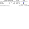
Comparison 7: 10‐day IV Meglumine Antimoniate 20mg/kg/day + 10‐day placebo versus 20‐day IV Meglumine Antimoniate in L. braziliensis and L. mexicana; FU: 1 year, Outcome 1: Complete cure
Primary outcomes: Adverse effects
Twenty‐three per cent of the participants (5/22) who received IVMA for 20 days developed six episodes of mild adverse reactions, which included four episodes of arthralgias and one episode each of anorexia and phlebitis at the site of injection. In the group receiving IVMA for 10 days, only one episode of arthralgia was observed in one participant. Shorter duration of treatment may decrease arthralgia risk, but the results are very imprecise due to the wide confidence interval (RR 0.25, 95% CI 0.03 to 2.06; Analysis 7.2).
7.2. Analysis.

Comparison 7: 10‐day IV Meglumine Antimoniate 20mg/kg/day + 10‐day placebo versus 20‐day IV Meglumine Antimoniate in L. braziliensis and L. mexicana; FU: 1 year, Outcome 2: Adverse effects: arthralgia
Secondary outcomes: Recurrence
Two of 22 participants (12.1%) receiving IVMA for 20 days did not respond to the treatment: one participant who was infected with L. braziliensis responded initially by 13 weeks but reactivation of the lesion occurred five months after the start of treatment; the other participant was removed from the study at 13 weeks. Two of the participants (12%; 2/22) receiving IVMA for 10 days had reactivations: one participant had an initial response but reactivation of the lesion occurred at 11 months, and the other participant was also removed at 13 weeks.
Intralesional antimony versus placebo
One RCT including 60 participants (Soto 2013) from Bolivia compared intralesional antimony (Sb) (1, 3 and 5 days) versus placebo.
Primary outcomes: Percentage of participants cured at least three months after the end of treatment
Cure rates were higher for participants treated with IL Sb 60% versus 13% for placebo (RR 5.00, 95% CI 1.94 to 12.89; Analysis 8.1)
8.1. Analysis.

Comparison 8: Intralesional antimony (650 μg/mm2) vs placebo in L. braziliensis,L. amazonensis,L. guyanensis and L. lainsoni; FU: 6 months, Outcome 1: Complete cure
Primary outcomes: Adverse effects
None of the participants experienced severe side effects. IL Sb was more painful (P ≤ 0.001), but this application showed a tendency toward less irritation (P = 0.06) than cream application.
Meglumine antimoniate plus tamoxifen versus meglumine antimoniate alone
A phase II pilot RCT including 38 participants with localised cutaneous leishmaniasis from Brazil (Machado 2018) compared oral (40 mg/day for 20 days) or topical tamoxifen (0.1% tamoxifen citrate for 20 days) combined with meglumine antimoniate (20 mg SbV/kg/day for 20 days) versus the standard SbV protocol (20 mg/kg/day for 20 days).
Primary outcomes: Percentage of participants cured at least three months after the end of treatment
The study found no significant differences in cure rates among the three options, either at three or at six months, since the 95% CI includes the possibility of both increased and reduced cure rates:
MA plus oral tamoxifen versus MA alone: at three months follow‐up cure rates accounted for 67% (8/12) and 53% (8/15), respectively (RR 1.25, 95% CI 0.67 to 2.32; Analysis 9.1); at six months follow‐up cure rates accounted for 58% (7/12) and 40% (6/15), respectively (RR 1.46, 95% CI 0.67 to 3.19; Analysis 9.1).
MA plus topical tamoxifen versus MA alone: at three months follow‐up cure rates accounted for 45% (5/11) and 53% (8/15), respectively (RR 0.85, 95% CI 0.38 to 1.90; Analysis 10.1); at six months follow‐up cure rates accounted for 36.4% (4/11) and 40% (6/15), respectively (RR 0.91, 95% CI 0.34 to 2.47; Analysis 10.1).
9.1. Analysis.

Comparison 9: Meglumine antimoniate 20 mg/kg/day plus oral tamoxifen 40 mg/day versus meglumine antimoniate alone in L. braziliensis; FU: 3‐6 months, Outcome 1: Complete cure at 3‐6 months
10.1. Analysis.

Comparison 10: Meglumine antimoniate 20 mg/kg/day plus topical tamoxifen for 20 days (0.1% citrate) versus meglumine antimoniate alone in L. braziliensis; FU: 3‐6 months, Outcome 1: Complete cure at 3‐6 months
Primary outcomes: Adverse effects
In 87% of participants, a similar prevalence of AEs were reported in the three groups (87%, 82% and 92% for SbV, SbV plus topical and SbV plus oral tamoxifen). These AEs were mild in general; arthralgia and myalgia (frequently linked to SbV use) were most commonly reported. A grade 3 headache and palpitation caused one participant to drop out of SbV plus oral tamoxifen treatment on the second day of treatment. In the SbV plus topical tamoxifen group, after the second SbV application, angio‐oedema led to one participant stopping therapy.
Secondary outcomes: Recurrence
The study found no significant differences in recurrence rates among the three options after six months, since the 95% CI includes the possibility of both increased and reduced recurrence rates:
MA plus oral tamoxifen versus MA alone: 8% (1/12) and 13% (2/15) (RR 0.59, 95% CI 0.05 to 7.43; Analysis 9.2)
MA plus topical tamoxifen versus MA alone: 9% (1/11) and 13% (2/15) (RR 0.68, 95% CI 0.07 to 6.61; Analysis 10.2)
9.2. Analysis.
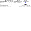
Comparison 9: Meglumine antimoniate 20 mg/kg/day plus oral tamoxifen 40 mg/day versus meglumine antimoniate alone in L. braziliensis; FU: 3‐6 months, Outcome 2: Recurrence at 6 months
10.2. Analysis.
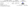
Comparison 10: Meglumine antimoniate 20 mg/kg/day plus topical tamoxifen for 20 days (0.1% citrate) versus meglumine antimoniate alone in L. braziliensis; FU: 3‐6 months, Outcome 2: Recurrence at 6 months
Meglumine antimoniate plus zinc versus meglumine antimoniate plus placebo
One pilot RCT including 29 participants (Guzman‐Rivero 2014) from Bolivia compared IMMA for 20 days plus 45 mg zinc daily for 60 days versus IMMA for 20 days plus placebo daily for 60 days.
Tertiary outcomes: Speed of healing
Authors reported that the time for reduction of lesion area did not differ significantly between placebo and zinc‐supplemented groups, but did not provide detailed numbers.
Intravenous meglumine antimoniate (IVMA) plus antihelminthic treatment versus IVMA plus placebo
One RCT, Newlove 2011, including 90 participants, all co‐infected with helminths and Leishmania braziliensis, from Brazil and all treated with intravenous Sbv (Glucantime) at 20 mg/kg/day for 20 days, compared adding oral placebo or antihelminthic treatment: albendazole (400 mg), ivermectin (200 μg/kg), and praziquantel (50 mg/kg) in an oral formulation at days 0 and 30 and day 60.
Primary outcomes: Percentage of participants cured at least three months after the end of treatment
There was no difference in cure rates between those treated with IVMA and antihelminthics or IVMA placebo (RR 0.77, 95% CI 0.48 to 1.25; Analysis 11.1).
11.1. Analysis.

Comparison 11: IV meglumine antimoniate (IVMA) plus antihelminthic treatment versus IVMA plus placebo in L.braziliensis; FU: 90 days, Outcome 1: Complete cure
Primary outcomes: Adverse effects
Adverse events were reported in 60% of participants in the treatment group and 60% of participants in the control group, with no statistically significant difference in the type or grade of events reported. Only grade 1 and 2 events were observed. Muscle pain (26%), headache (16%), leg pain (14%), weakness (14%), fever (13%), joint pain (12%), and dizziness (12%) were the most frequently reported symptoms.
Tertiary outcomes: Speed of healing
The median time to cure was 88 days in the control group versus 98 days in the treatment group, but authors state that this result was not statistically significant.
1.2 Stibogluconate
IM sodium stibogluconate (IMSSG) versus no treatment
One RCT including 61 participants (Guderian 1991) from Ecuador compared IMSSG for 20 days versus no treatment. Complete cure occurred in 90% (27/30) and 60% (9/15) of participants in the respective groups 1½ months after treatment (short‐term primary outcome, excluded from analysis).
IM sodium stibogluconate versus IM meglumine antimoniate
Primary outcomes: Percentage of participants cured at least three months after the end of treatment
One RCT including 114 participants (Soto 2004a) from Bolivia and Colombia compared IMSSG (branded and generic) for 20 days versus IMMA for 20 days. Six months after treatment, there was no difference in cure rates between IMSSG and IMMA (RR 1.07, 95% CI 0.88 to 1.30; Analysis 12.1). Similarly, there was no difference in cure rates between branded and generic IMSSG (RR 1.11, 95% CI 0.82 to 1.51) in L. panamensis infections.
12.1. Analysis.

Comparison 12: IM Sodium Stibogluconate 20 mg/kg/d for 20d vs IM Meglumine Antimoniate (20 mg/kg/d for 20d) in L. panamensis; FU: 6 months, Outcome 1: Complete cure
One RCT including 59 participants (Saenz 1987) from Panama compared IMSSG for 20 days versus IMMA for 20 days. Complete cure occurred in 46.7% (14/30) and 72.4% (21/29) of participants in the respective groups at the end of treatment (short‐term primary outcome, excluded from analysis).
Primary outcomes: Adverse effects
One RCT including 114 participants (Soto 2004a) reported myalgias in both SSG and MA groups respectively (RR 0.78, 95% CI 0.50 to 1.22; Analysis 12.3), but incidence of myalgia was higher for branded SSG compared to the generic SSG drug (RR 1.93, 95% CI 1.04 to 3.58; Analysis 13.2). There was no clear difference in headache between the SSG and MA groups (RR 0.68, 95% CI 0.37 to 1.26; Analysis 12.3) or between the branded and generic SSG groups (RR 1.67, 95% CI 0.65 to 4.25; Analysis 13.2). A metallic taste was observed more in the SSG than in the MA group (RR 0.49, 95% CI 0.27 to 0.92; Analysis 12.3) but the difference observed between the branded and generic SSG groups was less clear, as the result was very imprecise (RR 2.14, 95% CI 0.79 to 5.82; Analysis 13.2). There was also no clear difference in abdominal pain in the SSG and MA groups due to imprecision (RR 0.78, 95% CI 0.32 to 1.94; Analysis 12.3) but there may be more abdominal pain with branded compared to generic SSG (RR 3.00, 95% CI 0.85 to 10.63; Analysis 13.2).
12.3. Analysis.

Comparison 12: IM Sodium Stibogluconate 20 mg/kg/d for 20d vs IM Meglumine Antimoniate (20 mg/kg/d for 20d) in L. panamensis; FU: 6 months, Outcome 3: Adverse effects
13.2. Analysis.
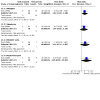
Comparison 13: IM Sodium Stibogluconate (branded) vs IM Sodium Stibogluconate (generic). Dose: 20 mg/kg/d for 20 d in L.panamensis; FU: 6 months, Outcome 2: Adverse effects
One RCT including 59 participants (Saenz 1987) reported that 63.3% (19/30) and 51.7% (15/29) of the participants in the SSG and MA groups respectively had mild‐to‐moderate adverse effects such as myalgias, arthralgias, headaches, pain at the site of injection, allergy and fever (RR 1.22, 95% CI 0.78 to 1.91; Analysis 12.2). There were no cases reporting hepatic, renal, haematologic or cardiac toxicity.
12.2. Analysis.

Comparison 12: IM Sodium Stibogluconate 20 mg/kg/d for 20d vs IM Meglumine Antimoniate (20 mg/kg/d for 20d) in L. panamensis; FU: 6 months, Outcome 2: Adverse effect Overall
Secondary outcomes: Recurrence
One RCT including 59 participants (Saenz 1987) reported that 2% (13/59), 23.3% (7/30) and 20.7% (6/29) of cured participants had reactivation of lesions after 6 to 12 months of follow‐up in the branded SSG, generic SSG and MA groups, respectively. Overall there was no clear difference in recurrence rates between SSG (branded and generic) and MA (RR 0.96, 95% CI 0.45 to 2.05; Analysis 12.4).
12.4. Analysis.

Comparison 12: IM Sodium Stibogluconate 20 mg/kg/d for 20d vs IM Meglumine Antimoniate (20 mg/kg/d for 20d) in L. panamensis; FU: 6 months, Outcome 4: Recurrence
Tertiary outcomes: Microbiological or histopathological cure of skin lesions
One RCT including 59 participants (Saenz 1987) reported that at the end of treatment, the cultures were parasitologically negative in 90% (27/30) and 89.7% (26/29) of the participants in the SSG and in the MA groups, respectively, showing no difference between groups (RR 1.00, 95% CI 0.85 to 1.19; Analysis 12.5).
12.5. Analysis.

Comparison 12: IM Sodium Stibogluconate 20 mg/kg/d for 20d vs IM Meglumine Antimoniate (20 mg/kg/d for 20d) in L. panamensis; FU: 6 months, Outcome 5: Microbiological or histopathological cure of skin lesions
IV sodium stibogluconate versus placebo
One RCT including 40 participants (Navin 1992) from Guatemala compared IVSSG for 20 days versus placebo.
Primary outcomes: Percentage of participants cured at least three months after the end of treatment
In L. braziliensis infections, complete cure occurred in 96% (24/25) and 20% (3/15) of participants in the respective groups two months after treatment. All participants (7/7) infected by L. mexicana in the SSG group were completely cured by six weeks, but two had subsequent reactivations two months after treatment (short‐term primary outcome, excluded from analysis).
Primary outcomes: Adverse effects
In the SSG group, 5/29 participants had nausea, 4/29 anorexia, 3/29 headache, 1/29 had rash, 6/29 had arthralgias and 10/29 had phlebitis. In the placebo group, 1/5 each had nausea and anorexia and 3/5 had abdominal pain.
Secondary outcomes: Recurrence
In the SSG group, none of the participants (24/25: 96%) infected with L. braziliensis and who responded to treatment had reactivations of their lesions between the 13‐ and 52‐week examinations. Sixty‐seven per cent of L. braziliensis‐infected participants (2/3) who received placebo that responded clinically had reactivations of their lesions, one at 14 weeks and the other at five months. None of the participants with L. mexicana had reactivations of their lesions between the 13‐ and 52‐weeks follow‐up examinations.
Different doses of IV sodium stibogluconate
One RCT including 40 participants (Ballou 1987) from the USA compared low‐dose IVSSG for 20 days versus high‐dose IVSSG for 20 days.
Primary outcomes: Percentage of participants cured at least three months after the end of treatment
Complete cure occurred in 76% (16/21) and 100% (19/19) of participants in the respective groups 1½ months after treatment (short‐term primary outcome, excluded from analysis).
Primary outcomes: Adverse effects
Mild‐to‐moderate muscle and joint stiffness were experienced by 62% (13/21) of participants in the low‐dose group and by 58% (11/19) of participants in the high‐dose group (RR 1.07, 95% CI 0.64 to 1.78; Analysis 14.1). Laboratory abnormalities were limited to increases in liver enzymes in 48% (10/21) and 53% (10/19), respectively (RR 0.90, 95% CI 0.49 to 1.68; Analysis 14.1); mild leucopenia in 9.5% (2/21) and 5.3% (1/19), respectively (RR 1.81, 95% CI 0.18 to 18.39; Analysis 14.1); and electrocardiographic abnormalities in 19% (4/21) and 21% (4/19), respectively (RR.0.90, 95% CI 0.26 to 3.12; Analysis 14.1).
14.1. Analysis.

Comparison 14: Low dose of IV sodium stibogluconate 20 days versus high doses in L.panamensis and L. chagasi; FU: 1 year, Outcome 1: Adverse effects
Different regimens of IV sodium stibogluconate
One RCT including 40 participants (Franke 1994) from Peru compared IVSSG for 28 days versus IVSSG for 40 days.
Primary outcomes: Percentage of participants cured at least three months after the end of treatment
One year after treatment, there was no clear difference in cure rates between 28 and 40 days of IVSSG (RR 0.83, 95% CI 0.47 to 1.47; Analysis 15.1) in L. braziliensis infections.
15.1. Analysis.

Comparison 15: IV Sodium Stibogluconate 20mg/kg for 28 days vs IV Sodium Stibogluconate for 40 days in L. braziliensis; FU: 1 year, Outcome 1: Complete cure
Primary outcomes: Adverse effects
None of the subjective complaints were severe enough to warrant cessation of treatment. Although more participants in the 40‐day regimen complained of arthralgias and myalgias, most complaints began before day 28. Side effects were arthralgias, myalgias, itch, rash, nausea, anorexia, abdominal pain, cough and headache.
Different doses and regimens of IV sodium stibogluconate
One RCT including 36 participants (Oster 1985) from the USA treated participants with IVSSG for 10 days at a dose of 600 mg a day by one of three schedules: once daily by rapid infusion for 10 days; a loading dose of 600 mg followed by a continuous infusion of 600 mg for 24 hours each day for nine days; or a loading dose of 600 mg followed by 200 mg every eight hours for nine days.
Primary outcome: Percentage of participants cured at least three months after the end of treatment
Complete cure occurred in 100% (12/12), 50% (6/12) and 42% (5/12) of participants in the respective groups at the end of treatment (short‐term primary outcome, excluded from analysis).
Primary outcomes: Adverse effects
Despite the lack of side‐effects reported by the participants, there was an equal distribution of mildly‐elevated liver enzymes, triglycerides and creatine phosphokinase (CPK) in the three groups.
Intralesional Stibogluconate (SB) versus Intralesional pentamidine (ILP)
Two RCTs in Bolivia by the same group (Soto 2016a; Soto 2016b), including a total of 120 participants, compared two topical intralesional treatments: ILSB (N‐methylglucamine five injections) versus ILP (three injections).
Primary outcomes: Percentage of participants cured at least three months after the end of treatment
No differences in cure rates for either treatment (RR 0.98, 95% CI 0.78 to 1.23, I2 = 0%, 2 studies, 120 participants, Analysis 16.1).
16.1. Analysis.

Comparison 16: IL Sodium Stibogluconate (650 μg; Sb 8 μL/mm2) vs IL pentamidine (240 μg; 8 μL/mm2) in L. braziliensis and L. braziliensis/amazonensis/lainsoni/guyanensis; FU: 6 months, Outcome 1: Complete cure
Primary outcomes: Adverse effects.
There were no clear differences between treatments for frequency of adverse effects): myalgia (RR 3.93, 95% CI 0.45 to 35.54); local irritation (RR 1.19, 95% CI 0.55 to 2.58); or local pain (RR 1.74, 95% CI 0.79 to 3.83) (Analysis 16.2).
16.2. Analysis.

Comparison 16: IL Sodium Stibogluconate (650 μg; Sb 8 μL/mm2) vs IL pentamidine (240 μg; 8 μL/mm2) in L. braziliensis and L. braziliensis/amazonensis/lainsoni/guyanensis; FU: 6 months, Outcome 2: Adverse effects
Secondary outcomes: Recurrence
Two participants out of 30 in the ILSB group had recurrence of the disease compared to none in the ILP group, but the results are very imprecise (RR 5.00, 95% CI 0.25 to 99.95; 1 study, 60 participants; Analysis 16.3).
16.3. Analysis.

Comparison 16: IL Sodium Stibogluconate (650 μg; Sb 8 μL/mm2) vs IL pentamidine (240 μg; 8 μL/mm2) in L. braziliensis and L. braziliensis/amazonensis/lainsoni/guyanensis; FU: 6 months, Outcome 3: Recurrence
2. Non‐antimonial systemic treatments
2.1 Oral antifungals
Ketoconazole versus IM meglumine antimoniate
One RCT including 41 participants (Saenz 1990) from Panama compared oral ketoconazole for 28 days versus IMMA for 20 days.
Primary outcomes: Percentage of participants cured at least three months after the end of treatment
Three months after treatment, there was no difference in cure rates between oral ketoconazole and 20 mg/kg/day IMMA for 20 days (RR 1.06, 95% CI 0.71 to 1.58; Analysis 17.1) in L. panamensis and L. mexicana infections.
17.1. Analysis.

Comparison 17: Oral ketoconazole 200 mg for 28 days vs IM Meglumine Antimoniate 20 mg/kg for 20 days in L. panamensis and L. mexicana; FU: 3 months, Outcome 1: Complete cure
Primary outcomes: Adverse effects
The laboratory abnormalities recorded in 27% (6/22) of ketoconazole‐treated participants were mild elevations of liver transaminase values that normalised during or after therapy. Subjective complaints consisted of headache (4/22), abdominal pain (2/22), fever (2/22), nausea (1/22) and malaise (1/22). Laboratory abnormalities were recorded in 47% (9/19) of participants in the IMMA group (RR 0.58, 95% CI 0.25 to 1.32; Analysis 17.2), consisting of mild elevations of liver enzymes which partially or completely resolved despite continued therapy in five participants. Eighty‐four per cent (16/19) complained of pain at the IMMA injection site. In addition, 58% (11/19) complained of myalgia, 21% (4/19) had headache or arthralgia, and 11% (2/19) had nausea or fever.
17.2. Analysis.

Comparison 17: Oral ketoconazole 200 mg for 28 days vs IM Meglumine Antimoniate 20 mg/kg for 20 days in L. panamensis and L. mexicana; FU: 3 months, Outcome 2: Adverse effects
Tertiary outcomes: Speed of healing
Fifty‐six per cent of participants (9/16) in the oral ketoconazole group versus 54% of participants (7/13) in the IMMA group demonstrated complete re‐epithelialisation of lesions by the end of approximately one month of therapy (RR 1.04, 95% CI 0.54 to 2.03; Analysis 17.3). In the ketoconazole group complete re‐epithelisation occurred by three months after the end of therapy.
17.3. Analysis.

Comparison 17: Oral ketoconazole 200 mg for 28 days vs IM Meglumine Antimoniate 20 mg/kg for 20 days in L. panamensis and L. mexicana; FU: 3 months, Outcome 3: Speed to healing (% of complete re‐epithelization of lesions at 1 month in cured patients)
Tertiary outcomes: Microbiological or histopathological cure of skin lesions
For the 73% (16/22) of participants who were cured by the end of therapy in the ketoconazole group, lesions were parasitologically sterile in all attempted tests for only 56% (9/16) of participants at the end of therapy. In the IMMA group 69% (9/13) of the participants who were cured had a negative diagnostic test result for leishmanial organisms at the end of therapy (RR 0.81, 95% CI 0.46 to 1.43; Analysis 17.4).
17.4. Analysis.
Comparison 17: Oral ketoconazole 200 mg for 28 days vs IM Meglumine Antimoniate 20 mg/kg for 20 days in L. panamensis and L. mexicana; FU: 3 months, Outcome 4: Miocrobiological cure of skin lesions (% in cured patients)
Ketoconazole versus IV sodium stibogluconate
One RCT including 48 participants (Navin 1992) from Guatemala compared oral ketoconazole for 28 days versus IVSSG for 20 days. Complete cure occurred in 52% (12/23) and 96% (24/25) of participants in the respective groups two months after treatment (short‐term primary outcome, excluded from analysis).
Primary outcomes: Adverse effects
In the ketoconazole group, 2/8 had nausea, abdominal pain and headache and 1/8 each had dizziness and rash. In the SSG group, 5/29 participants had nausea, 4/29 anorexia, 3/29 headache, 1/29 had rash, 6/29 had arthralgias and 10/29 had phlebitis.
Secondary outcomes: Recurrence
In the ketoconazole group, 17% (2/12) responders infected with L. braziliensis had reactivations of their lesions, one at 17 weeks and one at 11 months. In the SSG group, none of the 96% (24/25) of the participants infected with L. braziliensis and who responded to treatment by 13 weeks had reactivations of their lesions at the 52‐week examinations. None of the participants with L. mexicana who had responded in all treatment groups had reactivations of their lesions at the 52‐week follow‐up examinations.
Ketoconazole versus placebo
Primary outcomes: Percentage of participants cured at least three months after the end of treatment
One RCT including 41 participants (Saenz 1990) from Panama compared oral ketoconazole for 28 days versus oral placebo. Three months after treatment, cure rates were significantly higher in the oral ketoconazole group compared with placebo (RR 17.22, 95% CI 1.13 to 262.82; Analysis 18.1) in L. panamensis and L. mexicana infections, although results were very imprecise due to the wide confidence interval.
18.1. Analysis.

Comparison 18: Oral ketoconazole 200 mg vs oral placebo for 28 days in L. panamensis and L. mexicana; FU: 3 months, Outcome 1: Complete cure
One RCT including 38 participants (Navin 1992) from Guatemala compared oral ketoconazole for 28 days versus placebo. Complete cure occurred in 52% (12/23) and 20% (3/15) of participants in their respective groups two months after treatment (short‐term primary outcome, excluded from analysis).
Primary outcomes: Adverse effects
In one study including 41 participants (Saenz 1990) the laboratory abnormalities recorded in 27% (6/22) of ketoconazole‐treated participants were mild elevations of liver transaminase values that normalised during or after therapy. Subjective complaints consisted of headache (4/22), abdominal pain (2/22), fever (2/22), nausea (1/22), and malaise (1/22).
The other study, including 38 participants (Navin 1992), reported that in the ketoconazole group 2/8 each had nausea, abdominal pain and headache, and 1/8 had dizziness and rash. In the placebo group, 1/5 had nausea and anorexia and 3/5 had abdominal pain.
Secondary outcomes: Recurrence
In a study of 48 participants (Navin 1992) 17% (2/12) responders in the ketoconazole group infected with L. braziliensis had reactivations of their lesions, one at 17 weeks and one at 11 months. Sixty‐seven per cent of L. braziliensis‐infected participants (2/3) who received placebo and responded clinically had reactivations of their lesions, one at 14 weeks and the other at five months. All participants with L. mexicana who had responded in the two treatment groups had no reactivations of their lesions.
Fluconazole versus intravenous Glucantime
Two RCTs conducted in Brazil (Alves Noroes 2015; Prates 2017), including 173 participants in total, compared oral fluconazole versus intravenous Sbv (Glucantime).
Primary outcomes: Percentage of participants cured at least three months after the end of treatment
Cure rates were lower with oral fluconazole (RR 0.60, 95% CI 0.37 to 0.96; I2 = 44%; 2 studies, 173 participants; Analysis 19.1), and the percentage of participants needing rescue therapy was higher for those treated with oral fluconazole (RR 2.20, 95% CI 1.09 to 4.46; 1 study, 53 participants; Analysis 19.3).
19.1. Analysis.

Comparison 19: 300‐450 mg oral Fluconazole vs 20mg/kg/d IV Meglumine Antimoniate 20 mg/kg in L. braziliensis; FU: 3‐6 months, Outcome 1: Complete cure
19.3. Analysis.

Comparison 19: 300‐450 mg oral Fluconazole vs 20mg/kg/d IV Meglumine Antimoniate 20 mg/kg in L. braziliensis; FU: 3‐6 months, Outcome 3: Needed rescue therapy
Primary outcomes: Adverse effects.
There was no clear difference between treatments in the percentage of participants who suffered adverse events (none severe) (RR 1.07, 95% CI 0.52 to 2.20; 1 study, 53 participants; Analysis 19.2).
19.2. Analysis.

Comparison 19: 300‐450 mg oral Fluconazole vs 20mg/kg/d IV Meglumine Antimoniate 20 mg/kg in L. braziliensis; FU: 3‐6 months, Outcome 2: Adverse effects
Tertiary outcomes: Speed of healing
Average healing time (days) was much higher for those treated with oral fluconazole (mean difference (MD) 40.40 days, 95% CI 11.27 to 69.53; 1 study, 53 participants; Analysis 19.4).
19.4. Analysis.

Comparison 19: 300‐450 mg oral Fluconazole vs 20mg/kg/d IV Meglumine Antimoniate 20 mg/kg in L. braziliensis; FU: 3‐6 months, Outcome 4: Speed to healing (days)
2.2 Oral allopurinol
Oral allopurinol versus allopurinol combined with IM meglumine antimoniate
One RCT including 60 participants (Martínez 1992) from Colombia compared oral allopurinol for 15 days versus oral allopurinol plus IMMA in the same regimen.
Primary outcome: Percentage of participants cured at least three months after the end of treatment
One year after treatment, there was no difference in cure rates between oral allopurinol alone and oral allopurinol in combination with IMMA (RR 1.08, 95% CI 0.82 to 1.42; Analysis 20.1) in L. panamensis infections.
20.1. Analysis.

Comparison 20: Oral Allopurinol 20 mg/kg/d (4 doses) for 15d vs Allopurinol + IM Meglumine Antimoniate 20 mg/kg (same regimen) in L. panamensis; FU: 12 months, Outcome 1: Complete cure
Secondary outcomes: Recurrence
When comparing the two groups, the difference after 12 months in relapse after cure was unclear, due to the highly imprecise results (RR 0.70, 95% CI 0.07 to 7.30; Analysis 20.2).
20.2. Analysis.

Comparison 20: Oral Allopurinol 20 mg/kg/d (4 doses) for 15d vs Allopurinol + IM Meglumine Antimoniate 20 mg/kg (same regimen) in L. panamensis; FU: 12 months, Outcome 2: Recurrence
Oral allopurinol versus IV meglumine antimoniate
Primary outcomes: Percentage of participants cured at least three months after the end of treatment
One RCT including 58 participants (Martínez 1992) from Colombia compared oral allopurinol for 15 days versus IVMA for 15 days. One year after treatment, cure rates were significantly higher in the oral allopurinol group compared with the IVMA group (RR 2.20, 95% CI 1.34 to 3.60; Analysis 21.1).
21.1. Analysis.

Comparison 21: Oral Allopurinol 20 mg/kg/d (4 doses) x 15d vs. IV Meglumine Antimoniate (same regimen) in L. panamensis; FU: 1 year, Outcome 1: Complete cure
One RCT including 34 participants (D'Oliveira 1997) from Brazil compared oral allopurinol versus IVMA, both for 20 days. There was no complete cure for the first nine participants of the allopurinol group two months after treatment. The other nine participants in this group were not included in the evaluation because the protocol was stopped due to some participants getting worse, with antimonial administered to some of this group. There was complete cure in 50% (8/16) of the MA group two months after treatment (short‐term primary outcome, excluded from analysis).
Primary outcomes: Adverse effects
D'Oliveira 1997 reported that 11.1% (1/9) of participants developed mucocutaneous disease within three months, although the group is not stated.
Secondary outcomes: Recurrence
Martínez 1992 reported that a 12‐month relapse after cure was seen in 4% (1/25) and 6% (2/33) of the allopurinol and MA groups respectively, but the result was very imprecise due to the wide confidence interval (RR 0.66, 95% CI 0.06 to 6.88; Analysis 21.2).
21.2. Analysis.

Comparison 21: Oral Allopurinol 20 mg/kg/d (4 doses) x 15d vs. IV Meglumine Antimoniate (same regimen) in L. panamensis; FU: 1 year, Outcome 2: Recurrence
Oral allopurinol combined with IV meglumine antimoniate versus IV meglumine antimoniate
One RCT including 60 participants (Martínez 1992) from Colombia compared oral allopurinol for 15 days combined with IVMA for 15 days versus IVMA monotherapy for 15 days.
Primary outcomes: Percentage of participants cured at least three months after the end of treatment
One year after treatment, oral allopurinol had a significant synergistic effect with IVMA for 15 days compared to IVMA alone (RR 2.04, 95% CI 1.25 to 3.34; Analysis 22.1).
22.1. Analysis.

Comparison 22: Oral Allopurinol 20 mg/kg/d + IV Meglumine Antimoniate (20 mg/kg/d (4 doses) for 15d) vs IV Meglumine Antimoniate (same regimen) in L. panamensis; FU: 1 year, Outcome 1: Complete cure
Secondary outcomes: Recurrence
Relapse after cure was similar between groups (RR 0.94, 95% CI 0.14 to 6.31; Analysis 20.2).
Oral allopurinol versus IM meglumine antimoniate
One RCT including 127 participants (Vélez 1997) from Colombia compared oral allopurinol for 28 days versus IMMA for 20 days. Another RCT including 75 participants (Chico 1995) from Ecuador compared allopurinol plus probenecid for 28 days versus IMMA for 20 days and versus no treatment, but the follow‐up was short, only up to 70 days.
Primary outcomes: Percentage of participants cured at least three months after the end of treatment
In the Vélez 1997 study, oral allopurinol showed lower cure rates compared with IMMA one year after treatment (RR 0.39, 95% CI 0.26 to 0.58; Analysis 23.1).
23.1. Analysis.

Comparison 23: Oral Allopurinol 300 mg for 28d vs IM Meglumine Antimoniate 20mg/kg/d for 20 d in L braziliensis and L. panamensis ; FU: 12 month, Outcome 1: Complete cure
In the Chico 1995 study, cure rates were much lower for allopurinol plus probenecid (20%: 6/30) compared to IMMA (100%: 28/28) (short‐term primary outcome, excluded from analysis).
Primary outcomes: Adverse effects
Twenty‐five per cent of participants in the allopurinol group (15/60) had moderate‐to‐severe side effects. The only side effects attributable to allopurinol were headache and epigastric pain. Seventy‐nine per cent of participants (53/67) in the IMMA group had moderate side effects and 52.2% (35/67) had severe adverse effects. Myalgias, arthralgias, anorexia, nausea, and headache were the common adverse effects.
Secondary outcomes: Recurrence
Relapse or mucocutaneous disease was seen in 5% (3/60) of the allopurinol group: one relapse occurred five months after healing and two mucocutaneous cases occurred at the end of treatment and 1½ months later. Similar relapse or mucocutaneous disease was seen 1½ to 3 months after healing in both groups (RR 1.68, 95% CI 0.29 to 9.69; Analysis 23.2).
23.2. Analysis.

Comparison 23: Oral Allopurinol 300 mg for 28d vs IM Meglumine Antimoniate 20mg/kg/d for 20 d in L braziliensis and L. panamensis ; FU: 12 month, Outcome 2: Recurrence
Oral allopurinol combined with IV sodium stibogluconate versus IV sodium stibogluconate
Primary outcomes: Percentage of participants cured at least three months after the end of treatment
One RCT including 100 participants (Martínez 1997) from Colombia compared oral allopurinol combined with IVSSG versus IVSSG alone, both for 15 days. One year after treatment, oral allopurinol had a significant synergistic effect with IVSSG compared to IVSSG alone (RR 1.82, 95% CI 1.23 to 2.70; Analysis 24.1).
24.1. Analysis.

Comparison 24: Oral Allopurinol 20 mg/k/d + IV Sodium Stibogluconate (20 mg/kg/d (4 doses) x 15d) vs IV Sodium Stibogluconate (same dose) in L. braziliensis; FU: 1 year, Outcome 1: Complete cure
When we pooled two RCTs including 168 participants (L. braziliensis) (Martínez 1992; Martínez 1997) where oral allopurinol combined with IV antimonials (20 mg/kg/day for 15 days) was compared to IV antimonials alone, the results showed that oral allopurinol had a significant synergistic effect with IV antimonials (RR 1.90, 95% CI 1.40 to 2.59; I2 = 0%; Analysis 24.2).
24.2. Analysis.

Comparison 24: Oral Allopurinol 20 mg/k/d + IV Sodium Stibogluconate (20 mg/kg/d (4 doses) x 15d) vs IV Sodium Stibogluconate (same dose) in L. braziliensis; FU: 1 year, Outcome 2: Complete cure; Oral AL plus IVSSG vs IVSSG
One RCT including 81 participants with MCL (Llanos‐Cuentas 1997) from Peru compared oral allopurinol combined with IVSSG versus IVSSG alone, both for 28 days. One year after treatment, IVSSG alone presented similar cure rates (RR 0.62, 95% CI 0.38 to 1.03; Analysis 25.1).
25.1. Analysis.

Comparison 25: Oral Allopurinol 20 m/k/d + IV Sodium Stibogluconate (20 mg/kg/d (4 doses) for 28d) vs IV Sodium Stibogluconate (same dose); FU: 12 months, Outcome 1: Complete cure
Primary outcomes: Adverse effects
One RCT with 100 participants (Martínez 1997) reported that clinically‐important side effects were observed only for the group of participants who received SSG monotherapy. Two per cent of participants (1/49) developed severe chemical hepatitis with neurological manifestations, and treatment was stopped after seven days. The cause of this adverse effect is unclear, but it was not believed to be related to antileishmanial therapy. There was an increase in the frequency of eosinophilia and rash in the group receiving allopurinol (18% (9/51) eosinophilia and 28% (14/51) rash). In the SSG‐alone group 2% (1/49) had eosinophilia and the same results for rash. The rashes were generally macular or erythematous. There was no urticaria or desquamation. These cutaneous manifestations were mild, did not require treatment, and were consistent with the known side effects of allopurinol. The other RCT including 81 participants with MCL (Llanos‐Cuentas 1997) reported that the more frequent symptoms were headache (81.5% of participants), arthralgia (75.3%), myalgia (67.9%), chills (42%), fever (39.5%), abdominal pain (33.3%), and anorexia (25.9%). Three participants developed Herpes Zoster (two in the allopurinol combined with SSG group and one in the SSG‐alone group), and were treated with acyclovir, but one developed partial blindness as a consequence. The most frequent laboratory adverse effect was haematologic abnormality: the rate of thrombocytopenia was higher among the allopurinol‐plus‐SSG group.
Secondary outcomes: Recurrence
Two studies assessed recurrence (Llanos‐Cuentas 1997; Martínez 1997); pooling their results, we found no differences in the recurrence rate between treatments (RR 1.16, 95% CI 0.73 to 1.85; 2 studies, 181 participants; I2 = 0; Analysis 25.2).
25.2. Analysis.

Comparison 25: Oral Allopurinol 20 m/k/d + IV Sodium Stibogluconate (20 mg/kg/d (4 doses) for 28d) vs IV Sodium Stibogluconate (same dose); FU: 12 months, Outcome 2: Recurrence
Oral allopurinol ribonucleoside combined with probenecid versus IM sodium stibogluconate
One RCT including 61 participants (Guderian 1991) from Ecuador compared oral allopurinol ribonucleoside combined with probenecid for 28 days versus IMSSG for 20 days.
Primary outcomes: Percentage of participants cured at least three months after the end of treatment
Complete cure occurred in 30% (9/30) and 60% (9/15) of participants in the respective groups 1½ months after treatment (short‐term primary outcome, excluded from analysis). The authors also compared the experimental intervention versus no treatment, and reported complete cure of 30% (9/30) and 90% (27/30) of participants in the respective groups 1½ months after treatment.
Oral allopurinol versus no treatment
One RCT including 42 participants (Martínez 1992) from Colombia compared oral allopurinol for 15 days versus no treatment.
Primary outcome: Percentage of participants cured at least three months after the end of treatment
One year after treatment, oral allopurinol had significantly higher cure rates compared with no treatment, although results are very imprecise due to the wide confidence interval (RR 28.38, 95% CI 1.83 to 439.72; Analysis 26.1).
26.1. Analysis.

Comparison 26: Oral Allopurinol 20 mg/kg/d (4 doses) for 15 d vs no treatment in L. panamensis; FU: 12 months, Outcome 1: Complete cure
Secondary outcomes: Recurrence
Martínez 1992 reported that at 12 months 1/25 participants in the allopurinol group versus 2/17 in the no‐treatment group had relapsed after cure (RR 0.34, 95% CI 0.03 to 3.46; Analysis 26.2).
26.2. Analysis.

Comparison 26: Oral Allopurinol 20 mg/kg/d (4 doses) for 15 d vs no treatment in L. panamensis; FU: 12 months, Outcome 2: Recurrence
Oral allopurinol versus placebo
One RCT including 61 participants (Vélez 1997) from Colombia compared oral allopurinol for 28 days versus placebo.
Primary outcomes: Percentage of participants cured at least three months after the end of treatment
One year after treatment, there was no difference in cure rates between oral allopurinol and placebo (RR 1.06, 95% CI 0.61 to 1.85; Analysis 27.1).
27.1. Analysis.

Comparison 27: Oral Allopurinol 300 mg 28 days vs placebo in L. braziliensis and L. panamensis; FU: 12 months, Outcome 1: Complete cure
Primary outcomes: Adverse effects
Moderate‐to‐severe side effects were observed in 25% (15/60) of the allopurinol group and in 10% (6/60) of the placebo group. The only side effects attributable to allopurinol were headache and epigastric pain.
Secondary outcomes: Recurrence
Relapse or mucocutaneous disease was seen in 5% (3/60) of the allopurinol group: one case of relapse was seen five months after healing and two mucocutaneous cases were seen at the end of treatment and 1½ months later. Relapse was seen in 1.67% (1/60) of the placebo group: this participant had developed mucocutaneous disease 12 months after healing. Results are very imprecise due to the wide confidence interval, and no differences were observed between treatment groups (RR 3.00, 95% CI 0.32 to 28.03; Analysis 27.2).
27.2. Analysis.
Comparison 27: Oral Allopurinol 300 mg 28 days vs placebo in L. braziliensis and L. panamensis; FU: 12 months, Outcome 2: Relapse
Oral allopurinol combined with IV meglumine antimoniate versus no treatment
One RCT including 52 participants (Martínez 1992) from Colombia compared oral allopurinol combined with IVMA for 15 days versus no treatment.
Primary outcome: Percentage of participants cured at least three months after the end of treatment
One year after treatment, oral allopurinol combined with IVMA had significantly higher cure rates than no treatment (RR 26.50, 95% CI 1.71 to 410.42; Analysis 28.1).
28.1. Analysis.

Comparison 28: Oral Allopurinol 20 mg/kg/d + IV Meglumine Antimoniate (20 mg/kg/ d in 4 doses for 15d) vs no treatment in L. panamensis; FU: 12 months, Outcome 1: Complete cure
Secondary outcomes: Recurrence
Recurrence after cure at 12 months was higher in the no‐treatment group, but the results are very imprecise due to the wide confidence interval (RR 0.49, 95% CI 0.07 to 3.16; Analysis 28.2).
28.2. Analysis.

Comparison 28: Oral Allopurinol 20 mg/kg/d + IV Meglumine Antimoniate (20 mg/kg/ d in 4 doses for 15d) vs no treatment in L. panamensis; FU: 12 months, Outcome 2: Recurrence
2.3 Oral miltefosine
Oral miltefosine versus placebo
One RCT including 133 participants (Soto 2004b) from Colombia and Guatemala compared oral miltefosine for 28 days versus placebo in L. braziliensis,L. panamensis and L. mexicana infections.
Primary outcomes: Percentage of participants cured at least three months after the end of treatment
Six months after treatment, oral miltefosine had significantly higher cure rates than placebo in the Colombian site (RR 2.18, 95% CI 1.28 to 3.71; Analysis 29.1) but the difference was not so strong for the Guatemalan site (RR 2.50, 95% CI 0.99 to 6.33; Analysis 29.1). There is moderate evidence about the difference in cure rate between oral miltefosine and placebo. Oral miltefosine probably improves the chance of healing, corresponding to 644 more participants completely cured per 1000 participants (95% CI 378 more to 1000 more) (RR 2.25, 95% CI 1.42 to 3.38) (Table 2).
29.1. Analysis.

Comparison 29: Oral miltefosine 50 mg for 28 d vs placebo (same regimen) in L. mexicana, L. panamensis and L. braziliensis; FU: 6 months, Outcome 1: Complete cure
Primary outcomes: Adverse effects
Nausea was observed more in the miltefosine group (RR 3.96, 95% CI 1.49 to 10.48; Analysis 29.2), and so was vomiting (RR 6.92, 95% CI 2.68 to 17.86; Analysis 29.2), but the difference in cases of diarrhoea was less clear across groups, due to imprecision (RR 2.47, 95% CI 0.57 to 10.80; Analysis 29.2), as well as motion sickness (RR 1.29, 95% CI 0.68 to 2.42; Analysis 29.2) and headache (RR 1.32, 95% CI 0.67 to 2.59; Analysis 29.2). The creatinine level increased towards the normal range in the miltefosine recipients (RR 3.58, 95% CI 1.34 to 9.56; Analysis 29.2). Aspartate aminotransferase levels were higher with placebo, but the 95% CI includes 1, showing there may be little or no difference (RR 0.43, 95% CI 0.17 to 1.12; Analysis 29.2). The difference between groups for alanine aminotransferase was less clear, due to imprecision (RR 0.89, 95% CI 0.32 to 2.50; Analysis 29.2). See Table 2.
29.2. Analysis.

Comparison 29: Oral miltefosine 50 mg for 28 d vs placebo (same regimen) in L. mexicana, L. panamensis and L. braziliensis; FU: 6 months, Outcome 2: Adverse effects
Secondary outcomes: Recurrence
Recurrence occurred within six months in 4.1% (2/49) and 0% (0/24) of the respective treatment group in the Colombian site, and 10% (4/40) and 5% (1/20) of the respective treatment groups from the Guatemala site (RR 2.97, 95% CI 0.37 to 23.89; Analysis 29.3; Table 2).
29.3. Analysis.

Comparison 29: Oral miltefosine 50 mg for 28 d vs placebo (same regimen) in L. mexicana, L. panamensis and L. braziliensis; FU: 6 months, Outcome 3: Recurrence
Oral miltefosine versus meglumine antimoniate
Seven RCTs including 676 participants in total compared both treatments in participants with mucosal (Garcia 2014; Sampaio 2019) and cutaneous leishmaniasis (Chrusciak‐Talhari 2011; Machado 2010; Rubiano 2012;
Soto 2008; Vélez 2010). Five of them followed the participants for six months after treatment cessation (Garcia 2014; Machado 2010; Rubiano 2012; Sampaio 2019; Vélez 2010) and two followed them up to 12 months (Chrusciak‐Talhari 2011; Soto 2008).
Primary outcomes: Percentage of participants cured at least three months after the end of treatment
There were no differences in cure rates among the treatments (RR 1.07, 95% CI 0.86 to 1.34; I2 = 67%; Analysis 30.1) The relatively high heterogeneity is due to the studies reporting less favourable results for the miltefosine treatment (Soto 2008; Vélez 2010), but we could not identify any clinical or methodological differences between these studies and the other studies included in the meta‐analysis.
30.1. Analysis.

Comparison 30: Oral Miltefosine vs Meglumine Antimoniate; FU: 6‐12 months, Outcome 1: Complete cure
A separate analysis of the two studies that included participants with mucosal leishmaniasis (Garcia 2014; Sampaio 2019) shows no differences in cure rates between the treatments (RR 1.04, 95% CI 0.81 to 1.34; 2 studies, 40 participants: I2 = 0%), since the 95% CI includes the possibility of both increased and reduced cure rates.
Pooling the results of the studies that included only participants with cutaneous leishmaniasis (Chrusciak‐Talhari 2011; Machado 2010; Rubiano 2012; Soto 2008; Vélez 2010) shows no differences in cure rates between the treatments (RR 1.06, 95% CI 0.87 to 1.29; 5 studies, 636 participants; I2 = 72%).
After pooling the results of the RCT including 116 children aged between 2 and 12 years (Rubiano 2012) and the results for 28 children of the same age (Chrusciak‐Talhari 2011), we found no relevant differences in cure rates between treatments (RR 1.19, 95% CI 0.98 to 1.46; I2 = 0; Analysis 30.2).
30.2. Analysis.

Comparison 30: Oral Miltefosine vs Meglumine Antimoniate; FU: 6‐12 months, Outcome 2: Complete cure in children 2 to 12 years old
Sampaio 2019, including 40 participants followed up to four years after treatment, found that both treatment groups had similar cure rates (16/20 versus 12/20: RR 0.66, 95% CI 0.33 to 1.32).
Please see Table 3, where we are not certain about differences in cure rate between oral miltefosine and meglumine antimoniate. Oral miltefosine may make little or no difference to the chance of healing, corresponding to 741 more participants with healing per 1000 participants (95% CI 616 to 893 more).
Primary outcomes: Adverse effects
Pooling the results from three RCTs including 464 participants with the cutaneous form (Machado 2010; Rubiano 2012; Vélez 2010) revealed that the risks of nausea (RR 2.45, 95% CI 1.72 to 3.49; I2 = 0% Analysis 30.3) and vomiting (RR 4.76, 95% CI 1.82 to 12.46; I2 = 48%; Analysis 30.3) were in both cases higher in the miltefosine group.
30.3. Analysis.

Comparison 30: Oral Miltefosine vs Meglumine Antimoniate; FU: 6‐12 months, Outcome 3: Adverse events
Please see Table 3, where we are moderately confident in the effect estimate. Oral miltefosine probably increases nausea and vomiting rates, corresponding to 360 more participants with nausea per 1000 (95% CI 252 more to 512 more) and 415 more participants with vomiting per 1000 (95% CI 159 more to 1000 more).
There were no reports of serious adverse effects in Chrusciak‐Talhari 2011, which included 58 participants, and those adverse effects that were reported did not require participants to discontinue therapy. Clinical adverse events of the gastrointestinal tract were mostly reported in the miltefosine group, and they normally happened in the first week of treatment. The most common adverse effect was vomiting, which 48.3% (28/58) of participants reported, and 41% (7/17) with CTC grade 1 and 45.5% (5/11) with CTC grade 2 were children.
In the phase II RCT including 19 participants with ML (Garcia 2014) from Argentina, eight of the nine participants treated with miltefosine presented gastrointestinal symptoms (all of them of low severity). For the antimoniate group, six of the 10 participants presented with adverse effects that were treated with anti‐inflammatory drugs in three of them (for asthenia, arthralgia and headache); two participants presented pain in the injection area, fever and myalgia; and one suffered cutaneous rash.
In the RCT including 90 participants (Machado 2010), the incidence of adverse events was similar in the Sbv and miltefosine groups (76.7% versus 78.3%). Vomiting (41.7%), nausea (40%), and abdominal pain (23.3%) were significantly more frequent in the miltefosine group, while arthralgias (20.7%), myalgias (20.7%) and fever (23.3%) were significantly more frequent in the Sbv group.
In the RCT including 116 children all aged between 2 and 12 years, Rubiano 2012 found that 95% of clinical adverse effects were grade 1 ("mild symptoms that do not interfere with regular activities"). Children who were treated with meglumine antimoniate more commonly experienced increased levels of hepatic enzymes aspartate transaminase (AST) (16/57 versus 5/57; P = 0.01) and alanine transaminase (ALT) (10/57 versus 2/57; P = 0.01), compared with children receiving miltefosine. Children receiving miltefosine experienced gastrointestinal symptoms, nausea (9/57 versus 2/57; P = 0.02) and vomiting (15/57 versus 2/57; P < 0.001), more frequently than those receiving meglumine antimoniate.
Sampaio 2019, which included 40 participants, found that gastrointestinal effects (i.e. nausea, vomiting, and epigastric pain) were the only significant differences found between the two groups: participants in the miltefosine group most frequently reported these effects (RR 2.97, 95% CI 1.05 to 8.38).
Soto 2008, which included 58 participants, found that gastrointestinal symptoms were the main adverse effect for the miltefosine group during treatment: 61% of participants experienced these symptoms for a median of three days (range: 1 to 10 days). In the antimony group, 72% of participants reported arthralgias or local pain, or both, at the injection site, which lasted for a median of seven days (range: 5 to 14 days).
In the RCT including 288 participants, Vélez 2010 found that "with the exception of gastrointestinal problems, reports of adverse effects were generally more frequent and serious in the group treated with meglumine antimoniate; [the] frequency of adverse effects, such as fever, myalgia, arthralgia, and cephalea, was higher in the group that received meglumine antimoniate". See Table 3.
Tertiary outcomes: Speed of healing.
One RCT including 58 participants (Soto 2008) from Bolivia found that antimony cured more rapidly, with cure rates by one month after therapy higher for antimony‐treated participants (RR 0.72, 95% CI 0.59 to 0.89; Analysis 30.4).
30.4. Analysis.

Comparison 30: Oral Miltefosine vs Meglumine Antimoniate; FU: 6‐12 months, Outcome 4: Speed to healing (% of complete re‐epithelization of lesions at 1 month in cured patients)
2.4 Aminosidine sulphate
Different regimens of aminosidine sulphate
One RCT including 60 participants (Soto 1994a) from Colombia compared aminosidine sulphate (AS) 12 mg/kg/day for seven days, and for 14 days, versus AS 18 mg/kg/day for 14 days.
Primary outcomes: Percentage of participants cured at least three months after the end of treatment
One year after treatment, AS 12 mg/kg/day for seven days had significantly lower cure rates than AS 12 mg/Kg/day for 14 days (RR 0.23, 95% CI 0.07 to 0.73; Analysis 31.1), and 18 mg/Kg/day for 14 days (RR 0.20, 95% CI 0.06 to 0.62; Analysis 31.2). There was no clear difference between AS 12 mg/Kg/day and AS 18 mg/Kg/day, both for 14 days (RR 0.87, 95% CI 0.50 to 1.49; Analysis 31.3) in L. panamensis infections.
31.1. Analysis.

Comparison 31: Different regimens of IM Aminosidine in L. panamensis; FU: 1 year, Outcome 1: Complete cure; AS 12‐g base x 7 days versus AS 12‐g base x 14 days
31.2. Analysis.

Comparison 31: Different regimens of IM Aminosidine in L. panamensis; FU: 1 year, Outcome 2: Complete cure; AS 12‐g base x 7d versus AS 18‐g base for 14 d
31.3. Analysis.

Comparison 31: Different regimens of IM Aminosidine in L. panamensis; FU: 1 year, Outcome 3: Complete cure; AS 12‐g base x 14 d versus AS 18‐g base x 14 d
Primary outcomes: Adverse effects
The AST value was at 50% above the upper limit of normal in 3.3% (1/30) of participants in the AS 12 mg/kg/day for 14 days group. Of the participants in the AS 12 mg/kg/day for seven days group, 6.6% (2/30) had AST values between 100% and 200% above the upper limit, although no differences were seen between groups (RR 2.00, 95% CI 0.19 to 20.90; Analysis 31.4).
31.4. Analysis.

Comparison 31: Different regimens of IM Aminosidine in L. panamensis; FU: 1 year, Outcome 4: Adverse effects: AST level 50% higher that normal
IV aminosidine sulphate versus IV sodium stibogluconate
One RCT including 34 participants (Hepburn 1994) conducted in British soldiers deployed in Belize compared IVAS versus IVSSG, both for 20 days.
Primary outcomes: Percentage of participants cured at least three months after the end of treatment
Complete cure occurred in 59% (10/17) and 88% (15/17) of participants in the respective groups 1½ months after treatment (short‐term primary outcome, excluded from analysis).
Primary outcomes: Adverse effects
SSG was not well tolerated, with all participants reporting aching muscles and joint stiffness which started after 12 ‐ 17 days of treatment and persisted for two to four days after treatment had stopped. Six soldiers reported loss of appetite and three reported headaches. One soldier developed an erythematous macular rash after 17 days of treatment, which resolved two days after the course finished.
Intramuscular aminosidine sulphate (IMAS) versus IM meglumine antimoniate
One RCT including 31 participants (Correia 1996) from Brazil compared IMAS versus IMMA, both for 20 days.
Primary outcomes: Percentage of participants cured at least three months after the end of treatment.
One year after treatment, cure rates may be slightly higher with IMAS 20 mg/kg/day for 20 days compared with IMMA 10 mg/kg/day for 20 days in L. braziliensis infections (RR 1.22, 95% CI 0.94 to 1.58; Analysis 32.1).
32.1. Analysis.

Comparison 32: IM Aminosidine 20mg/kg/day for 20 days vs IM Meglumine Antimoniate 10mg/kg/day for 20 days in L. braziliensis; FU: 1 year, Outcome 1: Complete cure
Primary outcomes: Adverse effects
More participants reported myalgias in the IMMA group but the 95% confidence interval includes 1, showing that there might be no difference between groups (RR 0.27, 95% CI 0.07 to 1.06; Analysis 32.2). A similar finding was shown for arthralgias (RR 0.10, 95% CI 0.01 to 1.61; Analysis 32.2) but the difference between groups in asthenia is less clear (RR 0.71, 95% CI 0.25 to 2.03; Analysis 32.2), and also for anorexia (RR 1.07, 95% CI 0.44 to 2.59; Analysis 32.2).
32.2. Analysis.

Comparison 32: IM Aminosidine 20mg/kg/day for 20 days vs IM Meglumine Antimoniate 10mg/kg/day for 20 days in L. braziliensis; FU: 1 year, Outcome 2: Adverse effects
IMAS versus Intramuscular pentamidine isethionate (IMPI)
One RCT including 30 participants (Correia 1996) from Brazil compared IMAS for 20 days versus IMPI for eight applications.
Primary outcomes: Percentage of participants cured at least three months after the end of treatment
One year after treatment, there was no clear difference in cure rates between IMAS 20 mg/kg/day for 20 days and IMPI 4 mg/kg every two days for eight doses (RR 1.15, 95% CI 0.91 to 1.44; Analysis 33.1) in L. braziliensis infections.
33.1. Analysis.

Comparison 33: IM Aminosidine for 20 days vs IM Pentamidine Isethionate x 8 applications in L. braziliensis; FU: 1 year, Outcome 1: Complete cure
Primary outcomes: Adverse effects
Fewer adverse effects were observed for myalgias (RR 0.33, 95% CI 0.08 to 1.39; Analysis 33.2), for anorexia (RR 0.86, 95% CI 0.38 to 1.95; Analysis 33.2), for asthenia (RR 0.80, 95% CI 0.27 to 2.41; Analysis 33.2), and for arthralgias (RR 0.20, 95% CI 0.01 to 3.85; Analysis 33.2) in the IMAS group, although results were imprecise due to the wide confidence interval (high uncertainty).
33.2. Analysis.
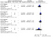
Comparison 33: IM Aminosidine for 20 days vs IM Pentamidine Isethionate x 8 applications in L. braziliensis; FU: 1 year, Outcome 2: Adverse effects
IM aminosidine sulphate versus IV meglumine antimoniate
One RCT including 38 participants with ML (Llanos‐Cuentas 2007) from Peru compared IMAS for 21 days versus IVMA for 28 days.
Primary outcomes: Percentage of participants cured at least three months after the end of treatment
One year after treatment, IMAS 14 mg/kg/day for 21 days had significantly lower cure rates than IVMA 20 mg/kg/ day for 28 days (RR 0.05, 95% CI 0.00 to 0.78; Analysis 34.1).
34.1. Analysis.

Comparison 34: IM Aminosidine 20 mg/kg/d for 28 d vs IV Meglumine Antimoniate 20 mg/kg for 28 d; L. braziliensis; FU: 1 year, Outcome 1: Complete cure
Secondary outcomes: Adverse effects
Participants from the IVMA group had transient and mild electrocardiograph abnormalities that did not need therapeutic intervention. Aminosidine sulphate was associated with pain at the injection site that improved with the application of local heat. Fever, chills, arthralgia, anorexia, and myalgia were seen equally in both groups.
2.5 Pentamidine isethionate
Intravenous pentamidine isethionate (IVPI) versus IV meglumine antimoniate
One RCT including 80 participants (Andersen 2005) from Peru compared IVPI for seven doses versus IVMA for 20 days.
Primary outcome: Percentage of participants cured at least three months after the end of treatment
Six months after treatment, IVPI 2 mg/kg on alternate days for seven doses showed significantly lower cure rates than IVMA 20 mg/kg/day for 20 days (RR 0.45, 95% CI 0.29 to 0.71; Analysis 35.1) in L. braziliensis infections.
35.1. Analysis.

Comparison 35: IV Pentamidine (2mg/kg) seven doses vs IV Meglumine Antimoniate (20 mg/kg) for 20 days in L. braziliensis; FU: 6 months, Outcome 1: Complete cure
Primary outcomes: Adverse effects
More gastrointestinal events were reported in the IVPI group (RR 1.44, 95% CI 0.90 to 2.29; Analysis 35.2). More participants reported musculoskeletal events with IVMA, but the result is imprecise, showing uncertainty (RR 0.80, 95% CI 0.49 to 1.31; Analysis 35.2). Headache was significantly higher in the IVMA group (RR 0.61, 95% CI 0.43 to 0.85; Analysis 35.2). Other minor side effects were lesion pain, paraesthesia, fever or chills, bad taste and cough.
35.2. Analysis.

Comparison 35: IV Pentamidine (2mg/kg) seven doses vs IV Meglumine Antimoniate (20 mg/kg) for 20 days in L. braziliensis; FU: 6 months, Outcome 2: Adverse effects
Secondary outcomes: Recurrence
The number of relapses at six months follow‐up was 12.5% (5/40) of the participants in each group.
Tertiary outcomes: Microbiological or histopathological cure of skin lesions
There were no parasites found in the IVMA group (0/40) but in the IVPI group 35% (14/40) had parasites at two weeks and 7.5% (3/40) at three months post‐therapy.
IM pentamidine isethionate versus IM meglumine antimoniate
Primary outcomes: Percentage of participants cured at least three months after the end of treatment
At one year of follow‐up, pooled results from three RCTs including 226 participants (Alves 2016; Correia 1996; Neves 2011) show no difference in cure rates between IMPI and IMMA (RR 0.95, 95% CI 0.81 to 1.13; I2 = 0%; Analysis 36.1).
Primary outcomes: Adverse effects
Two RCTs including 156 participants (Correia 1996; Neves 2011) found statistically significant differences in adverse effects for arthralgias (RR 0.27, 95% CI 0.11 to 0.69; I2 = 0%; Analysis 36.2) with a smaller risk for pentamidine, but the difference was less clear for myalgias (RR 0.73, 95% CI 0.35 to 1.53; I2 = 0%; Analysis 36.2) and for asthenia or weakness (RR 0.77, 95% CI 0.34 to 1.76; I2 = 0%; Analysis 36.2).
36.2. Analysis.

Comparison 36: IM Pentamidine vs IM Meglumine Antimoniate for 20 days in L. braziliensis; FU: 1 year, Outcome 2: Adverse effects
One RCT including 31 participants (Correia 1996) from Brazil found no clear differences for anorexia (RR 1.24, 95% CI 0.54 to 2.86; Analysis 36.2).
One RCT including 125 participants (Neves 2011) found that mild or moderate adverse effects were reported by 74 (40%) participants, with a higher risk for pain (RR 53.84, 95% CI 3.35 to 864.51; Analysis 36.2) and induration at the site of injection (RR 17.27, 95% CI 1.02 to 292.90; Analysis 36.2) in the pentamidine group, although results were very imprecise (wide confidence intervals).
One RCT including 70 participants (Alves 2016) from Brazil reported non‐significant differences in the percentage of participants with adverse effects among those treated with IMPI compared with IMMA (RR 1.06, 95% CI 0.60 to 1.88). The most common side effects in those treated with IMPI were pain at the injection site, paraesthesias in both legs and increase of CPK; and in the IMMA group were arthralgia, pain at the injection site, myalgia, increase of amylase, headache and ECG alterations.
Pentamidine isethionate 7 days versus pentamidine isethionate 4 days
One RCT including 163 participants (Hu 2015) from Suriname compared both treatment schedules.
Primary outcomes: Percentage of participants cured at least three months after the end of treatment
At 12‐week follow‐up they found no significant differences in the proportion of participants clinically cured (RR 1.14, 95% CI 0.84 to 1.56; Analysis 37.1) among seven‐ and four‐day treatments.
37.1. Analysis.

Comparison 37: Pentamidine Isethionate 7 mg/Kg 4 days vs Pentamidine Isethionate 4 mg/kg 7 days; FU: 12 weeks, Outcome 1: Complete cure
Primary outcomes: Adverse effects
At 12 weeks follow‐up there was no serious toxicity in any group, and none of the reported side effects required discontinuation of treatment in any participant.
Pentamidine isethionate: single dose versus 2 doses versus 3 doses
One RCT including 159 participants with cutaneous leishmaniasis from Brazil (Gadelha 2018), 120 with L. guyanensis, compared a single dose, two or three doses of 7 mg/kg body weight, intramuscularly, with an interval of seven days between each dose.
Primary outcomes: Percentage of participants cured at least three months after the end of treatment
At six months after the end of treatment, cure rates were lower in the single‐dose group (45.3%: 24/53) than in the two‐dose group (81.1%: 43/53; RR 0.56, 95% CI 0.40 to 0.77; Analysis 38.1), and lower than in the three‐dose group (96.2%: 51/53; RR 0.47, 95% CI 0.35 to 0.64; Analysis 38.1).
38.1. Analysis.

Comparison 38: Pentamidine Isethionate (7mg/kg): single dose versus two doses versus three doses in L. guyanensis; FU: 6 months, Outcome 1: Complete cure at 6 months
Cure rates were lower in the two‐dose group than in the three‐dose group: RR 0.84, 95% CI 0.73 to 0.97; Analysis 38.1.
The results were analysed separately for 122 men and 37 women, finding no differences in the effect of the treatments between male and female participants.
Primary outcomes: Adverse effects
Quoted from Gadelha 2018: "No serious adverse events (SAE) occurred and none of the reported adverse events (AE) required discontinuation of therapy in any [participant. Some participants] presented erythema and swelling at the injection site. Asthenia, fever, malaise, and headache were also reported, [and] were more often reported by [participants] treated with three PI doses than by those treated with one or two doses. Pain was the most frequent AE, 128 participants experienced grade 1 and 8 participants grade 2. Twenty‐three participants reported no AE. A 54‐year‐old male participant with a family history of diabetes developed type 2 diabetes mellitus one month after the treatment was concluded. This participant was treated with three PI doses with 1,764 mg PI.
Leukocytosis and discrete CPK, ALP, urea, and creatinine increase were observed one week after the treatment in all the participants. These values returned to normal one month after the treatment. The blood glucose level, measured 30 minutes before and after the injections, showed a significant reduction in groups treated with two and three PI doses."
2.6 Azithromycin versus meglumine antimoniate
Primary outcomes: Percentage of participants cured at least three months after the end of treatment
One RCT including 45 participants (Krolewiecki 2007) from Argentina compared oral azithromycin, 500 mg/day versus intramuscular meglumine antimoniate, 10 mg Sb/kg/day, both for 28 days, with a second cycle of 15 days if necessary.
One RCT including 48 participants (Toledo 2014) from Brazil compared injectable meglumine antimoniate (15 mg/kg/day up to 1215 mg) versus oral azithromycin (AZ) (500 mg/day) during 20 consecutive days.
Pooling both studies, cure rates were lower for those treated with azithromycin (RR 0.51, 95% CI 0.34 to 0.76; I2 = 0%; 2 studies, 93 participants; Analysis 39.1).
39.1. Analysis.

Comparison 39: 500 mg oral Azithromycin vs 1.5 g parenteral Meglumine Antimoniate in L. braziliensis; FU: 6‐12 months, Outcome 1: Complete cure
Please see Table 4, where we rate the certainty of the evidence for this outcome as moderate, which means we are moderately certain about the difference in cure rates between azithromycin and MA at six to 12 months' follow‐up. The true effect is likely to be close to the estimate of the effect, but there is a possibility that it is substantially different.
Primary outcomes: Adverse effects
Toledo 2014 found more adverse effects in the meglumine antimoniate group (49 events) than the azithromycin group (13 events), but there were no significant differences between the groups. The adverse effects were varied between the groups: in the meglumine antimoniate group, the most common were myalgia (22.4%), arthralgia (18.4%), and malaise (8.1%); in the azithromycin group, the most common were diarrhoea (30.8%) and abdominal pain (23.1%). When assessing by severity, most of the adverse effects (51/62; 82.5%) were deemed mild, nine were deemed moderate (14.5%), and two (3.2%) severe (e.g. malaise and vomiting).
Krolewiecki 2007 "found significant differences in tolerance between drugs, with 18 (78%) of 23 participants treated with MA reporting moderate or severe musculoskeletal symptoms (local and general myalgias, arthralgias or injection site pain, or both), with 11 participants (47.8%) requiring a change to the intravenous route to complete therapy." In participants treated with azithromycin, mild‐to‐moderate gastrointestinal issues were most common and occurred in six participants (27%). One participant experienced mild rash that resolved with oral antihistamines. See Table 4.
2.7 Amphotericin B plus oral rehydration solution versus amphotericin B plus normal saline solution (new comparison)
One RCT including 48 participants (Echevarria 2006) from Peru compared adding oral rehydration solution (ORS) versus adding an intravenous saline solution (SS) to treatment with intravenous amphotericin B, aiming to prevent nephrotoxicity.
Primary outcomes: Percentage of participants cured at least three months after the end of treatment
Not assessed.
Primary outcomes: Adverse effects
They only assessed renal function, and found no difference in serum creatinine, creatinine clearance, serum urea, and serum sodium values during treatment, but serum potassium values were lower in the SS group than in the ORS group. Hypokalaemia was much less frequent in the group treated with oral rehydration solution (RR 0.39, 95% CI 0.18 to 0.85; Analysis 40.1).
40.1. Analysis.

Comparison 40: Oral rehydration solution vs intravenous saline solution for patients treated with amphotericine B in L. braziliensis; FU: 42 days, Outcome 1: Adverse effects: hypokaliemia
3. Non‐antimonial topical or intralesional therapies
3.1 Topical paromomycin (aminosidine)
Paromomycin versus placebo
Primary outcomes: Percentage of participants cured at least three months after the end of treatment
One RCT including 76 participants (Arana 2001) from Guatemala compared topical 15% paromomycin in 12% MBCL ointment (PR‐MBCL) for 20 days versus placebo. One year after treatment, topical paromomycin in MBCL had significantly higher cure rates than placebo (RR 2.38, 95% CI 1.50 to 3.80; Analysis 42.1) in L. braziliensis and L. mexicana infections.
42.1. Analysis.

Comparison 42: Topical Paramomycin PR‐MBCL TD for 20d vs placebo TD for 20d in L. panamensis and L. mexicana; FU: 12 months, Outcome 1: Complete cure
One RCT including 53 participants, 48 of them under 18 years old, (Neva 1997) from Honduras compared topical 15% paromomycin in 10% urea ointment (PR‐U) for four weeks versus placebo. Complete cure occurred in 4.3% (1/23) and 3.3% (1/30) of participants in the respective groups 2½ months (11 weeks) after treatment (short‐term primary outcome, excluded from analysis).
Primary outcomes: Adverse effects
One study including 76 participants (Arana 2001) reported that of the 38 participants receiving PR‐MBCL ointment, 57.9% (22) had 30 adverse effects. These effects included local itch 42.1% (16/38), sensation of burning 28.9% (11/38), local pain 21% (8/38) and local oedema 2.6% (1/38). All adverse effects disappeared within one week after finishing the treatment. In Neva 1997 (including 53 participants) no untoward effect of either the paromomycin or placebo ointment was reported or observed.
Secondary outcomes: Recurrence
One study including 76 participants (Arana 2001) reported that between weeks 13 and 52, only 3.1% (1/32) of participants in the paromomycin group and none (0/13) in the placebo group with healed clinical lesions at the 13‐week follow‐up examination experienced reactivation of the lesion; reactivation occurred in the one affected individual around 26 weeks.
In Neva 1997 (53 participants), 10 participants recruited to the trial had already had the condition for nine months. Their lesions persisted, regardless of whether they received drug or placebo, although there was no change in size.
Paromomycin plus gentamicin versus paromomycin alone
Two RCTs from Panama compared topical 15% paromomycin plus 0.5% gentamicin versus 15% paromomycin for 20 days. Sosa 2013 included only 30 participants, was a phase II exploratory study with a very small sample size designed to be a preliminary estimation of the initial clinical cure rate as a basis for calculating sample sizes for the other study, a phase III trial with 399 participants (Sosa 2019).
Primary outcomes: Percentage of participants cured at least three months after the end of treatment
Paromomycin could result in higher rates of final clinical cure of all lesions, but the effects of paromomycin vary and it is possible that it worsens cure rates (RR 1.19, 95% CI 0.74 to 1.91; P = 0.45; I2 = 72%; 2 studies, 429 participants; Analysis 41.1).
41.1. Analysis.

Comparison 41: Topical Paramomycin (15%) + gentamicin (0.5%) vs topical Paramomycin (15%) alone once daily in L. panamensis; FU: 6 months, Outcome 1: Complete cure
Sosa 2019 found no differences between treatments (RR 0.99, 95% CI 0.88 to 1.10; P = 0.94). Sosa 2013 found that paromomycin could result in higher rates of final clinical cure of all lesions, but the 95% confidence interval indicates that it may also make little or no difference (RR 1.63, 95% CI 0.97 to 2.72).
The high statistical heterogeneity found in this meta‐analysis could be due to chance in the results of Sosa 2013, with a very small sample of only 15 participants in each compared group. Small changes in the number of participants cured in one group could result in large changes in the heterogeneity when combining the two studies.
Sosa 2019 performed separate analysis by age of participants and found no differences in cure rates, either in participants aged under 12 years (RR 0.86, 95% CI 0.74 to 1.01) nor in participants aged 12 to 17 years (RR 1.16, 95% CI 0.95 to 1.43; Analysis 41.2).
41.2. Analysis.

Comparison 41: Topical Paramomycin (15%) + gentamicin (0.5%) vs topical Paramomycin (15%) alone once daily in L. panamensis; FU: 6 months, Outcome 2: Complete cure in children
Primary outcomes: Adverse effects
Sosa 2013 found one severe adverse effect ‐ migraine headache ‐ in a participant treated with paromomycin plus 0,5% gentamicin and none in the paromomycin‐alone group.
In Sosa 2019 two participants among those treated with paromomycin plus 0.5% gentamicin suffered severe adverse effects: one participant had a second‐degree burn and the other an appendectomy; one participant in the paromomycin‐alone group had an infection of the surgical site. Minor adverse effects were sustained by more participants in the combined therapy group: contact dermatitis (99/201), pruritus (60/201), application site injury (22/201), application site dermatitis (21/201), application site pain (14/201), application site pruritus (11/201). For the paromomycin‐alone group: contact dermatitis (97/198), pruritus (53/198), application site injury (38/198), application site pain (16/198), application site dermatitis (13/198), and application site pruritus (8/198).
Tertiary outcomes: Speed of healing
In Sosa 2019 median times to initial clinical cure for index lesions were 36 days (95% CI 35 to 49) for paromomycin plus 0.5% gentamicin and 48 days (95% CI 36 to 49) for the paromomycin‐alone group.
Paromomycin‐MBCL versus IM meglumine antimoniate versus PR‐U
One RCT including 120 participants (Armijos 2004) from Ecuador compared topical PR‐MBCL for 30 days, topical PR‐U for 30 days, versus IMMA for 10 days.
Primary outcomes: Percentage of participants cured at least three months after the end of treatment
Complete cure occurred in 47.5% (19/40), 47.5% (19/40) and 70% (28/40) of participants in the respective groups two months after treatment (short‐term primary outcome, excluded from analysis).
Primary outcomes: Adverse effects
Inflammation and soreness were only reported in the paromomycin groups. However, the adverse effects reported in the MA group were epigastric pain, anxiety, nausea, dizziness, joint discomfort, shortness of breath, abdominal and muscular pain. All three groups experienced a number of side effects, including local application‐site reactions such as itch, burning, redness, heat and exudation, and systemic reactions including headache and weakness.
Secondary outcomes: Recurrence
During the 52‐week observation period, 10% (4/40), 5% (2/40) and 12.5% (5/40) of participants experienced infection relapse in the PR‐MBCL, PR‐U and MA groups respectively.
No differences were observed between PR‐MBCL and MA groups (RR 0.80, 95% CI 0.23 to 2.76; Analysis 43.1) nor between PR‐MCBL and PR‐U (RR 2.00, 95% CI 0.39 to 10.31; Analysis 43.1).
43.1. Analysis.

Comparison 43: Paromomycin 15% plus methylbenzonium chloride (PR–MBCL) 30 days versus meglumine antimoniate (MA) 20 mg/kg/day 10 days. FU: 3 months, Outcome 1: Recurrence
Tertiary outcomes: Speed of healing
Speed of healing was longer for PR‐MBCL compared to MA (MD 13.60, 95% CI 7.75 to 19.45; Analysis 43.2), but there were no differences between PR‐MBCL and PR‐U (MD −0.40, 95% CI −7.30 to 6.50; Analysis 43.2).
43.2. Analysis.

Comparison 43: Paromomycin 15% plus methylbenzonium chloride (PR–MBCL) 30 days versus meglumine antimoniate (MA) 20 mg/kg/day 10 days. FU: 3 months, Outcome 2: Speed to healing
The days required for initial healing were 43.1 ± 14.4 (mean ± standard deviation (SD)), 43.5 ± 17 and 29.5 ± 12.2 in the PR‐MBCL, PR‐U and MA groups respectively (the original paper reported that the time to cure was faster for participants treated with IMMA compared to PR‐MBCL (P = 0.001) or to PR‐U (P = 0.002) by the Students' T‐ test).
Paromomycin‐ MBCL combined with 7 days of IM/IV meglumine antimoniate versus paromomycin in MBCL combined with 3 days of IM/IV meglumine antimoniate
One RCT including 89 participants (Soto 1998) from Colombia compared PR‐MBCL for 10 days combined with a short course of IVMA for seven days versus PR‐MBCL for 10 days combined with a short course of IVMA for three days.
Primary outcomes: Percentage of participants cured at least three months after the end of treatment
One year after treatment, PR‐MBCL plus IVMA for seven days had significantly higher cure rates compared with PR‐MBCL plus IVMA for three days (RR 2.88, 95% CI 1.36 to 6.09; Analysis 44.1) in L. braziliensis and L. panamensis infections.
44.1. Analysis.

Comparison 44: Topical Paromomycin PR‐MBCL (TD x 10d) + IV Meglumine Antimoniate x 7 d vs Paromomycin PR‐MBCL + IV Meglumine Antimoniate x 3 d in L. braziliensis and L. panamensis; FU: 1 year, Outcome 1: Complete cure
Paromomycin‐MBCL combined with 7 days of IM/IV meglumine antimoniate versus 7 days of IM/IV meglumine antimoniate
In the same RCT including 89 participants (Soto 1998) from Colombia, PR‐MBCL for 10 days combined with a short course of IVMA for seven days was compared to IVMA for seven days.
Primary outcome: Percentage of participants cured at least three months after the end of treatment
One year after treatment, there was no significant difference in cure rates between PR‐MBCL plus IVMA for seven days and IVMA for seven days (RR 1.08, 95% CI 0.72 to 1.61; Analysis 45.1).
45.1. Analysis.

Comparison 45: Topical Paromomycin PR‐MBCL (TD x 10d) + IV Meglumine Antimoniate x 7 d vs Placebo + IV Meglumine Antimoniate x 7 d in L. braziliensis and L. panamensis; FU: 1 year, Outcome 1: Complete cure
Paromomycin‐MBCL combined with three days of IM/IV meglumine antimoniate versus seven days of IM/IV meglumine antimoniate
This RCT including 89 participants (Soto 1998) from Colombia also compared topical PR‐MBCL for 10 days combined with a short course of IVMA for three days versus IVMA for seven days.
Primary outcomes: Percentage of participants cured at least three months after the end of treatment
One year after treatment, PR‐MBCL plus IVMA for three days had significantly lower cure rates than IVMA for seven days (RR 0.38, 95% CI 0.17 to 0.83; Analysis 46.1).
46.1. Analysis.

Comparison 46: Topical Paromomycin PR‐MBCL (TD x 10d) + IV Meglumine Antimoniate x 3 d vs Placebo + IV Meglumine Antimoniate x 7 d in L. braziliensis and L. panamensis; FU: 1 year, Outcome 1: Complete cure
Paromomycin combined with IV meglumine antimoniate (for 3 and 7 days) versus IM/IV meglumine antimoniate
The same RCT including 89 participants (Soto 1998) from Colombia compared topical PR‐MBCL for 10 days combined with a short course of IVMA for seven days, versus topical PR‐MBCL for 10 days combined with a short course of IVMA for three days versus IVMA for 20 days.
Primary outcome: Percentage of participants cured at least three months after the end of treatment
One year after treatment, IVMA for 20 days had significantly higher cure rates than topical PR‐MBCL plus IVMA for seven days (RR 0.69, 95% CI 0.53 to 0.90; 90 participants; Analysis 47.1) or topical PR‐MBCL plus IVMA for three days (RR 0.24, 95% CI 0.11 to 0.50; 61 participants; Analysis 48.1) in L. braziliensis and L. panamensis infections.
47.1. Analysis.

Comparison 47: Topical Paromomycin PR‐MBCL (TD x 10d) + IV Meglumine Antimoniate x 7 d vs IV Meglumine Antimoniate for 20 d in L. braziliensis and L. panamensis; FU: 1 year, Outcome 1: Complete cure
48.1. Analysis.

Comparison 48: Topical Paromomycin PR‐MBCL (TD x 10d) + IV Meglumine Antimoniate x 3 d vs IV Meglumine Antimoniate for 20 d in L. braziliensis and L. panamensis; FU: 1 year, Outcome 1: Complete cure
Paromomycin in aquaphilic versus intralesional pentamidine
One RCT including 80 participants (Soto 2019) with cutaneous leishmaniasis, L. braziliensis, in Bolivia compared paromomycin‐aquaphilic applied topically daily for 20 days, intralesional pentamidine administered on days 1, 3, and 5, and aquaphilic‐vehicle, applied topically daily for 20 days.
Primary outcomes: Percentage of participants cured at least three months after the end of treatment
Cure rates after six months of follow‐up were similar for paromomycin‐aquaphilic (77.5%: 31/40) and pentamidine (70%: 14/20) for intralesional pentamidine (RR 1.11, 95% CI 0.79 to 1.54; Analysis 49.1), since the confidence interval includes the possibilities of both increased and reduced cure rates.
49.1. Analysis.

Comparison 49: Paromomycin (15%) in Aquaphilic versus intralesional pentamidine (30 mg/ ml) versus Aquaphilic vehicle in L. braziliensis; FU: 6 months, Outcome 1: Complete cure at 6 months
Cure rates were higher for paromomycin‐aquaphilic (77.5%: 31/40) than for aquaphilic vehicle (10%: 2/20), although results are very imprecise (RR 7.75, 95% CI 2.06 to 29.17; Analysis 49.1).
Primary outcomes: Adverse effects
Quote from Soto 2019: "There were several instances of erythema and pruritis in each group of participants, but all were grade 1. IL‐pentamidine was less well tolerated, as expected of an intralesional injection. Although superficial necrosis was obviated by subcutaneous rather than intradermal administration, 40% of participants reported pain; 40% demonstrated erythema; and a lesser percentage had swelling, hard edema, and pruritis."
3.2 Topical aminoglycosides
Formulation of aminoglycosides (WR279396) versus placebo
One RCT including 45 participants (Soto 2002) from Colombia compared topical WR279396 for 20 days versus placebo.
Primary outcomes: Percentage of participants cured at least three months after the end of treatment
Complete cure occurred in 51.5% (17/33) and 41.7% (5/12) of participants in the respective groups two months (70 days) after treatment (short‐term primary outcome, excluded from analysis).
Primary outcomes: Adverse effects
No relevant differences between treatments were observed (RR 1.64, 95% CI 0.69 to 3.86; Analysis 50.1).
50.1. Analysis.

Comparison 50: Topical Aminoglycoside WR279396 versus placebo in L. panamensis; FU: 6 months, Outcome 1: Adverse effects: mild side effects
Tertiary outcomes: Speed of healing
The speed of healing was shorter for the WR279396 group (MD −21.00, 95% CI −38.39 to −3.61; Analysis 50.2).
50.2. Analysis.
Comparison 50: Topical Aminoglycoside WR279396 versus placebo in L. panamensis; FU: 6 months, Outcome 2: Speed to healing
3.3 Topical 3% amphotericin B cream twice a day versus three times a day
A phase II RCT including 80 participants from Colombia (López 2018) compared topical 3% amphotericin B cream twice a day versus three times a day for four weeks.
Primary outcomes: Percentage of participants cured at least three months after the end of treatment.
There were no differences in the rates of final clinical cure of all lesions between treatments, either at three months (RR 1.08, 95% CI 0.57 to 2.08; Analysis 51.1), or at six months (RR 1.12, 95% CI 0.60 to 2.08; Analysis 51.2).
51.1. Analysis.

Comparison 51: Topical 3% amphotericin B cream twice a day versus three times a day in L. panamensis and L. braziliensis; FU: 6 months., Outcome 1: Complete cure at 3 months
51.2. Analysis.

Comparison 51: Topical 3% amphotericin B cream twice a day versus three times a day in L. panamensis and L. braziliensis; FU: 6 months., Outcome 2: Complete cure at 6 months
Primary outcomes: Adverse effects
Only five participants, two in the twice‐a‐day group and three in the three‐times‐a‐day group, reported adverse events related to the cream. The adverse events were all mild (burning sensation, itching and rash) and affected the area around the lesion where the cream was applied.
Tertiary outcomes: Speed of healing
Median time to cure was 57 days for the twice‐a‐day treatment and 62 days for the three‐times‐a‐day regimen.
3.4 Nitric oxide patch
Nitric oxide patch (NOP) versus meglumine antimoniate
One RCT including 143 participants (Lopez‐Jaramillo 2010) from Colombia compared a topical nanofiber NOP (≈ 3.5 μmol NO/cm2/day for 20 days, NOP) with intramuscular meglumine antimoniate (Glucantime, 20 mg/kg/day for 20 days).
Primary outcomes: Percentage of participants cured at least three months after the end of treatment
Complete cure percentages were much lower in participants treated with the NOP (38%) than in those treated with meglumine antimoniate (95%) (RR 0.40, 95% CI 0.29 to 0.55; Analysis 52.1).
52.1. Analysis.

Comparison 52: Nitric oxide patch (≈3.5 μmol NO/cm2 /day, NOP) + IM placebo vs IM Meglumine Antimoniate (20 mg/kg/day) + placebo patch, Outcome 1: Complete cure
Primary outcomes: Adverse effects
Participants treated with NOP had significantly less minor adverse effects, such as fever, headache, myalgia, and arthralgia. More participants had symptoms (itching or pain) at the site of the lesion in the NOP group compared with those treated with meglumine antimoniate.
3.5 Topical imiquimod
Imiquimod combined with IV/IM meglumine antimoniate versus IV/IM meglumine antimoniate
Primary outcomes: Percentage of participants cured at least three months after the end of treatment
In an RCT of 14 participants (Arévalo 2007) from Peru, topical 7.5% imiquimod cream combined with IVMA was compared to IVMA, both for 20 days. Three months after treatment, there was no clear difference in cure rates between topical 7.5% imiquimod plus 20 mg/kg/d IVMA for 20 days and 20 mg/kg/d IVMA for 20 days (RR 1.67, 95% CI 0.88 to 3.15; Analysis 53.1) in L. braziliensis, L. amazonensis, L. mexicana, and L. peruviana infections.
53.1. Analysis.

Comparison 53: 7.5% Imiquimod cream x 20 days + IV Meglumine Antimoniate for 20 days vs IV Meglumine Antimoniate x 20 days in L. braziliensis, L. peruviana, L. mexicana and L. amazonensis; FU: 3 months, Outcome 1: Complete cure
One RCT from Peru (Miranda‐Verástegui 2005), including 40 participants who had previously failed treatment with pentavalent antimony, compared topical 5% imiquimod cream combined with MA (IM in children and IV in older participants) to MA (IM in children and IV in older participants), both for 20 days. One year after treatment, there was no significant difference in cure rates between topical 5% imiquimod plus 20 mg/kg/d IMMA for 20 days and 20 mg/kg/d IMMA for 20 days (RR 0.87, 95% CI 0.58 to 1.30; Analysis 54.1) in L. peruviana and L. braziliensis infections, since the confidence interval includes the possibilities of both increased and reduced cure rates.
54.1. Analysis.

Comparison 54: Topical Imiquimod 5% + IV Meglumine Antimoniate vs placebo + IM/IV Meglumine Antimoniate in L. braziliensis, L. guyanensis and L. peruviana; FU: 1 year, Outcome 1: Complete cure
One RCT from Peru (Miranda‐Verástegui 2009) including 80 participants who had not been previously treated, compared topical imiquimod cream (125 ‐ 250 mg per lesion three times a week) combined with IVMA 20 mg/kg/d IVMA for 20 days versus IVMA plus placebo. One year after treatment, there was no significant difference in cure rates (RR 1.30, 95% CI 0.95 to 1.80; Analysis 54.1), since the confidence interval includes the possibility of both increased and reduced cure rates. Over the study period, only one adverse effect (rash) was recorded, in the imiquimod arm.
Pooled results showed high statistical heterogeneity (I2 = 58%), probably due to differences in the inclusion criteria for participants.
Adding topical imiquimod to IVMA probably makes little or no difference to the chance of healing in L. braziliensis, L. guyanensis and L.peruviana (see Table 5), corresponding to 750 more participants completely cured per 1000 participants (95% CI 435 to 975 more).
Primary outcomes: Adverse effects
In one study including 14 participants (Arévalo 2007), among participants treated with imiquimod, 77% (10/13) reported mild adverse effects (localised itch, erythema and oedema). In participants treated with MA, adverse effects were more severe, as 86% (12/14) reported arthralgia, myalgia, and flu‐like symptoms. Nine of the 14 participants treated with imiquimod had elevated liver enzyme levels, none of which resulted in the discontinuation of therapy. However, one participant voluntarily discontinued treatment with MA on day 15 of re‐treatment because of flu‐like symptoms, arthralgia, and myalgia.
In Miranda‐Verástegui 2005 (40 participants), there was no clear difference in adverse effects between groups for oedema (RR 0.88, 95% CI 0.39 to 1.95; Analysis 54.2), itching (RR 0.67, 95% CI 0.12 to 3.57; Analysis 54.2) or burning (RR 3.00, 95% CI 0.34 to 26.45; Analysis 54.2), since the confidence intervals include the possibilities of both increased and reduced adverse effects. Local pain was reported with equal frequency by participants treated with imiquimod and those treated with the placebo cream. Only mild erythema was more common among participants in the imiquimod group and was evident during most of the 20‐day treatment period, although results are very imprecise due to the wide confidence interval (RR 2.75, 95% CI 1.05 to 7.20; Analysis 54.2). In Miranda‐Verástegui 2009, including 80 participants, they found no differences in adverse effects between imiquimod and placebo creams. See Table 5, where we found low‐certainty evidence for these outcomes.
54.2. Analysis.

Comparison 54: Topical Imiquimod 5% + IV Meglumine Antimoniate vs placebo + IM/IV Meglumine Antimoniate in L. braziliensis, L. guyanensis and L. peruviana; FU: 1 year, Outcome 2: Adverse effects in patients in which previous treatment failed
Imiquimod versus IV meglumine antimoniate
One RCT including 13 participants (Arévalo 2007) from Peru compared topical 7.5% imiquimod cream versus IVMA, both for 20 days.
Primary outcomes: Percentage of participants cured at least three months after the end of treatment
Three months after treatment, cure rates were higher with 20 mg/kg/day IVMA (4/7 participants) versus topical 7.5% imiquimod (0/6 participants) for 20 days, but the results were very imprecise (RR 0.13, 95% CI 0.01 to 1.97; Analysis 55.1), since the confidence interval includes the possibility of both increased and reduced cure rates.
55.1. Analysis.

Comparison 55: 7.5% Imiquimod cream x 20 days vs IV Meglumine Antimoniate x 20 days in L. braziliensis, L. peruviana, L. mexicana and L. amazonensis; FU: 3 months, Outcome 1: Complete cure
Primary outcomes: Adverse effects
Among participants treated with imiquimod, adverse effects are described above. In addition, one participant voluntarily discontinued treatment with MA on day 15 of re‐treatment because of flu‐like symptoms, arthralgia, and myalgia.
4. Physical therapies
4.1 Thermotherapy
Thermotherapy versus placebo
One RCT including 44 participants (Navin 1990) from Guatemala applied three treatments of localised heat at 50 ºC for 30 seconds, at seven‐day intervals compared with placebo.
Primary outcomes: Percentage of participants cured at least three months after the end of treatment
Complete cure occurred in 73% (16/22) and 27% (6/22) of participants in the respective groups two months after treatment (short‐term primary outcome, excluded from analysis).
Primary outcomes: Adverse effects
No participant complained of symptoms related to treatment. Four participants developed moderately‐severe local cellulitis during heat, despite routine treatment with dicloxacillin one hour before and three days after each heat application. Participants treated with heat usually had superficial second‐degree burns where the electrodes were applied.
Tertiary outcomes: Microbiological or histopathological cure of skin lesions
The rate of participants parasitologically‐negative for Leishmania were higher for the thermotherapy group (RR 2.67, 95% CI 1.29 to 5.53; Analysis 56.1).
56.1. Analysis.

Comparison 56: Thermotherapy versus placebo in L.braziliensis and L. mexicana. FU: 13 weeks., Outcome 1: Microbiological or histopathological cure of skin lesions at 13 weeks
Thermotherapy versus IM meglumine antimoniate
Primary outcomes: Percentage of participants cured at least three months after the end of treatment
One RCT including 44 participants (Navin 1990) from Guatemala applied three treatments of localised heat at 50 ºC for 30 seconds, at seven‐day intervals compared to IMMA for 15 days. Complete cure occurred in 59% (13/22) and 73% (16/22) of participants in the respective groups two months after treatment (short‐term primary outcome, excluded from analysis).
One RCT including 292 participants (Vélez 2010) from Colombia compared a single thermotherapy session, which involved the application of 50 ºC, via a device, at the centre and active edge of each lesion to IMMA given for 20 days. Complete cure occurred in 58% of those treated with thermotherapy and 72% of participants treated with IMMA (RR 0.80, 95% CI 0.68 to 0.95; Analysis 57.1). Thermotherapy reduced the chance of healing in L. panamensis and L. braziliensis at six‐month follow‐up, corresponding to 576 fewer participants completely cured per 1000 participants (95% CI 490 fewer to 684 fewer). See Table 6 where we found high‐certainty evidence.
Primary outcomes: Adverse effects
In Navin 1990, no participant complained of symptoms related to treatment. Four participants developed moderately‐severe local cellulitis during heat, despite routine treatment with dicloxacillin one hour before and three days after each heat application. Participants treated with heat usually had superficial second‐degree burns where the electrodes were applied.
In Vélez 2010 from Colombia, the only side effect of thermotherapy was pain at the area four days after the end of treatment.
Tertiary outcomes: Microbiological or histopathological cure of skin lesions
Navin 1990: In the heat‐treated group, by week 13 73% (16/22) of participants were parasitologically negative for Leishmania. By week 9, 73% (16/22) of participants in the MA group had negative cultures (RR 2.67, 95% CI 1.29 to 5.53; Analysis 57.2).
57.2. Analysis.
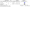
Comparison 57: Thermotherapy (at 50º for 30 seconds) vs Meglumine Antimoniate (20 mg Sb5/kg/day) in L. panamensis and L. braziliensis; FU: 6 months, Outcome 2: Microbiological or histopathological cure of skin lesions
Thermotherapy versus miltefosine
One RCT including 294 participants (Vélez 2010) from Colombia compared a single thermotherapy session involving the application of 50 ºC for 30 seconds over the lesion and surrounding area of each lesion to oral miltefosine given for 28 days.
Primary outcome: Percentage of participants cured at least three months after the end of treatment
There were no differences between treatments, with complete cure occurring in 58.5% of those treated with thermotherapy and 59.4% in participants treated with miltefosine (RR 0.98, 95% CI 0.81 to 1.20; Analysis 58.1), since the confidence interval includes the possibility of both increased and reduced cure rates.
58.1. Analysis.

Comparison 58: Thermotherapy (at 50º for 30 seconds) vs oral Miltefosine (total dose of 4,200 mg) in L. panamensis and L. brazililensis; FU: 6 months, Outcome 1: Complete cure
Secondary outcomes: Adverse effects
Primarily pain at the lesion site after treatment for thermotherapy and gastrointestinal adverse effects for miltefosine, including one participant that developed haematemesis.
Thermotherapy combined with IV meglumine antimoniate versus IV meglumine antimoniate
One RCT including 37 participants (Lobo 2006) from Brazil compared heat therapy given in a single session combined with IVMA after day 28 versus IVMA, both for 20 consecutive days.
Primary outcomes: Percentage of participants cured at least three months after the end of treatment
Complete cure occurred in 5.9% (1/17) and 10% (2/20) of participants in the respective groups at the end of treatment (short‐term primary outcome, excluded from analysis).
Primary outcomes: Adverse effects
No significant adverse effect was seen or reported by participants who submitted to heat therapy, except for secondary bacterial infection after treatment (seven in the heat therapy group and one in the MA group).
4.2 Cryotherapy
Cryotherapy versus placebo cream
One RCT including 50 participants (Soto 2013) from Bolivia compared two sessions of cryotherapy (days 1 and 14) with placebo cream (daily for 20 days).
Primary outcomes: Percentage of participants cured at least three months after the end of treatment
There were no statistically significant differences between treatments; complete cure occurred in 20% of those treated with cryotherapy and in 17% of participants treated with placebo cream (RR 1.20, 95% CI 0.37 to 3.93; Analysis 59.1).
59.1. Analysis.

Comparison 59: Cryotherapy (5–20 seconds) vs placebo cream in L.braziliensis,L. amazonensis, L. guyanensis and L. lainsoni; FU: 6 months, Outcome 1: Complete cure
Primary outcomes: Adverse effects
No participants experienced severe side effects. Cryotherapy was more painful and created more irritation and more vesicles/bullae than topical cream.
Cryotherapy versus IL sodium stibogluconate (ILSB)
One RCT including 50 participants (Soto 2013) from Bolivia compared two sessions of cryotherapy (days 1 and 14) with ILSB (1, 3 and 5 days).
Primary outcomes: Percentage of participants cured at least three months after the end of treatment
Cure rates were higher for participants treated with ILSB (70%) versus only 20% for cryotherapy (RR 0.29, 95% CI 0.12 to 0.71; Analysis 60.1).
60.1. Analysis.

Comparison 60: Cryotherapy (5–20 seconds) vs IL SB (0.008 μL)/mm2 in L.braziliensis,L. amazonensis, L. guyanensis and L. lainsoni; FU: 6 months, Outcome 1: Complete cure
Primary outcomes: Adverse effects
No participants experienced severe side effects. Cryotherapy was more painful and created more irritation and more vesicles/bullae than ILSB.
5. Immuno‐chemotherapy
5.1 Vaccines
Vaccine versus IM meglumine antimoniate
Primary outcomes: Percentage of participants cured at least three months after the end of treatment
Two RCTs from Venezuela including 277 participants (Convit 1987; Convit 1989) compared intradermal vaccine of the L. mexicana amazonensis strain combined with Bacille Calmette Guerin (BCG) versus IMMA. Six months after treatment, there was no difference in cure rates between the vaccine and IMMA (RR 0.96, 95% CI 0.90 to 1.04; I2 = 0%; Analysis 61.1) in L. braziliensis infections.
61.1. Analysis.

Comparison 61: Vaccine three doses vs IM Meglumine Antimoniate (50 mg/kg in 2‐3 series of 20 daily injections) in L. braziliensis; FU: 6 months, Outcome 1: Complete cure
Primary outcomes: Adverse effects
One study of 102 participants (Convit 1987) reported that for the vaccine group 5.2% (3/58) reported slight side effects (shallow necrosis and ulceration at the inoculation site between 1.5 and 1.9 cm in diameter). For the IMMA group 50% (22/44) reported side effects, and some were severe. The commonest moderate side effects were bone and muscle pain, headache and fever. The nine participants with severe side effects had one or more of the following: severe bone and muscle pain (five), hypotension (three), alteration of cardiac rhythm (one), severe colic (one), and paraesthesia (one).
One study of 175 participants (Convit 1989) reported that side effects in the vaccine group occurred in approximately 5% of participants and were limited to local lesions > 10 mm at injection sites or slight fever. Forty‐nine per cent of the participants (25/51) receiving IMMA showed secondary effects, including bone and muscle pain, headache and fever. The severe side effects observed in 17.6% (9/51) of participants included one or more of the following: cardiovascular alterations, such as hypotension or alterations in heart rhythm (four participants), paraesthesia and colic (one participant), and severe osteomuscular pain (five participants). Temporary suspension of treatment was required in the participants with severe side effects.
Secondary outcomes: Recurrence
One study of 175 participants (Convit 1989) reported that no relapses were seen in the two treatment groups.
Tertiary outcomes: Speed of healing
Convit 1987 (102 participants) reported that the average time from start of treatment to cure was 18.3 weeks for the vaccine group and 16.1 weeks for the IMMA group (the original paper reported that the time to cure was not significant (P > 0.05) by the Students' t‐test). In Convit 1989 (175 participants) the average time required for healing were 18.3 weeks in the vaccine group and 16.1 weeks in the IMMA group (the original paper reported that the difference was not statistically significant by variance analysis).
Tertiary outcomes: Development of cell‐mediated immunity
Convit 1987 reported that both groups showed changes in immunological reactivity after treatment, but the differences between them were not statistically significant. Montenegro skin test reactions increased from a mean of 21.88 mm before treatment to 26.8 mm in the vaccine group and from 20.50 mm to 24.7 mm in the IMMA group. In Convit 1989 the average size of the Montenegro reaction increased slightly in the two groups (from 21.6 mm before treatment to 25.4 mm in the vaccine‐treated group and from 20.4 mm to 20.8 mm in the IMMA group) but the differences between the groups and within each group were not statistically significant. While these increases are not significant, they clearly suggest stimulation of the participants' immune system.
Vaccine combined with IM meglumine antimoniate versus IM meglumine antimoniate plus placebo
In one RCT (Machado‐Pinto 2002) from Brazil, subcutaneous vaccination of L. amazonensis strain combined with IMMA was compared to IMMA plus placebo, both for 10 days followed by 10 days of rest. Complete cure occurred in 92.15% (47/51) and 7.84% (4/51) of participants in the respective groups at the end of treatment (short‐term primary outcome, excluded from analysis).
Primary outcomes: Adverse effects
Apart from occasional complaints of pain at the site of injection, no side effects were observed in either group.
Secondary outcomes: Recurrence
No relapses were observed at one year after cessation of treatment.
Tertiary outcomes: Speed of healing
The time taken to be cured was 43 days (CI 40 to 47) in the vaccine combined with IMMA group compared with 102 days (CI 97 to 107) in the IMMA plus placebo group (the original paper reported that the time to cure was faster for participants treated with vaccine plus IMMA (P < 0.0001) by the log rank test).
Intradermal vaccine of biological LEISH‐F2 + MPL‐SE versus sodium stibogluconate
One Phase II RCT from Chile (NCT01011309) compared an intradermal vaccine of biological LEISH‐F2 + MPL‐SE versus sodium stibogluconate and followed participants up to 336 days.
Primary outcomes: Percentage of participants cured at least three months after the end of treatment
At 84 days follow‐up clinical cure rates without rescue treatment at that time were smaller for the vaccine group (4/19; 21%) than for stibogluconate (15/16; 94%) (P = 0.00001).
Primary outcomes: Adverse effects
At 336 days follow‐up they found one serious adverse effect in the 24 participants treated with vaccine (hospitalisation due to grade 3 cellulitis) and one in the 21 treated with stibogluconate (hospitalisation due to antimonial toxicity).
At 336 days follow‐up, the rates of participants affected by non‐serious adverse effects were similar in both groups (RR 0.96, 95% CI 0.86 to 1.08; Analysis 62.1): 23/24 for immunotherapy participants and 21/21 for chemotherapy participants. More frequent non‐serious adverse effects for the immunotherapy participants were injection site problems (erythema or induration or pain, 102 events), increase in ALT (11/24 participants) and blood bilirubin increase (3/24 participants). More frequent non‐serious adverse event for the chemotherapy participants were white blood cell (WBC) decrease (10/21 participants), increase in ALT (9/21 participants), blood haemoglobin decrease (7/21 participants), blood potasium decrease (4/21 participants) and blood bilirubin increase (3/21 participants).
62.1. Analysis.

Comparison 62: Intradermal vaccine of biological LEISH‐F2 + MPL‐SE versus sodium stibogluconate in L. Peruvian. FU: up to 335 days, Outcome 1: Adverse effects not serious
5.2 Intradermal Bacillus Calmette‐Guerin (BCG)
BCG versus IM meglumine antimoniate
One RCT including 93 participants (Convit 1989) from Venezuela compared three doses of intradermal BCG versus IMMA.
Primary outcome: Percentage of participants cured at least three months after the end of treatment
Six months after treatment, intradermal BCG had significantly lower cure rates than IMMA (RR 0.46, 95% CI 0.32 to 0.65; Analysis 63.1) in L. braziliensis infections.
63.1. Analysis.

Comparison 63: BCG (three doses) vs IM Meglumine Antimoniate (50 mg/kg /day 40‐60 injections) in L. braziliensis; FU: 6 months, Outcome 1: Complete cure
Primary outcomes: Adverse effects
For the BCG‐alone group approximately 5% of participants experienced side effects which were limited to local lesions > 10 mm at injection sites or slight fever. In the group receiving IMMA, 48.9% (25/51) of the participants showed secondary effects, including bone and muscle pain, headache and fever. The serious side effects observed in 17.6% (9/51) of participants included one or more of the following: cardiovascular alterations, such as hypotension or alterations in heart rhythm (four participants), paraesthesia and colic (one participant), and severe osteomuscular pain (five participants). Temporary suspension of treatment was required in the participants with severe side effects.
Secondary outcomes: Recurrence
Only one relapse was observed in the BCG‐alone group between three months and 2½ years. No relapses were seen in the other group.
Tertiary outcomes: Development of cell‐mediated immunity
The average size of the Montenegro reaction increased slightly in the two groups (from 18.6 mm to 22.4 mm in the BCG group, and from 20.4 mm to 20.8 mm in the IMMA group), but the differences between groups and within each group were not statistically significant. While these increases are not significant, they clearly suggest stimulation of the participants' immune system.
5.3 Oral pentoxifylline
Pentoxifylline combined with IV sodium stibogluconate versus IV sodium stibogluconate
In one RCT including 23 participants with mucosal leishmaniasis (Machado 2007) from Brazil oral pentoxifylline combined with IVSSG was compared to IVSSG, both for 30 days.
Primary outcomes: Percentage of participants cured at least three months after the end of treatment
Four months after treatment, oral pentoxifylline had a significant synergistic effect with IVSSG 20 mg/Kg/day for 30 days (RR 1.66, 95% CI 1.03 to 2.69; Analysis 64.1) in L. braziliensis infections.
64.1. Analysis.

Comparison 64: Oral pentoxifylline 400 mg 3 times daily for 30d + IV Sodium Stibogluconate 20 mg/kg /d vs placebo + IV Sodium Stibogluconate in L. braziliensis; FU: 4 months, Outcome 1: Complete cure
Primary outcomes: Adverse effects
Mild adverse effects were observed more frequently in the pentoxifylline combined with SSG group, including nausea (three participants), arthralgias (one), and dizziness, abdominal pain, and diarrhoea (one). In the SSG group, one participant complained of anorexia, nausea, and myalgias. No participants in either group discontinued treatment because of these adverse effects.
Tertiary outcomes: Speed of healing
The speed of healing was shorter in the pentoxifylline combined with SSG group (MD −62.00, 95% CI −121.92 to −2.08; Analysis 64.2).
64.2. Analysis.
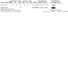
Comparison 64: Oral pentoxifylline 400 mg 3 times daily for 30d + IV Sodium Stibogluconate 20 mg/kg /d vs placebo + IV Sodium Stibogluconate in L. braziliensis; FU: 4 months, Outcome 2: Speed to healing
Pentoxifylline combined with IM meglumine antimoniate versus IM meglumine antimoniate plus placebo
One RCT including 70 participants with cutaneous leishmaniasis (Cossio‐Duque 2015) from Colombia compared intramuscular MA (20 mg/ kg /day x 20 days) plus oral PTX 400 mg thrice daily versus IMMA plus placebo.
Primary outcomes: Percentage of participants cured at least three months after the end of treatment
Cure rates were lower for added pentoxifylline (65% versus 75%), but the confidence interval indicated there might be little or no difference between groups (RR 0.86, 95% CI 0.63 to 1.18; Analysis 65.1).
65.1. Analysis.

Comparison 65: Oral Pentoxifylline (1200 mg/day) + IM Meglumine Antimoniate (20 mg/ kg /day) vs IM Meglumine Antimoniate + placebo; FU: 26 weeks, Outcome 1: Complete cure
Adding pentoxifylline to IMMA probably makes little or no difference to the chance of healing, corresponding to 645 more participants completely cured per 1000 participants (95% CI 473 more to 885 more). Please see Table 7.
Primary outcomes: Adverse effects
No differences between overall frequency and severity of adverse effects were found (Pentoxyfilline = 142 events versus placebo = 140 events). See Table 7.
Pentoxifylline combined with IM meglumine antimoniate versus IM meglumine antimoniate plus placebo
Two RCTs including 197 participants in total (Brito 2014; Brito 2017a) from Brazil compared pentavalent antimony given at a dose of 20 mg/kg a day plus oral pentoxifylline (400 mg) or placebo three times a day for 20 days.
Primary outcomes: Percentage of participants cured at least three months after the end of treatment
There were no differences in complete cure rates among the treatments: 47% pentoxifylline group and 43% placebo group (RR 1.08, 95% CI 0.80 to 1.47; I2 = 0%; Analysis 66.1).
66.1. Analysis.

Comparison 66: Oral Pentoxifylline (1200 mg) + IM Meglumine Antimoniate (20mg/kg) vs IM Meglumine Antimoniate (20mg/kg) + placebo L. braziliensis; FU: 90‐180 days, Outcome 1: Complete cure
Primary outcomes: Adverse effects
In one RCT including 164 participants (Brito 2017a) adverse effects were more common in the pentoxifylline group (37.8% of participants), versus 23% in the placebo group. However, serious AEs were more common in the placebo group (9 participants) compared to the pentoxifylline group (one participant). The most common side effects observed in the pentoxifylline group were myalgia (11), headache (9), nausea (7) and arthralgia (7). In the placebo group it was arthralgia (10) and myalgia (6). No cardiological AE was documented in either group.
In one RCT including 33 participants (Brito 2014), temporary, mild side effects, like arthralgia (four participants), headache (two participants), fever (two participants), and lack of appetite (two participants), were prevalent in the IMMA plus pentoxifylline group (five participants) as well as occurring in the IMMA plus placebo group. Neither group reported nausea, vomiting, or diarrhoea.
5.4 Topical or intralesional granulocyte macrophage colony‐stimulating factor (GM‐CSF)
GM‐CSF combined with IV meglumine antimoniate versus placebo
In one RCT including 22 participants (Santos 2004) from Brazil topical GM‐CSF (for a total of nine applications over three weeks) combined with IVMA was compared to IVMA alone, both for 20 days.
Primary outcomes: Percentage of participants cured at least three months after the end of treatment
Complete cure occurred in 91% (10/11) and 45.5% (5/11) of participants in the respective groups 40 days after treatment (short‐term primary outcome, excluded from analysis).
Primary outcomes: Adverse effects
No side effects were detected in participants in the GM‐CSF combined with IVMA group.
Tertiary outcomes: Speed of healing
The speed of healing occurred in 43 ± 14 (mean ± SD) days in the GM‐CSF combined with IVMA group and 104 ± 79 days in the placebo group (the original paper reported that the time to cure was faster for participants treated with topical GM‐CSF (P = 0.043) by the Mann‐Whitney U test).
GM‐CSF combined with IV sodium stibogluconate versus IV sodium stibogluconate
One RCT including 20 participants (Almeida 1999) from Brazil compared intralesional GM‐CSF combined with IVSSG versus IVSSG, both for 20 days.
Complete cure occurred in 70% (7/10) and 10% (1/10) of participants in the respective groups 20 days after treatment (short‐term primary outcome, excluded from analysis).
Tertiary outcomes: Speed of healing
The speed of healing was shorter in the GM‐CSF group (MD −61.00, 95% CI −104.25 to −17.75; Analysis 67.1).
67.1. Analysis.

Comparison 67: GM‐CSF combined with IV sodium stibogluconate versus IV sodium stibogluconate in L. braziliensis. FU 6 months, Outcome 1: Speed to healing
5.5 Subcutaneous interferon‐gamma (IFN‐γ)
10‐day IV meglumine antimoniate combined with subcutaneous IFN‐γ versus 10‐day and 20‐day IV meglumine antimoniate
One RCT including 44 participants (Arana 1994) from Guatemala compared IVMA for 20 days, IVMA for 10 days versus IVMA for 10 days combined with 10 days of IFN‐γ.
Primary outcomes: Percentage of participants cured at least three months after the end of treatment
There may be little or no difference in cure rates between 10‐day adjuvant subcutaneous IFN‐γ to both 10‐day IVMA therapy (RR 1.22, 95% CI 0.99 to 1.50; Analysis 68.1) and 20‐day IVMA therapy (RR 1.15, 95% CI 0.96 to 1.39; Analysis 68.2).
68.1. Analysis.

Comparison 68: Subcutaneous interferon‐Gamma plus IV MA versus IVMA alone in L. braziliensis and L. mexicana; FU: 1 year, Outcome 1: Complete cure; 10‐day IV MA+10‐day IFN‐γ versus 10‐day IV MA+10‐day placebo
68.2. Analysis.

Comparison 68: Subcutaneous interferon‐Gamma plus IV MA versus IVMA alone in L. braziliensis and L. mexicana; FU: 1 year, Outcome 2: Complete cure; 10‐day IV MA+ 10‐day IFN‐γ versus 20‐day IV MA
Primary outcomes: Adverse effects
A total of 50% (11/22) participants receiving IFN‐γ complained of mild malaise, headache, fever, and/or chills.
Results from the MEDLINE search for adverse effects
We conducted a MEDLINE search for adverse or side effects combined with therapeutic terms, which resulted in 204 hits. However, we could only find general papers reporting known adverse effects.
Discussion
Summary of main results
The studies included in this review addressed varied clinical questions and were methodologically diverse in terms of settings, the range of interventions and comparators, included participants, Leishmania species involved in different geographical areas, and outcome measures.
The heterogeneity in study design, moderate‐to‐high risk of bias, missing standard deviations, and a mix of comparators and dosing regimens did not, in general, enable data to be pooled, permit the accurate and direct comparisons of a considerable number of interventions, or facilitate conclusions about comparative benefit. Nonetheless, this review documents the effectiveness of treatments of existing RCTs and relevant evidence that can be used for policy, practice and future research.
The 75 RCTs included in the review covered 68 comparisons, broadly categorised into five main groups (antimonials, non‐antimonial systemic treatments, non‐antimonial topical or intralesional therapies, physical therapies, immuno‐chemotherapy). The most assessed class of treatment was non‐antimonial systemics, followed by antimonials, of which meglumine antimoniate was the most‐assessed intervention (15 studies). Non‐antimonial topical or intralesional therapies or immuno‐chemotherapy were assessed by similar numbers of studies, and of these, paromomycin, vaccines, pentoxifylline (in combination with other treatments) were most often investigated. The following interventions were assessed by single studies only: amphotericin B, amphotericin B cream, nitric oxide patch, cryotherapy, Bacillus Calmette‐Guerin (BCG), and IFN‐y. None of the included studies tested lipid‐based drugs, deoxycholate formulations, antimonial drugs combined with cytokines and free radicals, immunomodulatory therapy with cytokines, or itraconazole.
Our primary outcomes, percentage of participants cured at least three months after the end of treatment and adverse effects, were well measured. Forty‐five per cent of the studies assessed recurrence, which we also deemed a key outcome. Just over half of the included studies did not report any of our secondary outcomes.
We created 'Summary of findings' tables for relevant comparisons that have been used in previous evidence‐informed guidelines on the America continent (PAHO 2015). The most relevant findings in this updated systematic review are described below.
Compared to placebo:
Intramuscular meglumine antimoniate (IMMA) may increase the likelihood of complete healing (moderate‐certainty evidence), but may also make little to no difference, since the 95% confidence interval (CI) includes the possibilities of both increased and decreased healing (complete cure); IMMA probably increases severe adverse effects such as arthralgia and myalgia (moderate‐certainty evidence), and may make little to no difference to the risk of recurrence, but the 95% CI includes the possibilities of both increased and decreased risk (low‐certainty evidence). These results pertain to L. braziliensis and L. panamensis infections in American cutaneous and mucocutaneous leishmaniasis (ACML); participants were followed up for one year; Table 1.
Oral miltefosine probably improves the likelihood of complete healing (moderate‐certainty evidence); probably increases the risk of nausea and vomiting (moderate‐certainty evidence); and may make little to no difference to recurrence rates (low‐certainty evidence), but the 95% CI includes the possibilities of both increased and decreased risk. These results pertain to L. mexicana, L. panamensis, and L. braziliensis infections in American cutaneous leishmaniasis (ACL); participants were followed up for six months; Table 2.
Compared to meglumine antimoniate (MA) (active treatment):
Oral miltefosine may make little to no difference to the likelihood of complete healing (low‐certainty evidence). Oral miltefosine probably increases the risk of nausea and vomiting (moderate‐certainty evidence). Recurrence was not reported. These results pertain to L. braziliensis, L. panamensis, L. guyanensis and L. amazonensis infections in ACML; participants were followed up for 6 to 12 months; Table 3.
Oral azithromycin probably reduces the likelihood of complete healing (moderate‐certainty evidence). Recurrence was not reported, and we could not calculate the risk of adverse effects. These results pertain to L. braziliensis infections in ACML; participants were followed up for 6 to 12 months; Table 4.
Topical imiquimod and IVMA combined probably makes little to no difference to the likelihood of complete healing (moderate‐certainty evidence). Risk of recurrence was not reported, and we could not calculate the risk of adverse effects. These results pertain to L. braziliensis, L. guyanensis and L.peruviana infections in ACL; participants were followed up for 1 year; Table 5.
Local thermotherapy reduces the likelihood of complete healing (high‐certainty evidence). Risk of recurrence was not reported, and we could not calculate the risk of adverse effects. These results pertain to L. panamensis and L. braziliensis infections in ACL; participants were followed up for six months; Table 6.
Oral pentoxifylline and IMMA combined probably makes little to no difference to the likelihood of complete healing (moderate‐certainty evidence). Risk of recurrence was not reported, and we could not calculate the risk of adverse effects. These results pertain to treatment of CL (the trial did not state the species); participants were followed up for 26 weeks; Table 7.
Some studies did report cure but were not statistically assessed because they did not report cure rates in our pre‐specified time period for the primary outcome. Thus, there was insufficient RCT evidence to evaluate the efficacy of topical aminoglycosides, vaccine combined with IMMA, and topical or intralesional granulocyte macrophage colony‐stimulating factor (GM‐CSF).
The secondary objective of this review was to ascertain whether response to treatment is species‐dependent or associated with their geographical distribution. Heterogeneity of treatments and regimens hindered our assessment of this objective. Nonetheless, we found no disparities of response to pentavalent antimonial drugs among the studied species, including L. braziliensis, L. amazonensis, and L. guyanensis, and neither did other species, such as L. mexicana, L. chagasi, L. peruviana, and L. panamensis, respond differently. However, they could not be analysed specifically because of the small numbers of studies. Moreover, some of the included studies did not identify the species responsible for the illness, precluding a detailed review of the benefits of each therapeutic option. Some other studies reported epidemiological information without any identification of the species causing the disease, which may be considered a methodological bias.
Overall completeness and applicability of evidence
This review was not able to fully address the main objective 'To assess the effects of interventions for all immuno‐competent people who have American Cutaneous and Mucocutaneous Leishmaniasis (ACML)'. Key reasons for this included the scarcity of studies in the mucocutaneous form and the diversity of interventions and Leishmania species.
The review is applicable to immuno‐competent participants. However, some studies did not require HIV‐testing or other tests above and beyond clinically‐evident diseases or chronic conditions referred by the research participants. Recent trials often performed HIV testing whereas older trials in general neither performed or mentioned it. Although most of the evidence came from trials conducted in Brazil, where HIV incidence is relevant, the Pan American Health Organization (PAHO) estimates that cutaneous leishmaniasis/HIV co‐infection incidence is low. As an example, 209 co‐infected cases from Latin America were reported in 2017, which represented a 27% increase compared with the 2016 figures. However, bearing in mind that almost 50,000 cases were reported in Latin America in 2017, it seems reasonable to consider that the presence of co‐infected participants in clinical trials is low (PAHO 2019a).
While most RCTs evaluated the cutaneous form (67 studies), the evidence for the treatment of the mucocutaneous form of leishmaniasis is limited, which hinders the conclusions we can make about the effectiveness and safety of interventions for this form of leishmaniasis. A better option for participants with mucosal leishmaniasis (ML) would be to refer them to a reference treatment centre where better management is possible, using e.g. nasal fibre‐optic examination and molecular tests for improving diagnosis, more rigorous monitoring of toxicity to avoid lethality associated with treatment, and the possibility of offering combined treatments conducted by teams with expertise in treating participants with ML.
Several studies included some participants who tested negative for Leishmania parasite; whilst some might have been real cases (false negatives), others might have been affected by other diseases and therefore might have affected the results. Furthermore, almost a third of the studies either did not mention the causative parasite or made assumptions based on endemic species or previous studies. Of the 54 studies that identified the species, approximately half identified the presence of two or more species. This made it difficult for us to ascertain whether response to treatment is species‐dependent or associated with their geographical distribution, which was our secondary objective. Parasite diversity has a crucial relevance for disease severity and duration, and for clinical manifestations. Supplementing with molecular studies carried out in the different areas of the countries could contribute to establishing the distribution of Leishmania species and strains in the region.
Not all the trials provided information about the primary, secondary and tertiary outcomes prespecified in our review. As previously discussed (González 2009), we consider that assessing the number of persons cured rather than the number of lesions healed is a stronger criterion because it gives a better idea of the real number of participants that achieved complete cure. Most of the studies assessed our primary outcomes, although the timing of the primary efficacy outcome was variable. Over half of the studies did not report any of our secondary outcomes.
A large number of outcomes were not addressed at all: no studies reported the degree of functional and aesthetic impairment, prevention of scarring, or quality of life. The only tertiary outcomes reported were speed of healing and microbiological or histopathological cure of skin lesions; no studies reported change in isolation or PCR of Leishmania and emergence of resistance; development of cell‐mediated immunity; or all‐cause mortality.
In this update, we identified new RCTs that evaluated healing therapies, topical amphotericin B and oral miltefosine treatments, but few or no studies assessing fluconazole, itraconazole, cryotherapy, alternative therapy, nitric oxide patch, photodynamic therapy or laser treatments. Overall, most interventions assessed in this review are likely to be used in regions where a low‐cost treatment is needed. Finally, it was difficult to establish whether response to treatment is age‐dependent, due to the lack of variation in the participant age range, with few participants being either very young or over 50.
Amphotericin B is a second‐line treatment which has been widely used in the lipid or deoxycholate formulations for those cutaneous leishmaniasis cases that have switched into the mucocutaneous form and are resistant to antimonial drugs. However, we found no RCTs comparing lipid‐based drugs and deoxycholate formulations. In fact, we found only one RCT (Neves 2011) comparing meglumine antimoniate, pentamidine and amphotericin B for the treatment of cutaneous leishmaniasis by L. guyanensis. One RCT (López 2018) compared different regimens of amphotericin B (twice versus three times a day) for four weeks for the treatment of CL by L. panamensis or L. braziliensis. Although the design of the study was not meant to determine differences in cure rates by Leishmania species and the number of participants in each group was small, cure rates were higher in participants infected by L. panamensis compared to those infected by L. braziliensis.
Although currently‐available medications are not affordable by low‐income populations, for most of the affected patients in most of the affected countries the medications are available without any direct charge from the national health authorities. Vaccine studies are far from a clinically‐useful product.
Quality of the evidence
Most included trials were poorly designed and reported. Poor reporting is a major issue, since it may affect reliability. Sixty studies (80%) had at least one domain rated as high risk. An adequate randomisation method was reported in 63% (47/75) of the studies. Only 21% (16/75) had an adequate reporting of allocation concealment. Only four studies (5.3%) were at low risk of performance and detection bias. Attrition bias was low in 59 studies (79%). None of the studies were at low risk in all seven domains.
Other factors affecting the quality of the studies are related to attrition bias. Calculation of sample size was performed in 36% (27/75) of the studies, which may affect statistical power and may therefore add to imprecision. Although one study acknowledged a plan of 150 participants, owing to slow recruitment and insufficient evidence of efficacy in the immunotherapy group, enrolment was closed early (NCT01011309). Overall, few participants withdrew or were lost to follow‐up. Most study authors stated that they had performed a compliance assessment but results were seldom shown in the assessed studies. In fact, only two studies reported an assessment of compliance (Sosa 2019; Soto 2004b).
We created 'Summary of findings' tables for seven of the comparisons, and conducted GRADE assessments on the certainty of evidence for the key outcomes. We considered the quality of the evidence as high in only one comparison (thermotherapy compared to MA in L. panamensis and L. braziliensis, Table 6), for complete cure and adverse effects.
We considered the outcomes measured by the other six comparisons to be based on moderate‐ or low‐quality evidence. The main reasons we downgraded the quality of the evidence were imprecision (wide confidence interval of risk ratios), which occurred in four comparisons mainly for complete cure and adverse events (Table 1; Table 2; Table 5; Table 7), high heterogeneity in the results from different studies in one comparison for complete cure (Table 3), and risks of bias for the effect of unblinded evaluators in two comparisons for complete cure and adverse events (Table 3; Table 4).
We found many errors in the write‐up of published study reports, and the quality of reporting was generally poor. It is therefore essential that submitted journal manuscripts undergo rigorous peer review. We agree with the updated systematic review on interventions in Old World Cutaneous Leishmaniasis (Heras‐Mosteiro 2017), that there is a pressing need to prioritise and conduct well‐designed clinical trials to minimise bias.
Potential biases in the review process
We conducted a thorough and comprehensive search with no language restriction to minimise selection bias. We searched for conference papers, theses and ongoing trials to minimise publication bias, and we also wrote to authors from endemic countries to identify further relevant trials. We decided to include only RCTs to further minimise selection bias, since they are designed in such a way that study groups are similar at baseline, allowing researchers the quantification of the effect of the intervention under study (minimising the risk of under‐ and overestimation of the effects of the intervention(s) under study). Selective reporting of dramatic effects from non‐randomised trials without any control group is likely to be very misleading. However, we are aware that RCTs are not methodologically flawless and are therefore not exempt from bias either. Bias can occur throughout the steps of an RCT, i.e. from the allocation of participants to the intervention groups, through the delivery of interventions to the outcome measurement. Studies that report more positive effects have an increased chance of publication than those with less definitive results (Bigby 2003). It seems that the initial published study of an intervention has an increased likelihood of showing positive results.
We know that the search we used for identifying adverse effects of treatments for American cutaneous and mucocutaneous leishmaniasis was not comprehensive. Furthermore, we are aware that Embase would have been a better choice than MEDLINE, and that recommended practice would have been to leave out terms for the condition of interest, regardless of this increasing the number of results retrieved. However, we decided to be consistent with the original plan for the review by using the original adverse effects strategy, as this was a pragmatic approach for managing the workload around sifting and discussing adverse effects information in relation to leishmaniasis. Differences between the protocol and the updated review may have led to bias. For example, we did not contact research centres to identify unpublished literature, although we think it unlikely that missing unpublished literature may have changed the conclusions of our review. In the protocol, we had planned to explore reasons for heterogeneity using sensitivity or subgroup analyses, or both, but we did not do this because there were too few studies to perform these analyses. However, in the updated version we applied the GRADE tool to assess the strength of the evidence and explore magnitude and direction of the estimates, and we followed the Cochrane MECIR standards.
The authors of reviews may introduce bias by misinterpreting the results of a study. To minimise bias, at least two authors of this review assessed the eligibility of the studies and extracted data independently. However, we cannot rule out some bias due to the review authors’ inability to resolve the information considered ambiguous or incomplete. At least two independent review authors assessed the quality of each of the included studies, to evaluate the direction and magnitude of bias relative to the effect of the interventions examined. Critical appraisal was examined by the seven‐domain 'Risk of bias' tool in order to provide direct decision‐makers with the best available evidence.
The fact that 10 studies are awaiting classification and have not yet been incorporated may be a potential source of bias.
Agreements and disagreements with other studies or reviews
Although leishmaniasis constitutes an important public health programme in Latin America, several reviews have found little reliable evidence on the effectiveness of new treatments for the diseases that go beyond the management of the disease with antimonial and several other treatments (Almeida 2011; Brito 2017b; Reveiz 2013). In a clinical practice guideline using the GRADE methodology, Aronson 2016 recommended initiating the systematic management of medication in people suffering from complex cutaneous leishmaniasis with options including amphotericin B deoxycholate, lipid formulations of amphotericin B, pentavalent antimonial (SbV) compounds, and pentamidine. They also suggested the use of miltefosine and antifungal compounds including ketoconazole as options for oral management. It is also important to note that the decision about which treatment and dosage regimen can be subjective for the health professional. Other guidelines consider the possibility of no treatment for uncomplicated CL caused by parasite species with low risk of ML development (Aronson 2016).
In a recent systematic review, the authors compared the use of antimony infiltration therapy versus its systematic use, finding similar efficacy; however, there is insufficient evidence about late mucosal complications and adverse effects. A systematic review that evaluated the effectiveness of thermotherapy through a meta‐analysis of eight studies (Cardona‐Arias 2015) found that the effectiveness of thermotherapy was similar to that of pentavalent antimonial drugs. The authors suggested that it should be used as the first treatment option in areas with low prevalence of mucocutaneous forms and in people with contra‐indications for systemic treatment. The effectiveness of azole therapies (fluconazole, ketoconazole and itraconazole) was evaluated in 37 studies (not only randomised clinical trials) that included 1259 participants in the New World (13 studies) and in the Old World. The authors concluded that the effectiveness in the New World was 62% (95% CI 43 to 77%) and that there were no differences between azole therapy. In their conclusions, they suggested that there is no evidence for the exclusive treatment of azole therapy (Galvão 2017). A similar systematic review, which compared studies worldwide and meta‐analysed eight RCTs (two from America) (López‐Carvajal 2016), evaluated the effectiveness of cryotherapy compared with pentavalent antimonial; although there are no significant differences between the treatments, the cure rates were 63.6% and 74.7% per protocol respectively.
Our review provides additional evidence related to the use of miltefosine, topical treatments, local and combination therapies, and to the efficacy of several treatments (for example, azithromycin). It highlights the differences in the response to the different treatments according to the geographical areas and the diversity of parasite species, among other aspects. However, in contrast to Aronson 2016, which recommends the use of amphotericin B in the treatment of MCL or CL cases associated with increased risk for ML, our review found one study, (Neves 2011) which failed to assess the efficacy of amphotericin B due to high dropout rates (> 75%), and another study (López 2018) whose results do not support its use for the treatment of CL. Aronson 2016 included case/series reports and retrospective studies in their evidence summaries to support their recommendation. In agreement with Galvão 2017, Cardona‐Arias 2015 and López‐Carvajal 2016, we were not able to find significant evidence from RCTs to fully support or refute the use of azoles, nor the use of cryotherapy.
Authors' conclusions
Implications for practice.
This updated review confirms the perception of the unfavourable therapeutic scenario faced by clinicians treating people with cutaneous or mucocutaneous leishmaniasis in Latin America. This is supported by the evident longevity of parenteral antimonials as the reference treatment, in spite of their unfavourable pharmacological and toxicity profile. This clearly reveals that American tegumentary leishmaniasis (ATL) remains a neglected disease, lacking modern therapeutic approaches which could overtake the antimonial era.
Importantly, the review demonstrates that the consolidation of antimonials as the reference treatment for ATL occurred during the 1980s and 1990s, and was based on small‐sized clinical trials, which precluded definite conclusions on their efficacy compared with placebo.
The ATL treatment scenario has been slowly modified since the first RCT, exploring the efficacy of miltefosine, and the emerging interest in the development of local or topical treatments, overcoming systemic toxicity. The combination of systemic and local or topical treatment approaches has been revisited, including immunomodulatory drugs, new formulations of old drugs, and physical measures.
The evidence compiled herein is heterogeneous and mostly not suitable for meta‐analysis, reflecting the influence of the response of each parasite species to treatments and probably to other unmeasured factors related to characteristics of human populations and environmental issues. Clinicians therefore need to be aware that evidence obtained in geographical regions with at least a different prevalent parasite species can not be directly applicable in all of the endemic regions.
The point estimate for systemic antimonial treatment in people with CL reveals that clinicians may expect a cure in up to 80% of cases. This is suboptimal, and a constant stimulus to producing new evidence with therapeutic alternatives. Data on systemic antimonials use in people with ML is scarce, with only two of the eight studies that included participants with ML or MCL assessing this class of treatment. The confidence intervals for effectiveness estimates from these study results were wide, precluding us from drawing any conclusions.
In L. mexicana, L. panamensis, and L. braziliensis infections in ACL compared to placebo, oral miltefosine probably improves the likelihood of complete cure and probably increases the risk of nausea and vomiting (moderate‐certainty evidence). However, miltefosine may make little to no difference to recurrence rates (low‐certainty evidence), but the 95% CI includes the possibility of both increased and decreased risk.
Based on low‐certainty evidence, when compared to meglumine antimoniate (MA), oral miltefosine may make little to no difference to the likelihood of complete cure. We found moderate‐certainty evidence that oral miltefosine probably increases the risk of nausea and vomiting. Recurrence was not reported. These results are based on L. braziliensis, L. panamensis, L. guyanensis and L. amazonensis infections in ACML.
We found moderate‐certainty evidence that azithromycin, a candidate for oral treatment, probably reduces the likelihood of complete cure in L. braziliensis infections in ACML. Recurrence was not reported, and we could not calculate the risk of adverse effects.
The most notable feature of the current review is the phenomenon of emerging topical, local and combination therapies, some of them revisiting old modalities such as injected antimonials or thermotherapy, and newer therapies such as the association of pentoxifylline with antimonials.
Moderate‐certainty evidence indicates that compared to MA, pentoxifylline combined with intramuscular meglumine antimoniate (IMMA) probably makes little to no difference to the likelihood of complete cure for CL. Risk of recurrence was not reported, and we could not calculate the risk of adverse effects.
Thermotherapy emerged as an alternative intervention, with the aim of avoiding systemic toxicity. We found high‐certainty evidence that thermotherapy reduces the likelihood of complete cure in L. panamensis and L. braziliensis infections in ACL when compared with IMMA. Risk of recurrence was not reported, and we could not calculate the risk of adverse effects.
Evidence on the use of topical or locally‐injected treatments for CL including: imiquimod, nitric oxide patch, intralesional GM‐CSF and subcutaneous interferon gamma are scarce, precluding us from drawing any conclusions.
Intralesional infiltration of antimonials has been used for more than three decades across the continent, and has been included as an acceptable treatment modality, despite the lack of standardisation of the procedures for its application and therapeutic scheme.
Moderate‐certainty evidence indicates that in L. braziliensis and L. panamensis infections in ACML, IMMA may increase the likelihood of complete cure, but may also make little to no difference, since the 95% CI includes the possibilities of both increased and decreased healing. IMMA probably leads to more severe adverse effects such as arthralgia and myalgia (moderate‐certainty evidence), and may make little to no difference to the risk of recurrence, but the 95% CI includes the possibilities of both increased and decreased risk (low‐certainty evidence).
In L. braziliensis, L. guyanensis and L.peruviana infections in ACL, topical imiquimod combined with intravenous meglumine antimoniate (IVMA) probably makes little to no difference to the likelihood of complete cure (moderate‐certainty evidence). The studies in this comparison did not measure risk of recurrence, and we could not calculate the risk of adverse effects.
The dearth of evidence on the treatment of the mucosal form of leishmaniasis precludes any conclusion on the comparative efficacy/effectiveness of the available therapeutic modalities. The referral of people with mucosal leishmaniasis to a reference treatment centre would allow clinicians to take advantage of the centre’s expertise in dealing with this rare clinical presentation. Our review highlights the need to explore prognosis in people with leishmaniasis because most of the studies, as is usual in RCTs, preclude the possibility of drawing conclusions applicable to younger children and the oldest adults, mainly those above 50 years. From the reviewed RCTs, there is no evidence of sex as a prognostic factor among people with CL or MCL.
The 10 studies in Studies awaiting classification, once assessed, may alter the conclusions of the review.
Implications for research.
This review identifies an urgent need to improve the quality of future clinical trials to correct the lacunae identified in most of the current evidence. The main pitfalls are the lack of accurate and reliable diagnosis of parasite species affecting study participants, low‐powered trials to prove efficacy/effectiveness, and lack of standardised endpoints measured at standardised follow‐up periods.
Parasite species identification remains a crucial point to understand the large heterogeneity of efficacy estimations for the same treatments in different geographic areas. It seems that some progress has been made to improve the molecular diagnostic assays to achieve this goal, but the challenge of reaching consensus on what specific methodologies and targets should be the standard reference for clinical trials is still lacking. Development and implementation of feasible point‐of‐care molecular assays should be prioritised. A consensus is urgently needed on this issue to overcome this barrier.
Adequately‐powered trials will be challenged by a growing understanding of the inappropriateness of using placebo in participants affected by American cutaneous leishmaniasis. The last Cochrane Review in this field concluded that “There is a need for more evidence of the effectiveness and safety of different anti‐Leishmania drugs compared with placebo in self‐healing forms of leishmaniasis or with traditional first‐line antimonials in the mucocutaneous form, as the base to recommend alternative safe, efficacious and affordable treatments.” Since then, a systematic review on the controversial approach of using placebo as the comparator in clinical trials evaluating efficacy of treatments against American leishmaniasis concluded that the disease had “a low spontaneous cure rate following no‐treatment or placebo use, confirming that this strategy for the control group in CL studies exposes patients to greater morbidity, especially for CL caused by L. braziliensis” (Cota 2016). Further developments will be impacted by the placebo issue, considering that trials conducted with the use of active comparators would require greater numbers of participants to prove efficacy/effectiveness, and longer follow‐up to assess the risk of relapse or risk for mucosal disease. However, the evidence cited above constitutes a strong point to sustain a recommendation against the use of placebo.
Fortunately, the heterogeneity (due to the lack of standardised parameters for endpoint definition) and time‐to‐endpoint follow‐up has recently been assessed, and a promising consensus has been achieved on cutaneous leishmaniasis (Olliaro 2018). Similar efforts should be prioritised for the more challenging mucosal disease presentation. It was clear that long‐term follow‐up looking at estimations of recurrence rate is currently absent. Although it is comprehensible that long‐term follow‐up means a huge financial and logistic investment for most researchers, precluding the likelihood of such endpoints, sponsors need to be receptive to such needs. Investments in better and effective surveillance systems co‐operating with research centres could also be explored.
The scarring process could leave disfiguring sequelae. This could be extremely relevant for people with facial lesions, potentially affecting their quality of life. However, studies of this specific aspect of the disease are scarce, reflecting again its neglected character. Also, standardised instruments evaluating quality‐of‐life aspects in people with American cutaneous leishmaniasis are not yet fully validated (Galvão 2019). Endpoints for quality of life could therefore be included in future RCTs, at least in Brazil, where a promising instrument has recently been developed specifically for cutaneous leishmaniasis (Galvão 2018). Our review has not found RCTs on methods used in promoting wound‐healing as a form of care of cutaneous leishmaniasis. The incorporation of evaluation tools such as the Burn Scar Index, often called the Vancouver Scar Scale (Baryza 1995), was not covered in the reported trials. The validation of those scales in the populations affected by leishmaniasis could allow their incorporation as effective tools for measuring long‐term endpoints linked to quality of life in future RCTs.
A thorough discussion on the adequacy of using antimonials as the active comparator in RCTs for both cutaneous and mucosal diseases should be raised, because of their obvious inadequate pharmacological and toxicity profile. The current evidence on miltefosine and local therapies against cutaneous leishmaniasis seems to merit at least starting the appropriate debate on better comparators for future RCTs.
Treatment effectiveness evaluation through large‐scale pragmatic trials should be considered as a medium‐term goal for the more promising interventions. This is an essential and currently unmet step in the final development of those interventions. As stated above, the co‐operation of researchers with other partners inside the health systems taking care of the affected populations could permit the conduct of that type of challenging trial. Those pragmatic trials would be extremely useful in producing real‐world estimations of effectiveness, and an opportunity for adequate costing procedures, answering the critics of the piggy‐back cost‐effectiveness studies based on data from conventional RCTs.
All the effort invested in better trials producing more accurate and reliable efficacy/effectiveness estimations will be wasted if future trials are not considered as an essential but not a sufficient part of a complete development project which starts with a well‐defined product target profile. This is a very challenging situation for the common scenario of drug re‐purposing, looking for therapeutic alternatives for leishmaniasis, and a strong rationale should be used when the intervention includes such an approach (Jin 2014).
Prognosis and access issues are essential and should also be considered as research targets. Prognostic factors or at least prognostic markers are poorly understood in cutaneous and mucosal disease in Latin America. RCTs constitute an opportunity to identify prognostic factors, although exclusion criteria limit the external validity of such studies nested in RCTs. Future trials, ideally pragmatic ones, should offer the opportunity of identifying the main prognostic factors. Among these, parasite resistance should also be explored. It is time for the establishment of an effective network for monitoring this phenomenon. Miltefosine and newer drugs should be properly monitored for the emergence of resistance. Finally, access deserves some dedicated research, in order to guarantee better options to make the therapies available to the affected population as soon as they are proved efficacious.
Last but not least, it is time to think about the development of safer alternatives for women of childbearing age, for those with co‐morbid conditions, and in immuno‐compromised patients. Pragmatic trials could bring the opportunity to increase knowledge about the effectiveness and safety profiles of the current therapeutic alternatives for these vulnerable populations, improving the equity of the research.
What's new
| Date | Event | Description |
|---|---|---|
| 26 August 2020 | New citation required and conclusions have changed | This update includes studies assessing new types of treatments including azithromycin, amphotericin B, nitric oxide patch, and cryotherapy. Miltefosine appears to be consolidated as a consistent alternative for treating cutaneous leishmaniasis cases associated with the most relevant parasite species (i.e. L. braziliensis, L. guyanensis, and L. panamensis) across South American countries, including Brazil, Colombia, and Bolivia. Finally, the emergence of topicals alone or combined with systemic (oral or parenteral) treatments seem to be the future developing trend for cutaneous leishmaniasis treatment. |
| 26 August 2020 | New search has been performed | A new search led to the addition of 37 new included studies, and we updated the review in line with GRADE and MECIR standards. |
History
Protocol first published: Issue 3, 2004 Review first published: Issue 2, 2009
| Date | Event | Description |
|---|---|---|
| 5 August 2015 | Amended | Author information (affiliation) updated |
| 16 May 2012 | Amended | The lead author's contact details have been edited. |
| 30 April 2008 | Amended | Converted to new review format. |
| 14 February 2007 | New citation required and conclusions have changed | Substantive amendment |
Acknowledgements
We would like to thank the people who co‐authored the previous version of this systematic review.
The Cochrane Skin editorial base wishes to thank Urbà González who was the Key Editor for this updated review; Ching‐Chi Chi who was the Methods Editor; Tarek Turk and Sandra Muvdi who were the clinical referees; Kate Cahill who copy‐edited the review; and Nicole Pitcher who wrote the plain language summary, with support from Elizabeth Royle.
Appendices
Appendix 1. Cochrane Skin Specialised Register (CRSW) search strategy
(Leish* and (mucocutan* or mucos* or american or new world or nose* or nariz or naso* or pharyn* or faring* or laring* or laryn* or paladar* or palat* or cartila* or ear* or oreja* or orelha* or tegument*)) or espundia
Appendix 2. CENTRAL (Cochrane Library) search strategy
#1 MeSH descriptor: [Leishmaniasis, Mucocutaneous] explode all trees #2 espundia:ti,ab,kw #3 #1 or #2 #4 MeSH descriptor: [Leishmaniasis, Cutaneous] explode all trees #5 leish*:ti,ab,kw #6 #4 or #5 #7 (mucocutan* or mucos* or american or new world or nose* or nariz or naso* or pharyn* or faring* or laring* or laryn* or paladar* or palat* or cartila* or ear* or oreja* or orelha* or tegument*):ti,ab,kw #8 #6 and #7 #9 #3 or #8
Appendix 3. MEDLINE (Ovid) search strategy
1. exp Leishmaniasis, Mucocutaneous/ 2. espundia.mp. 3. or/1‐2 4. exp Leishmaniasis, Cutaneous/ 5. leish$.mp. 6. 4 or 5 7. (mucocutan$ or mucos$ or american or new world or nose$ or nariz or naso$ or pharyn$ or faring$ or laring$ or laryn$ or paladar$ or palat$ or cartila$ or ear$ or oreja$ or orelha$ or tegument$).mp. 8. 6 and 7 9. 3 or 8 10. randomized controlled trial.pt. 11. controlled clinical trial.pt. 12. randomized.ab. 13. placebo.ab. 14. clinical trials as topic.sh. 15. randomly.ab. 16. trial.ti. 17. 10 or 11 or 12 or 13 or 14 or 15 or 16 18. exp animals/ not humans.sh. 19. 17 not 18 20. 9 and 19
[Lines 10‐19: Cochrane Highly Sensitive Search Strategy for identifying randomized trials in MEDLINE: sensitivity‐ and precision‐maximizing version (2008 revision); Ovid format, from section 3.6.1 in Lefebvre C, Glanville J, Briscoe S, Littlewood A, Marshall C, Metzendorf M‐I, Noel‐Storr A, Rader T, Shokraneh F, Thomas J, Wieland LS. Technical Supplement to Chapter 4: Searching for and selecting studies. In: Higgins JPT, Thomas J, Chandler J, Cumpston MS, Li T, Page MJ, Welch VA (eds). Cochrane Handbook for Systematic Reviews of Interventions Version 6. Cochrane, 2019. Available from: www.training.cochrane.org/handbook]
Appendix 4. Embase (Ovid) search strategy
1. exp skin leishmaniasis/ 2. leish$.mp. 3. 1 or 2 4. (mucocutan$ or mucos$ or american or new world or nose$ or nariz or naso$ or pharyn$ or faring$ or laring$ or laryn$ or paladar$ or palat$ or cartila$ or ear$ or oreja$ or orelha$ or tegument$).mp. 5. 3 and 4 6. espundia.mp. 7. 5 or 6 8. crossover procedure.sh. 9. double‐blind procedure.sh. 10. single‐blind procedure.sh. 11. (crossover$ or cross over$).tw. 12. placebo$.tw. 13. (doubl$ adj blind$).tw. 14. allocat$.tw. 15. trial.ti. 16. randomized controlled trial.sh. 17. random$.tw. 18. or/8‐17 19. exp animal/ or exp invertebrate/ or animal experiment/ or animal model/ or animal tissue/ or animal cell/ or nonhuman/ 20. human/ or normal human/ 21. 19 and 20 22. 19 not 21 23. 18 not 22 24. 7 and 23
[Lines 8‐18: Based on terms suggested for identifying RCTs in Embase (section 3.6.2) in Lefebvre C, Glanville J, Briscoe S, Littlewood A, Marshall C, Metzendorf M‐I, Noel‐Storr A, Rader T, Shokraneh F, Thomas J, Wieland LS. Technical Supplement to Chapter 4: Searching for and selecting studies. In: Higgins JPT, Thomas J, Chandler J, Cumpston MS, Li T, Page MJ, Welch VA (eds). Cochrane Handbook for Systematic Reviews of Interventions Version 6. Cochrane, 2019. Available from: www.training.cochrane.org/handbook]
Appendix 5. CINAHL (EBSCO) search strategy
S1 TI espundia OR AB espundia S2 TI mucocutaneous leishmaniasis or AB mucocutaneous leishmaniasis S3 TI leish* OR AB leish* S4 TI ( (mucocutan* or mucos* or american or new world or nose* or nariz or naso* or pharyn* or faring* or laring* or laryn* or paladar* or palat* or cartila* or ear* or oreja* or orelha* or tegument*) ) OR AB ( (mucocutan* or mucos* or american or new world or nose* or nariz or naso* or pharyn* or faring* or laring* or laryn* or paladar* or palat* or cartila* or ear* or oreja* or orelha* or tegument*) ) S5 (TI ( (mucocutan* or mucos* or american or new world or nose* or nariz or naso* or pharyn* or faring* or laring* or laryn* or paladar* or palat* or cartila* or ear* or oreja* or orelha* or tegument*) ) OR AB ( (mucocutan* or mucos* or american or new world or nose* or nariz or naso* or pharyn* or faring* or laring* or laryn* or paladar* or palat* or cartila* or ear* or oreja* or orelha* or tegument*) )) AND (S3 AND S4) S6 ((TI ( (mucocutan* or mucos* or american or new world or nose* or nariz or naso* or pharyn* or faring* or laring* or laryn* or paladar* or palat* or cartila* or ear* or oreja* or orelha* or tegument*) ) OR AB ( (mucocutan* or mucos* or american or new world or nose* or nariz or naso* or pharyn* or faring* or laring* or laryn* or paladar* or palat* or cartila* or ear* or oreja* or orelha* or tegument*) )) AND (S3 AND S4)) AND (S1 OR S2 OR S5) S7 (MH "Clinical Trials+") S8 PT clinical trial S9 TX (clinic* n1 trial*) S10 (MH "Random Assignment") S11 TX random* allocat* S12 TX placebo* S13 (MH "Placebos") S14 (MH "Quantitative Studies") S15 TX allocat* random* S16 "randomi#ed control* trial*" S17 TX ( (singl* n1 blind*) or (singl* n1 mask*) ) or TX ( (doubl* n1 blind*) or (doubl* n1 mask*) ) or TX ( (tripl* n1 blind*) or (tripl* n1 mask*) ) or TX ( (trebl* n1 blind*) or (trebl* n1 mask*) ) S18 S7 or S8 or S9 or S10 or S11 or S12 or S13 or S14 or S15 or S16 or S17 S19 S6 AND S18
[Lines S7‐S18 are an adaptatation of the SIGN filter for RCTs for CINAHL via EBSCO]
Appendix 6. LILACS search strategy
(Leish$ and (mucocutan$ or mucos$ or american or new world or nose$ or nariz or naso$ or pharyn$ or faring$ or laring$ or laryn$ or paladar$ or palat$ or cartila$ or ear$ or oreja$ or orelha$ or tegument$)) or espundia
These terms were combined with the Controlled clinical trials topic‐specific query filter.
Appendix 7. MEDLINE (Ovid) Adverse effects search strategy
1. exp product surveillance, postmarketing/ or exp adverse drug reaction reporting systems/ or exp clinical trials, phase iv/ 2. adverse events.mp. 3. adverse effects.mp. 4. exp hypersensitivity/ or exp drug hypersensitivity/ or exp drug eruptions/ or exp hypersensitivity, delayed/ or exp hypersensitivity, immediate/ 5. exp hypersensitivity, immediate/ or exp anaphylaxis/ or exp conjunctivitis, allergic/ or exp dermatitis, atopic/ or exp food hypersensitivity/ or exp respiratory hypersensitivity/ or exp urticaria/ 6. side effect$.mp. 7. exp Poisoning/ 8. exp Substance‐Related Disorders/ 9. exp Drug Toxicity/ 10. exp Abnormalities, Drug‐Induced/ 11. exp Teratogens/ 12. exp Mutagens/ 13. exp Carcinogens/ 14. exp dermatitis, contact/ or exp dermatitis, allergic contact/ or exp dermatitis, irritant/ or exp dermatitis, phototoxic/ 15. photoallergic reactions.mp. 16. exp dermatitis, allergic contact/ or exp dermatitis, photoallergic/ 17. sensitization.mp. 18. fetal abnormalities.mp. 19. exp Drug Monitoring/ 20. harm$ effects.mp. 21. (toxic effects or drug effects).mp. 22. undesirable effect$.mp. 23. (safe or safety).mp. 24. toxicity.mp. 25. noxious.mp. 26. serious reaction$.mp. 27. complication$.mp. 28. tolerability.mp. 29. (adverse adj3 (effect$ or reaction$ or event$ or outcome$)).mp. 30. Tachyphylaxis/ci, de [Chemically Induced, Drug Effects] 31. *Itraconazole/ 32. *Ketoconazole/ 33. *Paromomycin/ 34. *Allopurinol/ 35. *Amphotericin B/ 36. aminosidine sulphate.mp. 37. pentamidine isethionate.mp. or *Pentamidine/ 38. *Aminoglycosides/ 39. miltefosine.mp. 40. thermotherapy.mp. 41. *Granulocyte‐Macrophage Colony‐Stimulating Factor/ 42. *Mefloquine/ 43. *Immunotherapy/ 44. *BCG Vaccine/ or bacillus calmette guerin.mp. 45. *Meglumine/ 46. sodium stibogluconate.mp. 47. meglumine antimoniate.mp. 48. imiquimod.mp. 49. IFN‐gamma.mp. 50. new world.mp. 51. American.mp. 52. cutaneous leishmaniasis.mp. or exp Leishmaniasis, Cutaneous/ 53. mucocutaneous leishmaniasis.mp. or exp Leishmaniasis, Mucocutaneous/ 54. 50 or 51 55. 52 and 54 56. 53 or 55 57. or/1‐30 58. or/31‐49 59. 56 and 57 and 58
Data and analyses
Comparison 1. IM Meglumine Antimoniate (20 mg/kg/d for 20 d) vs placebo (3 tablets/4 times a day for 28 d) in L. braziliensis and L. panamensis; FU: 3 months and 1 year.
| Outcome or subgroup title | No. of studies | No. of participants | Statistical method | Effect size |
|---|---|---|---|---|
| 1.1 Complete cure | 2 | 157 | Risk Ratio (M‐H, Random, 95% CI) | 4.23 [0.84, 21.38] |
| 1.2 Adverse effects (FU one year) | 1 | 134 | Risk Ratio (M‐H, Random, 95% CI) | 1.51 [1.17, 1.96] |
| 1.3 Recurrence (FU one year) | 1 | 127 | Risk Ratio (M‐H, Random, 95% CI) | 1.79 [0.17, 19.26] |
Comparison 2. 10‐day IM Meglumine Antimoniate 20 mg/Kg/day vs 20‐day IM Meglumine Antimoniate in L. braziliensis and L. panamensis; FU: 1 year.
| Outcome or subgroup title | No. of studies | No. of participants | Statistical method | Effect size |
|---|---|---|---|---|
| 2.1 Complete cure | 1 | 136 | Risk Ratio (M‐H, Random, 95% CI) | 1.17 [0.76, 1.79] |
| 2.2 Complete cure in children | 1 | 54 | Risk Ratio (M‐H, Random, 95% CI) | 0.87 [0.58, 1.30] |
| 2.2.1 Under 5 years old | 1 | 17 | Risk Ratio (M‐H, Random, 95% CI) | 0.44 [0.05, 4.02] |
| 2.2.2 5 to 15 years old | 1 | 37 | Risk Ratio (M‐H, Random, 95% CI) | 0.89 [0.59, 1.34] |
| 2.3 Adverse effects | 1 | Risk Ratio (M‐H, Random, 95% CI) | Subtotals only | |
| 2.3.1 Anorexia | 1 | 136 | Risk Ratio (M‐H, Random, 95% CI) | 1.00 [0.52, 1.94] |
| 2.3.2 Headache | 1 | 136 | Risk Ratio (M‐H, Random, 95% CI) | 0.55 [0.29, 1.01] |
| 2.3.3 Myalgias | 1 | 136 | Risk Ratio (M‐H, Random, 95% CI) | 1.08 [0.55, 2.12] |
| 2.3.4 Malaise | 1 | 136 | Risk Ratio (M‐H, Random, 95% CI) | 0.56 [0.27, 1.18] |
| 2.3.5 Arthralgias | 1 | 136 | Risk Ratio (M‐H, Random, 95% CI) | 0.36 [0.14, 0.94] |
Comparison 3. IV Meglumine Antimoniate 20 mg/kg/d for 15 d vs no treatment in L. panamensis; FU: 12 months.
| Outcome or subgroup title | No. of studies | No. of participants | Statistical method | Effect size |
|---|---|---|---|---|
| 3.1 Complete cure | 1 | 50 | Risk Ratio (M‐H, Random, 95% CI) | 13.24 [0.83, 210.87] |
| 3.2 Recurrence | 1 | 50 | Risk Ratio (M‐H, Random, 95% CI) | 1.55 [0.35, 6.85] |
Comparison 4. IV Meglumine Antimoniate for 7 days + placebo topically TD for 10 d vs IV Meglumine Antimoniate for 20 d in L. braziliensis & L. panamensis; FU: 1 year.
| Outcome or subgroup title | No. of studies | No. of participants | Statistical method | Effect size |
|---|---|---|---|---|
| 4.1 Complete cure | 1 | 61 | Risk Ratio (M‐H, Random, 95% CI) | 0.64 [0.44, 0.92] |
Comparison 5. IV Meglumine Antimoniate 15% (14 mg/kg/d) vs IV Meglumine Antimoniate 30% (28 mg/kg/d) ; FU: 2 years.
| Outcome or subgroup title | No. of studies | No. of participants | Statistical method | Effect size |
|---|---|---|---|---|
| 5.1 Complete cure (CL plus MCL) | 1 | 43 | Risk Ratio (M‐H, Random, 95% CI) | 0.67 [0.39, 1.14] |
| 5.2 Complete cure CL form | 1 | 26 | Risk Ratio (M‐H, Random, 95% CI) | 1.50 [0.81, 2.78] |
| 5.3 Complete cure MCL form | 1 | 17 | Risk Ratio (M‐H, Random, 95% CI) | 1.43 [0.53, 3.86] |
Comparison 6. Meglumine Antimoniate low dosage (5mg/kg/day) (30 to 120 days) up to vs high dosage (20‐30 mg+/kg/day) (20‐30 days) in L. braziliensis; FU: 12‐45 months.
| Outcome or subgroup title | No. of studies | No. of participants | Statistical method | Effect size |
|---|---|---|---|---|
| 6.1 Complete cure | 2 | 89 | Risk Ratio (M‐H, Random, 95% CI) | 1.10 [0.77, 1.58] |
| 6.2 Adverse effects | 1 | 23 | Risk Ratio (M‐H, Random, 95% CI) | 3.27 [0.83, 12.95] |
Comparison 7. 10‐day IV Meglumine Antimoniate 20mg/kg/day + 10‐day placebo versus 20‐day IV Meglumine Antimoniate in L. braziliensis and L. mexicana; FU: 1 year.
| Outcome or subgroup title | No. of studies | No. of participants | Statistical method | Effect size |
|---|---|---|---|---|
| 7.1 Complete cure | 1 | 44 | Risk Ratio (M‐H, Random, 95% CI) | 0.95 [0.73, 1.23] |
| 7.2 Adverse effects: arthralgia | 1 | 44 | Risk Ratio (M‐H, Random, 95% CI) | 0.25 [0.03, 2.06] |
Comparison 8. Intralesional antimony (650 μg/mm2) vs placebo in L. braziliensis,L. amazonensis,L. guyanensis and L. lainsoni; FU: 6 months.
| Outcome or subgroup title | No. of studies | No. of participants | Statistical method | Effect size |
|---|---|---|---|---|
| 8.1 Complete cure | 1 | 60 | Risk Ratio (M‐H, Random, 95% CI) | 5.00 [1.94, 12.89] |
Comparison 9. Meglumine antimoniate 20 mg/kg/day plus oral tamoxifen 40 mg/day versus meglumine antimoniate alone in L. braziliensis; FU: 3‐6 months.
| Outcome or subgroup title | No. of studies | No. of participants | Statistical method | Effect size |
|---|---|---|---|---|
| 9.1 Complete cure at 3‐6 months | 1 | 54 | Risk Ratio (M‐H, Random, 95% CI) | 1.33 [0.82, 2.16] |
| 9.1.1 Cure at 3 months | 1 | 27 | Risk Ratio (M‐H, Random, 95% CI) | 1.25 [0.67, 2.32] |
| 9.1.2 Cure at 6 months | 1 | 27 | Risk Ratio (M‐H, Random, 95% CI) | 1.46 [0.67, 3.19] |
| 9.2 Recurrence at 6 months | 1 | 27 | Risk Ratio (M‐H, Random, 95% CI) | 0.62 [0.06, 6.09] |
Comparison 10. Meglumine antimoniate 20 mg/kg/day plus topical tamoxifen for 20 days (0.1% citrate) versus meglumine antimoniate alone in L. braziliensis; FU: 3‐6 months.
| Outcome or subgroup title | No. of studies | No. of participants | Statistical method | Effect size |
|---|---|---|---|---|
| 10.1 Complete cure at 3‐6 months | 1 | 52 | Risk Ratio (M‐H, Random, 95% CI) | 0.87 [0.47, 1.63] |
| 10.1.1 Cure at 3 months | 1 | 26 | Risk Ratio (M‐H, Random, 95% CI) | 0.85 [0.38, 1.90] |
| 10.1.2 Cure at 6 months | 1 | 26 | Risk Ratio (M‐H, Random, 95% CI) | 0.91 [0.34, 2.47] |
| 10.2 Recurrence at 6 months | 1 | 26 | Risk Ratio (M‐H, Random, 95% CI) | 0.68 [0.07, 6.61] |
Comparison 11. IV meglumine antimoniate (IVMA) plus antihelminthic treatment versus IVMA plus placebo in L.braziliensis; FU: 90 days.
| Outcome or subgroup title | No. of studies | No. of participants | Statistical method | Effect size |
|---|---|---|---|---|
| 11.1 Complete cure | 1 | 90 | Risk Ratio (M‐H, Random, 95% CI) | 0.77 [0.48, 1.25] |
Comparison 12. IM Sodium Stibogluconate 20 mg/kg/d for 20d vs IM Meglumine Antimoniate (20 mg/kg/d for 20d) in L. panamensis; FU: 6 months.
| Outcome or subgroup title | No. of studies | No. of participants | Statistical method | Effect size |
|---|---|---|---|---|
| 12.1 Complete cure | 1 | 114 | Risk Ratio (M‐H, Random, 95% CI) | 1.07 [0.88, 1.30] |
| 12.2 Adverse effect Overall | 1 | 59 | Risk Ratio (M‐H, Random, 95% CI) | 1.22 [0.78, 1.91] |
| 12.3 Adverse effects | 1 | Risk Ratio (M‐H, Random, 95% CI) | Subtotals only | |
| 12.3.1 Myalgias | 1 | 114 | Risk Ratio (M‐H, Random, 95% CI) | 0.78 [0.50, 1.22] |
| 12.3.2 Headache | 1 | 114 | Risk Ratio (M‐H, Random, 95% CI) | 0.68 [0.37, 1.26] |
| 12.3.3 Metallic taste | 1 | 114 | Risk Ratio (M‐H, Random, 95% CI) | 0.49 [0.27, 0.92] |
| 12.3.4 Abdominal pain | 1 | 114 | Risk Ratio (M‐H, Random, 95% CI) | 0.78 [0.32, 1.94] |
| 12.4 Recurrence | 1 | 119 | Risk Ratio (M‐H, Random, 95% CI) | 0.96 [0.45, 2.05] |
| 12.5 Microbiological or histopathological cure of skin lesions | 1 | 59 | Risk Ratio (M‐H, Random, 95% CI) | 1.00 [0.85, 1.19] |
Comparison 13. IM Sodium Stibogluconate (branded) vs IM Sodium Stibogluconate (generic). Dose: 20 mg/kg/d for 20 d in L.panamensis; FU: 6 months.
| Outcome or subgroup title | No. of studies | No. of participants | Statistical method | Effect size |
|---|---|---|---|---|
| 13.1 Complete cure | 1 | 64 | Risk Ratio (M‐H, Random, 95% CI) | 1.11 [0.82, 1.51] |
| 13.2 Adverse effects | 1 | Risk Ratio (M‐H, Random, 95% CI) | Subtotals only | |
| 13.2.1 Myalgias | 1 | 64 | Risk Ratio (M‐H, Random, 95% CI) | 1.93 [1.04, 3.58] |
| 13.2.2 Headache | 1 | 64 | Risk Ratio (M‐H, Random, 95% CI) | 1.67 [0.65, 4.25] |
| 13.2.3 Metallic taste | 1 | 64 | Risk Ratio (M‐H, Random, 95% CI) | 2.14 [0.79, 5.82] |
| 13.2.4 Abdominal pain | 1 | 64 | Risk Ratio (M‐H, Random, 95% CI) | 3.00 [0.85, 10.63] |
13.1. Analysis.

Comparison 13: IM Sodium Stibogluconate (branded) vs IM Sodium Stibogluconate (generic). Dose: 20 mg/kg/d for 20 d in L.panamensis; FU: 6 months, Outcome 1: Complete cure
Comparison 14. Low dose of IV sodium stibogluconate 20 days versus high doses in L.panamensis and L. chagasi; FU: 1 year.
| Outcome or subgroup title | No. of studies | No. of participants | Statistical method | Effect size |
|---|---|---|---|---|
| 14.1 Adverse effects | 1 | Risk Ratio (M‐H, Random, 95% CI) | Subtotals only | |
| 14.1.1 Muscle‐joint stiffness | 1 | 40 | Risk Ratio (M‐H, Random, 95% CI) | 1.07 [0.64, 1.78] |
| 14.1.2 Increses in liver enzymes | 1 | 40 | Risk Ratio (M‐H, Random, 95% CI) | 0.90 [0.49, 1.68] |
| 14.1.3 Mild leukopenia | 1 | 40 | Risk Ratio (M‐H, Random, 95% CI) | 1.81 [0.18, 18.39] |
| 14.1.4 Electrographic abnormalities | 1 | 40 | Risk Ratio (M‐H, Random, 95% CI) | 0.90 [0.26, 3.12] |
Comparison 15. IV Sodium Stibogluconate 20mg/kg for 28 days vs IV Sodium Stibogluconate for 40 days in L. braziliensis; FU: 1 year.
| Outcome or subgroup title | No. of studies | No. of participants | Statistical method | Effect size |
|---|---|---|---|---|
| 15.1 Complete cure | 1 | 40 | Risk Ratio (M‐H, Random, 95% CI) | 0.83 [0.47, 1.47] |
Comparison 16. IL Sodium Stibogluconate (650 μg; Sb 8 μL/mm2) vs IL pentamidine (240 μg; 8 μL/mm2) in L. braziliensis and L. braziliensis/amazonensis/lainsoni/guyanensis; FU: 6 months.
| Outcome or subgroup title | No. of studies | No. of participants | Statistical method | Effect size |
|---|---|---|---|---|
| 16.1 Complete cure | 2 | 120 | Risk Ratio (M‐H, Random, 95% CI) | 0.98 [0.78, 1.23] |
| 16.2 Adverse effects | 2 | Risk Ratio (M‐H, Random, 95% CI) | Subtotals only | |
| 16.2.1 Myalgia | 2 | 120 | Risk Ratio (M‐H, Random, 95% CI) | 3.93 [0.45, 34.54] |
| 16.2.2 Local irritation | 2 | 120 | Risk Ratio (M‐H, Random, 95% CI) | 1.19 [0.55, 2.58] |
| 16.2.3 Local pain | 2 | 120 | Risk Ratio (M‐H, Random, 95% CI) | 1.74 [0.79, 3.83] |
| 16.3 Recurrence | 1 | 60 | Risk Ratio (M‐H, Random, 95% CI) | 5.00 [0.25, 99.95] |
Comparison 17. Oral ketoconazole 200 mg for 28 days vs IM Meglumine Antimoniate 20 mg/kg for 20 days in L. panamensis and L. mexicana; FU: 3 months.
| Outcome or subgroup title | No. of studies | No. of participants | Statistical method | Effect size |
|---|---|---|---|---|
| 17.1 Complete cure | 1 | 41 | Risk Ratio (M‐H, Random, 95% CI) | 1.06 [0.71, 1.58] |
| 17.2 Adverse effects | 1 | Risk Ratio (M‐H, Random, 95% CI) | Subtotals only | |
| 17.2.1 Laboratory abnormalities | 1 | 41 | Risk Ratio (M‐H, Random, 95% CI) | 0.58 [0.25, 1.32] |
| 17.3 Speed to healing (% of complete re‐epithelization of lesions at 1 month in cured patients) | 1 | 29 | Risk Ratio (M‐H, Random, 95% CI) | 1.04 [0.54, 2.03] |
| 17.4 Miocrobiological cure of skin lesions (% in cured patients) | 1 | 29 | Risk Ratio (M‐H, Random, 95% CI) | 0.81 [0.46, 1.43] |
Comparison 18. Oral ketoconazole 200 mg vs oral placebo for 28 days in L. panamensis and L. mexicana; FU: 3 months.
| Outcome or subgroup title | No. of studies | No. of participants | Statistical method | Effect size |
|---|---|---|---|---|
| 18.1 Complete cure | 1 | 33 | Risk Ratio (M‐H, Random, 95% CI) | 17.22 [1.13, 262.82] |
Comparison 19. 300‐450 mg oral Fluconazole vs 20mg/kg/d IV Meglumine Antimoniate 20 mg/kg in L. braziliensis; FU: 3‐6 months.
| Outcome or subgroup title | No. of studies | No. of participants | Statistical method | Effect size |
|---|---|---|---|---|
| 19.1 Complete cure | 2 | 173 | Risk Ratio (M‐H, Random, 95% CI) | 0.60 [0.37, 0.96] |
| 19.2 Adverse effects | 1 | 53 | Risk Ratio (M‐H, Random, 95% CI) | 1.07 [0.52, 2.20] |
| 19.3 Needed rescue therapy | 1 | 53 | Risk Ratio (M‐H, Random, 95% CI) | 2.20 [1.09, 4.46] |
| 19.4 Speed to healing (days) | 1 | 53 | Mean Difference (IV, Random, 95% CI) | 40.40 [11.27, 69.53] |
Comparison 20. Oral Allopurinol 20 mg/kg/d (4 doses) for 15d vs Allopurinol + IM Meglumine Antimoniate 20 mg/kg (same regimen) in L. panamensis; FU: 12 months.
| Outcome or subgroup title | No. of studies | No. of participants | Statistical method | Effect size |
|---|---|---|---|---|
| 20.1 Complete cure | 1 | 60 | Risk Ratio (M‐H, Random, 95% CI) | 1.08 [0.82, 1.42] |
| 20.2 Recurrence | 1 | 60 | Risk Ratio (M‐H, Random, 95% CI) | 0.70 [0.07, 7.30] |
Comparison 21. Oral Allopurinol 20 mg/kg/d (4 doses) x 15d vs. IV Meglumine Antimoniate (same regimen) in L. panamensis; FU: 1 year.
| Outcome or subgroup title | No. of studies | No. of participants | Statistical method | Effect size |
|---|---|---|---|---|
| 21.1 Complete cure | 1 | 58 | Risk Ratio (M‐H, Random, 95% CI) | 2.20 [1.34, 3.60] |
| 21.2 Recurrence | 1 | 58 | Risk Ratio (M‐H, Random, 95% CI) | 0.66 [0.06, 6.88] |
Comparison 22. Oral Allopurinol 20 mg/kg/d + IV Meglumine Antimoniate (20 mg/kg/d (4 doses) for 15d) vs IV Meglumine Antimoniate (same regimen) in L. panamensis; FU: 1 year.
| Outcome or subgroup title | No. of studies | No. of participants | Statistical method | Effect size |
|---|---|---|---|---|
| 22.1 Complete cure | 1 | 68 | Risk Ratio (M‐H, Random, 95% CI) | 2.04 [1.25, 3.34] |
| 22.2 Recurrence | 1 | 68 | Risk Ratio (M‐H, Random, 95% CI) | 0.94 [0.14, 6.31] |
22.2. Analysis.

Comparison 22: Oral Allopurinol 20 mg/kg/d + IV Meglumine Antimoniate (20 mg/kg/d (4 doses) for 15d) vs IV Meglumine Antimoniate (same regimen) in L. panamensis; FU: 1 year, Outcome 2: Recurrence
Comparison 23. Oral Allopurinol 300 mg for 28d vs IM Meglumine Antimoniate 20mg/kg/d for 20 d in L braziliensis and L. panamensis ; FU: 12 month.
| Outcome or subgroup title | No. of studies | No. of participants | Statistical method | Effect size |
|---|---|---|---|---|
| 23.1 Complete cure | 1 | 127 | Risk Ratio (M‐H, Random, 95% CI) | 0.39 [0.26, 0.58] |
| 23.2 Recurrence | 1 | 127 | Risk Ratio (M‐H, Random, 95% CI) | 1.68 [0.29, 9.69] |
Comparison 24. Oral Allopurinol 20 mg/k/d + IV Sodium Stibogluconate (20 mg/kg/d (4 doses) x 15d) vs IV Sodium Stibogluconate (same dose) in L. braziliensis; FU: 1 year.
| Outcome or subgroup title | No. of studies | No. of participants | Statistical method | Effect size |
|---|---|---|---|---|
| 24.1 Complete cure | 1 | 100 | Risk Ratio (M‐H, Random, 95% CI) | 1.82 [1.23, 2.70] |
| 24.2 Complete cure; Oral AL plus IVSSG vs IVSSG | 2 | 168 | Risk Ratio (M‐H, Random, 95% CI) | 1.90 [1.40, 2.59] |
Comparison 25. Oral Allopurinol 20 m/k/d + IV Sodium Stibogluconate (20 mg/kg/d (4 doses) for 28d) vs IV Sodium Stibogluconate (same dose); FU: 12 months.
| Outcome or subgroup title | No. of studies | No. of participants | Statistical method | Effect size |
|---|---|---|---|---|
| 25.1 Complete cure | 1 | 81 | Risk Ratio (M‐H, Random, 95% CI) | 0.62 [0.38, 1.03] |
| 25.2 Recurrence | 2 | 181 | Risk Ratio (M‐H, Random, 95% CI) | 1.16 [0.73, 1.85] |
Comparison 26. Oral Allopurinol 20 mg/kg/d (4 doses) for 15 d vs no treatment in L. panamensis; FU: 12 months.
| Outcome or subgroup title | No. of studies | No. of participants | Statistical method | Effect size |
|---|---|---|---|---|
| 26.1 Complete cure | 1 | 42 | Risk Ratio (M‐H, Random, 95% CI) | 28.38 [1.83, 439.72] |
| 26.2 Recurrence | 1 | 42 | Risk Ratio (M‐H, Random, 95% CI) | 0.34 [0.03, 3.46] |
Comparison 27. Oral Allopurinol 300 mg 28 days vs placebo in L. braziliensis and L. panamensis; FU: 12 months.
| Outcome or subgroup title | No. of studies | No. of participants | Statistical method | Effect size |
|---|---|---|---|---|
| 27.1 Complete cure | 1 | 120 | Risk Ratio (M‐H, Random, 95% CI) | 1.06 [0.61, 1.85] |
| 27.2 Relapse | 1 | 120 | Risk Ratio (M‐H, Random, 95% CI) | 3.00 [0.32, 28.03] |
Comparison 28. Oral Allopurinol 20 mg/kg/d + IV Meglumine Antimoniate (20 mg/kg/ d in 4 doses for 15d) vs no treatment in L. panamensis; FU: 12 months.
| Outcome or subgroup title | No. of studies | No. of participants | Statistical method | Effect size |
|---|---|---|---|---|
| 28.1 Complete cure | 1 | 52 | Risk Ratio (M‐H, Random, 95% CI) | 26.50 [1.71, 410.42] |
| 28.2 Recurrence | 1 | 52 | Risk Ratio (M‐H, Random, 95% CI) | 0.49 [0.07, 3.16] |
Comparison 29. Oral miltefosine 50 mg for 28 d vs placebo (same regimen) in L. mexicana, L. panamensis and L. braziliensis; FU: 6 months.
| Outcome or subgroup title | No. of studies | No. of participants | Statistical method | Effect size |
|---|---|---|---|---|
| 29.1 Complete cure | 1 | 133 | Risk Ratio (M‐H, Random, 95% CI) | 2.25 [1.42, 3.58] |
| 29.1.1 Colombia | 1 | 73 | Risk Ratio (M‐H, Random, 95% CI) | 2.18 [1.28, 3.71] |
| 29.1.2 Guatemala | 1 | 60 | Risk Ratio (M‐H, Random, 95% CI) | 2.50 [0.99, 6.33] |
| 29.2 Adverse effects | 1 | Risk Ratio (M‐H, Random, 95% CI) | Subtotals only | |
| 29.2.1 Nausea | 1 | 133 | Risk Ratio (M‐H, Random, 95% CI) | 3.96 [1.49, 10.48] |
| 29.2.2 Motion sickness | 1 | 133 | Risk Ratio (M‐H, Random, 95% CI) | 1.29 [0.68, 2.42] |
| 29.2.3 Headache | 1 | 133 | Risk Ratio (M‐H, Random, 95% CI) | 1.32 [0.67, 2.59] |
| 29.2.4 Vomiting | 1 | 133 | Risk Ratio (M‐H, Random, 95% CI) | 6.92 [2.68, 17.86] |
| 29.2.5 Diarrhoea | 1 | 133 | Risk Ratio (M‐H, Random, 95% CI) | 2.47 [0.57, 10.80] |
| 29.2.6 Creatinine | 1 | 133 | Risk Ratio (M‐H, Random, 95% CI) | 3.58 [1.34, 9.56] |
| 29.2.7 Aspartate aminotransferase | 1 | 133 | Risk Ratio (M‐H, Random, 95% CI) | 0.43 [0.17, 1.12] |
| 29.2.8 Alanine aminotransferase | 1 | 133 | Risk Ratio (M‐H, Random, 95% CI) | 0.89 [0.32, 2.50] |
| 29.3 Recurrence | 1 | 133 | Risk Ratio (M‐H, Random, 95% CI) | 2.97 [0.37, 23.89] |
Comparison 30. Oral Miltefosine vs Meglumine Antimoniate; FU: 6‐12 months.
| Outcome or subgroup title | No. of studies | No. of participants | Statistical method | Effect size |
|---|---|---|---|---|
| 30.1 Complete cure | 7 | 676 | Risk Ratio (M‐H, Random, 95% CI) | 1.05 [0.90, 1.23] |
| 30.1.1 6 months FU | 5 | 534 | Risk Ratio (M‐H, Random, 95% CI) | 1.07 [0.86, 1.34] |
| 30.1.2 12 month FU | 2 | 142 | Risk Ratio (M‐H, Random, 95% CI) | 1.07 [0.70, 1.62] |
| 30.2 Complete cure in children 2 to 12 years old | 2 | 144 | Risk Ratio (M‐H, Random, 95% CI) | 1.19 [0.98, 1.46] |
| 30.3 Adverse events | 3 | Risk Ratio (M‐H, Random, 95% CI) | Subtotals only | |
| 30.3.1 Nausea | 3 | 464 | Risk Ratio (M‐H, Random, 95% CI) | 2.45 [1.72, 3.49] |
| 30.3.2 Vomiting | 3 | 464 | Risk Ratio (M‐H, Random, 95% CI) | 4.76 [1.82, 12.46] |
| 30.4 Speed to healing (% of complete re‐epithelization of lesions at 1 month in cured patients) | 1 | 60 | Risk Ratio (M‐H, Random, 95% CI) | 0.72 [0.59, 0.89] |
Comparison 31. Different regimens of IM Aminosidine in L. panamensis; FU: 1 year.
| Outcome or subgroup title | No. of studies | No. of participants | Statistical method | Effect size |
|---|---|---|---|---|
| 31.1 Complete cure; AS 12‐g base x 7 days versus AS 12‐g base x 14 days | 1 | 60 | Risk Ratio (M‐H, Random, 95% CI) | 0.23 [0.07, 0.73] |
| 31.2 Complete cure; AS 12‐g base x 7d versus AS 18‐g base for 14 d | 1 | 60 | Risk Ratio (M‐H, Random, 95% CI) | 0.20 [0.06, 0.62] |
| 31.3 Complete cure; AS 12‐g base x 14 d versus AS 18‐g base x 14 d | 1 | 60 | Risk Ratio (M‐H, Random, 95% CI) | 0.87 [0.50, 1.49] |
| 31.4 Adverse effects: AST level 50% higher that normal | 1 | 60 | Risk Ratio (M‐H, Random, 95% CI) | 2.00 [0.19, 20.90] |
Comparison 32. IM Aminosidine 20mg/kg/day for 20 days vs IM Meglumine Antimoniate 10mg/kg/day for 20 days in L. braziliensis; FU: 1 year.
| Outcome or subgroup title | No. of studies | No. of participants | Statistical method | Effect size |
|---|---|---|---|---|
| 32.1 Complete cure | 1 | 31 | Risk Ratio (M‐H, Random, 95% CI) | 1.22 [0.94, 1.58] |
| 32.2 Adverse effects | 1 | Risk Ratio (M‐H, Random, 95% CI) | Subtotals only | |
| 32.2.1 Myalgias | 1 | 31 | Risk Ratio (M‐H, Random, 95% CI) | 0.27 [0.07, 1.06] |
| 32.2.2 Anorexia | 1 | 31 | Risk Ratio (M‐H, Random, 95% CI) | 1.07 [0.44, 2.59] |
| 32.2.3 Asthenia | 1 | 31 | Risk Ratio (M‐H, Random, 95% CI) | 0.71 [0.25, 2.03] |
| 32.2.4 Arthralgias | 1 | 31 | Risk Ratio (M‐H, Random, 95% CI) | 0.10 [0.01, 1.61] |
Comparison 33. IM Aminosidine for 20 days vs IM Pentamidine Isethionate x 8 applications in L. braziliensis; FU: 1 year.
| Outcome or subgroup title | No. of studies | No. of participants | Statistical method | Effect size |
|---|---|---|---|---|
| 33.1 Complete cure | 1 | 30 | Risk Ratio (M‐H, Random, 95% CI) | 1.15 [0.91, 1.44] |
| 33.2 Adverse effects | 1 | Risk Ratio (M‐H, Random, 95% CI) | Subtotals only | |
| 33.2.1 Myalgias | 1 | 30 | Risk Ratio (M‐H, Random, 95% CI) | 0.33 [0.08, 1.39] |
| 33.2.2 Anorexia | 1 | 30 | Risk Ratio (M‐H, Random, 95% CI) | 0.86 [0.38, 1.95] |
| 33.2.3 Asthenia | 1 | 30 | Risk Ratio (M‐H, Random, 95% CI) | 0.80 [0.27, 2.41] |
| 33.2.4 Arthralgias | 1 | 30 | Risk Ratio (M‐H, Random, 95% CI) | 0.20 [0.01, 3.85] |
Comparison 34. IM Aminosidine 20 mg/kg/d for 28 d vs IV Meglumine Antimoniate 20 mg/kg for 28 d; L. braziliensis; FU: 1 year.
| Outcome or subgroup title | No. of studies | No. of participants | Statistical method | Effect size |
|---|---|---|---|---|
| 34.1 Complete cure | 1 | 38 | Risk Ratio (M‐H, Random, 95% CI) | 0.05 [0.00, 0.78] |
Comparison 35. IV Pentamidine (2mg/kg) seven doses vs IV Meglumine Antimoniate (20 mg/kg) for 20 days in L. braziliensis; FU: 6 months.
| Outcome or subgroup title | No. of studies | No. of participants | Statistical method | Effect size |
|---|---|---|---|---|
| 35.1 Complete cure | 1 | 80 | Risk Ratio (M‐H, Random, 95% CI) | 0.45 [0.29, 0.71] |
| 35.2 Adverse effects | 1 | Risk Ratio (M‐H, Random, 95% CI) | Subtotals only | |
| 35.2.1 Gastrointestinal events | 1 | 80 | Risk Ratio (M‐H, Random, 95% CI) | 1.44 [0.90, 2.29] |
| 35.2.2 Musculoskeletal events | 1 | 80 | Risk Ratio (M‐H, Random, 95% CI) | 0.80 [0.49, 1.31] |
| 35.2.3 Headache | 1 | 80 | Risk Ratio (M‐H, Random, 95% CI) | 0.61 [0.43, 0.85] |
Comparison 36. IM Pentamidine vs IM Meglumine Antimoniate for 20 days in L. braziliensis; FU: 1 year.
| Outcome or subgroup title | No. of studies | No. of participants | Statistical method | Effect size |
|---|---|---|---|---|
| 36.1 Complete cure | 3 | 226 | Risk Ratio (M‐H, Random, 95% CI) | 0.95 [0.81, 1.13] |
| 36.2 Adverse effects | 2 | Risk Ratio (M‐H, Random, 95% CI) | Subtotals only | |
| 36.2.1 Myalgias | 2 | 156 | Risk Ratio (M‐H, Random, 95% CI) | 0.73 [0.35, 1.53] |
| 36.2.2 Anorexia | 1 | 31 | Risk Ratio (M‐H, Random, 95% CI) | 1.24 [0.54, 2.86] |
| 36.2.3 Asthenia | 2 | 155 | Risk Ratio (M‐H, Random, 95% CI) | 0.77 [0.34, 1.76] |
| 36.2.4 Arthralgias | 2 | 156 | Risk Ratio (M‐H, Random, 95% CI) | 0.27 [0.11, 0.69] |
| 36.2.5 Pain at the injection site | 1 | 125 | Risk Ratio (M‐H, Random, 95% CI) | 53.84 [3.35, 864.51] |
| 36.2.6 Induration | 1 | 125 | Risk Ratio (M‐H, Random, 95% CI) | 17.27 [1.02, 292.90] |
Comparison 37. Pentamidine Isethionate 7 mg/Kg 4 days vs Pentamidine Isethionate 4 mg/kg 7 days; FU: 12 weeks.
| Outcome or subgroup title | No. of studies | No. of participants | Statistical method | Effect size |
|---|---|---|---|---|
| 37.1 Complete cure | 1 | 163 | Risk Ratio (M‐H, Random, 95% CI) | 1.14 [0.84, 1.56] |
Comparison 38. Pentamidine Isethionate (7mg/kg): single dose versus two doses versus three doses in L. guyanensis; FU: 6 months.
| Outcome or subgroup title | No. of studies | No. of participants | Statistical method | Effect size |
|---|---|---|---|---|
| 38.1 Complete cure at 6 months | 1 | Risk Ratio (M‐H, Random, 95% CI) | Subtotals only | |
| 38.1.1 Single dose versus two doses | 1 | 106 | Risk Ratio (M‐H, Random, 95% CI) | 0.56 [0.40, 0.77] |
| 38.1.2 Single dose versus three doses | 1 | 106 | Risk Ratio (M‐H, Random, 95% CI) | 0.47 [0.35, 0.64] |
| 38.1.3 Two doses versus three doses | 1 | 106 | Risk Ratio (M‐H, Random, 95% CI) | 0.84 [0.73, 0.97] |
Comparison 39. 500 mg oral Azithromycin vs 1.5 g parenteral Meglumine Antimoniate in L. braziliensis; FU: 6‐12 months.
| Outcome or subgroup title | No. of studies | No. of participants | Statistical method | Effect size |
|---|---|---|---|---|
| 39.1 Complete cure | 2 | 93 | Risk Ratio (M‐H, Random, 95% CI) | 0.51 [0.34, 0.76] |
| 39.1.1 Follow‐up: 1 year | 1 | 45 | Risk Ratio (M‐H, Random, 95% CI) | 0.55 [0.34, 0.90] |
| 39.1.2 Follow‐up 6 months | 1 | 48 | Risk Ratio (M‐H, Random, 95% CI) | 0.44 [0.22, 0.87] |
Comparison 40. Oral rehydration solution vs intravenous saline solution for patients treated with amphotericine B in L. braziliensis; FU: 42 days.
| Outcome or subgroup title | No. of studies | No. of participants | Statistical method | Effect size |
|---|---|---|---|---|
| 40.1 Adverse effects: hypokaliemia | 1 | 48 | Risk Ratio (M‐H, Random, 95% CI) | 0.39 [0.18, 0.85] |
Comparison 41. Topical Paramomycin (15%) + gentamicin (0.5%) vs topical Paramomycin (15%) alone once daily in L. panamensis; FU: 6 months.
| Outcome or subgroup title | No. of studies | No. of participants | Statistical method | Effect size |
|---|---|---|---|---|
| 41.1 Complete cure | 2 | 429 | Risk Ratio (M‐H, Random, 95% CI) | 1.19 [0.74, 1.91] |
| 41.2 Complete cure in children | 1 | 184 | Risk Ratio (M‐H, Random, 95% CI) | 0.99 [0.74, 1.33] |
| 41.2.1 Children under 12 years | 1 | 107 | Risk Ratio (M‐H, Random, 95% CI) | 0.86 [0.74, 1.01] |
| 41.2.2 Children 12 to 17 years | 1 | 77 | Risk Ratio (M‐H, Random, 95% CI) | 1.16 [0.95, 1.43] |
Comparison 42. Topical Paramomycin PR‐MBCL TD for 20d vs placebo TD for 20d in L. panamensis and L. mexicana; FU: 12 months.
| Outcome or subgroup title | No. of studies | No. of participants | Statistical method | Effect size |
|---|---|---|---|---|
| 42.1 Complete cure | 1 | 76 | Risk Ratio (M‐H, Random, 95% CI) | 2.38 [1.50, 3.80] |
Comparison 43. Paromomycin 15% plus methylbenzonium chloride (PR–MBCL) 30 days versus meglumine antimoniate (MA) 20 mg/kg/day 10 days. FU: 3 months.
| Outcome or subgroup title | No. of studies | No. of participants | Statistical method | Effect size |
|---|---|---|---|---|
| 43.1 Recurrence | 1 | 160 | Risk Ratio (M‐H, Random, 95% CI) | 1.12 [0.42, 3.00] |
| 43.1.1 PR‐MBCL versus MA | 1 | 80 | Risk Ratio (M‐H, Random, 95% CI) | 0.80 [0.23, 2.76] |
| 43.1.2 PR‐MBCL versus PR‐U | 1 | 80 | Risk Ratio (M‐H, Random, 95% CI) | 2.00 [0.39, 10.31] |
| 43.2 Speed to healing | 1 | 160 | Mean Difference (IV, Random, 95% CI) | 6.73 [‐6.99, 20.44] |
| 43.2.1 PR‐MBCL versus MA | 1 | 80 | Mean Difference (IV, Random, 95% CI) | 13.60 [7.75, 19.45] |
| 43.2.2 PR‐MBCL versus PR‐U | 1 | 80 | Mean Difference (IV, Random, 95% CI) | ‐0.40 [‐7.30, 6.50] |
Comparison 44. Topical Paromomycin PR‐MBCL (TD x 10d) + IV Meglumine Antimoniate x 7 d vs Paromomycin PR‐MBCL + IV Meglumine Antimoniate x 3 d in L. braziliensis and L. panamensis; FU: 1 year.
| Outcome or subgroup title | No. of studies | No. of participants | Statistical method | Effect size |
|---|---|---|---|---|
| 44.1 Complete cure | 1 | 89 | Risk Ratio (M‐H, Random, 95% CI) | 2.88 [1.36, 6.09] |
Comparison 45. Topical Paromomycin PR‐MBCL (TD x 10d) + IV Meglumine Antimoniate x 7 d vs Placebo + IV Meglumine Antimoniate x 7 d in L. braziliensis and L. panamensis; FU: 1 year.
| Outcome or subgroup title | No. of studies | No. of participants | Statistical method | Effect size |
|---|---|---|---|---|
| 45.1 Complete cure | 1 | 89 | Risk Ratio (M‐H, Random, 95% CI) | 1.08 [0.72, 1.61] |
Comparison 46. Topical Paromomycin PR‐MBCL (TD x 10d) + IV Meglumine Antimoniate x 3 d vs Placebo + IV Meglumine Antimoniate x 7 d in L. braziliensis and L. panamensis; FU: 1 year.
| Outcome or subgroup title | No. of studies | No. of participants | Statistical method | Effect size |
|---|---|---|---|---|
| 46.1 Complete cure | 1 | 60 | Risk Ratio (M‐H, Random, 95% CI) | 0.38 [0.17, 0.83] |
Comparison 47. Topical Paromomycin PR‐MBCL (TD x 10d) + IV Meglumine Antimoniate x 7 d vs IV Meglumine Antimoniate for 20 d in L. braziliensis and L. panamensis; FU: 1 year.
| Outcome or subgroup title | No. of studies | No. of participants | Statistical method | Effect size |
|---|---|---|---|---|
| 47.1 Complete cure | 1 | 90 | Risk Ratio (M‐H, Random, 95% CI) | 0.69 [0.53, 0.90] |
Comparison 48. Topical Paromomycin PR‐MBCL (TD x 10d) + IV Meglumine Antimoniate x 3 d vs IV Meglumine Antimoniate for 20 d in L. braziliensis and L. panamensis; FU: 1 year.
| Outcome or subgroup title | No. of studies | No. of participants | Statistical method | Effect size |
|---|---|---|---|---|
| 48.1 Complete cure | 1 | 61 | Risk Ratio (M‐H, Random, 95% CI) | 0.24 [0.11, 0.50] |
Comparison 49. Paromomycin (15%) in Aquaphilic versus intralesional pentamidine (30 mg/ ml) versus Aquaphilic vehicle in L. braziliensis; FU: 6 months.
| Outcome or subgroup title | No. of studies | No. of participants | Statistical method | Effect size |
|---|---|---|---|---|
| 49.1 Complete cure at 6 months | 1 | Risk Ratio (M‐H, Random, 95% CI) | Subtotals only | |
| 49.1.1 Paromomycin versus Pentamidine | 1 | 60 | Risk Ratio (M‐H, Random, 95% CI) | 1.11 [0.79, 1.54] |
| 49.1.2 Paromomycin versus Aquafilic vehicle | 1 | 60 | Risk Ratio (M‐H, Random, 95% CI) | 7.75 [2.06, 29.17] |
Comparison 50. Topical Aminoglycoside WR279396 versus placebo in L. panamensis; FU: 6 months.
| Outcome or subgroup title | No. of studies | No. of participants | Statistical method | Effect size |
|---|---|---|---|---|
| 50.1 Adverse effects: mild side effects | 1 | 45 | Risk Ratio (M‐H, Random, 95% CI) | 1.64 [0.69, 3.86] |
| 50.2 Speed to healing | 1 | 45 | Mean Difference (IV, Random, 95% CI) | ‐21.00 [‐38.39, ‐3.61] |
Comparison 51. Topical 3% amphotericin B cream twice a day versus three times a day in L. panamensis and L. braziliensis; FU: 6 months.
| Outcome or subgroup title | No. of studies | No. of participants | Statistical method | Effect size |
|---|---|---|---|---|
| 51.1 Complete cure at 3 months | 1 | 80 | Risk Ratio (M‐H, Random, 95% CI) | 1.08 [0.57, 2.08] |
| 51.2 Complete cure at 6 months | 1 | 67 | Risk Ratio (M‐H, Random, 95% CI) | 1.12 [0.60, 2.08] |
Comparison 52. Nitric oxide patch (≈3.5 μmol NO/cm2 /day, NOP) + IM placebo vs IM Meglumine Antimoniate (20 mg/kg/day) + placebo patch.
| Outcome or subgroup title | No. of studies | No. of participants | Statistical method | Effect size |
|---|---|---|---|---|
| 52.1 Complete cure | 1 | 143 | Risk Ratio (M‐H, Random, 95% CI) | 0.40 [0.29, 0.55] |
Comparison 53. 7.5% Imiquimod cream x 20 days + IV Meglumine Antimoniate for 20 days vs IV Meglumine Antimoniate x 20 days in L. braziliensis, L. peruviana, L. mexicana and L. amazonensis; FU: 3 months.
| Outcome or subgroup title | No. of studies | No. of participants | Statistical method | Effect size |
|---|---|---|---|---|
| 53.1 Complete cure | 1 | 14 | Risk Ratio (M‐H, Random, 95% CI) | 1.67 [0.88, 3.15] |
Comparison 54. Topical Imiquimod 5% + IV Meglumine Antimoniate vs placebo + IM/IV Meglumine Antimoniate in L. braziliensis, L. guyanensis and L. peruviana; FU: 1 year.
| Outcome or subgroup title | No. of studies | No. of participants | Statistical method | Effect size |
|---|---|---|---|---|
| 54.1 Complete cure | 2 | 120 | Risk Ratio (M‐H, Random, 95% CI) | 1.09 [0.73, 1.62] |
| 54.1.1 Patients with previous treatment failure | 1 | 40 | Risk Ratio (M‐H, Random, 95% CI) | 0.87 [0.58, 1.30] |
| 54.1.2 Previously untreated patients | 1 | 80 | Risk Ratio (M‐H, Random, 95% CI) | 1.30 [0.95, 1.80] |
| 54.2 Adverse effects in patients in which previous treatment failed | 1 | Risk Ratio (M‐H, Random, 95% CI) | Subtotals only | |
| 54.2.1 Edema | 1 | 40 | Risk Ratio (M‐H, Random, 95% CI) | 0.88 [0.39, 1.95] |
| 54.2.2 Itching | 1 | 40 | Risk Ratio (M‐H, Random, 95% CI) | 0.67 [0.12, 3.57] |
| 54.2.3 Burning | 1 | 40 | Risk Ratio (M‐H, Random, 95% CI) | 3.00 [0.34, 26.45] |
| 54.2.4 Erythema | 1 | 40 | Risk Ratio (M‐H, Random, 95% CI) | 2.75 [1.05, 7.20] |
Comparison 55. 7.5% Imiquimod cream x 20 days vs IV Meglumine Antimoniate x 20 days in L. braziliensis, L. peruviana, L. mexicana and L. amazonensis; FU: 3 months.
| Outcome or subgroup title | No. of studies | No. of participants | Statistical method | Effect size |
|---|---|---|---|---|
| 55.1 Complete cure | 1 | 13 | Risk Ratio (M‐H, Random, 95% CI) | 0.13 [0.01, 1.97] |
Comparison 56. Thermotherapy versus placebo in L.braziliensis and L. mexicana. FU: 13 weeks.
| Outcome or subgroup title | No. of studies | No. of participants | Statistical method | Effect size |
|---|---|---|---|---|
| 56.1 Microbiological or histopathological cure of skin lesions at 13 weeks | 1 | 44 | Risk Ratio (M‐H, Random, 95% CI) | 2.67 [1.29, 5.53] |
Comparison 57. Thermotherapy (at 50º for 30 seconds) vs Meglumine Antimoniate (20 mg Sb5/kg/day) in L. panamensis and L. braziliensis; FU: 6 months.
| Outcome or subgroup title | No. of studies | No. of participants | Statistical method | Effect size |
|---|---|---|---|---|
| 57.1 Complete cure | 1 | 292 | Risk Ratio (M‐H, Random, 95% CI) | 0.80 [0.68, 0.95] |
| 57.2 Microbiological or histopathological cure of skin lesions | 1 | 44 | Risk Ratio (M‐H, Random, 95% CI) | 2.67 [1.29, 5.53] |
Comparison 58. Thermotherapy (at 50º for 30 seconds) vs oral Miltefosine (total dose of 4,200 mg) in L. panamensis and L. brazililensis; FU: 6 months.
| Outcome or subgroup title | No. of studies | No. of participants | Statistical method | Effect size |
|---|---|---|---|---|
| 58.1 Complete cure | 1 | 294 | Risk Ratio (M‐H, Random, 95% CI) | 0.98 [0.81, 1.20] |
Comparison 59. Cryotherapy (5–20 seconds) vs placebo cream in L.braziliensis,L. amazonensis, L. guyanensis and L. lainsoni; FU: 6 months.
| Outcome or subgroup title | No. of studies | No. of participants | Statistical method | Effect size |
|---|---|---|---|---|
| 59.1 Complete cure | 1 | 50 | Risk Ratio (M‐H, Random, 95% CI) | 1.20 [0.37, 3.93] |
Comparison 60. Cryotherapy (5–20 seconds) vs IL SB (0.008 μL)/mm2 in L.braziliensis,L. amazonensis, L. guyanensis and L. lainsoni; FU: 6 months.
| Outcome or subgroup title | No. of studies | No. of participants | Statistical method | Effect size |
|---|---|---|---|---|
| 60.1 Complete cure | 1 | 50 | Risk Ratio (M‐H, Random, 95% CI) | 0.29 [0.12, 0.71] |
Comparison 61. Vaccine three doses vs IM Meglumine Antimoniate (50 mg/kg in 2‐3 series of 20 daily injections) in L. braziliensis; FU: 6 months.
| Outcome or subgroup title | No. of studies | No. of participants | Statistical method | Effect size |
|---|---|---|---|---|
| 61.1 Complete cure | 2 | 277 | Risk Ratio (M‐H, Random, 95% CI) | 0.96 [0.90, 1.04] |
Comparison 62. Intradermal vaccine of biological LEISH‐F2 + MPL‐SE versus sodium stibogluconate in L. Peruvian. FU: up to 335 days.
| Outcome or subgroup title | No. of studies | No. of participants | Statistical method | Effect size |
|---|---|---|---|---|
| 62.1 Adverse effects not serious | 1 | 45 | Risk Ratio (M‐H, Random, 95% CI) | 0.96 [0.86, 1.08] |
Comparison 63. BCG (three doses) vs IM Meglumine Antimoniate (50 mg/kg /day 40‐60 injections) in L. braziliensis; FU: 6 months.
| Outcome or subgroup title | No. of studies | No. of participants | Statistical method | Effect size |
|---|---|---|---|---|
| 63.1 Complete cure | 1 | 93 | Risk Ratio (M‐H, Random, 95% CI) | 0.46 [0.32, 0.65] |
Comparison 64. Oral pentoxifylline 400 mg 3 times daily for 30d + IV Sodium Stibogluconate 20 mg/kg /d vs placebo + IV Sodium Stibogluconate in L. braziliensis; FU: 4 months.
| Outcome or subgroup title | No. of studies | No. of participants | Statistical method | Effect size |
|---|---|---|---|---|
| 64.1 Complete cure | 1 | 23 | Risk Ratio (M‐H, Random, 95% CI) | 1.66 [1.03, 2.69] |
| 64.2 Speed to healing | 1 | 23 | Mean Difference (IV, Random, 95% CI) | ‐62.00 [‐121.92, ‐2.08] |
Comparison 65. Oral Pentoxifylline (1200 mg/day) + IM Meglumine Antimoniate (20 mg/ kg /day) vs IM Meglumine Antimoniate + placebo; FU: 26 weeks.
| Outcome or subgroup title | No. of studies | No. of participants | Statistical method | Effect size |
|---|---|---|---|---|
| 65.1 Complete cure | 1 | 70 | Risk Ratio (M‐H, Random, 95% CI) | 0.86 [0.63, 1.18] |
Comparison 66. Oral Pentoxifylline (1200 mg) + IM Meglumine Antimoniate (20mg/kg) vs IM Meglumine Antimoniate (20mg/kg) + placebo L. braziliensis; FU: 90‐180 days.
| Outcome or subgroup title | No. of studies | No. of participants | Statistical method | Effect size |
|---|---|---|---|---|
| 66.1 Complete cure | 2 | 197 | Risk Ratio (M‐H, Random, 95% CI) | 1.08 [0.80, 1.47] |
Comparison 67. GM‐CSF combined with IV sodium stibogluconate versus IV sodium stibogluconate in L. braziliensis. FU 6 months.
| Outcome or subgroup title | No. of studies | No. of participants | Statistical method | Effect size |
|---|---|---|---|---|
| 67.1 Speed to healing | 1 | 20 | Mean Difference (IV, Random, 95% CI) | ‐61.00 [‐104.25, ‐17.75] |
Comparison 68. Subcutaneous interferon‐Gamma plus IV MA versus IVMA alone in L. braziliensis and L. mexicana; FU: 1 year.
| Outcome or subgroup title | No. of studies | No. of participants | Statistical method | Effect size |
|---|---|---|---|---|
| 68.1 Complete cure; 10‐day IV MA+10‐day IFN‐γ versus 10‐day IV MA+10‐day placebo | 1 | 44 | Risk Ratio (M‐H, Random, 95% CI) | 1.22 [0.99, 1.50] |
| 68.2 Complete cure; 10‐day IV MA+ 10‐day IFN‐γ versus 20‐day IV MA | 1 | 44 | Risk Ratio (M‐H, Random, 95% CI) | 1.15 [0.96, 1.39] |
Characteristics of studies
Characteristics of included studies [ordered by study ID]
Almeida 1999.
| Study characteristics | ||
| Methods |
Study design: randomised, double‐blind, placebo‐controlled clinical trial Setting/location: Ambulatory clinic in an endemic region in Brazil Period of study: not stated Unit of randomisation: participant Unit of analysis: participant Sample size calculation: not stated |
|
| Participants |
Type of Leishmania: Previous studies have showed L. braziliensis to be the aetiologic agent in this area Inclusion criteria: age between 10 and 50 years, presence of a single typical CL ulcer for ˂ 60 days duration, and confirmation of CL by compatible histology and either a positive serology or positive intradermal skin test for Leishmania antigen Exclusion criteria: pregnancy, other associated acute or chronic illnesses, and history of allergy to GM‐CSF and/or antimonial. Randomized: 20 Withdrawals: 0 Patients assessed: GM‐CSF + antimony group: 10, placebo + antimony group: 10 Age (years): GM‐CSF + antimony group: 31.80 ± 12.48, placebo + antimony group: 27.70 ± 12.94 Sex: M/F: 13/7: GM‐CSF + antimony group: M/F: 8/2; Placebo + antimony group: M/F: 5/5 Baseline imbalances: no Severity Illness: Mean size of the lesions before treatment in the group treated with GM‐CSF 1 antimony was 18.80 ± 5.75 mm and the range was 13 ‐ 30 mm. Mean size in the group treated with placebo + antimony was 17.90 ± 4.86 mm and the range was 13 ‐ 29 mm |
|
| Interventions |
Type of interventions: Topical miltefosine and glucantime intralesional injection
All participants received intravenous pentavalent antimonial, at 20 mg per kg of body weight, daily for 20 days Duration of intervention: 20 days Co‐interventions: None Rescue Therapy: All participants who had evidence of an active ulcer at 90 days after initiating the first treatment were defined as treatment failures and received an additional course of pentavalent antimonial treatment (20 mg/kg 21/day21 IV for 20 days) |
|
| Outcomes |
Definition:
Time points reported: The participants were evaluated minimally on days 30, 60, 90, 120, and 180 after treatment onset Adverse effects: Participants were questioned about expected adverse effects for 3 days (Days 5 – 7) following administration of the doses. These were considered drug‐related if they were not reported at presentation |
|
| Notes |
Ethical approval needed/obtained for study: The study was approved by the ethics committee at Hospital Universitário Prof. Edgard Santos Informed consent obtained: Written consent was obtained from all participants older than 18 years and from the parents of younger participants Study funding sources: This work was supported by National Institutes of Health grant AI‐30639 and by Financiadora de Estudos e Projetos, Centro de Apoio ao Desenvolvimento da Ciência e Tecnologia, and Programa de Apoio aos Núcleos de Excelência. Possible conflicts of interest: not stated |
|
| Risk of bias | ||
| Bias | Authors' judgement | Support for judgement |
| Random sequence generation (selection bias) | Unclear risk | Quote: “The selected patients were randomized into 2 study groups.” Method of generation of randomization sequence was not reported. |
| Allocation concealment (selection bias) | Unclear risk | Not stated |
| Blinding of participants and personnel (performance bias) All outcomes | Low risk | Quote: “The research team was blinded to the administered drug.” Comment: study is refered to as double‐blind so assume participants were also blinded as it was placebo‐controlled |
| Blinding of outcome assessment (detection bias) All outcomes | Low risk | Quote: “To maintain the double‐blind nature of the experiment, questions on possible side effects of the treatment were deferred to a third medical doctor who was conversant with the known side reactions of GM‐CSF, such as general malaise and muscle ache.” Comment: assessment of side effects was blinded, which is likely to be affected by unblinded assessments |
| Incomplete outcome data (attrition bias) All outcomes | Low risk | No losses to follow‐up |
| Selective reporting (reporting bias) | High risk | Side effects were claimed to have been assessed but were not reported in the Results section |
| Other bias | High risk | Leishmania sp was not confirmed and sample size calculation was not adequately reported |
Alves 2016.
| Study characteristics | ||
| Methods |
Study design: randomised, open‐label, controlled trial Setting/location: Faculty of Medicine of Ribeirao Preto ‐ University of Sao Paulo, Ribeirao Preto ‐ SP, Brazil Period of study: not reported Sample size calculation: not reported |
|
| Participants |
Type of Leishmania: In the PCR exams, Leishmania (Viannia) braziliensis was identified in 15/15 (100%) participants of the intervention group and in 14/14 (100%) participants of the control group Inclusion criteria: Patients suffering from American tegumentary leishmaniasis. The diagnosis was constituted by clinical history, epidemiological and physical examination compatible with ATL, and it has been confirmed by positive results of the following diagnostic methods: MST, histopathology and smear (with presence of amastigotes forms) and culture, indirect IFT and PCR Exclusion criteria: any previous treatment Randomised: 70 (intervention group (N = 34) and control (N = 36)) Withdrawals: 0 Patients assessed: 70 Age (years) and sex: not reported Baseline data: MST was positive in 31 (91.2%) patients in the intervention group, and in 33 (91.7%) in the control group. IFI, smear, culture, histopathology and PCR were positive in 7/10 (70.0%), 5/15 (33.3%), 7/15 (46.6%), 8/19 (42.1%) and 15/15 (100%) participants in the intervention group; and 15/15 (100%), 3/11 (27.2%), 3/12 (25%), 7/20 (35%) and 14/14 (100%) in the control group, respectively. |
|
| Interventions |
Type of interventions:
Duration of intervention: 20 days Co‐interventions: not reported Duration of follow‐up: not reported |
|
| Outcomes |
Definition:
Adverse effects: not described in Methods but reported in Results: “Side effects were observed in 14/34 (41.1%) patients in group I and in 14/36 (38.8%) in group II (P ¼.42). The most common side effects observed in the intervention group were pain in the injection site, paraesthesias in both legs and increase of CPK. In the control group, arthralgia, pain the local application, myalgia, increase of amylase, headache and ECG alterations". Time points reported: Clinical cure was assessed 180 days after the end of each treatment. Clinical assessments for secondary endpoints were carried out 1, 2, 3, 6, and 12 months after treatment |
|
| Notes |
Ethical approval needed/obtained for study: NR Informed consent obtained: NR Baseline imbalances: “The statistical analysis has demonstrated homogeneity between groups regarding gender, age, number of lesions, location of all lesions and evolution time”. Study funding sources: not reported, although no commercial support was identified Possible conflicts of interest: NR |
|
| Risk of bias | ||
| Bias | Authors' judgement | Support for judgement |
| Random sequence generation (selection bias) | Low risk | Quote: “Allocation was made following a random assignment in fixed block sizes of 4 patients.” Comment: randomisation sequence considered adequate |
| Allocation concealment (selection bias) | Unclear risk | Not stated |
| Blinding of participants and personnel (performance bias) All outcomes | High risk | Open‐label study |
| Blinding of outcome assessment (detection bias) All outcomes | Unclear risk | No information provided |
| Incomplete outcome data (attrition bias) All outcomes | Low risk | No losses to follow‐up |
| Selective reporting (reporting bias) | Low risk | Study protocol not available, but all outcomes described in the Methods section were reported in the Results |
| Other bias | High risk | Sample size calculation was not adequately reported |
Alves Noroes 2015.
| Study characteristics | ||
| Methods |
Study design: parallel, randomised clinical trial Setting/location: Brazil. Patients proceeded from Tropical Pathology Clinic of the Medical School of the Federal University of Ceará, in Barbalha. The Clinic is part of the health structure of the Department of Health of Barbalha, South Municipality of Ceará, located in the metropolitan region of Cariri, on the banks of Araripe. Period of study: from July 2009 to December 2011 Sample size calculation: The calculation of the sample (n) was based on the Central Limit Theorem, which advocates samples from more than 30 observations for continuous variables; the distribution of mean is Gaussian ‐ normal. |
|
| Participants |
Type of Leishmania:Leishmania (V.) braziliensis confirmed by identification of the parasite in at least 1 of 3 parasitological methods used and the Montenegro intradermal reaction. Inclusion criteria: men or women aged above 18 years, presenting lesion with parasitological confirmation for leishmaniasis and without prior treatment to agree to participate in the study by signing the Instrument of Consent Exclusion criteria: men or women under 18, pregnant or breastfeeding, patients with heart disease, liver disease (or only increases in enzymes, SGOT, SGPT), kidney disease, HIV carriers; and patients who had already developed a mucosal form of LTA and did not agree to participate Randomised: 120 (Fluconazole (N = 60) and Glucantime (N = 60)) Withdrawals: 0 Patients assessed: 120 Age (years) and sex: M/F: 56/64, average age between 30 and 40 years (Intervention group, mean of 41 years; Control group, mean 33.7 years)
Baseline data: Fluconazole group: Size of lesions: ranged from a minimum of 8.2 mm and a maximum of 113.5 mm Lower limb 21 (35.0%) Top member 21 (35.0%) Head 05 (8.3%) Joint 09 (15.0%) Trunk 04 (6.7%) Mixed 40 (66.7%) Glucantime group: Size of lesions: ranged from a minimum of 8.0 mm and a maximum of 190.6 mm Lower limb 19 (31.7%) Top member 22 (36.6%) Head 06 (10.0%) Joint 06 (10.0%) Trunk 07 (11.7%) Mixed 45 (75.0%) |
|
| Interventions |
Type of interventions:
Duration of intervention: 20 consecutive days (control) and 42 consecutive days (intervention) Co‐interventions: Lesions with secondary infections were treated with topical antibiotics or, when necessary, a systemic antibiotic (azithromycin) Duration of follow‐up: after 20, 40, 60 and 90 days |
|
| Outcomes |
Definition:
Speed of healing: not reported Adverse effects: The observed side effects were recorded at each visit Time points reported: at 90 days |
|
| Notes |
Ethical approval needed/obtained for study: Informed consent obtained: The original project was submitted and approved (Protocol 4/2008) to the Ethics Committee of Hospital São José of Infectious Diseases ‐ HSJ/Health Secretary of the State of Ceará and appreciated by the Ethics Committee of the Medical Program of Research in Barbalha, the Federal University of Ceará (UFC) Baseline imbalances: There were more women in the intervention group. Study funding sources: not reported Possible conflicts of interest: not reported |
|
| Risk of bias | ||
| Bias | Authors' judgement | Support for judgement |
| Random sequence generation (selection bias) | Low risk | Quote: “Was performed exchanged randomization in blocks. In a box, were deposited 20 records; each half of the records had different color. The blue color was defined as Group I and the red as Group II. Each patient who met the inclusion criteria withdrew, randomly, a record to define in which group it would be included". Comment: randomisation method considered adequate |
| Allocation concealment (selection bias) | Unclear risk | Not stated |
| Blinding of participants and personnel (performance bias) All outcomes | High risk | One treatment intravenous and the other oral capsules. Placebo drugs not used Comment: it is unlikely that participants/personnel were blind to the treatment |
| Blinding of outcome assessment (detection bias) All outcomes | Low risk | Quote: “Patients were evaluated by the principal researcher for the clinical follow‐up, using the protocol itself and by a blinded observer (clinical doctor's clinic), which recorded the clinical outcome and side effects in the conventional patient record.” Comment: assessment blinded |
| Incomplete outcome data (attrition bias) All outcomes | Low risk | No losses to follow‐up |
| Selective reporting (reporting bias) | Low risk | Protocol not available. Study not included in a RCT public registry. However, all outcomes described in the Methods section were reported in the Results |
| Other bias | Low risk | All information was provided |
Andersen 2005.
| Study characteristics | ||
| Methods |
Study design: Open‐label, randomised study Setting/location: Cuzco, Peru Period of study: 01 March 2001 to 17 June 2002 Sample size calculation: not described |
|
| Participants |
Type of Leishmania: Cutaneous leishmaniasis (CL) caused by L. braziliensis Inclusion criteria: age between 18 and 60 years; a parasitologic diagnosis of cutaneous leishmaniasis from a lesion; no evidence of mucosal involvement of the oropharynx; no previous use of anti‐leishmanial drugs; no previously confirmed leishmaniasis (by scar or clinically‐compatible history); no use of hypoglycaemic, nephrotoxic or pancreatitis‐inducing drugs; no acute or chronic medical condition Exclusion criteria: being pregnant or nursing Randomised: 80 Withdrawals: 0 Patients assessed: 40 participants administered glucantime and 40 participants administered pentamidine Age (years) and sex: ‐ Glucantime: age 28 (8.5), 34 men, 6 women ‐ Pentamidine: age 31 (10), 31 men, 9 women Baseline data: ‐ Glucantime: weight 56 kg (7.5), number of lesions 2.1 (1.7), 35% on arms, 46% on legs, duration of lesions 137 days (133) ‐ Pentamidine: weight 57 kg (5.1), number of lesions 2.3 (1.8), 34% on arms, 52% on legs, duration of lesions 119 days (163) |
|
| Interventions |
Type of interventions:
Duration of intervention: 7 ‐ 20 days Co‐interventions: not described Duration of follow‐up: 6 months follow‐up |
|
| Outcomes |
‐ Complete clinical response: was defined as 100% re‐epithelialisation of the lesion ‐ Clinical improvement: was defined as 75 − 99% re‐epithelialisation of the lesion compared with the previous measurement ‐ Clinical failure: was defined as > 50% enlargement of the lesion at any time in comparison to the previous measurement ‐ Clinical relapse: was an enlargement of a previously clinically‐responsive or clinically‐improved lesion, a new lesion at the original site, or a new lesion along the lymphatic drainage of the original lesion ‐ Parasitologic cure: was defined as the inability to culture or stain parasites from the lesion and parasitologic failure was the presence of culturable or stainable parasites Definitions of lesion cure and failure were based on both clinical and parasitologic criteria ‐ Failure: was defined as lesions that demonstrated clinical failure, clinical improvement with parasitologic failure, clinical relapse, or the lack of complete clinical response at 6 months ‐ Cure was the opposite of failure. Any lesion that did not meet the definition of failure prior to the 6‐month follow‐up and was completely re‐epithelialised by that time was considered to be cured. For a participant to be considered cured, all lesions had to be evaluated as cured ‐ Adverse effects Time points reported: 2 weeks, 3 months, and 6 months |
|
| Notes |
Ethical approval needed/obtained for study: The trial was reviewed and approved by the ethical committees of the Ministry of Health, Peru, and the University of Peru Cayetano Heredia. In addition, the study protocol was reviewed and approved by the Naval Medical Research Center Institutional Review Board (Protocol no. Naval Medical Research Center Detachmen2001.0012 (Department of Defense 31525) in compliance with all Federal regulations governing the protection of human subjects Informed consent obtained: NR Baseline imbalances: not described Study funding sources: This work was supported by United States Navy Work Unit Number no. 100401 000 9MPE B0018 Possible conflicts of interest: "The opinions and assertions contained herein are the private ones of the authors and are not to be construed as official or reflecting the views of the Navy Department or the naval service at large." |
|
| Risk of bias | ||
| Bias | Authors' judgement | Support for judgement |
| Random sequence generation (selection bias) | Low risk | Quote: "Volunteers were randomized to receive pentamidine isethionate (Pentam; Fujisawa, Deerfield IL) or meglumine antimoniate (Glucantime; Rhone Poulenc Rorer, Paris, France) in a 1:1 allocation." Comment: randomisation done |
| Allocation concealment (selection bias) | Unclear risk | Not stated |
| Blinding of participants and personnel (performance bias) All outcomes | High risk | Quote:"This study was an open‐label, randomized comparison of meglumine antimoniate (Glucantime) to pentamidine isethionate (pentamidine) in 80 patients with Peruvian cutaneous leishmaniasis" |
| Blinding of outcome assessment (detection bias) All outcomes | Unclear risk | No further information about blinding of outcome assessment was provided |
| Incomplete outcome data (attrition bias) All outcomes | Low risk | No missing results data |
| Selective reporting (reporting bias) | Low risk | Relevant outcomes were reported |
| Other bias | High risk | Sample size calculation was not adequately reported |
Arana 1994.
| Study characteristics | ||
| Methods |
Study design: Randomised, prospective, double‐blind trial Setting/location: Guatemala Period of study: not stated Unit of randomisation: participant Unit of analysis: participant Sample size calculation: not stated |
|
| Participants |
Type of Leishmania:Leishmania braziliensis, Leishmania mexicana Inclusion criteria: male Guatemalan soldiers with parasitologically‐proven cutaneous Leishmaniasis Exclusion criteria: not stated Randomized: 66 Withdrawals: 3. 2 participants, who had both received meglumine for 10 days, were lost to follow‐up after their 26‐week examinations. The other participant, who had received meglumine for 20 days, was lost to follow‐up after his 13‐week examination. In all cases, the participants were free of disease at their last examination Patients assessed: meglumine for 20 days: 22; meglumine for 10 days: 22; and meglumine for 10 days plus IFN‐γ: 22 Age (years), (mean, range): meglumine for 20 days: 20.1 ± 0.5; meglumine for 10 days: 18.9 ± 0.5; and meglumine for 10 days plus IFN‐γ: 19.6 ± 0.5 Sex: All men Baseline imbalances: no Severity Illness: The mean area of ulcers was: meglumine for 20 days: 1.0 ± 0.2 cm2; meglumine for 10 days: 0.8 ± 0.1 cm2; and meglumine for 10 days plus IFN‐γ: 1.4 ± 0.4 cm2 |
|
| Interventions |
Type of interventions:
In each case, meglumine, at a dose of 20 mg of pentavalent antimony/(kg of body weight/d), was given as an intravenous infusion over ~ 15 minutes Duration of intervention: 10 ‐ 20 days Co‐interventions: None Rescue therapy: Participants whose lesions were not completely re‐epithelialised by the 13‐week examination were removed from the study and treated with additional meglumine |
|
| Outcomes |
Definition:
Time points reported: Participants were examined for adverse effects and response to treatment at the following times after beginning treatment: 10 and 20 days and 6, 9, 13, 26, and 52 weeks. |
|
| Notes |
Ethical approval needed/obtained for study: not stated Informed consent obtained: Informed consent was obtained from the participants Study funding sources: The investigation was supported by the United Nations Development Programme/World Bank/WHO Special Programme for Research and Training in Tropical Diseases and by Boehringer Ingelheim Possible conflicts of interest: none declared |
|
| Risk of bias | ||
| Bias | Authors' judgement | Support for judgement |
| Random sequence generation (selection bias) | Unclear risk | Comment: insufficient detail was reported about the method used to generate the allocation sequence |
| Allocation concealment (selection bias) | Unclear risk | Not stated |
| Blinding of participants and personnel (performance bias) All outcomes | Low risk | Quote:“Treatments were given in a double‐blinded fashion.” Comment: participants and personnel probably blinded |
| Blinding of outcome assessment (detection bias) All outcomes | Unclear risk | No information provided |
| Incomplete outcome data (attrition bias) All outcomes | Low risk | 3/60 (5%) losses to follow‐up: Group 1 (MA 20 days): 1 participant was lost to follow‐up after his 13‐week examination Group 2 (MA 10 days): 2 participants were lost to follow‐up after their 26‐week examination Group 3 (MA 10 days + IFN‐γ): 0 |
| Selective reporting (reporting bias) | Low risk | All outcomes were reported in Results |
| Other bias | High risk | Sample size calculation was not adequately reported |
Arana 2001.
| Study characteristics | ||
| Methods |
Study design: Double‐blind, randomised trial Setting/location: Guatemala Period of study: not described Sample size calculation: not described |
|
| Participants |
Type of Leishmania: cutaneous leishmaniasis (CL). L. braziliensis and L. mexicana reported in previous studies Inclusion criteria: either male or female, aged 10 – 60 years, and parasitologically confirmed CL Exclusion criteria: > 4 lesions or an active lesion measuring > 5 cm in diameter; previous use of antimony‐containing drugs; serious concomitant medical problems; and evidence of mucosal involvement of leishmaniasis Randomised: 76 Withdrawals: All participants except 4 received their treatment without interruption. The 4 participants who did not finish their treatment, and 4 more participants who were lost after their 21‐day clinical evaluation were not included in the final analysis. Out of the 68 who completed their evaluation at the 13‐week examination, 35 belonged to the treatment group (PR‐MBCL ointment) and 33 to the placebo group Patients assessed: The treatment group included 35 participants, and the placebo group 33 participants Age (years) and sex:
Baseline data:
|
|
| Interventions |
Type of interventions:
The ointment was applied topically twice a day for 20 days across the lesions in 2 different directions at 90 ° to each other. Participants were instructed to wash their lesions with soap and water before applying the ointment. After the applications, the lesion was left uncovered. The amount of ointment used during each application varied depending on the lesion size, but a 14‐g tube per participant was enough to treat all the participants Duration of intervention: 20 days Co‐interventions: not described Duration of follow‐up: 12 months |
|
| Outcomes |
Definition:
Time points reported: All participants were evaluated at the end of weeks 1, 2, 3, 4, 6, 9, 13, 26, and 52 after therapy |
|
| Notes |
Baseline imbalances: Sex not reported Ethical approval needed/obtained for study: The Ethical Review Committee of the Universidad del Valle de Guatemala approved the study. Informed consent obtained: Informed consent was obtained from all adult participants and from parents or legal guardians of minors Study funding sources: This investigation received financial support from the UNDP/World Bank/WHO Special Programme for Research and Training in Tropical Diseases (TDR) Possible conflicts of interest: not described |
|
| Risk of bias | ||
| Bias | Authors' judgement | Support for judgement |
| Random sequence generation (selection bias) | Low risk | Quote: The PR ointment, paromomycin sulphate 15% plus MBCL 12%, and the placebo ointment tubes, which contained only soft paraffin, were prepared and randomly numbered by Teva Pharmaceutical Industries, Petach Tikva, Israel." Comment: randomisation method was described |
| Allocation concealment (selection bias) | Low risk | Quote: "The codes identifying the contents of each tube were kept in Geneva by the TDR/WHO representative (F. M.) until the study was completed." Comment: likely that allocation was concealed |
| Blinding of participants and personnel (performance bias) All outcomes | Low risk | Quote: "Neither the researchers nor the patients knew if the active substance or the placebo were in the tube. The codes identifying the contents of each tube were kept in Geneva by the TDR/WHO representative (F. M.) until the study was completed." Comment: blinding achieved |
| Blinding of outcome assessment (detection bias) All outcomes | Unclear risk | No further information about blinding of outcome assessment was provided |
| Incomplete outcome data (attrition bias) All outcomes | Low risk | Results for sex are missing |
| Selective reporting (reporting bias) | Unclear risk | Relevant outcomes were reported |
| Other bias | High risk | Leishmania sp was not confirmed and sample size calculation was not adequately reported |
Armijos 2004.
| Study characteristics | ||
| Methods |
Study design: Randomised, controlled, double‐blinded study Setting/location: The study sample was recruited from a pool of patients with parasitologically‐confirmed CL infection who attended the National Leishmaniasis Reference Laboratory clinic located in the Central University of Ecuador School of Medicine (Quito, Ecuador) Period of study: 24‐month period (February 1998 – January 2000). Unit of randomisation: participant Unit of analysis: participant Sample size calculation: not stated |
|
| Participants |
Type of Leishmania: All but 10% of participants were found to be infected with the subgenus Vianna; the remainder belonged to the subgenus Leishmania Inclusion criteria: Patients with the Leishmania parasite in their ulcerated lesions; with lesion evolution time ≥ 4 months prior to enrolment in the study; they had 1 – 3 CL lesions; age between 5 and 60 years, and gave their informed written consent Exclusion criteria: pregnant or lactating, had ≥ 3 lesions, lesions that were of a non‐ulcerative form, showed evidence of mucocutaneous or disseminated leishmaniasis infection, had active tuberculosis or PPD hyperreactivity (> 20 mm induration at 48 h), other serious infections (e.g. malaria, dengue, and fever), chronic illnesses (e.g. hypertension, diabetes) or immunosuppression (e.g. AIDS), had prior CL infection, were being treated with steroid or other immunosuppressant drugs, and had acute malnutrition Randomised: 120 participants to paromomycin plus methylbenzonium chloride (PR–MBCL); paromomycin plus urea (PR–U); meglumine antimoniate (MA) Withdrawals: 4 participants in the MA control group, 11 in the PR‐MBCL group and 10 in the PR‐U group. The failure to complete treatment was due to non‐compliance in all but 1 participant belonging to the PR–MBCL group who migrated to Spain to seek employment Patients assessed: PR–MBCL group: 40; PR–U group: 40 and MA control group: 40 Age (years): mean age PR–MBCL group: 20.6 ± 15.3; PR–U group: 18.0 ± 11.7; and MA control group: 21.3 ± 11.0 Sex: not stated Baseline imbalances: no Severity Illness: Mean lesion number: PR–MBCL group (1.5 ± 0.74); PR–U group (1.8 ± 1.1) and MA control group (1.7 ± 0.85). Mean lesion duration (mos): PR–MBCL group (3.0 ± 2.5); PR–U group (3.2 ± 2.6) and MA control group (2.7 ± 1.6). Mean lesion size (mm2): PR–MBCL group (259 ± 351); PR–U group (308 ± 529) and MA control group (418 ± 391) |
|
| Interventions |
Type of interventions: paromomycin plus methylbenzonium chloride (PR–MBCL); paromomycin plus urea (PR–U); meglumine antimoniate (MA).
Duration of intervention: 30‐day and 10‐day treatment periods, respectively, for the paromomycin and MA groups Co‐interventions: None Rescue therapy: Prior to each application in the intervention group, the clinicians cleaned lesions with hydrogen peroxide. They then placed a cross of ointment over each lesion and rubbed it in gently. Treated lesions were protected by a new gauze and adhesive tape dressing |
|
| Outcomes |
Definition:
Time points reported: The lesions were evaluated every 15 days during the first 3 months after the start of treatment. Thereafter, they were re‐examined once every 4 weeks during study weeks 16 – 24, and once again at weeks 36 and 52 Adverse effects: Potential treatment‐related toxicity was evaluated during the initial treatment period, on every fourth day, by physical examination, a symptom questionnaire, laboratory examination of blood and urine, and an electrocardiogram |
|
| Notes |
Ethical approval needed/obtained for study: The study protocol and informed consent form were approved by the Universidad Central (UCE) del Ecuador Medical Research Committee Informed consent obtained: The participants were enrolled in the study after signing written informed consent forms Study funding sources: The study was funded by PAHO (HDP/HDR/RG/ECU/1218) Possible conflicts of interest: not stated |
|
| Risk of bias | ||
| Bias | Authors' judgement | Support for judgement |
| Random sequence generation (selection bias) | Low risk | Quote: “The 120 subjects were randomized to one of three experimental groups using a computer‐generated random numbers table.” Comment: randomisation method described |
| Allocation concealment (selection bias) | Low risk | Quote: “The study codes for the three subject groups were unknown to clinicians, laboratory technologists, and other study personnel involved in selection, treatment and follow‐up. These were kept secured in a locked file cabinet until data analysis." Comment: allocation was concealed throughout the study |
| Blinding of participants and personnel (performance bias) All outcomes | Unclear risk | Quote: “Subjects in the two paromomycin treatment groups and the clinicians treating them were blinded with respect to the identity of the topical treatment received. However, it was not possible to blind the control subjects who received intramuscular (IM) meglumine antimoniate treatment.” Comment: 2 treatment groups blinded, but not control group |
| Blinding of outcome assessment (detection bias) All outcomes | Low risk | Comment: Clinicians treating participants were blinded and were likely to also assess the outcomes |
| Incomplete outcome data (attrition bias) All outcomes | High risk | Losses to follow‐up reported (25/120 = 20.83%) but reason was not provided: MA group: 4/40 (10%) P‐MBCL: 11/40 (27.5%) P‐U: 10/40 (25%) |
| Selective reporting (reporting bias) | Low risk | All expected/predicted outcomes were reported in the Results section |
| Other bias | Unclear risk | There was insufficient information to evaluate bias |
Arévalo 2007.
| Study characteristics | ||
| Methods |
Study design: Randomised 3‐arm pilot study Setting/location: This study was carried out at Cayetano Heredia Hospital in Lima, Peru Period of study: from August 2005 to October 2005 Unit of randomisation: participant Unit of analysis: participant Sample size calculation: not stated |
|
| Participants |
Type of Leishmania: 20 participants with cutaneous leishmaniasis (CL) where L. braziliensis, L. peruviana, L. mexicana, and L. amazonensis were endemic Inclusion criteria: Adults (> 18 years of age) with a confirmed diagnosis of CL and who had been newly referred to the outpatient Leishmania clinic. The patients were from cities in Peru where CL is endemic Exclusion criteria: Patients with mucosal involvement, other known diseases (e.g., AIDS, tuberculosis, bartonellosis, leprosy, or sporotrichosis), immunodeficiency, lesions 125 cm2 in area, and those with a history of previous treatment for leishmaniasis were excluded, as were women who were breast‐feeding or pregnant Randomised: 20 Withdrawals: 0 Patients assessed: meglumine antimonite group: 7; imiquimod cream group: 6; and meglumine antimoniate and imiquimod cream group: 7 Age (years): mean 34.9 ± 15.9 and median 32 (18 – 87) Sex: 11 (55%) male and 9 (45%) female Baseline imbalances: no Severity Illness: lesion area (cm2): meglumine antimonite group: mean 7.1 ± 8.7 and median 1.5 (0.18–25.5); imiquimod cream group: mean 4.2 ± 2.07 and median 2.7 (0.4 – 12.5; meglumine antimoniate and imiquimod cream group: mean 8.1 ± 10.4 and median 5.0 (0.9 – 33) |
|
| Interventions |
Type of interventions:
Duration of intervention: 20 days Co‐interventions: If bacterial superinfection of a lesion was observed, the participant was administered a regimen of daily cleansing and an oral antibiotic prior to the start of study medication. At the termination of the treatment period or during follow‐up visits, participants for whom therapy had failed were offered outpatient treatment with a second course of intravenous meglumine antimoniate, according to the established guidelines of the Peruvian Ministry of Health |
|
| Outcomes |
Definition:
Time points reported: The clinical outcome was recorded on days 0, 10, and 20 of active treatment and at each follow‐up visit Adverse effects: The following scale for evaluating adverse effects was used: mild, defined as causing no significant interference with daily activities; moderate, defined as causing mild interference with daily activities but not requiring treatment; and severe, defined as moderate or severe interference with daily activities, requiring treatment or intervention |
|
| Notes |
Ethical approval needed/obtained for study: This study was approved by the Institutional Review Board of New York University School of Medicine (New York, New York) and the Ethical Committee of the Universidad Peruana Cayetano Heredia (Lima, Peru) Informed consent obtained: the participants were enrolled in the study after signing written informed consent forms. Study funding sources: This study was funded, in part, by a grant from the American Society of Tropical Medicine and Hygiene‐Burroughs Wellcome Fund and by the Division of Pediatric Infectious Diseases, New York University School of Medicine. Imiquimod 7.5% was provided without cost by Dutriec SRL (Lima, Peru). I.A. is the recipient of the American Society of Tropical Medicine and Hygiene‐Burroughs Wellcome Funds Possible conflicts of interest: All authors: no conflicts declared |
|
| Risk of bias | ||
| Bias | Authors' judgement | Support for judgement |
| Random sequence generation (selection bias) | Unclear risk | Quote: "Patients were recruited and assigned randomly to 1 of the following 3 treatment groups" Comment: insufficient detail was reported about the method used to generate the allocation sequence |
| Allocation concealment (selection bias) | Unclear risk | Not stated |
| Blinding of participants and personnel (performance bias) All outcomes | Unclear risk | No information provided |
| Blinding of outcome assessment (detection bias) All outcomes | Unclear risk | No information provided |
| Incomplete outcome data (attrition bias) All outcomes | Low risk | No losses to follow‐up |
| Selective reporting (reporting bias) | Low risk | All outcomes were reported in Results section |
| Other bias | Unclear risk | There was insufficient information to evaluate bias |
Ballou 1987.
| Study characteristics | ||
| Methods |
Study design: Double‐blind, randomised controlled trial Setting/location: Walter Reed Army Medical Center, USA Period of study: between November 1984 and June 1986. Sample size calculation: The study was designed to enrol patients until either a statistically significant difference between the efficacies of the 2 treatment arms was identified or a total of 100 participants had been recruited |
|
| Participants |
Type of Leishmania: American cutaneous leishmaniasis caused by L. panamensis and L. chagasi Inclusion criteria: diagnosis established by culture of promastigotes from lesion aspirates or biopsy specimens (26 patients), by identification of amastigotes and granulomatous inflammation in biopsy material (26 patients, including 13 with positive cultures), or by identification of granulomatous inflammation in biopsy material without demonstrable parasites but with a serum IFA titre > 1:8 (1 1 patient); no evidence of underlying cardiac, hepatic, or renal diseases; no previous treatment with pentavalent antimonials; at least 18 years of age; and informed consent to participation in the trial Exclusion criteria: not described Randomised: 40 participants meeting these criteria were randomly assigned to group P20 or group P10 Withdrawals: not described Patients assessed: 21 participants were randomly assigned to 10 mg Sb/kg daily (group P10) and 19 participants to 20 mg Sb/kg daily (group P20) Age (years) and sex: All participants were men ‐ Group P10: mean age 27.9 ‐ Group P20: mean age 28.5 Baseline data: ‐ Group P10: mean weight 72.9 kg; mean number of lesions 2.9; mean duration 10.8 week; location extremity 31, head and neck 18, trunk 10 ‐ Group P20: mean weight 77.8 kg; mean number of lesions 2.7; mean duration 9.7 week; location extremity 38, head and neck 12, trunk 3 |
|
| Interventions |
Type of interventions:
Once daily for 20 consecutive days Duration of intervention: 10 or 20 days Co‐interventions: not described Duration of follow‐up: 12 months after completion of treatment |
|
| Outcomes |
Definition:
Time points reported: 3, 6, and 12 months after completion of treatment. |
|
| Notes |
Baseline imbalances: not stated Ethical approval needed/obtained for study: The study (work unit 1908) was approved by an institutional review board Informed consent obtained: yes Study funding sources: not stated Possible conflicts of interest: not stated |
|
| Risk of bias | ||
| Bias | Authors' judgement | Support for judgement |
| Random sequence generation (selection bias) | Unclear risk | Quote: "A double‐blind, randomised study was conducted at the Walter Reed Army Medical Center between November, 1984 and June, 1986. The study was designed to enrol patients until either a statistically significant difference between the efficacies of the two treatment arms was identified or a total of 100 patients had been recruited". Comment: insufficient detail was reported about the method used to generate the allocation sequence |
| Allocation concealment (selection bias) | Unclear risk | Not stated |
| Blinding of participants and personnel (performance bias) All outcomes | Low risk | Quote: "A double‐blind, randomised study was conducted…" Comment: participants and personnel were probably blinded |
| Blinding of outcome assessment (detection bias) All outcomes | Unclear risk | No information provided |
| Incomplete outcome data (attrition bias) All outcomes | Unclear risk | No information provided |
| Selective reporting (reporting bias) | Low risk | Relevant outcomes were reported |
| Other bias | Low risk | All information was provided |
Brito 2014.
| Study characteristics | ||
| Methods |
Study design: Single‐centre, pilot randomised and double‐blind clinical trial Setting/location: Health Post of Corte de Pedra, Bahia, Brazil Period of study: Not provided Sample size calculation: Not calculated |
|
| Participants |
Type of Leishmania:L. braziliensis Inclusion criteria: Presented 1 ‐ 3 cutaneous ulcers, had a duration of illness between 1 and 3 months, and had documentation of L. braziliensis infection by parasite isolation or real‐time PCR Exclusion criteria: None stated. Randomized: 36 Withdrawals: 3 Patients assessed: 33 Age (years) and sex: age 34 years ± 10 (SD) placebo group; 29 years ± 5 (SD) pentoxifylline group;M/F: 23/10 Baseline data: Number of lesions: 1 ± 0 SD placebo group; 1.7 ± 0.5 SD pentoxifylline group Size of lesions: 25 x 22 placebo group; 25 x 19 pentoxifylline group |
|
| Interventions |
Type of interventions:
Duration of intervention: 20 days Co‐interventions: not stated Duration of follow‐up: 90 days |
|
| Outcomes |
Definition:
Time points reported: 90 days |
|
| Notes |
Ethical approval needed/obtained for study: not stated Informed consent obtained: not stated Baseline imbalances: “There was no difference between the two groups regarding age, sex, or number and size of the lesions.” Study funding sources: INCT‐DT 573839/2008‐5 and ICIDR grant AI088650 Possible conflicts of interest: Nothing declared |
|
| Risk of bias | ||
| Bias | Authors' judgement | Support for judgement |
| Random sequence generation (selection bias) | Low risk | Quote page 617: "Patients were assigned to receive Sbv plus pentoxifylline (study group) or Sbv plus placebo (control group) by a randomization table obtained at www.randomization.com." |
| Allocation concealment (selection bias) | Unclear risk | Not stated |
| Blinding of participants and personnel (performance bias) All outcomes | Low risk | Quote page 617: “Double‐blind pilot trial” Comment: participants and personnel were probably blinded |
| Blinding of outcome assessment (detection bias) All outcomes | Unclear risk | Quote page 617: “Double‐blind pilot trial”. Unclear whether evaluators were the same doctors (personnel) |
| Incomplete outcome data (attrition bias) All outcomes | Unclear risk | No ITT analyses were performed. Losses to follow‐up: 3/36 (8.3%). Quote page 617: “Three patients were excluded because of loss to follow‐up or absence for the second immunological evaluation.” Comment: No detailed information on treatment group or reason |
| Selective reporting (reporting bias) | Low risk | Study protocol not available but main relevant outcomes reported (cure rates and secondary effects) No clinical trial identifier provided |
| Other bias | High risk | Sample size calculation was not adequately reported |
Brito 2017a.
| Study characteristics | ||
| Methods |
Study design: Randomised, double‐blind and placebo‐controlled trial Setting/location: Health post of Corte de Pedra, Bahia, Brazil Period of study: December 2010 to October 2013 Sample size calculation: For this superiority trial, the sample size was calculated assuming an expected 25% difference between groups with α = 0.05 and a power of 80% |
|
| Participants |
Type of Leishmania: PCR identified L. braziliensis in most participants (62%) Inclusion criteria: presence of 1 ‐ 3 ulcerated lesions measuring 1 ‐ 5 cm diameter with < 90 days, in a patient of 18 – 50 years Exclusion criteria: evidence of severe underlying disease (cardiac, renal, hepatic, or pulmonary), including serious infection other than CL; immunodeficiency or antibody to HIV; pregnancy or lactation Randomised: 164 (IVMA plus oral pentoxifylline group (N = 82) and IVMA plus oral placebo (N = 82)) Withdrawals: 0 Patients assessed: 164 Age (years) and sex: age range 18 to 62 years (MA+placebo: 33.4 ± 11.2; MA + pentoxifylline: 33.4 ± 10.4); MA+placebo (M/F): 59/23; MA + pentoxifylline (M/F): 51/31 Baseline data: MA+placebo group:
MA + pentoxifylline group:
|
|
| Interventions |
Type of interventions:
Duration of intervention: 20 days Co‐interventions: not reported Duration of follow‐up: 2 weeks, 1, 2, 3 and 6 months post‐therapy |
|
| Outcomes |
Definition:
Adverse effects: were graded according to the Common Terminology Criteria for Adverse Event v3.0 (CTCAE) of the National Cancer Institute (ctep.cancer.gov/protocolDevelopment/electronic_applications/docs/ctcaev3.pdf). Speed of healing: not reported Time points reported: Clinical cure was assessed 2 and 6 months after the end of each treatment |
|
| Notes |
Ethical approval needed/obtained for study: Ethics Committee of the Federal University of Bahia, Salvador, Brazil(CEP/MCO/UFBA‐Par/Res 078/2009) Informed consent obtained: consent was obtained from all participats before enrolment Baseline imbalances: predominance of male sex in the groups Study funding sources: INCT‐DT 573839/2008‐5 and ICIDR grant AI088650 Possible conflicts of interest: The authors declare that they have no conflict of interest |
|
| Risk of bias | ||
| Bias | Authors' judgement | Support for judgement |
| Random sequence generation (selection bias) | Low risk | Quote: "Patients were assigned to receive Sbv plus pentoxifylline (study group) or Sbv plus placebo (control group) by a randomization table obtained at www.randomization.com." Comment: randomisation method described |
| Allocation concealment (selection bias) | Unclear risk | Not stated |
| Blinding of participants and personnel (performance bias) All outcomes | Low risk | Described as double‐blind, with placebo and pentoxifylline looking identical |
| Blinding of outcome assessment (detection bias) All outcomes | Low risk | 2 physicians who were unaware of the group assignment independently examined the participants at all visits |
| Incomplete outcome data (attrition bias) All outcomes | Low risk | ITT analyses were performed No losses to follow‐up |
| Selective reporting (reporting bias) | Low risk | All outcomes were reported and the protocol was available (trial registered) |
| Other bias | Low risk | All information was provided |
Chico 1995.
| Study characteristics | ||
| Methods |
Study design: random study Setting/location: Ecuador Period of study: not reported Sample size calculation: convenience, not described. |
|
| Participants |
Type of Leishmania: 75 Ecuadorian patients with cutaneous leishmaniasis, complexes braziliensis, mexicana, panamensis, guyanensis, amazonensis Inclusion criteria: laboratory diagnosis of leishmania in lesions by: smears (Giemsa staining), histopathology (haematoxylin‐eosin), direct immunofluorescence with specific monoclonal antibodies (DIFMATB) Exclusion criteria: mucocutaneous lesions, severe concomitant diseases, abnormalities in laboratory tests (CBC, ESR, glucose, urea, creatinine, uric acid, total bilirubin, direct and indirect, SGOT, SGPT, phosphatase alkaline, Na, K, CI, EMO), or X‐ray and ECG were performed before inclusion Randomised: Group 1: 30 participants, treated with allopurinol riboside (1500 mg/6 h, 4 times per day) plus probenecid (500 mg/6 h, 4 times per day) for 28 days; Group 2: 30 participants, positive control, treated with pentostam (Sb) (20 mg/kg/day IM for 20 days); Group 3: 15 participants, untreated control group Withdrawals: only 62 completed the protocol with 365 days of follow‐up (22 of 30 participants treated with allopurinol riboside (RA); 28 of 30 participants treated with pentostam (Sb) and 12 of 15 patients untreated) Patients assessed: 75. Allopurinol riboside group: 30, pentostam group: 30, control group: 15 Age (years) and sex: average age allopurinol riboside: 28, pentostam: 29, untreated: 34; male/female ratio allopurinol riboside: 17/13, pentostam: 16/14, untreated: 8/7. Baseline data: allopurinol riboside: 1 lesion: 18, 2 lesions: 9, 3 lesions: 3; < 3 months of evolution: 15, 3 ‐ 6 months of evolution: 14, > 6 months of evolution: 1; in head: 6, in torso: 3, in extremities: 18, mixed: 3; average lesion diameter: 4,4 cm. Pentostam: 1 lesion: 16, two lesions: 7, three lesions: 7; < 3 months of evolution: 15, 3 ‐6 months of evolution: 10, > 6 months of evolution: 5; in head: 8, torso: 0, in extremities: 18, mixed: 4; average lesion diameter: 3,8 cm. Untreated: 1 lesion: 11, 2 lesions: 2, 3 lesions: 2; < 3 months of evolution: 6, 3 ‐ 6 months of evolution: 7, > 6 months of evolution: 2; in head: 2, torso: 2, in extremities: 11, mixed: 0; average lesion diameter: 1.3 cm |
|
| Interventions |
Type of interventions:
Duration of intervention: G1: 28 days and G2: 30 days Co‐interventions: None Rescue therapy: All failed cases were subsequently successfully treated with pentostam Duration of follow‐up: 1 year post‐treatment |
|
| Outcomes |
Definition:
Time points reported: days 7, 14, 70, 90, 180 and 365 |
|
| Notes |
Ethical approval needed/obtained for study: not stated Informed consent obtained: Included after previous consent Baseline imbalances: average diameter of lesions was lower in the untreated control group compared to others Study funding sources: Suported by Burroughs Wellcome Co. and Dember Foundation, Inc Possible conflicts of interest: not stated |
|
| Risk of bias | ||
| Bias | Authors' judgement | Support for judgement |
| Random sequence generation (selection bias) | Unclear risk | No information provided |
| Allocation concealment (selection bias) | Unclear risk | No information provided |
| Blinding of participants and personnel (performance bias) All outcomes | Unclear risk | No information provided |
| Blinding of outcome assessment (detection bias) All outcomes | Unclear risk | No information provided |
| Incomplete outcome data (attrition bias) All outcomes | Low risk | Losses to follow‐up: 6/75 (8%). Reasons not provided |
| Selective reporting (reporting bias) | Unclear risk | No information provided |
| Other bias | High risk | Sample size calculation was not adequately reported |
Chrusciak‐Talhari 2011.
| Study characteristics | ||
| Methods |
Study design: Single‐centre phase II/III randomised clinical trial Setting/location: dermatology outpatient clinic at the Fundação de Medicina Tropical—Amazonas, Manaus, AM, Brazil Period of study: February 2007 to December 2008 Sample size calculation: “The sample size was calculated assuming an expected 30% difference between groups (effectiveness of at least 50% for meglumine and 80% for miltefosine), 95% confidence interval (CI) and a power of 80%.” |
|
| Participants |
Type of Leishmania: 86 L. (V.) guyanensis; 3 L. (V.) braziliensis; 1 L. (V.) lainsoni Inclusion criteria: 1) clinical diagnosis of CL with 1 – 5 lesions with at least 1 ulcerated lesion with a diameter of 1 – 5 cm; 2) illness duration of < 3 months; 3) visualisation of Leishmania amastigotes on Giemsa Diff‐Quick, Dade Behring, Newark, EUA stained imprint from lesion biopsies; 4) no previous leishmania treatment Exclusion criteria: 1) evidence of immunodeficiency or antibodies to HIV; 2) pregnancy or patients not willing or unable to use contraceptives during and 3 months after the end of therapy; 3) ALT, AST ≥ 3× normal reference values, Billirubin ≥ 2× reference values, and creatinine and blood urea nitrogen (BUN) ≥ 1.5× normal reference values; 4) any evidence of serious underlying disease (cardiac, renal, hepatic, or pulmonary) including serious infection other than CL Randomised: 90 (60 Miltefosine, 30 Glucantime) Withdrawals: “Only three patients were lost in follow‐up: two in the miltefosine group (second‐ and fourth‐month visits) and one in the antimonial group (fourth‐month visit)”. Caution: In Table 2 it appears that the 3 withdrawals occurred in the miltefosine group Patients assessed: 84. “Six patients were not included in the intention to treat efficacy analysis: two patients in each treatment group were excluded because of different leishmania species and another two in the miltefosine group were excluded in the first week of treatment: one because of emigration and the other because of concomitant Plasmodium falciparum malaria. Therefore, only 84 patients were considered for drug efficacy analysis. Study compliance was very good" Age (years) and sex: “Subjects enrolled were from both genders, with ages ranging from 4 to 62 years of age.” Baseline data: N of lesions (Miltefosine/Glucantime): 1 lesion (32/15), 2 lesions (9/6), 3 lesions (9/4), 4 lesions (8/3), 5 lesions (2/2). Parasitology (Miltefosine/Glucantime): L. (V.) guyanensis (58/28), L. (V.) braziliensis (1/2), L. (V.) lainsoni (1/0). |
|
| Interventions |
Type of interventions:
Compliance to treatments was determined as follows: Glucantime was administered at local primary health clinics and injection records kept in clinics were checked weekly by the study physician Participants receiving miltefosine had to return the empty blister packs to receive the subsequent weekly dose Both drugs were delivered weekly to the study site Duration of intervention: Each participant received enough drugs for 7 days at a time Co‐interventions: None Duration of follow‐up: 6 months after the end of treatment |
|
| Outcomes |
Definition:
Primary study endpoints were calculated at 6 months follow‐up visits (definitive cures) or when criteria defined previously were fulfilled for clinical failure Time points reported: 1, 2, 4, and 6 months post‐therapy |
|
| Notes |
Ethical approval needed/obtained for study: The study protocol was reviewed and approved by the Institutional Review Board of the Fundação de Medicina Tropical ‐ Amazonas and by the Brazilian National Council of Ethics on Research (CONEP) Informed consent obtained: Informed consent was obtained from all study participants and/or guardians before enrolment Baseline imbalances: “There was no difference between treatment arms regarding gender, age, duration of illness, and number of lesions”. Study funding sources: FINEP/Brazil (project no. 3726/05) Possible conflicts of interest: “None of the authors of this manuscript have an association with a commercial or other entity that may pose a conflict of interest.” |
|
| Risk of bias | ||
| Bias | Authors' judgement | Support for judgement |
| Random sequence generation (selection bias) | Low risk | Quote page 256: "“Random numbers in a 2:1 allocation for miltefosine were obtained using StataCorp LP 9 (College Station, TX).” “Patients who met the entry criteria were randomly allocated (2:1) to oral miltefosine for 28 days or parenteral antimony for 20 days. In addition, patients were stratified according to age groups: 2–12 y/o and 13–65 y/o.” Comment: randomisation method described |
| Allocation concealment (selection bias) | Unclear risk | No information provided |
| Blinding of participants and personnel (performance bias) All outcomes | High risk | Quote: "This phase II/III prospective open label active‐control trial was designed to evaluate the efficacy and safety of miltefosine” Comment: not blinded |
| Blinding of outcome assessment (detection bias) All outcomes | Unclear risk | No information provided |
| Incomplete outcome data (attrition bias) All outcomes | Unclear risk | In Table 2 it appears that at 6‐month follow‐up 2 withdrawals occurred in the miltefosine group and 1 in the control group. Caution: It depends on the time points of assessment |
| Selective reporting (reporting bias) | Low risk | The article presents all relevant primary and secondary outcomes stated in the study protocol, available at ClinicalTrials.gov. ClinicalTrials.gov identifier NCT0060054 |
| Other bias | Low risk | All information was provided |
Convit 1987.
| Study characteristics | ||
| Methods |
Study design: Randomised observer‐blind study Setting/location: Venezuela Period of study: 12‐month period (1985 to 1986) Unit of randomisation: participant Unit of analysis: participant Sample size calculation: they assumed a minimum sample of 34 for 2 independent groups with 95% cure for the group with chemotherapy and with groups large enough to render 15% differences as significant, with an alpha error of 0.05 and beta error of 0.50 |
|
| Participants |
Type of Leishmania:L. braziliensis Inclusion criteria: All participants were selected from the same endemic area, over 12 years of age, localised clinical form of leishmaniasis < 1 year in evolution, written agreement to participate in the trial, and no contraindication to either chemotherapy or immunotherapy Exclusion criteria: not stated Randomised: 113 Withdrawals: Of the 113 participants initially selected, 11 were excluded because the diagnosis could not be confirmed by parasitology or because of previous ill effects from chemotherapy. Then, 6 participants had to be eliminated from the chemotherapy group and 2 from the immunotherapy group because they did not keep appointments or because they did not comply with the treatment regimens Patients assessed: 94; Immunotherapy: 52, and chemotherapy: 42 Age (years): Immunotherapy: 12 ‐ 19 y (26.92%), 20 ‐ 39 y (36.55%), 40 ‐ 59 y (15.38%), ≥ 60 y (21.15%); chemotherapy: 12 ‐ 19 y (28.57%), 20 ‐ 39 y (40.48%), 40 ‐ 59 y (21.43%), ≥ 60 y (9.52%) Sex: M/F: 61/33; Immunotherapy: female 36.54%, male 63.46%; chemotherapy: female 33.33% male 66.67% Baseline imbalances: no Severity Illness: Location of lesions: immunotherapy (lower limbs 55.77%, upper limbs 28.85%, ears 5.77%, others 11.31%), chemotherapy (lower limbs 50.00%, upper limbs 40.48%, ears 7.14%, others 14.29%). Mean size MNT (mm): immunotherapy 21.8 ± 9.14, and chemotherapy: 20.50 ± 8.02 |
|
| Interventions |
Type of interventions:
All participants were injected with a Leishmania antigen prepared with L mexicana promastigotes cultured in minimal essential medium (GIBCO) supplemented with fetal calf serum and vitamins and nucleotides. After culture, the parasites were concentrated, washed with PBS, standardised to a concentration of 6.25 x 10⁶ promastigotes/ml, and heat‐killed by autoclaving. This antigen was injected in 0.1 ml doses intradermally on the volar surface of the left forearm and read 48 h later in mm of induration (ball‐point‐pen technique) Duration of intervention: 27 weeks Co‐interventions: When there was no improvement of the infectious process after 15 days, systemic antibiotics were given according to bacterial sensitivities |
|
| Outcomes |
Definition:
Time points reported: All participants were evaluated every 2 weeks. Once clinical cure was established, all participants were re‐evaluated clinically (1 ‐ 3 months later) and by MNT, lymphocyte transformation tests, and ELISA. At present, the observation period for individual participants varies between 3 and 12 months after cure
|
|
| Notes |
Ethical approval needed/obtained for study: not stated Informed consent obtained: not stated Study funding sources: This work was partly financed by the Pan American Health Organization (PAHOjWHO); Petroleos de Venezuela, S.A.; Ministerio de Sanidad y Asistencia Social; and Universidad Central de Venezuela Possible conflicts of interest: none declared |
|
| Risk of bias | ||
| Bias | Authors' judgement | Support for judgement |
| Random sequence generation (selection bias) | Unclear risk | Quote: "Of the 113 patients initially selected, 11 were excluded because the diagnosis could not be confirmed by parasitology or because of previous ill‐effects from chemotherapy. The remainder were assigned at random to the two therapy groups, when the preliminary studies were completed." Comment: insufficient detail was reported about the method used to generate the allocation sequence |
| Allocation concealment (selection bias) | Unclear risk | Not stated |
| Blinding of participants and personnel (performance bias) All outcomes | High risk | Participants were not blinded |
| Blinding of outcome assessment (detection bias) All outcomes | Low risk | Observer‐blind study Quote:“Both groups were examined every two weeks and classified, as cured or not, by an experienced dermatologist who did not know to which group they belonged. For this examination the patients were asked to keep their deltoid regions covered and not to mention the kind of treatment they were receiving; health workers were trained to assist in this blinding.” |
| Incomplete outcome data (attrition bias) All outcomes | Unclear risk | Losses to follow‐up: 19/113 (16.81%) Reasons were provided but no ITT analysis was carried out |
| Selective reporting (reporting bias) | Low risk | All expected/predicted outcomes were reported in the Results section |
| Other bias | Low risk | All information was provided |
Convit 1989.
| Study characteristics | ||
| Methods |
Study design: Randomised clinical trial Setting/location: Miranda State, Venezuela Period of study: not reported Unit of randomisation: participant Unit of analysis: participant Sample size calculation: The minimum number of participants per group that would reveal a 20% difference in response at a 95% confidence level was 35 |
|
| Participants |
Type of Leishmania: localised cutaneous leishmaniasis (LCL). All isolates of parasites from these patients were identified as L. braziliensis Inclusion criteria: clinical disease classified as LCL with < 1 y of evolution, no contraindications for the use of immunotherapy or chemotherapy, age > 12 y, and informed written consent of the patient or a legal representative Exclusion criteria: existence of cardiovascular, renal, or hepatic lesions. Hyperreactivity to PPD (purified protein derivative of tuberculin), defined as reactions > 30 mm at 48 h, and the use of steroids or other immunosuppressant were contraindications for immunotherapy. Pregnant women and people suffering from malnutrition or other diseases affecting the general state of health were not included in the study Randomised: 217 Withdrawals: 0 Patients assessed: Immunotherapy: 124, BCG: 42, and chemotherapy: 51 Age (years): Immunotherapy: 12 ‐ 19 y (28.0%), 20 ‐ 39 y (40.2%), 40 ‐ 59 y (22.0%), ≥ 60 y (9.8%); BCG: 12 ‐ 19 y (23.3%), 20 ‐ 39 y (51.1%), 40 ‐ 59 y (23.3%), ≥ 60 y (2.3%); chemotherapy: 12 ‐ 19 y (31.1%), 20 ‐ 39 y (40.0%), 40 ‐ 59 y (20.0%), ≥ 60 y (8.9%) Sex: M/F:147/70; Immunotherapy: female 29.9%, male 70.1%; BCG: female 32.6%, male 67.4%; chemotherapy: female 37.8% male 62.2%, Baseline imbalances: no Severity Illness: Mean size (mm) of the lesions before treatment in the group treated with immunotherapy: 21.6 ± 10.1, BCG: 18.6 ± 6.6 and chemotherapy: 20.4 ± 7.9 |
|
| Interventions |
Type of interventions: Topical miltefosine and glucantime intralesional injection
All participants received intravenous pentavalent antimonial, at 20 mg per kg of body weight, daily for 20 days Duration of intervention: 20 days Co‐interventions: All participants in the 3 groups were instructed in local treatment of their lesions with soapy water, removal of scabs, and application of antiseptics 3 times a day. Local antiseptics included Alibour solution (250 mg of CUS04, 875 mg of ZnS04 and 25 mg of boric acid in 200 ml of H20) or iodinepolyvinylpyrollidone. Systemic antibiotics were administered on the basis of antibiograms if infectious foci did not respond to local treatment within 15 d Some participants with intermediate and all with 10 diffuse clinical forms of leishmaniasis were treated simultaneously with combined immunotherapy and chemotherapy, on the protocols described above |
|
| Outcomes |
Definition:
Time points reported: The participants were evaluated at 2‐week intervals by a clinician who was unaware of their group assignment. Follow‐up consists of observations at 6‐month intervals to detect relapses, reinfection, or the appearance of mucosal lesions
|
|
| Notes |
Ethical approval needed/obtained for study: not stated Informed consent obtained: Written consent was obtained from all participants Study funding sources: Funded by the Petróleos de Venezuela, Ministry of Health, Central University of Venezuela, Cámara Venezolana de la Indústria de Cerveza, the US National Institutes of Health, and the Pan American Health Organization Possible conflicts of interest: not stated |
|
| Risk of bias | ||
| Bias | Authors' judgement | Support for judgement |
| Random sequence generation (selection bias) | High risk | Quote: “The 217 patients who met the criteria for entry into the trial were given serial numbers and were assigned randomly to one of the three study groups. Because the minimum number of participants per group that would reveal a 20% difference in response at a 95% confidence level was 35, slightly more than that (42 and 51) were assigned to the 2 control groups (BCG only and chemotherapy only, respectively). The rest (124) were assigned to the experimental group (combined immunotherapy) to increase the likelihood that differences would be detected” Comment: Number of participants assigned to the groups was uneven |
| Allocation concealment (selection bias) | Unclear risk | It was not stated |
| Blinding of participants and personnel (performance bias) All outcomes | Unclear risk | No information provided |
| Blinding of outcome assessment (detection bias) All outcomes | Low risk | Quote: “The patients were evaluated at 2‐w intervals by a clinician who was unaware of their group assignment. This was ensured by covering the vaccination site with a plaster bandage before examination.” Comment: outcome assessment was blinded |
| Incomplete outcome data (attrition bias) All outcomes | Low risk | No losses to follow‐up |
| Selective reporting (reporting bias) | Low risk | All expected/predicted outcomes were reported in the Results section |
| Other bias | Low risk | All information was provided |
Correia 1996.
| Study characteristics | ||
| Methods |
Study design: Randomised, prospective, open‐label study Setting/location: Corte de Pedra‐Bahía, Brazil Period of study: October 1992 ‐ January 1993 Unit of randomisation: participant Unit of analysis: participant Sample size calculation: not stated |
|
| Participants |
Type of Leishmania: American Cutaneous Leishmaniasis (ACL) caused by L. braziliensis Inclusion criteria: primary cutaneous lesions compatible with ACL, aged 12 to 60, maximum of 5 ulcers and < 6 months Exclusion criteria: not stated Randomised: 46 patients were dynamically allocated into 3 groups, 2 of 15 (pentamidina and aminosidime) and a 16 (glucantime). Withdrawals: 0. Patients assessed: Pentamidine: 15 (32.6%), aminosidime: 15 (32.6%), and glucantime: 15 (35.0%) Age (years): mean age: Pentamidine 23.2 ± 8.3, aminosidime 29.8 ± 13.2 and glucantime 24.8 ± 12.5 Sex: Pentamidine: 12 male, 3 female; aminosidime: 9 male, 6 female; glucantime: 9 male, 7 female. Baseline imbalances: no Severity Illness: number of lesions: 1 lesion (pentamidine 12 (80%), aminosidime 10 (66.7%), glucantime 14 (87.5%)); 2 lesions (pentamidine 3 (20%), aminosidime 5 (33.3%), glucantime 2 (12.5%)). Location of lesions: lower limbs (pentamidine 12 (80%), aminosidime 11 (73.3%), glucantime 10 (62.5%)); others (pentamidine 3 (20%), aminosidime 4 (26.7%), glucantime 6 (37.5%)) |
|
| Interventions |
Type of interventions:
Duration of intervention: 20 days Co‐interventions: None |
|
| Outcomes |
Definition:
Time points reported: before and after treatment and every 3 months for 1 year Adverse effects: They were not detected |
|
| Notes |
Ethical approval needed/obtained for study: The protocol was approved by the ethics committee of the Universidade de Brasília and Universidade Federal da Bahia Informed consent obtained: the consent was obtained in writing, from participants or their guardians, or both Study funding sources: Núcleo de Medicina Tropical e Nutrição. Convênio FUB/OMS‐28100121; NIH Grant: Al‐30639 e PCDEN/Ministério da Saúde do Brasil Possible conflicts of interest: not stated |
|
| Risk of bias | ||
| Bias | Authors' judgement | Support for judgement |
| Random sequence generation (selection bias) | Unclear risk | Quote: "...a field study was conducted on randomized treatment of patients with primary cutaneous leishmaniasis" Comment: insufficient detail was reported about the method used to generate the allocation sequence |
| Allocation concealment (selection bias) | Unclear risk | Not stated |
| Blinding of participants and personnel (performance bias) All outcomes | High risk | Quote:"Realizou‐se um estudo prospectivo, aberto, em face à toxicidade dos compostos..." Translated quote: Prospective, open‐label study was carried out to assess the toxicity of the compounds Comment: Open‐label study |
| Blinding of outcome assessment (detection bias) All outcomes | Unclear risk | Open |
| Incomplete outcome data (attrition bias) All outcomes | Low risk | No losses to follow‐up |
| Selective reporting (reporting bias) | Low risk | All expected/predicted outcomes were reported in the Results section. |
| Other bias | High risk | Sample size calculation was not adequately reported |
Cossio‐Duque 2015.
| Study characteristics | ||
| Methods |
Study design: Randomised, double‐blind, "Add on" placebo‐controlled trial Setting/location: In 2 centres in Colombia Period of study: not reported Sample size calculation: not reported |
|
| Participants |
Type of Leishmania: cutaneous leishmaniasis Inclusion criteria: Age 18 ‐ 65 years; lesion > 1 month evolution; multiple lesions or single lesion ≥ 3 cm Exclusion criteria: not reported Randomised: 75 (but 73 assigned to intramuscular Meglumine antimoniate (MA) + pentoxifylline (N = 36) and intramuscular MA + placebo (N = 37)) . Withdrawals: 2 (unknown group) Patients assessed: 70 participants were assessed by ITT analysis (intramuscular MA + pentoxifylline (N = 2/36) and intramuscular MA + placebo (N = 1/37) lost), and 48 per protocol (PP) Age (years) and sex: 18 ‐ 65 years but sex unknown Baseline data: not reported |
|
| Interventions |
Type of interventions:
Duration of intervention: 20 days Co‐interventions: not reported Duration of follow‐up: 26 weeks |
|
| Outcomes |
Definition:
Adverse effects: not defined. Time points reported: end of treatment 5, 7, 13 and 26 weeks |
|
| Notes |
Baseline imbalances: not reported Ethical approval needed/obtained for study: The protocol was approved and monitored by the institutional ethical committee Informed consent obtained: not stated Study funding sources: The study was financed by COLCIENCIAS Contracts 253 ‐ 2010 Possible conflicts of interest: Not reported Abstract only |
|
| Risk of bias | ||
| Bias | Authors' judgement | Support for judgement |
| Random sequence generation (selection bias) | Low risk | Quote: “Seventy‐five parasitologically diagnosed patients were randomly allocated by computer” Comment: randomisation method was probably adequate |
| Allocation concealment (selection bias) | Unclear risk | Not stated |
| Blinding of participants and personnel (performance bias) All outcomes | Low risk | Double‐blinded |
| Blinding of outcome assessment (detection bias) All outcomes | Unclear risk | No information provided |
| Incomplete outcome data (attrition bias) All outcomes | Low risk | Analyses were reported by ITT and by protocol. Reasons for dropouts were not provided MA+ pentoxifylline group: 2 participants were not analysed. MA +placebo group: 1 participant was not analysed |
| Selective reporting (reporting bias) | Low risk | Study protocol not available but all outcomes described in the Methods section were reported in the Results. |
| Other bias | Unclear risk | There was insufficient information to evaluate the risk of bias |
D'Oliveira 1997.
| Study characteristics | ||
| Methods |
Study design: Open controlled trial Setting/location: Corte de Pedra, Salvador Bahia, Brasil Period of study: Not described Sample size calculation: Not described |
|
| Participants |
Type of Leishmania: cutaneous leishmaniasis caused by L. braziliensis Inclusion criteria: aged 12 to 45 years, with leishmania‐positive skin test and a maximum of 3 ulcerated lesions (with a minimum lesion diameter of 10 mm and a maximum diameter of 50 mm) who had received no previous treatment Exclusion criteria: chronic disease, use of other drugs, history of allergy to allopurinol, pregnancy, breast feeding, or forms of leishmaniasis other than cutaneous Randomized: 34: 18 in the allopurinol group and 16 in the antimonial group Withdrawals: 8 (50%) of the 16 participants in the antimonial group had completely healed ulcers after the 90‐day follow‐up period. In contrast, when the results for the first 9 participants in the allopurinol group were analysed, none of the participants had completely healed lesions within 3 months. One of these 9 participants had progressed to develop mucosal disease. The other 9 participants in the allopurinol group were not included in the evaluation because the protocol was broken and antimonial was administered before 90 days of treatment Patients assessed: 25 Age (years) and sex: Allopurinol group: age 24.8 years Antimonial group: age 27.7 years Baseline data: Allopurinol group: Disease duration prior to treatment 38.0 days; site of the lesion: above diaphragm 4, below diaphragm 18; number of lesions per participant: 15 had 1, 2 had 2, 1 had 3; lesion area prior to treatment 371.8 mm2 Antimonial group: Disease duration prior to treatment 31.9 days; site of the lesion: above diaphragm 12, below diaphragm 11; number of lesions per participant: 10 had 1, 5 had 2, 1 had 3; lesion area prior to treatment 341.4 mm2 |
|
| Interventions |
Type of interventions: 20 mg/kg of allopurinol orally, 3 times a day, or 10 mg/kg of intravenous antimoniate of meglumine, once a day. Both groups used the drugs for a period of 20 days Duration of intervention: 20 days Co‐interventions: Not described. Duration of follow‐up: 1 year |
|
| Outcomes |
Definition:
Adverse reactions: Not described. Time points reported: Participants were re‐examined on days 10 and 20 and at monthly intervals for 1 year following the beginning of treatment |
|
| Notes |
Baseline imbalances: not stated Ethical approval needed/obtained for study: not stated Informed consent obtained: Informed consent was obtained from all participants after the nature of the procedures had been fully explained to them Study funding sources: The Financiadora de Pesquisas (FINEP) and NIH Grant A.I. 30639 provided financial support Possible conflicts of interest: not stated |
|
| Risk of bias | ||
| Bias | Authors' judgement | Support for judgement |
| Random sequence generation (selection bias) | Unclear risk | Quote: "Randomly selected patients received 20 mg/kg of allopurinoi orally, or 10 mg/kg of intravenous antimoniate of meglumine." Comment: insufficient detail was reported about the method used to generate the allocation sequence |
| Allocation concealment (selection bias) | Unclear risk | No information about allocation concealment was provided |
| Blinding of participants and personnel (performance bias) All outcomes | High risk | Quote: "This open controlled trial included 34 patients..." Comment: Open controlled trial |
| Blinding of outcome assessment (detection bias) All outcomes | Unclear risk | Open controlled trial |
| Incomplete outcome data (attrition bias) All outcomes | Unclear risk | Quote: “The study protocol was stopped prematurely because of the clear difference in cure rates observed between the two patient groups after 90 days of follow‐up, and the development of mucosal disease in two (11%) of the 18 patients (mucosal leishmaniasis was only documented in 3% of patients with a previous history of cutaneous ulcer treated with antimonials” Comment: study ended early so information on attrition bias for whole length of study not known |
| Selective reporting (reporting bias) | Unclear risk | Not described. |
| Other bias | Unclear risk | There was insufficient information to evaluate the risk of bias |
Echevarria 2006.
| Study characteristics | ||
| Methods |
Study design: Open and randomised trial Setting/location: Cuzco, Peru Period of study: April 1994 to January 1995 Sample size calculation: The sample size was estimated in 48 participants, 24 participants in each study group, to detect a difference in the incidence of hypokalaemia of 40% between study groups with an alpha error of 0.05 and a beta error of 0.2 |
|
| Participants |
Type of Leishmania:Leishmania braziliensis Inclusion criteria: Eligible patients were adults between 18 and 60 years of age with clinically‐suspected muco‐cutaneous leishmaniasis presumably caused by Leishmania braziliensis v. braziliensis with indication to receive amphotericin B deoxycholate (AB), either because they had failed conventional treatment with 2 regimens of pentavalent antimonials, or because they had extensive muco‐cutaneous disease with laryngeal involvement. Attempts to microbiologically confirm the diagnosis were made using a Giemsa stain of an aspirate from a mucosal site, culture of an aspirate or tissue obtained by biopsy, or by a specific PCR applied to a tissue sample Exclusion criteria: People with history of allergy to AB or who had received AB in the week before recruitment were excluded, as well as pregnant or nursing women, people with severe underlying medical conditions including renal disease, cardiac disease, chronic liver disease, alcohol abuse, tuberculosis, and HIV infection. Patients receiving other nephrotoxic drugs, such as amino glycosides, antivirals, nonsteroidal anti‐inflammatory drugs, and cyclosporine were also excluded. We also excluded patients who had baseline creatinine values above 1.5 mg/dl, haemoglobin levels below 10 gr/dl, and serum albumin concentration below 3 gr/dl Randomized: 48 patients were included (ORS: 25, SS: 23) Withdrawals: 0 Patients assessed: 48 patients were included in the study, 23 in the SS group and 25 in the ORS group. Age (years) and sex: All patients were male. Age in years, mean ± SD: SS group: 35.8 ± 11.4 ORS group: 38.8 ± 10.6 Baseline data: Weight in kg, mean ± SD: SS group 53.1 ± 7.0, ORS group 53.7 ± 3.2. Duration in months of mucosal involvement, mean ± SD: SS group 78.4 ± 65.2, ORS group 67.5 ± 49.0. Previous treatment with pentavalent antimonials, n (%): SS group 18 (78), ORS group 17 (68). Laryngeal involvement, n (%): SS group 22 (96), ORS group 22 (88). Parasitological confirmation, n (%): SS group 22 (96), ORS group 22 (88). Haemoglobin, g/dl, mean ± SD: SS group 15.6 ± 1.4, ORS group 15.4 ± 1.4. White blood cells, cells/l, mean ± SD: SS group 7.7 ± 1.1, ORS group 7.6 ± 1.5. creatinine, mg/dl, mean ± SD: SS group 0.7 ± 0.2, ORS group 0.7 ± 0.1. Blood urea, mg/dl, mean ± SD: SS group 25.2 ± 6.7, ORS group 22.2 ± 7.3. creatinine clearance, mil/min, mean ± SD: SS group 73.6 ± 14.0, ORS group 73.0 ± 19.5. Serum sodium, mEq/L, mean ± SD: SS group 140.0 ± 14.5, ORS group 136.1 ± 13.8. Serum potassium, mEq/L, mean ± SD: SS group 5.6 ± 1.7, ORS group 6.1 ± 1.7 |
|
| Interventions |
Type of interventions:
Duration of intervention: 7 months Co‐interventions: Patients in both groups received AB (Fungizone, Bristol Myers Squibb, Bedfordview, NJ) for the treatment of leishmaniasis, at a daily dose of 0.6 mg/kg until attaining a cumulative dose of 25.2 mg/kg Duration of follow‐up: 42 days |
|
| Outcomes |
Definition: The primary outcome of the study was the effect of the 2 interventions on renal function while receiving AB therapy. Renal function was evaluated with periodic measurements of serum creatinine, urea, creatinine clearance (collecting 24‐hour urine), and electrolytes, including serum sodium and potassium at baseline, and on treatment days 8,16, 24, 32, and 42 Time points reported: days 8,16, 24, 32, and 42 |
|
| Notes |
Ethical approval needed/obtained for study: Ethical approval from Universidad Peruana Cayetano Heredia’s Institutional Review Board was obtained Informed consent obtained: Participants gave written consent to participate in the study Baseline imbalances: All participants were male. Baseline values for serum potassium were unexpectedly high but comparable in both treatment groups Study funding sources: Instituto de Medicina Tropical Alexander von Humboldt Possible conflicts of interest: not stated |
|
| Risk of bias | ||
| Bias | Authors' judgement | Support for judgement |
| Random sequence generation (selection bias) | Low risk | Quote: "Eligible patients were randomly allocated to the two study groups using a computer generated random table.” Comment: randomisation sequence method described. |
| Allocation concealment (selection bias) | Unclear risk | Not stated |
| Blinding of participants and personnel (performance bias) All outcomes | High risk | Quote: "it was conducted in a randomized but open fashion. Double masking the two interventions was not possible to achieve.” Comment: not blinded |
| Blinding of outcome assessment (detection bias) All outcomes | Unclear risk | No information provided |
| Incomplete outcome data (attrition bias) All outcomes | Unclear risk | Comment: Insufficient reporting of attrition/exclusions to permit judgement of Low risk or High risk (e.g. number randomised not stated), no reasons for missing data provided |
| Selective reporting (reporting bias) | Unclear risk | Not described |
| Other bias | Unclear risk | There was insufficient information to evaluate the risk of bias |
Ferreira 2014.
| Study characteristics | ||
| Methods |
Study design: Controlled clinical trial randomised, double‐blind and phase III Setting/location: Rio de Janeiro, Brasil Period of study: 2008 ‐ 2013 Sample size calculation: not stated |
|
| Participants |
Type of Leishmania:Leishmania (Viannia) braziliensis. Inclusion criteria: people with mucocutaneous leishmaniasis (MCL) Exclusion criteria: pregnant women, people on immunosuppressive therapy, presence of severe basal clinical alteration similar to adverse effect (AE) grade ≥ 3 (G3); laboratory anomaly grade ≥ 2 (G2) or corrected basal QT interval ≥ 0.46 seconds, equivalent to grade 3 (G3) Randomised: 20 participants with MCL (High dose n = 10; Low dose n = 10) Withdrawals: Of the 20 eligible patients, 3 were excluded for breach of the treatment protocol and 5 did not finish treatment due to adverse affects Patients assessed: 17 studied participants and 12 completed treatment Age (years) and sex: 35 to 77 years, 80% were male (M/F: 16/4) Baseline data: 94.1% acquired infection in the Southeast, The mucosal lesion location was in decreasing order, 82.4% in the nasal cavity, 58.8% in the oropharyngeal cavity and 17.6% in the larynx. The nasal cavity was the only structure affected in 41.2% cases. Nasal obstruction was reported by 11 participants; crust, bleeding and rhinorrhoea by 10; odynophagia and dysphagia by 6 and cough and dysphonia by 4 participants. The distribution of the social‐demographic and clinical characteristics was homogeneous between the 2 treatment groups. |
|
| Interventions |
Type of interventions:
Duration of intervention: high dose for 30 days, low dose for maximum 120 days Co‐interventions: not reported Duration of follow‐up: 1 year |
|
| Outcomes |
Definition:
Time points reported: every 10 days during treatment and 1, 3, 6, 9 and 12 months after the end of treatment |
|
| Notes |
Ethical approval needed/obtained for study: This study was approved by the Ethics Committee on Research /IPEC under the number ‐005.0.009.000‐07) Informed consent obtained: All participants signed an informed consent form Baseline imbalances: 80% of participants were male Study funding sources: FIOCRUZ Possible conflicts of interest: not stated |
|
| Risk of bias | ||
| Bias | Authors' judgement | Support for judgement |
| Random sequence generation (selection bias) | Unclear risk | Quote: "Randomly divided into groups of “high dose” and “low dose"" Comment: Does not describe the process of randomisation |
| Allocation concealment (selection bias) | Unclear risk | Not stated |
| Blinding of participants and personnel (performance bias) All outcomes | Low risk | Quote: “Randomized controlled trial, double‐blind” Comment: assume participants and personnel were blinded |
| Blinding of outcome assessment (detection bias) All outcomes | Unclear risk | Quote: “Randomized controlled trial, double‐blind” Quote: “To keep the blind quality of the test in relation to treatment efficiency, an otolaryngologist of the team evaluated patients before beginning treatment” Comment: not clear if all outcome assessment was blinded |
| Incomplete outcome data (attrition bias) All outcomes | Unclear risk | 60% of participants completed treatment |
| Selective reporting (reporting bias) | Low risk | All of the study’s prespecified (primary and secondary) outcomes that are of interest in the review have been reported in the prespecified way |
| Other bias | High risk | Leishmania sp was not confirmed and sample size was not adequately reported |
Figueiredo 1999.
| Study characteristics | ||
| Methods |
Study design: Randomised, double‐blind Setting/location: Brazil. The treatment of cutaneous form was performed in a medical post in Caritinga, located in the Vale do Rio Doce; most participants were located at a maximum of 100 kilometres from the city. The treatment of the mucocutaneous form was performed at the Hospital Santa Casa de Misericordia de Belo Horizonte or the Hospital das Clinicas UFMG; patients came from various regions of the state of Minas Gerais and three other states. Period of study: August 1981 to March 1986 Unit of randomisation: participant Unit of analysis: participant Sample size calculation: not stated |
|
| Participants |
Type of Leishmania: cutaneous and mucocutaneous leishmaniasis (sp unknown) Inclusion criteria: aged between 15 and 60 years, without specific treatment, considered healthy Exclusion criteria: kidney, liver, heart and central nervous system diseases, has MNT reaction negative, pregnant women Randomised: 26 participants with cutaneous leishmaniasis and 17 mucocutaneous leishmaniasis were randomised into 2 groups with different therapeutic dose for 14 and 28 mg/kg/day Withdrawals: 0 Patients assessed: 43 participants: 26 cutaneous form and 17 mucocutaneous form Age (years): not stated Sex: not stated Baseline imbalances: no Severity Illness: NR |
|
| Interventions |
Type of interventions:
Duration of intervention: 2 series of 20 days for cutaneous leishmaniasis and 3 series of 20 days to mucocutaneous leishmaniasis Co‐interventions: not reported Rescue therapy: Healed lesions of the cutaneous form were subjected to histopathological examination control 2 months after treatment and mucosal lesions healed after 6 months |
|
| Outcomes |
Definition:
Time points reported: Participants were observed for at least 2 years after treatment Adverse effects: the drug toxicity was evaluated in the cutaneous form before the start of treatment on day 8 of the series 1, day 11 interval and then after finishing the second set. Patients in the mucocutaneous form were subjected to the same tests before treatment and at the beginning and end of each series |
|
| Notes |
Ethical approval needed/obtained for study: not stated Informed consent obtained: not stated Study funding sources: UNDP/World Bank/WHO Special Program for Research and Training for Tropical Diseases Possible conflicts of interest: not stated |
|
| Risk of bias | ||
| Bias | Authors' judgement | Support for judgement |
| Random sequence generation (selection bias) | Unclear risk | Comment: insufficient detail was reported about the method used to generate the allocation sequence |
| Allocation concealment (selection bias) | Unclear risk | Not stated, but it was stated that after 2 years the random generated sequence was revealed (“Ao término dos dos anos de acompanhamento, aberto o segredo do studio, verificou‐se….”) |
| Blinding of participants and personnel (performance bias) All outcomes | Low risk | Described as "Double‐blinded", and the drugs were identical in appearance |
| Blinding of outcome assessment (detection bias) All outcomes | Unclear risk | No information provided |
| Incomplete outcome data (attrition bias) All outcomes | Low risk | No losses to follow‐up |
| Selective reporting (reporting bias) | Low risk | All expected/predicted outcomes were reported in the Results section |
| Other bias | Unclear risk | There was insufficient information to evaluate the risk of bias |
Franke 1994.
| Study characteristics | ||
| Methods |
Study design: Randomised clinical trial Setting/location: Villages of Oncongate and Sicuani in the Department of Cuzco, Peru Period of study: not stated Unit of randomisation: participaent Unit of analysis: participant Sample size calculation: not stated |
|
| Participants |
Type of Leishmania: mucosal leishmaniasis caused by L. braziliensis Inclusion criteria: if cultures prepared by inoculating aspirates from mucosal lesions into Senekji´s blood agar medium were positive for Leishmania Exclusion criteria: patients who received antimonials for treatment of leishmaniasis in the previous 12 months, had significant concomitant disease of any organ, or had abnormalities on subsequent baseline test (complete blood count; serum levels of glucose, glutamate‐pyruvate transaminase, bilirubin, urea nitrogen, and creatinine: electro‐cardiogram; chest radiograph) Randomised: 40 were randomised to receive either the (P28)‐ or (P40)‐day regimen of Pentostam Withdrawals: treatment was prematurely terminated due to thrombocytopenia in 3 participants, and 2 participants did not complete 6 months of follow‐up Patients assessed: P28: 20; P40: 20 Age (years): P28 mean age 33.7 ± 7.3 (24 ‐ 47); P40 mean age 30.7 ± 6.3 (22 ‐ 42) Sex: All participants were male, because it is more common for men to work in the jungles Baseline imbalances: no Severity Illness: duration in years of the mucosal disease: P28 mean 2.9 ± 2.1 (0.3 ‐ 8.5); P40 mean 2.9 ± 2.6 (0.2 ‐ 10) |
|
| Interventions |
Type of interventions: Receive either 28 days (P28) or 40 days (P40) of sodium stibogluconate (Pentostam)
The daily dose of Pentostam was administered in 50 ml of 5% dextrose in water by intravenous infusion over a 30 ‐ 45‐min period Duration of intervention: 28 or 40 days Co‐interventions: not stated |
|
| Outcomes |
Definition:
Time points reported: the nasal, oral, pharyngeal and laryngeal areas were examined at the end of therapy and the lesions were re‐cultured at this time. The participants were re‐examined at 1, 3, 6, 9, and 12 months after the end of therapy, at which times cultures were taken when clinically indicated Adverse effects: Patients were asked daily during treatment for symptomatic complaints including headache, dizziness, insomnia, nervousness, palpitation, abdominal pain, nausea, vomiting, diarrhoea, anorexia, itching, backache, arthralgias and myalgias |
|
| Notes |
Ethical approval needed/obtained for study: The protocol for this study was approved by the Peruvian Ministry of Health by the scientific and ethical review committees of the U.S. Naval Medical Research Institute Detachment and by the U.S: Food and Drug Administration Informed consent obtained: Each participant gave informed consent Study funding sources: This work was supported in part by the U.S. Naval Medical Research and Development Command, Department of the Navy work unit no. M1620A80AN521, and the U.S Army Medica. Research and Development Command Project no. 89PP9920 Possible conflicts of interest: "none declared" |
|
| Risk of bias | ||
| Bias | Authors' judgement | Support for judgement |
| Random sequence generation (selection bias) | Unclear risk | Quote: "Patients were randomized to receive either the 28‐ or 40‐day regimen of Pentostam." Comment: insufficient detail was reported about the method used to generate the allocation sequence |
| Allocation concealment (selection bias) | Unclear risk | Not stated |
| Blinding of participants and personnel (performance bias) All outcomes | Unclear risk | No information provided |
| Blinding of outcome assessment (detection bias) All outcomes | Unclear risk | No blinding of outcome assessment was described |
| Incomplete outcome data (attrition bias) All outcomes | Low risk | 5 participants dropped out: 4 in P28 and 1 in P40. Reasons were reported: 3 participants (P28: 2 and P40: 1) had thrombocytopenia and 2 participants (P28 group) did not appear for the 6 ‐ 12 months follow‐ups. |
| Selective reporting (reporting bias) | Low risk | All expected/predicted outcomes were reported in the Results section |
| Other bias | High risk | Sample size was not adequately reported |
Gadelha 2018.
| Study characteristics | ||
| Methods |
Study design: an open‐label, randomised, and controlled phase‐II clinical trial Setting/location: at the outpatient clinic of the Service of Dermatology at FMT‐HVD in Manaus, Amazonas, Brazil Period of study: From November 2013 to December 2015 Sample size calculation: The sample size was calculated by using the difference between proportions test by considering the alpha and beta errors. To achieve statistical significance, 53 individuals were sufficient for each group. The cure rate estimated for the group treated with three PI doses was 80%, and for the group treated with a single PI dose it was 58.1% at a power of 80% and a confidence level of 95% |
|
| Participants |
Type of Leishmania: The aetiologic agent was identified in 120 cases and distributed as follows: L. guyanensis (114 participants), L. naifi (4 participants), and L. braziliensis (2 participants). The diagnosis of the 39 remaining participants was confirmed through positive skin smear without species identification PCR was performed to amplify a fragment of the Hsp 70 gene and of miniexon of Leishmania sp Inclusion criteria: Individuals aged 16 – 64 years; 1 ‐ 6 lesions; confirmed CL based on amastigotes visualisation in direct examination of Giemsa stained of dermal scraping from the border of the lesion; no previous treatment for CL; no abnormal values for liver enzymes: amylase, creatine phosphokinase (CPK), ALP, ALT, AST, creatinine, and glucose Exclusion criteria: People with history of diabetes, cardiac, renal, and hepatic disease; Inability to attend one of the study visits; Pregnancy or lactation; people with CL treated in the previous 3 months; protein–calorie malnutrition Randomised: 159: Group 1: 53; Group 2: 53; Group 3: 53 Withdrawals: 0 Patients assessed: 159 Age (years) and sex: M/F: 122 /37 Age: The average age was 32 years old. Number of participants per age group: < 18: Group 1: 1 (1.9), Group 2: 1 (1.9), Group 3: 2 (3.8); 18 – 36: Group 1: 30 (56.6), Group 2: 21 (50.9), Group 3: 29 (54.7); 36 – 54: Group 1: 20 (37.7), Group 2: 17 (37.7), Group 3: 20 (37.7); > 54: Group 1: 2 (3.8), Group 2: 4 (9.4), Group 3: 2 (3.8). Children accounted for 4/159 (2.5%) Baseline data: 84 participants had a single lesion, 34 had 2 lesions, 22 had 3 lesions, 10 had 4 lesions, 5 had 5 lesions, and 4 had 6 lesions. Most of the lesions were located in the upper limbs No. of lesions (%): 1: Group 1: 33 (62.3), Group 2: 26 (49.1), Group 3: 25 (47.2); 2: Group 1: 11 (20.8), Group 2: 10 (18.9), Group 3: 13 (24.5); 3: Group 1: 8 (15.1), Group 2: 6 (11.3), Group 3: 8 (15.1); 4: Group 1: 1 (1.9), Group 2: 8 (15.1), Group 3: 1 (1.9); 5: Group 2: 3 (5.7), Group 3: 2 (3.8); 6: Group 3: 4 (7.5) |
|
| Interventions |
Type of interventions:
Duration of intervention: single dose, 2 and 3 weeks Co‐interventions: not reported Rescue therapy: All participants were instructed to eat carbohydrate‐rich food before receiving the PI injection. Rescue therapy for clinical failure was the administration of 20 mg/Sb (meglumine antimoniate)/kg body weight per day for 20 days according to the BMH recommendation Duration of follow‐up: Clinical evaluation was conducted at enrolment, during the treatment visits, and during the follow‐up visits 1, 4, 8, and 24 weeks after treatment |
|
| Outcomes |
Definition: Primary outcome Number of participants with complete healing in the diameters of the ulcers and lesions skin six months after the end of the treatment Clinical and laboratory adverse effects were graded according to the Common Terminology Criteria for Adverse Events (CTCAE version 5.0) of the National Cancer Institute (ctep.cancer.gov/reporting/ctc.html). CTCAE consider grade 1: asymptomatic or mild; grade 2: moderate, non‐invasive medical intervention indicated; grade 3: severe or medically significant but not immediately life‐threatening; grade 4: life‐threatening; grade 5: death Secondary outcomes A 50% reduction in lesion diameters 2 months after the end of the treatment Clinical failure was defined as the emergence of new lesions or a 50% increase in previously‐documented lesions 8 weeks after the treatment was concluded Time points reported: 6 months after the end of the treatment |
|
| Notes |
Ethical approval needed/obtained for study: “The study was approved by the Research and Ethics Committee of the FMT‐HVD.” Informed consent obtained: “Written informed consent was obtained from the patients enrolled in the study. For patients under 18 years old, written informed consent was obtained from parents or legal guardians.” Baseline imbalances: More men than women although no imbalances were seen between groups Study funding sources: “The author(s) received no specific funding for this work” Possible conflicts of interest: “The authors have declared that no competing interests exist” |
|
| Risk of bias | ||
| Bias | Authors' judgement | Support for judgement |
| Random sequence generation (selection bias) | Low risk | Quote: “Randomization was performed by a statistician with no clinical involvement in the trial using a random allocation sequence generated by the open software available at www.randomization.com.” |
| Allocation concealment (selection bias) | Low risk | Quote: “The allocation sequence was concealed in sequentially numbered, sealed envelopes until interventions were assigned. Patients chose one envelope and accordingly [was] assigned to one of the group.” Comment: We believe that envelopes were opaque |
| Blinding of participants and personnel (performance bias) All outcomes | Unclear risk | Quote: “Injections were administered by a nurse aware of the intervention allocation. Treatment assignment could not be masked to subjects due to the intramuscular injections.” Comment: there is no reason to believe that participants or nurse professionals would behave differently in a way that could bias |
| Blinding of outcome assessment (detection bias) All outcomes | Unclear risk | Not reported |
| Incomplete outcome data (attrition bias) All outcomes | Low risk | No losses to follow‐up |
| Selective reporting (reporting bias) | Low risk | All outcomes were reported in the study protocol registered at ClinicalTrials.gov (NCT02919605). |
| Other bias | Low risk | All information was provided |
Garcia 2014.
| Study characteristics | ||
| Methods |
Study design: Randomised clinical trial, phase II Setting/location: Institute of Experimental Pathology (IPE) / Salta province, Argentina Period of study: February 2010 to December 2012 Sample size calculation: no information provided. |
|
| Participants |
Type of Leishmania: Only available for 3 participants (two L. braziliensis and one L. amazonensis) Inclusion criteria: patients affected by mucosal Leishmaniasis, older than 12 years, with at least the previous 3 months without treatment for leishmaniasis Exclusion criteria: expected lack of adherence to treatment or for follow‐up; contraindications for miltefosine or meglumine antimoniate; concomitant diseases Randomised: 19 Withdrawals: 1 participant abandoned the study for personal reasons Patients assessed: 18 Age (years) and sex: Mean (SD): MF group: 38 y (20); MA group: 54 y (12); M/F: 14/5 Baseline data: Severity score, mean (SD); MF: 17 (14), MA: 20 (3); time of evolution of disease in years, mean (SD): MF: 10.9 (14), MA: 22.7 (10.4); number of sites affected, mean (SD): MF: 3.3 (1.2), MA: 3.7 (0.7) |
|
| Interventions |
Type of interventions:
Duration of intervention: from 28 to 35 days Co‐interventions: not reported Duration of follow‐up: 15 days after treatment, at 2, 6, 9 and 12 months |
|
| Outcomes |
Definition:
Adverse effects: *mucosal lesions severity score based on clinical symptoms (presence and intensity of erythema, oedema, infiltration and/or erosion; score 0 = no symptoms; 1 = light symptoms; 2 = moderate; 3 = serious) Time points reported: 12 months after treatment |
|
| Notes |
Ethical approval needed/obtained for study: Yes (Ministerio de Salud Pública de Salta) Informed consent obtained: Yes Baseline imbalances: none relevant Study funding sources: Consejo Nacional de Investigaciones Científicas y Técnicas, Fundación Bunge y Born, Agencia Nacional de Promoción Científica y Tecnológica Possible conflicts of interest: “None declared” |
|
| Risk of bias | ||
| Bias | Authors' judgement | Support for judgement |
| Random sequence generation (selection bias) | Low risk | Quote: "Random number table (GraphPad software)" Comment: method given |
| Allocation concealment (selection bias) | Unclear risk | Quote p 373: "Random sequence unknown for researchers" Comment: They stated that allocation on the randomisation list was unknown to the investigators but the method of concealment is not described |
| Blinding of participants and personnel (performance bias) All outcomes | High risk | Referred to as "Un‐blinded" |
| Blinding of outcome assessment (detection bias) All outcomes | Unclear risk | Referred to as "Un‐blinded" |
| Incomplete outcome data (attrition bias) All outcomes | Low risk | ITT was not performed but losses to follow‐up were < 20% (1/19 = 5.3%). 1 participant in the miltefosine group dropped out due to personal reasons and was not included in the 12 month‐ post‐treatment analysis |
| Selective reporting (reporting bias) | Low risk | No, but protocol not available. They report side effects Clinical Registration Number: none. |
| Other bias | High risk | Sample size was not adequately reported |
Guderian 1991.
| Study characteristics | ||
| Methods |
Study design: randomized, clinical trial Setting/location: Ecuador Period of study: April to August 1998 Unit of randomisation: participant Unit of analysis: participant Sample size calculation: not stated |
|
| Participants |
Type of Leishmania: cutaneous leishmaniasis (CL) caused by L. panamensis,L. guyanensis,L. braziliensis, and L. mexicana. The clinical diagnosis was parasitologically confirmed by culture, histopathologic analysis, Giemsa stain, and a direct immunofluorescent monoclonal antibody stain (DIFMA) Inclusion criteria: Participants were initially examined between April and August 1988 and were included in the study if Leishmania parasites were identified Exclusion criteria: Participants were excluded if they had facial or mucosal lesions, significant concomitant disease of any organ, or abnormalities on subsequent baseline laboratory tests Randomised: patients were randomised into 3 groups: standard therapy with Pentostam, allopurinol ribonucleoside plus probenecid (AR), and untreated controls. Because it was anticipated that few untreated patients would be cured, all participants were randomised in a ratio of 2:2:1 Withdrawals: 75 persons were eligible for the study; 61 of these completed treatment and at least 6 months of follow‐up, and were evaluated for this report. 2 of the 30 Pentostam participants, 9 of the 30 AR participants, and 3 of the 15 untreated participants were lost Patients assessed: Pentostam 28; AR 21, and untreated 12 Age (years): Pentostam; mean 29; AR: mean 25, and untreated: mean 36 years. Sex: M/F: 29/32 Baseline imbalances: no Severity Illness: The mean duration of at least 1 lesion self‐reported by the participants was 3.6 months. For the groups, the mean duration was: Pentostam (3.9 months), AR (3.5 months), and untreated (3.2 months) |
|
| Interventions |
Type of interventions:
Duration of intervention: Pentosmtam 20 days and allopurinol ribonucleoside 28 days. For the untreated controls the end of therapy was defined as 20 days after entering into the study Co‐interventions: not reported |
|
| Outcomes |
Definition:
Time points reported: The long and short axes of each lesion were measured by 1 observer to the nearest millimetre at the following times: prior to and after 1, 2 and 3 weeks of therapy, and after 4 weeks of therapy with allopurinol ribonucleoside. Participants in all groups were seen at 1½, 3, 6, and 12 months after the end of therapy Adverse effects: NR |
|
| Notes |
Ethical approval needed/obtained for study: The protocol for this study was approved by the Central University of Ecuador and by the US Food and Drug Administration Informed consent obtained: participants signed informed consent Study funding sources: not stated Possible conflicts of interest: not stated |
|
| Risk of bias | ||
| Bias | Authors' judgement | Support for judgement |
| Random sequence generation (selection bias) | Unclear risk | Quote: “All patients were randomized in a ratio of 2:2:1” Comment: insufficient detail was reported about the method used to generate the allocation sequence |
| Allocation concealment (selection bias) | Unclear risk | Not stated |
| Blinding of participants and personnel (performance bias) All outcomes | Unclear risk | No information provided |
| Blinding of outcome assessment (detection bias) All outcomes | Unclear risk | No blinding of outcome assessment was described |
| Incomplete outcome data (attrition bias) All outcomes | Unclear risk | Losses to follow‐up: 14 out of 75 (18.67%). Reasons were not reported Pentostam group: 2/30; AR group: 9/30; Untreated: 3/15 |
| Selective reporting (reporting bias) | Low risk | All expected/predicted outcomes were reported in the Results section. |
| Other bias | High risk | Sample size calculation was not adequately reported |
Guzman‐Rivero 2014.
| Study characteristics | ||
| Methods |
Study design: Pilot randomised clinical trial Setting/location: healthcare centres of Isiboro‐Secure park,Villa Tunari Hospital Cochabamba province; Bolivia Period of study: not stated Sample size calculation: not stated |
|
| Participants |
Type of Leishmania: Not reported but in the introduction they stated that L. braziliensis was endemic in Bolivia Inclusion criteria: age 15 ‐ 50 years, diagnosis of cutaneous leishmaniasis by any of the 2 laboratory tests described below, and no history of previous leishmaniasis episodes Exclusion criteria: mucosal or mucocutaneous leishmaniasis, presence of more than 2 cutaneous lesions, pregnancy, lactation, use of nutritional supplements, presence of diabetes mellitus, chronic renal failure, or liver disease Randomized: 34 Withdrawals: 5 Patients assessed: 29 Age (years) and sex: Median age (y): Women: 24 (15 ‐ 47), Men: 22 (15 ‐ 37) M/F: 18/11 Baseline data: no relevant differences between groups for plasma concentration of nutrient‐related compounds, haematological parameters or inflammatory markers |
|
| Interventions |
Type of interventions: All participants received for 20 days daily intramuscular injections of pentavalent antimony (Glucantime, Sanofi Aventis Farmaceutica Ltda, Sao Paulo, Brazil), 20 mg Sb/kg/day. The physicians in the healthcare centres of Isiboro‐Secure park administered the injection
1 capsule a day (zinc or placebo) was taken after a meal coinciding with the time of antimony injection during the therapy period and continued at the same time thereafter. Compliance was assessed by daily reporting of given capsule by the physicians Duration of intervention: 60 days for placebo or zinc capsule Co‐interventions: not stated Duration of follow‐up: 60 days after supplementation with zinc or placebo |
|
| Outcomes |
Definition:
Adverse effects: NR Time points reported: after 20 days at the end of antimony treatment (T1) and after 60 days of supplementation with zinc or placebo (T2) |
|
| Notes |
Ethical approval needed/obtained for study: Ethics permission for procedures involving human volunteers was obtained from the Bolivian Ethics Committee of the Medical Faculty, Universidad Mayor de San Simón and the Regional Ethics Committee, Lund, Sweden (no. 2009/171) Informed consent obtained: All participants completed a health questionnaire prior to entering the study and signed a consent form for inclusion into the study Baseline imbalances: None relevant Study funding sources: collaborative program between Universidad Mayor de San Simón and Lund University on Health and Nutrition supported by SIDA (Swedish International Development Agency). Further support was obtained from the EU project ECNIS2 Possible conflicts of interest: The authors declare no conflict of interests |
|
| Risk of bias | ||
| Bias | Authors' judgement | Support for judgement |
| Random sequence generation (selection bias) | Unclear risk | Quote: “Patients were randomly allocated to receive zinc or placebo coded capsules for 60 days.” Comment: No detailed information provided |
| Allocation concealment (selection bias) | Unclear risk | No detailed information provided |
| Blinding of participants and personnel (performance bias) All outcomes | High risk | Quote: “Open‐label”. Comment: No detailed information provided |
| Blinding of outcome assessment (detection bias) All outcomes | Unclear risk | No detailed information provided |
| Incomplete outcome data (attrition bias) All outcomes | Unclear risk | No ITT analysis were performed Losses to follow‐up: IMMA + Zinc: 1/15 (6.7%) (due to low adherence to the clinical follow‐up); IMMA + Placebo: 4/19 (21%) (due to low adherence to the supplementation and clinical follow‐up (3) and low adherence to the antimony treatment (1)) |
| Selective reporting (reporting bias) | Unclear risk | Study protocol not available |
| Other bias | High risk | Leishmania sp was not confirmed and sample size calculation was not adequately reported |
Hepburn 1994.
| Study characteristics | ||
| Methods |
Study design: Open, randomised and prospective study Setting/location: the army medical facility in Edinburgh, UK Period of study: Not described Sample size calculation: Not described |
|
| Participants |
Type of Leishmania: cutaneous leishmaniasis caused by L. braziliensis and L. mexicana Inclusion criteria: patients were British soldiers who had contracted CL in Belize and who had not received any anti‐leishmanial therapy for at least 3 months Exclusion criteria: Not described Randomised: Soldiers were randomly allocated to receive either aminosidine or sodium stibogluconate Withdrawals: Not described Patients assessed: 34 soldiers were enrolled into the study: 17 received aminosidine and 17 received sodium stibogluconate Age (years) and sex: Aminosidine: age 23.8 years (3.6); Sodium stibogluconate: age 23.5 years (3.8) Baseline data: Aminosidine: weight 72.8kg (7.1); number of lesions 1.58 (1 ‐ 5); size of lesion at start of treatment: ulcer 18.6 (14.5), induration 28.0 (17.9); site of lesion: head and neck 4, trunk 1 limbs 12 Sodium stibogluconate: weight 73.3kg (6.6); number of lesions 1.76 (1 ‐ 3); size of lesion at start of treatment: ulcer 11.8 (5.8), induration 25.4 (13.5); site of lesion: head and neck 4, trunk 1 limbs 12 |
|
| Interventions |
Type of interventions:
Duration of intervention: 20 days Co‐interventions: Not described. Duration of follow‐up: the participants were followed for at least 6 months to ensure the lesion did not reactivate |
|
| Outcomes |
Definition: Lesions were considered to have clinically healed when the ulcer had completely re‐epithelialised and the scar was flat and non‐indurated. Parasitological cure was defined as the absence of amastigotes and a negative culture Adverse effects Time points reported: during treatment, and again 2 weeks after treatment had finished. 6 weeks after the end of treatment |
|
| Notes |
Ethical approval needed/obtained for study: EIt was approved by the Army Medical Services Research and Ethics Committee Informed consent obtained: All the participants gave their written informed consent Study funding sources: not stated Possible conflicts of interest: not stated |
|
| Risk of bias | ||
| Bias | Authors' judgement | Support for judgement |
| Random sequence generation (selection bias) | Unclear risk | Quote: "Soldiers were randomly allocated to receive either aminosidine 14 mg/kg/d (max. 1 g daily) or sodium stibogluconate 20 mg/kg/d." Comment: insufficient detail was reported about the method used to generate the allocation sequence |
| Allocation concealment (selection bias) | Unclear risk | Not described |
| Blinding of participants and personnel (performance bias) All outcomes | High risk | The study was open |
| Blinding of outcome assessment (detection bias) All outcomes | Unclear risk | The study was open |
| Incomplete outcome data (attrition bias) All outcomes | Low risk | No missing results data |
| Selective reporting (reporting bias) | Unclear risk | Not described |
| Other bias | High risk | Sample size calculation was not adequately reported |
Hu 2015.
| Study characteristics | ||
| Methods |
Study design: Randomised, single‐blinded non‐inferiority trial Setting/location: outpatient clinic of the Dermatology Service in Paramaribo. Suriname Period of study: 03 January 2010 ‐ 30 April 2013 Sample size calculation: 70 participants per group were required to be 80% sure that the lower limit of a 90% 2‐sided confidence interval will exclude a difference in favour of the standard 3‐day regimen of more than 15% (non‐inferiority margin), assuming a 85% clinical cure for both groups. The 15% non‐inferiority margin was determined by consensus of a panel of dermatologists experienced in the treatment of CL and is in line with a similar study |
|
| Participants |
Type of Leishmania: Not confirmed but reported that In Suriname CL is endemic and mainly caused by L. guyanensis Inclusion criteria: Eligible individuals were ≥ 16 years with laboratory‐confirmed CL (histopathology and/or Giemsa smear of biopsy) who could be contacted by phone Exclusion criteria: CL patients treated in the previous 6 months, pregnancy or lactation, unable to attend 1 of the study visits, medical history of diabetes mellitus, cardiac, renal and hepatic disease, abnormal baseline values for amylase, AST, ALT, creatinine, glucose, haemoglobin, leucocytes, thrombocytes, and patients with known allergy to PI. Randomised: 163 Withdrawals: Study compliance was lower in the 3‐day regimen group: at 6 weeks follow‐up 32.9% (26/79) was either lost to follow‐up (23) or did not show up (3) compared to 17.9% (15/84) in the 7‐day regimen group, the difference being statistically significant (P = 0.013). At 12‐week visit 40.5% (32/79) and 29.8% (25/84) respectively were lost to follow‐up, the difference not being statistically significant (P = 0.074) Patients assessed: 84 + 79 analysed by ITT at 6 and 12 weeks Age (years) and sex: average age 7‐day regimen: 33 y (16 ‐ 59); 3‐day regimen: 30 (18 ‐ 75); M/F: 150/13 Baseline data: No. of lesions per participant (%): 7‐day regimen, 3‐day regimen: 1: 44 (52.4%) 40 (50.6%); 2: 15 (17.9%) 14 (17.7%); ≥ 3: 25 (29.8%) 25 (31.7%) Median no. of lesions (range): 7‐day regimen 1 (1 – 101), 3‐day regimen 1 (1 – 81) |
|
| Interventions |
Type of interventions:
Duration of intervention: 3 ‐ 7 days Co‐interventions: none Duration of follow‐up: 12 weeks after treatment. |
|
| Outcomes |
Definition: Primary endpoint was clinical cure six weeks after end of treatment Clinical cure was defined as complete re‐epithelisation and absence of inflammatory signs (infiltration, erythema and/or scaling). Therapy failure was observed in case of incomplete re‐epithelisation and/or inflammatory signs The secondary endpoints were clinical cure at 12 weeks, parasitological cure at 6 and 12 weeks, adverse and drug‐related toxicity events 1 week after the end of treatment and HRLQ differences before treatment and at 6 weeks follow‐up visit Time points reported: 6 weeks and 12 weeks |
|
| Notes |
Ethical approval needed/obtained for study: This study was approved by the Medical Ethical Commission of the Ministry of Health Suriname (VG 006‐2009) Informed consent obtained: Written informed consent has been obtained from all participants Baseline imbalances: none relevant. Study funding sources: Netherlands Foundation for Scientific Research/ Foundation for the Advancement of Tropical Research – Science for Global Development (NWO/WOTRO) [grant number WO16531300]. Possible conflicts of interest: The authors have declared that no competing interests exist |
|
| Risk of bias | ||
| Bias | Authors' judgement | Support for judgement |
| Random sequence generation (selection bias) | Low risk | Quote: "Randomization was performed by a computerized balanced block randomization scheme that was stratified on disease severity based upon the presence or absence of clinical loco regional lymphadenitis." Comment: randomisation method described |
| Allocation concealment (selection bias) | Unclear risk | Quote: "Injections were administered by the dermatologist aware of the intervention allocation." Comment: not described |
| Blinding of participants and personnel (performance bias) All outcomes | High risk | Unblinded for both |
| Blinding of outcome assessment (detection bias) All outcomes | Low risk | Quote: "Two independent blinded dermatologists with at least four years of diagnostic CL experience determined clinical cure (using standardized photographs of lesions). In case of disagreement a third blinded dermatologist passed the final judgment." Comment: |
| Incomplete outcome data (attrition bias) All outcomes | Unclear risk | ITT analysis were performed. Losses to follow‐up: Intervention (3‐day regimen): 33/79 (41.7%); Control (7‐day regimen): 27/84 (32.1%) Lost to follow‐up in both groups but the difference was not statistically significant |
| Selective reporting (reporting bias) | Low risk | All expected relevant outcomes reported. Dutch Trial Register (NTR 2076). |
| Other bias | High risk | Leishmania sp was not confirmed |
Krolewiecki 2007.
| Study characteristics | ||
| Methods |
Study design: Randomised open‐label study Setting/location: ambulatory setting; patients who came to the Instituto de Investigaciones en Enfermedades Tropicales at the Universidad Nacional de Salta in Orán, Argentina Period of study: March 2003 to September 2005 Sample size calculation: Calculations for sample size and confirmatory analysis were determined with an alpha of 0.05 and a power of 0.80, assuming a cure rate of 95% for MA and 65% for azithromycin according to the protocol definition of clinical cure and a 1:1 randomisation between groups |
|
| Participants |
Type of Leishmania:Leishmania (Viannia) braziliensis identified in 17 patients Inclusion criteria: Patients with parasitologically‐proven cutaneous leishmaniasis were eligible to participate in the study if they were ≥ 14 years of age, had lesions not longer than 3 months, and had stable residency in the area Exclusion criteria: if they had received any drug with activity against Leishmania in the previous 3 months, if they had mucosal lesions, electrocardiographic abnormalities that would pose a risk for the use of antimonial drugs, were pregnant or breastfeeding, or had other diseases or laboratory abnormalities that would compromise the analysis, such as elevated levels of transaminases (> 3 times the upper normal limit), active tuberculosis or immunodeficiencies (patients infected with HIV were excluded if they had CD4 cell counts < 200 cells/µL) Randomized: 45 Withdrawals: 0 Patients assessed: 45 Age (years) and sex: Age in years (mean ± SD): MA: 37 ± 11, AZ: 33 ± 13; M/F: 39/6 Baseline data: 33 participannts 1 lesion only, 7 participants 2 lesions, 4 participants ≥ 3 lesions |
|
| Interventions |
Type of interventions:
Duration of intervention: 28 days Co‐interventions: not stated Duration of follow‐up: 12 months |
|
| Outcomes |
Definition:
Time points reported: after 1 and 2 cycles, and at 1 year after completing the therapy |
|
| Notes |
Ethical approval needed/obtained for study: The study protocol and the informed consent form were reviewed and approved by an independent ethics committee and by Provincial, National and University authorities Informed consent obtained: Yes (see above) Baseline imbalances: None relevant Study funding sources: supported by an educational grant from Pfizer and by research grant Ramón Carrillo‐Arturo Oñativia 2002. CONAPRIS Res. 170/02 from the Ministerio de Salud‐ Presidencia de la Nación Argentina Possible conflicts of interest: Nothing declared |
|
| Risk of bias | ||
| Bias | Authors' judgement | Support for judgement |
| Random sequence generation (selection bias) | Low risk | Quote page 641: "The randomization sequence was obtained from a computer‐ generated random number table, the details of the series were unknown to the investigators and were contained in sealed envelopes sequentially numbered, each having on the outside only the name of the study, the strata, and the number." Comment: randomisation method described |
| Allocation concealment (selection bias) | Low risk | Quote page 641: "After acceptance of the patient to participate in the study and completion of the screening procedures, the appropriately numbered envelope was opened and the card indicated which treatment the patient would receive." Comment: allocation was likely concealed |
| Blinding of participants and personnel (performance bias) All outcomes | High risk | Open‐label |
| Blinding of outcome assessment (detection bias) All outcomes | Unclear risk | Quote page 641: “All the authors participated in the design of the study, had access to all study data and take responsibility for data analysis.” Comment: Open‐label. But the determination of the outcomes of treatment were performed by the trial physicians and by the physicians at the local hospital, who were not involved with the study |
| Incomplete outcome data (attrition bias) All outcomes | Low risk | ITT analyses were performed, with the exception of cure at 6 mo for the MA group Losses to follow‐up < 20%: 1/45 (2.2%). At 12 months of follow‐up, a participant receiving MA moved away from the area and was lost to follow‐up, although he had healed by the second month after completing therapy |
| Selective reporting (reporting bias) | Low risk | No study protocol available, but they report all relevant outcomes No clinical trials register number |
| Other bias | Low risk | All information was provided |
Llanos‐Cuentas 1997.
| Study characteristics | ||
| Methods |
Study design: randomized, open, controlled, comparative clinical trial. Setting/location: Subjects were recruited from two patients associations and two medical center in Cusco, Peru Period of study: January 1989 to February 1992. Unit of randomisation: patient Unit of analysis: patient Sample size calculation: ‐ Phase 1: patients with severe and moderate. They calculated a sample size of 44 patients to detect a difference of efficacy of at least 40% (we expected a cure rate of 50% to be associated with sodium stibogluconate plus allopurinol). ‐ Phase 2: only patients with moderate disease. The sample size was calculated as 59 patients to detect a difference of efficacy of 40% between both regimens (they expected a cure rate of 40% to be associated with sodium stibogluconate alone and a cure rate of 80% to be associated with sodium stibogluconate plus allopurinol), with an a error of 0.05 and a β error of 0.80 (considering losses of 5%). |
|
| Participants |
Type of Leishmania: mucocutaneous leishmaniasis (MCL) caused by Leishmania braziliensis complex. Inclusion Criteria: Patients with severe or moderate MCL with age between 15 to 60 deemed sufficiently serious or when the patient developed any years of age were eligible for study enrolment if they had a systemic disease. Exclusion Criteria: Clinically similar diseases (such as tuberculosis, leprosy, lymphoma, and paracoccidioidimycosis), series concomitant diseases, pregnancy, known or suspected allergy to Sb5+ or allopurinol, and use of Sb5+, allopurinol, amphotericin B, or ketoconazole in the last 6 months before the study. Randomized: 81 Withdrawals: 0 Patients assessed: 22 patients in phase 1 and 59 in phase 2 were randomized to either group A (sodium stiboglu stibogluconate alone) or group B (sodium stibogluconate plus allopurinol). Phase 1: 11 group A and 11 group B; phase 2: 30 group A and 29 group B. Age (years): mean age phase 1: group A 34.3±6 and group B 36.1±8.6; phase 2: group A 33.0±7.9 and group B 32.8±8.9. Sex: All patients were males. Baseline imbalances: not stated Severity Illness: mean duration of disease in months phase 1: group A 105.1±55 and group B 71.9±23.4; phase 2: group A79.9±53.1 and group B 75.9±53.1. Mean duration of mucosal disease phase 1: group A 33.6±18.9 and group B 34.9±25.6; phase 2: group A 34.2±28.1 and group B 37.9±41.9 |
|
| Interventions |
Type of interventions: The study was divided into two phases because the rate of cure of MCL varies with the severity of the disease.
Duration of intervention: 28 days Co‐interventions: not stated |
|
| Outcomes |
Definition:
Time points reported: 12‐month follow‐up period
|
|
| Notes |
Ethical approval needed/obtained for study: All changes in the study design were made and formalized by the I‐CHEM Steering Committee of the Special Programme for Research and Training in Tropical Diseases of the World Health Organization (Geneva) and the Ethical Committee of Universidad Peruana Cayetano Heredia (Lima, Peru). Informed consent obtained: patients gave written informed Study funding sources: This work was supported by the UNDP/World Bank/WHO Special Programme for Research and Training in Tropical Diseases (ID maniasis in Kenya was variable, with cure rates ranging from project 880174). Possible conflicts of interest: not stated |
|
| Risk of bias | ||
| Bias | Authors' judgement | Support for judgement |
| Random sequence generation (selection bias) | Low risk | Quote: “At enrolment time, the patients were randomized according to a permuted‐blocks scheme with a block size of 10 patients.” Comment: randomisation methods considered adequate. |
| Allocation concealment (selection bias) | Unclear risk | It was not stated |
| Blinding of participants and personnel (performance bias) All outcomes | High risk | Open study |
| Blinding of outcome assessment (detection bias) All outcomes | Unclear risk | Open study |
| Incomplete outcome data (attrition bias) All outcomes | Low risk | Quote: “Two of 22 patients in phase 1 withdrew from the study, one because of severe thrombocytopenia and one because of anaemia. Nine of the 59 patients in phase 2 withdrew from the study because of toxicity related to the treatment. Therefore, 20 patients in phase 1 and 50 in phase 2 were included in the analysis of efficacy. Ninety percent of patients adhered to follow‐up procedures, but all were evaluated at the 12‐month follow‐up” Comment: attrition bias unlikely. |
| Selective reporting (reporting bias) | Low risk | All expected/predicted outcomes were reported in the Results section. |
| Other bias | Low risk | All information was provided |
Llanos‐Cuentas 2007.
| Study characteristics | ||
| Methods |
Study design: Randomised open trial Setting/location: Cuzco, Peru Period of study: from October 1993 to May 1994 Sample size calculation: the sample size was calculated as 48 participants per study group to detect a difference in clinical cure rates of 30%, estimated as 70% with meglumine antimoniate and 40% with aminosidine sulphate, with an alfa error of 0.05 and beta error of 0.2 |
|
| Participants |
Type of Leishmania: Mucocutaneous leishmaniasis (MCL) caused by Leishmania (Viannia) braziliensis Inclusion criteria: adults between 18 and 60 years of age with moderate MCL, defined as involvement of the nasal and pharyngeal mucosa with or without laryngeal infection but without respiratory distress and with proven presence of parasites by culture, histology, and/or PCR on a biopsy specimen Exclusion criteria: patients who had received treatment in the previous 6 months with antileishmanial agents or who had failed on a course of treatment with amphotericin B, patients with known or suspected allergy to aminoglycosides or antimonials, pregnant or nursing women, and patients not willing to return for follow‐up evaluations. Patients with severe concurrent illnesses such as tuberculosis, renal, liver, or heart disease, or alcoholism Randomised: 38 eligible participants were randomly allocated to the 2 study groups: aminosidine sulphate or meglumine antimonate Withdrawals: 0 Patients assessed: 38: Aminosidine sulphate: 21 and Meglumine antimoniate: 17 Age (years) and sex: All were men in both intervention groups. Mean age was 32.6 ± 8.4 in the aminosidine group and 33.2 ± 8.3 in the meglumine group Baseline data: Aminosidine group: mean of weight 55.0 ± 6.5 kg, mean duration of residence in an endemic area was 20.1 ± 32.0 months; mean duration of mucosal disease was 43.3 ± 52.2 months; active cutaneous disease 4; extension of mucosal involvement to nose, pharynx, and palate 5; nose, pharynx, palate, and epiglottis 5; and nose, pharynx, palate, epiglottis, and vocal cords 11. Meglumine antimonate group: mean weight 55.7 ± 6.4 kg, mean duration of residence in an endemic area was 19.4 ± 26.2 months; mean duration of mucosal disease was 33.2 ± 26.3 months; active cutaneous disease 3; extension of mucosal involvement to nose, pharynx, and palate 9; nose, pharynx, palate, and epiglottis 4; and nose, pharynx, palate, epiglottis, and vocal cords 4 |
|
| Interventions |
Type of interventions:
Duration of intervention: Aminosidine sulphate group: 21 days and Meglumine antimonate group: 28 days Co‐interventions: not stated Rescue therapy: Participants were hospitalised throughout the period of treatment. No other antileishmanial drugs were allowed. A detailed history and complete physical examination was performed on admission Duration of follow‐up: 1 year post‐treatment |
|
| Outcomes |
Definition:
Time points reported: Each day participants were questioned for adverse effects and were examined physically. Mucosal lesions were re‐assessed at the end of treatment and every 3 months for 1 year. Parasitologic examination was repeated if lesions persisted |
|
| Notes |
Baseline imbalances: all participants were men Ethical approval needed/obtained for study: Ethical approval was obtained from the Special Program for Research and Training in Tropical Diseases of the World Health Organization and from Universidad Peruana Cayetano Heredia’s Institutional Review Board Informed consent obtained: Participants gave written consent to participate Study funding sources: this study was funded by a research grant from the UNDP/WOLRD Bank/WHO Special Programme for Research and Training in Tropical Diseases (TDR) 930033 Possible conflicts of interest: the authors disclosed no conflict of interest |
|
| Risk of bias | ||
| Bias | Authors' judgement | Support for judgement |
| Random sequence generation (selection bias) | Low risk | Quote: "Computer‐generated random table in a 1:1 ratio" Comment: randomisation method described |
| Allocation concealment (selection bias) | Unclear risk | Not described |
| Blinding of participants and personnel (performance bias) All outcomes | Unclear risk | No information provided |
| Blinding of outcome assessment (detection bias) All outcomes | Unclear risk | No further information about blinding of outcome assessment was provided |
| Incomplete outcome data (attrition bias) All outcomes | Low risk | No losses to follow‐up |
| Selective reporting (reporting bias) | Low risk | Relevant outcomes were reported |
| Other bias | Low risk | All information was provided |
Lobo 2006.
| Study characteristics | ||
| Methods |
Study design: Randomised, clinical trial Setting/location: Bahía, Brazil Period of study: March 1997 to December 2000 Unit of randomisation: participant Unit of analysis: participant Sample size calculation: not stated |
|
| Participants |
Type of Leishmania: cutaneous leishmaniasis (CL). L. braziliensis (endemic) Inclusion criteria: patients had to be at least 18 years old, have no more than 2 cutaneous lesions and none larger than 10 cm. They had to have no signs and/or symptoms of mucous leishmanial involvement and no previous history of leishmaniasis or specific leishmanial treatment. They had to be willing to return for clinical and laboratory evaluation 14 and 28 d after initiating treatment and continue for clinical follow‐up Exclusion criteria: Pregnant women were excluded, as were people who had contraindications for meglutamine antimonate (Glucantime; Rhodia) treatment, such as severe renal or cardiovascular disease Randomised: 37 participants were randomly placed into 2 groups, 17 receiving heat therapy and 20 receiving Glucantime Withdrawals: 1 participant treated with heat therapy was excluded from analysis because he had put gunpowder over his ulcer and burned it several days after day 14 evaluation Patients assessed: 17 receiving heat therapy and 20 receiving Glucantime Age (years): heat therapy: mean 34 ± 14.6, range 18 ‐ 65; Glucantime mean 36 ± 17.2, range 18 ‐ 67 Sex: M/F: 24/13; heat therapy: men 9 (53%) and women 8 (47%); Glucantime: men 15 (75%) and women 3 (15%) Baseline imbalances: no Severity Illness: number of lesions: 1 (heat therapy 16 (94) and Glucantime 13 (65)); 2 (heat therapy 1 (6) and Glucantime 7 (35)). Ulcer location: heat therapy (trunk 2 (12), upper limbs 3 (18), lower limbs 12 (70)); Glucantime (face 3 (15), trunk 2 (10), upper limbs 3 (15), lower limbs 12 (60)). Mean ulcer size: heat therapy 23 ± 9.3 and Glucantime 21 ± 10.2 |
|
| Interventions |
Type of interventions:
On day 14, the lesion was measured and signs of healing or secondary infection noted, and at that time another biopsy was taken for immunohistochemistry. The lesions were re‐evaluated on day 28. Blood samples were taken for cytokine analysis on days 14 and 28 Duration of intervention: 28 days Co‐interventions: If the lesion had a secondary infection, it was treated with local antibiotics before therapy was initiated |
|
| Outcomes |
Definition:
Adverse effects: not stated Time points reported: 14 and 28 days after initiating treatment |
|
| Notes |
Ethical approval needed/obtained for study: The protocol was approved by the IRB of the FIOCRUZ, Federal University of Bahia and Harvard School of Public Health Informed consent obtained: After receiving a detailed explanation of the procedures to be carried out, the participants signed an informed consent form Study funding sources: support of grants from NIH NIAID and the Fundaçao Bahiana de Infectologia in Salvador, Bahia Possible conflicts of interest: "The authors have no conflicts of interest concerning the work reported in this paper" |
|
| Risk of bias | ||
| Bias | Authors' judgement | Support for judgement |
| Random sequence generation (selection bias) | Unclear risk | Quote: “At day 0, patients included in the study with a presumptive diagnosis of CL (as determined by a positive skin test and positive serology) were randomly assigned to the heat therapy or Glucantime treatment groups” Comment: insufficient detail was reported about the method used to generate the allocation sequence |
| Allocation concealment (selection bias) | Unclear risk | Not stated |
| Blinding of participants and personnel (performance bias) All outcomes | Unclear risk | No information provided |
| Blinding of outcome assessment (detection bias) All outcomes | Unclear risk | No information about blinding of personnel was provided but it is not likely to add risk of bias, being oral administration |
| Incomplete outcome data (attrition bias) All outcomes | Low risk | Losses to follow‐up: 1 out of 37 (2.7%) |
| Selective reporting (reporting bias) | Low risk | All expected/predicted outcomes were reported in the Results section |
| Other bias | High risk | Leishmania sp was not confirmed and sample size calculation was not adequately reported |
Lopez‐Jaramillo 2010.
| Study characteristics | ||
| Methods |
Study design: Double‐blind, randomised placebo‐controlled clinical trial Setting/location: Patients admitted to the local hospitals in Santander (El Carmen de Chucuri, San Vicente de Chucuri, Rionegro, El Playon, Lebrija, Cimitarra,and Landazuri), and Tolima (Rovira, Ortega, and Rio Blanco), Colombia Period of study: March 2007 to August 2008 Sample size calculation: not performed |
|
| Participants |
Type of Leishmania:L. panamensis Inclusion criteria: male and female ≥ 10 years of age; a parasitological diagnosis of CL with demonstration of Leishmania amastigotes on smears or promastigotes in culture Exclusion criteria: any history of anti‐Leishmania therapy in the last 3 months; presence of > 5 lesions; presence of lesions in the perimeter (< 2 cms) of mucosal areas, eyes, nose, mouth, or genitals Randomised: 178 (90 in the MA group and 88 in the NOP group) Withdrawals: 35 (13 in the MA group and 22 in the NOP group) Patients assessed: 143 (77 in the MA group and 66 in the NOP group) Age (years) and sex: M/F: 109/69
Baseline data: 43.8% of participants in the Glucantime group had 2 or more lesions, in comparison with 46.5% of participants in the NOP group (P = 0.9). There were no significant differences between groups in the initial size (26.4 ± 31.3 cm 2 versus 21.7 ± 33.4 cm 2, P = 0.35) or the evolution time (48.8 ± 28.6 days versus 49.0 ± 35.2 days, P = 0.67) of the lesions |
|
| Interventions |
Type of interventions:
Duration of intervention: 20 days Co‐interventions: not stated Rescue Therapy: When therapeutic failure occurred, participants were treated with intramuscular Glucantime at doses of 20 mg/kg/d for 20 days Duration of follow‐up: 90 days after the beginning of the treatment |
|
| Outcomes |
Definition:
Adverse effects: Each day during the treatment period, participants were questioned about symptoms suggesting possible drug side effects, including fever, myalgia, arthralgia, nausea, vomiting, abdominal discomfort, hyporexia, local rash or pain, and headache Time points reported: at Day 21 (1 day after the end of treatment) and at Days 45 and 90 after the beginning of the treatment |
|
| Notes |
Ethical approval needed/obtained for study: This study was approved by the institutional review board at the Cardiovascular Foundation of Colombia, and the Health Departments of Santander and Tolima localities Informed consent obtained: Informed consent was obtained from all participants or parents of minors before enrolment Baseline imbalances: No statistically significant differences between groups for age, sex, body mass index and history of leishmaniasis Study funding sources: This study was supported by a grant from the Institute for Science and Technology “COLCIENCIAS” (grant: 6566‐04‐18090). Melvin Yesid Rincon Acelas was also supported from a young investigator fellowship award from COLCIENCIAS Possible conflicts of interest: not stated |
|
| Risk of bias | ||
| Bias | Authors' judgement | Support for judgement |
| Random sequence generation (selection bias) | Low risk | Quote: "Randomization list was prepared using a computer program" Comment: randomisation method described |
| Allocation concealment (selection bias) | Low risk | Quote: "The randomization process was blinded and centralized: once eligibility of a patient was established, the investigators informed the study headquarters. The assigned code was reported to the monitoring nurse who had no contact with the participants." Comment: allocation was likely concealed |
| Blinding of participants and personnel (performance bias) All outcomes | Low risk | Quote: "Assigned code was reported to the monitoring nurse who had no contact with the participant" Quote: “This study was a double‐blind, randomized clinical trial comparing…” Comment: assume participants were blinded |
| Blinding of outcome assessment (detection bias) All outcomes | Unclear risk | Quote: “This study was a double‐blind, randomized clinical trial comparing…” Comment: Not specified whether the outcome assessor was blinded or not |
| Incomplete outcome data (attrition bias) All outcomes | Low risk | Comment: Missing outcome data balanced in numbers across intervention groups, with similar reasons for missing data across groups ITT was not performed
|
| Selective reporting (reporting bias) | Low risk | Study protocol not available but all outcomes described in the Methods section were reported in the Results |
| Other bias | High risk | Sample size calculation was not adequately reported |
López 2018.
| Study characteristics | ||
| Methods |
Study design: Open‐label, randomised, non‐comparative phase Ib/II clinical trial Setting/location: recovery centre or adults attending the PECET Clinic, both locations in Colombia Period of study: February 2014 to July 2016 Sample size calculation: “It was calculated that a sample size of 36 subjects per treatment arm (36 three times per day and 36 twice per day) would provide a precision estimate of 15% with 95% CI, based on an anticipated cure rate at Day 90 of 70%. Accounting for 10% subjects lost during follow up, four more subjects were added resulting in sample size of 40 subjects per regimen, or 80 subjects in total.” |
|
| Participants |
Type of Leishmania: CL caused by either L. panamesis or L. braziliensis (using PCR ± restriction fragment length polymorphism (PCR‐RFLP)) L. braziliensis:12; L. panamesis: 66; Both: 2 Inclusion criteria: Men and women, aged ≥ 18 and ≤ 60 years old, confirmed parasitological diagnosis of CL, people with ≤ 3 ulcerative lesions of ≥ 0.5 cm and ≤ 3 cm (longest diameter) not located on the ear, face, close to mucosal membranes, joints, or on a location that in the opinion of the principal investigator was difficult to maintain application of the study drug topically Exclusion criteria: women with a positive serum pregnancy test, breast‐feeding, or of a fertile age but not agreeing to take appropriate contraception during treatment period up to Day 45; history of clinically‐significant medical problems as determined by history or laboratory studies; previous use of antileishmanial drugs (within 8 weeks); or abnormal laboratory values at baseline (Hb < 10g; serum creatinine above normal level; ALT / AST 3 times above normal range) Randomised: 80 Withdrawals: 6: Anfoleish twice a day: 2; Anfoleish thrice a day: 4 Patients assessed: 79 Age (years) and sex: Age (Years) Median (IQR) 24 (21 ± 29); Anfoleish twice a day: 24 (21±29); Anfoleish thrice a day: 24 (21 ± 29) M/F: 78/2: Anfoleish twice a day: 39/1; Anfoleish thrice a day: 39/1 Baseline data: Lesions were ulcerative, and most participants had only 1 lesion (n = 72, 90%) Size: Ulcer D1 (mm2) Median (IQR): Anfoleish twice a day: 85.8 (37.3 ± 262.5); Anfoleish thrice a day: 59.4 (28.5 ± 174.2) Anatomical location: Head and neck (%) twice a day: 6 (13.6); thrice a day: 6 (13.6); Thorax (%) twice a day: 2 (4.6); thrice a day: 5 (11.4); Upper limbs (%) twice a day: 30 (68.2); thrice a day: 25 (56.8); Lower limbs (%) twice a day: 6 (13.6); thrice a day: 8 (18.2) |
|
| Interventions |
Type of interventions:
Duration of intervention: 4 weeks (28 days) Co‐interventions: not stated Rescue therapy: Meglumine antimoniate at doses of 20 mg/SbV /kg body weight per day for 20 days as recommended by Colombian Ministry of Health guidelines was provided free of charge to all participants who met the failure criteria and those who, for whatever reason, decided to withdraw from the study Duration of follow‐up: 6 months Evaluated on a weekly basis during the treatment, at the end of treatment (day 28) and then on day 45 ± 5 days and on Days 90 ± 14 and 180 ± 14 to assess initial and final cure respectively |
|
| Outcomes |
Definition:
Time points reported: 180 days |
|
| Notes |
Ethical approval needed/obtained for study: "Approvals from Army's Research Unit and their Institutional Ethics Committee was also obtained." Informed consent obtained: "Recognizing the influences of the military command structure (in Colombia), the study consent was obtained by a study staff not affiliated to the army. The presence of army officers or any superior (in Colombia), at the time of the recruitment or during the consenting process was not allowed. Before entry into the study, investigators obtained written informed consent from all participants." Baseline imbalances: Apart from lesion size, randomisation successfully allocated participants with similar characteristics, into both treatment groups. Lesions in participants assigned to the twice‐a‐day group were significantly larger than the lesions of those assigned to the thrice‐a‐day group (P = 0.04) Study funding sources: “This study was supported by Drugs for Neglected Diseases initiative (DNDi). The founders were involved in study design, publish and preparation of the manuscript.” Possible conflicts of interest: “The authors have declared that no competing interests exist.” |
|
| Risk of bias | ||
| Bias | Authors' judgement | Support for judgement |
| Random sequence generation (selection bias) | Low risk | Quote: “A list of treatments, generated randomly in blocks of six (EpiInfo, version 3.1, CDC, Atlanta, GA), was used to assign each subject to a treatment group.” Comment: randomisation method described |
| Allocation concealment (selection bias) | Low risk | Quote: “Numbered opaque envelopes were used to conceal the random allocation sequence. Only the study coordinator had access to the list and was in charge of assigning the treatments.” Comment: allocation was likely concealed |
| Blinding of participants and personnel (performance bias) All outcomes | High risk | Open trial |
| Blinding of outcome assessment (detection bias) All outcomes | Unclear risk | Open trial ‐ but not sure if outcome assessment was blinded |
| Incomplete outcome data (attrition bias) All outcomes | Low risk | Losses to follow‐up: 7/80 (8.75%) of which 6 were lost during the follow‐up period (2 and 4 in the twice‐ and thrice‐a‐day groups, respecitvely), and 3 participants were removed from the study because of the appearance of new lesions The efficacy of the treatments was calculated by ITT and per protocol (PP) |
| Selective reporting (reporting bias) | Low risk | All outcomes described in Methods were reported in the Results section. Registered ClinicalTrials.gov NCT01845727 |
| Other bias | Low risk | All information was provided |
Machado 2007.
| Study characteristics | ||
| Methods |
Study design: Double‐blind, placebo‐controlled trial Setting/location: Corte de Pedra, Salvador‐Bahia, Brazil Period of study: not described Sample size calculation: not described |
|
| Participants |
Type of Leishmania: mucosal leishmaniasis. L. braziliensis (endemic) Inclusion criteria: patients aged 18 – 65 years and had severe mucosal leishmaniasis (defined as the presence of deep mucosal ulcers or septal infiltration or perforation, or both) Exclusion criteria: patients who had superficial mucosal ulcers (mild mucosal leishmaniasis), prior therapy for mucosal disease, diabetes, or co‐infection with HIV, or who were unavailable for follow‐up Randomised: 23 participants Withdrawals: 0 Patients assessed: 11 participants in the pentoxifylline with Sbv group and 12 in the Sbv treatment alone (placebo) Age (years) and sex: Placebo plus Sbv: median age 40, mean 42 ± 14; 8% women Pentoxifylline plus Sbv: median age 32, mean 37 ± 15; 27% women Baseline data: Placebo plus Sbv: 75% participants with previous cutaneous leishmaniasis; median duration of symptoms 12, mean 50 ± 79; median diameter measurement 22 mm, mean 25 ± 10; median follow‐up 27 months, mean 28 ± 9 months. Median time to cure 105 days, mean 145 ± 99. Pentoxifylline plus Sbv: 64% participants with previous cutaneous leishmaniasis; median duration of symptoms 6, mean 18 ± 36; median diameter measurement 20 mm, mean 19 ± 4; median follow‐up 23 months, mean 27 ± 10 months. Median time to cure 75 days, mean 83 ± 36 |
|
| Interventions |
Type of interventions:
Duration of intervention: 30 days Co‐interventions: not stated Rescue therapy: Participants who became worse or who had no change in the characteristics of their lesions received a second course of Sbv. Participants who demonstrated partial but not complete healing at 90 days after initiation of therapy were further evaluated for another 30 – 60 days, and those who did not experience complete healing underwent another round of treatment Duration of follow‐up: 2 years post‐treatment. |
|
| Outcomes |
Definition:
Time points reported: All participants were evaluated every 30 days by a blinded otolaryngology specialist. An interim analysis was performed at 90 days after initiation of therapy |
|
| Notes |
Ethical approval needed/obtained for study: This study was approved by the ethical committee for research of the Hospital Universitário Prof. Edgard Santos, Salvador‐Bahia, Brazil Informed consent obtained: yes Study funding sources: Howard Hughes Medical Institute (International Scholars Research Grant) and Fundação de Amparo a Pesquisa do Estado da Bahia Possible conflicts of interest: All authors: no conflicts |
|
| Risk of bias | ||
| Bias | Authors' judgement | Support for judgement |
| Random sequence generation (selection bias) | Low risk | Quote: "After consent was obtained, patients were randomized, through the use of a randomization table, to the combined treatment group or the control group" Comment: randomisation method described |
| Allocation concealment (selection bias) | Unclear risk | Quote: "This randomized, double‐blind, placebo‐controlled trial" Comment: No further information about allocation concealment was provided |
| Blinding of participants and personnel (performance bias) All outcomes | Low risk | Both the otolaryngologist and participants were blinded to treatment assignment during all the steps of the study |
| Blinding of outcome assessment (detection bias) All outcomes | Low risk | Both the otolaryngologist and participants were blinded including the follow‐up period |
| Incomplete outcome data (attrition bias) All outcomes | Low risk | No losses to follow‐up |
| Selective reporting (reporting bias) | Low risk | Relevant outcomes were reported |
| Other bias | High risk | Leishmania sp was not confirmed and sample size calculation was not adequately reported |
Machado 2010.
| Study characteristics | ||
| Methods |
Study design: Randomised, open‐label, controlled clinical trial Setting/location: Bahia, Brazil Period of study: From July 2007 to August 2008 Sample size calculation: Sample size of 90 participants was obtained by calculating the number of participants needed for 80% power (ß = 0.2) to detect an absolute difference as large as 25% in the rate of cure between the 2 treatment groups with a statistical significance of 5% (α = 0.05) |
|
| Participants |
Type of Leishmania:Leishmania (Viannia) braziliensis Inclusion criteria: age between 2 and 65 years; a maximum of 5 ulcers with no more than 2 body regions involved; lesion size between 10 and 50 mm in a single dimension; and a period of < 90 days from the onset of the first ulcer Exclusion criteria: patients with a prior history of CL or antimony use, patients with evidence of mucosal or disseminated disease, pregnant or breastfeeding mothers, and patients with HIV or any systemic severe disease Randomised: 90 participants (60 to receive miltefosine, 30 to receive Sb) Withdrawals: 3 participants. 2 (1 from miltefosine group and the other from Sbv group) were lost for follow‐up after the end of the treatment. 1 participant in the miltefosine group was excluded by irregular use of the medication Patients assessed: 90 participants were randomly assigned in a rate of 2:1 Age (years) and sex: Male/female: 61/29; Age (years) ± SD (range): miltefosine group 22.7 ± 14.7 (7 – 65), Sbv group 22.0 ± 15.2 (4 – 59); No. of lesions: 1 (69), ≥ 2 (21); Area of lesion (mm2): miltefosine group 410.6, Sbv group 461.2 Baseline data: NR |
|
| Interventions |
Type of interventions:
Duration of intervention: Miltefosine for 28 days, MA for 20 days Co‐interventions: not reported Duration of follow‐up: 6 months |
|
| Outcomes |
Definition:
Primary endpoint: cure at 6 months after the end of therapy.
Secondary endpoints: cure at 2 months after the end of therapy; data from clinical and laboratory adverse effects.
All lesions were also categorised as either active or healed (cured) at follow‐up visits. Only lesions with complete re‐epithelialisation, without raised borders, infiltrations or crusts were considered healed. Adverse effects Time points reported: 2 weeks, 1, 2, 4 and 6 months. |
|
| Notes |
Ethical approval needed/obtained for study: This study was approved by the Ethics Committee of the Federal University of Bahia, in Salvador, Brazil (CEP/MCO/UFBA‐Par/Res 034/2007) Informed consent obtained: A written informed consent was obtained for all adult participants, and from parents or guardians of minors Baseline imbalances: overall, there was a predominance of male participants (67.8% vs 32.2%) Study funding sources: this study is part of a National Multicenter Clinical Trial for the evaluation of miltefosine in the treatment of cutaneous leishmaniasis caused by L. (V.) braziliensis and L. (V.) guyanensis in Brazil Possible conflicts of interest: the authors have declared that no competing interests exist. |
|
| Risk of bias | ||
| Bias | Authors' judgement | Support for judgement |
| Random sequence generation (selection bias) | Low risk | Quote: “randomization table was obtained with Statacorp LP 9, Texas. USA.” Comment: method described |
| Allocation concealment (selection bias) | Unclear risk | Not stated. |
| Blinding of participants and personnel (performance bias) All outcomes | High risk | Quote: “This study is a randomized, open‐label” Comment: participants not blinded |
| Blinding of outcome assessment (detection bias) All outcomes | Unclear risk | Quote: “This study is a randomized, open‐label” Comment: not clear if outcome assessments were blinded |
| Incomplete outcome data (attrition bias) All outcomes | Low risk | Comment: 90 participants were included in the trial and completed the treatment, and 87 were followed for the entire 6 months after therapy |
| Selective reporting (reporting bias) | Unclear risk | Not described |
| Other bias | Low risk | All information was provided |
Machado 2018.
| Study characteristics | ||
| Methods |
Study design: Multi‐arm, phase II, randomised and controlled study Setting/location: Patients were spontaneously seeking medical attention at the health centre of Corte de Pedra, in the state of Bahia, Brazil Period of study: November 2015 to November 2016 Sample size calculation: not stated |
|
| Participants |
Type of Leishmania: positive culture or positive PCR for L. braziliensis Inclusion criteria: untreated CL with 1 – 3 months of active disease, with diagnostic confirmation through positive identification of amastigotes in histopathological examination or positive culture or positive PCR for L. braziliensis. Recruitment required individuals to be 18 – 65 years of age, with a number of lesions ranging from 1 ‐ 5, with the presence of ulcerated lesions with sizes varying between 1 and 5 cm in diameter Exclusion criteria: pregnant or breastfeeding women, childbearing‐age women unwilling to adhere to contraceptive measures during treatment and until 2 months after the end of treatment; previous history of leishmaniasis treatment; malnutrition; concomitant diseases such as cardiac, pulmonary, hepatic, cancer, tuberculosis, malaria, AIDS, any other infectious disease; laboratory evidence of liver or kidney disease Randomised: 38: Group 1: 15; Group 2: 11; Control: 12 Withdrawals: 2: Group 1: 1; Group 2: 0; Control: 1 Patients assessed: 38: Group 1: 15; Group 2: 11; Control: 12 Age (years) and sex: Age (years): Group 1: 35 (24 – 47); Group 2: 43 (32 – 53); Group 3 (Control): 29 (19 – 44) M/F: 23/15 Baseline data: Most participants presented with only 1 lesion (74%, 28/38) with mean duration of disease of at least 1 month Number of lesions Single: Group 1: 9 (75%); Group 2: 8 (73%); Control: 11 (73%) > 1: Group 1: 3 (25%); Group 2: 3 (27%); Control: 4 (27%) Area of the lesion (mm2): Group 1: 208 (538); Group 2: 144 (245); Control: 165 (345) |
|
| Interventions |
Type of interventions: All groups were treated with the standard regimen of SbV (meglumine antimoniate—Glucantime) 20 mg SbV/kg/ day, intravenously, daily for 20 days
Duration of intervention: 20 days Co‐interventions: not stated Duration of follow‐up: 6 months |
|
| Outcomes |
Definition: Primary outcome
Secondary outcomes
Time points reported: 90 and 210 days (3 and 7 months) after recruitment into the study |
|
| Notes |
Ethical approval needed/obtained for study: “All procedures involving human subjects were approved by the Human Research Ethics Committee of the Biomedical Sciences Institute of the University of Sao Paulo and by the Human Research Ethics Committee of Hospital Universitario Prof. Edgard Santos of the University Federal da Bahia” Informed consent obtained: “A signed term of informed consent was obtained from all subjects.” Baseline imbalances: The frequency of lymphadenopathy in association with the cutaneous lesion was different between groups, being detected in 93% of participants treated only with SbV and observed in 64% and 33% of participants treated with the topical or oral association, respectively. Other characteristics did not vary significantly between groups Study funding sources: “This work was supported by Fundaçao de Amparo à Pesquisa do Estado de Sao Paulo (FAPESP 2015/09080‐2). SRBU is the recipient of a senior researcher scholarship from CNPq.” Possible conflicts of interest: Not reported |
|
| Risk of bias | ||
| Bias | Authors' judgement | Support for judgement |
| Random sequence generation (selection bias) | Low risk | Quote: “Patients were randomised by www.randomization.com and allocated at a rate of 1:1:1 into three groups: oral tamoxifen plus SbV, topical tamoxifen plus SbV and SbV monotherapy. Randomisation codes were generated by MEFD in a single block (block size = 38).” |
| Allocation concealment (selection bias) | Low risk | Quote: “Sequentially coded numbers associated with intervention arm and allocation were kept under the responsibility of MEFD, and kept in opaque and sealed envelopes.” |
| Blinding of participants and personnel (performance bias) All outcomes | High risk | Quote: “Participants and care providers were not blinded because interventions were not similar.” |
| Blinding of outcome assessment (detection bias) All outcomes | Low risk | Quote: “Outcome assessment was performed based on physical examination and without collecting any information regarding use of medications or side effects by PRLM and by EMC (blinded).” |
| Incomplete outcome data (attrition bias) All outcomes | Low risk | Losses to follow‐up: 2/38 (5.3%) ITT analysis was performed 4 participants did not complete the study: 1 from the SbV, 1 from the SbV plus topical tamoxifen group and 2 from the oral tamoxifen group. 2 of these were lost to follow‐up, and 2 had severe AEs with irregular use of medication |
| Selective reporting (reporting bias) | Low risk | The trial was registered at Plataforma Brasil (plataformabrasil.saude.gov.br) under the certificate CAAE: 42930015.6.3001.0049 |
| Other bias | High risk | Sample size calculation was not adequately reported |
Machado‐Pinto 2002.
| Study characteristics | ||
| Methods |
Study design: Double‐blind, placebo‐controlled trial Setting/location: Minas Gerais, Brazil Period of study: May 1998 to January 1999 Unit of randomisation: participant Unit of analysis: participant Sample size calculation: not stated |
|
| Participants |
Type of Leishmania: American cutaneous leishmaniasis (ACL). L. braziliensis (endemic) Inclusion criteria: age over 5 years, a parasitologically‐confirmed diagnosis of cutaneous leishmaniasis, and an informed consent form signed by the patient or the parents/guardians of those under 16 years of age Exclusion criteria: NR Randomised: 102 Withdrawals: 3 were excluded due to previously‐diagnosed cardiac arrhythmias and 3 were lost to follow‐up (4 from group 1 and 2 from group 2) Patients assessed: vaccine 47 and placebo 49 Age (years): Vaccine: median 16 (5 ‐ 65); placebo: median 29 (7 ‐ 82) Sex: M/F: 57/39; Vaccine: 53.2% male and 46.8% female. Placebo: 65.3% male and 34.7% female Baseline imbalances: no Severity Illness: Median duration of disease: 60 days in both groups. Median number of lesions: vaccine 1 (1 ‐ 8) and placebo 1 (1 ‐ 7). Mean lesion size mm: vaccine 34.2 ± 25.8 and placebo 34.5 ± 25.6 |
|
| Interventions |
Type of interventions: they treated 102 participants with ACL using either a combination of a single‐strain Leishmania amazonensis killed promastigote vaccine plus a half‐dose of meglumine antimoniate, or placebo plus the same half‐dose regimen of meglumine antimoniate, in 10‐day series followed by 10‐day intervals
The pentavalent antimonial, Glucantime (N‐methyl‐glucamine antimoniate), produced by Rhodia in Brazil, was used. Glucantime is marketed in 5 mL ampoules containing 425 mg pentavalent antimony (the equivalent of 85 mg Sb(V)/mL) Group 1: daily subcutaneous injection of 0.5 mL of the vaccine plus 8.5 mg/kg (0.5 mL/5 kg body weight) intramuscular injection of antimonial Glucantime for 10 days followed by 10 days of rest Group 2: daily subcutaneous injection of 0.5 mL of placebo plus 8.5 mg/kg (0.5 mL/5 kg body weight) intramuscular injection of antimonial Glucantime for 10 days followed by 10 days of rest Duration of intervention: 10 days Co‐interventions: not stated Rescue therapy: Participants were re‐evaluated every 20 days and, if not cured (complete re‐epithelialisation and no infiltration), a new cycle of treatment was started. Those who had not reached cure after 4 series of treatment were switched to a full‐dose treatment schedule with antimonials (17 mg/kg/day without vaccine/placebo) |
|
| Outcomes |
Definition:
Time points reported: Participants were re‐evaluated every 20 days |
|
| Notes |
Ethical approval needed/obtained for study: not stated Informed consent obtained: The informed consent form signed by the participant or the parents/guardians of those under 16 years of age Study funding sources: Conselho Nacional de Pesquisas (CNPq), Fundação Nacional de Saude (FNS), Secretaria Municipal de Saude de Caratinga, and Santa Casa de Belo Horizonte, Brazil provided support. Possible conflicts of interest: not stated |
|
| Risk of bias | ||
| Bias | Authors' judgement | Support for judgement |
| Random sequence generation (selection bias) | Unclear risk | Quote: “double‐blind, placebo‐controlled trial was performed in which patients were allocated by chance to one of the two study arms” Comment: insufficient detail was reported about the method used to generate the allocation sequence |
| Allocation concealment (selection bias) | Unclear risk | Not stated |
| Blinding of participants and personnel (performance bias) All outcomes | Low risk | Quote: “double‐blind, controlled trial” Comment: participants were likely blinded |
| Blinding of outcome assessment (detection bias) All outcomes | Unclear risk | No blinding of outcome assessment was described |
| Incomplete outcome data (attrition bias) All outcomes | Low risk | Losses to follow‐up: 6 out of 102 (6%) |
| Selective reporting (reporting bias) | High risk | Outcomes were at times not reported by group |
| Other bias | High risk | Leishmania sp was not confirmed and sample size calculation was not adequately reported |
Martínez 1992.
| Study characteristics | ||
| Methods |
Study design: Open‐label, randomised clinical trial Setting/location: Lopez de Micay on the southern Pacific coast of Colombia Period of study: 1988 ‐ 1990 Sample size calculation: not described |
|
| Participants |
Type of Leishmania: American Cutaneous Leishmaniasis caused by Leishmania braziliensis panamensis Inclusion criteria: patients who had disease proved by examination of a smear, culture, or biopsy and who had received no previous therapy were included in the study. In addition, to minimise the possibility of bacterial superinfection often associated with lesions of the lower extremities, patients were included in the study only if their lesions were confined to the upper portion of the trunk or the arms. Patients with a body weight within 20 percent of the ideal weight for their height Exclusion criteria: patients were excluded if they or their parents did not give written informed consent; if they had a known or suspected allergy to antimony or allopurinol; if they were pregnant or nursing; if they had serious concomitant diseases or any disease other than leishmaniasis requiring treatment; or if they had a pre‐existing skin rash or another disease of the skin Randomised: 110 Withdrawals: 0 Patients assessed: 110: meglumine antimoniate: 33; combination of allopurinol and meglumine antimoniate: 35; allopurinol: 25 and untreated: 17 Age (years) and sex: Except for 2 girls 8 and 10 years of age, the participants were male, ranging in age from 11 to 40. meglumine antimoniate: 33 men; combination of allopurinol and meglumine antimoniate: 35 men; allopurinol: 23 men and 2 female and untreated: 17 men Baseline data: Meglumine antimonia t e: 1 lesion: 15; 2 lesions: 7; 3 lesions: 8; 4 lesions: 1; > 4 lesions: 2; lesion on face: 4; arms: 20; trunk: 2; < 2 mm diameter: 16; 2 ‐ 4 mm: 14; 4 ‐ 6mm: 3; > 6mm: 0 Combination of allopurinol and meglumine antimoniate: 1 lesion: 14; 2 lesions: 8; 3 lesions: 10; 4 lesions: 2; > 4 lesions: 1; lesion on face: 5; arms: 12; trunk: 3; < 2 mm diameter: 18; 2 ‐ 4 mm: 12; 4 ‐ 6 mm: 4; > 6 mm: 1 Allopurinol: 1 lesion: 3; 2 lesions: 6; 3 lesions: 5; 4 lesions: 3; > 4 lesions: 0; lesion on face: 3; arms: 18; trunk: 1; < 2 mm diameter: 13; 2 ‐ 4 mm: 11; 4 ‐ 6 mm: 1; > 6mm: 0 Untreated: 1 lesion: 7; 2 lesions: 7; 3 lesions: 3; 4 lesions: 0; > 4 lesions: 0; lesion on face: 2; arms: 14; trunk: 1; < 2 mm diameter: 9; 2 ‐ 4 mm: 6; 4 ‐ 6 mm: 2; > 6 mm: 0 |
|
| Interventions |
Type of interventions:
Duration of intervention: 15 days Co‐interventions: not stated Duration of follow‐up: 2 years |
|
| Outcomes |
Definition:
Time points reported: Clinical evaluations were performed at intervals of 1, 3, 6, and 12 months after the administration of the study drug. Evaluation of the cutaneous lesions, including culture, biopsy, and measurements of diameter, was performed before each participant’s admission to the study, on days 7 and 15, and at intervals thereafter |
|
| Notes |
Baseline imbalances: not reported. Ethical approval needed/obtained for study: not stated Informed consent obtained: not stated Study funding sources: not stated Possible conflicts of interest: not stated |
|
| Risk of bias | ||
| Bias | Authors' judgement | Support for judgement |
| Random sequence generation (selection bias) | Low risk | Quote: "A master randomization list was generated by a computer. Corresponding packets of consecutively numbered envelopes were provided, each of which contained a card indicating the treatment assignment." Quote: "Several patients elected not to participate, since they did not wish to receive injections. Others elected not to be treated at all. The patients in the former group received allopurinol alone, and those in the latter group were followed as untreated controls. Thus, the patients in these two groups were not randomized but were self‐selected." Comment: randomisation method was described in detail |
| Allocation concealment (selection bias) | Unclear risk | Quote: "A master randomization list was generated by a computer. Corresponding packets of consecutively numbered envelopes were provided, each of which contained a card indicating the treatment assignment." Comment: No further information about allocation concealment was provided |
| Blinding of participants and personnel (performance bias) All outcomes | High risk | This study was open‐label |
| Blinding of outcome assessment (detection bias) All outcomes | Unclear risk | Blinding of outcome assessment was not described |
| Incomplete outcome data (attrition bias) All outcomes | Low risk | No losses to follow‐up. |
| Selective reporting (reporting bias) | Low risk | Relevant outcomes were reported |
| Other bias | High risk | Sample size calculation was not adequately reported |
Martínez 1997.
| Study characteristics | ||
| Methods |
Study design: Open‐label, randomised clinical trial Setting/location: southern Colombia Period of study: May 1989 to December 1991 Unit of randomisation: participant Unit of analysis: participant Sample size calculation: not stated |
|
| Participants |
Type of Leishmania: cutaneous leishmaniasis (CL) caused by L. braziliensis Inclusion criteria: age between 10 and 50 years, presence of a single typical patient with cutaneous and mucocutaneous leishmaniasis Exclusion criteria: if they did not give written informed consent; if they had a known or suspected allergy to antimony or allopurinol; if they were pregnant or nursing; if they had serious concomitant diseases or any disease other than leishmaniasis requiring treatment; or if they had a pre‐existing rash or another disease of the skin. To be enrolled, patients had to have body weights within 20% of the ideal weights for their height Randomised: 100; stibogluconate alone (49), and 51 received the combination regimen (stibogluconate + allopurinol) Withdrawals: 0 Patients assessed: stibogluconate 49 and stibogluconate + allopurinol 51 Age (years): 18 ‐ 57 years. Sex: M/F: 86/14; Stibogluconate: 44 (90) men and 5 (10) women; Stibogluconate + allopurinol: 42 (82) men and 9 (18) womenBaseline imbalances: no Severity Illness: number of lesions in Stibogluconate group: 1: 32 (65%); 2: 9 (18%); 3: 6 (12%); 4: 0; 5: 2 (4%). number of lesions in Stibogluconate + allopurinol group: 1: 32 (63%); 2: 8 (16%); 3: 6 (10%); 4: 6 (12); 5: 0. |
|
| Interventions |
Type of interventions:
Duration of intervention: 15 days Co‐interventions: not stated |
|
| Outcomes |
Definition:
Participants with multiple lesions were not considered to be cured unless all lesions were healed Adverse effects: Participants were questioned about expected adverse effects Time points reported: Clinical evaluations were performed at intervals of 1 month, 3 months, 6 months, and 12 months after the administration of the study drug |
|
| Notes |
Ethical approval needed/obtained for study: The protocol was approved by the Tropical Disease Research Section of the World Health Organization Informed consent obtained: Participants gave written informed consent Study funding sources: not stated Possible conflicts of interest: not stated |
|
| Risk of bias | ||
| Bias | Authors' judgement | Support for judgement |
| Random sequence generation (selection bias) | Low risk | Quote: “A master randomization list was generated by computer at the Department of Statistics at Cauca University” Comment: randomisation method described |
| Allocation concealment (selection bias) | Unclear risk | Not stated |
| Blinding of participants and personnel (performance bias) All outcomes | High risk | Open‐label |
| Blinding of outcome assessment (detection bias) All outcomes | Unclear risk | Open‐label |
| Incomplete outcome data (attrition bias) All outcomes | Low risk | Participants lost to follow‐up: SSG: 1/49; SSG + Allopurinol: 2/51;
Withdrawal because of toxicity: SSG: 1/49; SSG + Allopurinol: 0/51 Total losses: 4/100 (4%); ITT analyses were performed |
| Selective reporting (reporting bias) | Low risk | All expected/predicted outcomes were reported in the Results section |
| Other bias | High risk | Sample size calculation was not adequately reported |
Miranda‐Verástegui 2005.
| Study characteristics | ||
| Methods |
Study design: Double‐blind, randomised trial Setting/location: provinces of Churin, Barranca, Yauyos, Satipo, Chachapoyas, Cuzco, and Madre de Dios, Peru Period of study: February 2001 to August 2002 Unit of randomisation: participant Unit of analysis: participant Sample size calculation: not stated |
|
| Participants |
Type of Leishmania: cutaneous leishmaniasis (CL): L. (V.) peruviana and L. (V.) braziliensis Inclusion criteria: each participat must have had parasitologically‐confirmed cutaneous leishmaniasis and a history of 1 failed course of treatment with meglumine antimoniate Exclusion criteria: mucosal involvement, pregnancy, breast‐feeding, 1 lesion with an area 125 cm2, a history of liver or renal disease, allergy to antimony or imiquimod, or the presence of another significant medical condition (e.g. liver failure, renal failure, AIDS, or tuberculosis) Randomised: 40 Withdrawals: 0. Patients assessed: Imiquimod group 20 and vehicle group 20 Age (years): Imiquimod group: mean 14.1 ± 2.9, median 8.0 (1 ‐ 41). Vehicle group: mean 19.25 ± 4.6, median 11 (1 ‐ 78) Sex: M/F: 23/17; Imiquimod group: 10 male (50%) and 10 female (50%). Vehicle group: 13 (65%) male and 7 (35%) female. Baseline imbalances: no Severity Illness: Location of lesion: imiquimod group (face 26, upper extremity 4, and lower extremity 5); vehicle group (face 30, upper extremity 5, lower extremity 5). Total area for lesion in cm2: Imiquimod group (mean 1.3 ± 3.0, median 8.8 (0.05 ‐ 6.3), vehicle group (mean 2.3 ± 4.5, median 5.2 (0.06 ‐ 15.2) |
|
| Interventions |
Type of interventions: All 40 participants enrolled in the study received standard therapy (20 mg/kg per day) with meglumine antimoniate (Glucantime; Aventis Pharma) for 20 days. This drug was administered intramuscularly to 9 children 5 years of age (5 from the imiquimod group and 4 from the vehicle control group) or by slow intravenous infusion (over 15 min) in older participants
A thin layer of cream was applied to each lesion every other day for 20 days (i.e. 10 applications). Each numbered treatment package had 10 sachets that were identical in appearance and contained either the imiquimod cream or the vehicle cream. Each lesion typically received 125 – 250 mg of cream every other day. The entire area of each lesion (including a 0.5 cm margin of normal skin) was treated. Cream was applied in the morning by the physician and was rubbed into the lesion(s) until no longer visible. Occlusive dressings were not used, but a sterile plastic “cap” was used for the lesions not on the face, to prevent cream removal by clothing. Participants were instructed to remove the cap after ∼ 1 h and to wash the lesions with soap and water after ∼ 8 h Duration of intervention: 20 days Co‐interventions: All participants had a full medical evaluation on enrolment. If the clinical presentation raised the possibility of a bacterial superinfection (e.g. surrounding cellulitis, actively weeping lesions, tissue maceration, and foul odour), treatment with local or systemic antibiotic therapy and daily cleaning of the lesion was initiated to resolve the infection prior to entry into the study Rescue therapy: At termination of the study (12 months), participants for whom therapy had failed were offered outpatient treatment with intravenous amphotericin B (0.5 mg/kg in 500 mL of 5% dextrose every other day, for a total cumulative dose of 7.5 – 15 mg/kg or a maximum of 1 g) |
|
| Outcomes |
Definition:
Adverse effects: semi‐quantitative grading scale for evaluating adverse effects was used: grade 1 was defined as no significant interference with daily activities; grade 2 was defined as mild interference with daily activities, but no treatment required; and grade 3 was defined as severe interference with daily activities and treatment or intervention required Time points reported: Lesions and adverse effects were evaluated during treatment and at 1, 2, 3, 6, and 12 months after the treatment period |
|
| Notes |
Ethical approval needed/obtained for study: The study protocol and consent form were approved by the Research Ethics Committees of McGill University and Universidad Peruana Cayetano Heredia (Lima, Peru) and followed the Helsinki Declaration of 2000 Informed consent obtained: All of the participants or their guardians provided written, informed consent Study funding sources: financial support of the World Health Organization/Tropical Disease Research and 3M Pharmaceuticals. G.M. and B.W. Possible conflicts of interest: All authors: no conflicts |
|
| Risk of bias | ||
| Bias | Authors' judgement | Support for judgement |
| Random sequence generation (selection bias) | Unclear risk | Quote: "Subjects were also randomized to receive either imiquimod 5% cream or placebo vehicle cream" Comment: insufficient detail was reported about the method used to generate the allocation sequence |
| Allocation concealment (selection bias) | Unclear risk | Not stated |
| Blinding of participants and personnel (performance bias) All outcomes | Low risk | Quote: "...we performed a randomized, double‐blind, placebo‐ controlled trial using ..." Quote: “Each numbered treatment package had 10 sachets that were identical in appearance and contained either the imiquimod cream or the vehicle cream.” Comment: participants and personnel were likely blinded to treatment assignment |
| Blinding of outcome assessment (detection bias) All outcomes | Unclear risk | No blinding of outcome assessment was described |
| Incomplete outcome data (attrition bias) All outcomes | Low risk | Losses to follow‐up: 2/40 (5%) |
| Selective reporting (reporting bias) | Low risk | All expected/predicted outcomes were reported in the Results section |
| Other bias | High risk | Leishmania sp was not confirmed and sample size calculation was not adequately reported |
Miranda‐Verástegui 2009.
| Study characteristics | ||
| Methods |
Study design: Randomised double‐blind clinical trial Setting/location: Lima and Cuzco, Peru. Period of study: 12 months. Sample size calculation: Sample size was estimated such that the log‐rank test for equality of survival curves would have 80% power to detect a statistically significant difference in proportions cured at 3 months of at least 32% (hazard ratio of 2.6) (estimates of proportions cured at 3 months were based on previously published data) |
|
| Participants |
Type of Leishmania: participants recruited in Lima are typically infected with L. peruviana, L. guyanensis, or L. braziliensis, while those recruited at the Cuzco site are infected predominantly with L. braziliensis Inclusion criteria: Male and female between 5 and 65 years of age; confirmed diagnosis of cutaneous leishmaniasis (i.e. presence of an active ulcerative cutaneous Leishmania lesion, and a positive identification of the parasite from the lesion. (smear microscopy, culture, or PCR); duration of disease > 4 weeks; No prior therapy with anti‐Leishmania drugs; female patients of childbearing age: a negative urine pregnancy test, not breastfeeding, required to use adequate contraception during the 20‐day treatment; informed written consent (self or parent for under‐18 year‐olds) for the trial and a separate additional consent for photos of lesions (at baseline and at follow‐up time points).; willing to participate in all treatment and follow‐up visits, and be reachable by study personnel Exclusion criteria: Lesion(s) 2500 mm2; mMore than 6 cutaneous lesions; mucosal lesion; previous exposure to imiquimod or anti‐Leishmania treatment; participation in another experimental protocol and/or had received investigational products within previous 30 days; history of any acute or chronic illness (other than cutaneous leishmaniasis) or medication that, in the opinion of the investigators, may interfere with the evaluation of the trial (e.g. history of heart or liver illness); history of significant psychiatric illness; history of previous anaphylaxis or severe allergic reaction to 1 or more of the proposed drugs; unlikely to co‐operate with the requirements of the study protocol; concomitant infection (i.e. bartonellosis, sporotrichosis, mycobacterial infection) Randomised: 80 participants (20 experimental and 20 control subjects at each site) Withdrawals: 5 participants (2 discontinued intervention; 3 los to follow‐up) Patients assessed: 75 completed the study (Control n = 36; experimental arm n = 39) Age (years) and sex: age (mean ± SD): experimental arm (25.0 ± 10.3), control arm (25.9 ± 10.4); male/female: 62/18 Baseline data: study site (no. of participants): Lima (40), Cuzco (40); Occupation (no. of participants): agriculture (48), professional (3), mining (8), student (3), tourism (13), other (5); Region where leishmaniasis was acquired (no. of participants): mountains (14), jungle (66) |
|
| Interventions |
Type of interventions:
Duration of intervention: Topical treatment was applied 3 times a week for a total of 9 applications during the 20‐day course Co‐interventions: not reported. Duration of follow‐up: 12 months post‐treatment period |
|
| Outcomes |
Definition: The primary outcome was
Time points reported: 1, 2, 3, 6, 9 and 12 months |
|
| Notes |
Ethical approval needed/obtained for study: Approvals from the ethics review boards of Universidad Peruana Cayetano Heredia UPCH, McGill University, and the National Institute of Health in Peru (INS‐Peru) were obtained Informed consent obtained: Written informed consent was obtained from each participant enrolled in the study and the participant/parent Baseline imbalances: Overall, there was a predominance of male participants. Lesions were on average larger in the Cuzco cohort Study funding sources: funded by the Drugs for Neglected Diseases Initiative (DNDi). The funders were involved in the study design but did not play a role in data collection and analysis, decision to publish, or preparation of the manuscript Possible conflicts of interest: the authors have declared that no competing interests exist |
|
| Risk of bias | ||
| Bias | Authors' judgement | Support for judgement |
| Random sequence generation (selection bias) | Low risk | Quote: "randomization list generated by 3M Pharmaceuticals Inc.” Comment: Each clinic recruited 20 experimental and 20 control participants and assigned treatment based on a 1:1 randomisation list generated by 3M Pharmaceuticals Inc |
| Allocation concealment (selection bias) | Unclear risk | Not stated |
| Blinding of participants and personnel (performance bias) All outcomes | Low risk | Quote: “Study I.D. numbers and corresponding treatment packages were prepared so that both subjects and study investigators were blind to treatment allocation throughout the study.” Comment: both participants and personnel were blinded to treatment group |
| Blinding of outcome assessment (detection bias) All outcomes | Unclear risk | Quote: “randomized double‐blind clinical trial.” Comment: not clear if outcome assessment was blinded |
| Incomplete outcome data (attrition bias) All outcomes | Low risk | No ITT analyses were performed Losses to follow‐up: 5/80 (6%) (reasons were provided) |
| Selective reporting (reporting bias) | Unclear risk | Not described |
| Other bias | Low risk | All information was provided |
Navin 1990.
| Study characteristics | ||
| Methods |
Study design: Randomised, placebo‐controlled clinical trial Setting/location: Guatemala Period of study: not stated Unit of randomisation: participant Unit of analysis: participant Sample size calculation: not stated |
|
| Participants |
Type of Leishmania: cutaneous leishmaniasis (CL): Leishmania braziliensis and Leishmania mexicana Inclusion criteria: age between 18 and 60 years and diagnosis of leishmania confirmed by positive thin smears of cultures, no previous treatment with antimonials, no serious concomitant medical problems, lesion < 25 cm2 in size, and no visual evidence of mucosal involvement Exclusion criteria: patients with lesions in locations that would have been difficult to treat with the heat device, such as lesions of the ear, near the eye, and on the finger. Patients with unilateral lymphadenopathy or subcutaneous nodules in an area of lymph drainage from the lesion Randomised: 66 Withdrawals: 14: 7 had lesions in difficult‐to‐treat locations (5 on the ear and 2 near the eye), 3 preferred not to receive experimental treatment, 2 had lymphadenopathy, and 2 had lesion area > 25 cm2. Patients assessed: 22 receiving meglumine antimoniate (Glucantime), 22 receiving localised controlled heat from a radio‐frequency generator, and 22 receiving treatment with placebo Age (years): participants were young men, average age 20 years Sex: all participants were male soldiers Baseline imbalances: no Severity Illness: participants had up to 6 lesions at the time of diagnosis, but most (68%) had only 1 lesion. The mean area of ulceration was 5.2 cm2 |
|
| Interventions |
Type of interventions:
The area to be treated with heat or placebo was anaesthetised with lidocaine HCL, thoroughly debrided, and moistened for 15 mins with cotton sponges soaked in 0.85% NaCl Duration of intervention: 15 days Co‐interventions: not stated Rescue therapy: If a participant's lesion was not completely re‐epithelialised by the 13‐week follow‐up examination, the participant was removed from the study and treated with meglumine antimoniate 850 mg each day for 15 days |
|
| Outcomes |
Definition:
Adverse effects: Participants were evaluated daily during treatment for adverse effects Time points reported: Clinical response was evaluated 2, 6, 9, 13, 26 and 52 weeks after the star of treatment |
|
| Notes |
Ethical approval needed/obtained for study: not stated Informed consent obtained: informed consent was obtained from each participant Study funding sources: not stated Possible conflicts of interest: not stated |
|
| Risk of bias | ||
| Bias | Authors' judgement | Support for judgement |
| Random sequence generation (selection bias) | Unclear risk | Quote: "subjects were assigned randomly to 1 of the 3 treatment groups..." Comment: insufficient detail was reported about the method used to generate the allocation sequence |
| Allocation concealment (selection bias) | Unclear risk | Not stated |
| Blinding of participants and personnel (performance bias) All outcomes | Low risk | Single‐blinded. Participants did not know whether they were receiving heat or sham treatments |
| Blinding of outcome assessment (detection bias) All outcomes | Unclear risk | No blinding of outcome assessment was described |
| Incomplete outcome data (attrition bias) All outcomes | Low risk | No losses to follow‐up |
| Selective reporting (reporting bias) | Low risk | All expected/predicted outcomes were reported in the Results section. |
| Other bias | High risk | Sample size calculation was not adequately reported |
Navin 1992.
| Study characteristics | ||
| Methods |
Study design: Randomised, comparative trial Setting/location: Guatemala Period of study: January 1988 to November 1989 Unit of randomisation: participant Unit of analysis: participant Sample size calculation: not stated |
|
| Participants |
Type of Leishmania: cutaneous leishmaniasis (CL): Leishmania braziliensis and L. mexicana Inclusion criteria: confirmed diagnosis of leishmaniasis, no previous treatment with antimonials or imidazoles, no serious concomitant medical problems, availability for follow‐up for 12 months, and no visible evidence of mucosal involvement Exclusion criteria: not stated Randomised: 120 participants were assigned randomly and equally to 1 of 3 treatment groups: sodium stibogluconate, ketoconazole, and placebo Withdrawals: 37 persons did not meet eligibility criteria (4 had received previous treatment and 33 were not available for 12 months of follow‐up), and 5 did not want to participate in the study Patients assessed: this study included 21 civilians and 99 soldiers. Stibogluconate 40, Ketoconazole 38 and placebo 40 Age (years): mean age±SD: Stibogluconate: 19.1 ± 0.6, Ketoconazole 20.0 ± 1.2 and placebo 21.3 ± 1.4 Sex: all participants were men Baseline imbalances: no Severity Illness: Mean area of ulceration (cm2): Stibogluconate 1.5 ± 0.3, Ketoconazole 2.2 ± 0.4, and placebo 2.0 ± 0.4. Number of lesions per participant: Stibogluconate 1.6 ± 0.2, Ketoconazole 1.5 ± 0.1, placebo 1.5 ± 0.2 |
|
| Interventions |
Type of interventions:
Duration of intervention: 20 or 28 days Co‐interventions: not stated Rescue therapy: Participants who were removed from the study were treated with meglumine antimoniate at 20 mg of antimony per kg per day for 20 days. Participants with clinically‐healed but parasitologically‐positive lesions at the 9‐week examination were not necessarily retreated |
|
| Outcomes |
Definition:
Adverse effects: participants reported the number of adverse effects during treatment Time points reported: participants were evaluated at 1, 2, 3, 4, 6, 9, 26, and 52 weeks after the start of therapy |
|
| Notes |
Ethical approval needed/obtained for study: not stated Informed consent obtained: not stated Study funding sources: US Army Medical Research and Development Command Possible conflicts of interest: Opinions. interpretations. conclusions, and recommendations are those of the authors and are not necessarily endorsed by the US Army |
|
| Risk of bias | ||
| Bias | Authors' judgement | Support for judgement |
| Random sequence generation (selection bias) | Low risk | Quote: “Patients were randomized according to a preexisting list produced by a computer program that differed from a random number generator only in that it assigned equal numbers of patients into each treatment group.” Comment: randomisation sequence method described |
| Allocation concealment (selection bias) | Unclear risk | Not stated |
| Blinding of participants and personnel (performance bias) All outcomes | Unclear risk | Quote: “Half the patients assigned to the placebo group received saline infusions similar to the stibogluconate infusions, and half received tablets similar in form to ketoconazole.” Comment: SSG was not blinded.Ketoconazole and placebo claimed to be similar, but not identical |
| Blinding of outcome assessment (detection bias) All outcomes | Low risk | Quote: “Clinical evaluation of all lesions was made by one physician (B.A.A.), who was not aware of what treatment a particular patient had received. In addition, two other physicians (F. A. Neva and C. Ponce), who also did not know what treatment patients had received, evaluated photographs of lesions before treatment and at the 9‐ or 13‐week follow‐up examination.” |
| Incomplete outcome data (attrition bias) All outcomes | Low risk | Losses to follow‐up: 7/120 (5.83%) Quote: “All but 2 of the 120 patients completed their treatments without interruption. Both patients who prematurely interrupted their treatments were receiving ketoconazole. Data on these two patients are not included in the analysis of response rates.” Quote: “During the 12 months of observation, only 5 (4%) patients were lost to follow‐up (3 in the stibogluconate group and 1 each in the ketoconazole and placebo groups)". |
| Selective reporting (reporting bias) | Low risk | All expected/predicted outcomes were reported in the Results section |
| Other bias | High risk | Sample size calculation was not adequately reported |
NCT01011309.
| Study characteristics | ||
| Methods |
Study design: Phase 2, randomised, open‐label, controlled study Setting/location: A medical clinic at the Instituto de Medicina Tropical ‘Alexander von Humboldt’, Universidad Peruana Cayetano Heredia, Lima, Peru Period of study: October 2009 ‐ December 2011 Sample size calculation: Not reported, but a total of 150 patients was planned, including age de‐escalation to adolescents after the first 60 adults enrolled. Owing to slow recruitment and insufficient evidence of efficacy in the immunotherapy group, enrolment was closed early |
|
| Participants |
Type of Leishmania:Leishmania peruviana (confirmed by PCR) Inclusion criteria: Male and female ≥ 12 years and < 70 years of age. In the first stage of the study, only adults aged ≥ 18 years and < 70 years will be enrolled. In the second stage, enrolment will also include adolescents aged ≥ 12 ‐ < 18 years; must have a clinical diagnosis of cutaneous leishmaniasis confirmed by positive identification of Leishmania parasite and identification of L. peruviana by PCR; lesions must be clear of any superinfection prior to enrolment; female patients of childbearing age must have a negative serum pregnancy test at screening, a negative urine pregnancy test within 24 hours before the first vaccination or initiation of chemotherapy, must not be breast‐feeding, and are required to use adequate contraception through Day 84 of the study. These precautions are necessary due to unknown effects that LEISH‐F2 + MPL SE, sodium stibogluconate might have in a fetus or newborn infant; the following laboratory blood tests must have values within the normal ranges at screening: sodium, potassium, urea, total bilirubin, ALT, AST, glucose, creatinine, alkaline phosphatase, total WBC count and platelet count. Haemoglobin may exceed the ULN since patients reside in the Andes at very high altitude (up to 20 g/dL); the following serology tests must be negative at screening: HIV‐1/2, hepatitis B surface antigen (HBsAg), and hepatitis C virus (HCV) antibody. All patients (or their parents) will receive HIV‐related counselling prior to testing. Patients with positive HIV test results will be referred for counselling and treatment as appropriate; potential study participants (or their guardians) must give written informed consent, be willing to be housed in Lima for a minimum of 20 days and up to 63 days, able to attend all required follow‐up visits, have a permanent address, and be reachable by study site personnel Exclusion criteria: Infection with species other than L.peruviana as confirmed by PCR; presence of 11 or more active cutaneous leishmaniasis lesions; the diameter of the ulcerated area of any single lesion is > 60 mm; presence of lesions with superinfection at time of enrolment; history of mucocutaneous leishmaniasis or diagnosis of mucocutaneous leishmaniasis at screening; history of previous exposure to Leishmania vaccines; known use of injected or oral corticosteroids within 6 weeks prior to the first vaccination or initiation of chemotherapy; participation in another experimental protocol or receipt of any investigational products within 30 days prior to the first vaccination or initiation of chemotherapy; history of autoimmune disease or other causes of immunosuppressive states; history or evidence of any acute or chronic illness that, in the opinion of the study clinician, may interfere with the evaluation of the safety or the immunogenicity of the vaccine. (Patients presenting with concomitant illness will be referred for standard clinical care); history of use of any medication that, in the opinion of the study clinician, may interfere with the evaluation of the safety or the immunogenicity of the vaccine; history of significant psychiatric illness; drug addiction including alcohol abuse; patients with a history of previous anaphylaxis, severe allergic reaction to vaccines or unknown allergens, or allergic reaction to eggs; patients who are unlikely to co‐operate with the requirements of the study protocol; ECG with evidence of ventricular arrhythmias ≥ 4 extra systoles per minute; known allergy or contraindication to chemotherapy (e.g. known reaction to pentavalent antimonials, cardiopathy, myocarditis) Randomised: 45: Group 1: Immunotherapy v1.4/1.5: 14; Group 2: Immunotherapy v1.6: 10; Group 3: Sodium stibogluconate (SSG): 21 Withdrawals: 10: Group 1: Immunotherapy v1.4/1.5: 2; Group 2: Immunotherapy v1.6: 3; Group 3: Sodium stibogluconate (SSG): 5 Patients assessed: 35: Group 1: Immunotherapy v1.4/1.5: 12; Group 2: Immunotherapy v1.6: 7; Group 3: Sodium stibogluconate (SSG): 16 Age (years) and sex: Age: 38.0 (14.0); Group 1: 8.3 (14.2); Group 2: 32.7 (6.8); Group 3: 40.2 (16.0) Between 18 and 65 years: 44 participants; ≥ 65 years: 1 participant (SSG group) M/F= 27/18, of which: Group 1 M/F: 9/5; Group 2 M/F: 5/5; Group 3 M/F: 13/8 Baseline data: No baseline data other than age and sex |
|
| Interventions |
Type of interventions:
Duration of intervention: 56, 28 and 20 days respectively Co‐interventions: Not reported Duration of follow‐up: 336 days |
|
| Outcomes |
Definition: Primary Outcome Measures:
Adverse effects: Grade 1 severity or higher occurring in ≥ 3 participants during active treatment phase of the study. (Day 0 through Day 336) ( designated as safety issue: Yes). Safety of immunotherapy with the vaccine was compared to the safety of chemotherapy with sodium stibogluconate. All adverse effects are listed regardless of relatedness Secondary Outcome Measures:
Time points reported: Days 0, 56 or 84, 168, and 336 |
|
| Notes |
Ethical approval needed/obtained for study: not stated Informed consent obtained: not stated Baseline imbalances: not stated Study funding sources: Study sponsored by IDRI (Infectious Disease Research Institute) Possible conflicts of interest: “Principal Investigators are NOT employed by the organization sponsoring the study”. Unpublished study. Results published in ClinicalTrials.gov web site Caveats: "A total of 150 patients was planned, including age de‐escalation to adolescents after the first 60 adults enrolled. Owing to slow recruitment and insufficient evidence of efficacy in the immunotherapy group, enrolment was closed early." |
|
| Risk of bias | ||
| Bias | Authors' judgement | Support for judgement |
| Random sequence generation (selection bias) | Unclear risk | Quote: “Randomized” Comment: insufficient detail was reported about the method used to generate the allocation sequence |
| Allocation concealment (selection bias) | Unclear risk | Not stated |
| Blinding of participants and personnel (performance bias) All outcomes | High risk | None (open‐label) |
| Blinding of outcome assessment (detection bias) All outcomes | Unclear risk | None (open‐label) |
| Incomplete outcome data (attrition bias) All outcomes | Unclear risk | Losses to follow‐up: 10/45 (22.2%) Analyses were not done by ITT Reasons were provided: Adverse effects: G1: 2; G2: 1; G3: 2; Lack of efficacy: G2: 1; Protocol violation: G2: 1; G3:3 |
| Selective reporting (reporting bias) | Low risk | All outcomes were reported and the trial was registered at ClinicalTrials.gov Registration. NCT01011309 |
| Other bias | High risk | In clinical.trials in the Certain agreements section appears that: "There IS an agreement between Principal Investigators and the Sponsor (or its agents) that restricts the PI's rights to discuss or publish trial results after the trial is completed.” |
Neva 1997.
| Study characteristics | ||
| Methods |
Study design: Double‐blind, placebo‐controlled clinical trial Setting/location: Village of San Juan Bautista, Municipality of Pespire, Department of Choluteca, and Village of Coyolito, Municipality of Ampala, Department of Valle, Honduras Period of study: not stated Unit of randomisation: participant Unit of analysis: participant Sample size calculation: not stated |
|
| Participants |
Type of Leishmania: cutaneous leishmaniasis (CL): Leishmania mexicana and L. chagasi Inclusion criteria: not stated Exclusion criteria: not stated Randomised: Each participant was given a number in consecutive order. Assignment to receive drug or placebo was determined from a list of random numbers generated by Epi‐Info software Withdrawals: 0 Patients assessed: 53 participants were enrolled in the study, 26 from San Juan Bautista and 27 from Coyolito Age (years):. Cases were equally divided by sex and ranged in age from 3 to 36 years. Only 4 participants were over the age of 20 years, 1 was 18, and the remainder 16 years or less Sex: not stated Baseline imbalances: no Severity Illness: Most cases (70% ‐ 90%) had only 1 or 2 lesions. Multiple lesions were somewhat more common in participants from Coyolito, 8 of the 27 having 3 or more |
|
| Interventions |
Type of interventions:
Parents and participants were instructed to apply the ointment to the lesions 3 times daily, including Saturdays and Sundays, for 4 weeks Duration of intervention: 4 weeks Co‐interventions: not stated |
|
| Outcomes |
Definition:
Adverse effects: not stated Time points reported: Participants were checked and their lesions were evaluated and photographed before and at 2 and 4 weeks after therapy was started. A final evaluation including photography, of lesions was carried out 11 weeks after completion of 4 weeks of topical treatment |
|
| Notes |
Ethical approval needed/obtained for study: The nature and purpose of the study was approved by a local research committee Informed consent obtained: not stated Study funding sources: Financial support for typing parasite strains was provided by the Walter Reed Army Institute of Research, USA Possible conflicts of interest: not stated |
|
| Risk of bias | ||
| Bias | Authors' judgement | Support for judgement |
| Random sequence generation (selection bias) | Low risk | Quote: “Each patient was given a number in consecutive order. Assignment to receive drug or placebo was determined from a list of random numbers generated by Epi‐Info software.” Comment: randomisation method was described |
| Allocation concealment (selection bias) | Low risk | Quote: “The code identifying the contents of each tube was know only to the Geneva participants (FM and PO)” Quote: “Disclosure of which ointment each patient had received was made only after this evaluation.” Comment: allocation was likely concealed |
| Blinding of participants and personnel (performance bias) All outcomes | Low risk | Quote: “Tubes prepared by Farmitalia Carlo Erba and provided to the WHO TDR programme, containing the drug or placebo were identical in appearance and marked only by a number” Comment: referred to as "Double‐blind" ‐ likely blinded |
| Blinding of outcome assessment (detection bias) All outcomes | Unclear risk | No blinding of outcome assessment was described |
| Incomplete outcome data (attrition bias) All outcomes | Low risk | No losses to follow‐up |
| Selective reporting (reporting bias) | Low risk | All expected/predicted outcomes were reported in the Results section |
| Other bias | High risk | Sample size was not adequately reported |
Neves 2011.
| Study characteristics | ||
| Methods |
Study design: Randomised clinical trial Setting/location: Manaus – Amazonas State, Brasilia – Federal District, Corte de Pedra – Bahia State, and Ribeirao Preto – Sao Paulo State Period of study: January 2009 ‐ Feburary 2010 Sample size calculation: not stated |
|
| Participants |
Type of Leishmania:L. guyanensis and L. brasiliensis Inclusion criteria: Weight > 8 kg; Gender: male or female patients; Clinical findings compatible with CL and positive direct examination (by smear) for Leishmania; disease duration: between 1 and 3 months; number of lesions: a maximum of 6 lesions (localised cutaneous leishmaniasis ‐ LCL); presence of at least 1 ulcerated lesion; lack of mucosal involvement and no history, confirmed or not, of cutaneous leishmanial lesion; signing the Informed Consent Form (ICF). Exclusion criteria: Prior treatment with pentavalent antimonials or leishmanicidal drugs in the last 6 months; clinical and/or laboratory evidence of cardiac abnormalities (pre‐treatment ECG changes); concomitant tuberculosis, leprosy, cancer, diabetes mellitus or other serious illness; uncontrolled hypertension (HTN ≥ 160/95 mmHg, verified at least 3 times on different days); evidence of peripheral vascular involvement (presence of varicose veins in the legs or ulcerated, flat, hyperpigmented, painful lesions, even in the absence of secondary infection); history of alcoholism; treatment with corticosteroids or other immunosuppressants; pregnancy; AST ≥ 3 times the upper limit of normal; ALT ≥ 3 times the upper limit of normal; serum creatinine or urea ≥ 1.5 times the upper limit of normal Randomised: 185 participants (NMG: 74 participants; Pentamidine 74 participants; Amphotericin B: 37 participants) Withdrawals: 5 participants from the antimonial group and 4 from the pentamidine group were lost during follow‐up. 2 participants in the antimonial group and 1 in the pentamidine group withdrew from the study after randomisation, as they preferred other medications; 28 Amphotericin B participants withdrew after randomisation Patients assessed: NMG: 58 participants; Pentamidine 58 patients ; Amhotericin B: 0 due to small number of patients (for clinical efficacy only patients with L. guyanensis were evaluated, with 11 participants being excluded from the antimonial group and 12 from the pentamidine group) Age (years) and sex: Both sexes: 44 women and 141 men, with ages ranging from 5 to 65 years Baseline data: location of the lesions: head (13), upper limbs (68), lower limbs (99), trunk (46) |
|
| Interventions |
Type of interventions:
Duration of intervention: 20 days Co‐interventions: not reported Duration of follow‐up: 6 months |
|
| Outcomes |
Definition:
Time points reported: 30, 60 and 180 days after the end of the treatment |
|
| Notes |
Ethical approval needed/obtained for study: not stated Informed consent obtained: The study included those who agreed to participate in the investigation by signing the Informed Consent Form Baseline imbalances: overall, there was a predominance of male participants Study funding sources: Financiadora de Estudos e Projetos (Research and Projects Financing) of the Ministry of Science and Technology ‐ FINEP Possible conflicts of interest: none |
|
| Risk of bias | ||
| Bias | Authors' judgement | Support for judgement |
| Random sequence generation (selection bias) | Low risk | Quote: “A list of random distribution was established for their allocation in the treatment groups. This list was generated by the biostatistician of the project.” Comment: randomisation method was described |
| Allocation concealment (selection bias) | Unclear risk | Not stated |
| Blinding of participants and personnel (performance bias) All outcomes | Unclear risk | No information provided |
| Blinding of outcome assessment (detection bias) All outcomes | Unclear risk | No information provided |
| Incomplete outcome data (attrition bias) All outcomes | Low risk | Missing outcome data balanced in numbers across intervention groups, with similar reasons facross groups. In group that was unbalanced, results to be analysed separately |
| Selective reporting (reporting bias) | Unclear risk | Not described |
| Other bias | High risk | Sample size calculation was not adequately reported |
Newlove 2011.
| Study characteristics | ||
| Methods |
Study design: Randomised, double‐blind, placebo‐controlled clinical trial (NCT00469495) Setting/location: Bahia, Brazil Period of study: 4 months Sample size calculation: The sample size of 90 provided 80% power to detect a difference of 25% in the rate of cure between the groups with α = 0.05. The assumption that there would be a clinical difference in cure rate of 25% was based on a prior study showing response rates of 70% and 95% in helminth‐positive versus helminth‐negative subjects, respectively |
|
| Participants |
Type of Leishmania:Leishmania braziliensis Inclusion criteria: age between 13 and 50 years; a maximum of 3 ulcers; lesion diameter between 5 and 50 mm; and a period of 15 to 60 days from the onset of the ulcer. Only patients with helminthic infection were included. Exclusion criteria: patients who had been treated for helminths within 6 months, patients with evidence of mucosal or disseminated disease, pregnant or breastfeeding mothers, and patients with diabetes mellitus Randomised: 90 (45 early‐treatment group; 45 control group) Withdrawals: 0 Patients assessed: 90; all 90 participants completed primary endpoint Age (years) and sex: Early treatment group: 34 male and 11 female between ages 15 ‐ 35; Control group: 32 male and 13 female between ages 15 ‐ 35 Baseline data: body mass index (interquartile range): treatment group 21.3 (19.4 – 24.7), control group 22.3 (20.7 – 24.0); No. (%) with 1 lesion: treatment group 34 (75.6%), control group 34 (75.6%); No. (%) with 2 lesions: treatment group 7 (16.7%), control group 8 (17.8%); No. (%) with 3 lesions: treatment group 4 (8.9%), control group 3 (6.7%); Lesion size, median (interquartile range), mm2: treatment group 180 (70 – 400), control group 198 (100 – 400) |
|
| Interventions |
Type of interventions:
Duration of intervention: 30 days. Co‐interventions: control: appropriate oral antihelminthic based on parasitological assay results on the 60‐day visit Duration of follow‐up: 90 days |
|
| Outcomes |
Definition:
Time points reported: 30, 60, and 90 days after initiation of Sb v therapy |
|
| Notes |
Ethical approval needed/obtained for study: This study was approved by the Ethics Committees of the Federal University of Bahia, Brazil and Weill Cornell Medical College Informed consent obtained: Written informed consent was obtained from all participants Baseline imbalances: Most CL participants (73.3%) were male Study funding sources: this study was supported by NIH/FIC grant D43 TW007127 and NIH/NIAID K24AI078884 Possible conflicts of interest: the authors do not have commercial or other associations that might pose a conflict of interest |
|
| Risk of bias | ||
| Bias | Authors' judgement | Support for judgement |
| Random sequence generation (selection bias) | Low risk | Quote: “a randomization table was used for group assignment.” Comment: method was described |
| Allocation concealment (selection bias) | Low risk | Quote: “a randomization table was used for group assignment and sealed envelopes were used for allocation concealment.” Comment: allocation was likely concealed |
| Blinding of participants and personnel (performance bias) All outcomes | Low risk | Quote: "randomized, double‐blind” Quote: "placebo manufactured by Federal University of Bahia Pharmacy that was identical in form, color, and number to a treatment group at Days 0 and 30." Comment: likely blinded |
| Blinding of outcome assessment (detection bias) All outcomes | Unclear risk | Quote: "randomized, double‐blind” Comment unclear if outcome assessment was blinded |
| Incomplete outcome data (attrition bias) All outcomes | Low risk | Comment: No missing outcome data |
| Selective reporting (reporting bias) | Low risk | Comment: relevant outcomes reported |
| Other bias | High risk | Leishmania sp was not confirmed |
Oliveira‐Neto 1997.
| Study characteristics | ||
| Methods |
Study design: Randomised, double‐blind, clinical trial Setting/location: Rio de Janeiro, Brazil Period of study: not stated Unit of randomisation: participant Unit of analysis: participant Sample size calculation: not stated |
|
| Participants |
Type of Leishmania: cutaneous leishmaniasis (CL) caused by L. braziliensis Inclusion criteria: clinical appearance of lesions, positive MST and the presence of parasites either in in‐prints, histological examination or isolation in culture Exclusion criteria: not stated Randomised: 23 participants were randomly assigned to receive either a high dose of antimony or a lower one Withdrawals: 0. Patients assessed: low‐dose: 12 participants and high‐dose: 11 participants Age (years): Ages ranged from 11 to 66 years. Mean age of participants: Low‐dose 25.25 ± 4.03 and high‐dose 25.72 ± 5.10 Sex: 14 were male and 9 female Baseline imbalances: no Severity Illness: Mean area of ulceration (cm2): low‐dose 3.73 ± 0.95 and high‐dose 3.89 ± 1.11. Number of lesion for participant: low‐dose 1 to 4 lesions and high‐dose 1 to 7 lesions. Mean duration of lesions (months): low‐dose 3.25 ± 0.79 and high‐dose 2.54 ± 0.38 |
|
| Interventions |
Type of interventions:
Medication was N‐methyl glucantime and each 5 ml ampoule contain 425 mg of pentavalent antimony. The doses were administered by intravenous route, diluted in distilled water always with the same final volume of 20 ml Duration of intervention: 30 days Co‐interventions: not stated |
|
| Outcomes |
Definition:
Adverse effects: Participants were examined for adverse side effects. Time points reported: Participants were examined for adverse side effects and response to treatment at the following times after the beginning of the treatment: weeks 1, 2, 3, 4, 6; months 2, 3, 4, 6 and 12 and then years |
|
| Notes |
Ethical approval needed/obtained for study: not stated Informed consent obtained: not stated Study funding sources: Faper J. (N‐E26/170‐825/95) Possible conflicts of interest: not stated |
|
| Risk of bias | ||
| Bias | Authors' judgement | Support for judgement |
| Random sequence generation (selection bias) | Unclear risk | Quote: "Patients were randomly and equally assigned to received either...." Comment: insufficient detail was reported about the method used to generate the allocation sequence |
| Allocation concealment (selection bias) | Unclear risk | Not stated |
| Blinding of participants and personnel (performance bias) All outcomes | Low risk | Quote: “Treatments were given in a double‐blind fashion" Comment: likely blinded |
| Blinding of outcome assessment (detection bias) All outcomes | Unclear risk | Blinding of outcome assessment was not described |
| Incomplete outcome data (attrition bias) All outcomes | Low risk | No losses to follow‐up |
| Selective reporting (reporting bias) | Low risk | All expected/predicted outcomes were reported in the Results section |
| Other bias | High risk | Sample size calculation was not adequately reported |
Oster 1985.
| Study characteristics | ||
| Methods |
Study design: Randomised clinical trial Setting/location: Walter Reed Army Medical Center, Washington, DC Period of study: February 1978 to January 1982 Sample size calculation: not described |
|
| Participants |
Type of Leishmania: American cutaneous leishmaniasis caused by L. braziliensis, L. mexicana, and L. chagasi Inclusion criteria: the patient had not previously been treated with antileishmanial drugs, and the patient was at least 18 years of age and gave informed consent to participate in the trial Exclusion criteria: not described Randomised: 36 Withdrawals: 0 Patients assessed: 36 participants were randomly assigned to 1 of 3 experimental treatment schedules, 12 participants in each group Age (years) and sex: not described Baseline data: lesion size in the groups A = 3.0 ± 0.4 cm (mean ± SE), B = 2.8 ± 0.5, C = 2.5 ± 0.4, STD = 2.5 ± 0.3 |
|
| Interventions |
Type of interventions:
Duration of intervention: 10 days Co‐interventions: not stated Rescue therapy: Participants failing this second course of therapy were then treated with the regimen they had not yet received. Participants in group STD received standard therapy when retreated for a failure Duration of follow‐up: 1 year post‐treatment |
|
| Outcomes |
Definition:
Time points reported: All participants were examined daily for signs of response to therapy and questioned daily about the occurrence of any new symptoms possibly. Patients were asked to return 1, 3, 6, and 12 months after apparent cure, at which time an interval history, physical examination, and lesion aspiration for culture were obtained |
|
| Notes |
Baseline imbalances: not described sex and age. Ethical approval needed/obtained for study: not stated Informed consent obtained: yes Study funding sources: not stated Possible conflicts of interest: not stated |
|
| Risk of bias | ||
| Bias | Authors' judgement | Support for judgement |
| Random sequence generation (selection bias) | Low risk | Quote: "Assignment to one of three treatment groups was made according to a predetermined randomized schedule which was balanced for every three patients." Comment: method was described |
| Allocation concealment (selection bias) | Unclear risk | Not stated |
| Blinding of participants and personnel (performance bias) All outcomes | Unclear risk | No information provided |
| Blinding of outcome assessment (detection bias) All outcomes | Unclear risk | No blinding of outcome assessment was described |
| Incomplete outcome data (attrition bias) All outcomes | Low risk | No losses to follow‐up |
| Selective reporting (reporting bias) | Unclear risk | Protocol not available |
| Other bias | Unclear risk | There was insufficient information to evaluate the risk of bias |
Palacios 2001.
| Study characteristics | ||
| Methods |
Study design: Randomised, controlled, clinical trial Setting/location: Centro Internacional de Entrenamiento e Investigaciones Médicas (CIDEIM) in Cali and Tumaco, Colombia Period of study: April 1996 to March 1997 Unit of randomisation: participant Unit of analysis: participant Sample size calculation: The sample size to detect a 25% or greater difference in efficacy between the 10‐ and 20‐day treatment groups with a 95% confidence level and a beta error of 20% was 42 patients per group. A 25% difference was considered clinically significant, taking into account the potential benefits of a 10‐day treatment as opposed to 20 days, the high efficacy of pentavalent antimonials reported in the literature for the treatment of New World cutaneous leishmaniasis (88% – 100%) and the 100% cure we obtained in a previous pilot study involving 10 adult patients treated with meglumine antimonate for 10 days (Ochoa MT, unpublished data). The number of participants in the total study population was increased to 136 to compensate for the expected loss to follow‐up |
|
| Participants |
Type of Leishmania:L. braziliensis and L. panamensis Inclusion criteria: patients who had a parasitological diagnosis of cutaneous leishmaniasis Exclusion criteria: Patients who had been treated previously with antimonials, ketoconazole, or another imidazole, amphotericin B or pentamidine, as well as those with mucosal leishmaniasis, severe cardiovascular, renal, hepatic, or pancreatic disease, and pregnant or nursing women Randomised: 136 Withdrawals: 54 (40%) were not included in the efficacy analysis: 6 received an inadequate dose, 32 were non‐adherent to treatment, 13 did not return for the 13‐week follow‐up examination, and 3 did not return for the 52‐week evaluation. A total of 46 in the 10‐day group and 36 in the 20‐day group were analysed for final response Patients assessed: 10 days of treatment with meglumine antimoniate: 68 and 20 days of treatment with meglumine antimoniate: 68 Age (years): median age 10 days: 10 years old, and 20 days: 11 years old Sex: M/F: 76/60; 10 days: male 40 (58.8%), female 28 (41.2%); and 20 days: male 36 (52.9%), female 32 (47.1%) Baseline imbalances: no Severity Illness: Lesion characteristics in the group of 10 days were: ulcer 119 (71%), plaque 29 (17%), nodule 10 (6%), papule 8 (5%), regional adenopathy 35 (29%), satellite lesion 39 (23%). Lesion characteristics in the group of 20 days were: ulcer 119 (77%), plaque 21 (14%), nodule 9 (5%), papule 5 (4%), regional adenopathy 27 (17%), satellite lesion 45 (29%). Median area of lesion: 10 days 391.8 mm2 and 20 days 430.9 mm2 |
|
| Interventions |
Type of interventions: Meglumine antimoniate was prescribed according to the current recommendations of the Pan‐American Health Organization/World Health Organization (PAHO/WHO), and the Ministry of Health of Colombia at a dosage of 20 mg Sb/Kg/day with no upper limit on the daily dose. Treatment was applied intramuscularly, once a day for 10 or 20 days, depending on the study group Duration of intervention: 10 and 20 days Co‐interventions: not stated Rescue therapy: Participants in either group who were classified as a clinical failure received a new treatment with meglumine antimoniate at the same dose for 20 days |
|
| Outcomes |
Definition:
Adverse effects: The adverse effects of antimonials were monitored over the treatment period using a structured questionnaire. Study personnel or health volunteers questioned participants about the presence of malaise, headache, myalgias, arthralgias, and anorexia. No laboratory tests were performed to evaluate toxicity of the treatments Time points reported: all participants were clinically evaluated at 13, 26, and 52 weeks after initiation of treatment |
|
| Notes |
Ethical approval needed/obtained for study: All protocols were reviewed and approved by the Institutional Review Board of CIDEIM in accordance with the guidelines of the United States Federal Policy for Protection of Human Subjects (Title 45, Code of Federal Regulations, Part 46) and the Colombian Ministry of Health Informed consent obtained: the signed consent was obtained from each participant or from the guardian in the case of children less than 18 years of age Study funding sources: This work was supported by the Ministry of Health of Colombia and by a Young Investigator Studentship Grant from Colciencias to Ricardo Palacios Possible conflicts of interest: not stated |
|
| Risk of bias | ||
| Bias | Authors' judgement | Support for judgement |
| Random sequence generation (selection bias) | Low risk | Quote: “Randomisation was performed using permuted block randomization” Comment: method was described |
| Allocation concealment (selection bias) | Unclear risk | It was not stated |
| Blinding of participants and personnel (performance bias) All outcomes | High risk | Quote: “Due to the ethical considerations of applying a placebo by intramuscular injection over 10 days, the administration of treatment was not masked.” Comment: Patients were not blinded |
| Blinding of outcome assessment (detection bias) All outcomes | Low risk | Quote: “all patients were clinically evaluated at 13, 26, and 52 weeks after initiation of treatment by a physician other than the one prescribing treatment and who was masked with respect to which study group the patient belonged.” Comment: masked examiners evaluated clinical responses |
| Incomplete outcome data (attrition bias) All outcomes | High risk | Losses to follow‐up: 54 out of 136 (39.7%): 10‐day group: 22/68; 20‐day group: 32/68 Reasons were provided in Fig 1 of the publication and included inadquate dose given, not adhering to treatment or missing follow‐up appointments. |
| Selective reporting (reporting bias) | Low risk | All expected/predicted outcomes were reported in the Results section |
| Other bias | Low risk | All information was provided |
Prates 2017.
| Study characteristics | ||
| Methods |
Study design: Parallel, single‐blind randomised controlled clinical trial Setting/location: Outpatients from the health post of Corte de Pedra, Bahia, Brazil Period of study: February 2014 to April 2015 Sample size calculation: The sample size of 70 patients was obtained considering a variation of 30% of the cure rate (55% in the control vs 85% in the study group), with an α value of 0.10% and 80% power |
|
| Participants |
Type of Leishmania: A positive PCR result for L. braziliensis was found in 94.3% (50 of 53) Inclusion criteria: diagnosis of CL based on case definition; illness duration >1 month and < 3 months; age 18 – 65 years; 1 – 3 ulcerated lesions; major ulcer diameter ranging from 10 to 50 mm Exclusion criteria: pregnancy or breastfeeding among women; any uncontrolled active infectious or severe disease; allergy to fluconazole or Sbv Randomised: 53: Fluconazole (N = 27) and Sbv (N = 26) Withdrawals: 2 in the fluconazole group Patients assessed: 53 (ITT analysis) Age (years) and sex: M/F: 35/18, and age ranged from 18 to 53 years. Baseline data: Fluconazole group: M/F: 15/27; No. of lesions (Mean ± SD): 1.2 (0.4); Lesions in inferior limbs (%) 26/27 ( 96.3); Ulcer area, mean (SD), 270.6 (247.6) mm2 Sbv group: M/F: 20/26; No. of lesions (Mean ± SD): 1.3 (0.6); Area of lesions (mm2) (M ± SD): 132 ± 248; Lesions in inferior limbs (%) 17/26 (65.4) Ulcer area, mean (SD), 393 (337.9) |
|
| Interventions |
Type of interventions:
Duration of intervention: 20 days (control) and 28 days (intervention) Co‐interventions: not stated Duration of follow‐up: at 2 weeks and 1, 2, and 6 months after therapy |
|
| Outcomes |
Definition:
Adverse effects: Clinical and laboratory AEs were graded according to the Common Terminology Criteria for Adverse Events of the National Cancer Institute Time points reported: Initial and definitive clinical cure were assessed 2 and 6 months after the end of each treatment, respectively. Interim analysis to access efficacy and safety was performed 3 times by the data safety and monitoring board, 8 months after the beginning of the study and then at 2‐month intervals |
|
| Notes |
Ethical approval needed/obtained for study: Informed consent obtained: The study was approved by the Ethics Committee of the Federal University of Bahia, Brazil (registration 296.392/2013). Baseline imbalances: Comparison between groups showed a lower mean age in the fluconazole group (P = 0.01). The groups also differed in the location of ulcerated lesions, with 26 of 27 participants (96.3%) in the fluconazole arm presenting with ulcers on the lower limbs, compared with 17 of 26 (65.4%) in the Sbv arm (P = 0.005) Study funding sources: This work was supported by the National Council of Scientific and Technological Development (MCTI/CNPq/MS‐SCTIE‐Decit 40/2012; Research for Neglected Diseases grant 404129/2012‐9) Possible conflicts of interest: The authors declare that they have no conflict of interest |
|
| Risk of bias | ||
| Bias | Authors' judgement | Support for judgement |
| Random sequence generation (selection bias) | Low risk | Quote: “Patients were randomized by a computer list obtained in www.randomization.com and allocated at a rate of 1:1 into 2 groups: fluconazole (intervention) and Sbv (control).” Comment: method was described |
| Allocation concealment (selection bias) | Unclear risk | Not stated |
| Blinding of participants and personnel (performance bias) All outcomes | Unclear risk | Quote: “A single‐ blind randomized controlled clinical trial was performed. Both clinicians and subjects were instructed to not exchange any information regarding the treatment." Comment: not clear if blinding was broken |
| Blinding of outcome assessment (detection bias) All outcomes | Low risk | Quote: “Clinicians blinded to the assignment group performed the physical examination to evaluate cure. Both clinicians and subjects were instructed to not exchange any information regarding the treatment.” Comment: outcome assessment was blinded |
| Incomplete outcome data (attrition bias) All outcomes | Low risk | ITT analyses were performed, although 1 participant in the fluconazole group discontinued treatment because of malaise, headache, and moderate dizziness 2 losses to follow‐up (< 5%) |
| Selective reporting (reporting bias) | Low risk | All outcomes were reported and the trial was registered at Clinical Trials Registration. NCT01953744 |
| Other bias | Low risk | All information was provided |
Ravis 2013.
| Study characteristics | ||
| Methods |
Study design: Randomised, double‐blind, 2‐group trial Setting/location: Lima, Peru, and Panama City, Panama Period of study: 20 days Sample size calculation: not stated |
|
| Participants |
Type of Leishmania: cutaneous leishmaniasis (sp unknown) Inclusion criteria: not stated Exclusion criteria: not stated Randomised: Adult: Paromomycin = 13; WR 279,3 = 13 Child: Paromomycin = 18 WR 279,3 = 16 Withdrawals: 0 Patients assessed: Day 20: Adult: Paromomycin = 13 WR 279,3 = 13 Child: Paromomycin = 18 WR 279,3 = 16 Age (years) and sex: adult/paromomycin: Age, yrs 37 (14); Sex, M/F 10/3. Adult/ WR 279,396: Age, yrs 36 (12); Sex, M/F 8/5. Child/paromomycin: Age, yrs 11.4 (4.1); Sex, M/F 14/4. Child/ WR 279,396: Age, yrs 10.3 (2.9); Sex, M/F 13/3 Baseline data: adult/paromomycin: Wt, kg 74 (19); Lesion size mm2 332 (317). Adult/ WR 279,396: Wt, kg 77 (15); Lesion size mm2 175 (163) Child/paromomycin: Wt, kg 40.6 (17.6); Lesion size mm2 155 (158). Child/ WR 279,396: Wt, kg 35.8 (12.7); Lesion size mm2 112 (175) |
|
| Interventions |
Type of interventions:
Duration of intervention: 20 days Co‐interventions: not described Duration of follow‐up: not reported |
|
| Outcomes |
Definition: The lower limits of quantitation(LLOQ)were 13.20 ng/ml for gentamicin C1, 12.25 ng/ml for gentamicin C1a, 21.25 ng/ml for gentamicin C2, and 50.0 ng/ml for gentamicin (total) and paromomycin. The total body clearance value was the mean of the 2 reported estimates for 12 mg/kg‐of‐body‐weight‐ and 15 mg/kg single‐intramuscular ‐paromomycin‐dose groups Time points reported: days 1 and 20 |
|
| Notes |
Ethical approval needed/obtained for study: In Peru, the protocol was approved by the Independent Ethics Committee (IEC) of the Universidad Peruana Cayetano Heredia (UPCH) and the IEC of Hospital Nacional Cayetano Heredia (HNCH) in Lima City. The Ministry of Health of Peru approved importation of the study drug. In Panama, the protocol was approved by the Panamanian National Committee of Bioethics for Research, Panama City, Panama. In the United States, the protocol was approved by the Human Research Protections Office, U.S. Army Medical Research and Materiel Command, Ft. Detrick, MD Informed consent obtained: Written informed consent was obtained from all adult participants and from the legal representatives of all minors. In addition, all minors provided witnessed assent Baseline imbalances: not described. Study funding sources: this work was supported by the Department of the Army Possible conflicts of interest: not stated |
|
| Risk of bias | ||
| Bias | Authors' judgement | Support for judgement |
| Random sequence generation (selection bias) | Unclear risk | Insufficient details were provided |
| Allocation concealment (selection bias) | Unclear risk | Not stated |
| Blinding of participants and personnel (performance bias) All outcomes | Low risk | Referred to as "double‐blind" so assume participants and personnel were blinded to treatment group |
| Blinding of outcome assessment (detection bias) All outcomes | Unclear risk | No information provided |
| Incomplete outcome data (attrition bias) All outcomes | Low risk | No losses to follow‐up |
| Selective reporting (reporting bias) | Low risk | Registered at ClinicalTrials.gov under registration no. NCT01032382 and NCT01083576. All outcomes described in the registration were reported |
| Other bias | Unclear risk | There was insufficient information to evaluate the risk of bias |
Rubiano 2012.
| Study characteristics | ||
| Methods |
Study design: Randomised, open‐label, non‐ inferiority clinical trial Setting/location: 3 geographic locations in Colombia: the municipalities of Chaparral (Tolima), Tumaco (Nariño) and Cali (Valle) Period of study: July 2007 to November 2009. The last 26‐week follow‐up was completed in June 2010 Sample size calculation: Sample size estimate assumed 20% treatment failure for meglumine antimoniate and 15% for miltefosine and a 15% maximum inferiority of miltefosine. 62 children per group were necessary to demonstrate this difference with an α value of 0.05 (1 tail) and a power of 90%. The 15% maximum difference was determined by consensus of a panel of physicians experienced in the treatment of leishmaniasis |
|
| Participants |
Type of Leishmania: Parasites were isolated and identified in 51.7% of participants (60/116) from samples obtained at the baseline visit. Most (71.6%) were L. panamensis, followed by L. guyanensis (26.6%) Inclusion criteria: children aged 2 – 12 years with parasitologically‐confirmed cutaneous leishmaniasis who were available to receive supervised treatment for up to 28 days and participate in follow‐up for 26 weeks Exclusion criteria: weight < 10 kg, mucocutaneous disease, use of anti‐Leishmania medications during the month prior to diagnosis, medical history of cardiac, renal, or hepatic disease, menarche, and baseline values for haemoglobin, amylase,AST, ALT, creatinine, and serum urea nitrogen outside the normal range Randomised: 116 Withdrawals: 5 (2 in the MA group and 3 in the miltefosine group) Patients assessed: 116 by ITT and 111 per protocol (56 in the MA group and 55 in the miltefosine group) Age (years) and sex: MA group: age, median (range),y: 7 (2 ‐ 11) and M/F: 31/27; Miltefosine group: age, median (range),y: 7 (2 ‐ 12) and M/F: 24/34 Baseline data: Lesions per person, median (range), No.: 2 (1 ‐ 7) in the MA group and 2 (1 ‐ 8) in the miltefosine group; Lesion size, median (range) mm2: MA group, 209 (28 ‐ 1764); Miltefosine group: 277 (2 ‐ 2441); Location of lesion, N°: Head and neck: 48 (MA) and 31 (Miltefosine), Upper limbs: 37 (MA) and 48 (Miltefosine); Lower limbs: 23 (MA) and 31 (Miltefosine); Trunk: 15 (MA) and 10 (Miltefosine) |
|
| Interventions |
Type of interventions:
When participants met definitions of therapeutic failure (at any of the follow‐up visits), follow‐up was concluded, and alternative treatment was provided Duration of intervention: miltefosine 28 days; antimoniate 28 days Co‐interventions: not stated Duration of follow‐up: 26 weeks after initiation of treatment |
|
| Outcomes |
Definition: Primary outcomes Initial therapeutic response at week 13:
Initial therapeutic response at week 26:
Adverse effects were identified by study personnel using a structured questionnaire to record constitutional and gastrointestinal symptoms. AEs were classified according to Common Terminology Criteria for Adverse Events, version 3.0 Secondary outcome Parasitologic response, defined as failure to culture parasites from lesion aspirates obtained at the end of treatment Time points reported: End of treatment, at 13 and 26 weeks after initiation of treatment |
|
| Notes |
Ethical approval needed/obtained for study: The study was approved by the institutional ethical review boards of Centro Internacional de Entrenamiento e Investigaciones Medicas (CIDEIM) and Centro Dermatologico Federico Lleras Acosta, the national reference center for dermatologic disease Informed consent obtained: Legal guardians of all participants provided written informed consent; patients aged ≥7 years provided written informed assent. Baseline imbalances: The only significant difference between groups for baseline characteristics was more frequent presence of palpable lymphatic involvement along the trajectory‐draining lesions in the miltefosine group Study funding sources: Colombian national Departamento Administrativo de Ciencia, Tecnología e Innovación (COLCIENCIAS) (grant 2229‐343‐19253). Capacity building for the ethical conduct of clinical trials at the study sites was supported by the National Institute of Allergy and Infectious Diseases International Collaborations in Infectious Disease Research Program (grant 1 U19AIO65866) and Fogarty Global Infectious Diseases Research Training Program (grant D43 TW006589). Possible conflicts of interest: MCM. joined Sanofi Pasteur in July 2009, a year after the start of this non‐inferiority trial. All other authors declare no potential conflicts |
|
| Risk of bias | ||
| Bias | Authors' judgement | Support for judgement |
| Random sequence generation (selection bias) | Low risk | Quote: "A computerized balanced block randomization scheme was used to generate group assignment, which was stratified according to study site and age group (2 to < 7 and 7–12 years)." Comment: randomisation method described |
| Allocation concealment (selection bias) | Low risk | Quote: "To ensure allocation concealment, treatment was assigned by the coordinating center (CIDEIM) via phone call from the study site at subject inclusion. Directly observed treatment was administered daily by study personnel." Comment: allocation was likely concealed |
| Blinding of participants and personnel (performance bias) All outcomes | High risk | Quote: “Double blinding was not undertaken because of the different routes of administration of the study medications and the unjustified and unethical risk of injection placebo. Comment: Open‐label study |
| Blinding of outcome assessment (detection bias) All outcomes | Low risk | Quote: “Masked evaluation” ”To eliminate ascertainment bias, treatment outcome was determined by a masked evaluator using standardized photographs of lesions. In case of disagreement between the clinical evaluation by study site physicians and the masked evaluator, the photos were evaluated by a second masked dermatologist. This occurred in 4 cases; outcome assessment by the 2 masked evaluators concurred in all cases.” Comment: outcome assessment was blinded |
| Incomplete outcome data (attrition bias) All outcomes | Low risk | Quote: “Subjects withdrawn from the study or unavailable for follow‐up were considered therapeutic failures for ITT analysis and excluded from PP analysis.” Comment: Few losses in follow‐up, distributed in both treatment groups (1 and 2) |
| Selective reporting (reporting bias) | Low risk | Reported all relevant outcomes that were planned in the Protocol Clinical Trial Registration: NCT00487253 |
| Other bias | Low risk | All information was provided |
Saenz 1987.
| Study characteristics | ||
| Methods |
Study design: Randomised study Setting/location: rural areas of the provinces of Panama and Colon Period of study: February 1983 to July 1985 Unit of randomisation: participant Unit of analysis: participant Sample size calculation: not stated |
|
| Participants |
Type of Leishmania: cutaneous leishmaniasis (CL) by L. braziliensis panamensis. Diagnostic confirmation by culture or biopsy Inclusion criteria: confirmed diagnosis of leishmaniasis Exclusion criteria: prior treatment for leishmaniasis Randomised: 59 participants were randomised into 2 groups: 29 received Glucantime and 30 received pentostam glucantime Withdrawals: 9 participants are excluded because they had < 6 months follow‐up Patients assessed: glucantime: 29 and pentostam: 30 Age (years): the average age was 29.6 y for glucantime and 26.4 y for pentostam Sex: M/F: 47/12; ratio m/f: glucantime 9:1 and pentostam 2:1 Baseline imbalances: no Severity Illness: type lesion was ulcerative in all participants |
|
| Interventions |
Type of interventions:
Duration of intervention: 20 days Co‐interventions: not described |
|
| Outcomes |
Definition:
Adverse effects: They evaluated adverse effects such as myalgia, arthralgia, headache, fever, malaise, pain at the injection site and allergy Time points reported: every 3 months to complete 1 year |
|
| Notes |
Ethical approval needed/obtained for study: not stated Informed consent obtained: not stated Study funding sources: Partly funded by UNDP/World Bank/WHO Special Program for Research and Training in Tropical diseases Possible conflicts of interest: not stated |
|
| Risk of bias | ||
| Bias | Authors' judgement | Support for judgement |
| Random sequence generation (selection bias) | Unclear risk | Quote: "Los pacientes fueron distribuidos al azar en dos grupos terapeuticos..." Translated quote: "The patients were randomized into two therapeutic groups..." Comment: insufficient detail was reported about the method used to generate the allocation sequence |
| Allocation concealment (selection bias) | Unclear risk | Not stated |
| Blinding of participants and personnel (performance bias) All outcomes | Unclear risk | No information provided |
| Blinding of outcome assessment (detection bias) All outcomes | Unclear risk | No blinding of outcome assessment was described |
| Incomplete outcome data (attrition bias) All outcomes | Unclear risk | Losses to follow‐up: 9 out of 59 (15.3%) |
| Selective reporting (reporting bias) | Low risk | All expected/predicted outcomes were reported in the Results section |
| Other bias | High risk | Sample size calculation was not adequately reported |
Saenz 1990.
| Study characteristics | ||
| Methods |
Study design: Randomised study Setting/location: Panama Period of study: March 1986 to March 1988 Unit of randomisation: participaent Unit of analysis: participant Sample size calculation: not stated |
|
| Participants |
Type of Leishmania: cutaneous leishmaniasis (CL) caused by L. panamensis and L. mexicana Inclusion criteria: Panamanians with cutaneous lesions clinically diagnosed as leishmaniasis and who gave informed consent Exclusion criteria: if they had facial or mucosal lesions, significant concomitant disease of any organ, or abnormalities on subsequent baseline tests (complete blood count, determination of serum levels of glucose, glutamic oxaloacetic transaminase, glutamic pyruvic transaminase, bilirubin, alkaline phosphatase, urea nitrogen, creatinine, cholesterol, and calcium; electrocardiogram; chest radiograph) Randomised: 41 Withdrawals: 0 Patients assessed: 22 participants were entered into the ketoconazole group, and 19 participants were randomised into the Pentostam group Age (years): Ketoconazole group: the age range was 16 to 48 years; the mean age was 25 years and 18 of 22 (82%) were between 16 and 30 years of age. Pentostam group: the age range was 17 to 67 years; the mean age was 34 years and 11 of 19 (58%) were 16 to 30 years of age Sex: All participants were male Baseline imbalances: no Severity Illness: There was a mean of 2.1 lesions per ketoconazole‐treated participant, and 23 of the 35 lesions (66%) were on the upper extremities. There was a mean of 2.6 lesions perpPentostam‐treated participant, with 25 of 49 lesions (51%) being on the upper extremities. For the ketoconazole group, there was a mean ± SD duration of disease of 8.2 ± 3.5 weeks; for the pentostam group, there was a duration of 12.5 ± 3.0 weeks. The mean ± SD area of the lesions on ketoconazole‐treated a was 333 ± 319 mm2. The size of the lesions on pentostam‐treated participants was 350 ± 470mm2 |
|
| Interventions |
Type of interventions:
Duration of intervention: 20 or 28 days Co‐interventions: not described Rescue therapy: Participants in whom ketoconazole, pentostam, or placebo therapy failed were retreated with the local standard of care, pentavalent antimony in the form of Glucantime or Pentostam (20 mg Sb/kg, with a maximum of 850 mg Sb/day, intramuscularly for 12 days), and all were cured |
|
| Outcomes |
Definition:
Therapy failed in a person if any lesion failed to respond to therapy or relapsed. Although the clinician who initially judged the clinical status of the lesion was aware of the participant’s treatment group, photographs of the lesions provided objective evidence of clinical status Adverse effects: During treatment, participants were asked daily for symptomatic complaints, and blood was drawn weekly for complete blood counts and serum chemistries Time points reported: In clinic 1,2,3,6, and 12 months after the end of treatment. At these times, any no‐ healed lesion was measured |
|
| Notes |
Ethical approval needed/obtained for study: not stated. Informed consent obtained: participant gave informed consent Study funding sources: not stated Possible conflicts of interest: not stated |
|
| Risk of bias | ||
| Bias | Authors' judgement | Support for judgement |
| Random sequence generation (selection bias) | Low risk | Quote: “Randomization was accomplished by card drawing” Comment: adequate method given |
| Allocation concealment (selection bias) | Unclear risk | Not stated |
| Blinding of participants and personnel (performance bias) All outcomes | Unclear risk | Clinicians were aware of the treatment, but we do not know whether participants were aware of the allocated treatment or not |
| Blinding of outcome assessment (detection bias) All outcomes | Low risk | Quote: “Although the clinician who initially judged the clinical status of the lesion was aware of the patient’s treatment group, photographs of the lesions provided objective evidence of clinical status.” Comment: outcomes unlikely to be affected (objective assessment) |
| Incomplete outcome data (attrition bias) All outcomes | Low risk | No losses to follow‐up |
| Selective reporting (reporting bias) | Low risk | All expected/predicted outcomes were reported in the Results section |
| Other bias | High risk | Sample size calculation was not adequately reported |
Saheki 2017.
| Study characteristics | ||
| Methods |
Study design: Phase III, randomised, controlled, single‐blind, non‐inferiority trial Setting/location: Evandro Chagas National Institute of Infectious Diseases (INI), Oswaldo Cruz Foundation (Fiocruz),Rio de Janeiro State, Brazil Period of study: October 2008 to July 2014 Sample size calculation: calculated “To calculate the sample size, we have used estimates from the largest clinically acceptable margin derived from historical data. Sample size calculations were inflated to allow for the possibility of up to a 10% withdrawal rate from the study. With these assumptions, a statistical power (1 ‐ β) of 80%, and α level of 5% (one‐sided test), a total of 72 patients would be required to determine non‐inferiority.” |
|
| Participants |
Type of Leishmania: 54 samples were identified as Leishmania (Viannia) braziliensis through species characterisation by multi‐locus enzyme electrophoresis (MLEE) Inclusion criteria: at least 13 years or older and had parasitological diagnosis of cutaneous leishmaniasis by at least one of the following methods: direct examination (scraping or imprint), histopathology, culture, immunohistochemistry or PCR Exclusion criteria: prior treatment with meglumine antimoniate (MA); concomitant mucosal leishmaniasis, lack of exposure in an endemic area of Rio de Janeiro State; women in reproductive age not using contraceptives; pregnancy; mmunosuppressive therapy; ongoing treatment for tuberculosis or leprosy; presence of severe or worse changes in baseline clinical evaluation; presence of moderate or worse changes in baseline laboratory evaluation; presence of moderate or worse changes in baseline electrocardiographic evaluation or baseline corrected QT interval (cQT) >460 ms Randomised: 72 (high‐dose MA (N = 36) and low‐dose MA (N = 36)) Withdrawals: 1 (High dose MA group) Patients assessed: 72 included in the ITT and 71 in the PP analyses Age (years) and sex: Low dose/high dose: 13 ‐ 20 years 6 (16.7)/ 4 (11.1); 21 ‐ 35 years 8 (22.2)/ 13 (36.1); 36 ‐ 50 years 11 (30.6)/ 8 (22.2); > 50 years 11 (30.6)/ 11 (30.6) Sex (M/F): 51/21: Low‐dose (25/11); High‐dose (26/10) Baseline data: Low dose/high dose: No. of lesions per participant 1.0 (1 ± 2.5)/ 1.0 (1.0 ± 1.0); Feature of the main lesion‐ulcerated, n (%) 34 (94.4)/ 33 (91.7); Site of the main lesion‐lower limbs, n (%) 11 (30.6)/ 18 (50.6); lymph node involvement, n (%) 9 (25.0)/ 11 (30.6); Mean diameter of the main lesion, mm 33.8 (10.1)/ 31.6 (17.3); mean diameter of the main ulcer, mm 22.7 (8.1)/ 19.9 (7.8); MST mm 14.1 (8.8)/ 18.2 (8.5); MST positivity, n/N (%) 29/33 (87.9)/ 29/30 (96.7); positive culture for Leishmania in skin biopsy, n (%) 32 (94.1)/ 31 (91.2) |
|
| Interventions |
Type of interventions:
Duration of intervention: 20 days (intervention 1) and 30 days (intervention 2) Co‐interventions: not described Rescue therapy: Participants received alternative therapies according to clinician's preferences and standard local practices if they had not achieved clinical cure or were unable to tolerate the allocated treatment Duration of follow‐up: 3.78 years (95% CI, 3.36 to 4.19). |
|
| Outcomes |
Definition:
Adverse effects: Major adverse effects were defined as presence of severe or worse changes in clinical evaluation, presence of moderate or worse changes in laboratory evaluation, presence of moderate or worse changes in electrocardiographic evaluation or corrected QT interval (cQT) > 460 ms Time points reported: Clinical cure at 360 days of follow‐up. Clinical assessment with enquiry about adverse effects was done every 10 days during treatment, and every month thereafter for 2 months |
|
| Notes |
Baseline imbalances: There were a few statistically non‐significant but noteworthy imbalances: male predominance, the high‐dose group had a lower median age, a greater percentage of subjects with diabetes and lesions located in lower limbs Ethical approval needed/obtained for study: Ethical approval was granted by the ethics committee at the Evandro Chagas National Institute of Infectious Diseases under number 0055.0.009.000‐07 on 17 October 2007 Informed consent obtained: Participants gave their informed consent Study funding sources: The authors received no specific funding for this work. However, this research was partially funded by Conselho Nacional de Desenvolvimento Científico e Tecnológico ‐ CNPq, grants 304335/ 14‐2, 304786/2013‐6 and 307090/2004‐3, and by Fundacão de Amparo à Pesquisa do Rio de Janeiro ‐ FAPERJ, grants E‐26/202.911/2015, E26/ 201.537/2014, E‐26/102.183/2013 and E26/101‐ 511/2010. AdOS is supported by CNPq, grant 304335/14‐2, and FAPERJ, grant E‐26/202.911/ 2015. MFM is supported by CNPq, grant 304786/ 2013‐6, and FAPERJ, grant E26/201.537/2014. CMVR is supported by FAPERJ, grant E‐26/ 102.183/2013. MCAM is supported by CNPq, grant 307090/2004‐3. SRLP is supported by FAPERJ, grant E26/101‐511/2010 Possible conflicts of interest: The authors have declared that no competing interests exist |
|
| Risk of bias | ||
| Bias | Authors' judgement | Support for judgement |
| Random sequence generation (selection bias) | Low risk | Quote: “Randomization was performed by a statistician with no clinical involvement in the trial using a random allocation sequence generated by Epi Info, version 6.04d, in blocks of size 12” Comment: method described |
| Allocation concealment (selection bias) | Low risk | Quote: “The allocation sequence was concealed in sequentially numbered, opaque and sealed envelopes until interventions were assigned. Envelopes were kept by an independent pharmacist in a safe deposit box at the pharmacy of INI. To ensure allocation concealment, after the written informed consents were obtained from eligible subjects, treatment was assigned by a second independent pharmacist at the pharmacy of INI." Comment: allocation likely concealed |
| Blinding of participants and personnel (performance bias) All outcomes | Unclear risk | Quote: “While treatment assignment obviously could not be masked to subjects due to the intramuscular injections, they were instructed before each visit not to discuss any aspects of the treatment with the examiners.” |
| Blinding of outcome assessment (detection bias) All outcomes | Low risk | Quote: “Clinicians involved in subjects' enrolment and adverse effects management were masked to lesion assessment and group assignment. Dermatologists who assessed the lesions were masked to group assignment, clinical data and adverse events.” Quote: “All clinical, dermatological, electrocardiographic (ECG) and laboratory assessments were performed masked with respect to the treatment allocation, which was not revealed until the database had been closed at the end of the trial.” Quote: “A second trial statistician responsible solely for undertaking the analyses was masked to the randomization sequence and to the treatment allocation until all analyses had been done.” Comment: outcome assessment blinded |
| Incomplete outcome data (attrition bias) All outcomes | Low risk | Analyses were reported by ITT and per protocol. Reasons for dropouts were not provided High‐dose group: 1 died |
| Selective reporting (reporting bias) | Low risk | Study protocol was available and registered (ClinicalTrials.gov: NCT01301924) Outcomes reported in Methods were reported in Results |
| Other bias | Low risk | All information was provided |
Sampaio 2019.
| Study characteristics | ||
| Methods |
Study design: Phase II, open‐label, randomised clinical trial Setting/location: Hospital Universitário de Brasília (HUB) in Brazil, to which patients are referred by primary care facilities for diagnostic confirmation of suspected cases of ATL Period of study: January 2010 to December 2016 Sample size calculation: “sample size of 40 patients was expected. This population was defined based on the availability of the tested drug, whichis not yet commercially available in Brazil” |
|
| Participants |
Type of Leishmania: The subgenus of the detected parasitewas identified using PCR‐restriction fragment length polymorphism (PCR‐RFLP). Amastigote forms were found in the histopathological examinations of 6% of participants. PCR using kinetoplastid DNA (kDNA) from nasal swabs was positive in 58.82% of participants, where Leishmania (V.) braziliensis was detected Inclusion criteria: clinical (the presence of any infiltration or ulceration in nasopharyngeal or oral structures) and an epidemiological history compatible with ML, in addition to parasite visualisation (culture, direct examination, histopathology), or at least2 of the following exams compatible with the diagnosis: MST, compatible histopathological infiltrate, and indirect immunofluorescence Exclusion criteria: > 70 years old or < 18 years old, who underwent specific treatment for leishmaniasis < 6 months before recruitment, who showed any evidence of immunosuppression (e.g. HIV, immunosuppressive drugs), or who had any clinical condition that contraindicated the use of medications (e.g. pregnancy, renal failure, cardiopathy) Randomised: 40: Intervention group: Control group: 20 Withdrawals: 8: Intervention group: 2; Control group: 6 Patients assessed: 32/40 (ITT analysis) "Quote: “we performed two analyses: a per‐protocol analysis including only patients who concluded treatment and excluding patients who lost the analyzed outcome, and an intention‐to‐treat‐analysis, performed at 90 days and four years after treatment, in which any patient that missed a follow‐up visit was considered a therapeutic failure.” Intervention group: 18; Control group: 14 Age (years) and sex: Age (years): Intervention group: 61.2 (SD 11.3); Control group: 50.8 (SD 13.0) M/F: 18/22: Intervention group: 9/11; Control group: 9/9 Baseline data: Active cutaneous lesions n (%): Intervention group: 3 (15.0); Control group: 1 (5.6) Disease time (months): Intervention group: 112.4 (SD 133.3); Control group: 141.5 (SD 152.5) |
|
| Interventions |
Type of interventions: Although there is no current consensus about the dose of MILT for the treatment of New World leishmaniasis, the WHO suggests a dose of 2 mg/kg/day, while the PAHO suggests 1.5 – 2 mg/kg/day. This drug is not commercialised in Brazil
Duration of intervention: 20 and 30 days respectively Co‐interventions: not stated Duration of follow‐up: 6 months and some up to 4 years |
|
| Outcomes |
Definition:
Any participant that missed a follow‐up visit was considered a therapeutic failure Time points reported: The participants were actively recruited at the hospital for clinical evaluation at 0, 30, 60, 90, and 180 days, as well as every 6 months up to 4 years after treatment. |
|
| Notes |
Ethical approval needed/obtained for study: “The study complied with the Declaration of Helsinki (1964 and subsequent revisions). The study was approved by the Ethics Committee of the Faculty of Medicine, University of Brasilia (076/2008).” Informed consent obtained: “All patients with a confirmed diagnosis of ML were consecutively included after signing an informed consent form” Baseline imbalances: Differences between the 2 experimental groups in characteristics, such as age and disease time, likely occurred due to chance Study funding sources: “The drug MILT (Impavido ®) was donated by Laboratorio Æterna Zentaris GmbH. This work was supported by grant number 478575/2008‐4 from Conselho Nacional de Desenvolvimento Científico e Tecnológico.” Possible conflicts of interest: “The authors declare no conflicts of interest” |
|
| Risk of bias | ||
| Bias | Authors' judgement | Support for judgement |
| Random sequence generation (selection bias) | Low risk | Quote: “Patient allocation was made following a random assignment in fixed block sizes of four patients. A staff member, who was different from the principal investigator, randomly created a list containing ten groups of four patients. Two patients from each group were allocated to each block.” |
| Allocation concealment (selection bias) | Low risk | Quote: “The generated list was kept by an administrative employee who was not involved in either the intervention or the outcome measurements” |
| Blinding of participants and personnel (performance bias) All outcomes | High risk | No blinding Quote: “The absence of blinding or sham intervention may have weakened the allocation concealment.” |
| Blinding of outcome assessment (detection bias) All outcomes | Unclear risk | No further information about blinding of outcome assessment was provided |
| Incomplete outcome data (attrition bias) All outcomes | Low risk | Losses to follow‐up: 8/40 (20%) However, both per protocol and ITT analyses were performed. Reasons: 2 losses in the MILT group and 6 losses in the N‐MA group. In the MILT group, 1 participant had their treatment suspended due to abdominal pain and elevation of serum amylase at 231 U/L (reference values: 20 – 160 U/L), while the second participant abandoned clinical follow‐up after treatment completion. In the N‐MA group, 2 participants did not complete the study due to treatment suspension related to an adverse prolonged corrected QTc interval. In the N‐MA group, 2 participants declined to participate in the study after randomisation prior to the first medication dose, alleging that the pentavalent antimonial side effects were too severe. 2 additional participants also abandoned clinical follow‐up after treatment completion. |
| Selective reporting (reporting bias) | Low risk | Study protocol was registered in ClinicalTrials.gov database (NCT01377974) |
| Other bias | Low risk | All information was provided |
Santos 2004.
| Study characteristics | ||
| Methods |
Study design: Randomised, double‐blind, placebo‐controlled study Setting/location: Corte de Pedra, Bahia, Brazil Period of study: not stated Unit of randomization: participant Unit of analysis: participant Sample size calculation: not stated |
|
| Participants |
Type of Leishmania: cutaneous leishmaniasis (CL). L. braziliensis (endemic) Inclusion criteria: age of 15 – 50 years and a diagnosis of CL within 60 days of the beginning of the cutaneous lesion, confirmed either by parasitologic (culture or histopathologic) examination or by positive results of at least 2 of the following: compatible histopathologic examination, serologic examination, or delayed‐type hypersensitivity test (also called “the Montenegro skin test”) to Leishmania antigen Exclusion criteria: pregnancy, an age of < 15 or > 50 years, other associated acute or chronic illness, and a history of allergy to GM‐CSF or antimony or both Randomised: 22 Withdrawals: 22 participants were initially assigned to the 2 groups. 2 participants (1 in each of the groups) were excluded, because the topical medication was administered by use of a different wound compress Patients assessed: 10 participants received antimony plus topical Granulocyte‐macrophage colony‐stimulating factor (GM‐CSF), and 10 received antimony plus placebo (saline) Age (years): mean age GM‐CSF + antimony group: 28 ± 14, placebo + antimony group: 29 ± 1 Sex: GM‐CSF + antimony group: included 7 male (70%) and 3 female (30%); placebo + antimony group: included 9 male (90%), and 1 female (10%) Baseline imbalances: no Severity Illness: Mean size of the lesions before treatment in the group treated with GM‐CSF + antimony was 25 ± 5.8 mm and in the group treated with placebo + antimony was 24 ± 5 mm |
|
| Interventions |
Type of interventions: antimony plus topical Granulocyte‐macrophage colony‐stimulating factor (GM‐CSF), and antimony plus placebo
All of the participants received intravenous pentavalent antimonial treatment (meglumin antimoniate; Roche) daily for 20 days, at 20 mg/ kg of body weight Duration of intervention: antimony 20 days and GM‐CSF 3 weeks Co‐interventions: not described Rescue therapy: participants with failed treatment received an additional course of intravenous pentavalent antimonial therapy (20 mg/kg of body weight daily for 20 days) |
|
| Outcomes |
Definition:
Adverse effects: To maintain the double‐blind nature of the study, questions on possible side effects of the treatment were deferred to a third medical doctor who was conversant with the known side effects of GM‐CSF (such as general malaise and myalgias) Time points reported: For 6 months after treatment was initiated, the participants were evaluated every 15 days; afterward, they were evaluated every 2 months until 1 year of follow‐up was completed |
|
| Notes |
Ethical approval needed/obtained for study: the study was approved by the Ethics Committee of the Hospital Universitário Professor Edgard Santos, Bahia, Brazil Informed consent obtained: Written, informed consent was obtained from all participants > 18 years old and from parents of younger participants Study funding sources: not stated Possible conflicts of interest: not stated |
|
| Risk of bias | ||
| Bias | Authors' judgement | Support for judgement |
| Random sequence generation (selection bias) | Low risk | Quote: “The patients who met the inclusion criteria were randomized by use of a randomization table; the randomization was performed by a statistician." Comment: randomisation method considered adequate |
| Allocation concealment (selection bias) | Unclear risk | Not stated |
| Blinding of participants and personnel (performance bias) All outcomes | Low risk | Quote: “Both the patients and the physicians who performed the clinical follow‐up were blinded.” Comment: double‐blinded study |
| Blinding of outcome assessment (detection bias) All outcomes | Low risk | Quote: “Two physicians independently examined the patients on all visits. Two independent physicians examined the participants on all visits and questions on possible side effects of the treatment were deferred to a third medical doctor who was conversant with the known side effects of GM‐CSF.” Comment: outcome assessment was blinded |
| Incomplete outcome data (attrition bias) All outcomes | Low risk | Quote: “Two patients (1 in each of the groups) were excluded, because the topical medication was administered by use of a different wound compress." |
| Selective reporting (reporting bias) | Low risk | All expected/predicted outcomes were reported in the Results section |
| Other bias | High risk | Leishmania sp was not confirmed and sample size calculation was not adequately reported |
Sosa 2013.
| Study characteristics | ||
| Methods |
Study design: Randomised, double‐blinded Phase 2 trial Setting/location: area surrounding Panama City, Panama, where L. panamensis is prevalent Period of study: February 2010 to 28 March 2011 Sample size calculation: Quote.”As the study was primarily a PK (pharmacokinetics) evaluation that was not designed to compare the efficacy of the two topical creams, the selection of the number of subjects (15 in each arm) was based on the following objectives: 1) to obtain PK data which, when combined with PK data from a similarly designed Phase 2 study in Peru, would provide a collective body of data to determine the extent of systemic drug exposure; and, 2) to obtain sufficient data to have a preliminary estimation of the initial clinical cure rate as a basis for calculating sample sizes for a possible larger trial.” |
|
| Participants |
Type of Leishmania: 14/26 positive to L. panamiensis Inclusion criteria: Eligible patients were male or non‐pregnant/non‐lactating women; ≥ 5 years of age; with ≤ 10 lesions; and with 1 of these lesions (the index lesion) having the following characteristics: ulcerative, at least 1 cm, and < 5 cm in greatest diameter of lesion, including induration, and confirmed to contain Leishmania by culture or microscopic examination of lesion material. The reason to designate 1 lesion as the index lesion is that the response of at least that lesion would reflect the efficacy of treatment on an ulcer known to be caused by Leishmania Exclusion criteria: signs of disseminated disease, against which a topical treatment would not be expected to be effective or recent treatment (within 8 weeks of starting study treatments) with a recognised antileishmanial Randomised: 30 Withdrawals: none Patients assessed: 30 Age (years) and sex: 6 (5 ‐ 11 years), 7 (12 ‐ 17 years), 17 adults; M/F: 24/6 Baseline data: Total number of lesions (N) WR: 34, PA: 30; Number of lesions per participant: mean (SD): WR: 2.3 (1.7), PA: 2.0 (1.0); Duration of disease before treatment (days), mean (SD): WR: 94 (97). PA: 68 (18) |
|
| Interventions |
Type of interventions:
Duration of intervention: Participants were treated once daily for 20 days with the topical creams Co‐interventions: not stated Duration of follow‐up: Blood samples for PKs were collected during the first 20 days, local application site toxicity was assessed daily, and lesion sizes were measured 5 times during the treatment period. Participants were followed at weekly intervals after completing treatment of safety and efficacy up to Day 63, and then had final follow‐up visits at Days 100 and 168 |
|
| Outcomes |
Definition:
Adverse effects: the safety endpoints were adverse events in general, application site reactions, and aminoglycoside renal toxicity determined by serum creatinine measurements at the end of therapy on Day 20 Time points reported: Participants were followed at weekly intervals after completing treatment of safety and efficacy up to Day 63, and then had final follow‐up visits at Days 100 and 168 |
|
| Notes |
Ethical approval needed/obtained for study: The protocol was approved by the Panamanian National Committee of Bioethics for Research, Panama City, Panama, and by the Human Research Protections Office, U.S. Army Medical Research and Materiel Command, Ft. Detrick, Maryland Informed consent obtained: Informed consent was obtained from all study participants and/or guardians before enrolment Baseline imbalances: none relevant Study funding sources: sponsored by the Office of the Surgeon General, Department of the Army, USA, and it is registered with ClinicalTrials.gov identifier NCT01083576 Possible conflicts of interest: Nothing declared. However, "The opinions or assertions contained herein are the private views of the authors, and are not to be construed as official, or as reflecting true views of the Department of the Army or the Department of Defense." |
|
| Risk of bias | ||
| Bias | Authors' judgement | Support for judgement |
| Random sequence generation (selection bias) | Low risk | Quote p 558: "After establishing eligibility for the study, a total of 30 patients were randomly assigned in 1:1 allocation to the two treatment groups in a blinded manner. To balance treatment assignments by age group, a permuted block randomization method was used to generate the treatment randomization within age groups. Subjects were stratified by age group: 5–11 years, 12–7 years, and ≥ 18 years of age. No more than 18 subjects could be randomized in any age range, so that there would be at least six subjects in each age stratum evaluable for the PK analysis." Comment: randomisation method described |
| Allocation concealment (selection bias) | Unclear risk | No information provided |
| Blinding of participants and personnel (performance bias) All outcomes | Low risk | Mentioned as “double‐blinded”. Assume participants and personnel were blinded |
| Blinding of outcome assessment (detection bias) All outcomes | Unclear risk | Mentioned as “double‐blinded” ‐ not clear if outcome assessors were blinded |
| Incomplete outcome data (attrition bias) All outcomes | Low risk | Quote page 559: " Six subjects in the Paromomycin Alone group and one subject in the WR 279,396 group were withdrawn from the study by the investigator before the final visit at Day 168 because of treatment failure. All of these subjects were included in the ITT analysis." Comment: Losses to follow‐up > 20%: Paromomycin alone: 6/15 (40%), WR 279,396: 1/15 (6.7%).Withdrawals were due to treatment failure. ITT analysis performed |
| Selective reporting (reporting bias) | Low risk | Efficacy and safety data were secondary endpoints in this study. Protocol available (ClinicalTrials.gov identifier: NCT 01083576) |
| Other bias | Low risk | All information was provided |
Sosa 2019.
| Study characteristics | ||
| Methods |
Study design: Phase 3, randomised, double‐blind study Setting/location: Instituto Conmemorativo Gorgas de Estudios de la Salud, Panama City, Panama. The study was condcuted at three sites in Panama: Penonome, Panama City, and Changuinola Period of study: May 2013 to January 2016 Sample size calculation: "The sample size of the study was adjusted to 400 total subjects, adding 50 subjects to each study arm to maintain at least 90% power with a two‐sided alpha of 0.05 to detect statistically significant superiority of paromomycin‐gentamicin over paromomycin alone for L. panamensis patients" |
|
| Participants |
Type of Leishmania: Whenever possible, infecting species of Leishmania were determined by PCR followed by restriction fragment length polymorphism (RFLP) using the heat shock protein 70 for discrimination of Leishmania species and isoenzyme analysis (multilocus enzyme electrophoresis (MLEE)). Of 399 participants, 398 were typed using PCR/RFLP. Of those, a total of 312 (78%) were identified as infected with L. panamensis, 78 (20%) with L. guyanensis, 8 (2%) with L.braziliensis, and 1(0.3%) with L. naiffi Inclusion criteria: Male or female aged at least 2; participant or legal guardian able to give written informed consent or assent, as appropriate; diagnosis of CL in at least 1 lesion by at least 1 of the following methods: positive culture for promastigotes,or microscopic identification of amastigotes in stained lesion tissue; at least 1 ulcerative lesion ≥ 1 cm and ≤ 5 cm that has a diagnosis of CL; willing to forego other forms of treatments for CL including other investigational treatments during the study; in the opinion of the investigator, participant (or their legal guardian), is capable of understanding and complying with the protocol; if female and of child‐bearing potential, must have a negative serum pregnancy test during screening and agree to use an acceptable method of birth control during the treatment phase and for 1 week after treatment is completed Exclusion criteria: Lesion due to leishmania that involves the nasal or oral mucosa or any signs of mucosal disease that might be due to Leishmania; only a single lesion on the ear with erosive cartilage; signs and symptoms of disseminated disease in the opinion of the investigator; > 10 lesions; woman who is breast‐feeding; significant organ abnormality, chronic disease such as diabetes, severe hearing loss, evidence of renal or hepatic dysfunction, or creatinine, AST, or ALT > 15% above the upper limit of normal (ULN) as defined by the clinical laboratory‐defined normal ranges; received treatment for leishmaniasis including any medication with pentavalent antimony including sodium stibogluconate (Pentostam™), meglumine antimoniate (Glucantime™); amphotericin B (including liposomal amphotericin B and amphotericin B deoxycholate); or other medications containing paromomycin (administered parenterally or topically) or methyl benzethonium chloride (MBCL); gentamicin; fluconazole; ketoconazole; pentamidine; miltefosine, azithromycin or allopurinol that was completed within 56 days of starting study treatments; history of known or suspected hypersensitivity or idiosyncratic reactions to aminoglycosides Randomised: 400, although 1 randomised participant in Group 2 (a minor) was later determined to not be properly consented (legal guardian could not provide documentation) and was not included in the analysis. Thus, 399 of which: Group 1: WR 279,396: 201; Group 2: Paromomycin alone: 198 (after excluding the minor) Withdrawals: 16: Group 1: WR 279,396: 9; Group 2: Paromomycin alone: 67 Patients assessed: 387: Group 1: WR 279,396: 195; Group 2: Paromomycin alone: 192 Age (years) and sex: Overall: 23 ± SD: 16 (2 – 78): G1: 23 ± 17 (2 – 78), G2: 24 ± 15 (2 – 73) ≤ 18 years: G1: 105; G2: 110 M/F= 250/149 of which: Group 1 M/F: 125/76; Group 2 M/F: 125/73 Baseline data: Total number of lesions: Group 1: 417; Group 2: 396 Area of all lesion ulcers (mm2): mean ± SD (range): Group 1: 120 ± 146 (0.2 – 1053); Group 2: 121 ± 152 (5.2 – 1158) Number of baseline lesions (standard deviation): Group 1: 2.3 ± 1.7 (1 – 10); Group 2 2.1 ± 1.6 (1 – 9) Length of time in days before treatment that lesions were first noticed: Group 1: 59.7 ± 53.6 (15 – 374); Group 2: 62.1 ± 57.7 (10 – 559) |
|
| Interventions |
Type of interventions:
A vehicle‐control group was not included as it was considered unethical to withhold treatment based on the standard of care in Panama and the results of the Phase 3 Tunisian study, which showed the statistical superiority of paromomycin‐gentamicin and paromomycin alone compared with the vehicle‐control Duration of intervention: once daily for 20 days Co‐interventions: not described Rescue therapy: “Subjects who fail therapy (see definition of failure below) will be taken off study and may be administered rescue therapy at the discretion of the subject's personal physician.” Duration of follow‐up: 168 days |
|
| Outcomes |
Definition: Primary Outcome Measures: Percentage of participants with final clinical cure. Final clinical cure was defined as: initial clinical cure (100% re‐epithelialisation of index lesion by nominal Day 63) or initial clinical improvement (> 50% re‐epithelialisation of index lesion by nominal Day 63) followed by 100% re‐epithelialisation of the index lesion on or before nominal Day 100. In addition, no relapse of index lesion by nominal Day 168 The safety endpoints were adverse effects (AEs) including application site reactions (pain, erythema, oedema, and vesicles) and increased creatinine and transaminases. Examination of the nasal and oral mucosa was performed at baseline and Days 63, 100, and 168 for evidence of mucosal disease. Evidence of mucosal leishmaniasis was also considered an adverse effect Secondary Outcome Measures: Percentage of participants with all lesions cured, defined as final clinical cure (as defined above) and cure of all other lesions by nominal Day 100 (100% re‐epithelialisation of all ulcerated lesions and resolution of all other types of lesions); and median time to initial clinical cure (100% re‐epithelialisation of the index lesion) Time points reported: Days 0, 2 ‐ 9 daily, day 20, day 35, 49, 63, 100, and 168 |
|
| Notes |
Ethical approval needed/ btained for study: The protocol was approved by the Gorgas Institutional Bioethics Committee, the National Committee of Bioethics for Research, Panama and by the Human Research Protections Office, U.S. Army Medical Research and Materiel Command. Informed cosent obtained: All participants or their legal representatives provided written informed consent, and minors also provided assent Baseline imbalances: There was a slightly higher proportion of males (62.7%) than females and adults constituted 53.9% of participants studied. Baseline characteristics were not significantly different between groups Study funding sources: This study was funded by the U.S. Army Medical Materiel Development Activity (USAMMDA), U.S. Army Medical Research and Materiel Command. USAMMDA also provided the investigational product for the study Possible conflicts of interest: “Principal Investigators are NOT employed by the organization sponsoring the study” Unpublished study. Results published in ClinicalTrials.gov website |
|
| Risk of bias | ||
| Bias | Authors' judgement | Support for judgement |
| Random sequence generation (selection bias) | Low risk | Quote: “If eligible, subjects will be randomized in a targeted 1:1 ratio (200 subjects per group) using site as a stratification variable” Comment: randomisation method seems adequate |
| Allocation concealment (selection bias) | Unclear risk | Not stated |
| Blinding of participants and personnel (performance bias) All outcomes | Low risk | Double (Participant, Investigator) |
| Blinding of outcome assessment (detection bias) All outcomes | Unclear risk | Not reported |
| Incomplete outcome data (attrition bias) All outcomes | Low risk | Quote: “The modified intention‐to‐treat (mITT) population (N = 399) and the safety population consisted of all subjects who received any administration of investigational product and was used as the primary analytic population for efficacy and safety analyses. The evaluable population (N = 387) included all subjects who received daily doses of investigational product for at least 18 of the total 20 days and did not have missing lesion measurements at day 63 and 168. Final clinical cure rates of the index lesion and all lesions (proportions) were compared between the two treatment groups by uncorrected chi‐square test using the mITT group.” Comment: Losses to follow up: 16/399 = 4.01% A total of 16 participants, 9 in the WR 279,396 group and 7 in the paromomycin‐alone group missed at least 1 day of application of investigational product. 2 participants missed treatment due to adverse effects of mild and transient hypoacusia or vomiting, neither of which were considered to be related to study cream |
| Selective reporting (reporting bias) | Low risk | All outcomes were reported and the trial was registered at Clinical Trials Registration. NCT01790659 |
| Other bias | Unclear risk | There was insufficient information to evaluate the risk of bias |
Soto 1994a.
| Study characteristics | ||
| Methods |
Study design: Randomised study Setting/location: Colombia Period of study: January to December 1992 Sample size calculation: 90 consecutive patients who met eligibility requirements |
|
| Participants |
Type of Leishmania: Cutaneous leishmaniasis (CL) caused by L. panamensis Inclusion criteria: adults between 18 and 60 years old, had cutaneous leishmaniasis proven parasitologically, had not used putative antileishmanial compounds in the previous 9 months, and gave written informed consent to participate Exclusion criteria: Patients were excluded from the study if there were serious concomitant medical problems in their history or abnormalities in baseline laboratory tests (blood levels of white cells or haemoglobin, serum levels of AST or urea nitrogen (BUN), urine analysis) Randomised: The 90 participants were randomly assigned to receive 1 of the 3 treatments with aminosidine sulphate. Because the original intention was to compare group (i) with group (ii), the first 60 participants were randomly allocated equally between those 2 groups. When 50 participants (25 in each group) had been entered, it became clear that aminosidine efficacy was less than expected. They therefore decided to add group (in). The final 40 participants of the study were randomly allocated to groups (i), (ii) and (iii) in the ratio of 5:5:30 Withdrawals: 1 participant in group (ii) was lost to follow‐up Patients assessed: There were 30 evaluable participants in groups (i) and (iii), and 29 in group (ii) Age (years) and sex: All participants were men and were aged 18 ‐ 60 Baseline data: the mean size of lesions in‐group (i) was 143 mm2, smaller than the mean sizes of lesions in groups (ii) (305 mm2) and (iii) (288 mm2) |
|
| Interventions |
Type of interventions: The 90 participants were randomly assigned to receive 1 of the following 3 treatments with aminosidine sulphate:
Duration of intervention: 7 ‐ 14 days Co‐interventions: not described Duration of follow‐up: 1½ months follow‐up |
|
| Outcomes |
Definition: The definitions of response were based on clinical criteria. Each lesion was judged to have enlarged, undergone no appreciable change, improved, or to have healed on the basis of comparison with its original size: enlarged if > 150% of original size, no change if 50% ‐ 150% of the original size, improved if l% ‐ 49% of the original size, and healed if the lesion was no longer present
Adverse effects: In all participants, the levels of white cells and haemoglobin in the blood and the levels of AST and BUN in the serum were within normal limits (AST: < 27 units/L; BUN: < 20 mgL) before starting therapy. There was no abnormal haematological value after therapy. In only 3 participants were the AST or BUN values after therapy > 25% above the upper limit of normal. 1 participant in group (ii) had an AST value 50% above the upper limit of normal, and 2 participants in group (i) had AST values 100% and 200% above the upper limit. No participant had impaired hearing ability after therapy Time points reported: 1½, 3, 6 and 12 months after the end of treatment, when the lesions were re‐measured |
|
| Notes |
Ethical approval needed/obtained for study: The study was approved by the Ethical Review Committee of Bogota Military Hospital. Informed consent obtained: yes Baseline imbalances: Do not describe mean or median age Study funding sources: Farmitalia Carlo Erba, Milano, Italy Possible conflicts of interest: Dr. Olliaro was employed at Farmitalia Carlo Erba during the initial phase of this work. |
|
| Risk of bias | ||
| Bias | Authors' judgement | Support for judgement |
| Random sequence generation (selection bias) | Low risk | Quote: "The 90 patients were randomly assigned to receive one of the 3 treatments with aminosidine sulphate"..."Because the original intention was to compare group (i) with group (ii), the first 60 patients were randomly allocated equally between those 2 groups. When 50 patients (25 in each group) had been entered, it became clear that aminosidine efficacy was less than expected. We therefore decided to add group (in). The final 40 patients of the study were randomly allocated to groups (i), (iii) and (iii) in the ratio of 5:5:30." Comment: randomisation method described |
| Allocation concealment (selection bias) | Unclear risk | It was not stated |
| Blinding of participants and personnel (performance bias) All outcomes | Unclear risk | No further information provided |
| Blinding of outcome assessment (detection bias) All outcomes | Unclear risk | No further information about blinding of outcome assessment was provided |
| Incomplete outcome data (attrition bias) All outcomes | Low risk | Only 1 participant in group (ii) was lost to follow‐up |
| Selective reporting (reporting bias) | Low risk | Relevant outcomes were reported |
| Other bias | High risk | Sample size calculation was not adequately reported |
Soto 1998.
| Study characteristics | ||
| Methods |
Study design: Randomised, partially double‐blind, controlled phase 3 trial Setting/location: Colombia Period of study: not described Sample size calculation: not described |
|
| Participants |
Type of Leishmania: American cutaneous leishmaniasis caused by L. braziliensis and L. panamensis Inclusion criteria: adults between 18 and 60 years old, had cutaneous leishmaniasis proven parasitologically, had not used putative antileishmanial compounds in the previous 9 months, and gave written informed consent to participate Exclusion criteria: serious concomitant medical problems in their history or abnormalities in baseline laboratory tests (blood levels of white cells or haemoglobin, serum levels of AST or urea nitrogen (BUN), urine analysis) Randomised: 150 Withdrawals: Because of a protocol error, 1 participant randomised to group 1 was instead treated with Sb for 20 days and was analysed as a member of group 4 Patients assessed: Group 1: 59 participants, group 2: 30 participants; group 3: 30 participants, group 4: 31 participants. Age (years) and sex: All participants were men and were aged 18 ‐ 60 Baseline data: the mean number lesions per participant in groups 1, 2 and 3 were 1.4, and 1.2 in group 4. The mean lesion size in group 1 was 224 ± 210 mm2, in group 2 was 202 ± 221, in group 3 was 302 ± 423 and in group 4 was 267 ± 331 |
|
| Interventions |
Type of interventions:
Duration of intervention: 3, 7, or 20 days. Co‐interventions: not described Duration of follow‐up: 12 months of follow‐up |
|
| Outcomes |
Definition:
Time points reported: 1.5, 9 and 12 months after the end of follow‐up Adverse effects were not reported |
|
| Notes |
Ethical approval needed/obtained for study: The study was approved by the Ethical Review Committee of the Universidad Militar Nueva Granada, Bogata Informed consent obtained: not stated Baseline imbalances: Do not describe mean or median age Study funding sources: This work was supported in part by the AB Foundation for Medical Research Possible conflicts of interest: not stated |
|
| Risk of bias | ||
| Bias | Authors' judgement | Support for judgement |
| Random sequence generation (selection bias) | Low risk | Quote: "Patients were randomly assigned in unequal allocation (2:1:1:1) to four groups" Quote: "Because of a protocol error, one patient who was randomized to group 1 was instead treated with Sb for 20 days and was analyzed as a memember of group 4" Comment: randomisation method described |
| Allocation concealment (selection bias) | Unclear risk | Not stated |
| Blinding of participants and personnel (performance bias) All outcomes | Low risk | Quote: "Since both groups 1 and 2 received a topical cream for 10 days in addition to Sb for 7 days, the study was double‐blinded for those two groups" "the study was double‐blinded for group 1 and 2" Comment: it is a partially double‐blind controlled phase III trial |
| Blinding of outcome assessment (detection bias) All outcomes | Unclear risk | No further information about blinding of outcome assessment was provided |
| Incomplete outcome data (attrition bias) All outcomes | Low risk | Because of a protocol error, 1 participant who was randomised to group 1 was instead treated with Sb for 20 days and was analysed as a member of group 4. No dropouts were reported |
| Selective reporting (reporting bias) | Low risk | Relevant outcomes were reported |
| Other bias | High risk | Sample size calculation was not adequately reported |
Soto 2002.
| Study characteristics | ||
| Methods |
Study design: Phase II pilot study Setting/location: Colombia Period of study: not described Sample size calculation: not described |
|
| Participants |
Type of Leishmania: American cutaneous leishmaniasis caused by L. panamensis Inclusion criteria: patients with mild‐to‐moderate but not severe disease, and with ulcerative disease. Total ulcer lesion size was < 2000 mm2, lymphadenopathy was < 1 cm in diameter, and there was no disease of the oronasal mucosa. In addition, screening laboratory values (such as serum levels of creatinine) had to be within normal limits and the patients had to be without concomitant medical problems Exclusion criteria: Patients with papular or nodular lesions Randomised: 45 Withdrawals: 8 Patients assessed: Of 45 participants, 33 were randomied to the WR279396 group (active group) and 12 to the placebo group Age (years) and sex: All participants were men aged ~ 25 years; mean age was 23 ± 2.6 in the WR279396 group and 26 ± 9 in the placebo group Baseline data: The pre‐therapy lesion sizes were a mean of 166 mm2. There was no statistical difference in pretreatment lesion sizes between the active and the placebo group (P = 0.4, t‐test). The mean number of lesions was 1.6 per participant |
|
| Interventions |
Type of interventions: Each ulcerative lesion was treated twice a day for 20 days with 0.0005 mL/mm2 of WR279396 or placebo Duration of intervention: 20 days. Co‐interventions: not described. Duration of follow‐up: 6 months of follow‐up |
|
| Outcomes |
Definition:
Determination of lesion cure and failure was made by a clinician blinded as to the treatment group. For a participant to be cured, all lesions had to resolve Adverse reactions: In the active group, 18 (55%) of 33 participants experienced local reactions, all of which were reported as having a pain grade of 1 and lasting a mean of 3.6 days, except for 1 participant, who had Grade 2 erythema for 1 day. 4 (33%) of 12 placebo participants reported a pain grade of 1 for a mean of 2.5 days each Time points reported: 20 days of therapy and at 1½, 3, and 6 months after the beginning of therapy |
|
| Notes |
Ethical approval needed/obtained for study: Approved by the Hospital Militar Central, the Walter Reed Army Institute of Research, and the Human Subject Review Board, Office of the Surgeon General, Department of the Army Informed consent obtained: yes Study funding sources: the financial support was of the U.S. Army Medical Research and Material Command and the A. B. Foundation for Medical Research Possible conflicts of interest: The opinions or assertions contained in this paper are those of the authors and are not to be construed as the official or reflecting the views of the Department of Defense or the United States Army |
|
| Risk of bias | ||
| Bias | Authors' judgement | Support for judgement |
| Random sequence generation (selection bias) | Low risk | Quote: "Admitted patients were randomly assigned to treatment with WR279396 or placebo (the base used in WR279396) in a 2:1 allocation. The reason for the lack of exact 2:1 assignment was that randomization was performed for a possible total of 60 patients to allow for drop‐outs, and a relatively large number of active treatments were randomized to the first 45 patients." Comment: randomisation method described |
| Allocation concealment (selection bias) | Unclear risk | Not stated |
| Blinding of participants and personnel (performance bias) All outcomes | Unclear risk | No information provided |
| Blinding of outcome assessment (detection bias) All outcomes | Unclear risk | No blinding of outcome assessment was described |
| Incomplete outcome data (attrition bias) All outcomes | Unclear risk | Quote: "The 5 nonassessable patients in the active group were so designated because they absconded after treatment (n=1), they self‐administered glucantime after treatment (n=1), they had initial disease of the lip and were admitted via a protocol violation (n=1), they demonstrated one cured lesion and one lesion that failed to cure (n=1), and the lesion size diminished by 90% but not by 100% (n1). The 3 nonassessable patients in the placebo group all had lesions that at the end of therapy were relatively unchanged but were parasitologically positive. These patients were removed from the protocol and treated with glucantime at the patients’ request". Comment: 8/45 (17.8%). Reasons for exclusion were provided |
| Selective reporting (reporting bias) | Low risk | Relevant outcomes were reported |
| Other bias | High risk | Sample size calculation was not adequately reported |
Soto 2004a.
| Study characteristics | ||
| Methods |
Study design: Randomised, double‐blinded comparative trial Setting/location: Bolivia and Colombia Period of study: October 2001 to February 2003 Sample size calculation: Not described. |
|
| Participants |
Type of Leishmania: American cutaneous leishmaniasis caused by L. panamensis Inclusion criteria: patients ≥ 18 years old with parasitologically‐proven cutaneous leishmaniasis Exclusion criteria: mucosal disease, previous treatment with antimonials, concurrent treatment with hepatotoxic, pancreaticotoxic, or cardiotoxic drugs, and any concurrent systemic medications except common drugs for symptomatic relief Randomised: 114 Withdrawals: 0 Patients assessed:
Age (years) and sex:
Baseline data: Bolivian participants: in 45 participants, the mean number of lesions per participant was 1.8 and the mean lesion size was 397 mm2
Colombian participants: In 69 participants, the mean number of lesions per participant was 1.9 and the mean lesion size was 328 mm2
|
|
| Interventions |
Type of interventions: Each agent was administered at a dose of 20 mg of Sb/kg/ day (there was no upper limit on the daily dose) intramuscularly for 20 consecutive days Duration of intervention: 20 days Co‐interventions: Not described. Duration of follow‐up: 6 months after the end of treatment |
|
| Outcomes |
Definition:
Time points reported: day 10 of therapy, at the end of therapy on day 20, and 15 days after therapy |
|
| Notes |
Ethical approval needed/obtained for study: The study protocol and amendments were reviewed and approved by the responsible authorities at the Bolivian and Colombian study sites: Colegio Medico in Bolivia and Comite de Etica en Investigacion, Hospital Militar Central, Bogota for Colombia Informed consent obtained: All participants provided informed consent Study funding sources: the AB Foundation supported this study Possible conflicts of interest: not stated |
|
| Risk of bias | ||
| Bias | Authors' judgement | Support for judgement |
| Random sequence generation (selection bias) | Low risk | Quote: "The patients were randomized by playing cards to receive either Pentostam or Glucantime at an allocation ratio of 1:1" Comment: randomisation method described |
| Allocation concealment (selection bias) | Unclear risk | Not stated |
| Blinding of participants and personnel (performance bias) All outcomes | Low risk | The study was a randomised, double‐blinded comparative trial. Assume participants and study personnel were blinded |
| Blinding of outcome assessment (detection bias) All outcomes | Unclear risk | Blinding of outcome assessment was not described |
| Incomplete outcome data (attrition bias) All outcomes | Low risk | No missing results data |
| Selective reporting (reporting bias) | Unclear risk | Protocol not available |
| Other bias | High risk | Sample size calculation was not adequately reported |
Soto 2004b.
| Study characteristics | ||
| Methods |
Study design: Randomised, placebo‐controlled, double‐blind multicentre trial Setting/location: In Colombia, the participants were both civilians and soldiers who acquired infection in the provinces of Uraba and Carmen de Chucuri and who were evaluated in local hospitals for diagnosis and treatments. In Guatemala, the participants were civilians who presented, received diagnoses, and were treated at 2 clinics operated by the Universidad del Valle de Guatemala, which is located in Poptun, El Peten, Guatemala. Study conducted in Colombia and Guatemala Period of study: June 2000 to December 2002 Sample size calculation: not stated |
|
| Participants |
Type of Leishmania: In Colombia, all parasites were L. panamensis via monoclonal antibody binding. In Guatemala, 63% were L. braziliensis, and 37% of speciated parasites were L. mexicana via PCR Inclusion criteria: Either sex; aged > 12 years; parasitologically‐confirmed CL; no mucosal involvement; previous treatment for the disease was permitted if the therapy had stopped > or 4 weeks earlier and the lesions were not improving Exclusion criteria: significant concomitant diseases were excluded by history and by the requirement for approximately normal complete blood cell counts (i.e. WBC count, haemoglobin level, and platelet count), liver transaminase levels (i.e. AST and ALT levels), and kidney function test results (i.e. creatinine and blood urea nitrogen level); pregnancy and lactation, and significant concomitant diseases Randomised: 133 (intervention group (N = 89) and control (N = 44)) Withdrawals: 8 (intervention group (N = 7) and control (N = 1)) Patients assessed: 125 (ITT and per‐protocol analyses were carried out) Age (years) and sex: Age, mean years ± SD: Miltefosine (Colombia): 24 ± 10; M/F 42/7; Miltefosine (Guatemala): 26 ± 10; M/F 18/6; Placebo (Colombia): 25 ± 13; M/F 39/1; Placebo (Guatemala): 28 ± 12; M/F 20/0 Overall M/F: 119/14 Baseline data: Median no. of lesions (range): Miltefosine (Colombia): 1 (1 – 8); Miltefosine (Guatemala): 1 (1 – 10); Placebo (Colombia): 1 (1 – 5); Placebo (Guatemala): 1 (1 – 3) Ulcer size, median mm2 (range): Miltefosine (Colombia): 171 (72 – 1775); Miltefosine (Guatemala): 165 (6 – 1650); Placebo (Colombia): 238 (6 – 2110); Placebo (Guatemala): 154 (6 – 3300) No. (%) of participants with previous therapy failure: Miltefosine (Colombia): 3 (6); Miltefosine (Guatemala): 10 (25); Placebo (Colombia): 2 (8) ;Placebo (Guatemala): 8 (40) |
|
| Interventions |
Type of interventions:
Duration of intervention: 28 days Co‐interventions: not stated Duration of follow‐up: 6 months |
|
| Outcomes |
Definition:
Adverse effects: Subjective and laboratory adverse events were graded according to the Common Toxicity Criteria (CTC) of the National Cancer Institute (ctep.cancer.gov/reporting/ctc.html) Time points reported: Cure was assessed at 2 weeks, 2 months, and 6 months after the end of therapy |
|
| Notes |
Ethical approval needed/obtained for study: The study protocol and amendments were approved by the responsible authority at the Colombian study site (Comite de Etica en Investigacion, Hospital Militar Central, Bogota, Colombia) and at the Guatemalan study site (Universidad del Valle Ethics Committee) Informed consent obtained: not stated Baseline imbalances: Prominence of men. In the Guatemalan site 40% of placebo participants had previous treatment failure vs 25% in the miltefosine group Study funding sources: Zentaris (to J.S. and B.A.A.) Possible conflicts of interest: not stated |
|
| Risk of bias | ||
| Bias | Authors' judgement | Support for judgement |
| Random sequence generation (selection bias) | Unclear risk | Quote:"This was a randomized, placebo controlled, double‐blind multicenter trial of miltefosine" Comment: insufficient detail was reported about the method used to generate the allocation sequence |
| Allocation concealment (selection bias) | Unclear risk | Not stated |
| Blinding of participants and personnel (performance bias) All outcomes | Low risk | Double‐blind ‐ assume participants and personnel were blinded |
| Blinding of outcome assessment (detection bias) All outcomes | Unclear risk | Blinding of outcome assessment was not described |
| Incomplete outcome data (attrition bias) All outcomes | Low risk | Losses to follow‐up: 8 out of 133 (6%). Reasons were provided ITT and PP analyses were carried out. |
| Selective reporting (reporting bias) | Low risk | All expected/predicted outcomes were reported in the Results section |
| Other bias | High risk | Sample size calculation was not adequately reported |
Soto 2008.
| Study characteristics | ||
| Methods |
Study design: Randomised, unblinded clinical trial Setting/location: Bolivian provinces of Beni or La Paz from which we recruited mucosal patients and treated them at the community clinic of Palos Blancos Period of study: November 2005 to March 2007 Sample size calculation: No. “The number of patients and patient allocation (2 miltefosine: 1 antimony) was chosen based on resource constraints and the desire to provide more patients for the experimental (miltefosine) group.” |
|
| Participants |
Type of Leishmania:Leishmania braziliensis Inclusion criteria: skin ulcer confirmed to be caused by leishmania by visualisation of parasites in lesion material by Giemsa staining; either sex; ≥ 12 years of age Exclusion criteria: mucosal disease or anti‐leishmanial therapy for at least 6 months; significant concomitant disease by history, physical examination, or blood tests; pregnancy or lactation Randomised: 62 Withdrawals: 5, 3 in miltefosine group and 2 in glucantime group Patients assessed: 57 Age (years) and sex: 25 – 30 years of age; 51 male and 11 female Baseline data: median of 1 ulcer per participant with an average area of ∼ 300 mm2 |
|
| Interventions |
Type of interventions:
Duration of intervention: 28 days miltefosine and 20 days antimony Co‐interventions: none. Duration of follow‐up: 6 months |
|
| Outcomes |
Definition:
Time points reported: 1, 3 and 6 months |
|
| Notes |
Ethical approval needed/obtained for study: The study was approved by the Comité de Etica, Colegio Médico, La Paz, Bolivia Informed consent obtained: All participants signed informed consent Baseline imbalances: none relevant Study funding sources: AB Foundation Possible conflicts of interest: “J. Berman is an officer of the AB Foundation, the funder of the study. This statement is made in the interest of full disclosure and not because the author considers this to be a conflict of interest.” |
|
| Risk of bias | ||
| Bias | Authors' judgement | Support for judgement |
| Random sequence generation (selection bias) | Unclear risk | Quote p 210: “The number of patients and patient allocation (2 miltefosine: 1 antimony) was chosen based on resource constraints and the desire to provide more patients for the experimental (miltefosine) group. The patients were randomized in a 2:1 allocation.” Comment: No information provided |
| Allocation concealment (selection bias) | Unclear risk | Not stated |
| Blinding of participants and personnel (performance bias) All outcomes | Unclear risk | No information provided |
| Blinding of outcome assessment (detection bias) All outcomes | Unclear risk | No information provided |
| Incomplete outcome data (attrition bias) All outcomes | Low risk | Losses to follow‐up (5 in total accounting for 8%): 3 in miltefosine group and 2 in glucantime group |
| Selective reporting (reporting bias) | Low risk | All relevant outcomes reported |
| Other bias | High risk | Leishmania sp was not confirmed and sample size calculation was not adequately reported |
Soto 2013.
| Study characteristics | ||
| Methods |
Study design: Randomised, open clinical trial Setting/location: Chapare province, Bolivia Period of study: May 2011 to January 2012. Sample size calculation: “The sample size, based on feasibility of accrual over 6 months, was adequate to differentiate putative cure rates of 80% (IL Sb group) vs 10% (placebo group).” |
|
| Participants |
Type of Leishmania: ILSb: L. braziliensis (15) and L. amazonensis (2); Cryotherapy: L. braziliensis (7) and L. guyanensis (1); Placebo; L. braziliensis (14), L. lainsoni (2) and L. guyanensis (1). Inclusion criteria: ≥ 12 years of age; 1 ulcerative lesion ≤ 30 mm in largest diameter, thus with a total lesion area of ≤ 900 mm2; parasitological diagnosis by visualisation in the direct smear or biopsy, or culture from a lesion aspirate Exclusion criteria: specific or putatively specific antileishmanial therapy (Sb, pentamidine, amphotericin B, miltefosine, imidazoles, allopurinol) in the last 3 months; mucosal lesions in the nose and mouth by physical examination; and history of concomitant diseases including immunosuppression that would be likely to interact, either positively or negatively, with IL Sb treatment Randomised: 80 Withdrawals: 3 participants lost to follow‐up, one in each group Patients assessed: 77 Age (years) and sex: Mean age 29 ± 12 y (ILSb: 29 ± 13 y, Cryotherapy: 26 ± 11 y, Placebo: 32 ± 13 y); sex information not provided Baseline data: Lesion size, mm2, mean (SD): ILSb: 259 (191), Cryo: 205 (118), Placebo: 188 (145); Lesion location, No. (%) IlSb, Cryo, Placebo: arms/hand: 3 (10%), 6 (30%), 8 (27%); head/neck: 3 (10%), 2 (10%), 6 (20%); chest/back: 2 (7%), 1 (5%), 0 (0%); leg: 22 (73%), 11 (55%), 16 (53%) |
|
| Interventions |
Type of interventions:
Duration of intervention: Intralesional Sb (N‐methylglucamine) was administered on each of days 1, 3, and 5; Cryotherapy was performed on days 1 and 14. Placebo was spread evenly over the lesion daily for 20 days. Application was administered by medical personnel 1 – 2 times a week during clinic visits at those times and by the participant on the other days Co‐interventions: For all experimental groups, apparent superinfection upon entrance into the study was treated with soap and water plus fusidic acid cream twice a day for 4 – 7 days, augmented by dicloxacillin if necessary (1.5 g orally for 7 days), prior to antileishmanial treatment Duration of follow‐up: 6 months after the end of treatment and seen x 3 times during that time: at 1 month, 3 months, and 6 months after the end of therapy |
|
| Outcomes |
Definition: The endpoint parameter was reduction in lesion size. Lesion size: defined as the area of the lesion ulcer, and was computed as maximum ulcer width × maximum ulcer length. Lesion size was measured at study entry, then at 1 month, 3 months, and 6 months after the end of therapy. The change in lesion size was calculated by expressing lesion sizes after therapy as a percentage of the lesion size prior to therapy Lesion failure: doubling of lesion size by 1 month after therapy, < 50% diminution in lesion size at 3 months after therapy, relapse (substantial enlargement after previous diminution), and not achieving a lesion size of 0 mm2 at 6 months after therapy. Any lesion that did not fail was considered to be cured. Thus, for a participant to be cured, the lesion could not have doubled soon after therapy (1 month), failed to make substantial progress toward healing (at least 50% resolution by 3 months), relapsed, or failed to completely re‐epithelialise at 6 months Adverse effects: Local and systemic Time points reported: 1 month, 3 months and 6 months |
|
| Notes |
Ethical approval needed/obtained for study: The study was approved by the Comité de Bioética de la Facultad de Medicina, Universidad Mayor de San Simón, Cochabamba, Bolivia Informed consent obtained: Participants in the Chapare province, Bolivia, catchment area were identified and, after signing informed consent and meeting entrance criteria, were treated at the Hospital Local, Chipiriri, Bolivia Baseline imbalances: none relevant. Study funding sources: AB Foundation Possible conflicts of interest: All authors: No reported conflicts. All authors have submitted the ICMJE Form for Disclosure of Potential Conflicts of Interest |
|
| Risk of bias | ||
| Bias | Authors' judgement | Support for judgement |
| Random sequence generation (selection bias) | Low risk | Quote: "Patients were assigned to the 3 groups via a randomized deck of cards in the ratio 3:2:3” Comment: method described |
| Allocation concealment (selection bias) | Unclear risk | No information provided |
| Blinding of participants and personnel (performance bias) All outcomes | High risk | Quote: “The original design was a 4‐arm, open‐label comparison” Comment: blinding not done |
| Blinding of outcome assessment (detection bias) All outcomes | Unclear risk | Not reported although they stated it is open‐label |
| Incomplete outcome data (attrition bias) All outcomes | Low risk | ITT analyses were performed Losses to follow‐up < 20%: 3/80 (3.8%), 1 in each group |
| Selective reporting (reporting bias) | Low risk | Protocol available (NCT01300975). All relevant outcomes reported |
| Other bias | Low risk | All information was provided |
Soto 2016a.
| Study characteristics | ||
| Methods |
Study design: Parallel, open‐label phase II, randomised clinical trial Setting/location: outpatients from Chapare, Los Yungas, and Santa Cruz, Bolivia Period of study: March 2013 ‐ November 2014. Sample size calculation: “Formal sample size calculations were not used for this hypothesis‐generating phase 2 study.” |
|
| Participants |
Type of Leishmania: L. braziliensis, L amazonensis,L. lainson and L. guyanensis. by PCR; L. braziliensis (17), Leishmania braziliensis/amazonensis/lainsoni/guyanensis (4) Inclusion criteria: to have 1 ulcerative lesion ≤ 30 mm in largest diameter and ≤ 500 mm2 in total area. Other entrance characteristics were identical to that for the previous study (see notes below): either gender, ≥ 12 years, parasitologically‐diagnosed, no antileishmanial therapy in the last 3 months, no mucosal lesions, and no history of concomitant diseases including immunosuppression Exclusion criteria: None specified Randomised: 90 Withdrawals: 2 Patients assessed: 88 Age (years) and sex: mean age 29 years; sex not available Age (years): mean (SD): ILSb‐3 injections: 28 10); ILSb‐5: 30 (13); ILPenta‐120‐3: 30 (11) Baseline data: 310 mm mean baseline lesion size. Mean weight 62 kg |
|
| Interventions |
Type of interventions:
Duration of intervention: from 5 to 11 days Co‐interventions: none reported. Duration of follow‐up: up to 6 months after the end of therapy |
|
| Outcomes |
Definitions:
Adverse effects: patients were evaluated for local pain and irritation (defined as erythema and/or edema and/or itching and/or burning sensation and/ or scaling), which could be caused by any agent. Each adverse effect other than pain was graded on a 0 – 3 scale defined as follows: 0 = absent, 1 = mild (present but treatment not required), 2 = moderate (present and needed specific treatment), and 3 = severe (present with such intensity that antileishmanial therapy had to be stopped). The 0 – 3 scale for pain was as follows: 0 = absent; 1 = mild (present but expected with injections, treatment not required); 2 = moderate (present and more than expected with injections, treatment not required); and 3 = severe (present with such intensity that analgesics were required) Time points reported: at 1 month, 3 months, and 6 months after the end of therapy |
|
| Notes |
Ethical approval needed/obtained for study: The study was approved by the Comité de Bioética de la Facultad de Medicina, Universidad Mayor de San Simón, Cochabamba, Bolivia Baseline imbalances: none detected Study funding sources: Funded by a grant from the AB Foundation to Jaime Soto Possible conflicts of interest: The authors declared they have no conflicts of interest |
|
| Risk of bias | ||
| Bias | Authors' judgement | Support for judgement |
| Random sequence generation (selection bias) | Low risk | Quote: “Through a randomized list generated by a computer program” Comment: method described |
| Allocation concealment (selection bias) | Unclear risk | No information was provided |
| Blinding of participants and personnel (performance bias) All outcomes | High risk | “Open label”. Study not blinded |
| Blinding of outcome assessment (detection bias) All outcomes | High risk | “Open label”. Study not blinded |
| Incomplete outcome data (attrition bias) All outcomes | Low risk | 2 losses to follow‐up (1 in each of 2 groups) Analyses were reported by ITT and per protocol. Reasons for dropouts were not provided, but < 5% were lost to follow‐up |
| Selective reporting (reporting bias) | Low risk | Study protocol not available although these trials were a continuation of the investigations of the prior publication, study procedures closely followed those previously reported in Soto 2013: NCT01300975 |
| Other bias | Low risk | All information was provided |
Soto 2016b.
| Study characteristics | ||
| Methods |
Study design: parallel, Open‐label phase II, randomised clinical trial Setting/location: outpatients from Chapare, Los Yungas, and Santa Cruz, Bolivia Period of study: March 2013 ‐ November 2014 Sample size calculation: “Formal sample size calculations were not used for this hypothesis‐generating phase 2 study.” |
|
| Participants |
Type of Leishmania: L. braziliensis by PCR: L. braziliensis (13), Leishmania sp (2) Inclusion Criteria: to have 1 ulcerative lesion ≤ 30 mm in largest diameter and ≤ 500 mm2 in total area. Other entrance characteristics were identical to that for the previous study (see notes below): either gender, ≥ 12 years, parasitologically‐diagnosed, no antileishmanial therapy in the last 3 months, no mucosal lesions, and no history of concomitant diseases including immunosuppression Exclusion Criteria: None specified Randomised: 60 Withdrawals: 1 in the ILPenta‐240‐3 group Patients assessed: 59. Age (years) and sex: mean age 27 years; sex not available Age (years): mean (SD): ILSb‐5: 25 (7); ILPenta‐240‐3: 28 (7) Baseline data: 260 mm mean baseline lesion size. Mean weight 60 kg |
|
| Interventions |
Type of interventions:
Duration of intervention: 5 to 11 days Co‐interventions: none reported. Duration of follow‐up: up to 6 months after the end of therapy |
|
| Outcomes |
Definitions:
Adverse effects: patients were evaluated for local pain and irritation (defined as erythema and/or oedema and/or itching and/or burning sensation and/ or scaling), which could be caused by any agent. Each adverse effect other than pain was graded on a 0 – 3 scale defined as follows: 0 = absent, 1 = mild (present but treatment not required), 2 = moderate (present and needed specific treatment), and 3 = severe (present with such intensity that antileishmanial therapy had to be stopped). The 0 – 3 scale for pain was as follows: 0 = absent; 1 = mild (present but expected with injections, treatment not required); 2 = moderate (present and more than expected with injections, treatment not required); and 3 = severe (present with such intensity that analgesics were required) Time points reported: at 1 month, 3 months, and 6 months after the end of therapy |
|
| Notes |
Ethical approval needed/obtained for study: The study was approved by the Comité de Bioética de la Facultad de Medicina, Universidad Mayor de San Simón, Cochabamba, Bolivia Baseline imbalances: none detected Study funding sources: Funded by a grant from the AB Foundation to Jaime Soto Possible conflicts of interest: The authors declared they have no conflicts of interest |
|
| Risk of bias | ||
| Bias | Authors' judgement | Support for judgement |
| Random sequence generation (selection bias) | Low risk | Quote: “Through a randomized list generated by a computer program” Comment: randomisation method described |
| Allocation concealment (selection bias) | Unclear risk | No information was provided |
| Blinding of participants and personnel (performance bias) All outcomes | High risk | “Open label”. Study not blinded |
| Blinding of outcome assessment (detection bias) All outcomes | High risk | “Open label”. Study not blinded |
| Incomplete outcome data (attrition bias) All outcomes | Low risk | 1 lost to follow‐up |
| Selective reporting (reporting bias) | Low risk | Study protocol not available although these trials were a continuation of the investigations of the prior publication, study procedures closely followed those reported” in a previous article (Soto 2013: NCT01300975) |
| Other bias | Low risk | All information was provided |
Soto 2019.
| Study characteristics | ||
| Methods |
Study design: Randomised double‐blind placebo‐controlled clinical trial Setting/location: Outpatients at Centro de Salud de la Asunta, Hospital Palos Blancos, Bolivia Period of study: April 2017 to April 2018 Sample size calculation: “The sample size calculation was based on the primary outcome variable (cure) for paromomycin‐Aquaphilic vs Aquaphilicvehicle. We assumed that the lowest cure rate that a product such as paromomycin‐Aquaphilic could have and still be clinically attractive is 65%. We also assumed that the negative (Aquaphilic‐vehicle) control cure rate could be as high as 25%. With a paromomycin‐Aquaphilic cure rate of 65% and an Aquaphilic‐vehicle cure rate of 25%, alpha of 0.05, beta of 0.80, and an allocation ratio of 2 (paromomycin‐Aquaphilic) to 1 (Aquaphilic‐vehicle), 34 patients were needed in paromomycin‐Aquaphilic and 17 patients were needed in Aquaphilic‐vehicle. The extra patients (6 in paromomycin‐Aquaphilic and 3 in Aquaphilic‐vehicle) were enrolled to protect against possible losses to follow‐up. We also assigned 20 patients to the positive control, IL‐pentamidine.” |
|
| Participants |
Type of Leishmania: cutaneous leishmaniasis: L. braziliensis (endemic), although only 7 patients had L. braziliensis (5 in the intervention group and 1 in each of the control groups) Inclusion criteria: 1 to 2 ulcerative lesions, each ≤ 30 mm in largest diameter and with a total lesion area ≤ 900 mm2; ≥ 12 years old; parasitologically‐diagnosed by visualisation of amastigotes or culture of promastigotes from lesion material; no antileishmanial therapy in the last 3 months; no mucosal lesions; and no history of significant concomitant diseases, including immunosuppression Exclusion criteria: Previous treatment for leishmaniasis with Sb, pentamidine, amphotericin B, miltefosine, imidazoles, allopurinol in the last 3 months; other diseases that would be likely in the PI's opinion to interact, either positively or negatively, with treatment Randomised: 80: Intervention: 40; active control: 20; placebo control: 20 Withdrawals: 6: Intervention: 2; active control: 2; placebo control: 2 Patients assessed: 80 (ITT analysis performed) Age (years) and sex: Age in years, as mean (SD): Overall: 28 (9.1): intervention: 27 (6.9); active control: 25 (8.5); placebo control: 32 (11) Male and female were included but the proportions were not reported Baseline data: Lesion size in mm2, as mean (SD): Overall 299 (138): intervention: 338 (108); active control: 310 (132); placebo control: 304 (134) |
|
| Interventions |
Type of interventions:
Duration of intervention: 20 days (paromomycin and placebo groups) Co‐interventions: Treatment for pruritus, erythema or swelling or both was cortisone 1% cream twice a day for 2 to 4 days. Treatment for pain was paracetamol 500 mg orally as 1 or 2 tablets a day for 1 to 3 days Duration of follow‐up: 6 months |
|
| Outcomes |
Definition:
Adverse effects: Local (erythema, swelling, hard oedema, superficial necrosis, pruritis, and pain) were assessed on treatment days when treatments were applied by study personnel. A 0 – 3 scale was used, where 0 meant absent; 1 meant mild (present but treatment not required); 2 meant moderate (present and needed specific treatment); and 3 meant severe (present with such intensity that antileishmanial therapy had to be stopped) Time points reported: 1, 3 and 6 months |
|
| Notes |
Ethical approval needed/obtained for study: “This study was approved by the Comité de Bioética de la Facultad de Medicina, Universidad Mayor de San Simón, Cochabamba, Bolivia.” Informed consent obtained: “After signing informed consent and meeting entrance criteria, the patients were treated at Centro de Salud de la Asunta, Hospital Palos Blancos, Bolivia” Baseline imbalances: “In spite of randomization, the entrance age was higher for the Aquaphilic group than for the other 2 groups (P = .02–0.06 in t‐test).” Study funding sources: “This work was supported by a grant from the AB Foundation to J. S.” Possible conflicts of interest: “J. S. has received research funding from the AB Foundation. J. B. is an officer of the AB Foundation. All other authors report no potential conflicts. All authors have submitted the ICMJE Form for Disclosure of Potential Conflicts of Interest. Conflicts that the editors consider relevant to the content of the manuscript have been disclosed.” |
|
| Risk of bias | ||
| Bias | Authors' judgement | Support for judgement |
| Random sequence generation (selection bias) | Unclear risk | Quote: “This was a randomized evaluation of 3 interventions.” Comment: insufficient detail was reported about the method used to generate the allocation sequence |
| Allocation concealment (selection bias) | Low risk | Quote: “Treatments were distributed by the study pharmacist to the study staff." Comments: Allocation likely concealed |
| Blinding of participants and personnel (performance bias) All outcomes | Low risk | Quote: “Staff and patients were blinded with respect to whether the patient was receiving Aquaphilic‐vehicle or paromomycin‐ Aquaphilic” Comment: Intralesional injection of pentamidine group was not masked |
| Blinding of outcome assessment (detection bias) All outcomes | Unclear risk | Not reported |
| Incomplete outcome data (attrition bias) All outcomes | Low risk | ITT analysis was performed Losses to follow‐up: 6/80 (7.5%), 2 in each intervention group although reasons were not provided |
| Selective reporting (reporting bias) | Low risk | Clinical trial was registered (NCT03096457). All outcomes described in the protocol were reported in the study |
| Other bias | Unclear risk | Leishmania sp was claimed to be endemic although it was confirmed in 7 participants. The method used to confirm the type of Leishmania was not described |
Souza 1998.
| Study characteristics | ||
| Methods |
Study design: Randomised comparative study Setting/location: Brazil Period of study: not reported Sample size calculation: not stated |
|
| Participants |
Type of Leishmania: American cutaneous leishmaniasis (sp unknown) Inclusion Criteria: 1 to 10 lesions for a maximum of 6 months Exclusion Criteria: not stated Randomised: 172: Group A ‐ 66 cases received pentamidine injections; Group B ‐ 54 cases received glucantime injections; Group C 52 cases were treated with glucantime injections Withdrawals: not stated Patients assessed: Number of participants assessed unknown Age (years) and sex: ages varied from 14 to 40 years old. M/F: 145/27 Baseline data: They presented 1 to 10 lesions, present for a maximum of 6 months |
|
| Interventions |
Type of interventions:
Duration of intervention: 6 ‐ 20 days. Co‐interventions: not stated Duration of follow‐up: Unknown |
|
| Outcomes |
Definition: not stated Time points reported: not stated |
|
| Notes |
Ethical approval needed/obtained for study: not stated Informed consent obtained: not stated Baseline imbalances: Males predominance (145 males and 27 females presented) Study funding sources: not stated Possible conflicts of interest: not stated |
|
| Risk of bias | ||
| Bias | Authors' judgement | Support for judgement |
| Random sequence generation (selection bias) | Unclear risk | Insufficient information about the sequence generation process to permit judgement of low risk or high risk |
| Allocation concealment (selection bias) | Unclear risk | Not stated |
| Blinding of participants and personnel (performance bias) All outcomes | Unclear risk | No information provided |
| Blinding of outcome assessment (detection bias) All outcomes | Unclear risk | Insufficient information to permit judgement of low risk or high risk; authors did not judge if the outcome is likely to be influenced by lack of blinding |
| Incomplete outcome data (attrition bias) All outcomes | Unclear risk | Neither the number of withdrawals nor the number of participants assessed were reported |
| Selective reporting (reporting bias) | Unclear risk | Not reported |
| Other bias | Unclear risk | There was insufficient information to evaluate the risk of bias |
Toledo 2014.
| Study characteristics | ||
| Methods |
Study design: Multicentre, randomised, open‐label, 2‐arm parallel‐group, non‐inferiority clinical trial Setting/location: All of the treatments were performed in an ambulatory setting.The René Rachou Research Centre Oswaldo Cruz Foundation (CPqRR/FIOCRUZ), and Montes Claros State University (Unimontes). Both centers are located in Minas Gerais, Brazil Period of study: August 2008 to March 2012 Sample size calculation: Initially, a sample size of 310 participants in each group was calculated using an accuracy method, with an α error = 0.05 and a power = 0.8, and considering a 60% cure rate in both arms. The inferior limit of the 95% confidence interval (1‐sided) between the azithromycin and meglumine antimoniate cure rates should not have been less than −10% in order to consider the azithromycin treatment as non‐inferior. The final sample size was 682 participants, which took into consideration a possible loss of follow‐up period of 10% |
|
| Participants |
Type of Leishmania:L. (Viannia) braziliensis was the species identified in DNA samples extracted from biopsy fragments of 29 participants. Leishmania (Viannia) braziliensis is the most common species found in the patients at the study reference centre (> 90%) (unpublished data) and in the study area (92.5%) Inclusion criteria: Treatment‐naïve patients with localised cutaneous leishmaniasis were included in the study after signing the informed consent form if they met the following inclusion criteria: age between 14 to 65 years; ≤ 6 apparent cutaneous lesions that were compatible with CL and a positive MST test (≥ 5mm), followed by parasitological or molecular confirmation of the CL infection (which included a direct exam, culture, pathological exam, and/or kDNA detection by PCR) Exclusion criteria: disseminated leishmaniasis; the presence of mucosal lesions; pregnancy (confirmed by β‐hCG in their blood sample); breast feeding; diseases that interfere with scar healing; primary or acquired immunodeficiency; immunosuppressive drug use; use of any topical or oral medication that could interfere with the healing process or with potential leishmanicidal action, including antibiotics; decompensated chronic diseases; any ECG abnormalities that contra‐indicated meglumine antimoniate use; use of any medication that could prolong the QTc interval; any diseases or conditions that may lead to non‐compliance with protocol, including alcohol abuse; and intolerance to azithromycin, macrolides or meglumine antimoniate Randomised: 48 (MA: 24, AZ: 24) Withdrawals: 4 participants from the meglumine antimoniate group withdrew during treatment, due to adverse effects. All of the participants enrolled in the azithromycin group completed their treatment. During the 3 months post‐treatment, 5 participants were lost to follow‐up, which included 3 from the azithromycin group and 2 from the meglumine antimoniate group Patients assessed: 43: 21 azithromycin group and 18 meglumine antimoniate group Age (years) and sex: M/F: 38/10 where most participants were male (38 ‐ 79.2%); ages ranged from 15 to 56 years (mean 34.5 ± 12.2) Baseline data: Almost 2/3 of the participants had only 1 lesion, and the time since the first lesion occurred ranged from 15 to 425 days (mean 98.6 ± 67.5). The lesions of 29 (61.7%) participants had ≤ 90 days of progression. The mean lesion area was 5.17 cm2 (SD =10.11). There were no baseline demographic and clinical characteristic differences between both groups |
|
| Interventions |
Type of interventions:
Duration of intervention: 20 consecutive days Co‐interventions: Nothing stated Duration of follow‐up: 3 and 6 months after completion of treatment |
|
| Outcomes |
Definition: The primary efficacy end point was CL cure rate by ITT and per protocol (PP) analyses.
The secondary endpoints were as follows:
All of the secondary efficacy endpoints were analysed by ITT and per protocol (PP). The safety analysis included all patients who received at least 1 dose of study drugs. Clinical, laboratory and ECG abnormalities were categorised according to the AIDS clinical trial group (ACTG) criteria Time points reported: Months 1, 2, 3, and 6 after treatment |
|
| Notes |
Ethical approval needed/obtained for study: The clinical study protocol and informed consent were reviewed and approved by the Rene Rachou Research Centre, Oswaldo Cruz Foundation [Centro de Pesquisa René Rachou, Fundação Oswaldo Cruz (CPqRR, FIOCRUZ)], (CAAE 0010.0.246.000‐06/CEPSH‐CPqRR 20/2006) and Montes Claros State University ‐ Unimontes (Comitê de Ética em Pesquisa da Universidade Estadual de Montes Claros 2050) ethics committees. The Brazilian National Council on Ethics in Research (CONEP) accredits these committees. The project has also been approved by the Ethics Review Committee of the University of Brasilia from where a third trial site was expected to be coordinated but was cancelled due to the study interruption Informed consent obtained: Written informed consent was obtained for every participant prior to enrolment. For participants younger than 18 years of age, a written informed consent was also obtained from their legal representative Baseline imbalances: There were no baseline demographic and clinical characteristic differences between the groups. However, 79% of participants had 1 lesion in the AZ group vs 50% in the MA group; the area of lesions (2.93% vs 13.22%) and the mean time (in days) since the first lesion (41.0% vs 85.3%) were lower in the AZ group Study funding sources: supported by Conselho Nacional de Desenvolvimento Científi co e Tecnológico (CNPq), Fundaçãode Amparo à Pesquisa do Estado de Minas Gerais (FAPEMIG) and Fundação Oswaldo Cruz (FIOCRUZ). The funders had no role in study design, data collection and analysis, decision to publish, or preparation of the manuscript Possible conflicts of interest: The authors declare that there is no conflict of interest |
|
| Risk of bias | ||
| Bias | Authors' judgement | Support for judgement |
| Random sequence generation (selection bias) | Low risk | Quote: “Randomization used blocks of six and a 1:1 allocation rates were generated by using the Web site Randomization.com [http://www.randomization.com].” Comment: randomisation method was described |
| Allocation concealment (selection bias) | Low risk | Quote: “Envelopes that were sequentially numbered, opaque and sealed were provided to the local clinical coordinator. Allocation followed the recruitment sequence, and the patient’s name was written on the envelope before it was opened.” Comment: allocation was likely concealed |
| Blinding of participants and personnel (performance bias) All outcomes | High risk | Open‐label ‐ participants and study personnel not blinded |
| Blinding of outcome assessment (detection bias) All outcomes | Unclear risk | Open‐label. However it is quite unlikely that lack of blinding of outcome evaluators could result in bias when assessing main outcome (cure was defined as complete lesion healing and re‐epithelialisation without inflammatory infiltration and erythema until 90 days after the treatment ended) |
| Incomplete outcome data (attrition bias) All outcomes | Low risk | Participants withdrawn from the study due to AEs or loss to follow‐up were considered treatment failures in the ITT analysis. 4 participants from the meglumine antimoniate group withdrew during treatment due to adverse events. During the 3 months post‐treatment, 5 participants were lost to follow‐up, which included 3 from the azithromycin group and 2 from the meglumine antimoniate group |
| Selective reporting (reporting bias) | Low risk | The article presents all relevant primary and secondary outcomes stated in the study protocol (ClinicalTrials.gov: NCT00682656) |
| Other bias | Low risk | All information was provided |
Vélez 1997.
| Study characteristics | ||
| Methods |
Study design: Randomised, controlled, and partially double‐blinded phase III study Setting/location: Colombia Period of study: April 1992 to November 1995 Unit of randomisation: participant Unit of analysis: participant Sample size calculation: not stated |
|
| Participants |
Type of Leishmania: cutaneous leishmaniasis (CL): L. panamensis and L. braziliensis Inclusion criteria: patients aged 6 to 60 years, had cutaneous leishmaniasis as confirmed by the presence of parasites, had not received treatment for leishmaniasis with recognised agents during the previous 6 months, did not have lesions close to the eyes or on the mucosa, had body weight that was appropriate for height, and were amenable to prolonged follow‐up Exclusion criteria: presence of concomitant diseases that required medical intervention, abnormalities in the complete blood count, abnormal glutamate oxaloacetate aminotransferase levels, abnormal creatinine levels, abnormal uric acid levels, and pregnancy Randomised: 187 Withdrawals: 5 of the original 187 randomly‐assigned participants were excluded from the study: 2 participants violated the study protocol, 1 had an uncertain parasitologic diagnosis, 1 had a clinical course that could not be interpreted, and 1 had co‐infection with Sporothrix schenckii. 4 participants were excluded from the placebo group and 1 from the glucantime group Patients assessed: Allopurinol group: 60; placebo group: 56; and glucantime group: 66 Age (years): mean age allopurinol group: 29 ± 12, placebo group: 25 ± 13 and glucantime group: 25 ± 13 Sex: M/F: 114/62; allopurinol group: 63% male, 37% female; placebo group: 63% male, 37% female; and glucantime group: 62% male and 38% female Baseline imbalances: no Severity Illness: mean lesions per participant: allopurinol group: 2.8 ± 2.8, placebo group: 3.3 ± 3.4 and glucantime group: 2.9 ± 3.8. Location of lesions: allopurinol group (upper body 32%, lower body 43%, upper and lower body 25%); placebo group: (upper body 45%, lower body 29%, upper and lower body 27%) and glucantime group: (upper body 38%, lower body 44%, upper and lower body 18%). |
|
| Interventions |
Type of interventions:
Duration of intervention: antimony and placebo: 28 days and glucantime: 20 days Co‐interventions: not described |
|
| Outcomes |
Definition:
A participant was considered cured if all of their lesions had a complete clinical response by the third month of therapy and no relapse had occurred by the 12‐month follow‐up appointment. Therapy was considered to have failed if any of the participant's lesions did not respond to therapy or relapsed Adverse effects: Toxicity was determined by 1 evaluator before the code was broken. The occurrence and severity of anticipated adverse effects were recorded at each monitoring session Time points reported: Lesions were examined before the start of therapy; at the end of therapy; and 1.5, 3, 6, 9, and 12 months after the end of therapy |
|
| Notes |
Ethical approval needed/obtained for study: The ethical review committee of the Antioquia University School of Medicine and Hospital San Vicente de Paul, Medellin, Colombia, approved the study Informed consent obtained: all participants gave written informed consent Study funding sources: By UNDP/World Bank/World Health Organization Special Programme for Research and Trainiing in Tropical Disease (Dr. Vélez) Possible conflicts of interest: not stated |
|
| Risk of bias | ||
| Bias | Authors' judgement | Support for judgement |
| Random sequence generation (selection bias) | Unclear risk | Quote: “The randomization code was broken after the end of follow‐up for the last patient.” Comment: method of generation of randomisation sequence (in this case the code) was not clearly described |
| Allocation concealment (selection bias) | Low risk | Quote: “The randomization code was broken after the end of FU for the last patient.” Comment: allocation was concealed |
| Blinding of participants and personnel (performance bias) All outcomes | Unclear risk | Quote: “partially double‐blinded phase III study” Comment: not clear who was blinded |
| Blinding of outcome assessment (detection bias) All outcomes | Low risk | Quote: “After the end of follow‐up for the last patient, three independent, blinded evaluators determined efficacy and reached a consensus for each patient.” Comment: outcome assessment blinded |
| Incomplete outcome data (attrition bias) All outcomes | Low risk | Losses to follow‐up: 5/187 (2.67%). Reasons were provided |
| Selective reporting (reporting bias) | Low risk | All expected/predicted outcomes were reported in the Results section |
| Other bias | High risk | Sample size calculation was not adequately reported |
Vélez 2010.
| Study characteristics | ||
| Methods |
Study design: Randomised, open‐label Phase III clinical trial Setting/location: 5 military health establishments located in central, northeast, and southern Colombia Period of study: June 2006 to April 2008 Sample size calculation: The sample size was calculated assuming an expected effectiveness of at least 78% for miltefosine and 90% for the pentavalent antimonials, 95% confidence interval (CI) and a power of 80%. An additional 20% was added to the calculated sample size to compensate for loss during the follow‐up period. On the basis of these figures, the sample size was calculated as 144 participants per group, for a total of 288 for both groups |
|
| Participants |
Type of Leishmania:L. (V.) panamensis (n = 86; MA group: 32, Thermotherapy group: 24, and MF group: 30). L. (V.) braziliensis (n = 162; MA group: 52, Thermotherapy group: 59, and MF group: 51) Inclusion criteria: a confirmed parasitological diagnosis of leishmaniasis; received no treatment for the current infection during the past 6 weeks; normal renal, hepatic, pancreatic, and haematological functions; volunteered to participate in the study Exclusion criteria: serious concomitant illnesses; lesions with mucosal involvement; disseminated cutaneous leishmaniasis (presence of 10 or more cutaneous lesions and a negative MST) Randomised: 437 (MF (N = 145), Thermotherapy (N = 149), and MA (N = 143)) Withdrawals: 60 (23 in the MF group, 15 in the Thermotherapy group, and 22 in the MA group) Patients assessed: 437 (all participants were assessed by ITT analysis) Age (years) and sex: Age (years) (median (min – max)): MA group. 23 (19 – 38), Thermotherapy group: 23 (19 ‐ 39), and MF group: 23 (19 – 37); adult men serving in the Colombian Army Baseline data: 68% only 1 lesion; 31% 2 or more. Type of lesion: 95% ulceration of lesions: 81% upper part of the body |
|
| Interventions |
Type of interventions:
Duration of intervention: 20 days antimoniate, 28 days miltefosine, and a single session in the thermotherapy group Co‐interventions: In the thermotherapy group after the the single session and over the next 10 days, an antibiotic ointment (fusidic acid) was applied over the lesions to prevent secondary infections Duration of follow‐up: 6 months |
|
| Outcomes |
Definition
Adverse effects: evaluated according to standard criteria used in therapy of cancer v.3 (CTCAE) Time points reported: end of treatment, 6 weeks, 3 months and 6 months after completion of treatment |
|
| Notes |
Ethical approval needed/obtained for study: The protocol was approved by the bioethics committee for research on humans in the Sede de Investigación Universitaria (CBEIH‐SIU) of the University of Antioquia and by the ethics committee of the General Health Directorate of the Colombian Army Informed consent obtained: All participants signed an informed consent form in the presence of 2 witnesses Baseline imbalances: none relevant. Study funding sources: Funding was provided by the Social Protection Ministry of the Republic of Colombia, which did not participate in the design, implementation, analysis or report of this project Possible conflicts of interest: “The authors hereby state they have no conflict of interest in this study.” |
|
| Risk of bias | ||
| Bias | Authors' judgement | Support for judgement |
| Random sequence generation (selection bias) | Low risk | Quote p 352: "Subjects were randomly assigned to the treatment groups. A list of treatments, generated randomly in blocks of eight (EpiInfo, version 3.1, CDC, Atlanta, GA), was used to assign each subject to a treatment group." |
| Allocation concealment (selection bias) | Low risk | Quote: “Only the clinical coordinator of the study had access to the list and was in charge of allocating treatments.” Comment: allocation was likely concealed |
| Blinding of participants and personnel (performance bias) All outcomes | High risk | “open‐label” ‐ so participants and study personnel were not blinded |
| Blinding of outcome assessment (detection bias) All outcomes | Unclear risk | “open‐label” ‐ unclear if outcome assessment was blinded |
| Incomplete outcome data (attrition bias) All outcomes | Low risk | Analyses were reported by ITT and per protocol. Reasons for dropouts were provided. MF group: 2 participants (1.4%) did not complete the treatment because of secondary effects and 21 (14.7%) were lost during the 6‐month follow‐up, so 122 (84.1%) completed the study according to the protocol Thermotherapy group: end of treatment (2) and 6 months follow‐up (13) MA group: 18 (12.6%) were lost during the 6‐month follow‐up, 2 (1.4%) left the army before completing the study, and 2 (1.4%) were killed in combat, so 121 participants (84.6%) completed the study according to the protocol |
| Selective reporting (reporting bias) | Low risk | Protocol available, all relevant outcomes were reported ClinicalTrials.gov: NCT00471705 |
| Other bias | Low risk | All information was provided |
ACML: American cutaneous and mucocutaneous leishmaniasis; AL: allopurinol; ALP: alkaline phosphatase; ALT: alanine aminotransferase; AS: aminosidine sulphate; AST: aspartate aminotransferase; ATL: American tegumentary leishmaniasis; C: Calculated; CL: cutaneous leishmaniasis; D: design; Dropouts: ND = no dropouts; ECG: electrocardiogram; Excl: exclusion criteria; FU: follow‐up; GM‐CSF: Granulocyte macrophage colony‐stimulating factor; h: hour(S); HRLQ: health‐related life quality; IFT: immunofluorescence test; IL: intralesional; IM: intramuscular; Incl: inclusion criteria; IV: intravenous; M/F: male/female ratio; MA: meglumine antimoniate; MBCL: methylbenzethonium chloride; MDLBT: Median duration of lesions before therapy; MNL: Median number of lesions; MSL: Median size of lesions; MST: Montenegro skin test; NC: Not calculated; NR: not reported; OD: once daily; PCR: polymerase chain reaction; PI: pentamidine isethionate; PR: paromomycin; Px: participants; Sample size: Small = < 50 participants; Medium = 51 ‐ 150 participants; Large = > 150 participants; SGOT: Serum glutamic oxaloacetic transaminase; SGPT: Serum glutamic pyruvic transaminase; SSG: sodium stibogluconate; T1: treatment 1; T2: Treatment 2; T3: treatment 3; T4: treatment 4.
Characteristics of excluded studies [ordered by study ID]
| Study | Reason for exclusion |
|---|---|
| Armijos 2004B | Excluded in the previous review (use of vaccines alone, aimed at preventing cutaneous leishmaniasis) |
| De Luca 1999 | Excluded in the previous review (use of vaccines alone, aimed at preventing cutaneous leishmaniasis) |
| De Luca 2001 | Excluded in the previous review (use of vaccines alone, aimed at preventing cutaneous leishmaniasis) |
| De Luca 2003 | Excluded in the previous review (use of vaccines alone, aimed at preventing cutaneous leishmaniasis) |
| Deps 2000 | Excluded in the previous review (inadequate method of randomisation sequence) |
| Fagundes 2007 | Excluded in the previous review (ineligible study design) |
| Falquete 2002 | Ineligible study design |
| Gardlo 2003 | Ineligible study design |
| Hendrickx 1998 | Ineligible study design |
| Hepburn 1993 | Ineligible study design |
| Hepburn 1994b | Excluded in the previous review (no outcome of interest) |
| Herwaldt 1992 | Ineligible study design |
| Krause 1999 | Ineligible study design |
| Laguna‐Torres 1999 | Excluded in the previous review (ineligible study design) |
| Llanos 1991 | Excluded in the previous review (no outcome of interest) |
| Llanos‐Cuentas 2010 | Ineligible comparator |
| Monjour 1994 | Excluded in the previous review (use of vaccines alone) |
| Motta 2012 | Ineligible study design |
| Nascimento 1990 | Excluded in the previous review (use of vaccines alone, aimed at preventing cutaneous leishmaniasis) |
| Oliveira‐Neto 2000 | Excluded in the previous review (ineligible study design) |
| Rodriguez 1995 | Excluded in the previous review (no outcomes of interest) |
| Saldanha 2000 | Excluded in the previous review (no outcome of interest) |
| Soto 1994b | Excluded in the previous review (inadequate method of randomisation sequence) |
| Soto 1995 | Ineligible study design |
| Soto‐Mancipe 1993 | Excluded in the previous review (inadequate method of randomisation sequence) |
| Urcuyo 1982 | Ineligible study design |
| Veiga 1985 | Ineligible study design |
| Vélez 2005 | Excluded in the previous review (use of vaccines alone, aimed at preventing cutaneous leishmaniasis) |
| Wortmann 2002 | Excluded in the previous review (mixed Old World and New World forms of CL) |
Characteristics of studies awaiting classification [ordered by study ID]
NCT00004755.
| Methods | Treatment, randomised, open label, parallel assignment, safety/efficacy study |
| Participants | Total enrolment: 375
Participants are followed at 3, 6, and 9 months, then annually for at least 5 years
Ages eligible for study: 12 years and above; genders eligible for study: both criteria
PROTOCOL ENTRY CRITERIA: ‐‐ Disease characteristics ‐‐ Parasitologically‐confirmed cutaneous leishmaniasis (lesion of < 3 months duration) No mucocutaneous leishmaniasis No prior leishmaniasis ‐‐Prior/concurrent therapy ‐‐ No prior treatment for leishmaniasis ‐‐ Px characteristics ‐‐ Hepatic: No clinical or laboratory evidence of hepatic disease Renal: No clinical or laboratory evidence of renal disease No hyperuricaemia or gout Cardiovascular: No clinical, electrocardiographic, or laboratory evidence of cardiac disease Other: No allergy or other contraindication to allopurinol or glucantime; no concurrent medication that might interact with study drugs, e.g.: probenecid, warfarin, azathioprine; no skin rash; no malnutrition; no other medical contra‐indication to protocol therapy; no pregnant or nursing women |
| Interventions | Group 1: IMMA daily. Px with less than a complete response on Day 21 continue treatment until lesions heal completely or for a maximum of 60 days. Px with progressive disease on Day 40 are removed from study Group 2: Daily oral allopurinol. Px with a partial response on Day 21 continue treatment until lesions heal completely. Px with stable or progressive disease on Day 21 or unhealed lesions on Day 56 cross to glucantime therapy. Accrual into this group was closed in 6/96.Group 3: Oral allopurinol plus IMMA |
| Outcomes | Not reported |
| Notes | Recruitment status: completed |
NCT00111514.
| Methods | Treatment, randomised, double‐blind, placebo‐control, parallel assignment, safety study |
| Participants | Total enrolment: 48
Ages eligible for study: 18 ‐ 60 years; genders eligible for study: both
Inclusion criteria: Px with mucocutaneous leishmaniasis confirmed by a positive smear, in vitro culture or PCR test Exclusion criteria: Mucocutaneous leishmaniasis must not involve the vocal cords or cause respiratory distress, and there must be no evidence of other disease |
| Interventions | This study is a phase 1, randomised, double‐blind, placebo‐controlled, sequential dose‐escalating trial to evaluate the safety and immunogenicity of 3 injections of 5, 10, or 20 µg of Leish‐111f protein + 25 µg of MPL‐SE adjuvant given at 4‐week intervals as an adjunct to standard chemotherapy with pentavalent antimony (20 mg/kg/day for 28 days) in Px with mucocutaneous leishmaniasis |
| Outcomes | Further study details as provided by Infectious Disease Research Institute: Primary outcome measures:
Secondary outcome measures:
|
| Notes | Recruitment status: completed |
NCT00111553.
| Methods | Treatment, randomised, double‐blind, active control, parallel assignment, safety study |
| Participants | Total enrolment: 45 Ages eligible for study: 18 ‐ 60 years; genders eligible for study: both Inclusion criteria:
Exclusion criteria:
|
| Interventions | This study is a phase 1, randomised, double‐blind, placebo controlled, sequential dose‐escalating trial to evaluate the safety and immunogenicity of 3 injections of 5, 10, or 20 µg of Leish‐111f protein + 25 µg of MPL‐SE adjuvant given at 4‐week intervals as an adjunct to the standard chemotherapy with Glucantime cycles, as described above in Px with CL |
| Outcomes | Further study details as provided by Infectious Disease Research Institute: Primary outcome measures:
Secondary outcome Measures:
|
| Notes | Recruitment status: completed |
NCT00317980.
| Methods | Treatment, randomised, single‐blind, active control, parallel assignment, safety/efficacy study |
| Participants | Total enrolment: 324 Ages eligible for study: 7 ‐ 50 years; genders eligible for study: both Accepts healthy volunteers Inclusion criteria:
Exclusion criteria:
|
| Interventions | Group1: IVMA (calculated dose based on the concentration of pentavalent antimony) 5 mg/kg/d Group2: IVMA (calculated dose based on the concentration of pentavalent antimony) 15 mg/kg/d Frequency: for 20 days. |
| Outcomes | The clinical outcomes of cure or failure will be evaluated until the third month of follow‐up Primary outcome measures:
Secondary outcome measures:
|
| Notes | Recruitment status: completed |
NCT00973128.
| Methods | Randomised, safety/efficacy study, parallel assignment, single‐blind (Investigator), treatment |
| Participants | Total enrolment: 40 Ages eligible for study: 15 ‐ 50 years; genders eligible for study: both Accepts healthy volunteers: No Inclusion criteria:
Exclusion criteria:
|
| Interventions | Intervention 1: Antimony (20 mg/daily for 10 days) plus GM‐CSF treatment: antimony (20 mg/daily for 10 days) plus GM‐CSF (400 µg, divided in 2 doses a week apart) Active comparator: Antimony (20 mg/daily for 20 days) plus saline administered in an identical fashion to the GM‐CSF |
| Outcomes | Primary outcome measures:
Secondary outcome measures:
|
| Notes | Recruitment status: completed |
NCT01380301.
| Methods | Randomised, safety/efficacy study, parallel assignment, open label, treatment |
| Participants | Total enrolment: 19 Ages eligible for study: 12 ‐ 75 years; genders eligible for study: both Accepts healthy volunteers: No Type of leishmaniasis: cutaneous leishmaniasis Inclusion criteria:
Exclusion criteria:
|
| Interventions | Experimental: Miltefosine and Antimony Miltefosine 1.5 to 2.5 mg/kg/d during 14 days simultaneously with meglumine antimoniate 20 mg/kg/d during 10 days Active comparator: Miltefosine alone: Miltefosine 1.5 to 2.5 mg/kg/d during 14 days |
| Outcomes | Primary outcome measures:
Secondary outcome measures:
|
| Notes | Recruitment status: Terminated (low efficacy rates) |
NCT01380314.
| Methods | Randomised, efficacy study, parallel assignment, double‐blind (participant, investigator), treatment |
| Participants | Total enrolment: 60 Ages eligible for study: 12 years and older; genders eligible for study: both Accepts healthy volunteers: No Type of leishmaniasis: cutaneous leishmaniasis Inclusion criteria:
Exclusion criteria:
|
| Interventions | Intervention 1: Miltefosine 150 mg x day during 28 days + Imiquimod 5% every other day during 3 weeks Placebo comparator: Miltefosine 150 mg x day during 28 days + Placebo every other day during 3 weeks |
| Outcomes | Primary outcome measures:
Secondary outcome measures:
|
| Notes | Recruitment status: completed |
NCT01464242.
| Methods | Randomised, safety/efficacy study, parallel assignment, double‐blind (participant, caregiver, investigator), treatment |
| Participants | Total enrolment: 100 Participants aged 18 to 65 years; genders eligible for study: both Accepts healthy volunteers: No Inclusion criteria:
Exclusion criteria:
|
| Interventions | Intervention group: Glucantime® 20 mg/kg/day intramuscular injection (IM) daily + pentoxifylline 400 mg orally 3 times a day Control group: Glucantime® 20 mg/kg/day IM each day + placebo 400 mg orally 3 times a day Frequency: 20 days |
| Outcomes | Primary outcome measures:
Secondary outcome measures:
|
| Notes | Recruitment status: completed |
NCT03294161.
| Methods | Single‐blinded, parallel, randomised clinical trial |
| Participants | N = 50 Ages eligible for study: 18 ‐ 65 years (Adult, Older Adult); gender eligible for study: both Accepts healthy volunteers: no Corte de Pedra Health Post, Bahia, Brazil Inclusion criteria:
Exclusion criteria:
|
| Interventions | Intervention: Immucillin DI4G Immucillin DI4G was administered by topical use at 2% concentration once a day for 20 days associated with Meglumine antimoniate administered by intravenous route at a dosage of 20 mg/kg/day, during 20 days.Other Name: Fourth‐generation Immucillin Derivative Active comparator: Meglumine antimoniate Placebo for topical use once a day at the ulcer for 20 days associated with Meglumine antimoniate administered as the standard treatment for cutaneous leishmaniasis by intravenous route at a dosage of 20 mg/kg/day, during 20 days.Other Name: Glucantime |
| Outcomes |
Primary outcomes:
Secondary outcomes:
|
| Notes | Recruitment status: completed |
Silva 2006.
| Methods | Double‐blind, randomised, double‐masked, placebo‐controlled clinical trial |
| Participants | Total enrolment: 620 Ages eligible for study: 18 ‐ 50 years; genders eligible for study: both Accepts healthy volunteers: No Inclusion criteria:
Exclusion criteria:
|
| Interventions | Group 1: For 20 days this group will receive simultaneously IMMA (Glucantime®) 20 mg/kg/day with a maximum dose of 3 ampoules a day; and a NOP placebo Group 2: For 20 days this group will receive simultaneously placebo of IMMA (5 ‐ 15 cc/day) and an active NOP |
| Outcomes | Primary outcome measures:
Secondary outcome measures:
|
| Notes | Recruitment status: Terminated (an interim analysis showed that nitric oxide patches are not effective) |
CL: cutaneous leishmaniasis; GM‐CSF: Granulocyte macrophage colony‐stimulating factor; HIV: human immunodeficiency viruses; HTLV‐1: Human T‐cell lymphotropic virus type 1; IgG: immunoglobulin G; IM: intramuscular; IV: intravenous; PCR: polymerase chain reaction; Px: participants; MA: meglumine antimoniate; NOP: Nitric oxide patch; NR: not reported.
Characteristics of ongoing studies [ordered by study ID]
NCT00537953.
| Study name | Efficacy and safety of a short course of the combination of miltefosine and antimony to treat cutaneous leishmaniasis in Bolivia |
| Methods | Interventional Treatment, randomised, open label, active control, parallel assignment, safety/efficacy study |
| Participants | Ages eligible forsStudy: 18 ‐ 65 years; genders eligible for study: male Accepts healthy volunteers: No Inclusion criteria:
Exclusion criteria:
|
| Interventions | Not reported |
| Outcomes | Not reported |
| Starting date | Not reported |
| Contact information | Jaime Soto, MD 571 348 2171 j.soto@medplus.org.co Julia Toledo, MD 571 347 6093 toledo_julia@yahoo.es Study ID Numbers: 2007‐Bol/LC‐1339 Study first received and last updated: 28 September 2007 ClinicalTrials.gov Identifier: NCT00537953 Health Authority: Bolivia: Ministry of Health |
| Notes | Recruitment status: unknown |
NCT01301937.
| Study name | Phase III clinical trial for mucosal or mucocutaneous leishmaniasis. Comparison between the standard and alternative antimonial schemes |
| Methods | Interventional Allocation: randomised Intervention model: parallel assignment Masking: triple (care provider, investigator, outcomes assessor) Primary purpose: treatment |
| Participants | Estimated enrolment: 76 Ages eligible for study: 13 years and older; gender: both Inclusion criteria:
Exclusion criteria:
|
| Interventions | Intervention 1: high continuous dose: IM Meglumine antimoniate 20 mg/kg/day for 30 continuous days Intervention 2: low continuous dose: IM Meglumine antimoniate 5 mg/kg/day for up to 120 continuous days according to clinical cure |
| Outcomes | Primary outcome measures:
This study is designed to evaluate the efficacy of high and low doses of meglumine antimoniate in the treatment of mucosal or mucocutaneous leishmaniasis Secondary outcome measures:
This study is designed to evaluate the safety of high and low doses of meglumine antimoniate in the treatment of mucosal or mucocutaneous leishmania |
| Starting date | October 2008 |
| Contact information | Contact: Armando O. Schubach, MD, PhD; and Claudia M. Valete‐Rosalino, MD, PhD
e‐mail: vigileish@ipec.fiocruz.br Tel: (55)(21)38659541 ClinicalTrials.gov Identifier: NCT01301937 Other Study ID Numbers: low dosage ML Study first received: 20 February 2011 Last updated: 20 May 2017 |
| Notes | Recruitment status: recruiting |
NCT02530697.
| Study name | The association of miltefosine and pentoxifylline to treat mucosal leishmaniasis: an open‐label, randomised clinical trial in Brazil |
| Methods | Randomised, safety/efficacy study, parallel assignment, open label, treatment |
| Participants | Total enrolment: 40 Participants aged 18 to 80 years; genders eligible for study: both Accepts healthy volunteers: No Inclusion criteria:
Exclusion criteria:
|
| Interventions | Intervention group 1: Miltefosine 2.5 mg/kg/day up to 50 mg 2x/daily. Pentoxifylline 20 mg/kg/day up to 400 mg 3x/daily Control group: Intervention group 1: 20 mgSb + 5 /kg/day meglumine antimoniate intravenous. Pentoxifylline 20 mg/kg/day up to 400 mg 3x/daily Frequency: 28 days |
| Outcomes | Primary outcome measures:
|
| Starting date | August 2015 |
| Contact information | Sofia S Martins, email: sofiasalesm@gmail.com Raimunda Sampaio, email: raimunda.sampaio@gmail.com ClinicalTrials.gov Identifier: NCT02530697 Other Study ID Numbers: 40068714.1.0000.5558 Study first received: 19 August 2015 Last updated: 22 May 2016 Health Authority: Brazil: National Committee of Ethics in Research |
| Notes | Recruitment status: recruiting |
NCT02687971.
| Study name | A randomised, open label, multicentre study to determine the efficacy and safety of combining thermotherapy and a short course of miltefosine for the treatment of uncomplicated cutaneous leishmaniasis in the New World |
| Methods | Study type: interventional Study design: allocation: randomised Intervention model: parallel assignment Masking: none (open label) Primary purpose: treatment |
| Participants | Estimated enrolment: 130 Ages eligible for study: 18 ‐ 60 years (adult); genders eligible for study: all Accepts healthy volunteers: no Inclusion criteria: Patient with a confirmed diagnosis of CL in at least 1 lesion by at least 1 of the following methods: microscopic identification of amastigotes in stained lesion tissue, or demonstration of Leishmania by PCR, or positive culture for promastigotes Patient has a lesion that satisfies the following criteria:
Exclusion criteria:
|
| Interventions | Active Comparator: Thermotherapy alone Local heat will be applied using a Localised Current Field radio‐frequency generating device manufactured by Thermo‐Med Technologies, Inc. A wand with 2 electrodes is connected to the main housing by a thin wire. The electrodes are applied to the skin. We will use electrodes 6 mm long, separated by 4 mm.1 single session at the site of the lesion(s) at 50 °C for 30‐second applications will be used. Depending on the size of the lesion, more than 1 application may be administered Experimental: Thermotherapy plus Miltefosine In addition to receiving 1 single session of thermotherapy as described above, participants will receive oral miltefosine 2 or 3 capsules a day, which is the equivalent of 100 to 150 mg respectively for 21 days. Miltefosine capsules will be taken after breakfast, lunch and dinner, i.e. after food. The daily dose of miltefosine will depend on the weight of each participant. According to dosage instructions if the participant is taking the miltefosine twice a day, it must be taken in the morning and at night (dose of 100 mg/Kg/day); if the participant is taking miltefosine 3 times a day, it must be taken in the morning, at noon and at night (dose of 150 mg/Kg/day) |
| Outcomes | Primary outcome measures:
Secondary outcome measures:
|
| Starting date | December 2016 |
| Contact information |
Colombia Programa de Estudios y Control de Enfermedades Tropicales (PECET), Universidad de Antioquia Recruiting, Medellin, Colombia Contact: Ivan Velez: idvelez@pecet‐colombia.org Principal Investigator: Ivan Dario Velez Peru IMT Alexander Von Humboldt Recruiting, Lima, Peru Contact: Alejandro Llanos‐Cuentas, Dr +51 1 482 7739 alejandro.llanos.c@upch.pe Contact: Braulio Valencia, MD +5114827739 braulio.valencia@upch.pe ClinicalTrials.gov Identifier: NCT02687971 Other Study ID Numbers: DNDi‐MILT‐07‐CL Study first received: 17 February 2016 Last updated: 25 April 2017 |
| Notes | Recruitment status: recruiting |
NCT03023111.
| Study name | Miltefosine and GM‐CSF in cutaneous leishmaniasis: a randomised and controlled trial |
| Methods | Study type: interventional (clinical trial) Allocation: randomised Intervention model: parallel assignment Masking: quadruple (participant, care provider, investigator, outcomes assessor) Primary purpose: treatment |
| Participants | Estimated enrolment: 300 participants Ages eligible for study: 18 ‐ 65 years (adult, older adult) Sexes eligible for study: all Accepts healthy volunteers: no Inclusion criteria:
Exclusion criteria:
|
| Interventions |
|
| Outcomes | Primary outcome measures
Secondary outcome measures
|
| Starting date | March 2017 |
| Contact information | Contact: Paulo RL Machado, MD, PhD 55‐71‐32377353 prlmachado@hotmail.com Contact: Edgar M Carvalho, MD, PhD 55‐71‐32377353 edgar@ufba.br |
| Notes | Recruitment status: not yet recruiting |
NCT03084952.
| Study name | A phase 2, randomised, unicentric clinical trial with dose scaling for safety, tolerability and efficacy assessment of 18‐Methoxycoronaridine administered to cutaneous leishmaniasis patients |
| Methods | Study type: interventional Study design: allocation: randomised Intervention model: sequential assignment Masking: none (open label) Primary purpose: treatment |
| Participants | Estimated enrolment: 52 Ages eligible for study: 18 ‐ 59 years (Adult); genders eligible for study: all Accepts healthy volunteers: no Inclusion criteria:
Exclusion criteria:
|
| Interventions | Experimental: 18‐Methoxycoronaridine
Active comparator: Glucantime |
| Outcomes | Primary outcome measures:
|
| Starting date | November 2017 |
| Contact information | Jan Carlo Delorenzi, PhD +55(11)989780869 jancarlo@hebron.com.br ClinicalTrials.gov Identifier: NCT03084952 History of Changes Other Study ID Numbers: HB/F2‐002/2016 Study first received: 14 March 2017 Last Updated: 15 August 2017 |
| Notes | Recruitment status: not yet recruiting |
NCT03829917.
| Study name | Oral miltefosine plus topical paromomycin In American cutaneous leishmaniasis |
| Methods | Study type: interventional (clinical trial) Allocation: randomised Intervention model: parallel assignment Masking: double (participant, investigator) Primary purpose: treatment |
| Participants | Estimated enrolment: 120 participants Ages eligible for study: 12 years and older (child, adult, older adult); genders eligible for study: all Accepts healthy volunteers: no Inclusion and exclusion criteria:
|
| Interventions |
|
| Outcomes | Primary outcome measures: Change in size of cutaneous ulcers at 2, 3, 4 and 6 months after the beginning of therapy Complete healing of all lesions by 6 months after the beginning of therapy. Thus for a participant to be cured: no lesion could enlarge by 50%, relapse, or heal incompletely; and no new Leishmania‐positive lesion can have appeared |
| Starting date | 01 February 2019 |
| Contact information | Contact: Patricia Gutierrez, Ms, 33515152 pgutierrezduenas@gmail.com Contact: Paula Soto, MD, 33515152 dra.paula.dermalaser@gmail.com |
| Notes | Recruitment status: recruiting |
NCT04072874.
| Study name | Evaluation of the safety and clinical activity of Curaleish lotion and cream in the topical treatment of cutaneous leishmaniasis in Colombia |
| Methods | Study type: interventional (clinical trial) Allocation: randomised Intervention model: parallel assignment Intervention model description: An open‐label, randomised, non‐comparative, two‐arm exploratory study. After all the screening evaluations have been completed, the principal investigator or his designee will confirm the participant's eligibility on Day 1, and the randomisation will be determined by IWRS where randomised identification of the participant will be provided. The treatment allocation will be performed according to a computer‐generated random code Masking: none (open label) Primary purpose: treatment |
| Participants | Estimated enrolment: 50 participants Ages eligible for study: 18 ‐ 60 years (adult); genders eligible for study: all Accepts healthy volunteers: no Inclusion criteria:
Exclusion criteria:
|
| Interventions | Active comparator: Regimen 1: Curaleish lotion applied 3 times a day in combination with Curaleish cream applied twice a day for 4 weeks For both treatments, the participant applies Curaleish lotion in the morning, afternoon, and evening, i.e. 3 times a day, and Curaleish cream in the morning and afternoon, i.e. twice a day. Drug: Experimental topical (Curaleish Topical) Active Comparator: Regimen 2: Curaleish lotion applied three times a day in combination with Curaleish cream applied twice a day for 6 weeks For both treatments, the participant applies Curaleish lotion in the morning, afternoon, and evening, i.e. 3 times a day, and Curaleish cream in the morning and afternoon, i.e. twice a day |
| Outcomes | Primary outcome measures
Secondary outcome measures
The local AEs that will be evaluated are:
The following evaluations will be made:
|
| Starting date | January 2020 |
| Contact information | Contact: Ivan D Velez +574 2196501 idvelez@pecet‐colombia.org Contact: Liliana Lopez +574 2196506 liliana.lopez@pecet‐colombia.org |
| Notes | Recruitment status: not yet recruiting |
NTR2076.
| Study name | Clinical, parasitological and pharmaco‐economical evaluation of a 3 days versus 7 days pentamidine isethionate regimen for cutaneous leishmaniasis in Suriname |
| Methods | 2‐arm parallel randomised controlled clinical trial |
| Participants | Age: NR Gender: NR Nº of participants: 220 Inclusion criteria:
Exclusion criteria:
|
| Interventions | 1. A standard course of pentamidine isethionate 4 mg/kg body weight IM at day 1, 4 and 7 (control/usual care arm) 2. A short course regimen of pentamidine isethionate 7 mg/kg body weight IM on day 1 and day 3 (case/intervention arm) |
| Outcomes | Primary outcomes: To establish if a short course of pentamidine isethionate 7 mg/kg body weight given intramuscular at days 1 and 3 (short course) is as effective as the standard course pentamidine isethionate 4 mg/kg intramuscular at days 1, 4 and 7 (standard course) in people with CL. CL is diagnosed by the detection of leishmaniasis organisms (in a skin smear or a biopsy by light microscope or by the detection of leishmaniasis nucleic acid sequences through NAAT) in a clinically‐ suspected lesion 1. Clinical relapse 2. Parasitological cure rate 6 and 12 weeks after completion of the treatment Secondary outcomes: 1. To establish if the short course has an equal rate of participant‐reported side effects and clinically‐determined drug‐related toxicity effects as the standard course 2. To establish if the short course is equal to the standard course for health‐related quality of life measured by validated self‐report questionnaires (Generic QoL measured by the EQ‐5D and EQ‐VAS questionnaires and disease‐specific QoL measured by the SKINDEX questionnaire) 3. To establish if the short course is equal to the standard course for cost effectiveness based on a cost survey questionnaire. The appropriate type of economic evaluation is conditional on the results of the primary objective (relapse rate) and health‐related quality of life (HR‐QoL) 4. To establish the effect on participant compliance of the short‐course regimen versus the standard‐course regimen |
| Starting date | 11 January 2009 |
| Contact information | Name: Ricardo Hu Address: Dienst dermatologie, Tourtonnelaan 5 Paramaribo Suriname Telephone: 597‐474315 Email: ricarhu@gmail.com |
| Notes | Recruitment status: recruiting |
PER‐007‐16.
| Study name | A randomised open‐label multicentre study to determine the efficacy and safety of combining thermotherapy and a short course of miltefosine for the treatment of uncomplicated cutaneous leishmaniasis in the New World |
| Methods | Randomised, open‐label, multicentre clinical superiority trial |
| Participants | Target sample size: 65 Age: 18 ‐ 60; genders: both Inclusion criteria:
Exclusion criteria:
|
| Interventions | Intervention 1: Thermotherapy (1 application session, 50 °C for 30 seconds) Intervention 2: Thermotherapy (1 application session, 50 °C for 30 seconds) + miltefosine 2.5 mg/kg/day for 21 days All participants will have a follow‐up visit at Days 7, 14, 21, 45, 63, 90 and 180 after the beginning of treatment to assess efficacy, as measured by the number who fulfil the initial and final cure criteria at Day 90 and Day 180, respectively |
| Outcomes | Primary outcome(s): The proportion of initial clinical cure rate in each regimen (TT & TT + miltefosine) measured at Day 90 Initial cure: Ulcerated lesions: 100% re‐epithelialisation* of the lesion(s) by Day 90. Non‐ulcerated lesions: flattening and/or no signs of induration of the lesion(s) by Day 90 Secondary outcome(s) The number of participants who fulfil the criteria of initial cure and have no relapse by Day 180 (final cure) Frequency and severity of adverse effects by treatment group The number of participants with lesions 100% re‐epithelialised/flattened at each measurement time point The number of participants with 100% re‐epithelialisation/flattening of lesions by Leishmania species over time |
| Starting date | 04 April 2016 |
| Contact information | Name: Alejandro Llanos Address: Avenida el Derby Nro. 250 Oficina 1204‐Santiago de Surco Santiago de Surco Lima Lima Perú Telephone: 994273050 Email: elmer.llanos@upch.pe Affiliation: Peruvian Clinical Research S.A.C |
| Notes | Recruitment status: active |
RBR‐5r93wn.
| Study name | Efficacy and safety of Miltefosin in comparison with Liposomal Anfotericin B for the treatment of Mucosal Leishmaniasis |
| Methods | Study type: intervention Study design: Open, randomised‐controlled, in parallel 3‐arms treatment study Phase: 3 |
| Participants | Target sample size: 116 Age: 18 years and older Inclusion criteria:
Exclusion criteria:
|
| Interventions |
|
| Outcomes | Primary outcome(s)
Secondary outcome(s)
|
| Starting date | 01 March 2018 |
| Contact information | Name: Gláucia Fernandes Cota Address: Av Augusto de Lima, 1715 30190‐002 Belo Horizonte Brazil Telephone: 553133497712 Email: cota@minas.fiocruz.br Affiliation: Centro de Pesquisa Rene Rachou, Fundação Oswaldo Cruz |
| Notes | Recruitment status: not yet recruiting |
RBR‐6mk5n4.
| Study name | Multicentre study evaluating the efficacy and safety of intralesional administration of Meglumine Antimoniate compared to systemic treatment for cutaneous leishmaniasis |
| Methods | Phase III clinical trial of treatment randomised‐controlled, parallel, 2‐arms open trial |
| Participants | Nº of participants: 250 from 7 recruiting centres Age: 13 ‐ 100 years Gender: NR Inclusion criteria:
Exclusion criteria:
|
| Interventions | Group 1 (intervention): 125 participants with confirmed cutaneous leishmaniasis will be treated with intralesional meglumine antimoniate in a total of 3 infiltrations with a 14‐day interval (with a maximum limit of 3 ampoules IL/day) Group 2 (control): 125 participants with confirmed cutaneous leishmaniasis will be treated with 10 ‐ 20 mg/kg/day meglumine antimoniate systemically for 20 days (a maximum limit of 3 ampoules a day) |
| Outcomes | Primary outcome: The efficacy of intralesional therapy with meglumine antimoniate for localised cutaneous leishmaniasis is expected to be no less than 20% as described with systemic therapy. The outcome will be evaluated within 180 days. Cure will be measured by the clinical evaluation of healed leishmaniasis lesions (complete epithelialisation and total involution of the infiltration interpreted as definitive healing) Secondary outcome: Frequency and severity of adverse effects 20% lower in the group treated with intralesional infiltration. The extent of adverse effects will be assessed by the number of adverse effects reported in the adverse event records by the physician on days 15, 20 and 45 of the start of treatment |
| Starting date | 01 December 2017 |
| Contact information | Name: Liliane Fátima Antonio Olivieira Address: Av Brasil, 4365 ‐ Manguinhos 21040‐360 RIO DE JANEIRO Brazil Telephone: 55(21)38659609 Email: lilianedefatima@gmail.com Affiliation: Instituto Nacional de Infectologia Evandro Chagas (INI) ‐Fiocruz |
| Notes | Recruitment status: recruiting Financial support: Organização Pan‐Americana de Saúde (OPAS) ‐ Brasília, DF, Brazil |
AE: adverse effects; CL: cutaneous leishmaniasis; D90: day 90; GM‐CSF: Granulocyte macrophage colony‐stimulating factor; HIV: human immunodeficiency viruses; IL: intralesional; IM: intramuscular; IWRS: Interactive Web Response System; LM: leishmaniasis; PCR: polymerase chain reaction; PI: principal investigator; Px = participants; MA: meglumine antimoniate; MC: Methoxycoronardine; NR: not reported; TT: thermotherapy.
Differences between protocol and review
Differences between the protocol and the current update
For differences between other published versions, please see the ‘Differences between protocol and review section’ within the original publications.
The protocol of this review was first entitled 'Interventions for mucocutaneous leishmaniasis' (González 2004). However, the clinical subject was split into two reviews, entitled 'Interventions for Old World cutaneous leishmaniasis' (Heras‐Mosteiro 2017) and 'Interventions for American cutaneous and mucocutaneous leishmaniasis' (González 2009). As reported in the update of the former, this decision stemmed from the fact that the Leishmania species in the geographical areas involving the Old World differ from those affecting the New World (Heras‐Mosteiro 2017). We adapted and updated the Background, and the Objectives of the present review are focused on the cutaneous and mucocutaneous forms of leishmaniasis (CL) in the New World.
Compared with the published protocol, there were some alterations in the tasks completed by review authors because none of the previous authors except for MP remain as authors of the updated version of this review.
Objectives: The original protocol focused on the mucocutaneous form of leishmaniasis. The objective had to be modified to include cutaneous leishmaniasis, as we included the cutaneous form. Because in the protocol only immuno‐competent people who had the disease were included, we modified the objective accordingly. Given that few Leishmania sp can produce the mucosal form, causing destruction of the nasopharyngeal mucosa, we thought it appropriate to assess variations in response to treatment attributable to exposure to different species.
Type of participants: As we were including the cutaneous form of leishmaniasis, we extended the definition to 'All immuno‐competent people who have cutaneous leishmaniasis or mucocutaneous leishmaniasis, or both'. We have also included parasite confirmation by smear tests.
Types of interventions: we added a list of interventions in response to referees' comments and to ease readability.
Types of outcome measures: We added the following sentence to justify that in the previous review, studies that did not report any of the outcomes of interest were excluded: "We included studies that reported at least one of the outcomes listed below. Studies that did not report any of the outcomes of interest were therefore excluded."
Types of outcome measures > Primary outcomes: The primary outcome originally stated in the protocol was the following "Percentage of participants 'cured' at three months after the end of treatment". For this updated review we included the term 'at least' to include cure at three months and beyond. We deemed as short‐term those studies that assessed cure prior to three months after cessation of treatment, and we deemed long‐term those studies assessing cure at three months and beyond. Studies reporting a cure within three months after the end of treatment (short‐term) were considered for inclusion, and their results, although reported narratively, were excluded from any meta‐analysis. We also moved adverse effects from a secondary to a primary outcome, following the MECIR C14 criterion.
Types of outcome measures > Tertiary outcomes: We added the term 'All‐cause mortality' for clarification, since it was unclear whether we were assessing mortality attributable to Leishmaniasis or to any cause of death. We have also added a definition for 'speed of healing' for clarification, as suggest by one external referee. Speed of healing is now defined as the average time from start of treatment to cure. We added a new outcome 'Development of cell‐mediated immunity', defined as any difference in the size of leishmanin skin test reaction.
Electronic searches: for this review update, we did not search the Cochrane Database of Abstracts of Reviews of Effectiveness (DARE) or MedCarib, as we replaced them with the following databases, which we considered relevant for the identification of ongoing trials.
The ISRCTN registry (www.controlled-trials.com)
The US National Institutes of Health Ongoing Trials Register ClinicalTrials.gov (www.clinicaltrials.gov)
The Australian New Zealand Clinical Trials Registry (www.anzctr.org.au)
The World Health Organization International Clinical Trials Registry Platform (apps.who.int/trialsearch/)
The EU Clinical Trials Register (www.clinicaltrialsregister.eu/)
Searching other resources > Unpublished literature: We did not contact the following Tropical Medicine Centres, due to lack of resources: Department of Infectious Diseases and Tropical Medicine at the University of Munich, Germany; Swiss Tropical Institute, Switzerland; Prince Leopold Institute of Tropical Medicine, Belgium; McGill Centre for Tropical Disease, Canada; Tulane University School of Public Health & Tropical Medicine, USA; London School of Hygiene & Tropical Medicine, UK; Tropical Medicine at the Liverpool School of Tropical Medicine, UK; Department of Public Health and Tropical Medicine James Cook, University of North Queensland, Australia; Institute Pasteur, France; Bernhard Nocht Institute, Germany; TropEdEurop, Spain; and Centro Dermatológico Federico Lleras Acosta, Colombia.
Searching other resources > Adverse effects: Although planned in the protocol, we did not conduct a search using the terms efeito$ colaterai$, efecto$ adverso$, adverse effect$, toxici$. The review team used the search strategy developed in Appendix 7, which was published in the previous review (González 2009), because it was more accurate.
Data collection and analysis: Since the protocol was published some time ago (2004), we have updated the methodology in line with Cochrane standards.
Measures of treatment effect: We planned in the protocol to express results as the number needed to treat for an additional beneficial outcome where appropriate, for a range of plausible control event rates. We did not do this, because the great variety found among different participant populations made it impossible to obtain a range of plausible control event rates. As the Cochrane Handbook for Systematic Reviews of Interventions says, "Risk ratios and relative risk reductions remain crucial because relative effect tends to be substantially more stable across risk groups than does absolute benefit" (Higgins 2011). We decided to describe hazard ratios (HRs) for time‐to‐event outcomes data when the studies did. We have followed the recommendation, "Conducting a meta‐analysis using summary information from published papers or trial reports is often problematic, as the most appropriate summary statistics are typically not presented", and we have not calculated them because we did not have enough information from studies (Higgins 2011).
Assessment of heterogeneity: In the protocol, we had planned to explore reasons for heterogeneity using sensitivity or subgroup analyses, or both, but we did not do this because there were too few studies to perform a sensitivity or subgroup analysis. We had not planned how to assess clinical heterogeneity in the protocol, but we have covered this in the update.
Data synthesis: Although not planned in the protocol, we decided to only undertake data synthesis if we were able to identify two or more studies investigating similar treatments and reporting data that could be pooled. We did this because the previous systematic review chose this approach, and we consider that defining a minimum number of studies is necessary to be informative in the data synthesis phase. Where it was not possible to perform a meta‐analysis, we summarised the data for each trial.
We decided not to meta‐analyse studies when I2 was above 75% and effect estimates crossed the no‐effect line. However, we did meta‐analyse studies with a high I2 if none of the confidence intervals crossed the line of no effect, and we discuss the reasons for such significant heterogeneity.
Although not planned in the protocol, where an ITT approach was not stated, we used the numbers originally randomised to the groups in order to calculate effect estimates. We did this to avoid overestimating the effect of the intervention (to reduce attrition bias). For losses to follow‐up, it was not always possible to determine within which arm the losses occurred, and therefore to perform ITT analyses.
Dealing with missing data: In the protocol, we did not specify how to deal with missing data; for this update, we therefore specified that we would treat missing data as treatment failures.
Reporting bias: In the protocol we did not specify if we would investigate reporting bias. In the review we planned to investigate it but were unable to do so, due to the low number of studies included in the meta‐analyses.
Subgroup analysis: Following clinical recommendations from an external referee we aimed to consider the age of participants in a subgroup analysis; separately children under five years, and over five years of age. In particular the clinical reason was given as: "Therapeutic failure and relapses are frequent in children with cutaneous leishmaniasis, especially with pentavalent antimonials and this may be due to differences in pharmacokinetics. It is important to report results separately for children and adults as this variable may influence the results." However, it was not possible to carry out the subgroup analysis as too few studies reported separate data for these age categories.
Contributions of authors
MP was the contact person with the editorial base and co‐ordinated contributions from the co‐authors. MP, LR screened papers against eligibility criteria. MP, LR obtained data on ongoing and unpublished studies. VE, KO, CEP, MP, JRR appraised the quality of papers. VE, KO, CEP, MP, JRR extracted data for the review and sought additional information about papers. MP, JRR entered data into RevMan. JRR analysed data. MP, JRR, LR, GAR interpreted data. MP, JRR worked on the Methods sections. ANE, GAR drafted the clinical sections of the Background. JT was the consumer co‐author and checked the review for readability and clarity, as well as ensuring outcomes are relevant to consumers. MP is the guarantor of the update.
Disclaimer
This project was supported by the National Institute for Health Research, via Cochrane Infrastructure funding to the Cochrane Skin Group. The views and opinions expressed therein are those of the authors and do not necessarily reflect those of the Systematic Reviews Programme, NIHR, NHS or the Department of Health.
Sources of support
Internal sources
No sources of support found, Germany
External sources
-
The National Institute for Health Research (NIHR), UK
The NIHR, UK, is the largest single funder of the Cochrane Skin Group.
Declarations of interest
Mariona Pinart: none known. José‐Ramón Rueda: none known. Gustavo AS Romero: author on included study Toledo 2014 but not involved in data extraction or assessment of risks of bias. Carlos Eduardo Pinzón‐Flórez: none known. Luz Karime Osorio Arango: none known. Ana Nilce Silveira Maia‐Elkhoury: none known. Ludovic Reveiz: none known. Ludovic Reveiz has contributed to this review in a personal capacity and during his spare time. The views and opinions expressed herein are those of the review authors and do not necessarily reflect those of the organisation where he works. Vanessa M Elias: none known. John A Tweed: none known
New search for studies and content updated (conclusions changed)
References
References to studies included in this review
Almeida 1999 {published data only}
- Almeida R, D'Oliveira A Jr, Machado P, Bacellar O, Ko AI, Jesus AR, et al. Randomized, double-blind study of stibogluconate plus human granulocyte macrophage colony stimulating factor versus stibogluconate alone in the treatment of cutaneous leishmaniasis. Journal of Infectious Diseases 1999;180(5):1735-7. [CENTRAL: CN-00266553] [DOI] [PubMed] [Google Scholar]
Alves 2016 {published data only}
- Alves FHC, E Silva JSF, De Araujo Pereira LI, De Paula CDR, Ribeiro RN, Gomes CM, et al. The efficacy of pentamidine in comparison to pentavalent antimonial in American tegumentary leishmaniasis: an open label, randomized, controlled trial. Journal of the American Academy of Dermatology 2016;74(5 Suppl 1):AB155. [CENTRAL: CN-01160654] [Google Scholar]
Alves Noroes 2015 {published data only}
- Alves Norões IJ, Moraes MO, Silva Feitosa TT, Bonilha de Souza LS, Gonçalves Júnior J, Rolim-Neto ML, et al. Avaliation the therapeutic efficacy of fluconazol. International Archives of Medicine 2015;8(135):1-16. [DOI: 10.3823/1734] [DOI] [Google Scholar]
- Lima da Silva CG. Avaliaçao da eficácia terapêutica do fluconazol na leishmaniose tegumentar humana [Doctoral hesis]. Faculdade de Medicina, Fortaleza, Brazil: Universidade Federal do Ceará, 2012. [Google Scholar]
Andersen 2005 {published data only}
- Andersen EM, Cruz-Saldarriaga M, Llanos-Cuentas A, Luz-Cjuno M, Echevarria J, Miranda-Verasategui C, et al. Comparison of meglumine and pentamidine for Peruvian cutaneous leishmaniasis. American Journal of Tropical Medicine and Hygiene 2005;72(2):133-7. [CENTRAL: CN-00502527] [PubMed] [Google Scholar]
Arana 1994 {published data only}
- Arana BA, Navin TR, Arana FE, Berman JD, Rosenkaimer F. Efficacy of a short course (10 days) of high-dose meglumine antimoniate with or without Interferon-gamma in treating cutaneous leishmaniasis in Guatemala. Clinical Infectious Diseases 1994;18(3):381-4. [CENTRAL: CN-00102358] [DOI] [PubMed] [Google Scholar]
Arana 2001 {published data only}
- Arana BA, Mendoza CE, Rizzo NR, Kroeger A. Randomized, controlled, double-blind trial of topical treatment of cutaneous leishmaniasis with the paromomycin plus methylbenzethonium chloride ointment in Guatemala. American Journal of Tropical Medicine and Hygiene 2001;65(5):466-70. [CENTRAL: CN-00375557] [DOI] [PubMed] [Google Scholar]
Arévalo 2007 {published data only}
- Arévalo I, Tulliano G, Quispe A, Spaeth G, Mathlashewski, Llanos-cuantas A, et al. Role of imiquimod and parenteral meglumine antimoniate in the intitial treatment of cutaneous lesihmaniasis. Clinical Infectious Diseases 2007;44(12):1549-54. [CENTRAL: CN-00589061] [DOI] [PubMed] [Google Scholar]
Armijos 2004 {published data only}
- Armijos RX, Weigel MM, Calvopiña M, Mancheno M, Rodriguez R. Comparison of the effectiveness of two topical paromomycin treatments versus meglumine antimoniate for New World cutaneous leishmaniasis. Acta Tropica 2004;91(2):153-60. [CENTRAL: CN-00480899] [DOI] [PubMed] [Google Scholar]
Ballou 1987 {published data only}
- Ballou WR, McClain JB, Gordon DM, Dhanks GD, Adujar J, Berman JD, et al. Safety and efficacy of high-dose sodium stibogluconate therapy of American cutaneous leishmaniasis. Lancet 1987;4(8549):13-6. [CENTRAL: CN-00048758] [DOI] [PubMed] [Google Scholar]
Brito 2014 {published data only}
- Brito G, Dourado M, Polari L, Celestino D, Carvalho LP, Queiroz A, et al. Clinical and immunological outcome in cutaneous leishmaniasis patients treated with pentoxifylline. American Journal of Tropical Medicine and Hygiene 2014;90(4):617-20. [CENTRAL: CN-00984312] [DOI] [PMC free article] [PubMed] [Google Scholar]
Brito 2017a {published data only}
- Brito G, Dourado M, Guimaraes L, Meireles E, Schriefer A, Carvalho E, et al. Oral pentoxifylline associated with pentavalent antimony: a randomized trial for cutaneous leishmaniasis. American Journal of Tropical Medicine and Hygiene 2017;96(5):1155-9. [CENTRAL: CN-01373250] [DOI] [PMC free article] [PubMed] [Google Scholar]
Chico 1995 {published data only}
- Chico M, Armijos R, Racines J, Mancero T, Guderian RH. Evaluation of allopurinol-riboside therapy against Ecuadorian cutaneous leishmaniasis [Evaluación del ribósido de alpurinol contra la leishmaniasis cutánea ecuatoriana]. Biomedica 1995;15(3):116-22. [DOI: 10.7705/biomedica.v15i3.868] [DOI] [Google Scholar]
Chrusciak‐Talhari 2011 {published data only}
- Chrusciak-Talhari A, Dietze R, Chrusciak Talhari C, Da Silva RM, Gadelha Yamashita EP, De Oliveira Penna G, et al. Randomized controlled clinical trial to access efficacy and safety of miltefosine in the treatment of cutaneous leishmaniasis caused by Leishmania (Viannia) guyanensis in Manaus, Brazil. American Journal of Tropical Medicine and Hygiene 2011;84(2):255-60. [CENTRAL: CN-00778313] [DOI] [PMC free article] [PubMed] [Google Scholar]
Convit 1987 {published data only}
- Convit J, Castellanos PL, Rondon A, Pinardi ME, Ulrich M, Castes M, et al. Immunotherapy versus chemotherapy in localised cutaneous leishmaniasis. Lancet 1987;21(8530):401-5. [CENTRAL: CN-00046375] [DOI] [PubMed] [Google Scholar]
Convit 1989 {published data only}
- Convit J, Castellanos PL, Ulrich M, Castés M, Rondón A, Pinardi ME, et al. Immunotherapy of localized, intermediate, and diffuse forms of American cutaneous leishmaniasis. Journal of Infectious Diseases 1989;160(1):104-15. [CENTRAL: CN-00060507] [DOI] [PubMed] [Google Scholar]
Correia 1996 {published data only}
- Correia D, Macêdo VO, Carvalho EM, Barral A, Magalhâes AV, De Abreu MV, et al. Comparative study of meglumine antimoniate, pentamidine isothionate and aminosidine sulphate in the treatment of primary skin lesions caused by Leishmania (viannia) braziliensis. Revista da Sociedade Brasilera de Medicina Tropical 1996;29(5):447-53. [CENTRAL: CN-00132502] [DOI] [PubMed] [Google Scholar]
Cossio‐Duque 2015 {published data only}
- Cossio-Duque A, Mar CM, Navas A, Valderrama L, Cuervo-Pardo L, Marquez R, et al. Effect of the addition of pentoxifylline on the therapeutic and inflammatory response in patients with cutaneous leishmaniasis: a randomized placebo controlled trial. Frontiers 2015;93(4 Suppl):536. [CENTRAL: CN-01249729] [Google Scholar]
D'Oliveira 1997 {published data only}
- D'Oliveira A Jr, Machado PR, Carvalho EM. Evaluating the efficacy of allopurinol for treatment of cutaneous leishmaniasis. International Journal of Dermatology 1997;36(12):938-40. [CENTRAL: CN-00147570] [DOI] [PubMed] [Google Scholar]
Echevarria 2006 {published data only}
- Echevarria J, Seas C, Cruz M, Chavez E, Campos M, Cieza J, et al. Oral rehydration solution to prevent nephrotoxicity of amphotericin B. American Journal of Tropical Medicine and Hygiene 2006;75(6):1108-12. [CENTRAL: CN-00577315] [PubMed] [Google Scholar]
Ferreira 2014 {unpublished data only}
- Ferreira Terceiro BR. Comparação entre o esquema padrão e alternativo de antimoniato de meglumina no tratamento da leishmaniose mucocutânea ou mucosa [Comparison of the standard scheme and alternative meglumine antimoniate in the treatment of leishmaniasis mucocutaneous or mucosal] [Masters thesis]. Rio de Janeiro: Instituto Nacional de Infectologia Evandro Chagas, 2014. [lil-751645] [Google Scholar]
Figueiredo 1999 {published data only}
- Figueiredo Kopke LF, Siviero do Vale EC, Grossi Araujo M, Araújo Magalhâes P, Furtado T. Treatment of American cutaneous leishmaniasis by N-methyl-glucamine. Double-blind study with dosages of 14 mg/kg/day and 28 mg/kg/day of antimony [Tratamento da leishmaniose tegumentar americana pelo antimoniato de N-metil-glucamina: Estudo duplo-cego com doses de 14 mg/kg/dia e 28 mg/kg/dia de antimônio]. Anais Brasileiros de Dermatologia 1991;66(2):87-94. [CENTRAL: CN-00187782] [EMBASE: 21169232] [Google Scholar]
Franke 1994 {published data only}
- Franke ED, Llanos-Cuentas A, Echevarria J, Cruz ME, Campos P, Tovar AA, et al. Efficacy of 28-day and 40-day regimens of sodium stibogluconate (pentostam) in the treatment of mucosal leishmaniasis. American Journal of Tropical Medicine and Hygiene 1994;51(1):77-82. [CENTRAL: CN-00103648] [DOI] [PubMed] [Google Scholar]
Gadelha 2018 {published data only}
- Gadelha EP, Ramasawmy R, Da Costa Oliveira B, Morais Rocha N, De Oliveira Guerra JA, Allan Villa Rouco da Silva G, et al. An open label randomized clinical trial comparing the safety and effectiveness of one, two or three weekly pentamidine isethionate doses (seven milligrams per kilogram) in the treatment of cutaneous leishmaniasis in the Amazon Region. PLOS Neglected Tropical Diseases 2018;12(10):e0006850. [DOI] [PMC free article] [PubMed] [Google Scholar]
Garcia 2014 {published data only}
- Garcia Bustos MF, Barrio A, Parodi C, Beckar J, Moreno S, Basombrio MA. Miltefosine versus meglumine antimoniate in the treatment of mucosal leishmaniasis. Medicina 2014;74(5):371-7. [CENTRAL: CN-01089869] [PubMed] [Google Scholar]
Guderian 1991 {published data only}
- Guderian RH, Chico ME, Rogers MD, Pattishall KM, Grogl M, Berman JD. Placebo controlled treatment of Ecuadorian cutaneous leishmaniasis. American Journal of Tropical Medicine and Hygiene 1991;45(1):92-7. [CENTRAL: CN-00077349] [DOI] [PubMed] [Google Scholar]
Guzman‐Rivero 2014 {published data only}
- Guzman-Rivero M, Rojas E, Verduguez-Orellana A, Pardo H, Torrico MC, Cloetens L, et al. Nutritional status in patients with cutaneous leishmaniasis and a study of the effects of zinc supplementation together with antimony treatment. Food & Nutrition Research 2014;58:23353. [CENTRAL: CN-01072233] [DOI: 10.3402/fnr.v58.23353] [DOI] [PMC free article] [PubMed] [Google Scholar]
Hepburn 1994 {published data only}
- Hepburn NC, Tidman MJ, Hunter JA. Aminosidine (paromomycin) versus sodium stibogluconate for the treatment of American cutaneous leishmaniasis. Transactions of the Royal Society of Tropical Medicine and Hygiene 1994;88(6):700-3. [CENTRAL: CN-00111782] [DOI] [PubMed] [Google Scholar]
Hu 2015 {published data only}
- Hu RV, Straetemans M, Kent AD, Sabajo LO, De Vries HJ, Fat RF. Randomized single-blinded non-inferiority trial of 7 mg/kg pentamidine isethionate versus 4 mg/kg pentamidine isethionate for cutaneous leishmaniaisis in Suriname. PLOS Neglected Tropical Diseases 2015;9(3):e0003592. [CENTRAL: CN-01256791] [DOI] [PMC free article] [PubMed] [Google Scholar]
Krolewiecki 2007 {published data only}
- Krolewiecki AJ, Romero HD, Cajal SP, Abraham D, Mimori T, Matsumoto T, et al. A randomized clinical trial comparing oral azithromycin and meglumine antimoniate for the treatment of American cutaneous leishmaniasis caused by Leishmania (Viannia) braziliensis. American Journal of Tropical Medicine and Hygiene 2007;77(4):640-6. [CENTRAL: CN-00620647] [PubMed] [Google Scholar]
Llanos‐Cuentas 1997 {published data only}
- Llanos-Cuentas A, Echevarria J, Cruz M, La Rosa A, Campos P, Campos M, et al. Efficacy of sodium stibogluconate alone and in combination with allopurinol for treatment of mucocutaneous leishmaniasis. Clinical Infectious Diseases 1997;25(3):677-84. [CENTRAL: CN-00143908] [DOI] [PubMed] [Google Scholar]
Llanos‐Cuentas 2007 {published data only}
- Llanos-Cuentas A, Echevarria J, Seas C, Chang E, Cruz M, Alvarez E, et al. Parenteral aminosidine is not effective for peruvian mucocutaneous leishmaniasis. American Journal of Tropical Medicine and Hygiene 2007;76(6):1128-31. [CENTRAL: CN-00703926] [PubMed] [Google Scholar]
Lobo 2006 {published data only}
- Lobo IM, Soares MB, Correia TM, De Freitas LA, Oliveira MI, Nakatani M, et al. Heat therapy for cutaneous leishmaniasis elicits a systemic cytokine response similar to that of antimonial (Glucantime) therapy. Transactions of the Royal Society of Tropical Medicine and Hygiene 2006;100(7):642-9. [CENTRAL: CN-00561435] [DOI] [PubMed] [Google Scholar]
López 2018 {published data only}
- López L, Vélez I, Asela C, Cruz C, Alves F, Robledo S, et al. A phase II study to evaluate the safety and efficacy of topical 3% amphotericin B cream (Anfoleish) for the treatment of uncomplicated cutaneous leishmaniasis in Colombia. PLOS Neglected Tropical Disiseases 2018;12(7):e0006653. [DOI] [PMC free article] [PubMed] [Google Scholar]
Lopez‐Jaramillo 2010 {published data only}
- Lopez-Jaramillo P, Rincon MY, Garcia RG, Silva SY, Smith E, Kampeerapappun P, et al. A controlled, randomized-blinded clinical trial to assess the efficacy of a nitric oxide releasing patch in the treatment of cutaneous leishmaniasis by Leishmania (V.) panamensis. American Journal of Tropical Medicine and Hygiene 2010;83(1):97-101. [CENTRAL: CN-00749347] [DOI] [PMC free article] [PubMed] [Google Scholar]
Machado 2007 {published data only}
- Machado PR, Lessa H, Lessa M, Guimaraes LH, Bang H, Ho JL, et al. Oral pentoxifylline combined with pentavalent antimony: a randomized trial for mucosal leishmaniasis. Clinical Infectious Diseases 2007;44(6):788-93. [CENTRAL: CN-00578520] [DOI] [PubMed] [Google Scholar]
Machado 2010 {published data only}
- Machado PR, Ampuero J, Guimaraes LH, Schriefer A, Carvalho EM, Talhari S, et al. Efficacy of miltefosine in the treatment of American cutaneous leishmaniasis caused by leishmania braziliensis in Brazil. PLOS Neglected Tropical Diseases 2010;4(12):e912. [DOI: 10.4269/ajtmh.2009.81.201] [DOI] [PMC free article] [PubMed] [Google Scholar]
Machado 2018 {published data only}
- Machado PR, Ribeiro CS, França-Costa J, Dourado ME, Trinconi CT, Yokoyama-Yasunaka JK, et al. Tamoxifen and meglumine antimoniate combined therapy in cutaneous leishmaniasis patients: a randomised trial. Tropical Medicine & International Health 2018;23(9):936-42. [DOI] [PubMed] [Google Scholar]
Machado‐Pinto 2002 {published data only}
- Machado-Pinto J, Pinto J, Da Costa CA, Genaro O, Marques MJ, Modabber F, et al. Immunochemotherapy for cutaneous leishmaniasis: a controlled trial using killed Leishmania (Leishmania) amazonensis vaccine plus antimonial. International Journal of Dermatology 2002;41(2):73-8. [CENTRAL: CN-00389013] [DOI] [PubMed] [Google Scholar]
Martínez 1992 {published data only}
- Martínez S, Marr J. Allopurinol in the treatment of American cutaneous leishmaniasis. New England Journal of Medicine 1992;326(11):741-4. [CENTRAL: CN-00081583] [DOI] [PubMed] [Google Scholar]
Martínez 1997 {published data only}
- Martínez S, Gonzalez M, Vernaza ME. Treatment of cutaneous leishmaniasis with allopurinol and stibogluconate. Clinical Infectious Diseases 1997;24(2):165-9. [CENTRAL: CN-00138905] [DOI] [PubMed] [Google Scholar]
Miranda‐Verástegui 2005 {published data only}
- Miranda-Verástegui C, Llanos-Cuentas A, Arévalo I, Ward BJ, Matlashewski G. Randomized, double-blind clinical trial of topical imiquimod 5% with parenteral meglumine antimoniate in the treatment of cutaneous leishmaniasis in Peru. Clinical Infectious Diseases 2005;40(10):1395-403. [CENTRAL: CN-00560657] [DOI] [PubMed] [Google Scholar]
Miranda‐Verástegui 2009 {published data only}
- Miranda-Verástegui C, Tulliano G, Gyorkos TW, Calderon W, Rahme E, Ward B, et al. First-line therapy for human cutaneous leishmaniasis in Peru using the TLR7 agonist imiquimod in combination with pentavalent antimony. PLOS Neglected Tropical Diseases 2009;3(7):e491. [CENTRAL: CN-00729032] [DOI] [PMC free article] [PubMed] [Google Scholar]
Navin 1990 {published data only}
- Navin TR, Arana BA, Arana FE, De Mérida AM, Castillo AL, Pozuelos JL. Placebo-controlled clinical trial of meglumine antimoniate (glucantime) versus localized controlled heat in the treatment of cutaneous leishmaniasis in Guatemala. American Journal of Tropical Medicine and Hygiene 1990;42(1):43-50. [CENTRAL: CN-00065499] [DOI] [PubMed] [Google Scholar]
Navin 1992 {published data only}
- Navin TR, Aran BA, Arana FE, Berman JD, Chajón JF. Placebo-controlled clinical trial of sodium stibogluconate (pentostam) versus ketoconazole for treating cutaneous leishmaniasis in Guatemala. Journal of Infectious Diseases 1992;165(3):528-34. [CENTRAL: CN-00081938] [DOI] [PubMed] [Google Scholar]
NCT01011309 {published data only}
- NCT01011309. A study of the efficacy and safety of the LEISH-F2 + MPL-SE vaccine for treatment of cutaneous leishmaniasis [A phase 2, randomized, open-label, controlled study to evaluate the efficacy, safety, and immunogenicity of the LEISH-F2 + MPL-SE vaccine in the treatment of patients with cutaneous leishmaniasis]. clinicaltrials.gov/ct2/show/study/NCT01011309 (first received 11 November 2009).
Neva 1997 {published data only}
- Neva FA, Ponce C, Ponce E, Kreutzer R, Modabber F, Olliaro P. Non-ulcerative cutaneous leishmaniasis in Honduras fails to respond to topical paromomycin. Transactions of the Royal Society of Tropical Medicine and Hygiene 1997;91(4):473-5. [CENTRAL: CN-00145412] [DOI] [PubMed] [Google Scholar]
Neves 2011 {published data only}
- Neves LO, Talhari AC, Gadelha EP, Silva Junior RM, Guerra JA, Ferreira LC, et al. A randomized clinical trial comparing meglumine antimoniate, pentamidine and amphotericin B for the treatment of cutaneous leishmaniasis by Leishmania guyanensis. Anais Brasileiros de Dermatologia 2011;86(6):1092-101. [CENTRAL: CN-00842795] [DOI] [PubMed] [Google Scholar]
Newlove 2011 {published data only}
- Newlove T, Guimaraes LH, Morgan DJ, Alcantara L, Glesby MJ, Carvalho EM, et al. Antihelminthic therapy and antimony in cutaneous leishmaniasis: a randomized, double-blind, placebo-controlled trial in patients co-infected with helminths and Leishmania braziliensis. American Journal of Tropical Medicine and Hygiene 2011;84(4):551-5. [CENTRAL: CN-00788444] [DOI] [PMC free article] [PubMed] [Google Scholar]
Oliveira‐Neto 1997 {published data only}
- Oliveira-Neto MP, Schubach A, Mattos M, Gonçalves-Costa SC, Pirmez C. Treatment of American cutaneous leishmaniasis: a comparison between low dosage (5mg/kg/day) and high dosage (20 mg/kg/day) antimony regimens [Traitement de la leishmanoise cutanee americaine: une comparison entre faible dose (5mg/kg/jour) et haute dose (20mg/kg/jour) d'antimoine]. Pathologie-biologie 1997;45(6):496-9. [CENTRAL: CN-00254046] [PubMed] [Google Scholar]
Oster 1985 {published data only}
- Oster CN, Chulay JD, Hendricks LD, Pamplin CL III, Ballou WR, Berman JD, et al. American cutaneous leishmaniasis: a comparison of three sodium stibogluconate treatment schedules. American Journal of Tropical Medicine and Hygiene 1985;34(5):856-60. [CENTRAL: CN-00039556] [DOI] [PubMed] [Google Scholar]
Palacios 2001 {published data only}
- Palacios R, Osorio LE, Grajales LF, Ochoa MT. Treatment failure in children in a randomized clinical trial with 10 and 20 days of meglumine antimoniate for cutaneous leishmaniasis due to leishmania viannia species. American Journal of Tropical Medicine and Hygiene 2001;64(3-4):187-93. [CENTRAL: CN-00349275] [DOI] [PubMed] [Google Scholar]
Prates 2017 {published data only}
- Prates FV, Dourado ME, Silva SC, Schriefer A, Guimarães LH, Brito MD, et al. Fluconazole in the treatment of cutaneous leishmaniasis caused by Leishmania braziliensis: a randomized controlled trial. Clinical Infectious Diseases 2017;64(1):67-71. [CENTRAL: CN-01444134] [DOI] [PubMed] [Google Scholar]
Ravis 2013 {published data only}
- Ravis WR, Llanos-Cuentas A, Sosa N, Kreishman-Deitrick M, Kopydlowski KM, Nielsen C, et al. Pharmacokinetics and absorption of paromomycin and gentamicin from topical creams used to treat cutaneous leishmaniasis. Antimicrobial Agents and Chemotherapy 2013;57(10):4809-15. [CENTRAL: CN-00916363] [DOI] [PMC free article] [PubMed] [Google Scholar]
Rubiano 2012 {published data only}
- Rubiano LC, Miranda MC, Muvdi Arenas S, Montero LM, Rodríguez-Barraquer I, Garcerant D, et al. Noninferiority of miltefosine versus meglumine antimoniate for cutaneous leishmaniasis in children. Journal of Infectious Diseases 2012;205(4):684-92. [CENTRAL: CN-00814449] [DOI] [PMC free article] [PubMed] [Google Scholar]
Saenz 1987 {published data only}
- Saenz RE, Paz HM, Johnson CM, Narvaez E, De Vasquez AM. Evaluation of the effectiveness and toxicity of pentostam and glucantime in the treatment of cutaneous leishmaniasis [Evaluación de la effectividad y toxicidad del pentostam y del glucantime en el tratamiento de la leishmaniasis cutanea]. Revista Médica de Panamá 1987;12(3):148-57. [CENTRAL: CN-01139764] [PubMed] [Google Scholar]
Saenz 1990 {published data only}
- Saenz RE, Paz H, Berman JD. Efficacy of ketoconazole against leishmania braziliensis panamensis cutaneous leishmaniasis. American Journal of Medicine 1990;89(2):147-55. [CENTRAL: CN-00069419] [DOI] [PubMed] [Google Scholar]
Saheki 2017 {published data only}
- Lyra MR. Phase III clinical trial for American cutaneous leishmaniasis. Equivalence between high and low dose schemes of meglumine antimoniate [Masters thesis] [Ensaio clínico fase iii para leishmaniose tegumentar americana forma cutânea. Equivalência entre esquemas de alta e baixa dose de antimoniato de meglumina]. www.arca.fiocruz.br/handle/icict/14378 (accessed prior to 1 February 2019).
- Ribeiro MN, Pimentel MI, Schubach Ade O, Oliveira R de V, Teixeira JL, Leite MP, et al. Factors associated with adherence to different treatment schemes with meglumine antimoniate in a clinical trial for cutaneous leishmaniasis. Revista do Instituto de Medicina Tropical de Sao Paulo 2014;56(4):291-6. [DOI] [PMC free article] [PubMed] [Google Scholar]
- Saheki MN, Lyra MR, Bedoya-Pacheco SJ, Antonio LF, Pimentel MIF, Salgueiro MM, et al. Low versus high dose of antimony for American cutaneous leishmaniasis: a randomized controlled blind non-inferiority trial in Rio de Janeiro, Brazil. PLOS One 2017;12(5):e0178592. [CENTRAL: CN-01413190] [DOI] [PMC free article] [PubMed] [Google Scholar]
- Saheki MN. A randomized, controlled, single blind, non-inferiority trial of low dose versus high dose meglumine antimoniate for American cutaneous leishmaniasis in Rio de Janeiro, Brazil [Masters thesis] [Ensaio clínico de não inferioridade, randomizado, controlado, cego,de antimoniato de meglumina em dose baixa versus dose alta paraleishmaniose cutânea americana no Rio de Janeiro, Brasil]. www.arca.fiocruz.br/handle/icict/12458 (accessed prior to 1 February 2019).
Sampaio 2019 {published data only}
- Sampaio RN, Silva Jsfe, Paula CD, Porto C, Motta Jocd, Pereira LI, et al. A randomized, open-label clinical trial comparing the long-term effects of miltefosine and meglumine antimoniate for mucosal leishmaniasis. Revista da Sociedade Brasileira de Medicina Tropical 2019;52:e20180292. [DOI] [PubMed] [Google Scholar]
Santos 2004 {published data only}
- Santos JB, De Jesus AR, Machado PR, Magalhâes A, Salgado K, Carvalho EM, et al. Antimony plus recombinant human granulocyte-macrophage colony-stimulating factor applied topically in low doses enhances healing of cutaneous leishmaniasis ulcers: a randomized, double-blind, placebo-controlled study. Journal of Infectious Diseases 2004;190(10):1793-6. [CENTRAL: CN-00492355] [DOI] [PubMed] [Google Scholar]
Sosa 2013 {published data only}
- Sosa N, Capitan Z, Nieto J, Nieto M, Calzada J, Paz H, et al. Randomized, double-blinded, phase 2 trial of wr 279,396 (paromomycin and gentamicin) for cutaneous leishmaniasis in Panama. American Journal of Tropical Medicine and Hygiene 2013;89(3):557-63. [CENTRAL: CN-00876215] [DOI] [PMC free article] [PubMed] [Google Scholar]
Sosa 2019 {published data only}
- NCT01790659. Phase 3 study of Walter Reed (WR) 279,396 and paromomycin alone for the treatment of cutaneous leishmaniasis in Panama [A randomized, double-blind, pivotal phase 3 study of WR 279,396 (paromomycin + gentamicin topical cream) and paromomycin alone topical cream for the treatment of cutaneous leishmaniasis in Panama]. clinicaltrials.gov/ct2/show/study/NCT01790659 (first received 13 February 2013).
- Sosa N, Pascale JM, Jimenez AI, Norwood JA, Kreishman-Detrick M, Weina PJ, et al. Topical paromomycin for New World cutaneous leishmaniasis. PLOS Neglected Tropical Diseases 2019;13(5):e0007253. [DOI] [PMC free article] [PubMed] [Google Scholar]
Soto 1994a {published data only}
- Soto J, Grogl M, Berman J, Olliaro P. Limited efficacy of injectable aminosidine as single-agent therapy for Colombian cutaneous leishmaniasis. Transactions of the Royal Society of Tropical Medicine and Hygiene 1994;88(6):695-8. [CENTRAL: CN-00111780] [DOI] [PubMed] [Google Scholar]
Soto 1998 {published data only}
- Soto J, Fuya P, Herrera R, Berman J. Topical paromomycin/methylbenzethonium chloride plus parenteral meglumine antimoniate as treatment for American cutaneous leishmaniasis: controlled study. Clinical Infectious Diseases 1998;26(1):56-8. [CENTRAL: CN-00147306] [DOI] [PubMed] [Google Scholar]
Soto 2002 {published data only}
- Soto JM, Toledo JT, Gutierrez P, Arboleda M, Nicholls RS, Padilla JR, et al. Treatment of cutaneous leishmaniasis with a topical antileishmanial drug (WR279396): phase 2 pilot study. American Journal of Tropical Medicine and Hygiene 2002;66(2):147-51. [CENTRAL: CN-00390421] [DOI] [PubMed] [Google Scholar]
Soto 2004a {published data only}
- Soto J, Valda-Rodriguez L, Toledo J, Vera-Navarro L, Luz M, Monasterios-Torrico H, et al. Comparison of generic to branded pentavalent antimony for treatment of new world cutaneous leishmaniasis. American Journal of Tropical Medicine and Hygiene 2004;71(5):577-81. [CENTRAL: CN-00504004] [PubMed] [Google Scholar]
Soto 2004b {published data only}
- Soto J, Arana BA, Toledo J, Rizzo N, Vega JC, Diaz A, et al. Miltefosine for new world cutaneous leishmaniasis. Clinical Infectious Diseases 2004;38(9):1266-72. [CENTRAL: CN-00470183] [DOI] [PubMed] [Google Scholar]
Soto 2008 {published data only}
- Soto J, Rea J, Balderrama M, Toledo J, Soto P, Valda L, et al. Efficacy of miltefosine for Bolivian cutaneous leishmaniasis. American Journal of Tropical Medicine and Hygiene 2008;78(2):210-1. [CENTRAL: CN-00629911] [PubMed] [Google Scholar]
Soto 2013 {published data only}
- Soto J, Rojas E, Guzman M, Verduguez A, Nena W, Maldonado M, et al. Intralesional antimony for single lesions of Bolivian cutaneous leishmaniasis. Clinical Infectious Diseases 2013;56(9):1255-60. [CENTRAL: CN-00877427] [DOI] [PubMed] [Google Scholar]
Soto 2016a {published data only}
- Soto J, Paz D, Rivero D, Soto P, Quispe J, Toledo J, et al. Intralesional pentamidine: a novel therapy for single lesions of Bolivian cutaneous leishmaniasis. American Journal of Tropical Medicine and Hygiene 2016;94(4):852-6. [CENTRAL: CN-01154109] [DOI] [PMC free article] [PubMed] [Google Scholar]
Soto 2016b {published data only}
- Soto J, Paz D, Rivero D, Soto P, Quispe J, Toledo J, et al. Intralesional pentamidine: a novel therapy for single lesions of Bolivian cutaneous leishmaniasis. American Journal of Tropical Medicine and Hygiene 2016;94(4):852-6. [CENTRAL: CN-01154109] [DOI] [PMC free article] [PubMed] [Google Scholar]
Soto 2019 {published data only}
- Soto J, Soto P, Ajata A, Luque C, Tintaya C, Paz D, et al. Topical 15% paromomycin-aquaphilic for Bolivian leishmania braziliensis cutaneous leishmaniasis: a randomized, placebo-controlled trial. Clinical Infectious Diseases 2019;68(5):844-9. [DOI] [PubMed] [Google Scholar]
Souza 1998 {published data only}
- Souza e Souza I, Lima IC, Alecrim JF. American cutaneous leishmaniasis: therapeutic study of pentamidine isotionate versus N-metil-glucantime. Journal of the European Academy of Dermatology and Venereology : JEADV 1998;11(Suppl 2):S153. [Google Scholar]
Toledo 2014 {published data only}
- Toledo Junior A, Daher AB, Amaral TA, Carvalho SF, Romero GA, Rabello A. Poor response to azithromycin in cutaneous leishmaniasis leading to a premature interruption of a multicentric phase III clinical trial in Brazil. Revista Da Sociedade Brasileira de Medicina Tropical 2014;47(6):756-62. [CENTRAL: CN-01048573] [DOI] [PubMed] [Google Scholar]
Vélez 1997 {published data only}
- Vélez I, Agudelo S, Hendrickx E, Puerta J, Grogl M, Modabber F, et al. Inefficacy of allopurinol as monotherapy for Colombian cutaneous leishmaniasis: a randomized, controlled trial. Annals of Internal Medicine 1997;126(3):232-6. [CENTRAL: CN-00135976] [DOI] [PubMed] [Google Scholar]
Vélez 2010 {published data only}
- López L, Cruz C, Godoy G, Robledo SM, Vélez ID. Thermotherapy effective and safer than miltefosine in the treatment of cutaneous leishmaniasis in Colombia. Revista do Instituto de Medicina Tropical de Sao Paulo 2013;55(3):S0036-46652013000300197. [DOI] [PubMed] [Google Scholar]
- López L, Robayo M, Vargas M, Vélez ID. Thermotherapy. An alternative for the treatment of American cutaneous leishmaniasis. Trials 2012;13:58. [DOI] [PMC free article] [PubMed] [Google Scholar]
- Vélez I, Lopez L, Sanchez X, Mestra L, Rojas C, Rodriguez E. Efficacy of miltefosine for the treatment of American cutaneous leishmaniasis. American Journal of Tropical Medicine and Hygiene 2010;83(2):351-6. [CENTRAL: CN-00760206] [DOI] [PMC free article] [PubMed] [Google Scholar]
References to studies excluded from this review
Armijos 2004B {published data only}
- Armijos RX, Weigel MM, Calvopina M, Hidalgo A, Cevallos W, Correa J. Safety, immunogeneicity, and efficacy of an autoclaved leishmania amazonensis vaccine plus BCG adjuvant against New World cutaneous leishmaniasis. Vaccine 2004;22(9-10):1320-6. [CENTRAL: CN-00471149] [DOI] [PubMed] [Google Scholar]
De Luca 1999 {published data only}
- De Luca PM, Mayrink W, Alves CR, Coutinho SG, Oliveira MP, Bertho AL, et al. Evaluation of the stability and immunogenicity of autoclaved and nonautoclaved preparations of a vaccine against American tegumentary leishmaniasis. Vaccine 1999;17(9-10):1179-85. [CENTRAL: CN-00305330] [DOI] [PubMed] [Google Scholar]
De Luca 2001 {published data only}
- De Luca PM, Mayrink W, Pinto JA, Coutinho SG, Santiago MA, Toledo VP, et al. A randomized double-blind placebo-controlled trial to evaluate the immunogenicity of a candidate vaccine against American tegumentary leishmaniasis. Acta Tropica 2001;80(3):251-60. [CENTRAL: CN-00375249] [DOI] [PubMed] [Google Scholar]
De Luca 2003 {published data only}
- De Luca PM, Mayrink W, Santiago MA, Nogueira R, Conceição-Silva F, Mélo G, et al. Randomized, double-blind, placebo-controlled study on the immunogenicity of the leishmanin skin test. Transactions of the Royal Society of Tropical Medicine and Hygiene 2003;97(6):709-12. [CENTRAL: CN-00551996] [DOI] [PubMed] [Google Scholar]
Deps 2000 {published data only}
- Deps PD, Viana MC, Falqueto A, Dietze R. Evaluation of the efficacy and toxicity of N-methyl-glucamine vs BP88 Sodium Stibogluconate in the treatment of localized cutaneous leishmaniasis. Revista da Sociedade Brasileira de Medicina Tropical 2000;33(6):535-43. [CENTRAL: CN-00399871] [DOI] [PubMed] [Google Scholar]
Fagundes 2007 {published data only}
- Fagundes A, Marzochi MC, Perez M, Schubach A, Ferreira A, Silva JP, et al. Skin reactivity to thimerosal and phenol-preserved Montenegro antigen in Brazil. Acta Tropica 2007;101(1):25-30. [CENTRAL: CN-00577747] [DOI] [PubMed] [Google Scholar]
Falquete 2002 {published data only}
- Falquete A, Sessa AP. Leishmaniose tegumentar americana. In: Veronesi R, Focaccia R, editors(s). Tratado de infectologia. Sâo Paulo: Atheneu, 2002:1241-53. [Google Scholar]
Gardlo 2003 {published data only}
- Gardlo K, Horska Z, Enk CD, Rauch L, Megahed M, Ruzicka T, et al. Treatment of cutaneous leishmaniasis by photodynamic therapy. Journal of the American Academy of Dermatology 2003;48(6):893-6. [CENTRAL: CN-00466020] [DOI] [PubMed] [Google Scholar]
Hendrickx 1998 {published data only}
- Hendrickx EP, Agudelo SP, Munoz DL, Puerta JA, Velez Bernal ID. Lack of efficacy of mefloquine in the treatment of New World cutaneous leishmaniasis in Colombia. American Journal of Tropical Medicine and Hygiene 1998;59(6):889-92. [PMID: ] [DOI] [PubMed] [Google Scholar]
Hepburn 1993 {published data only}
- Hepburn NC, Tidman MJ, Hunter JA. Cutaneous leishmaniasis in British troops from Belize. British Journal of Dermatology 1993;128(1):63-8. [PMID: ] [DOI] [PubMed] [Google Scholar]
Hepburn 1994b {published data only}
- Hepburn NC, Siddique I, Howie AF, Beckett GJ, Hayes PC. Hepatotoxicity of sodium stibogluconate therapy for American cutaneous leishmaniasis. Transactions of the Royal Society of Tropical Medicine and Hygiene 1994;88(4):453-5. [CENTRAL: CN-00119186] [DOI] [PubMed] [Google Scholar]
Herwaldt 1992 {published data only}
- Herwaldt BL, Arana BA, Navin TR. The natural history of cutaneous leishmaniasis in Guatemala. Journal of Infectious Diseases 1992;165(3):518-27. [PMID: ] [DOI] [PubMed] [Google Scholar]
Krause 1999 {published data only}
- Krause G, Kroeger A. Topical paromomycin/methylbenzethonium chloride plus parenteral meglumine antimonate as treatment of American cutaneous leishmaniasis: controlled study. Clinical Infectious Diseases 1999;29(2):466-7. [PMID: ] [DOI] [PubMed] [Google Scholar]
Laguna‐Torres 1999 {published data only}
- Laguna-Torres VA, Silva CA, Correia D, Carvalho EM, Magalhâes AV, De Oliveira Macêdo V. Efficacy of mefloquine in the treatment of skin leishmaniasis in an endemic area of leishmania (viannia) braziliensis. Revista da Sociedade Brasileira de Medicina Tropical 1999;32(5):529-32. [CENTRAL: CN-00414710] [DOI] [PubMed] [Google Scholar]
Llanos 1991 {published data only}
- Llanos A, Cieza J, Bernardo J, Echevarria J, Biaggioni I, Sabra R, et al. Effect of salt supplementation on amphotericin B nephrotoxicity. Kidney International 1991;40(2):302-8. [CENTRAL: CN-00079305] [DOI] [PubMed] [Google Scholar]
Llanos‐Cuentas 2010 {published data only}
- Llanos-Cuentas A, Calderon W, Cruz M, Ashman JA, Alves FP, Coler RN, et al. A clinical trial to evaluate the safety and immunogenicity of the LEISH-F1+MPL-SE vaccine when used in combination with sodium stibogluconate for the treatment of mucosal leishmaniasis. Vaccine 2010;28(46):7427-35. [CENTRAL: CN-00768896] [DOI] [PubMed] [Google Scholar]
Monjour 1994 {published data only}
- Monjour L, Neogy AB, Vouldoukis I, Silva OA, Boisnic S, Brito ME, et al. Exploitation of parasite derived antigen in therapeutic success of human cutaneous leishmaniasis in Brazil. Memórias do Instituto Oswaldo Cruz 1994;89(3):479-83. [CENTRAL: CN-00118323] [DOI] [PubMed] [Google Scholar]
Motta 2012 {published data only}
- Motta JO, Sampaio RN. A pilot study comparing low-dose liposomal amphotericin B with N-methyl glucamine for the treatment of American cutaneous leishmaniasis. Journal of the European Academy of Dermatology and Venereology : JEADV 2012;26(3):331-5. [CENTRAL: CN-00882917] [DOI] [PubMed] [Google Scholar]
Nascimento 1990 {published data only}
- Nascimento E, Mayrink W, Da Costa CA, Michalick MS, Melo MN, Barros GC, et al. Vaccination of humans against cutaneous leishmaniasis: cellular and humoral inmune responses. Infection and Immunity 1990;58(7):2198-203. [CENTRAL: CN-00068740] [DOI] [PMC free article] [PubMed] [Google Scholar]
Oliveira‐Neto 2000 {published data only}
- Oliveira-Neto MP, Mattos M, Primez C, Fernandes O, Gonçalves-Costa SC, Souza CF, et al. Mucosal leishmaniasis ("espundia") responsive to low dose of N-methyl glucantime in Rio de Janeiro, Brazil. Revista do Instituto de Medicina Tropical de São Paulo 2000;42(6):321-5. [CENTRAL: CN-00372731] [DOI] [PubMed] [Google Scholar]
Rodriguez 1995 {published data only}
- Rodriguez LV, Dedet JP, Paredes V, Mendoza C, Cardenas F. A randomized trial of amphotericin B alone or in combination with itraconazole in the treatment of mucocutaneous leishmaniasis. Memórias do Instituto Oswaldo Cruz 1995;90(4):525-8. [CENTRAL: CN-00121054] [DOI] [PubMed] [Google Scholar]
Saldanha 2000 {published data only}
- Saldanha AC, Romero GA, Guerra C, Merchan-Hamann E, Macedo Vde O. Comparative study between sodium stibogluconate BP 88 and meglumine antimoniate in cutaneous leishmaniasis treatment. II. Biochemical and cardiac toxicity. Revista da Sociedade Brasileira de Medicina Tropical 2000;33(4):383-8. [PMID: ] [DOI] [PubMed] [Google Scholar]
Soto 1994b {published data only}
- Soto J, Buffet P, Grogl M, Berman J. Successful treatment of Colombian cutaneous leishmaniasis with four injections of pentamidine. American Journal of Tropical Medicine and Hygiene 1994;50(1):107-11. [CENTRAL: CN-00098861] [DOI] [PubMed] [Google Scholar]
Soto 1995 {published data only}
- Soto J, Hernandez N, Mejia H, Grogl M, Berman J. Successful treatment of New World cutaneous leishmaniasis with a combination of topical paromomycin/methylbenzethonium chloride and injectable meglumine antimonate. Clinical Infectious Diseases 1995;20(1):47-51. [CENTRAL: CN-00113359] [DOI] [PubMed] [Google Scholar]
Soto‐Mancipe 1993 {published data only}
- Soto-Mancipe J, Grogl M, Berman JD. Evaluation of pentamidine for the treatment of cutaneous leishmaniasis in Colombia. Clinical Infectious Diseases 1993;16(3):417-25. [CENTRAL: CN-00091785] [DOI] [PubMed] [Google Scholar]
Urcuyo 1982 {published data only}
- Urcuyo FG, Zaias N. Oral ketoconazole in the treatment of leishmaniasis. International Journal of Dermatology 1982;21(7):414-6. [PMID: ] [DOI] [PubMed] [Google Scholar]
Veiga 1985 {published data only}
- Veiga JP, Rosa TT, Kimachi T, Wolff ER, Sampaio RN, Gagliardi AR, et al. [Renal function in patients with mucocutaneous leishmaniasis treated with pentavalent antimony compounds]. Revista do Instituto de Medicina Tropical de Sao Paulo 1985;27(6):298-302. [PMID: ] [DOI] [PubMed] [Google Scholar]
Vélez 2005 {published data only}
- Vélez ID, Gilchrist K, Arbelaez MP, Rojas CA, Puerta JA, Antunes CM, et al. Failure of a killed Leishmania amazonensis vaccine against American cutaneous leishmaniasis in Colombia. Transactions of the Royal Society of Tropical Medicine and Hygiene 2005;99(8):593-8. [CENTRAL: CN-00521907] [DOI] [PubMed] [Google Scholar]
Wortmann 2002 {published data only}
- Wortmann G, Scoot Miller R, Oster C, Jackson J, Aronson N. A randomized, double-blind study of the efficacy of a 10- or 20-day course of sodium stibogluconate for treatment of cutaneous leishmaniasis in United States military personnel. Clinical Infectious Diseases 2002;35(3):261-7. [PMID: ] [DOI] [PubMed] [Google Scholar]
References to studies awaiting assessment
NCT00004755 {published data only}
- NCT00004755. Allopurinol, glucantime, or allopurinol/glucantime for cutaneous leishmaniasis in Brazil [Phase II randomized study of allopurinol versus glucantime versus allopurinol/glucantime for cutaneous leishmaniasis in Brazil]. clinicaltrials.gov/ct2/show/record/NCT00004755 (first received 25 February 2000).
NCT00111514 {published data only}
- NCT00111514. Study to evaluate the Leish-111F + MPL-SE vaccine in the treatment of mucosal leishmaniasis [A phase 1, randomized, double-blind, placebo-controlled, dose-escalating study to evaluate safety, tolerability, and immunogenicity of Leish-111f + MPL-SE vaccine in combination with pentavalent antimony in treatment of mucosal leishmaniasis]. clinicaltrials.gov/ct2/show/study/NCT00111514 (first received 23 May 2005).
NCT00111553 {published data only}
- NCT00111553. Study to evaluate the Leish-111F + MPL-SE vaccine in the treatment of cutaneous leishmaniasis [Randomized, double-blind, adjuvant- and placebo-controlled, dose-escalating study to evaluate safety, tolerability, and immunogenicity of Leish-111f + MPL-SE vaccine with meglumine antimoniate (glucantime) in cutaneous leishmaniasis]. clinicaltrials.gov/ct2/show/study/NCT00111553 (first received 24 May 2005).
NCT00317980 {published data only}
- NCT00317980. Safety and efficacy of low-dose pentavalent antimony for treatment of cutaneous leishmaniasis (Lowdosesb) [Phase IV randomized controlled clinical trial to evaluate the safety and efficacy of low-dose pentavalent antimony compared to the standard dose in patients with cutaneous leishmaniasis caused by Leishmania (Viannia) braziliensis]. clinicaltrials.gov/ct2/show/study/NCT00317980 (first received 25 April 2006).
NCT00973128 {published data only}
- NCT0973128. Reduced doses of antimony plus ranulocyte monocyte colony stimulating factor (GM-CSF) for cutaneous leishmaniasis (GMCSFSbv) [Reduced doses of antimony plus recombinant human GM-CSF compared with antimony in standard doses for cutaneous leishmaniasis: a randomized, single-blind, placebo-controlled, pilot study]. clinicaltrials.gov/ct2/show/study/NCT0973128 (first received 9 September 2009).
NCT01380301 {published data only}
- NCT01380301. Treatment of cutaneous leishmaniasis with a combination of miltefosine and antimony [Treatment of Bolivian cutaneous leishmaniasis with a combination of short courses of miltefosine and antimony]. clinicaltrials.gov/ct2/show/study/NCT01380301 (first received 27 June 2011).
NCT01380314 {published data only}
- NCT01380314. Oral miltefosine plus topical imiquimod to treat cutaneous leishmaniasis [Treatment of Bolivian cutaneous leishmaniasis with a combination of oral miltefosine plus topical imiquimod 5%]. clinicaltrials.gov/ct2/show/record/NCT01380314 (first received 27 June 2011).
NCT01464242 {published data only}
- NCT01464242. Add-on study of pentoxifylline in cutaneous leishmaniasis (GT) [Therapeutic gain of adding the immunomodulator pentoxifylline to the treatment of cutaneous leishmaniasis]. clinicaltrials.gov/ct2/show/record/NCT01464242 (first received 3 November 2011).
NCT03294161 {published data only}
- NCT03294161. Fourth-generation immucillin derivative DI4G associated therapy in cutaneous leishmaniasis [Transition-state analog inhibitor of human purine nucleoside phosphorylase as an adjunct in cutaneous leishmaniasis therapy: a randomized and controlled trial]. clinicaltrials.gov/ct2/show/study/NCT03294161 (first received 26 September 2017).
Silva 2006 {published data only}
- Silva SY, Rueda LC, Lopez M, Velez ID, Rueda-Clausen CF, Smith DJ, et al. Double blind, randomized controlled trial, to evaluate the effectiveness of a controlled nitric oxide releasing patch versus meglumine antimoniate in the treatment of cutaneous leishmaniasis [NCT00317629]. Trials 2006;7:14. [PMID: ] [DOI] [PMC free article] [PubMed] [Google Scholar]
References to ongoing studies
NCT00537953 {published data only}
- NCT00537953. Short course of miltefosine and antimony to treat cutaneous leishmaniasis in Bolivia [Efficacy and safety of a short course of the combination of miltefosine and antimony to treat cutaneous leishmaniasis in Bolivia]. clinicaltrials.gov/ct2/show/study/NCT00537953 (first received 2 October 2007).
NCT01301937 {published data only}
- NCT01301937. Low antimonial dosage in American mucosal leishmaniasis [Phase III clinical trial for mucosal or mucocutaneous leishmaniasis. Comparison between the standard and alternative antimonial schemes]. clinicaltrials.gov/ct2/show/study/NCT01301937 (first received 23 February 2011).
NCT02530697 {published data only}
- NCT02530697. The association of miltefosine and pentoxifylline to treat mucosal leishmaniasis: a clinical trial in Brazil. clinicaltrials.gov/ct2/show/study/NCT02530697 (first received 21 August 2015).
NCT02687971 {published data only}
- NCT02687971. Thermotherapy + a short course of miltefosine for the treatment of uncomplicated cutaneous leishmaniasis in the New World. clinicaltrials.gov/ct2/show/study/NCT02687971 (first received 23 February 2016).
NCT03023111 {published data only}
- NCT03023111. Miltefosine and GM-CSF in cutaneous leishmaniasis: a randomized and controlled trial. clinicaltrials.gov/ct2/show/NCT03023111 (first received 18 January 2017).
NCT03084952 {published data only}
- NCT03084952. Phase 2 trial to evaluate 18-methoxycoronaridine efficacy, safety and tolerability in cutaneous leishmaniasis patients. clinicaltrials.gov/ct2/show/study/NCT03084952 (first received 21 March 2017).
NCT03829917 {published data only}
- NCT03829917. Oral miltefosine plus topical paromomycin In American cutaneous leishmaniasis. clinicaltrials.gov/show/nct03829917 (first received 1 August 2019).
NCT04072874 {published data only}
- NCT04072874. Evaluation of the safety and clinical activity of Curaleish lotion and cream in the topical treatment of cutaneous leishmaniasis in Colombia. clinicaltrials.gov/ct2/show/NCT04072874 (first received 4 September 2019).
NTR2076 {published data only}
- NTR2076. Treatment of cutaneous leishmaniasis with pentamidine isethionate in Suriname; a comparison study between two treatment regimes, 3 days vs 7 days [Clinical, parasitological and pharmaco-economical evaluation of a 3 days versus 7 days pentamidine isethionate regimen for cutaneous leishmaniasis in Suriname]. www.trialregister.nl/trialreg/admin/rctview.asp?TC=2076 (first received 25 September 2009).
PER‐007‐16 {published data only}
- PER-007-16. A randomized, open label multicenter study to determine the efficacy and safety of combining thermotherapy and a short course of miltefosine for the treatment of uncomplicated cutaneous leishmaniasis in the New World. www.ins.gob.pe/ensayosclinicos/rpec/recuperarECPBNuevo.asp?ver=EN&numEc=007-16 (first received 5 July 2016).
RBR‐5r93wn {published data only}
- RBR-5r93wn. Comparison of the effect and toxicity between two options for the treatment of Mucosal Leishmaniasis: miltefosin and Liposomal Anfotericin B. apps.who.int/trialsearch/Trial2.aspx?TrialID=RBR-5r93wn (first received 15 January 2018).
RBR‐6mk5n4 {published data only}
- RBR-6mk5n4. Multicentre study evaluating the efficacy and safety of Intralesional administration of Meglumine Antimoniate compared to systemic treatment for cutaneous leishmaniasis. www.ensaiosclinicos.gov.br/rg/RBR-6mk5n4/ (first received 8 November 2017).
Additional references
Abdeladhim 2014
- Abdeladhim M, Kamhawi S, Valenzuela JG. What's behind a sand fly bite? The profound effect of sand fly saliva on host hemostasis, inflammation and immunity. Infection, Genetics and Evolution 2014;28:691-703. [DOI] [PMC free article] [PubMed] [Google Scholar]
Alcais 1997
- Alcais A, Abel L, David C, Torrez ME, Flandre P, Dedet JP. Risk factors for onset of cutaneous and mucocutaneous leishmaniasis in Bolivia. American Journal of Tropical Medicine and Hygiene 1997;57(1):79-84. [DOI] [PubMed] [Google Scholar]
Alexander 2009
- Alexander B, Agudelo LA, Navarro JF, Ruiz J F, Molina J, Aguilera G, et al. Relationship between coffee cultivation practices in Colombia and exposure to infection with Leishmania. Transactions of the Royal Society of Tropical Medicine and Hygiene 2009;103(12):1263-8. [DOI] [PubMed] [Google Scholar]
Almeida 2005
- Almeida RP, Brito J, Machado PL, De Jesus AR, Schriefer A, Guimaraes LH, et al. Successful treatment of refractory cutaneous leishmaniasis with GM-CSF and antimonials. American Journal of Tropical Medicine and Hygiene 2005;73(1):79-81. [PubMed] [Google Scholar]
Almeida 2011
- Almeida OL, Santos JB. Advances in the treatment of cutaneous leishmaniasis in the new world in the last ten years: a systematic literature review. Anais Brasileiros de Dermatologia 2011;86(3):497-506. [DOI] [PubMed] [Google Scholar]
Alvar 2012
- Alvar J, Vélez ID, Bern C, Herrero M, Desjeux P, Cano J, et al. Leishmaniasis worldwide and global estimates of its incidence. PLOS One 2012;7(5):e35671. [DOI] [PMC free article] [PubMed] [Google Scholar]
Alvar 2013
- Alvar J, Croft S L, Kaye P, Khamesipour A, Sundar S, Reed SG. Case study for a vaccine against leishmaniasis. Vaccine 2013;31(Suppl 2):B244-9. [DOI] [PubMed] [Google Scholar]
Amato 2007
- Amato VS, Tuon FF, Siqueira AM, Nicodemo AC, Neto VA. Treatment of mucosal leishmaniasis in Latin America: systematic review. American Journal of Tropical Medicine and Hygiene 2007;77(2):266-74. [PubMed] [Google Scholar]
Ampuero 2006
- Ampuero J, Macedo V, Marsden P. Clinical findings of tegumentary leishmaniasis in children under five years of age in an endemic area of Leishmania (Viannia) braziliensis. Revista da Sociedade Brasileira de Medicina Tropical 2006;39(1):22-6. [DOI] [PubMed] [Google Scholar]
Arevalo 2007
- Arevalo I, Tulliano G, Quispe A, Spaeth G, Matlashewski G, Llanos-Cuentas A, et al. Role of imiquimod and parenteral meglumine antimoniate in the initial treatment of cutaneous leishmaniasis. Clinical Infectious Diseases 2007;44(12):1549-54. [DOI] [PubMed] [Google Scholar]
Aronson 2016
- Aronson N, Herwaldt BL, Libman M, Pearson R, Lopez-Velez R, Weina P, et al. Diagnosis and treatment of leishmaniasis: clinical practice guidelines by the Infectious Diseases Society of America (IDSA) and the American Society of Tropical Medicine and Hygiene (ASTMH). Clinical Infectious Diseases 2016;63(12):e202-64. [DOI] [PubMed] [Google Scholar]
Badaro 2001
- Badaro R, Lobo I, Nakatani M, Muinos A, Netto EM, Coler RN, et al. Successful use of a defined antigen/GM-CSF adjuvant vaccine to treat mucosal Leishmaniasis refractory to antimony: a case report. Brazilian Journal of Infectious Diseases 2001;5(4):223-32. [DOI] [PubMed] [Google Scholar]
Barral 1995
- Barral A, Costa JM, Bittencourt AL, Barral-Netto M, Carvalho EM. Polar and subpolar diffuse cutaneous leishmaniasis in Brazil: clinical and immunopathologic aspects. International Journal of Dermatolology 1995;34(7):474-9. [DOI] [PubMed] [Google Scholar]
Baryza 1995
- Baryza MJ, Baryza GA. The Vancouver Scar Scale: an administration tool and its interrater reliability. Journal of Burn Care & Rehabilitation 1995;16(5):535-38. [DOI] [PubMed] [Google Scholar]
Beaumier 2013
- Beaumier CM, Gillespie PM, Hotez PJ, Bottazzi ME. New vaccines for neglected parasitic diseases and dengue. Translational Research 2013;162(3):144-55. [DOI] [PubMed] [Google Scholar]
Becker 1999
- Becker I, Volkow P, Velasco-Castrejon O, Salaiza-Suazo N, Berzunza-Cruz M, Dominguez JS, et al. The efficacy of pentamidine combined with allopurinol and immunotherapy for the treatment of patients with diffuse cutaneous leishmaniasis. Parasitology Research 1999;85(3):165-70. [DOI] [PubMed] [Google Scholar]
Berman 1981
- Berman JD, Neva FA. Effect of temperature on multiplication of Leishmania amastigotes within monocyte-derived macrophages in vitro. American Journal of Tropical Medicine and Hygiene 1981;30:318-21. [DOI] [PubMed] [Google Scholar]
Bern 2008
- Bern C, Maguire JH, Alvar J. Complexities of assessing the disease burden attributable to leishmaniasis. PLoS Neglected Tropical Diseases 2008;2(10):e313. [DOI] [PMC free article] [PubMed] [Google Scholar]
Beyrer 2007
- Beyrer C, Villar JC, Suwanvanichkij V, Singh S, Baral SD, Mills EJ. Neglected diseases, civil conflicts, and the right to health. Lancet 2007;370(9587):619-27. [DOI] [PubMed] [Google Scholar]
Biagi 1953
- Biagi F. Synthesis of 70 case histories of cutaneous leishmaniasis of Mexico (ulcer of the chicle gatherers). Medicina (Mex) 1953;33(682):385-96. [PubMed] [Google Scholar]
Bigby 2003
- Bigby M, Williams H. Appraising systematic reviews and meta-analyses. Archives of Dermatology 2003;139(6):795-8. [DOI] [PubMed] [Google Scholar]
Boggild 2008
- Boggild AK, Miranda-Verastegui C, Espinosa D, Arevalo J, Martinez-Medina D, Llanos-Cuentas A, et al. Optimization of microculture and evaluation of miniculture for the isolation of Leishmania parasites from cutaneous lesions in Peru. American Journal of Tropical Medicine and Hygiene 2008;79(6):847-52. [PubMed] [Google Scholar]
Boggild 2010
- Boggild AK, Ramos AP, Espinosa D, Valencia BM, Veland N, Miranda-Verastegui C, et al. Clinical and demographic stratification of test performance: a pooled analysis of five laboratory diagnostic methods for American cutaneous leishmaniasis. American Journal of Tropical Medicine and Hygiene 2010;83(2):345-50. [DOI] [PMC free article] [PubMed] [Google Scholar]
Bomfim 2007
- Bomfim G, Andrade BB, Santos S, Clarencio J, Barral-Netto M, Barral A. Cellular analysis of cutaneous leishmaniasis lymphadenopathy: insights into the early phases of human disease. American Journal of Tropical Medicine and Hygiene 2007;77(5):854-9. [PubMed] [Google Scholar]
Brito 2017b
- Brito NC, Rabello A, Cota GF. Efficacy of pentavalent antimoniate intralesional infiltration therapy for cutaneous leishmaniasis: A systematic review. PLoS One 2017;12(9):e0184777. [DOI] [PMC free article] [PubMed] [Google Scholar]
Buates 1999
- Buates S, Matlashewski G. Treatment of experimental leishmaniasis with the immunomodulators imiquimod and S-28463: efficacy and mode of action. Journal of Infectious Diseases 1999;179(6):1485-94. [DOI] [PubMed] [Google Scholar]
Cardona‐Arias 2015
- Cardona-Arias JA, Vélez ID, Lopez-Carvajal L. Efficacy of thermotherapy to treat cutaneous leishmaniasis: a meta-analysis of controlled clinical trials. PLOS One 2015;10(5):e0122569. [DOI] [PMC free article] [PubMed] [Google Scholar]
Carvalho 2015
- Carvalho AM, Cristal JR, Muniz AC, Carvalho LP, Gomes R, Miranda JC, et al. Interleukin 10-dominant immune response and increased risk of cutaneous leishmaniasis after natural exposure to lutzomyia intermedia sand flies. Journal of Infectious Diseases 2015;212(1):157-65. [DOI] [PMC free article] [PubMed] [Google Scholar]
Consigli 2006
- Consigli J, Danielo C, Gallerano V, Papa M, Guidi A. Cutaneous leishmaniasis: successful treatment with itraconazole. International Journal of Dermatolology 2006;45(1):46-9. [DOI] [PubMed] [Google Scholar]
Costa 1986
- Costa JM, Marsden PD, Llanos-Cuentas EA, Netto EM, Carvalho EM, Barral A, et al. Disseminated cutaneous leishmaniasis in a field clinic in Bahia, Brazil: a report of eight cases. Journal of Tropical and Medicine Hygiene 1986;89(6):319-23. [PubMed] [Google Scholar]
Cota 2016
- Cota GF, De Sousa MR, Fereguetti TO, Saleme PS, Alvarisa TK, Rabello A. The cure rate after placebo or no therapy in American cutaneous leishmaniasis: a systematic review and meta-analysis. PLOS One 2016;11(2):e0149697. [DOI] [PMC free article] [PubMed] [Google Scholar]
Covidence [Computer program]
- Veritas Health Innovation Covidence. Melbourne, Australia: Veritas Health Innovation, accessed prior to 9 April 2019. Available at covidence.org.
Cummings 1997
- Cummings AM, Hedge JL, Laskey J. Ketoconazole impairs early pregnancy and the decidual cell response via alterations in ovarian function. Fundamental and Applied Toxicology 1997;40(2):238-46. [DOI] [PubMed] [Google Scholar]
Cupolillo 1994
- Cupolillo E, Grimaldi G Jr, Momen H. A general classification of New World Leishmania using numerical zymotaxonomy. American Journal of Tropical Medicine and Hygiene 1994;50(3):296-311. [DOI] [PubMed] [Google Scholar]
Cupolillo 2001
- Cupolillo E, Aguiar Alves F, Brahim LR, Naiff MF, Pereira LO, Oliveira-Neto MP, et al. Recent advances in the taxonomy of the New World leishmanial parasites. Medical Microbiology and Immunology 2001;190(1-2):57-60. [DOI] [PubMed] [Google Scholar]
Dantas 2014
- Dantas ML, Oliveira JM, Carvalho L, Passos ST, Queiroz A, Guimaraes LH, et al. Comparative analysis of the tissue inflammatory response in human cutaneous and disseminated leishmaniasis. Memórias do Instituto Oswaldo Cruz 2014;109(2):202-9. [DOI] [PMC free article] [PubMed] [Google Scholar]
Davies 1997
- Davies CR, Llanos-Cuentas EA, Campos P, Monge J, Villaseca P, Dye C. Cutaneous leishmaniasis in the Peruvian Andes: risk factors identified from a village cohort study. American Journal of Tropical Medicine and Hygiene 1997;56(1):85-95. [DOI] [PubMed] [Google Scholar]
Davies 2000
- Davies CR, Reithinger R, Campbell-Lendrum D, Feliciangeli D, Borges R, Rodriguez N. The epidemiology and control of leishmaniasis in Andean countries. Cadernos de Saude Publica / Ministerio da Saude, Fundacao Oswaldo Cruz, Escola Nacional de Saude Publica 2000;16(4):925-50. [DOI] [PubMed] [Google Scholar]
De Lima 2009
- De Lima H, Rodriguez N, Feliciangeli MD, Barrios MA, Sosa A, Agrela I, et al. Cutaneous leishmaniasis due to Leishmania chagasi/Le. infantum in an endemic area of Guarico State, Venezuela. Transactions of the Royal Society of Tropical Medicine and Hygiene 2009;103(7):721-6. [DOI] [PubMed] [Google Scholar]
De Macedo‐Silva 2013
- De Macedo-Silva ST, Urbina JA, De Souza W, Rodrigues JC. In vitro activity of the antifungal azoles itraconazole and posaconazole against Leishmania amazonensis. PLOS One 2013;8(12):e83247. [DOI] [PMC free article] [PubMed] [Google Scholar]
Deeks 2019
- Deeks JJ, Higgins JP, Altman DG (editors). Chapter 10: Analysing data and undertaking meta-analyses. In: Higgins JP, Thomas J, Chandler J, Cumpston M, Li T, Page MJ, et al (editors). Cochrane Handbook for Systematic Reviews of Interventions version 6.0 (updated July 2019). The Cochrane Collaboration, 2019. Available from www.training.cochrane.org/handbook.
Duthie 2012
- Duthie MS, Raman VS, Piazza FM, Reed SG. The development and clinical evaluation of second-generation leishmaniasis vaccines. Vaccine 2012;30(2):134-41. [DOI] [PMC free article] [PubMed] [Google Scholar]
Edwards 2000
- Edwards L. Imiquimod in clinical practice. Journal of the American Academy of Dermatology 2000;43(1 Pt 2):S12-7. [DOI] [PubMed] [Google Scholar]
Egger 1997
- Egger M, Davey Smith G, Schneider M, Minder C. Bias in meta-analysis detected by a simple, graphical test. BMJ 1997;315(7109):629–34. [DOI] [PMC free article] [PubMed] [Google Scholar]
Enk 2015
- Enk CD, Nasereddin A, Alper R, Dan-Goor M, Jaffe CL, Wulf HC. Cutaneous leishmaniasis responds to daylight-activated photodynamic therapy: proof of concept for a novel self-administered therapeutic modality. British Journal of Dermatology 2015;172(5):1364-70. [DOI] [PubMed] [Google Scholar]
Evangelou 2011
- Evangelou G, Krasagakis K, Giannikaki E, Kruger-Krasagakis S, Tosca A. Successful treatment of cutaneous leishmaniasis with intralesional aminolevulinic acid photodynamic therapy. Photodermatology, Photoimmunology and Photomedicine 2011;27(5):254-6. [DOI] [PubMed] [Google Scholar]
FDA 2011
- Food And Drug Administration Drug Safety Communication. Use of long-term, high-dose Diflucan (fluconazole) during pregnancy may be associated with birth defects in infants. www.fda.gov/Drugs/DrugSafety/ucm266030.htm (accessed prior to 23 January 2019).
FDA 2013
- Food And Drug Administration Drug Safety Communication. Nizoral (ketoconazole) oral tablets: Potentially fatal liver injury, risk of drug interactions and adrenal gland problems. www.fda.gov/drugs/drug-safety-and-availability/fda-drug-safety-communication-fda-limits-usage-nizoral-ketoconazole-oral-tablets-due-potentially (accessed 09 August 2020). [PubMed]
FDA 2014
- Food And Drug Administration Drug Safety Communication. Sporanox (itraconazole) Oral Solution - Detailed view: Safety labeling changes approved by FDA Center for Drug Evaluation and Research (CDER). www.accessdata.fda.gov/drugsatfda_docs/label/2012/020657s027lbl.pdf (accessed prior to 23 January 2019).
FDA 2016
- Food And Drug Administration Drug Safety Communication. FDA evaluating study examining use of oral fluconazole (Diflucan) in pregnancy. www.fda.gov/drugs/drug-safety-and-availability/fda-drug-safety-communication-fda-review-study-examining-use-oral-fluconazole-diflucan-pregnancy (accessed prior to 23 January 2019).
Fernandez 2014
- Fernandez OL, Diaz-Toro Y, Ovalle C, Valderrama L, Muvdi S, Rodriguez I, et al. Miltefosine and antimonial drug susceptibility of Leishmania Viannia species and populations in regions of high transmission in Colombia. PLOS Neglected Tropical Diseases 2014;8(5):e2871. [DOI] [PMC free article] [PubMed] [Google Scholar]
Fontenele e Silva 2013
- Fontenele e Silva JS, Galvao TF, Pereira MG, Silva MT. Treatment of American tegumentary leishmaniasis in special populations: a summary of evidence. Revista da Sociedade Brasileira de Medicina Tropical 2013;46(6):669-77. [DOI] [PubMed] [Google Scholar]
Fraga 2012
- Fraga J, Veland N, Montalvo AM, Praet N, Boggild AK, Valencia BM, et al. Accurate and rapid species typing from cutaneous and mucocutaneous leishmaniasis lesions of the New World. Diagnostic Microbiology and Infectious Disease 2012;74(2):142-50. [DOI] [PubMed] [Google Scholar]
Gallis 1990
- Gallis HA, Drew RH, Pickard WW. Amphotericin B: 30 years of clinical experience. Revews of Infectious Diseases 1990;12(2):308-29. [DOI] [PubMed] [Google Scholar]
Galvão 2017
- Galvão EL, Rabello A, Cota GF. Efficacy of azole therapy for tegumentary leishmaniasis: a systematic review and meta-analysis. PLOS One 2017;12(10):e0186117. [DOI] [PMC free article] [PubMed] [Google Scholar]
Galvão 2018
- Galvão EL, Pedras MJ, Cota GF, Simões TC, Rabello A. Development and initial validation of a cutaneous leishmaniasis impact questionnaire. PLOS One 2018;13(8):e0203378. [DOI] [PMC free article] [PubMed]
Galvão 2019
- Galvão EL, Pedras MJ, Cota GF, Rabello A, Simões TC. How cutaneous leishmaniasis and treatment impacts in the patients' lives: a cross-sectional study. PLOS One 2019;14(1):e0211374. [DOI] [PMC free article] [PubMed] [Google Scholar]
Gomes 2015
- Gomes CM, Mazin SC, Santos ER, Cesetti MV, Bachtold GA, Cordeiro JH, et al. Accuracy of mucocutaneous leishmaniasis diagnosis using polymerase chain reaction: systematic literature review and meta-analysis. Memórias do Instituto Oswaldo Cruz 2015;110(2):157-65. [DOI] [PMC free article] [PubMed] [Google Scholar]
González 2010
- González U, Pinart M, Reveiz L, Rengifo-Pardo M, Tweed J, Macaya A, et al. Designing and reporting clinical trials on treatments for cutaneous leishmaniasis. Clinical Infectious Diseases 2010;51(4):409-19. [DOI] [PubMed] [Google Scholar]
González 2015
- González U, Pinart M, Sinclair D, Firooz A, Enk C, Vélez ID, et al. Vector and reservoir control for preventing leishmaniasis. Cochrane Database of Systematic Reviews 2015, Issue 8. Art. No: CD008736. [DOI: 10.1002/14651858.CD008736.pub2] [DOI] [PMC free article] [PubMed] [Google Scholar]
Graca 2012
- Graca GC, Volpini AC, Romero GA, Oliveira Neto MP, Hueb M, Porrozzi R, et al. Development and validation of PCR-based assays for diagnosis of American cutaneous leishmaniasis and identification of the parasite species. Memórias do Instituto Oswaldo Cruz 2012;107(5):664-74. [DOI] [PubMed] [Google Scholar]
Guyatt 2008
- Guyatt GH, Oxman AD, Vist GE, Kunz R, Falck-Ytter Y, Alonso-Coello P, et al. GRADE: an emerging consensus on rating quality of evidence and strength of recommendations. BMJ 2008;336(7650):924-6. [DOI] [PMC free article] [PubMed] [Google Scholar]
Handler 2015
- Handler MZ, Patel PA, Kapila R, Al-Qubati Y, Schwartz RA. Cutaneous and mucocutaneous leishmaniasis: clinical perspectives. Journal of the American Academy of Dermatology 2015;73(6):897-908; quiz 909-10. [DOI] [PubMed] [Google Scholar]
Hashiguchi 2016
- Hashiguchi Y, Gomez EL, Kato H, Martini LR, Velez LN, Uezato H. Diffuse and disseminated cutaneous leishmaniasis: clinical cases experienced in Ecuador and a brief review. Tropical Medicine and Health 2016;44:2. [DOI] [PMC free article] [PubMed] [Google Scholar]
Heras‐Mosteiro 2017
- Heras-Mosteiro J, Monge-Maillo B, Pinart M, Lopez Pereira P, Reveiz L, Garcia-Carrasco E, et al. Interventions for Old World cutaneous leishmaniasis. Cochrane Database of Systematic Reviews 2017, Issue 12. Art. No: CD005067. [DOI: 10.1002/14651858.CD005067.pub5] [DOI] [PMC free article] [PubMed] [Google Scholar]
Higgins 2003
- Higgins JP, Thompson SG, Deeks JJ, Altman DG. Measuring inconsistency in meta-analyses. BMJ 2003;327(7414):557-60. [DOI] [PMC free article] [PubMed] [Google Scholar]
Higgins 2011
- Higgins JP, Green S, editor(s). Cochrane Handbook for Systematic Reviews of Interventions Version 5.1.0 (updated March 2011). The Cochrane Collaboration, 2011. Available from handbook.cochrane.org.
Hoeltzenbein 2013
- Hoeltzenbein M, Stieler K, Panse M, Wacker E, Schaefer C. Allopurinol use during pregnancy - outcome of 31 prospectively ascertained cases and a phenotype possibly indicative for teratogenicity. PLOS One 2013;8(6):e66637. [DOI] [PMC free article] [PubMed] [Google Scholar]
Jin 2014
- Jin G, Wong ST. Toward better drug repositioning: prioritizing and integrating existing methods into efficient pipelines. Drug Discovery Today 2014;19(5):637-44. [DOI] [PMC free article] [PubMed]
Karimkhani 2016
- Karimkhani C, Wanga V, Coffeng LE, Naghavi P, Dellavalle RP, Naghavi M. Global burden of cutaneous leishmaniasis: a cross-sectional analysis from the Global Burden of Disease Study 2013. Lancet Infectious Diseases 2016;16(5):584-91. [DOI] [PubMed] [Google Scholar]
Kim 2009
- Kim DH, Chung HJ, Bleys J, Ghohestani RF. Is paromomycin an effective and safe treatment against cutaneous leishmaniasis? A meta-analysis of 14 randomized controlled trials. PLOS Neglected Tropical Diseases 2009;3(2):e381. [DOI] [PMC free article] [PubMed] [Google Scholar]
Lainson 1994
- Lainson R, Shaw JJ, Silveira FT, De Souza AA, Braga RR, Ishikawa EA. The dermal leishmaniases of Brazil, with special reference to the eco-epidemiology of the disease in Amazonia. Memórias do Instituto Oswaldo Cruz 1994;89(3):435-43. [DOI] [PubMed] [Google Scholar]
Lee 2002
- Lee N, Bertholet S, Debrabant A, Muller J, Duncan R, Nakhasi HL. Programmed cell death in the unicellular protozoan parasite Leishmania. Cell Death Differentiation 2002;9(1):53-64. [DOI] [PubMed] [Google Scholar]
Lessa 2012
- Lessa HA, Lessa MM, Guimaraes LH, Lima CM, Arruda S, Machado PR, et al. A proposed new clinical staging system for patients with mucosal leishmaniasis. Transactions of the Royal Society of Tropical Medicine and Hygiene 2012;106(6):376-81. [DOI] [PMC free article] [PubMed] [Google Scholar]
Llanos‐Cuentas 2008
- Llanos-Cuentas A, Tulliano G, Araujo-Castillo R, Miranda-Verastegui C, Santamaria-Castrellon G, Ramirez L, et al. Clinical and parasite species risk factors for pentavalent antimonial treatment failure in cutaneous leishmaniasis in Peru. Clinical Infectious Diseases 2008;46(2):223-31. [DOI] [PubMed] [Google Scholar]
Lopez 2012
- Lopez L, Robayo M, Vargas M, Vélez ID. Thermotherapy. An alternative for the treatment of American cutaneous leishmaniasis. Trials 2012;13:58. [DOI] [PMC free article] [PubMed] [Google Scholar]
López‐Carvajal 2016
- López-Carvajal L, Cardona-Arias JA, Zapata-Cardona MI, Sánchez-Giraldo V, Vélez ID. Efficacy of cryotherapy for the treatment of cutaneous leishmaniasis: meta-analyses of clinical trials. BMC Infectous Diseases 2016;16:360. [DOI: 10.1186/s12879-016-1663-3] [DOI] [PMC free article] [PubMed] [Google Scholar]
Machado 2015
- Machado PR, Rosa ME, Guimaraes LH, Prates FV, Queiroz A, Schriefer A, et al. Treatment of disseminated leishmaniasis with liposomal amphotericin B. Clinical Infectious Diseases 2015;61(6):945-9. [DOI] [PubMed] [Google Scholar]
Mansueto 2014
- Mansueto P, Seidita A, Vitale G, Cascio A. Leishmaniasis in travelers: a literature review. Travel Medicine and Infectious Disease 2014;12(6 Pt A):563-81. [DOI] [PubMed] [Google Scholar]
Marsden 1979
- Marsden PD. Current concepts in parasitology. Leishmaniasis. New England Journal of Medicine 1979;300(7):350-2. [DOI] [PubMed] [Google Scholar]
Marsden 1994
- Marsden PD. Mucosal leishmaniasis due to Leishmania (Viannia) braziliensis L(V)b in Tres Bracos, Bahia-Brazil. Revista da Sociedade Brasileira de Medicina Tropical 1994;27(2):93-101. [DOI] [PubMed] [Google Scholar]
Marsden 1998
- Marsden PD, Lessa HA, Oliveira MR, Romero GA, Marotti JG, Sampaio RN, et al. Clinical observations of unresponsive mucosal leishmaniasis. American Journal of Tropical Medicine and Hygiene 1998;59(4):543-5. [DOI] [PubMed] [Google Scholar]
Mishra 2007
- Mishra J, Saxena A, Singh S. Chemotherapy of leishmaniasis: past, present and future. Current Medicinal Chemistry 2007;14(10):1153-69. [DOI] [PubMed] [Google Scholar]
Modabber 2007
- Modabber F, Buffet PA, Torreele E, Milon G, Croft SL. Consultative meeting to develop a strategy for treatment of cutaneous leishmaniasis. Institute Pasteur, Paris. 13-15 June, 2006. Kinetoplastid Biology and Disease 2006;6:3. [DOI] [PMC free article] [PubMed] [Google Scholar]
Mondragon‐Shem 2015
- Mondragon-Shem K, Al-Salem WS, Kelly-Hope L, Abdeladhim M, Al-Zahrani MH, Valenzuela JG, et al. Correction: Severity of Old World cutaneous leishmaniasis Is influenced by previous exposure to sandfly bites in Saudi Arabia. PLoS Neglected Tropical Diseases 2015;9(12):e0004294. [DOI] [PMC free article] [PubMed] [Google Scholar]
Monteiro 2009
- Monteiro WM, Neitzke-Abreu HC, Ferreira ME, Melo GC, Barbosa Md, Lonardoni MV, et al. Population mobility and production of American tegumentary leishmaniasis in the State of Parana, southern Brazil. Revista da Sociedade Brasileira de Medicina Tropical 2009;42(5):509-14. [DOI] [PubMed] [Google Scholar]
Neto 2006
- Neto VL, Véras MM. American tegumentary leishmaniasis: fluconazole therapy. Revista Brasileira de Medicina de Família e Comunidade 2006;2(7):228-34. [Google Scholar]
Oliveira 2011
- Oliveira LF, Schubach AO, Martins MM, Passos SL, Oliveira RV, Marzochi MC, et al. Systematic review of the adverse effects of cutaneous leishmaniasis treatment in the New World. Acta Tropica 2011;118(2):87-96. [DOI] [PubMed] [Google Scholar]
Oliveira 2015
- Oliveira MB, Calixto G, Graminha M, Cerecetto H, Gonzalez M, Chorilli M. Development, characterization, and in vitro biological performance of fluconazole-loaded microemulsions for the topical treatment of cutaneous leishmaniasis. BioMed Research International 2015;2015:396894. [DOI] [PMC free article] [PubMed] [Google Scholar]
Olliaro 2013
- Olliaro P, Vaillant M, Arana B, Grogl M, Modabber F, Magill A, et al. Methodology of clinical trials aimed at assessing interventions for cutaneous leishmaniasis. PLOS Neglected Tropical Diseases 2013;7(3):e2130. [DOI] [PMC free article] [PubMed] [Google Scholar]
Olliaro 2018
- Olliaro P, Grogl M, Boni M, Carvalho EM, Chebli H, Cisse M, et al. Harmonized clinical trial methodologies for localized cutaneous leishmaniasis and potential for extensive network with capacities for clinical evaluation. PLOS Neglected Tropical Diseases 2018;12(1):e0006141. [DOI] [PMC free article] [PubMed]
Osorio 1998
- Osorio LE, Castillo CM, Ochoa MT. Mucosal leishmaniasis due to Leishmania (Viannia) panamensis in Colombia: clinical characteristics. American Journal of Tropical Medicine and Hygiene 1998;59(1):49-52. [DOI] [PubMed] [Google Scholar]
Page 2019
- Page MJ, Higgins JP, Sterne JA. Chapter 13: Assessing risk of bias due to missing results in a synthesis. In: Higgins JP, Thomas J, Chandler J, Cumpston M, Li T, Page MJ, et al (editors). Cochrane Handbook for Systematic Reviews of Interventions version 6.0 (updated July 2019). The Cochrane Collaboration, 2019. Available from www.training.cochrane.org/handbook.
PAHO 2015
- Pan American Health Organization. Leishmaniasis in the Americas: Treatment recommendations [Leishmaniasis en las Américas. recomendaciones para el tratamiento]. www.paho.org/hq/index.php?option=com_topics&view=rdmore&cid=6123&item=leishmaniasis&cat=scientific_technical&type=guidelines-6123&Itemid=40754&lang=en (accessed prior to 23 January 2019). [ISBN: 978-92-75-11752-1]
PAHO 2018
- Pan American Health Organization. Leishmaniasis in the Americas: Treatment recommendations. www.paho.org/hq/index.php?option=com_topics&view=rdmore&cid=6123&item=leishmaniasis&cat=scientific_technical&type=guidelines-6123&Itemid=40754&lang=en (accessed prior to 14 July 2020).
PAHO 2019a
- Pan American Health Organization. Leishmaniasis: Epidemiological Report in the Americas. iris.paho.org/xmlui/bitstream/handle/123456789/50505/Leishreport2019_eng.pdf (accessed prior to 25 February 2020).
PAHO 2019b
- Pan American Health Organization. Manual of Procedures for Leishmaniases Surveillance and Control in the Americas. Washington, D.C: PAHO, 2019. [ISBN: 9789275120637] [Google Scholar]
Perez‐Florez 2016
- Perez-Florez M, Ocampo CB, Valderrama-Ardila C, Alexander N. Spatial modeling of cutaneous leishmaniasis in the Andean region of Colombia. Memórias do Instituto Oswaldo Cruz 2016;111(7):433-42. [DOI: 10.1590/0074-02760160074] [DOI] [PMC free article] [PubMed] [Google Scholar]
Peterson 2003
- Peterson AT, Shaw J. Lutzomyia vectors for cutaneous leishmaniasis in Southern Brazil: ecological niche models, predicted geographic distributions, and climate change effects. International Journal of Parasitology 2003;33(9):919-31. [DOI] [PubMed] [Google Scholar]
Ponte‐Sucre 2003
- Ponte-Sucre A. Physiological consequences of drug resistance in Leishmania and their relevance for chemotherapy. Kinetoplastid Biology and Disease 2003;2(1):14. [DOI] [PMC free article] [PubMed] [Google Scholar]
Prestes 2015
- Prestes SR, Guerra JA, Romero GA, Magalhaes LK, Santana RA, Maciel MG, et al. Polymerase chain reaction-based method for the identification of Leishmania (Viannia) braziliensis and Leishmania (Viannia) guyanensis in mucosal tissues conserved in paraffin. Revista da Sociedade Brasileira de Medicina Tropical 2015;48(5):555-9. [DOI] [PubMed] [Google Scholar]
Ramos 1996
- Ramos H, Valdivieso E, Gamargo M, Dagger F, Cohen BE. Amphotericin B kills unicellular leishmanias by forming aqueous pores permeable to small cations and anions. Journal of Membrane Biology 1996;152(1):65-75. [DOI] [PubMed] [Google Scholar]
Reed 2016
- Reed SG, Coler RN, Mondal D, Kamhawi S, Valenzuela JG. Leishmania vaccine development: exploiting the host-vector-parasite interface. Expert Review of Vaccines 2016;15(1):81-90. [DOI] [PMC free article] [PubMed] [Google Scholar]
Reveiz 2013
- Reveiz L, Maia-Elkhoury AN, Nicholls RS, Romero GA, Yadon ZE. Interventions for American cutaneous and mucocutaneous leishmaniasis: a systematic review update. PLOS One 2013;8(4):e61843. [DOI] [PMC free article] [PubMed] [Google Scholar]
Review Manager 2014 [Computer program]
- Nordic Cochrane Centre, The Cochrane Collaboration Review Manager 5 (RevMan 5). Version 5.3. Copenhagen: Nordic Cochrane Centre, The Cochrane Collaboration, 2014.
Rojas 2002
- Rojas CA, Weigle KA, Barrera L, Collazos C. Surveillance and screening of American cutaneous leishmaniasis by Colombian primary health care workers using a clinical prediction rule. Transactions of the Royal Society of Tropical Medicine and Hygiene 2002;96(4):405-10. [DOI] [PubMed] [Google Scholar]
Romero 2001a
- Romero GA, Guerra MV, Paes MG, Macedo VO. Comparison of cutaneous leishmaniasis due to Leishmania (Viannia) braziliensis and L. (V.) guyanensis in Brazil: therapeutic response to meglumine antimoniate. American Journal of Tropical Medicine and Hygiene 2001;65(5):456-65. [DOI] [PubMed] [Google Scholar]
Romero 2001b
- Romero GA, Vinitius De Farias Guerra M, Gomes Paes M, De Oliveira Macedo V. Comparison of cutaneous leishmaniasis due to Leishmania (Viannia) braziliensis and L. (V.) guyanensis in Brazil: clinical findings and diagnostic approach. Clinical Infectious Diseases 2001;32(9):1304-12. [DOI] [PubMed] [Google Scholar]
Romero 2005
- Romero GA, De la Gloria Orge M, De Farias Guerra MV, Paes MG, De Oliveira Macedo V, De Carvalho EM. Antibody response in patients with cutaneous leishmaniasis infected by Leishmania (Viannia) braziliensis or Leishmania (Viannia) guyanensis in Brazil. Acta Tropica 2005;93(1):49-56. [DOI] [PubMed] [Google Scholar]
Sacks 1983
- Sacks DL, Barral A, Neva F. Thermosensitivity patterns of Old vs. New World cutaneous strains of Leishmania growing within mouse peritoneal macrophages in vitro. American Journal of Tropical Medicine and Hygiene 1983;32(2):300-4. [DOI] [PubMed] [Google Scholar]
Salaiza‐Suazo 1999
- Salaiza-Suazo N, Volkow P, Tamayo R, Moll H, Gillitzer R, Perez-Torres A, et al. Treatment of two patients with diffuse cutaneous leishmaniasis caused by Leishmania mexicana modifies the immunohistological profile but not the disease outcome. Tropical Medicine and International Health 1999;4(12):801-11. [DOI] [PubMed] [Google Scholar]
Sands 1985
- Sands M, Kron MA, Brown RB. Pentamidine: a review. Reviews of Infectious Diseases 1985;7(5):625-34. [DOI] [PubMed] [Google Scholar]
Santrich 1990
- Santrich C, Segura I, Arias AL, Saravia NG. Mucosal disease caused by Leishmania braziliensis guyanensis. American Journal of Tropical Medicine and Hygiene 1990;42(1):51-5. [DOI] [PubMed] [Google Scholar]
Savioli 2006
- Savioli L, Engels D, Daumerie D, Jannin J, Alvar J, Asiedu K, et al. Response from Savioli and colleagues from the Department of Neglected Tropical Diseases, World Health Organization. PLOS Medicine 2006;3(6):e283. [DOI] [PMC free article] [PubMed] [Google Scholar]
Schonian 2010
- Schonian G, Mauricio I, Cupolillo E. Is it time to revise the nomenclature of Leishmania? Trends in Parasitology 2010;26(10):466-9. [DOI] [PubMed] [Google Scholar]
Sereno 2001
- Sereno D, Holzmuller P, Mangot I, Cuny G, Ouaissi A, Lemesre JL. Antimonial-mediated DNA fragmentation in Leishmania infantum amastigotes. Antimicrobial Agents and Chemotherapy 2001;45(7):2064-9. [DOI] [PMC free article] [PubMed] [Google Scholar]
Sharifi 1998
- Sharifi I, FeKri AR, Aflatonian MR, Khamesipour A, Nadim A, Mousavi MR, et al. Randomised vaccine trial of single dose of killed Leishmania major plus BCG against anthroponotic cutaneous leishmaniasis in Bam, Iran. Lancet 1998;351(9115):1540-3. [DOI] [PubMed] [Google Scholar]
Shaw 1988
- Shaw JJ. Animal reservoirs of Leishmania in different ecological situations and their importance in the epidemiology of the disease. Memórias do Instituto Oswaldo Cruz 1988;83(Suppl 1):486-90. [DOI] [PubMed] [Google Scholar]
Sousa 2011
- Sousa AQ, Frutuoso MS, Moraes EA, Pearson RD, Pompeu MM. High-dose oral fluconazole therapy effective for cutaneous leishmaniasis due to Leishmania (Vianna) braziliensis. Clinical Infectious Diseases 2011;53(7):693-5. [DOI] [PubMed] [Google Scholar]
Sudhandiran 2003
- Sudhandiran G, Shaha C. Antimonial-induced increase in intracellular Ca2+ through non-selective cation channels in the host and the parasite is responsible for apoptosis of intracellular Leishmania donovani amastigotes. Journal of Biological Chemistry 2003;278(27):25120-32. [DOI] [PubMed] [Google Scholar]
Sundar 2006
- Sundar S, Chatterjee M. Visceral leishmaniasis-current therapeutic modalities. Indian Journal of Medicine Research 2006;123(3):345-52. [PubMed] [Google Scholar]
Sundar 2008
- Sundar S, Chakravarty J. Paromomycin in the treatment of leishmaniasis. Expert Opinion on Investigational Drugs 2008;17(5):787-94. [DOI] [PubMed] [Google Scholar]
Tojal da Silva 2006
- Tojal da Silva AC, Cupolillo E, Volpini AC, Almeida R, Romero GA. Species diversity causing human cutaneous leishmaniasis in Rio Branco, state of Acre, Brazil. Tropical Medicine & International Health 2006;11(9):1388-98. [DOI] [PubMed] [Google Scholar]
Toledo 2013
- Toledo AC Jr, Da Silva RE, Carmo RF, Amaral TA, Luz ZM, Rabello A. Assessment of the quality of life of patients with cutaneous leishmaniasis in Belo Horizonte, Brazil, 2009-2010. A pilot study. Transactions of the Royal Society of Tropical Medicine 2013;107(5):335-6. [DOI] [PubMed] [Google Scholar]
Tracy 2001
- Tracy JW, Webster LT Jr. Drugs used in the chemotherapy of protozoal infections. In: Hardman JG, Limbird LE, Gilman AG, editors(s). The Pharmacological Basis of Therapeutics. 10th edition. New York: McGraw-Hill, 2001. [Google Scholar]
Turetz 2002
- Turetz ML, Machado PR, Ko AI, Alves F, Bittencourt A, Almeida RP, et al. Disseminated leishmaniasis: a new and emerging form of leishmaniasis observed in northeastern Brazil. Journal of Infectious Diseases 2002;186(12):1829-34. [DOI] [PubMed] [Google Scholar]
Verma 2004
- Verma NK, Dey CS. Possible mechanism of miltefosine-mediated death of Leishmania donovani. Antimicrobial Agents and Chemotherapy 2004;48(8):3010-5. [DOI] [PMC free article] [PubMed] [Google Scholar]
Weigle 1987
- Weigle KA, De Davalos M, Heredia P, Molineros R, Saravia NG, D'Alessandro A. Diagnosis of cutaneous and mucocutaneous leishmaniasis in Colombia: a comparison of seven methods. American Journal of Tropical Medicine and Hygiene 1987;36(3):489-96. [DOI] [PubMed] [Google Scholar]
Weigle 1991
- Weigle KA, Valderrama L, Arias AL, Santrich C, Saravia NG. Leishmanin skin test standardization and evaluation of safety, dose, storage, longevity of reaction and sensitization. American Journal of Tropical Medicine and Hygiene 1991;44(3):260-71. [DOI] [PubMed] [Google Scholar]
Weigle 1993
- Weigle KA, Escobar M, Arias AL, Martinez F, Rojas C. A clinical prediction rule for American cutaneous leishmaniasis in Colombia. International Journal of Epidemiology 1993;22(3):548-58. [DOI] [PubMed] [Google Scholar]
WHO 2010
- World Health Organization. Control of the leishmaniasis: report of a meeting of the WHO Expert Committee on the Control of Leishmaniases, Geneva, 22-26 March 2010. apps.who.int/iris/handle/10665/44412 (accessed prior to 23 January 2019).
Wortmann 2010
- Wortmann G, Zapor M, Ressner R, Fraser S, Hartzell J, Pierson J, et al. Lipsosomal amphotericin B for treatment of cutaneous leishmaniasis. American Journal of Tropical Medicine and Hygiene 2010;83(5):1028-33. [DOI] [PMC free article] [PubMed] [Google Scholar]
Yadon 2003
- Yadon ZE, Rodrigues LC, Davies CR, Quigley MA. Indoor and peridomestic transmission of American cutaneous leishmaniasis in northwestern Argentina: a retrospective case-control study. American Journal of Tropical Medicine and Hygiene 2003;68(5):519-26. [DOI] [PubMed] [Google Scholar]
Yang 2012
- Yang YG, Zou XB, Zhao H, Zhang YJ, Li HJ. Photodynamic therapy of condyloma acuminata in pregnant women. Chinese Medical Journal (Engl) 2012;125(16):2925-8. [PubMed] [Google Scholar]
Zajtchuk 1989
- Zajtchuk JT, Casler JD, Netto EM, Grogl M, Neafie RC, Hessel CR, et al. Mucosal leishmaniasis in Brazil. Laryngoscope 1989;99(9):925-39. [DOI] [PubMed] [Google Scholar]
References to other published versions of this review
González 2004
- González U, Pinart M, Rengifo-Pardo M, Macaya A, Alvar J. Interventions for mucocutaneous leishmaniasis. Cochrane Database of Systematic Reviews 2004, Issue 3. Art. No: CD004834. [DOI: 10.1002/14651858.CD004834] [DOI] [PubMed] [Google Scholar]
González 2009
- González U, Pinart M, Rengifo-Pardo M, Macaya A, Alvar J, Tweed JA. Interventions for American cutaneous and mucocutaneous leishmaniasis. Cochrane Database of Systematic Reviews 2009, Issue 2. Art. No: CD004834. [DOI: 10.1002/14651858.CD004834.pub2] [DOI] [PubMed] [Google Scholar]