PLOS ONE logoLink to PLOS ONE
. 2021 Jun 2;16(6):e0252121. doi: 10.1371/journal.pone.0252121

Proof of concept for real-time detection of SARS CoV-2 infection with an electronic nose

Kobi Snitz 1,*,#, Michal Andelman-Gur 1,#, Liron Pinchover 1, Reut Weissgross 1, Aharon Weissbrod 1, Eva Mishor 1, Roni Zoller 1, Vera Linetsky 1, Abebe Medhanie 1, Sagit Shushan 1,2, Eli Jaffe 3, Noam Sobel 1,*
Editor: Matthieu Louis4
PMCID: PMC8172018  PMID: 34077435

Abstract

Rapid diagnosis is key to curtailing the Covid-19 pandemic. One path to such rapid diagnosis may rely on identifying volatile organic compounds (VOCs) emitted by the infected body, or in other words, identifying the smell of the infection. Consistent with this rationale, dogs can use their nose to identify Covid-19 patients. Given the scale of the pandemic, however, animal deployment is a challenging solution. In contrast, electronic noses (eNoses) are machines aimed at mimicking animal olfaction, and these can be deployed at scale. To test the hypothesis that SARS CoV-2 infection is associated with a body-odor detectable by an eNose, we placed a generic eNose in-line at a drive-through testing station. We applied a deep learning classifier to the eNose measurements, and achieved real-time detection of SARS CoV-2 infection at a level significantly better than chance, for both symptomatic and non-symptomatic participants. This proof of concept with a generic eNose implies that an optimized eNose may allow effective real-time diagnosis, which would provide for extensive relief in the Covid-19 pandemic.

Introduction

Viruses alone don’t produce volatile organic compounds (VOCs), but virus-infected cells do [1], and these can be targeted for VOC-based disease detection [2]. Such detection can be conducted with trained animals such as dogs [3, 4], but large-scale animal deployment is a challenge. In turn, electronic noses (eNoses) are machines that mimic the animal olfactory system [57], and these can be deployed at scale. eNoses typically contain an array of sensors, each optimized for a different chemical range [8], and the readout of their resultant multi-sensor pattern can be "trained" to identify targets ranging from viral or bacterial infections [913], to non-infectious diseases [14, 15].

In an ideal setting, one would first use analytical equipment such as gas-chromatography mass-spectrometry (GCMS) to identify the VOCs of interest, and then optimize the sensors within the eNose (typically by selectively coating them) so as to best detect those target VOCs [5]. The problem with this ideal path is that it takes time, and time is one thing we don’t have in the Covid-19 pandemic. Given initial data suggesting dogs may be able to smell Covid-19 patients [16], it has been suggested that using eNoses to do the same may provide for a much-needed aid in the fight against the Covid-19 pandemic [17]. With this in mind, we set out to test whether a generic eNose could be used to detect SARS CoV-2 infection. Aiming for application in a real-world setting, we were faced with deciding what body-odor source to sample. Much of the eNose diagnostics effort in the literature is focused on exhaled breath analysis. Such breath sampling and analysis has standard protocols [18, 19], and has reached at achievements in cases such as identifying pneumonia [20], tuberculosis [11, 21], asthma and COPD [22], respiratory infections [23] and lung malignancies [2426], and recently indeed for COVID-19 [27, 28]. Moreover, eNose measurements of exhaled breath may inform on non-respiratory conditions as well, such as neurodegenerative illnesses [15]. Here, however, we opted not to target exhaled breath analysis per se. Rather, we observe that the nasal passage has been implicated as a site of SARS CoV-2 infection [29, 30]. Therefore, our goal is to "smell" the inner nasal passage itself. From our perspective, breath, and its associated lung-derived VOCs, are an inevitable source of noise in the nasal passage, but not our intended target. Thus, we set out to develop methods that differ from the standard breath sampling and analysis typically applied in the field. Moreover, we then applied these methods in a real-world uncontrolled field environment. We found that despite various sources of noise, we could detect SARS CoV-2 infection at above chance levels. This implies there is a signal in this source, but only if optimized in the future, may this approach have clinical value.

Methods

Participants

We placed our experiment in-line at a national testing station ran by Magen David Adom, the Israeli equivalent of the Red Cross, in Tel Aviv, Israel. People were sent to the station by a national referring system that assigned tests to individuals who had a lengthy exposure to a verified Covid-19 patient, or were experiencing persistent Covid-19 symptoms. These were the only selection criteria applied. We further excluded minors, as we did not have ethical approval to include them in our study. With these criteria, we tested 503 individuals (229F, 267M, 4 unidentified sex, mean age 38.9), of which 27 (12F, mean age 35.85) were later deemed SARS CoV-2 positive (5.4%) by RT-PCR. RT-PCR was conducted in one of several nationally certified labs, and we followed up all positive diagnoses with phone-interviews to verify the result and again verify the lack or presence of symptoms. All participants provided informed written consent to procedures approved by the Weizmann Institute of Science Institutional Review Board (IRB). The individuals pictured in Figs 1 and 2 and in S1 Video have provided written informed consent (as outlined in PLOS consent form) to publish their image alongside the manuscript.

Fig 1. A mobile eNose platform was deployed at a drive-through testing station.

Fig 1

A. The detailed logic of this set-up is described in the Methods under “electronic nose set-up”. Components not drawn to scale. 1. The individual disposable sampling valve. 2. One-way flow valve. 3. 1-meter long disposable Tygon tube. 4. The T junction with quick-connect. 5. The eNose inlet tube. 6. The cleaning overflow pull away line. 7. The PEN 3 eNose. 8. Plexiglass mini-hood. 9. Vacuum line pulling from hood. 10. eNose exhaust line. 11. Liquid trap. 12. Ethanol canisters. 13. Filter. 14. Medical grade breathable air canister. 15. Clean air line into air bag. 16. Air bag. 17. Clean reference air inlet to eNose. 18. Laptop. 19. USB line from laptop to eNose. B. The image is a screenshot from the S1 Video, depicting the experimenter handing a sampling valve to a participant. Visible system components numbered as in A. The person in the car is a co-author demonstrating, and not a participant, and informed consent for publication of identifying images in an online open-access journal was obtained.

Fig 2. A one-way disposable sampling valve protected participants.
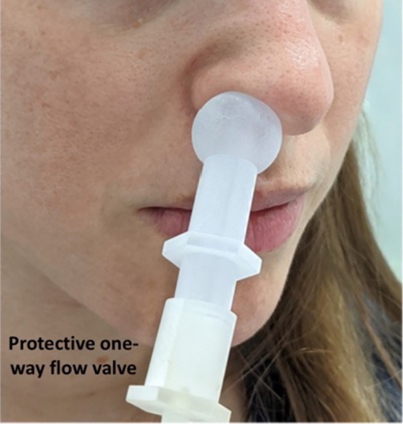
Fig 2

The sampling valve fits snugly against the nostril, providing an air-tight connection for pulling air from within the nostril, independent of exhalation. The person in the image is a co-author demonstrating, and not a participant, and informed consent for publication of identifying images in an online open-access journal was obtained.

Electronic nose set-up

We used a PEN3 eNose (AIRSENSE Analytics GmbH, Schwerin, Germany). The PEN3 is a compact (92 × 190 × 255 mm) lightweight (2.3 kg) device, consisting of a gas sampling unit and a sensor array. The sensor array is composed of 10 different thermo-regulated metal oxide sensors, positioned in a stainless-steel chamber (volume: 1.8 ml, temperature: 110°C). Each sensor is uniquely coated, rendering it particularly sensitive to a restricted class of chemical compounds. When a compound interacts with the sensor, this results in an oxygen exchange that leads to a change in electrical conductivity [31]. The specific sensitivities of the sensors are in Table 1. We used the PEN3 with its native sampling software (WinMuster), and the following settings: Chamber flow = 400ml/min, Flush time = 40s, Zero-point trim time = 10s, Measurement time = 80s. In the following paragraph, numbers in parenthesis relate to the numbered elements in Fig 1. For the current experiment we placed the entire sensing apparatus on the chassis of an electric wheelchair, to as to provide for system mobility. At the front of the wheelchair we secured an electric lift (FA-200-TR-24-60, Firgelli, Laverton North, Australia), and placed the eNose (Fig 1, #7) on the lift shelf. This allowed adjusting eNose height to individual car window height. On the wheelchair we also secured a canister of pressurized medical-grade breathing air (Fig 1, #14). This air was used to continuously inflate a large breathing bag (Xenon-133 Rebreathing System, Biodex, Shirley NY, USA) (Fig 1, #16), and this served as the reference air source for the eNose. This arrangement assured a consistent reference regardless of any environmental changes that may have occurred at the testing station over time. Finally, we took several steps to assure the safety of both the experimenters and participants, and protect them from the risk of infection from our system. The PEN3 has a sample exhaust port at its back. We directed this potentially infected exhaust through a one-way flow valve into a liquid trap (to protect the eNose from back-flow) (Fig 1, #11) and then directly into the bottom of a 1-liter 70% ethanol canister (Fig 1, #12). The exhausted air bubbled through the ethanol, and continued into the bottom of a second 1-liter 70% ethanol canister (Fig 1, #12). After the exhausted air bubbled through the second canister, it passed through a glass-microfiber filter (GasVent 2000 01, GVS, Morecambe, UK) (Fig 1, #13), and was directly vacuumed to a pump situated about ~30 meters outside of the testing station tent. There the exhausted air passed through an additional filter, before mixing with the outside environment. This flow path promised that air sampled from the participants was both treated and distanced. In addition, we considered the unlikely possibility that the PEN3 might have some internal leak. To address this remote possibility, we enclosed the entire device in a plexiglass box (Fig 1, #8), that had a 1/2-inch tube (Fig 1, #9) pulling ~30 LPM from the box top to the same distant location, ~30 meters outside of the testing station tent. In other words, the PEN3 was within a closed mini-hood. Moreover, in its cleaning phase, the PEN3 can push overflow air out through its inlet port. To address this possible source of contamination, we placed a "T" at the tip of the inlet tube (Fig 1, #4), with two all-Teflon ¼-inch one-way flow valves (CV-4-4-P-05, iPolymer, Irvine, CA, USA) (Fig 1, #2). One valve prevented flow from the device towards the participant, and the other valve directed such overflow cleaning air into the same line that led ~30 meters outside of the testing station tent. Finally, at the "T" we had a quick-connector, to which we attached the disposable unit used for each participant. This disposable unit included one-meter long ¼-inch Tygon tubing (Fig 1, #3), ending at a sampling valve (Fig 1, #1, and Fig 2). This 3D printed valve was shaped so as to fit snugly against the nostril from which it pulled air. This valve also contained a final one-way flow valve, so that if somehow something went wrong at the eNose apparatus, perhaps a breakdown following extended use, each participant was in this way nevertheless behind an added individual layer of protection (Fig 2). This entire system was reviewed by the device safety unit at the Israeli Ministry of Health, and was granted safety approval.

Table 1. eNose sensor functionalization.

Sensor number Sensor name Object substances for sensing Limit of detection
Sensor 1 W1C Aromatics 5 ppm
Sensor 2 W5S Ammonia and aromatic molecules 1 ppm
Sensor 3 W3C Broad-nitrogen oxide 5 ppm
Sensor 4 W6S Hydrogen 5 ppm
Sensor 5 W5C Methane, propane, and aliphatics 1 ppm
Sensor 6 W1S Broad-methane 5 ppm
Sensor 7 W1W Sulfur-containing organics 0.1 ppm
Sensor 8 W2S Broad-alcohols, broad-carbon chains 5 ppm
Sensor 9 W2W Aromatics, sulfur- and chlorine-containing organics 1 ppm
Sensor 10 W3S Methane and aliphatics 5 ppm

The specific functionalization of each of the 10 sensors in the PEN3 eNose as defined by the manufacturer.

Procedures

Cars were typically queued up at the testing station. An experimenter in full personal protective equipment (PPE) approached the car, and through a slightly open window explained the purpose of the experiment and its procedures, and requested participation. If the person in the car agreed, he/she was handed informed consent documents that included spaces for their Israeli national ID number (this number is used by the health system for obtaining results), their phone number, questions on age, sex, and whether the person was experiencing symptoms (yes/no without details). In an effort to minimize the transfer of potentially infected materials back to the experimenters, we did not collect the informed consent documents, but rather photographed the signature and information page through the car window. The participant was then handed the sampling valve, and instructed to hold it snugly against a nostril opening for 80 seconds. The shape of the sampling valve (Fig 2) ensured an air-tight application to the nose, such that outside air was not directly sampled. The participants were told to breath normally, but only through their open mouth, during these 80 seconds. After the sample, the disposable sampling valve and tube were discarded, and the participants advanced about 10 meters to the RT-PCR swabbing station. In other words, this experiment, by design, was double-blind.

Analysis

All analyses were conducted using Matlab software (Mathworks, USA). We applied the long short-term memory (LSTM) deep-learning classifier algorithm [32] to the entire time-series. We then applied a leave-one-out cross validation: We randomly selected 26 out of 27 positive samples, trained the classifier on these 26 positive samples, and then tested whether it can select between the one remaining positive sample versus one randomly selected negative sample (Fig 3). Chance performance at this task is 50%. We repeated this process 500 times, each time testing a different sample, in order to obtain a true-positive rate. Finally, because no selection of 500 is privileged, we repeated this entire process 100 times in order to obtain a distribution of possible true-positive rates. To estimate the statistical significance of the outcome, we repeated the entire process 600 times, but here first arbitrarily deemed a random 27 negative samples as "positive". In this manner, we can ask what is the chance probability of obtaining a similar result. The entire analysis code, with explanatory comments, is in the S1 Materials folder, that contains all the raw data, and all the code used in the analysis of this manuscript.

Fig 3. Analysis path.

Fig 3

We randomly selected 26 out of 27 positive samples, trained the classifier on these 26 positive samples, and then tested whether it can select between the one remaining positive sample versus one randomly selected negative sample. Chance performance at this task is 50%. We repeated this process 500 times, each time testing a different sample, in order to obtain a true-positive rate. Finally, because no selection of 500 is privileged, we repeated this entire process 100 times in order to obtain a distribution of possible true-positive rates.

Results

We successfully deployed at a drive-through testing station [33] in Tel Aviv, Israel (Fig 1B, S1 Video). Here, individuals being tested never exited their car, but rather drove through one of several lanes housing a testing team that swabbed them through an open window for later analysis by reverse-transcription polymerase chain reaction (RT-PCR) test for pathogen identification [34]. Individuals were referred to the testing station only if they had exposure to a verified Covid-19 patient, or were persistently experiencing Covid-19 symptoms. We placed our testing station in line with one such lane, directly before swabbing, and offered drivers to participate in our experiment. After providing written informed consent to procedures carried out in accordance with relevant guidelines and regulations, they were handed a disposable sampling valve (object #1 in Figs 1 and 2) that was linked to the eNose inlet port by a disposable flexible tube. They were instructed to secure this sampling valve air-tight to their nostril for 80 seconds while the eNose pulled sampled air at 400cc per minute (S1 Video). They then continued directly to RT-PCR testing.

Compliance was very high, with about 81% agreeing to participate. In 22 days of deployment, we obtained 503 samples (229F, 267M, 4 unreported sex, mean age 38.9), of which 27 (12F, 15M, mean age 35.85) were later deemed SARS CoV-2 positive (5.4%) by RT-PCR.

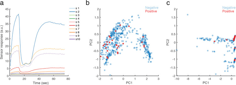
To ask whether we could use the eNose to identify SARS CoV-2 infection, we enacted the following analysis scheme: Initially, we visually inspected the data and observed that sensors #1, #3, and #5 never responded to any sample (Fig 4A). We therefore discarded these sensors from further analysis (this information may help guide a future search for COVID-19 related VOCs, considering the sensitivity specification of each sensor [31]). Next, we observe that PEN3 sensors each have a dynamic response pattern over about 65 seconds from sampling onset, before reaching equilibrium (Fig 4A). To ask whether relevant information was either in the entire time-series, or at the point of equilibrium alone, we conducted two principal component analyses (PCA) [35]: one using equilibrium end-values alone, and the other using a 3rd degree polynomial fit of the entire time-series. We observe no evidence for clustering of positive and negative test results in the equilibrium-value PCA (Fig 4B), but evidence for clusters in the time-series PCA (Fig 4C).

Fig 4. eNose measurements cluster SARS CoV-2 positive participants.

Fig 4

A. An example of the raw eNose measurement from one participant. Each line is one of 10 sensors. B. PCA of the equilibrium end-values only. C. PCA of the 3rd degree polynomial fit of the entire time-series. n = 503. The 27 positive samples are in red.

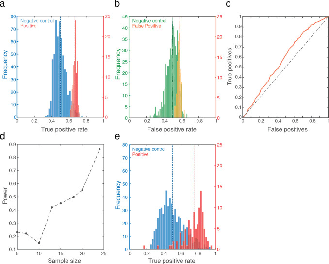
With this encouraging observation in hand, we applied the long short-term memory (LSTM) deep-learning classifier algorithm [18] to the entire time-series. We then applied a leave-one-out cross validation: We randomly selected 26 out of 27 positive samples, trained the classifier on these 26 positive samples, and then tested whether it can select between the one remaining positive sample versus one randomly selected negative sample (Fig 3). Chance performance at this task is 50%. We repeated this process 500 times, each time testing a different sample, in order to obtain a true-positive rate. Finally, because no selection of 500 is privileged, we repeated this entire process 100 times in order to obtain a distribution of possible true-positive rates. We observe a true positive rate ranging between 61% and 71%, with mean at 66.7% (Std Dev = 2%) (Fig 5A in red). The associated mean false negative rate was 33.3%, and the mean false positive rate was 57% (Std Dev = 2%) (Fig 5B in yellow). This combines for a receiver operating characteristic curve (ROC) that is modestly but consistently above the diagonal line, with an area under the curve (AUC) of 0.58 (Fig 5C). In relation to results with RT-PCR [36], the 66.7% true positive rate may be seen as promising, but considering the shallow ROC curve, is it significantly different from 50% chance? To assess this, we repeated the entire above process, but now first randomly selected 27 negative samples, and arbitrarily labeled them as the positive samples. We repeated this 600 times, and observe a true positive rate that indeed centered at 50% (Std Dev = 7%), and only 11 times in 600 repetitions reached 66.7% (Fig 5A in blue). This assigns a Bootstrap p value of less than 0.02 to our result (Hedges’ g effect size = 2.48). Moreover, a post-hoc analysis of power implied that at 27 positive samples, this result has 94% power (Fig 5D, Supplementary Fig 1 in S1 File). Finally, could have we been detecting general malaise rather than SARS CoV-2 infection? We observe that of the 27 positive participants, 14 were non-symptomatic. We therefore repeated the entire analysis scheme, now using only these 14 non-symptomatic positive samples instead of the 27 samples. We obtained a mean ROC AUC of 0.63, making for a true positive rate ranging from 47.4% to 94.4%, with mean at 75.8% (Std Dev = 12%) (we disregarded 2 outliers out of the 100, who were at 16.4% and 33.4%, i.e., more than 3 Std Dev from the mean) (Fig 5E in red). To test whether this is significantly different from chance, we again arbitrarily assign 14 negative sample as positive, and repeat the analysis 600 times. In only 34 cases did we obtain a similar outcome, thus assigning a Bootstrap p value of 0.057 to this result (Hedges’ g effect size = 2.0) (Fig 5E in blue). We conclude that it is unlikely that we were merely detecting general malaise rather than SARS CoV-2 infection.

Fig 5. An eNose can smell SARS CoV-2 infection.

Fig 5

A. In red, histogram of 100 true-positive values generated by the classifier (each value is the result of 500 selections of one of 27 positive and one of 476 negative), with the mean success rate (66.7%) in dashed red line. In blue, the control analysis: Histogram of 600 true-positive values generated by the classifier (each value is the result of 500 selections of one of 27 negatives now randomly assigned as positive, and one of 449 remaining negatives), with the mean (50%) in dashed blue line. B. The same analysis as in A, here depicting false positives (yellow) and random control (green). C. ROC curve. D. Power analysis on increasing sample sizes (see Supplementary Fig 1 in S1 File). E. Same as A, but using only the 14 non-symptomatic positive participants. Note two discarded outliers at 16.4% and 33.4%.

Discussion

This manuscript had a modest goal: to test the hypothesis that there is a body-odor, and more specifically an intra-nasal-passage body-odor, associated with SARS CoV-2 infection detectable by eNose. To this we provide a positive answer, providing proof of concept for VOC-based real-time detection of SARS CoV-2 infection. We note that results to this effect have also recently been obtained by others: In one study [28], the authors used a commercial metal-oxide-sensor-based eNose, albeit different from ours (Aeonose, The Aeonose Company, Zutphen, the Netherlands). They sampled oral exhaled breath from 219 participants, of which 57 were COVID-19 positive. These participants were tested at two locations, all the negative participants were tested at an outpatient clinic, and most all of the positive patients were tested at a COVID-19 nursing unit. This group obtained an ROC AUC of 0.74, compared to our ROC AUCs of 0.58 and 0.63. In a second study [27], the authors used a proprietary nanomaterial-based hybrid sensor array eNose. This group also sampled oral exhaled breath, from 140 participants, of which 49 were COVID-19 positive, 58 were COVID-19 negative, and 33 were other lung disease participants. These participants were tested at multiple locations, with all the COVID-19 positive participants coming from the same intensive care unit at a hospital in Wuhan, China. This group obtained an ROC AUC of 0.81, again compared to our ROC AUCs of 0.58 and 0.63. Thus, the two current published efforts that we are aware of obtained results significantly better than ours. What underlies these differences? The likely explanation is that these groups used a better device, and applied it using better sampling methods. We do not say this facetiously. In turn, we would like to highlight one strength of our effort that we think is meaningful: We sampled in a naturalistic setting where, unlike in the two above examples, sources of variance where equally distributed across groups. A limitation of eNose diagnostics is that we often don’t know the precise molecular identity of the signal. Thus, had we tested a homogenous hospitalized cohort, did we then detect COVID-19, or did we detect the food/laundry-detergent/any other commonality of the jointly housed COVID-19 positive participants? Here, by going directly to the very difficult end-test, namely the extremely noisy (olfaction-wise) and highly variable environment of the drive-through testing station, we provide a powerful inherently double-blind proof of concept for our hypothesis. Here, one SARS CoV-2 positive person may have just eaten a tuna-fish sandwich, another may have just brushed their teeth, and a third may have just doused themselves with their favorite perfume. Unlike an olfaction-wise homogenous group of patients in a hospital (who eat the same food, wear commonly laundered clothing, etc.), the only thing these people have in common is a later-obtained SARS CoV-2 positive test. With this noise in mind, we nevertheless obtain statistically significant classification. In contrast to this strength, our study has several weaknesses. First, our sampling strategies clearly introduce even added noise. For example, air from the car interior can enter through the unsampled nostril and mouth, and make its way to the sampling apparatus. Beyond this, the primary weakness that limits our effort to the status of proof of concept alone is the level of false positives. Although as noted our statistically significant 66.7% true positive rate (or 75.8% for non-symptomatic) is not far off of RT-PCR true positive rates [28], and we obtain this result instantaneously rather than days later, our 57% false positive rate prevents this method from deployment in its current form. Thus, our result is a basic-science proof of concept, and not a clinical tool. We think, however, that this shortcoming is technical rather than conceptual, and we have identified several steps towards addressing this limitation. For example, a major source of olfactory noise in our apparatus was the individual sampling valve that we used (Fig 2). Its manufacturing process included a disinfection procedure that introduced significant added noise to our measurements. Additional improvements may also include optimization of sensor coating based on analysis of Covid-19 volatiles. Given our current results with a generic eNose, we speculate that an optimized eNose may be able to provide effective real-time diagnoses in locations such as airports, the work-place, and cultural events, and in this potentially contribute to social and economic recovery in the COVID-19 pandemic.

Supporting information

S1 Video. A video demonstrating activity at the drive-through.

The person being tested is an experimenter demonstrating, and not a participant.

(MP4)

S1 Materials. A folder containing all the raw data, the code, and a readme file explaining how to run it.

(ZIP)

S1 File

(PDF)

Acknowledgments

We thank Ofer Perl for photography in Fig 2. We thank Shoval Silbert and Stratasys for their gracious help at 3D printing, Dr. Nadav Sheffer at the Ministry of Health, and Miki Segal, Efi Levav, and Ilan Klein from MDA for their gracious hospitality and help at the drive-through station.

Data Availability

All relevant data are within the manuscript and its Supporting Information files.

Funding Statement

This study was supported by pilot grants from MAFAT: The Israeli Ministry of Defense Directorate of Defense Research and Development, and from Sonia T. Marschak. This effort also relied on ongoing Sobel lab resources, provided by a European Research Council AdG. grant #670798 (SocioSmell) and Horizon 2020 FET Open project #662629 (NanoSmell). The funders had no role in study design, data collection and analysis, decision to publish, or preparation of the manuscript.

References

  • 1.Aksenov AA, Sandrock CE, Zhao W, Sankaran S, Schivo M, Harper R, et al. Cellular scent of influenza virus infection. Chembiochem: a European journal of chemical biology. 2014;15(7):1040. 10.1002/cbic.201300695 [DOI] [PMC free article] [PubMed] [Google Scholar]
  • 2.Broza YY, Mochalski P, Ruzsanyi V, Amann A, Haick H. Hybrid volatolomics and disease detection. Angewandte Chemie International Edition. 2015;54(38):11036–48. 10.1002/anie.201500153 [DOI] [PubMed] [Google Scholar]
  • 3.Pickel D, Manucy GP, Walker DB, Hall SB, Walker JC. Evidence for canine olfactory detection of melanoma. Applied Animal Behaviour Science. 2004;89(1–2):107–16. [Google Scholar]
  • 4.Seo I-S, Lee H-G, Koo B, Koh CS, Park H-Y, Im C, et al. Cross detection for odor of metabolic waste between breast and colorectal cancer using canine olfaction. PLoS one. 2018;13(2):e0192629. 10.1371/journal.pone.0192629 [DOI] [PMC free article] [PubMed] [Google Scholar]
  • 5.Pearce TC, Schiffman SS, Nagle HT, Gardner JW. Handbook of machine olfaction: electronic nose technology: John Wiley & Sons; 2006. [Google Scholar]
  • 6.Doleman BJ, Lewis NS. Comparison of odor detection thresholds and odor discriminablities of a conducting polymer composite electronic nose versus mammalian olfaction. Sensors and Actuators B: Chemical. 2001;72(1):41–50. [Google Scholar]
  • 7.Röck F, Barsan N, Weimar U. Electronic nose: current status and future trends. Chemical reviews. 2008;108(2):705–25. 10.1021/cr068121q [DOI] [PubMed] [Google Scholar]
  • 8.Freund MS, Lewis NS. A chemically diverse conducting polymer-based" electronic nose". Proceedings of the National Academy of Sciences. 1995;92(7):2652–6. 10.1073/pnas.92.7.2652 [DOI] [PMC free article] [PubMed] [Google Scholar]
  • 9.van Geffen WH, Bruins M, Kerstjens HA. Diagnosing viral and bacterial respiratory infections in acute COPD exacerbations by an electronic nose: a pilot study. Journal of breath research. 2016;10(3):036001. 10.1088/1752-7155/10/3/036001 [DOI] [PubMed] [Google Scholar]
  • 10.Gardner JW, Shin HW, Hines EL. An electronic nose system to diagnose illness. Sensors and Actuators B: Chemical. 2000;70(1–3):19–24. [Google Scholar]
  • 11.Saktiawati AM, Stienstra Y, Subronto YW, Rintiswati N, Gerritsen J-W, Oord H, et al. Sensitivity and specificity of an electronic nose in diagnosing pulmonary tuberculosis among patients with suspected tuberculosis. PLoS One. 2019;14(6):e0217963. 10.1371/journal.pone.0217963 [DOI] [PMC free article] [PubMed] [Google Scholar]
  • 12.Lewis JM, Savage RS, Beeching NJ, Beadsworth MB, Feasey N, Covington JA. Identifying volatile metabolite signatures for the diagnosis of bacterial respiratory tract infection using electronic nose technology: a pilot study. PLoS One. 2017;12(12):e0188879. 10.1371/journal.pone.0188879 [DOI] [PMC free article] [PubMed] [Google Scholar]
  • 13.Shafiek H, Fiorentino F, Merino JL, López C, Oliver A, Segura J, et al. Using the electronic nose to identify airway infection during COPD exacerbations. PLoS One. 2015;10(9):e0135199. 10.1371/journal.pone.0135199 [DOI] [PMC free article] [PubMed] [Google Scholar]
  • 14.Brekelmans MP, Fens N, Brinkman P, Bos LD, Sterk PJ, Tak PP, et al. Smelling the diagnosis: the electronic nose as diagnostic tool in inflammatory arthritis. A case-reference study. PloS one. 2016;11(3):e0151715. 10.1371/journal.pone.0151715 [DOI] [PMC free article] [PubMed] [Google Scholar]
  • 15.Bach J-P, Gold M, Mengel D, Hattesohl A, Lubbe D, Schmid S, et al. Measuring compounds in exhaled air to detect Alzheimer’s disease and Parkinson’s disease. PloS one. 2015;10(7):e0132227. 10.1371/journal.pone.0132227 [DOI] [PMC free article] [PubMed] [Google Scholar]
  • 16.Jendrny P, Schulz C, Twele F, Meller S, von Köckritz-Blickwede M, Osterhaus ADME, et al. Scent dog identification of samples from COVID-19 patients–a pilot study. BMC Infectious Diseases. 2020;20(1):1–7. 10.1186/s12879-020-05281-3 [DOI] [PMC free article] [PubMed] [Google Scholar]
  • 17.Lamote K, Janssens E, Schillebeeckx E, Lapperre TS, De Winter BY, Van Meerbeeck JP. The scent of COVID-19: viral (semi-) volatiles as fast diagnostic biomarkers? Journal of breath research. 2020. 10.1088/1752-7163/aba105 [DOI] [PubMed] [Google Scholar]
  • 18.Van der Schee M, Fens N, Brinkman P, Bos L, Angelo M, Nijsen T, et al. Effect of transportation and storage using sorbent tubes of exhaled breath samples on diagnostic accuracy of electronic nose analysis. Journal of breath research. 2012;7(1):016002. 10.1088/1752-7155/7/1/016002 [DOI] [PubMed] [Google Scholar]
  • 19.Scarlata S, Pennazza G, Santonico M, Pedone C, Antonelli Incalzi R. Exhaled breath analysis by electronic nose in respiratory diseases. Expert review of molecular diagnostics. 2015;15(7):933–56. 10.1586/14737159.2015.1043895 [DOI] [PubMed] [Google Scholar]
  • 20.Schnabel R, Boumans M, Smolinska A, Stobberingh E, Kaufmann R, Roekaerts P, et al. Electronic nose analysis of exhaled breath to diagnose ventilator-associated pneumonia. Respiratory medicine. 2015;109(11):1454–9. 10.1016/j.rmed.2015.09.014 [DOI] [PubMed] [Google Scholar]
  • 21.Bruins M, Rahim Z, Bos A, van de Sande WW, Endtz HP, van Belkum A. Diagnosis of active tuberculosis by e-nose analysis of exhaled air. Tuberculosis. 2013;93(2):232–8. 10.1016/j.tube.2012.10.002 [DOI] [PubMed] [Google Scholar]
  • 22.Fens N, Van der Schee M, Brinkman P, Sterk P. Exhaled breath analysis by electronic nose in airways disease. Established issues and key questions. Clinical & Experimental Allergy. 2013;43(7):705–15. 10.1111/cea.12052 [DOI] [PubMed] [Google Scholar]
  • 23.Joensen O, Paff T, Haarman EG, Skovgaard IM, Jensen PØ, Bjarnsholt T, et al. Exhaled breath analysis using electronic nose in cystic fibrosis and primary ciliary dyskinesia patients with chronic pulmonary infections. PLoS One. 2014;9(12):e115584. 10.1371/journal.pone.0115584 [DOI] [PMC free article] [PubMed] [Google Scholar]
  • 24.Dragonieri S, Van Der Schee MP, Massaro T, Schiavulli N, Brinkman P, Pinca A, et al. An electronic nose distinguishes exhaled breath of patients with Malignant Pleural Mesothelioma from controls. Lung Cancer. 2012;75(3):326–31. 10.1016/j.lungcan.2011.08.009 [DOI] [PubMed] [Google Scholar]
  • 25.Machado RF, Laskowski D, Deffenderfer O, Burch T, Zheng S, Mazzone PJ, et al. Detection of lung cancer by sensor array analyses of exhaled breath. American journal of respiratory and critical care medicine. 2005;171(11):1286–91. 10.1164/rccm.200409-1184OC [DOI] [PMC free article] [PubMed] [Google Scholar]
  • 26.Swanson B, Fogg L, Julion W, Arrieta MT. Electronic Nose Analysis of Exhaled Breath Volatiles to Identify Lung Cancer Cases: A Systematic Review. Journal of the Association of Nurses in AIDS Care. 2020;31(1):71–9. 10.1097/JNC.0000000000000146 [DOI] [PubMed] [Google Scholar]
  • 27.Shan B, Broza YY, Li W, Wang Y, Wu S, Liu Z, et al. Multiplexed nanomaterial-based sensor array for detection of COVID-19 in exhaled breath. ACS nano. 2020;14(9):12125–32. 10.1021/acsnano.0c05657 [DOI] [PubMed] [Google Scholar]
  • 28.Wintjens AG, Hintzen KF, Engelen SM, Lubbers T, Savelkoul PH, Wesseling G, et al. Applying the electronic nose for pre-operative SARS-CoV-2 screening. Surgical endoscopy. 2020:1–8. 10.1007/s00464-020-08169-0 [DOI] [PMC free article] [PubMed] [Google Scholar]
  • 29.Brann DH, Tsukahara T, Weinreb C, Lipovsek M, Van den Berge K, Gong B, et al. Non-neuronal expression of SARS-CoV-2 entry genes in the olfactory system suggests mechanisms underlying COVID-19-associated anosmia. Science advances. 2020;6(31):eabc5801. 10.1126/sciadv.abc5801 [DOI] [PMC free article] [PubMed] [Google Scholar]
  • 30.Meinhardt J, Radke J, Dittmayer C, Franz J, Thomas C, Mothes R, et al. Olfactory transmucosal SARS-CoV-2 invasion as a port of central nervous system entry in individuals with COVID-19. Nature neuroscience. 2021;24(2):168–75. 10.1038/s41593-020-00758-5 [DOI] [PubMed] [Google Scholar]
  • 31.Baietto M, Wilson AD, Bassi D, Ferrini F. Evaluation of three electronic noses for detecting incipient wood decay. Sensors. 2010;10(2):1062–92. 10.3390/s100201062 [DOI] [PMC free article] [PubMed] [Google Scholar]
  • 32.Graves A. Generating sequences with recurrent neural networks. arXiv preprint arXiv:13080850. 2013. [Google Scholar]
  • 33.Shah A, Challener D, Tande AJ, Mahmood M, O’Horo JC, Berbari E, et al., editors. Drive-through testing: a unique, efficient method of collecting large volume of specimens during the SARS-CoV-2 (COVID-19) pandemic. Mayo Clinic Proceedings; 2020: Elsevier. [DOI] [PMC free article] [PubMed] [Google Scholar]
  • 34.Tang Y-W, Schmitz JE, Persing DH, Stratton CW. Laboratory diagnosis of COVID-19: current issues and challenges. Journal of clinical microbiology. 2020;58(6). 10.1128/JCM.00512-20 [DOI] [PMC free article] [PubMed] [Google Scholar]
  • 35.Jeon H-M, Lee J-Y, Jeong G-M, Choi S-I. Data reconstruction using iteratively reweighted L1-principal component analysis for an electronic nose system. PloS one. 2018;13(7):e0200605. 10.1371/journal.pone.0200605 [DOI] [PMC free article] [PubMed] [Google Scholar]
  • 36.Fang Y, Zhang H, Xie J, Lin M, Ying L, Pang P, et al. Sensitivity of chest CT for COVID-19: comparison to RT-PCR. Radiology. 2020;296(2):E115–E7. 10.1148/radiol.2020200432 [DOI] [PMC free article] [PubMed] [Google Scholar]

Decision Letter 0

Matthieu Louis

26 Oct 2020

PONE-D-20-27895

Proof of Concept for Real-Time Detection of SARS CoV-2 Infection with an Electronic Nose

PLOS ONE

Dear Dr. Sobel,

Thank you for submitting your manuscript to PLOS ONE. After careful consideration, we feel that it has merit but does not fully meet PLOS ONE’s publication criteria as it currently stands. Therefore, we invite you to submit a revised version of the manuscript that addresses the points raised during the review process.

The reviewers raised several issues that must be addressed to validate the methodology behind your work:

1. Please answer the concerns of reviewer #2 about the necessity to use of a sealed mask to prevent the detection of environmental contaminants, as well as the possibility that "normal" breathing patterns might not be adequate to detect endogenous VOCs. 

2. Please clarify the discrepancy between the expectation of reviewer #2 that the sensor’s conductivity should increase with the exhaled gas concentration whereas you appear to be observing the opposite. 

3. As requested by reviewer #1, it would be important to report any information you might have about the parameters of exhaled breath (humidity) affecting the sensor's readout.

In your revised manuscript, you're encouraged (i) to add to your introduction a review of published applications of the electronic nose technology to the diagnosis of lung disease; (ii) to include a discussion of any specific biomarkers you found associated with the VOCs of SARS CoV-2.

Please submit your revised manuscript by Dec 10 2020 11:59PM. If you will need more time than this to complete your revisions, please reply to this message or contact the journal office at plosone@plos.org. When you're ready to submit your revision, log on to https://www.editorialmanager.com/pone/ and select the 'Submissions Needing Revision' folder to locate your manuscript file.

Please include the following items when submitting your revised manuscript:

  • A rebuttal letter that responds to each point raised by the academic editor and reviewer(s). You should upload this letter as a separate file labeled 'Response to Reviewers'.

  • A marked-up copy of your manuscript that highlights changes made to the original version. You should upload this as a separate file labeled 'Revised Manuscript with Track Changes'.

  • An unmarked version of your revised paper without tracked changes. You should upload this as a separate file labeled 'Manuscript'.

If you would like to make changes to your financial disclosure, please include your updated statement in your cover letter. Guidelines for resubmitting your figure files are available below the reviewer comments at the end of this letter.

If applicable, we recommend that you deposit your laboratory protocols in protocols.io to enhance the reproducibility of your results. Protocols.io assigns your protocol its own identifier (DOI) so that it can be cited independently in the future. For instructions see: http://journals.plos.org/plosone/s/submission-guidelines#loc-laboratory-protocols

We look forward to receiving your revised manuscript.

Kind regards,

Matthieu Louis

Academic Editor

PLOS ONE

Journal Requirements:

When submitting your revision, we need you to address these additional requirements.

1. Please ensure that your manuscript meets PLOS ONE's style requirements, including those for file naming. The PLOS ONE style templates can be found at

https://journals.plos.org/plosone/s/file?id=wjVg/PLOSOne_formatting_sample_main_body.pdf and

https://journals.plos.org/plosone/s/file?id=ba62/PLOSOne_formatting_sample_title_authors_affiliations.pdf

2. We note that Figure 1 and Online Video SnitzMovie includes images of participants in the study. 

As per the PLOS ONE policy (http://journals.plos.org/plosone/s/submission-guidelines#loc-human-subjects-research) on papers that include identifying, or potentially identifying, information, the individual(s) or parent(s)/guardian(s) must be informed of the terms of the PLOS open-access (CC-BY) license and provide specific permission for publication of these details under the terms of this license. Please download the Consent Form for Publication in a PLOS Journal (http://journals.plos.org/plosone/s/file?id=8ce6/plos-consent-form-english.pdf). The signed consent form should not be submitted with the manuscript, but should be securely filed in the individual's case notes. Please amend the methods section and ethics statement of the manuscript to explicitly state that the patient/participant has provided consent for publication: “The individual in this manuscript has given written informed consent (as outlined in PLOS consent form) to publish these case details”.

If you are unable to obtain consent from the subject of the photograph, you will need to remove the figure and any other textual identifying information or case descriptions for this individual.

3.  We note that you have a patent relating to material pertinent to this article. Please provide an amended statement of Competing Interests to declare this patent (with details including name and number), along with any other relevant declarations relating to employment, consultancy, patents, products in development or modified products etc. Please confirm that this does not alter your adherence to all PLOS ONE policies on sharing data and materials, as detailed online in our guide for authors http://journals.plos.org/plosone/s/competing-interests by including the following statement: "This does not alter our adherence to  PLOS ONE policies on sharing data and materials.” If there are restrictions on sharing of data and/or materials, please state these. Please note that we cannot proceed with consideration of your article until this information has been declared.

This information should be included in your cover letter; we will change the online submission form on your behalf.

Please know it is PLOS ONE policy for corresponding authors to declare, on behalf of all authors, all potential competing interests for the purposes of transparency. PLOS defines a competing interest as anything that interferes with, or could reasonably be perceived as interfering with, the full and objective presentation, peer review, editorial decision-making, or publication of research or non-research articles submitted to one of the journals. Competing interests can be financial or non-financial, professional, or personal. Competing interests can arise in relationship to an organization or another person. Please follow this link to our website for more details on competing interests: http://journals.plos.org/plosone/s/competing-interests

4. Please include captions for your Supporting Information files at the end of your manuscript, and update any in-text citations to match accordingly. Please see our Supporting Information guidelines for more information: http://journals.plos.org/plosone/s/supporting-information.

[Note: HTML markup is below. Please do not edit.]

Reviewers' comments:

Reviewer's Responses to Questions

Comments to the Author

1. Is the manuscript technically sound, and do the data support the conclusions?

The manuscript must describe a technically sound piece of scientific research with data that supports the conclusions. Experiments must have been conducted rigorously, with appropriate controls, replication, and sample sizes. The conclusions must be drawn appropriately based on the data presented.

Reviewer #1: Yes

Reviewer #2: No

**********

2. Has the statistical analysis been performed appropriately and rigorously?

Reviewer #1: Yes

Reviewer #2: No

**********

3. Have the authors made all data underlying the findings in their manuscript fully available?

The PLOS Data policy requires authors to make all data underlying the findings described in their manuscript fully available without restriction, with rare exception (please refer to the Data Availability Statement in the manuscript PDF file). The data should be provided as part of the manuscript or its supporting information, or deposited to a public repository. For example, in addition to summary statistics, the data points behind means, medians and variance measures should be available. If there are restrictions on publicly sharing data—e.g. participant privacy or use of data from a third party—those must be specified.

Reviewer #1: Yes

Reviewer #2: No

**********

4. Is the manuscript presented in an intelligible fashion and written in standard English?

PLOS ONE does not copyedit accepted manuscripts, so the language in submitted articles must be clear, correct, and unambiguous. Any typographical or grammatical errors should be corrected at revision, so please note any specific errors here.

Reviewer #1: Yes

Reviewer #2: Yes

**********

5. Review Comments to the Author

Please use the space provided to explain your answers to the questions above. You may also include additional comments for the author, including concerns about dual publication, research ethics, or publication ethics. (Please upload your review as an attachment if it exceeds 20,000 characters)

Reviewer #1: The authors present a proof-of-concept of SARS CoV-2 detection with a commercially available e-nose. They present a very detailed description of in-line sampling methods at a national testing station in Tel Aviv, Israel. Many details are provided on the strategy deployed to operate in a safe environment and avoid contamination during the test of each volunteer. This is valuable contribution for future studies aimed to develop and test e-noses for the detection of such infection.

The data analysis was carried out correctly with machine-learning methods. The authors also provide a critical assessment of points of strength and weaknesses of both sampling method and data analysis and recognize that the main limit of their method is the level of false positives, which hinders the present method to be brought beyond the status of proof-of-concept. Nevertheless they try to identify some steps to overcome to present limitations.

In spite of the many positive aspects outlined above, the manuscript need some improvements, listed below.

1. The Introduction is written is a rather popularized style. I would have expected a brief review of the existing literature on this topic. I understand that this is quite a new case-study of volatolomics but there is no presentation of recent papers such as ACS Nano 2020, 14, 9, 12125–12132, which might be an interesting paper to be contrasted and compared with the present one. At least, a brief account of recent papers dealing with the application of e-noses to lung diseases should be provided.

2. It is claimed that virus-infected cells produce VOCs that can be targeted for VOC-based disease detection. However, there no discussion about possible VOCs in the case of SARS CoV-2 infection. Is there any idea about the kind of biomarkes that can be related to VOCs? Could they be pneumonia biomarkers, one of the most severe consequences of SARS CoV-2 infection?

3. Though the sampling has been detailed, information about the exhaled breath is missing, such as humidity, that may affect the sensors readout. Typically, humidity in breath would require some type of filter prior to admitting the sampled breath to the sensor array; of course, one needs to make sure not to filter the analyte too. May the sampled breath of infected patients display a humidity content different from that of healthy volunteers?

4. Did the authors consider the potential effect of lung function variations on the VOCs levels in exhaled breath and, consequently, on sensor response? This is quite a general issue in e-nose applications to respiratory medicine and needs to be addressed. Data on FEV1 (forced expiratory volume in one second) and FVC (forced vital capacity) might be useful, if available, for the discrimination of volunteers.

In conclusion I believe that the paper can be published provided that the authors address the points above.

Reviewer #2: Review comments may be found on the attachment. In papers of this type, the protocols used are extremely important. If poor protocols are used, the conclusions may be deemed invalid. The reviewer believes that there is indeed an excellent case for the use of specific VOC's and their concentrations as a function of time to provide a rapid, sensitive and selective diagnosis; however, this paper fails to do so.

**********

6. PLOS authors have the option to publish the peer review history of their article (what does this mean?). If published, this will include your full peer review and any attached files.

If you choose “no”, your identity will remain anonymous but your review may still be made public.

Do you want your identity to be public for this peer review? For information about this choice, including consent withdrawal, please see our Privacy Policy.

Reviewer #1: No

Reviewer #2: No

[NOTE: If reviewer comments were submitted as an attachment file, they will be attached to this email and accessible via the submission site. Please log into your account, locate the manuscript record, and check for the action link "View Attachments". If this link does not appear, there are no attachment files.]

While revising your submission, please upload your figure files to the Preflight Analysis and Conversion Engine (PACE) digital diagnostic tool, https://pacev2.apexcovantage.com/. PACE helps ensure that figures meet PLOS requirements. To use PACE, you must first register as a user. Registration is free. Then, login and navigate to the UPLOAD tab, where you will find detailed instructions on how to use the tool. If you encounter any issues or have any questions when using PACE, please email PLOS at figures@plos.org. Please note that Supporting Information files do not need this step.

Attachment

Submitted filename: EnoseReview.pdf

PLoS One. 2021 Jun 2;16(6):e0252121. doi: 10.1371/journal.pone.0252121.r002

Author response to Decision Letter 0


24 Apr 2021

Dear Editor and Referees,

Thank you for the detailed response and for the reviewers’ comments. We have carefully reviewed the comments and have revised the manuscript accordingly. Our point-by-point responses are given below.

Editor comments:

1. Please answer the concerns of reviewer #2 about the necessity to use of a sealed mask to prevent the detection of environmental contaminants, as well as the possibility that "normal" breathing patterns might not be adequate to detect endogenous VOCs.

We have answered this at two levels: First, likely due to poor wording on our part, the Referee misunderstood our aim as to conduct exhaled breath analysis, and with this in mind, had concerns regarding departure from common practice in breath sampling. However, our aim was not exhaled breath analysis, but rather smelling of the intranasal passage. We aimed for this because the nasal passage may be a primary sight of infection. Thus, we were not trying to “smell the lungs”, but rather “smell the inside of the nose”. Here, exhaled breath is in fact an unavoidable source of noise that we had to accept, but it was not our target. We have now made this very clear in the manuscript, so as to avoid misunderstandings. Second, we have added Figure 2, that clearly shows how our system is air-tight, and samples from the inside of the nose, and not environmental air. We should have likely had this figure in the original version, and hope it now clarifies things.

2. Please clarify the discrepancy between the expectation of reviewer #2 that the sensor’s conductivity should increase with the exhaled gas concentration whereas you appear to be observing the opposite.

The sensor response can be either an increase in resistance or a decrease in resistance depending on whether the gas is a reducing gas or an oxidizing gas and whether the sensor is an N type or a P type sensor (Berna, 2010): “For p type oxides, an increase in the resistance is found in the presence of reducing gases, while the resistance decreases in response to oxidizing gases; n-type oxides show opposite behavior. Examples of n type oxides are SnO2 and WO3; and a p-type oxide is CTO.” However, in the specific e-nose that we used (commercial e-nose PEN3), the conductivity usually decreases after an oxygen exchange (Baietto et al., 2010). We have now also detailed all this in the manuscript.

3. As requested by reviewer #1, it would be important to report any information you might have about the parameters of exhaled breath (humidity) affecting the sensor's readout.

We do not have any humidity measure. We acknowledge of course that added measures (humidity, temperature, etc) may have provided for an even better tool, and therefore throughout this manuscript we think we maintain extreme modesty regarding our claims. We never suggest that this is the best possible system for the job, we merely suggest that the eNose data alone captures meaningful variance, even in this challenging “open field” setting.

In your revised manuscript, you're encouraged (i) to add to your introduction a review of published applications of the electronic nose technology to the diagnosis of lung disease; (ii) to include a discussion of any specific biomarkers you found associated with the VOCs of SARS CoV-2.

We added to our introduction a short review of the e-nose application to lung diseases, and to COVID-19. We also commented on VOC specificity where relevant, albeit we have no data on this from our effort.

Reviewer 1:

Reviewer #1: The authors present a proof-of-concept of SARS CoV-2 detection with a commercially available e-nose. They present a very detailed description of in-line sampling methods at a national testing station in Tel Aviv, Israel. Many details are provided on the strategy deployed to operate in a safe environment and avoid contamination during the test of each volunteer. This is valuable contribution for future studies aimed to develop and test e-noses for the detection of such infection.

We thank the Referee for the kind words.

The data analysis was carried out correctly with machine-learning methods. The authors also provide a critical assessment of points of strength and weaknesses of both sampling method and data analysis and recognize that the main limit of their method is the level of false positives, which hinders the present method to be brought beyond the status of proof-of-concept. Nevertheless they try to identify some steps to overcome to present limitations.

We again thank the Referee for the kind words.

In spite of the many positive aspects outlined above, the manuscript need some improvements, listed below.

1. The Introduction is written is a rather popularized style. I would have expected a brief review of the existing literature on this topic. I understand that this is quite a new case-study of volatolomics but there is no presentation of recent papers such as ACS Nano 2020, 14, 9, 12125–12132, which might be an interesting paper to be contrasted and compared with the present one. At least, a brief account of recent papers dealing with the application of e-noses to lung diseases should be provided.

Consistent with this comment, we have added the following paragraph to the introduction:

Much of the eNose diagnostics effort in the literature is focused on exhaled breath analysis. Such breath sampling and analysis has standard protocols [1, 2], and has reached at achievements in cases such as identifying pneumonia [3], tuberculosis [4, 5], asthma and COPD [6], respiratory infections [7] and lung malignancies [8-10], and recently indeed for COVID-19 [11, 12]. Moreover, eNose measurements of exhaled breath may inform on non-respiratory conditions as well, such as neurodegenerative illnesses [13]. Here, however, we are not proposing exhaled breath analysis per se. Rather, we observe that the nasal passage has been implicated as a site of SARS CoV-2 infection [20-21]. Therefore, our goal is to "smell" the inner nasal passage itself. From our perspective, breath, and its associated lung-derived VOCs, are an inevitable source of noise in the nasal passage, but not our intended target. Thus, we set out to develop methods that differ from the standard breath sampling and analysis typically applied in the field.

Moreover, in further consideration of: “might be an interesting paper to be contrasted and compared with the present one”, in the discussion we now add:

We note that results to this effect have recently been obtained by others: In one study [12], the authors used a commercial metal-oxide-sensor-based eNose, albeit different from ours (Aeonose, The Aeonose Company, Zutphen, the Netherlands). They sampled oral exhaled breath from 219 participants, of which 57 were COVID-19 positive. These participants were tested at two locations, all the negative participants were tested at an outpatient clinic, and most all of the positive patients were tested at a COVID-19 nursing unit. This group obtained an ROC AUC of 0.74, compared to our ROC AUCs of 0.58 and 0.63.. In a second study [11], the authors used a proprietary nanomaterial-based hybrid sensor array eNose. This group also sampled oral exhaled breath, from 140 participants, of which 49 were COVID-19 positive, 58 were COVID-19 negative, and 33 were other lung disease participants. These participants were tested at multiple locations, with all the COVID-19 positive participants coming from the same intensive care unit at a hospital in Wuhan, China. This group obtained an ROC AUC of 0.81, again compared to our ROC AUCs of 0.58 and 0.63.. Thus, the two current published efforts that we are aware of obtained results significantly better than ours. What underlies these differences? The likely explanation is that these groups used a better device, and applied it using better sampling methods. We do not say this facetiously. That said, we would like to highlight one strength of our effort that we think is meaningful: We sampled in a naturalistic setting where, unlike in the two above examples, sources of variance where equally distributed across groups. A limitation of eNose diagnostics is that we don’t know the identity of the signal. Thus, did the above efforts detect COVID-19, or did they detect the food/laundry-detergent/any other commonality of the jointly housed COVID-19 positive participants? Here, by going directly to the very difficult end-test, namely the extremely noisy (olfaction-wise) and highly variable environment of the drive-through testing station, we provide a powerful inherently double-blind proof of concept for our hypothesis. Here, one SARS CoV-2 positive person may have just eaten a tuna-fish sandwich, another may have just brushed their teeth, and a third may have just doused themselves with their favorite perfume. Unlike an olfaction-wise homogenous group of patients in a hospital (who eat the same food, wear commonly laundered clothing, etc.), the only thing these people have in common is a later-obtained SARS CoV-2 positive test. With this noise in mind, we nevertheless obtain statistically significant classification.

2. It is claimed that virus-infected cells produce VOCs that can be targeted for VOC-based disease detection. However, there no discussion about possible VOCs in the case of SARS CoV-2 infection. Is there any idea about the kind of biomarkes that can be related to VOCs? Could they be pneumonia biomarkers, one of the most severe consequences of SARS CoV-2 infection?

This is indeed an important point. As we clearly acknowledge throughout the manuscript, we have no VOC-specific data. The only thing we can point to in this respect is the lack of contribution from sensors #1, #3, and #5, thus giving some indication on the classes of molecules not involved in this classification. That said, we do not think pneumonia was playing a role here, as these participants were mostly non-symptomatic, and indeed, our device worked better in the non-symptomatic sub-cohort.

3. Though the sampling has been detailed, information about the exhaled breath is missing, such as humidity, that may affect the sensors readout. Typically, humidity in breath would require some type of filter prior to admitting the sampled breath to the sensor array; of course, one needs to make sure not to filter the analyte too. May the sampled breath of infected patients display a humidity content different from that of healthy volunteers?

We have no data on humidity from a dedicated humidity sensor. That COVID-19 positive participants may have different humidity than COVID-19 negative participants is a keen thought, yet we think this is not the case, as humidity is primarily evident in Sensor #4, yet this sensor was not the major player in the response.

4. Did the authors consider the potential effect of lung function variations on the VOCs levels in exhaled breath and, consequently, on sensor response? This is quite a general issue in e-nose applications to respiratory medicine and needs to be addressed. Data on FEV1 (forced expiratory volume in one second) and FVC (forced vital capacity) might be useful, if available, for the discrimination of volunteers.

We agree with the referee that lung function might be a useful predictor of COVID-19, and may affect the volume of exhaled air. We did not, and could not obtain these measures in this field-study. That said, we reiterate that here we were not measuring exhaled air in the typical sense. Participants did not exhale into our device, and our measure was not governed by the respiratory pattern. We had a constant vacuum from the nose, while participants were breathing through their mouth. We have now better clarified this in the manuscript.

In conclusion I believe that the paper can be published provided that the authors address the points above.

Than you.

Reviewer 2

1. Page 4 lines 8-9 state, ”When a compound interacts with the sensor, this results in an oxygen exchange that leads to decreased electrical conductivity.” This would appear to be inaccurate, as the sensor’s conductivity increases as the target gas concentration increases which may be confirmed by simply referring to the sensor manufacturer’s technical data sheet. Also, the usefulness of this paper would be greatly increased by specifically stating the VOCs in question and providing some technical data sheet information on the sensors as supplemental material.

We thank the referee for raising this point. Indeed, the sensor response can be either an increase in resistance or a decrease in resistance depending on whether the gas is a reducing gas or an oxidizing gas and whether the sensor is an N type or a P type sensor (Berna, 2010): “For p type oxides, an increase in the resistance is found in the presence of reducing gases, while the resistance decreases in response to oxidizing gases; n-type oxides show opposite behavior. Examples of n type oxides are SnO2 and WO3; and a p-type oxide is CTO.” However, in the specific e-nose that we used (commercial e-nose PEN3), the conductivity usually decreases after an oxygen exchange (Baietto et al., 2010). The following chart, supplied by the manufacturer provides the most detailed available specification of each sensor’s response:

Sensor number Sensor name Object substances for sensing Limit of detection

Sensor 1 W1C Aromatics 5 ppm

Sensor 2 W5S Ammonia and aromatic molecules 1 ppm

Sensor 3 W3C Broad-nitrogen oxide 5 ppm

Sensor 4 W6S Hydrogen 5 ppm

Sensor 5 W5C Methane, propane, and aliphatics 1 ppm

Sensor 6 W1S Broad-methane 5 ppm

Sensor 7 W1W Sulfur-containing organics 0.1 ppm

Sensor 8 W2S Broad-alcohols, broad-carbon chains 5 ppm

Sensor 9 W2W Aromatics, sulfur- and chlorine-containing organics 1 ppm

Sensor 10 W3S Methane and aliphatics 5 ppm

Per the Referee suggestion, we have now added this table to the manuscript. Beyond this very general classification, we have no detailed information on VOCs measured, as we did not use any additional analytical method (e.g., GCMS).

2. It is currently well known in the art of breath analysis detection that a unique VOC pattern is emitted from healthy individuals as well as individuals having a disease. It is also well known in breath collection protocols that environmental contamination of breath samples is a key problem that is required to be addressed in breath sample collection for maintaining the integrity of the collected sample and for facilitating accuracy of the study results.The authors teach participant’s breath samples were taken in the participant’s vehicle. Page 6, Lines 19-21 teach, “The participant was then handed the sampling valve, and instructed to hold it snugly against a nostril opening for 80 seconds. The participants were told to breathe normally through their open mouth during these 80 seconds”. The problem with this procedure set forth by the authors is that a normal breathing pattern, in my opinion, is not conducive to accurately detect endogenous VOCs, whereby, a deep breath with a long exhalation will ensure that critical VOCs retained deep within the lungs are expelled during the collection.

We completely agree with this comment, and its statement suggests that we had failed to convey a primary aspect of our effort. We were not aiming to measure exhaled breath, but rather sample the intranasal space. This is because the nasal passage is itself a primary site of infection (Brann et al., 2020; Meinhardt et al., 2021). Thus, our device was not dependent in any way on exhalation, but rather pulled a constant vacuum from the nasal passage. The connection to the nose was air-tight, such that the vacuum pulled from the nose, not from the car interior, etc. In the revised manuscript we have better clarified all this, and have added new Figure 2 that shows the air-tight sampling unit.

4. Further, a breath sample from a single nostril presents opportunity of contaminants from entering the breath collection vessel from environmental contaminants being inhaled through the second nostril and/or the mouth, which was taught by authors to have been exposed to environmental air which inherently contain contaminants such as including, but not limited to, car exhaust, air fresheners, and/or breath from a second individual sitting in the same vehicle. Both the second nostril and the mouth should have been retained in an enclosed device such as a sealed mask to be completely sealed off from the aforesaid environmental contaminants. This would, in my opinion, been a much better protocol and would not call the results into question.

We do not dispute that we could have conducted an even better effort. The shortcoming described here stands, and there are likely more. Moreover, we now explicitly reiterate this specific shortcoming in the manuscript. In the discussion:

“our study has several weaknesses. First, our sampling strategies clearly introduce noise. For example, air from the car interior can enter through the unsampled nostril and mouth, and make its way to the sampling apparatus.”

However, we humbly submit that the possibility that we could have built a better system, does not preclude publication of our results within the very limited claims we make. The fact is that we have statistically significant classification, and we make all our raw data publicly available. We submit that although not perfect, this effort and data are valuable for the community.

The reviewer has determined the Authors have completely disregarded standard breath collection protocols. Thus, it is my opinion that the integrity of this study has been compromised, rendering the data presented to be unsatisfactory for its intended purpose.

As noted, we acknowledge that our goal was not “breath collection” as previously practiced. We present an alternative approach, we post all the raw data, and reach at a statistically significant result. We do not present it as a medical solution, but rather merely as proof of concept for a body odor associated with COVID-19. We humbly submit that we meet this modest standard.

1. Van der Schee M, Fens N, Brinkman P, Bos L, Angelo M, Nijsen T, et al. Effect of transportation and storage using sorbent tubes of exhaled breath samples on diagnostic accuracy of electronic nose analysis. Journal of breath research. 2012;7(1):016002.

2. Scarlata S, Pennazza G, Santonico M, Pedone C, Antonelli Incalzi R. Exhaled breath analysis by electronic nose in respiratory diseases. Expert review of molecular diagnostics. 2015;15(7):933-56.

3. Schnabel R, Boumans M, Smolinska A, Stobberingh E, Kaufmann R, Roekaerts P, et al. Electronic nose analysis of exhaled breath to diagnose ventilator-associated pneumonia. Respiratory medicine. 2015;109(11):1454-9.

4. Bruins M, Rahim Z, Bos A, van de Sande WW, Endtz HP, van Belkum A. Diagnosis of active tuberculosis by e-nose analysis of exhaled air. Tuberculosis. 2013;93(2):232-8.

5. Saktiawati AM, Stienstra Y, Subronto YW, Rintiswati N, Gerritsen J-W, Oord H, et al. Sensitivity and specificity of an electronic nose in diagnosing pulmonary tuberculosis among patients with suspected tuberculosis. PLoS One. 2019;14(6):e0217963.

6. Fens N, Van der Schee M, Brinkman P, Sterk P. Exhaled breath analysis by electronic nose in airways disease. Established issues and key questions. Clinical & Experimental Allergy. 2013;43(7):705-15.

7. Joensen O, Paff T, Haarman EG, Skovgaard IM, Jensen PØ, Bjarnsholt T, et al. Exhaled breath analysis using electronic nose in cystic fibrosis and primary ciliary dyskinesia patients with chronic pulmonary infections. PLoS One. 2014;9(12):e115584.

8. Dragonieri S, Van Der Schee MP, Massaro T, Schiavulli N, Brinkman P, Pinca A, et al. An electronic nose distinguishes exhaled breath of patients with Malignant Pleural Mesothelioma from controls. Lung Cancer. 2012;75(3):326-31.

9. Machado RF, Laskowski D, Deffenderfer O, Burch T, Zheng S, Mazzone PJ, et al. Detection of lung cancer by sensor array analyses of exhaled breath. American journal of respiratory and critical care medicine. 2005;171(11):1286-91.

10. Swanson B, Fogg L, Julion W, Arrieta MT. Electronic Nose Analysis of Exhaled Breath Volatiles to Identify Lung Cancer Cases: A Systematic Review. Journal of the Association of Nurses in AIDS Care. 2020;31(1):71-9.

11. Shan B, Broza YY, Li W, Wang Y, Wu S, Liu Z, et al. Multiplexed nanomaterial-based sensor array for detection of COVID-19 in exhaled breath. ACS nano. 2020;14(9):12125-32.

12. Wintjens AG, Hintzen KF, Engelen SM, Lubbers T, Savelkoul PH, Wesseling G, et al. Applying the electronic nose for pre-operative SARS-CoV-2 screening. Surgical endoscopy. 2020:1-8.

13. Bach J-P, Gold M, Mengel D, Hattesohl A, Lubbe D, Schmid S, et al. Measuring compounds in exhaled air to detect Alzheimer's disease and Parkinson’s disease. PloS one. 2015;10(7):e0132227.

Attachment

Submitted filename: SnitzRefereeReply.pdf

Decision Letter 1

Matthieu Louis

11 May 2021

Proof of Concept for Real-Time Detection of SARS CoV-2 Infection with an Electronic Nose

PONE-D-20-27895R1

Dear Dr. Sobel,

We’re pleased to inform you that your manuscript has been judged scientifically suitable for publication and will be formally accepted for publication once it meets all outstanding technical requirements.

Within one week, you’ll receive an e-mail detailing the required amendments. When these have been addressed, you’ll receive a formal acceptance letter and your manuscript will be scheduled for publication.

An invoice for payment will follow shortly after the formal acceptance. To ensure an efficient process, please log into Editorial Manager at http://www.editorialmanager.com/pone/, click the 'Update My Information' link at the top of the page, and double check that your user information is up-to-date. If you have any billing related questions, please contact our Author Billing department directly at authorbilling@plos.org.

If your institution or institutions have a press office, please notify them about your upcoming paper to help maximize its impact. If they’ll be preparing press materials, please inform our press team as soon as possible -- no later than 48 hours after receiving the formal acceptance. Your manuscript will remain under strict press embargo until 2 pm Eastern Time on the date of publication. For more information, please contact onepress@plos.org.

Kind regards,

Matthieu Louis

Academic Editor

PLOS ONE

Additional Editor Comments (optional):

Reviewers' comments:

Reviewer's Responses to Questions

Comments to the Author

1. If the authors have adequately addressed your comments raised in a previous round of review and you feel that this manuscript is now acceptable for publication, you may indicate that here to bypass the “Comments to the Author” section, enter your conflict of interest statement in the “Confidential to Editor” section, and submit your "Accept" recommendation.

Reviewer #1: All comments have been addressed

**********

2. Is the manuscript technically sound, and do the data support the conclusions?

The manuscript must describe a technically sound piece of scientific research with data that supports the conclusions. Experiments must have been conducted rigorously, with appropriate controls, replication, and sample sizes. The conclusions must be drawn appropriately based on the data presented.

Reviewer #1: Yes

**********

3. Has the statistical analysis been performed appropriately and rigorously?

Reviewer #1: Yes

**********

4. Have the authors made all data underlying the findings in their manuscript fully available?

The PLOS Data policy requires authors to make all data underlying the findings described in their manuscript fully available without restriction, with rare exception (please refer to the Data Availability Statement in the manuscript PDF file). The data should be provided as part of the manuscript or its supporting information, or deposited to a public repository. For example, in addition to summary statistics, the data points behind means, medians and variance measures should be available. If there are restrictions on publicly sharing data—e.g. participant privacy or use of data from a third party—those must be specified.

Reviewer #1: Yes

**********

5. Is the manuscript presented in an intelligible fashion and written in standard English?

PLOS ONE does not copyedit accepted manuscripts, so the language in submitted articles must be clear, correct, and unambiguous. Any typographical or grammatical errors should be corrected at revision, so please note any specific errors here.

Reviewer #1: Yes

**********

6. Review Comments to the Author

Please use the space provided to explain your answers to the questions above. You may also include additional comments for the author, including concerns about dual publication, research ethics, or publication ethics. (Please upload your review as an attachment if it exceeds 20,000 characters)

Reviewer #1: (No Response)

**********

7. PLOS authors have the option to publish the peer review history of their article (what does this mean?). If published, this will include your full peer review and any attached files.

If you choose “no”, your identity will remain anonymous but your review may still be made public.

Do you want your identity to be public for this peer review? For information about this choice, including consent withdrawal, please see our Privacy Policy.

Reviewer #1: No

Acceptance letter

Matthieu Louis

24 May 2021

PONE-D-20-27895R1

Proof of Concept for Real-Time Detection of SARS CoV-2 Infection with an Electronic Nose

Dear Dr. Sobel:

I'm pleased to inform you that your manuscript has been deemed suitable for publication in PLOS ONE. Congratulations! Your manuscript is now with our production department.

If your institution or institutions have a press office, please let them know about your upcoming paper now to help maximize its impact. If they'll be preparing press materials, please inform our press team within the next 48 hours. Your manuscript will remain under strict press embargo until 2 pm Eastern Time on the date of publication. For more information please contact onepress@plos.org.

If we can help with anything else, please email us at plosone@plos.org.

Thank you for submitting your work to PLOS ONE and supporting open access.

Kind regards,

PLOS ONE Editorial Office Staff

on behalf of

Dr Matthieu Louis

Academic Editor

PLOS ONE

Associated Data

    This section collects any data citations, data availability statements, or supplementary materials included in this article.

    Supplementary Materials

    S1 Video. A video demonstrating activity at the drive-through.

    The person being tested is an experimenter demonstrating, and not a participant.

    (MP4)

    S1 Materials. A folder containing all the raw data, the code, and a readme file explaining how to run it.

    (ZIP)

    S1 File

    (PDF)

    Attachment

    Submitted filename: EnoseReview.pdf

    Attachment

    Submitted filename: SnitzRefereeReply.pdf

    Data Availability Statement

    All relevant data are within the manuscript and its Supporting Information files.


    Articles from PLoS ONE are provided here courtesy of PLOS

    RESOURCES