Abstract
Alzheimer’s disease (AD) is a neurodegenerative disorder causing 70% of dementia cases. However, the mechanism of disease development is still elusive. Despite the availability of a wide range of biological data, a comprehensive understanding of AD's mechanism from machine learning (ML) is so far unrealized, majorly due to the lack of needed data density. To harness the AD mechanism's knowledge from the expression profiles of postmortem prefrontal cortex samples of 310 AD and 157 controls, we used seven predictive operators or combinations of RapidMiner Studio operators to establish predictive models from the input matrix and to assign a weight to each attribute. Besides, conventional fold-change methods were also applied as controls. The identified genes were further submitted to enrichment analysis for KEGG pathways. The average accuracy of ML models ranges from 86.30% to 91.22%. The overlap ratio of the identified genes between ML and conventional methods ranges from 19.7% to 21.3%. ML exclusively identified oxidative phosphorylation genes in the AD pathway. Our results highlighted the deficiency of oxidative phosphorylation in AD and suggest that ML should be considered as complementary to the conventional fold-change methods in transcriptome studies.
Subject terms: Machine learning, Neurological disorders
Introduction
Alzheimer's disease (AD) is a neurodegenerative disease that usually starts gradually around the age of 65 and causes around 70% of dementia cases. Over 20 years, the Aβ amyloid hypothesis dominated the direction of research and drug development in AD. Briefly, APP excision by β- and γ-secretases sequentially yields 40 and 42 amino Aβ monomers, which in turn accumulate into amyloid fibrils and causes downstream tau hyperphosphorylation and neurotoxicity, under the condition of insufficient degradation of Aβ. Although Aβ amyloid and tau hypotheses are still the major focuses of clinical trials1, the high failure rate (205 phase 3 trials completed, terminated, withdrawn, and only one approved by FDA up to Feb 2020, http://clinicaltrials.gov) pushed the research community for the reappraisal of the Aβ-centered etiology2,3.
According to Gong et al. 2018, the collective effects of multiple genes/insults may lead to the development and onset of AD2. Thus, multifactorial diagnosis and personalized treatment were emphasized since different combinations of etiological genes/insults may present in each individual. However, due to insufficient knowledge of AD's full spectrum, there is an urgent need to decipher the mechanism and risk factors of AD.
Machine learning (ML) is the process that computer systems use algorithms and statistical models to perform a prediction relying on patterns and inference without using explicit instructions. The application of ML on AD is focused on the diagnosis of AD from neuroimaging4. Despite the fact that the emergence of a wide range of biological data of AD, including genomic profiling and electronic health records, a comprehensive understanding of AD's mechanism from ML is so far unrealized, majorly due to the lack of needed data density5. We have previously identified MMP14 and dystonin potentially modulate the crosstalk between diabetes and AD by meta-analysis6,7. In this study, we applied ML to a publically available transcriptome dataset from AD postmortem to uncover the complex genetic network and compare the results with conventional fold-change (FC) methods.
Methods
Data source
The gene expression profile of the prefrontal cortex brain tissues of 310 AD patients and 157 non-demented control samples were retrieved from the GSE33000 dataset8 of the National Center for Biotechnology Information (NCBI) Gene Expression Omnibus (GEO) database. This dataset was selected. The processed data, which have been adjusted for the age, gender, RIN, pH, PMI, batch, and preservation of the samples, were downloaded from the Sample table. This dataset contains 39,279 detected probes, of which 13,798 were annotated, and a total of 9969 genes were profiled, while 31 probes were omitted due to mapping to more than one gene.
Another publically available microarray dataset GSE844229, which profiled PFC from 56 postmortems with varying degrees of AD pathological abnormalities, was utilized as the unseen dataset to verify our models. The samples were classified into control or AD by CDR, Braak, and CERAD. Notably, due to the difference of microarray used, out of the 9966 attribute genes of the training dataset, 3680 genes were not profiled in the testing dataset. To conduct the testing, these 3680 gene profiles were artificially added with FC assigned as "1" for all samples.
Machine learning
RapidMiner Studio version 9.5 (WIN64 platform) was registered to Jack Cheng and was executed under the Windows 10 operating system with Intel Core i3-3220 CPU and 16 GB RAM. In addition to the samples' age and sex, the 9969 profiled genes were assigned as the regular attributes (potential contributing factors to be analyzed in modeling operator) in the modeling. The disease status (1 = AD; 0 = non-AD CTRL) was assigned as the Label attribute (the predicted class in modeling operator). The sample ID was assigned as the ID attribute (assigning the identity of the sample). The input matrix is supplied as Supplementary File 1.
Seven predictive operators or combinations of RapidMiner Studio operators were used to establish predictive models from the input matrix and assign a weight to each attribute. They were (1) AdaBoost + Decision Tree, (2) AdaBoost + Rule Induction, (3) AdaBoost + Decision Stump, (4) Generalized Linear Model, (5) Logistic Regression, (6) Gradient Boosted Trees, and (7) Random Forest + Weight by Tree Importance. The parameters of these operators are listed in the Parameters sheet of Supplementary File 2. Notably, in the Random Forest model, the number of trees was 500, and the depth of split was set to '-1', which means the maximal depth parameter puts no bound on the depth of the trees. Moreover, the Generalized Linear Model is a regularized GLM, and the elastic net penalty was used for parameter regularization. Other operators under the category Models / Predictive were abandoned in this study due to the reasons listed in the Models sheet of Supplementary File 2.
The model's performance was estimated by cross-validation of models, which contains two subprocesses: a training subprocess and a testing subprocess. The training subprocess produces a trained model to be applied to the testing subprocess for the performance evaluation. In this study, the samples were randomly divided into ten subsets, with an equal number of samples. Each of the ten subsets was iterationaly used in the testing subprocess to evaluate the trained model from the other nine subsets. The convergence of each model's iteration was recorded and summarized in Supplementary File 3, which describes how genes were aggregated from these iterations. The performance of a model can be evaluated by its accuracy, precision, and recall, where accuracy = (TP + TN)/(TP + FP + FN + TN), precision = TP/(TP + FP), recall = TP/(TP + FN), T = true, F = false, P = positive, and N = negative. The setup diagrams of the seven predictive models are illustrated in Supplementary File 4.
Conventional fold-change method
The fold-change (FC) was defined as the average of gene expression of AD samples relative to that of control samples. Student’s T-test was used to calculate the significance of FC. Non-significant FCs (p > 0.05) were neglected.
Gene enrichment analysis
The gene list was used as the input to STRING: functional protein association networks10 (https://string-db.org/). For the global enrichment analysis, gene symbols with weight/expression levels were submitted to the “Proteins with Values / Ranks” module. For KEGG11,12 enrichment analysis, gene symbols were submitted to the “Multiple Proteins by Names / Identifiers” module.
Results
Identifying AD-predictive genes by ML
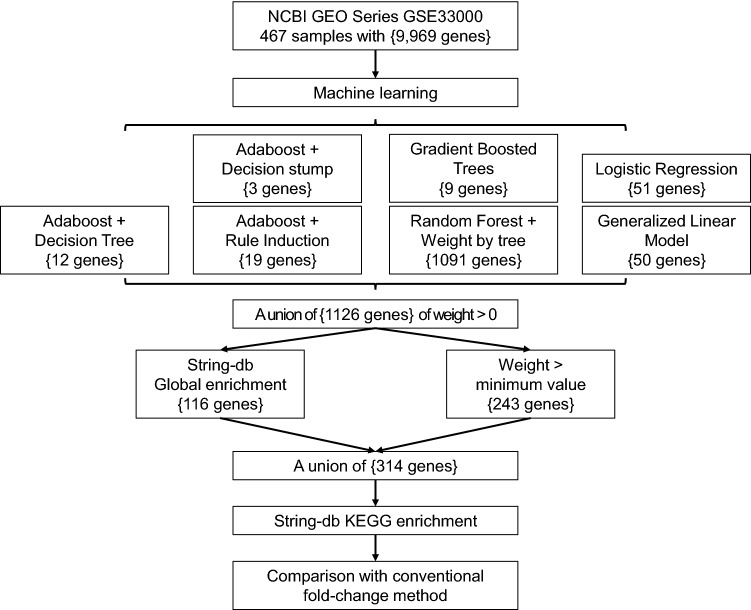
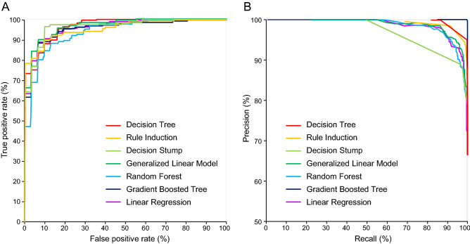
We developed a workflow (Fig. 1) to identify AD-predictive genes by ML, and each of the seven predictive operators or combinations of operators produced a gene list along with the weight of predictive contribution. The full lists are provided in the sheets of Generalized Linear Model, Logistic Regression, Rule Induction, Decision Stump, Decision Tree, Gradient Boosted Trees, and Weight of Random Forest of Supplementary File 2. The average accuracy of these models ranges from 86.30% to 91.22%, and the Performance sheet of Supplementary File 2 summarizes the accuracy, precision, and recall of each model, while ROC curves and precision recall curves are shown in Fig. 2. Combing the genes from the seven models, we got a union of 1126 non-redundant genes with weight > 0 (the Non-redundant Genes sheet of Supplementary File 2). To further extract the more representative genes, those genes satisfying both conditions, 1) genes with the weight of the minimum value (i.e., 0.001), and 2) genes without a presence in the global enrichment analysis (the Global Enrichment sheet of Supplementary File 2), were filtered out. Finally, we reached a list of 314 genes (the Genes sheet of Supplementary File 5).
Figure 1.
The study design and workflow of identifying AD-predictive genes by ML. The curly brackets indicate the number of genes that passed the criteria or were identified in the ML models.
Figure 2.
The performance of various ML models. A) ROC curves and B) precision recall (PR) curves of the ML models used in this study.
We conducted the analysis of variance (ANOVA) test to determine the probability for the null hypothesis of the equal performance of the different ML models. The ANOVA result (f = 1.558, prob = 0.174, alpha = 0.050) could not reject the null hypothesis, indicating that the difference between the performance of the different ML models is not significant. The process was exported as “ANOVA.rmp” and was uploaded to GitHub at https://github.com/JackCheng-TW/RapidMiner-files/Process/.
To check if our findings are not unique to a single dataset, we took another microarray-profiled PFC dataset GSE84422 as an unseen dataset to verify our models. Although nearly one-third of the training genes are missing in the test dataset, GSE84422 is currently the 2nd largest one after GSE33000. Upon testing, the accuracy was 28.57%, 58.93%, 76.79%, 82.14%, 71.43%, 44.64%, and 69.64% for decision tree, random forest, gradient boosted tree, generalized linear model, linear regression, decision stump, and rule induction, respectively. Since there are only a few attributes in decision tree/stump, missing one or two may largely limit the model performance. In contrast, models with more attributes like GLM outperform the others. The results indicate some models' generalizability and the difficulty of applying ML models on cross-platform datasets. The testing dataset "GSE84422_testing.xls" and exported processes were uploaded to GitHub at https://github.com/JackCheng-TW/RapidMiner-files/testing/.
ML compensates conventional FC methods in gene identification
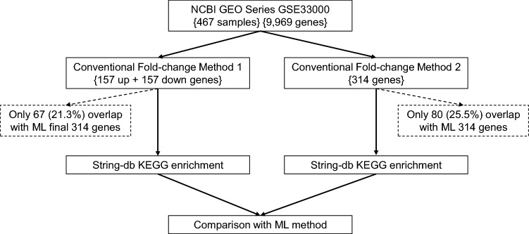
To compare the differences in gene identification between ML and conventional FC-based methods, we adopted two independent strategies, as illustrated in Fig. 3. In one way, the uppermost 157 genes and the bottommost 157 genes of fold change were selected (Genes sheet of Supplementary File 6). In the other way, we selected 314 DEGs by firstly filtering with the fold change cutoffs 1.2, and followed by the rank of the p values (Genes sheet of Supplementary File 7). Surprisingly, there were only 67 (21.3%) or 80 (25.5%) genes overlapped with the ML-derived 314 genes for the two conventional FC-based methods, respectively.
Figure 3.
The workflow of identifying dysregulated genes by the conventional FC method. The curly brackets indicate the number of genes that passed the criteria or were identified in each step. The overlap of identified genes between ML and the FC method is shown in dashed squares.
Next, to figure out the differences in enriched pathways, the final gene list from ML and those from the two conventional FC-based methods were submitted to KEGG pathway enrichment analysis, respectively. The top 15 enriched KEGG pathways are summarized in Table 1, while the KEGG sheets in Supplementary Files 5, 6, 7 provide the full results. As anticipated, most of the pathways enriched by ML-derived genes are not redundant to conventional FC-based methods. Interestingly, the KEGG Alzheimer's pathway (hsa05010) was only enriched by the ML-derived genes, not conventional methods. However, this does not imply that ML is superior to or can replace the conventional methods since the latter also exclusively enriched several AD-related pathways, such as complement cascades (hsa04610), cytokine-cytokine receptor interaction (hsa04060), and phagosome (hsa04145). The mutual exclusivity of critical pathways demonstrates that ML compensates the conventional FC methods in gene identification.
Table 1.
Enriched KEGG pathways of genes identified by machine learning and conventional fold-change (FC) methods, respectively.
| Machine learning | FC method 1 | FC method 2 | |
|---|---|---|---|
| 1 | Alzheimer's disease | Complement and coagulation cascades | GABAergic synapse |
| 2 | Parkinson's disease | Staphylococcus aureus infection | Morphine addiction |
| 3 | Huntington's disease | Phagosome | MAPK signaling pathway |
| 4 | Thermogenesis | Pertussis | Retrograde endocannabinoid signaling |
| 5 | Oxidative phosphorylation | Legionellosis | Nicotine addiction |
| 6 | Neurotrophin signaling pathway | Rheumatoid arthritis | Butanoate metabolism |
| 7 | MAPK signaling pathway | Malaria | |
| 8 | Acute myeloid leukemia | Systemic lupus erythematosus | |
| 9 | Non-alcoholic fatty liver disease (NAFLD) | Prion diseases | |
| 10 | Retrograde endocannabinoid signaling | Cytokine-cytokine receptor interaction | |
| 11 | FoxO signaling pathway | TNF signaling pathway | |
| 12 | Endometrial cancer | Kaposi's sarcoma-associated herpesvirus infection | |
| 13 | Alcoholism | MAPK signaling pathway | |
| 14 | Influenza A | Ras signaling pathway | |
| 15 | Serotonergic synapse | Influenza A |
ML highlights oxidative phosphorylation genes in the AD pathway
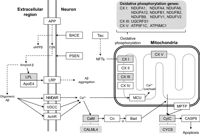
When we looked into ML-derived genes, which enriched the pathways, we found a considerable overlap of genes between the ML-exclusive pathways (Table 2). These genes are ATP5C1, ATP5G1, NDUFA1, NDUFA4, NDUFA6, NDUFA12, NDUFB1, NDUFB2, NDUFB9, NDUFV1, NDUFV2, and UQCRFS1. They belong to the oxidative phosphorylation pathway (hsa00190), which is also a part of the KEGG Alzheimer’s pathway (Fig. 4). Among them, NDUFA1, NDUFA4, NDUFA6, NDUFA12, NDUFB1, NDUFB2, NDUFB9, NDUFV1, NDUFV2 belong to the OXPHOS protein complexes (CX) I of the electron transport chain (ETC); while UQCRFS1 belongs to CX III of ETC. Moreover, ATP5C1 and ATP5G1 belong to the ATP synthase (CX V).
Table 2.
Overlapping of ML-identified genes in the enriched KEGG pathways. “O” denotes the presence of the gene.
| KEGG gene | Alzheimer's disease | Thermogenesis | Oxidative phosphorylation | Non-alcoholic fatty liver disease (NAFLD) |
|---|---|---|---|---|
| ATP5C1 | O | O | O | |
| ATP5G1 | O | O | O | |
| CALML4 | O | |||
| CYCS | O | O | ||
| LPL | O | |||
| NDUFA1 | O | O | O | O |
| NDUFA12 | O | O | O | O |
| NDUFA4 | O | O | O | O |
| NDUFA6 | O | O | O | O |
| NDUFB1 | O | O | O | O |
| NDUFB2 | O | O | O | O |
| NDUFB9 | O | O | O | O |
| NDUFV1 | O | O | O | O |
| NDUFV2 | O | O | O | O |
| UQCRFS1 | O | O | O | O |
| ACTB | O | |||
| RPS6KA1 | O | |||
| SMARCA4 | O | |||
| SOS1 | O |
Figure 4.
The ML-identified genes enrich the KEGG AD pathway. Proteins or protein complexes are shown in squares. Only a part of the KEGG AD pathway is shown. Dash line arrows indicate transformation, while solid line arrows indicate a reaction. The enriched proteins or protein complexes are drawn in grey, with the ML-identified genes noted at the open bracket near it.
Co-predictive partners of the CX genes
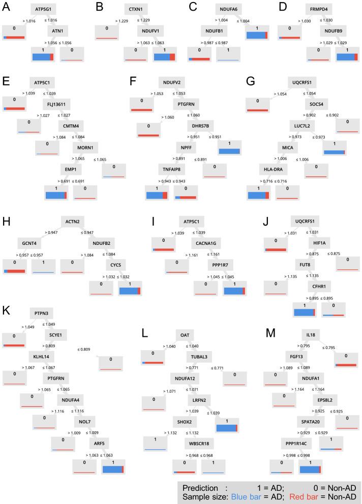
Random Forest produces decision trees, which use combinations of the Attribute value, i.e., expression of genes, to predict the sample Label, i.e., AD or not. Figure 5 shows 13 decision trees involving ETC complexes subunit genes, and Table 3 summarizes the 12 CX genes and other 37 predictive genes in these trees. Notably, 32 out of the 37 genes are relevant to AD. The AD-relevance is established by association studies of the expression, genomics, or metabolomics, respectively, with references listed in Table 3.
Figure 5.
Slightly down-regulated oxidative phosphorylation genes predict AD. (A) to (M) The decision trees from the random forest model containing NDUFA1, NDUFA4, NDUFA6, NDUFA12, NDUFB1, NDUFB2, NDUFB9, NDUFV1, NDUFV2, UQCRFS1, ATP5F1C, and ATP5MC1. The prediction outcome is denoted by 0 and 1, where 1 = AD, and 0 = Non-AD. The sample size is denoted by the thickness of the bar, while the sample type is denoted by blue or red, where blue bar = AD, and red bar = Non-AD.
Table 3.
Oxidative phosphorylation genes and their companions identified in machine learning.
| Gene | Full name | AD | Association | Refs. | ||
|---|---|---|---|---|---|---|
| E | G | M | ||||
| ATP5F1C* | ATP synthase subunit gamma, mitochondrial | O | ||||
| ATP5MC1* | ATP synthase F(0) complex subunit C1, mitochondrial | O | ||||
| NDUFA1 | NADH dehydrogenase [ubiquinone] 1 alpha subcomplex subunit 1 | O | ||||
| NDUFA12 | NADH dehydrogenase [ubiquinone] 1 alpha subcomplex subunit 12 | O | ||||
| NDUFA4 | Cytochrome c oxidase subunit NDUFA4 | O | ||||
| NDUFA6 | NADH dehydrogenase [ubiquinone] 1 alpha subcomplex subunit 6 | O | ||||
| NDUFB1 | NADH dehydrogenase [ubiquinone] 1 beta subcomplex subunit 1 | O | ||||
| NDUFB2 | NADH dehydrogenase [ubiquinone] 1 beta subcomplex subunit 2, mitochondrial | O | ||||
| NDUFB9 | NADH dehydrogenase [ubiquinone] 1 beta subcomplex subunit 9 | O | ||||
| NDUFV1 | NADH dehydrogenase [ubiquinone] flavoprotein 1, mitochondrial | O | ||||
| NDUFV2 | NADH dehydrogenase [ubiquinone] flavoprotein 2, mitochondrial | O | ||||
| UQCRFS1 | Cytochrome b-c1 complex subunit Rieske, mitochondrial | O | ||||
| ACTN2 | Alpha-actinin-2 | O | O | 64,65 | ||
| ARF5 | ADP-ribosylation factor 5 | O | 66 | |||
| ATN1 | Atrophin-1 | O | 14 | |||
| CACNA1G | Voltage-dependent T-type calcium channel subunit alpha-1G | O | 67 | |||
| CFHR1 | Complement factor H-related protein 1 | O | 68 | |||
| CMTM4 | CKLF like MARVEL transmembrane domain containing 4 | O | 69 | |||
| CTXN1 | Cortexin-1 | O | 16 | |||
| DHRS7B | Dehydrogenase/reductase SDR family member 7B | O | 70 | |||
| EMP1 | Epithelial membrane protein 1 | O | 71 | |||
| EPS8L2 | Epidermal growth factor receptor kinase substrate 8-like protein 2 | O | 72 | |||
| FGF13 | Fibroblast growth factor 13 | O | 67 | |||
| FLJ13611* | Trafficking protein particle complex 13 | O | 73 | |||
| FRMPD4 | FERM and PDZ domain-containing protein 4 | O | 18 | |||
| FUT8 | Alpha-(1,6)-fucosyltransferase | O | 74 | |||
| GCNT4 | Beta-1,3-galactosyl-O-glycosyl-glycoprotein beta-1,6-N-acetylglucosaminyltransferase 4 | |||||
| HIF1A | Hypoxia-inducible factor 1-alpha | O | O | 75,76 | ||
| HLA-DRA | HLA class II histocompatibility antigen, DR alpha chain | O | O | 67,77 | ||
| IL18 | Interleukin-18 | O | 78 | |||
| KLHL14 | Kelch-like protein 14 | |||||
| LRFN2 | Leucine-rich repeat and fibronectin type-III domain-containing protein 2 | O | 79 | |||
| LUC7L2 | Putative RNA-binding protein Luc7-like 2 | O | 80 | |||
| MICA | MHC class I polypeptide-related sequence A | |||||
| MORN1 | MORN repeat containing 1 | O | 81 | |||
| NOL7 | Nucleolar protein 7 | O | 82 | |||
| NPFF | Pro-FMRFamide-related neuropeptide FF | O | 83 | |||
| OAT | Ornithine aminotransferase, mitochondrial | O | 45 | |||
| PPP1R14C | Protein phosphatase 1 regulatory subunit 14C | O | 84 | |||
| PPP1R7 | Protein phosphatase 1 regulatory subunit 7 | O | 85 | |||
| PTGFRN | Prostaglandin F2 receptor negative regulator | O | 86 | |||
| PTPN3 | Tyrosine-protein phosphatase non-receptor type 3 | O | 18 | |||
| SCYE1* | Aminoacyl tRNA synthase complex-interacting multifunctional protein 1 | |||||
| SHOX2 | Short stature homeobox protein 2 | |||||
| SOCS4 | Suppressor of cytokine signaling 4 | O | 87 | |||
| SPATA20 | Spermatogenesis-associated protein 20 | |||||
| TNFAIP8 | Tumor necrosis factor alpha-induced protein 8 | O | 88 | |||
| TUBAL3 | Tubulin alpha chain-like 3 | O | ||||
| WBSCR18* | DNAJC30 interacts with ATP synthase and links mitochondria to brain development | O | 89 | |||
An “O” denotes the present knowledge that supports the involvement or association of the gene with Alzheimer’s disease. The category item “AD” indicates the involvement of the gene in the KEGG AD pathway, while “E”, “G”, and “M” indicate the evidence of association studies of the expression, genomics, and metabolomics. An * sign indicates the presence of another preferred name in the String-db. The alternative names are shown in the parentheses. ATP5F1C (ATP5C1); ATP5MC1 (ATP5G1); FLJ13611 (TRAPPC13); SCYE1 (AIMP1); WBSCR18 (DNAJC30).
Figure 5A shows that the expression of ATP5G1 and ATN1 predicts AD. Although the exact function of ATN1 is unknown, it may act as a transcriptional co-repressor in neurons13. Moreover, alternative splicing of ATN1 was significantly detected in the frontal lobe of AD postmortem14. Figure 5B shows that the expression of NDUFV and CTXN1 predicts AD. CTXN1 encodes cortexin-1 and may mediate signaling of cortical neurons during forebrain development15, and it is highly dysregulated in the aging brain16. Figure 5C shows that slightly downregulation of two CX I genes, NDUFA6 and NDUFB1 predicts AD. Figure 5D shows that the expression of NDUFB9 and FRMPD4 predicts AD. FRMPD4 positively regulates dendritic spine morphogenesis and involves in excitatory synaptic transmission17. Besides, the expression of FRMPD4 was found to be significantly altered in the AD hippocampus18. Other AD-predictive genes in these decision trees will be discussed in groups according to their biological functions.
Discussion
We conducted machine learning (ML) analyses to train AD case/control classifiers using transcriptomic data and then compared the ML-derived gene features with that from the conventional differential expression analysis. ML exclusively highlighted oxidative phosphorylation but could not fully include the findings from the conventional methods. The pathways involving the identified genes and the limitation of the study are discussed below.
Oxidative phosphorylation
Oxidative phosphorylation in eukaryotes takes place at the electron transport chain in the mitochondrion. The oxidation of NADH or succinate from the citric acid cycle is the energy source of ATP synthase. During this process, several mitochondrial inner-membrane-embedded complexes, including CX I and CX III, pump protons out from the inner membrane to establish proton gradient, while CX V utilizes the energy of the influx of protons to generate ATP from ADP19.
Abnormal mitochondrial morphology and functions, including glucose metabolism and ROS production, have been identified as early hallmarks of AD20,21. These phenotypes are directly related to the disruption of glycolytic processes and the impairment of the ETC complexes. In the '90 s, most research efforts have been devoted to investigating the role of CX IV in AD22,23. However, the evidence is not conclusive on whether dysregulation of any single ETC complex dominates AD progress. For example, besides expression, several mutations in ETC complex subunit genes may impair the complex activity24. Moreover, the ETC complex's dysregulation seems to be brain-region dependent, e.g., CX IV has no significant decrease in the temporal lobe, and CX I–III are decreased at certain cortex locations of AD24.
Recent studies also highlighted CX I's role, especially its deregulation, is tau-dependent in contrast to the Aβ-dependent CX IV25. Moreover, an SNP association study demonstrated the AD association for complex I genes but not for complexes II–V26. Furthermore, from a postmortem study of 18 AD and 44 controls, the downregulation of CX I-V in the hippocampus was identified27. However, the expression of CX I genes may not be monotonic during the AD progression. From a postmortem study of twelve AD and six controls, CX I genes are reported to decrease in the early stage and increase in the frontal cortex of definite AD patients28.
When we only see the symptom, most things look complex, especially the case for ETC complexes in AD. Could ML guide us through this misty forest with the aid of the Random Forest model by finding out potential partners of CX genes in predicting AD?
Neural maintenance or transmission
Among the AD-predictive genes, CACNA1G, FGF13, LRFN2, NPFF, and SHOX2 participate in neural maintenance or transmission. CACNA1G encodes voltage-dependent T-type calcium channel subunit alpha-1G. FGF13 is a fibroblast growth factor and plays a critical role in neuron polarization and migration29. LRFN2 promotes neurite outgrowth and increases the expression of the NMDA receptor30. NPFF is a neuropeptide, while SHOX2 may be a growth regulator in the neural system and involves processing somatosensory information31. In AD, pathological hallmarks include synaptic failure and neuronal loss32. Moreover, the critical role of mitochondria in supporting synaptic, as well as the evidence of dysfunction of mitochondria from both clinical postmortem33 and animal models34 of AD, support the mitochondria-synapse hypothesis of AD. Our findings that simultaneous dysregulation of CX and neuronal genes predict AD supports this hypothesis.
Immune system
The innate immunity, especially neuroinflammation mediated by microglia, is considered a hallmark of AD, whereas the role of the adapted immunity in AD is not conclusive35. Among the AD-predictive genes, CFHR1, CMTM4, HLA-DRA, IL18, MICA, MORN1, SCYE1, and SOCS4 participate in immunity. CMTM4 regulates PD-L1 protein36, which binds to PD-1 and suppresses the T-cells' adaptive arm, while HLA-DRA presents the extracellular-protein-derived peptides to, and MICA presents the stress-induced self-antigen to T-cells, respectively37. Moreover, MORN1 modulates functional Ca2+ influx in T cells upon activation of T-cell receptors38. IL18 and SCYE1 (AIMP1) are pro-inflammatory cytokines, while SOCS4 is part of a negative feedback system that regulates cytokine signal transduction39. CFHR1 is an inhibitor of the complement pathway that blocks C5 convertase and controls complement activation along with complement factor H40. Our results indicate that the dysregulation of both innate and adaptive immunity genes may cooperate with CX genes to advance AD progression.
Phosphatase regulators
In AD, hyperphosphorylation of the microtubule-associated proteins, especially tau, disrupts the microtubules' assembly in neurons. Moreover, significantly lower type 1 phosphatase (PP1) activity in AD brains suggests the critical role of dysfunctional phosphatases in AD41. Among the AD-predictive genes, PPP1R14C and PPP1R7 belong to PP1 regulatory subunit 14 and subunit 7, respectively. Our results indicate that the dysregulation of PPP1R14C and PPP1R7, along with CX genes, may further advance AD progression by aggravating the microtubule-associated proteins' hyperphosphorylation.
Protein glycosylation
Protein glycosylation is a ubiquitous posttranslational modification of site-specific attachment of glycans and regulates the protein's folding and function. During the protein transport from Endoplasmic Reticulum to the Golgi apparatus, a series of attachment of oligosaccharides maturates a wide variety of complex N- or O-glycans. An N-glycosylation denotes the glycan's attachment to the amide nitrogen of an asparagine residue of the protein, whereas an O-glycosylation denotes the attachment to the oxygen atom of serine or threonine residues. Abnormal N- and O-glycosylation has been reported in AD42,43. Among the AD-predictive genes, FUT8 and GCNT4 mediate glycosylation in the Golgi apparatus. FUT8 catalyzes the addition of fucose to the GlcNAc residue, while GCNT4 is a glycosyltransferase mediating O-glycan branching44. Thus, the dysregulation of FUT8 and GCNT4 may aggravate AD progression by abnormal glycosylation under the condition of CX deficiency.
Other mitochondria machinery
Notably, among the AD-predictive genes, there are two mitochondrial genes besides the CX: Ornithine aminotransferase (OAT) and DnaJ homolog subfamily C member 30 (DNAJC30/ WBSCR18). OAT converts ornithine into pyrroline-5-carboxylate (P5C), which can serve as the precursor of proline and glutamate. Furthermore, since ornithine is an intermediate product in the urea cycle, OAT dysregulation may lead to abnormalities of both energy production machinery and the supply of neural transmitters. Recently, the OAT substrate ornithine has been proposed as an early diagnostic biomarker of AD45, and altered expression of the urea cycle enzymes have been identified in sporadic AD brains46. Our finding that simultaneous downregulation of OAT and CX I predicts AD indicates that the deficiency of the urea cycle and CX may co-operate to advance AD.
Meanwhile, DNAJC30 has been recently identified as an auxiliary component of ATP-synthase machinery in the mitochondria47. The removal of Dnajc30 in mice resulted in hypofunctional mitochondria, decreased integrity of CXs, and abnormal neocortical pyramidal neurons47. Our finding that the simultaneous downregulation of DNAJC30 and CX I predicts AD also supports the mitochondria deficiency hypothesis of AD.
Slightly dysregulated CX genes predict AD
From an overall observation on the CX-related decision trees (Fig. 5), a combination of down-regulated CX components and one or several partner genes mentioned above predicts AD. Notably, the margin is not conventional twofold, 1.5-fold, or even 1.2-fold. The margin is very subtle, and this is why the conventional FC method cannot identify them. With the criteria of p < 0.05 and FC 1.2, 1.5, or 2, the numbers of DEGs of the model dataset GSE33000 are 418, 10, and 0, respectively, as shown in Supplementary File 10. We compared the 418 DEGs with the ML 314 genes in Supplementary File 5 and found the number of intersection genes to be 60 (19.1%), which was compatible with the results of the conventional method 1 (21.3%) and the conventional method 2 (25.5%). Furthermore, it is difficult to identify complicated rules by conventional methods. Therefore, we suggest adopting machine-learning algorithms, especially decision trees, rule induction, and random forest, as complementary methods in transcriptome studies.
Limitations
There are several limitations to the interpretation of the results. (1) The samples are primarily of Caucasian ancestry. The biased sample race may limit the results to be applied to other races. (2) The samples are from the postmortem of a specific brain region. Since expressional heterogeneity, this may limit the results to be applied to other brain regions. (3) Due to the same reason, the results can hardly be applied to patient diagnosis purposes. (4) For the future application of the study pipeline, at least hundreds of samples might be required due to ML's nature. (5) ML models predict the patient disease labels but not the involvement of genes in disease, and additional genetic evidence is required to delineate any possible causal/reactive roles of these gene features in AD. (6) The performance difference in the independent dataset could be attributed to the detectable genes of different chip systems and the within-dataset variations. The absence of 36.9% attributes (genes) in the test set largely limited the performance of some models. Moreover, the limitation may also come from the difference in the sampling quality, which is reflected by the within-dataset variation (the average STD/INT were 8.2% and 21% for the modeling set and test set, respectively).
Rapidminer models have also been used to identify the transcriptomic bio-signature of an infectious disease condition in the mammary gland of the cow48, with the performance ranging from 53 to 87%, which is compatible with the performance of this study. The differences in strategy majorly lay in whether pre-screening attributes (the so-called feature selection) before applying ML49. The benefits of feature selection include simplifying models, shorter training times, and avoidance of high dimensionality problems; however, the feature selection step using the entire dataset may strongly bias all downstream prediction, even when cross-validation is used50. Therefore, in this study, we decided to skip the feature selection step to achieve an unbiased understanding of AD.
Since decision trees were the final models to identify potential novel genes in this study, whether the data size is big enough is crucial. According to Vabalas et al.51, we conducted a series of train/test split to validate whether arbitrary partial subsets of data could generate decision trees to predict the “unseen” counterpart, with the same parameters used in this study. As shown in Supplementary File 8, the recall rates were saturated at n = 94, i.e., 20% of the total samples, which may imply the sample size was sufficient to conduct this study.
Although we did not combine datasets in this study, appropriate methods used for reducing the batch effect and differences between experiments52 should be applied when combing datasets in future studies. We also noticed that random forest analysis dominated the identified gene features, indicating that future similar studies might focus on random forest first. However, other models may supply other 10% genetic cues on the investigator’s demand.
Hypotheses developed from ML models
To discover and characterize the underlying pathophysiological pathways of AD are the main objectives of genetic research, including this ML study. Based on our findings, we postulate that two novel players, i.e., RNF157 and KIAA1715, may independently participate in AD pathophysiology by mediating the mitogen-activated protein kinase (MAPK) signaling pathway. MAPKs are serine/threonine protein kinases regulating cellular processes in response to environmental stimuli and participate in hallmark events of AD, including tau phosphorylation, Aβ deposition, and chronic inflammation53,54.
In the #2 model of RF (Supplementary File 2), RNF157, EPHA2, and hCG_1776018 (also known as PIRT, an uncharacterized phosphoinositide-interacting protein) co-predict AD. EPHA2 is a membrane receptor tyrosine kinase, which regulates migration, adhesion, and blood–brain barrier through MAPK signaling55. RNF157 is an E3 ubiquitin ligase that acts as a downstream effector of PI3K/MAPK signaling56 and regulates the survival of neurons by ubiquitinating APBB157. Presently, there is no knowledge about the roles of these three genes in AD. We hypothesize that RNF157 may act as the downstream of EPHA2 and hCG_1776018, and regulate neural death upon cellular stress in the AD microenvironment. We also postulate that RNF157 agonist may act as a symptomatic treatment in AD.
In the #230 model of RF (Supplementary File 2), KIAA1715 and MAP3K9 co-predict AD. MAP3K9 is a serine/threonine kinase that is activated by environmental stress and acts as an upstream activator of the MKK/JNK signal transduction cascade regulating apoptosis58. MAP3K9 dysregulation has been proposed as a possible marker in AD59. KIAA1715 (also known as LNPK) is an endoplasmic reticulum (ER) membrane protein, which stabilizes ER curvature and ER tubular junction network60,61. Mutations in KIAA1715 cause neurodevelopmental syndromes, such as intellectual disability and epilepsy61. Notably, disruption of ER-mitochondria contact has recently been found in AD postmortem62, while restoring ER-mitochondria contact rescues AD animal model63. However, there is no knowledge about the role of KIAA1715 in AD. We hypothesize that under the pro-inflammatory microenvironment of AD, KIAA1715 deficiency may lead to instability of ER structure, leading to disruption of ER-mitochondria contact and eventually aggravate AD progression.
Conclusion
Our study using machine learning techniques on the gene expression profile of the postmortem of the prefrontal cortex brain tissues of AD and controls highlighted the oxidative phosphorylation genes in the AD pathway. These genes were exclusively identified in ML but not in the conventional counterpart. Our results imply that ML should be considered complementary to the conventional FC methods in transcriptome studies. More importantly, we show that hypotheses underlying pathophysiological pathways of AD could be developed by further looking into ML models.
Supplementary Information
Abbreviations
- AD
Alzheimer's disease
- ML
Machine learning
- OXPHOS
Oxidative phosphorylation
- CX
OXPHOS protein complex
- FC
Fold-change
Author contributions
W.Y.L. and F.J.T. initiated and supervised this study. J.C. and H.P.L. contributed to the acquisition, analysis, and interpretation of data. All authors discussed and drafted the manuscript.
Funding
This work was supported by grants from the Ministry of Science and Technology in Taiwan (MOST107-2314-B-039-042-MY2, MOST106-2314-B-039-009-, MOST108-2320-B-039-031-MY3, MOST 109-2314-B-039-030) and grants from China Medical University & Hospital (CMU109-MF-85, CMU108-MF-68, CMU108-MF-61, CMU107-S-08, DMR-109-150, DMR-106-119).
Data availability
All data in this study are included in the supplementary data. The raw data used for machine learning and traditional expression analysis in the CSV format was uploaded as Supplementary File 1. Besides, it is also available from https://github.com/JackCheng-TW/RawData. The independent dataset was uploaded as Supplementary File 9.
Code availability
The machine learning platform RapidMiner Studio is available at https://rapidminer.com/. The process files in Rapidminer format (.rmp) of this study and the generated models were uploaded to GitHub at https://github.com/JackCheng-TW/RapidMiner-files.
Competing interests
The authors declare no competing interests.
Footnotes
Publisher's note
Springer Nature remains neutral with regard to jurisdictional claims in published maps and institutional affiliations.
These authors contributed equally: Jack Cheng and Hsin-Ping Liu.
Contributor Information
Wei-Yong Lin, Email: linwy@mail.cmu.edu.tw.
Fuu-Jen Tsai, Email: d0704@mail.cmuh.org.tw.
Supplementary Information
The online version contains supplementary material available at 10.1038/s41598-021-93085-z.
References
- 1.Cummings J, Lee G, Ritter A, Sabbagh M, Zhong K. Alzheimer's disease drug development pipeline: 2019. Alzheimer’s & Dement. 2019;5:272–293. doi: 10.1016/j.trci.2019.05.008. [DOI] [PMC free article] [PubMed] [Google Scholar]
- 2.Gong C-X, Liu F, Iqbal K. Multifactorial hypothesis and multi-targets for Alzheimer’s disease. J. Alzheimers Dis. 2018;64:S107–S117. doi: 10.3233/JAD-179921. [DOI] [PubMed] [Google Scholar]
- 3.Hölscher C. Moving towards a more realistic concept of what constitutes Alzheimer's disease. EBioMedicine. 2019;39:17–18. doi: 10.1016/j.ebiom.2018.12.030. [DOI] [PMC free article] [PubMed] [Google Scholar]
- 4.Tanveer M, et al. Machine learning techniques for the diagnosis of Alzheimer’s disease: A review. ACM Trans. Multimed. Comput. Commun. Appl. 2019;16:1–35. [Google Scholar]
- 5.Perakslis E, Riordan H, Friedhoff L, Nabulsi A, Pich EM. A call for a global ‘bigger’data approach to Alzheimer disease. Nat. Rev. Drug Discov. 2019;18:319. doi: 10.1038/nrd.2018.86. [DOI] [PubMed] [Google Scholar]
- 6.Cheng J, et al. Matrix metalloproteinase 14 modulates diabetes and Alzheimer’s disease cross-talk: A meta-analysis. Neurol. Sci. 2018;39:267–274. doi: 10.1007/s10072-017-3166-4. [DOI] [PubMed] [Google Scholar]
- 7.Cheng J, et al. Dystonin/BPAG1 modulates diabetes and Alzheimer’s disease cross-talk: A meta-analysis. Neurol. Sci. 2019;40:1577–1582. doi: 10.1007/s10072-019-03879-3. [DOI] [PubMed] [Google Scholar]
- 8.Narayanan M, et al. Common dysregulation network in the human prefrontal cortex underlies two neurodegenerative diseases. Mol. Syst. Biol. 2014;10:743. doi: 10.15252/msb.20145304. [DOI] [PMC free article] [PubMed] [Google Scholar]
- 9.Wang M, et al. Integrative network analysis of nineteen brain regions identifies molecular signatures and networks underlying selective regional vulnerability to Alzheimer’s disease. Genome Med. 2016;8:1–21. doi: 10.1186/s13073-015-0257-9. [DOI] [PMC free article] [PubMed] [Google Scholar]
- 10.Szklarczyk D, et al. STRING v11: Protein–protein association networks with increased coverage, supporting functional discovery in genome-wide experimental datasets. Nucleic Acids Res. 2019;47:D607–D613. doi: 10.1093/nar/gky1131. [DOI] [PMC free article] [PubMed] [Google Scholar]
- 11.Kanehisa M, Goto S. KEGG: Kyoto encyclopedia of genes and genomes. Nucleic Acids Res. 2000;28:27–30. doi: 10.1093/nar/28.1.27. [DOI] [PMC free article] [PubMed] [Google Scholar]
- 12.Kanehisa M. Toward understanding the origin and evolution of cellular organisms. Protein Sci. 2019;28:1947–1951. doi: 10.1002/pro.3715. [DOI] [PMC free article] [PubMed] [Google Scholar]
- 13.Wood JD, et al. Atrophin-1, the dentato-rubral and pallido-luysian atrophy gene product, interacts with ETO/MTG8 in the nuclear matrix and represses transcription. J. Cell Biol. 2000;150:939–948. doi: 10.1083/jcb.150.5.939. [DOI] [PMC free article] [PubMed] [Google Scholar]
- 14.Twine NA, Janitz K, Wilkins MR, Janitz M. Whole transcriptome sequencing reveals gene expression and splicing differences in brain regions affected by Alzheimer's disease. PLoS ONE. 2011;6:e16266. doi: 10.1371/journal.pone.0016266. [DOI] [PMC free article] [PubMed] [Google Scholar]
- 15.Coulter PM, Bautista EA, Margulies JE, Watson JB. Identification of cortexin: A novel, neuron-specific, 82-residue membrane protein enriched in rodent cerebral cortex. J. Neurochem. 1993;61:756–759. doi: 10.1111/j.1471-4159.1993.tb02183.x. [DOI] [PubMed] [Google Scholar]
- 16.Wang J, et al. Chromosome 19p in Alzheimer's disease: When genome meets transcriptome. J. Alzheimers Dis. 2014;38:245–250. doi: 10.3233/JAD-130917. [DOI] [PubMed] [Google Scholar]
- 17.Lee HW, et al. Preso, a novel PSD-95-interacting FERM and PDZ domain protein that regulates dendritic spine morphogenesis. J. Neurosci. 2008;28:14546–14556. doi: 10.1523/JNEUROSCI.3112-08.2008. [DOI] [PMC free article] [PubMed] [Google Scholar]
- 18.Hokama M, et al. Altered expression of diabetes-related genes in Alzheimer's disease brains: The Hisayama study. Cereb. Cortex. 2014;24:2476–2488. doi: 10.1093/cercor/bht101. [DOI] [PMC free article] [PubMed] [Google Scholar]
- 19.Zorova LD, et al. Mitochondrial membrane potential. Anal. Biochem. 2018;552:50–59. doi: 10.1016/j.ab.2017.07.009. [DOI] [PMC free article] [PubMed] [Google Scholar]
- 20.Lin MT, Beal MF. Mitochondrial dysfunction and oxidative stress in neurodegenerative diseases. Nature. 2006;443:787–795. doi: 10.1038/nature05292. [DOI] [PubMed] [Google Scholar]
- 21.Zhu X, Perry G, Smith MA, Wang X. Abnormal mitochondrial dynamics in the pathogenesis of Alzheimer's disease. J. Alzheimers Dis. 2013;33:S253–S262. doi: 10.3233/JAD-2012-129005. [DOI] [PMC free article] [PubMed] [Google Scholar]
- 22.Kish SJ, et al. Brain cytochrome oxidase in Alzheimer's disease. J. Neurochem. 1992;59:776–779. doi: 10.1111/j.1471-4159.1992.tb09439.x. [DOI] [PubMed] [Google Scholar]
- 23.Mutisya EM, Bowling AC, Beal MF. Cortical cytochrome oxidase activity is reduced in Alzheimer's disease. J. Neurochem. 1994;63:2179–2184. doi: 10.1046/j.1471-4159.1994.63062179.x. [DOI] [PubMed] [Google Scholar]
- 24.Shoffner JM. Oxidative phosphorylation defects and Alzheimer's disease. Neurogenetics. 1997;1:13–19. doi: 10.1007/s100480050002. [DOI] [PubMed] [Google Scholar]
- 25.Rhein V, et al. Amyloid-β and tau synergistically impair the oxidative phosphorylation system in triple transgenic Alzheimer's disease mice. Proc. Natl. Acad. Sci. USA. 2009;106:20057–20062. doi: 10.1073/pnas.0905529106. [DOI] [PMC free article] [PubMed] [Google Scholar]
- 26.Biffi A, et al. Genetic variation of oxidative phosphorylation genes in stroke and Alzheimer's disease. Neurobiol. Aging. 2014;35:1956.e1951–1956.e1958. doi: 10.1016/j.neurobiolaging.2014.01.141. [DOI] [PMC free article] [PubMed] [Google Scholar]
- 27.Mastroeni D, et al. Nuclear but not mitochondrial-encoded oxidative phosphorylation genes are altered in aging, mild cognitive impairment, and Alzheimer's disease. Alzheimers Dement. 2017;13:510–519. doi: 10.1016/j.jalz.2016.09.003. [DOI] [PMC free article] [PubMed] [Google Scholar]
- 28.Manczak M, Park BS, Jung Y, Reddy PH. Differential expression of oxidative phosphorylation genes in patients with Alzheimer’s disease. NeuroMol. Med. 2004;5:147–162. doi: 10.1385/NMM:5:2:147. [DOI] [PubMed] [Google Scholar]
- 29.Smallwood PM, et al. Fibroblast growth factor (FGF) homologous factors: New members of the FGF family implicated in nervous system development. Proc. Natl. Acad. Sci. USA. 1996;93:9850–9857. doi: 10.1073/pnas.93.18.9850. [DOI] [PMC free article] [PubMed] [Google Scholar]
- 30.Wang C-Y, et al. A novel family of adhesion-like molecules that interacts with the NMDA receptor. J. Neurosci. 2006;26:2174–2183. doi: 10.1523/JNEUROSCI.3799-05.2006. [DOI] [PMC free article] [PubMed] [Google Scholar]
- 31.Blaschke RJ, et al. SHOT, a SHOX-related homeobox gene, is implicated in craniofacial, brain, heart, and limb development. Proc. Natl. Acad. Sci. USA. 1998;95:2406–2411. doi: 10.1073/pnas.95.5.2406. [DOI] [PMC free article] [PubMed] [Google Scholar]
- 32.Guo L, Tian J, Du H. Mitochondrial dysfunction and synaptic transmission failure in Alzheimer’s disease. J. Alzheimers Dis. 2017;57:1071–1086. doi: 10.3233/JAD-160702. [DOI] [PMC free article] [PubMed] [Google Scholar]
- 33.Parker WD, Parks J, Filley CM, Kleinschmidt-DeMasters B. Electron transport chain defects in Alzheimer's disease brain. Neurology. 1994;44:1090–1090. doi: 10.1212/WNL.44.6.1090. [DOI] [PubMed] [Google Scholar]
- 34.Du H, et al. Cyclophilin D deficiency attenuates mitochondrial and neuronal perturbation and ameliorates learning and memory in Alzheimer's disease. Nat. Med. 2008;14:1097–1105. doi: 10.1038/nm.1868. [DOI] [PMC free article] [PubMed] [Google Scholar]
- 35.Van Eldik LJ, et al. The roles of inflammation and immune mechanisms in Alzheimer's disease. Alzheimer's & Dement. 2016;2:99–109. doi: 10.1016/j.trci.2016.05.001. [DOI] [PMC free article] [PubMed] [Google Scholar]
- 36.Mezzadra R, et al. Identification of CMTM6 and CMTM4 as PD-L1 protein regulators. Nature. 2017;549:106–110. doi: 10.1038/nature23669. [DOI] [PMC free article] [PubMed] [Google Scholar]
- 37.Davis MM, Bjorkman PJ. T-cell antigen receptor genes and T-cell recognition. Nature. 1988;334:395–402. doi: 10.1038/334395a0. [DOI] [PubMed] [Google Scholar]
- 38.Woo JS, et al. Junctophilin-4, a component of the endoplasmic reticulum–plasma membrane junctions, regulates Ca2+ dynamics in T cells. Proc. Natl. Acad. Sci. USA. 2016;113:2762–2767. doi: 10.1073/pnas.1524229113. [DOI] [PMC free article] [PubMed] [Google Scholar]
- 39.Kedzierski L, et al. Suppressor of cytokine signaling 4 (SOCS4) protects against severe cytokine storm and enhances viral clearance during influenza infection. PLoS Pathog. 2014;10:e1004134. doi: 10.1371/journal.ppat.1004134. [DOI] [PMC free article] [PubMed] [Google Scholar]
- 40.Heinen S, et al. Factor H–related protein 1 (CFHR-1) inhibits complement C5 convertase activity and terminal complex formation. Blood. 2009;114:2439–2447. doi: 10.1182/blood-2009-02-205641. [DOI] [PubMed] [Google Scholar]
- 41.Gong CX, Singh TJ, Grundke-Iqbal I, Iqbal K. Phosphoprotein phosphatase activities in Alzheimer disease brain. J. Neurochem. 1993;61:921–927. doi: 10.1111/j.1471-4159.1993.tb03603.x. [DOI] [PubMed] [Google Scholar]
- 42.Kanninen K, Goldsteins G, Auriola S, Alafuzoff I, Koistinaho J. Glycosylation changes in Alzheimer’s disease as revealed by a proteomic approach. Neurosci. Lett. 2004;367:235–240. doi: 10.1016/j.neulet.2004.06.013. [DOI] [PubMed] [Google Scholar]
- 43.Zhu Y, Shan X, Yuzwa SA, Vocadlo DJ. The emerging link between O-GlcNAc and Alzheimer disease. J. Biol. Chem. 2014;289:34472–34481. doi: 10.1074/jbc.R114.601351. [DOI] [PMC free article] [PubMed] [Google Scholar]
- 44.Schwientek T, et al. Control of O-glycan branch formation molecular cloning and characterization of a novel thymus-associated core 2 β1, 6-N-acetylglucosaminyltransferase. J. Biol. Chem. 2000;275:11106–11113. doi: 10.1074/jbc.275.15.11106. [DOI] [PubMed] [Google Scholar]
- 45.Liang Q, et al. Metabolomics-based screening of salivary biomarkers for early diagnosis of Alzheimer's disease. RSC Adv. 2015;5:96074–96079. doi: 10.1039/C5RA19094K. [DOI] [Google Scholar]
- 46.Jęśko H, et al. Altered expression of urea cycle enzymes in amyloid-β protein precursor overexpressing PC12 cells and in sporadic Alzheimer’s disease brain. J. Alzheimers Dis. 2018;62:279–291. doi: 10.3233/JAD-170427. [DOI] [PubMed] [Google Scholar]
- 47.Tebbenkamp AT, et al. The 7q11. 23 protein DNAJC30 interacts with ATP synthase and links mitochondria to brain development. Cell. 2018;175:1088–1104. doi: 10.1016/j.cell.2018.09.014. [DOI] [PMC free article] [PubMed] [Google Scholar]
- 48.Sharifi S, et al. Integration of machine learning and meta-analysis identifies the transcriptomic bio-signature of mastitis disease in cattle. PLoS ONE. 2018;13:e0191227. doi: 10.1371/journal.pone.0191227. [DOI] [PMC free article] [PubMed] [Google Scholar]
- 49.Cheng J, Liu H-P, Lin W-Y, Tsai F-J. Identification of contributing genes of Huntington’s disease by machine learning. BMC Med. Genom. 2020;13:1–11. doi: 10.1186/s12920-020-00822-w. [DOI] [PMC free article] [PubMed] [Google Scholar]
- 50.Ambroise C, McLachlan GJ. Selection bias in gene extraction on the basis of microarray gene-expression data. Proc. Natl. Acad. Sci. USA. 2002;99:6562–6566. doi: 10.1073/pnas.102102699. [DOI] [PMC free article] [PubMed] [Google Scholar]
- 51.Vabalas A, Gowen E, Poliakoff E, Casson AJ. Machine learning algorithm validation with a limited sample size. PLoS ONE. 2019;14:e0224365. doi: 10.1371/journal.pone.0224365. [DOI] [PMC free article] [PubMed] [Google Scholar]
- 52.Mohammadi-Dehcheshmeh M, et al. Unified transcriptomic signature of arbuscular mycorrhiza colonization in roots of Medicago truncatula by integration of machine learning, promoter analysis, and direct merging meta-analysis. Front. Plant Sci. 2018;9:1550. doi: 10.3389/fpls.2018.01550. [DOI] [PMC free article] [PubMed] [Google Scholar]
- 53.Zhu X, Lee H-G, Raina AK, Perry G, Smith MA. The role of mitogen-activated protein kinase pathways in Alzheimer’s disease. Neurosignals. 2002;11:270–281. doi: 10.1159/000067426. [DOI] [PubMed] [Google Scholar]
- 54.Lee JK, Kim N-J. Recent advances in the inhibition of p38 MAPK as a potential strategy for the treatment of Alzheimer’s disease. Molecules. 2017;22:1287. doi: 10.3390/molecules22081287. [DOI] [PMC free article] [PubMed] [Google Scholar]
- 55.Darling TK, et al. EphA2 contributes to disruption of the blood-brain barrier in cerebral malaria. PLoS Pathog. 2020;16:e1008261. doi: 10.1371/journal.ppat.1008261. [DOI] [PMC free article] [PubMed] [Google Scholar]
- 56.Dogan T, et al. Role of the E3 ubiquitin ligase RNF157 as a novel downstream effector linking PI3K and MAPK signaling pathways to the cell cycle. J. Biol. Chem. 2017;292:14311–14324. doi: 10.1074/jbc.M117.792754. [DOI] [PMC free article] [PubMed] [Google Scholar]
- 57.Matz A, et al. Regulation of neuronal survival and morphology by the E3 ubiquitin ligase RNF157. Cell Death Differ. 2015;22:626–642. doi: 10.1038/cdd.2014.163. [DOI] [PMC free article] [PubMed] [Google Scholar]
- 58.Durkin JT, et al. Phosphoregulation of mixed-lineage kinase 1 activity by multiple phosphorylation in the activation loop. Biochemistry. 2004;43:16348–16355. doi: 10.1021/bi049866y. [DOI] [PubMed] [Google Scholar]
- 59.Zhang L, et al. Potential hippocampal genes and pathways involved in Alzheimer’s disease: A bioinformatic analysis. Genet. Mol. Res. 2015;14:7218–7232. doi: 10.4238/2015.June.29.15. [DOI] [PubMed] [Google Scholar]
- 60.Shemesh T, et al. A model for the generation and interconversion of ER morphologies. Proc. Natl. Acad. Sci. USA. 2014;111:E5243–E5251. doi: 10.1073/pnas.1419997111. [DOI] [PMC free article] [PubMed] [Google Scholar]
- 61.Breuss MW, et al. Mutations in LNPK, encoding the endoplasmic reticulum junction stabilizer lunapark, cause a recessive neurodevelopmental syndrome. Am. J. Hum. Genet. 2018;103:296–304. doi: 10.1016/j.ajhg.2018.06.011. [DOI] [PMC free article] [PubMed] [Google Scholar]
- 62.Lau DH, et al. Disruption of endoplasmic reticulum-mitochondria tethering proteins in post-mortem Alzheimer's disease brain. Neurobiol. Dis. 2020;143:105020. doi: 10.1016/j.nbd.2020.105020. [DOI] [PMC free article] [PubMed] [Google Scholar]
- 63.Garrido-Maraver J, Loh SH, Martins LM. Forcing contacts between mitochondria and the endoplasmic reticulum extends lifespan in a Drosophila model of Alzheimer's disease. Biol. Open. 2020;9:bio47530. doi: 10.1242/bio.047530. [DOI] [PMC free article] [PubMed] [Google Scholar]
- 64.Lanke V. Integrative Analysis of Gene Expression Profiles in Aging and Alzheimer’s Disease. International Institute of Information Technology; 2019. [Google Scholar]
- 65.Ramanan VK, et al. Genome-wide pathway analysis of memory impairment in the Alzheimer’s Disease Neuroimaging Initiative (ADNI) cohort implicates gene candidates, canonical pathways, and networks. Brain Imaging Behav. 2012;6:634–648. doi: 10.1007/s11682-012-9196-x. [DOI] [PMC free article] [PubMed] [Google Scholar]
- 66.Canchi S, et al. Integrating gene and protein expression reveals perturbed functional networks in Alzheimer’s disease. Cell Rep. 2019;28:1103–1116. doi: 10.1016/j.celrep.2019.06.073. [DOI] [PMC free article] [PubMed] [Google Scholar]
- 67.Antonell A, et al. A preliminary study of the whole-genome expression profile of sporadic and monogenic early-onset Alzheimer's disease. Neurobiol. Aging. 2013;34:1772–1778. doi: 10.1016/j.neurobiolaging.2012.12.026. [DOI] [PubMed] [Google Scholar]
- 68.Shi L, et al. A decade of blood biomarkers for Alzheimer’s disease research: An evolving field, improving study designs, and the challenge of replication. J. Alzheimers Dis. 2018;62:1181–1198. doi: 10.3233/JAD-170531. [DOI] [PMC free article] [PubMed] [Google Scholar]
- 69.Mamoor S. The Middle Temporal Gyrus is Transcriptionally Altered in Patients with Alzheimer’s Disease. OSF; 2020. [Google Scholar]
- 70.Pang X, et al. The bioinformatic analysis of the dysregulated genes and microRNAs in entorhinal cortex, hippocampus, and blood for Alzheimer’s disease. BioMed Res. Int. 2017 doi: 10.1155/2017/9084507. [DOI] [PMC free article] [PubMed] [Google Scholar]
- 71.Furney S, et al. Genome-wide association with MRI atrophy measures as a quantitative trait locus for Alzheimer's disease. Mol. Psychiatry. 2011;16:1130–1138. doi: 10.1038/mp.2010.123. [DOI] [PMC free article] [PubMed] [Google Scholar]
- 72.Tseveleki V, et al. Comparative gene expression analysis in mouse models for multiple sclerosis, Alzheimer's disease and stroke for identifying commonly regulated and disease-specific gene changes. Genomics. 2010;96:82–91. doi: 10.1016/j.ygeno.2010.04.004. [DOI] [PMC free article] [PubMed] [Google Scholar]
- 73.Neuner SM, et al. Systems genetics identifies modifiers of Alzheimer’s disease risk and resilience. BioRxiv. 2017;2017:225714. [Google Scholar]
- 74.Shi Y, et al. Transcriptomic analyses for identification and prioritization of genes associated with Alzheimer’s disease in humans. Front. Bioeng. Biotechnol. 2020;8:31. doi: 10.3389/fbioe.2020.00031. [DOI] [PMC free article] [PubMed] [Google Scholar]
- 75.Lanke V, Moolamalla S, Roy D, Vinod P. Integrative analysis of hippocampus gene expression profiles identifies network alterations in aging and Alzheimer’s disease. Front. Aging Neurosci. 2018;10:153. doi: 10.3389/fnagi.2018.00153. [DOI] [PMC free article] [PubMed] [Google Scholar]
- 76.Szymanski M, Wang R, Fallin MD, Bassett SS, Avramopoulos D. Neuroglobin and Alzheimer's dementia: Genetic association and gene expression changes. Neurobiol. Aging. 2010;31:1835–1842. doi: 10.1016/j.neurobiolaging.2008.10.003. [DOI] [PMC free article] [PubMed] [Google Scholar]
- 77.Swaminathan S, et al. Analysis of copy number variation in Alzheimer’s disease in a cohort of clinically characterized and neuropathologically verified individuals. PLoS ONE. 2012;7:e50640. doi: 10.1371/journal.pone.0050640. [DOI] [PMC free article] [PubMed] [Google Scholar]
- 78.Ojala J, et al. Expression of interleukin-18 is increased in the brains of Alzheimer's disease patients. Neurobiol. Aging. 2009;30:198–209. doi: 10.1016/j.neurobiolaging.2007.06.006. [DOI] [PubMed] [Google Scholar]
- 79.Sherva R, et al. Genome-wide association study of the rate of cognitive decline in Alzheimer's disease. Alzheimers Dement. 2014;10:45–52. doi: 10.1016/j.jalz.2013.01.008. [DOI] [PMC free article] [PubMed] [Google Scholar]
- 80.Kong W, et al. The construction of common and specific significance subnetworks of Alzheimer’s Disease from multiple brain regions. BioMed Res. Int. 2015 doi: 10.1155/2015/394260. [DOI] [PMC free article] [PubMed] [Google Scholar]
- 81.Rahman MR, et al. Identification of common molecular biomarker signatures in blood and brain of Alzheimer’s disease. BioRxiv. 2019;2019:482828. [Google Scholar]
- 82.Pradeep C, Prerna D, Lukiw W. An exploratory analysis of conservation of co-expressed genes across Alzheimer’s disease progression. J. Comput. Sci. Syst. Biol. 2013;6:215–227. [Google Scholar]
- 83.Floudas CS, Um N, Kamboh MI, Barmada MM, Visweswaran S. Identifying genetic interactions associated with late-onset Alzheimer’s disease. BioData Min. 2014;7:35. doi: 10.1186/s13040-014-0035-z. [DOI] [PMC free article] [PubMed] [Google Scholar]
- 84.Baye TM, et al. Candidate gene discovery procedure after follow-up confirmatory analyses of candidate regions of interests for Alzheimer's disease in the NIMH sibling dataset. Dis. Markers. 2008;24:293–309. doi: 10.1155/2008/736409. [DOI] [PMC free article] [PubMed] [Google Scholar]
- 85.Seyfried NT, et al. A multi-network approach identifies protein-specific co-expression in asymptomatic and symptomatic Alzheimer’s disease. Cell Syst. 2017;4:60–72. doi: 10.1016/j.cels.2016.11.006. [DOI] [PMC free article] [PubMed] [Google Scholar]
- 86.Muraoka S, et al. Proteomic profiling of extracellular vesicles derived from cerebrospinal fluid of Alzheimer’s disease patients: A pilot study. Cells. 2020;9:1959. doi: 10.3390/cells9091959. [DOI] [PMC free article] [PubMed] [Google Scholar]
- 87.Walker D, Whetzel A, Lue L-F. Expression of suppressor of cytokine signaling genes in human elderly and Alzheimer’s disease brains and human microglia. Neuroscience. 2015;302:121–137. doi: 10.1016/j.neuroscience.2014.09.052. [DOI] [PMC free article] [PubMed] [Google Scholar]
- 88.Lee YH, Song GG. Genome-wide pathway analysis of a genome-wide association study on Alzheimer’s disease. Neurol. Sci. 2015;36:53–59. doi: 10.1007/s10072-014-1885-3. [DOI] [PubMed] [Google Scholar]
- 89.Puthiyedth N, Riveros C, Berretta R, Moscato P. Identification of differentially expressed genes through integrated study of Alzheimer’s disease affected brain regions. PLoS ONE. 2016;11:e0152342. doi: 10.1371/journal.pone.0152342. [DOI] [PMC free article] [PubMed] [Google Scholar]
Associated Data
This section collects any data citations, data availability statements, or supplementary materials included in this article.
Supplementary Materials
Data Availability Statement
All data in this study are included in the supplementary data. The raw data used for machine learning and traditional expression analysis in the CSV format was uploaded as Supplementary File 1. Besides, it is also available from https://github.com/JackCheng-TW/RawData. The independent dataset was uploaded as Supplementary File 9.
The machine learning platform RapidMiner Studio is available at https://rapidminer.com/. The process files in Rapidminer format (.rmp) of this study and the generated models were uploaded to GitHub at https://github.com/JackCheng-TW/RapidMiner-files.


