Key Points
Question
Does a slower infusion rate compared with a control rate affect 90-day survival of critically ill patients requiring fluid challenges?
Findings
In this randomized clinical trial that included 10 520 patients in intensive care units, treatment with fluid boluses at 333 mL/h vs 999 mL/h resulted in 90-day mortality of 26.6% vs 27.0%, a difference that was not statistically significant.
Meaning
Among critically ill patients requiring fluid challenges, infusing at a slower rate compared with a faster rate did not reduce 90-day mortality.
Abstract
Importance
Slower intravenous fluid infusion rates could reduce the formation of tissue edema and organ dysfunction in critically ill patients; however, there are no data to support different infusion rates during fluid challenges for important outcomes such as mortality.
Objective
To determine the effect of a slower infusion rate vs control infusion rate on 90-day survival in patients in the intensive care unit (ICU).
Design, Setting, and Participants
Unblinded randomized factorial clinical trial in 75 ICUs in Brazil, involving 11 052 patients requiring at least 1 fluid challenge and with 1 risk factor for worse outcomes were randomized from May 29, 2017, to March 2, 2020. Follow-up was concluded on October 29, 2020. Patients were randomized to 2 different infusion rates (reported in this article) and 2 different fluid types (balanced fluids or saline, reported separately).
Interventions
Patients were randomized to receive fluid challenges at 2 different infusion rates; 5538 to the slower rate (333 mL/h) and 5514 to the control group (999 mL/h). Patients were also randomized to receive balanced solution or 0.9% saline using a factorial design.
Main Outcomes and Measures
The primary end point was 90-day survival.
Results
Of all randomized patients, 10 520 (95.2%) were analyzed (mean age, 61.1 years [SD, 17.0 years]; 44.2% were women) after excluding duplicates and consent withdrawals. Patients assigned to the slower rate received a mean of 1162 mL on the first day vs 1252 mL for the control group. By day 90, 1406 of 5276 patients (26.6%) in the slower rate group had died vs 1414 of 5244 (27.0%) in the control group (adjusted hazard ratio, 1.03; 95% CI, 0.96-1.11; P = .46). There was no significant interaction between fluid type and infusion rate (P = .98).
Conclusions and Relevance
Among patients in the intensive care unit requiring fluid challenges, infusing at a slower rate compared with a faster rate did not reduce 90-day mortality. These findings do not support the use of a slower infusion rate.
Trial Registration
ClinicalTrials.gov Identifier: NCT02875873
This factorial clinical trial examined whether infusing critically ill patients in the intensive care unit requiring fluid challenges at a slower rate than a faster rate would reduce 90-day mortality.
Introduction
Fluid challenges are a mainstay of therapy for critically ill patients with signs of poor perfusion.1,2 In children, use of fluid bolus may be associated with worse outcomes that may be related to more cardiovascular collapse in children receiving boluses than in those receiving continuous infusions.3,4 There is no clear guidance on the infusion rate to be applied during fluid challenges among patients in the intensive care unit (ICU).2 Higher rates are suggested to improve macrohemodynamic parameters (mean arterial pressure, cardiac output) faster.5 However, fast infusion rates might rapidly expand the intravascular space resulting in more fluid entering the tissues, worsening tissue edema, and reducing fluid reabsorption from interstitial to intravascular space, leading to worse organ failure.6,7,8 Furthermore, any differences in outcomes attributable to fluid composition might be magnified by faster infusion rates because biochemical perturbations in plasma brought on by nonphysiological fluids would tend to attenuate as fluid distributes over time.
Although most resuscitation guidelines recommend fluid challenges, there is no consensus on what the optimal infusion rate in critically ill patients is.2 A large multicenter observational cohort conducted in 2013 reported that fluid boluses are usually made with 500 mL aliquots over 20 to 30 minutes but reported major variation in practice among centers.9 Despite several proposed triggers for fluid challenge use, the decision to give fluid bolus remains mostly empirical.9
The Balanced Solutions in Intensive Care Study (BaSICS),10,11 a factorial trial that evaluated whether a slower fluid bolus rate (333 mL/h) vs control rate (999 mL/h), was conducted to assess whether slower infusion rates would be associated with improved 90-day survival in critically ill patients.
Methods
Study Design and Oversight
This clinical trial was conducted at 75 ICUs in Brazil. The study protocol and statistical analysis plan were published before analyses began10,11 and are available in Supplement 1 and Supplement 2, respectively. All patients or their next of kin provided written informed consent. In accordance with Brazilian law, consent could be obtained after enrollment, due to the need to start the intervention as soon as possible. This study was a factorial trial that assessed both the effects of 2 different infusion rates to be used during fluid challenges (reported in this article) and the effects of 2 fluid types (Plasma-Lyte 148 vs 0.9% saline, results reported separately).12 The trial was approved by the ethics committee of the coordinating site (HCor) and all enrolling sites prior to enrollment.
Patients
Patients were randomized if they needed at least 1 fluid expansion, were not expected to be discharged the next day after enrollment, and met at least 1 of the following criteria: (1) older than 65 years; (2) hypotensive (mean arterial pressure <65 mm Hg or systolic blood pressure <90 mm Hg, or use of vasopressors); (3) sepsis (defined as suspected or confirmed infection plus acute organ dysfunction); (4) use of mechanical ventilation or noninvasive mechanical ventilation or high-flow nasal cannula for at least 12 hours; (5) early signs of kidney dysfunction (oliguria or serum creatinine level >1.2 mg/d for women or >1.4 mg/dL for men); or (6) had liver cirrhosis or acute liver failure. Patients with acute kidney injury using or expected to require kidney replacement therapy in the next 6 hours after admission were excluded, as were patients with severe electrolyte disturbance (serum sodium level ≤120 mmol/L or ≥160 mmol/L), those whose death was considered imminent in the next 24 hours, those with suspected or confirmed brain death, patients receiving palliative or comfort care only, and patients previously enrolled in the trial. (To convert creatinine from mg/dL to μmol/L, multiply by 88.4.)
Randomization
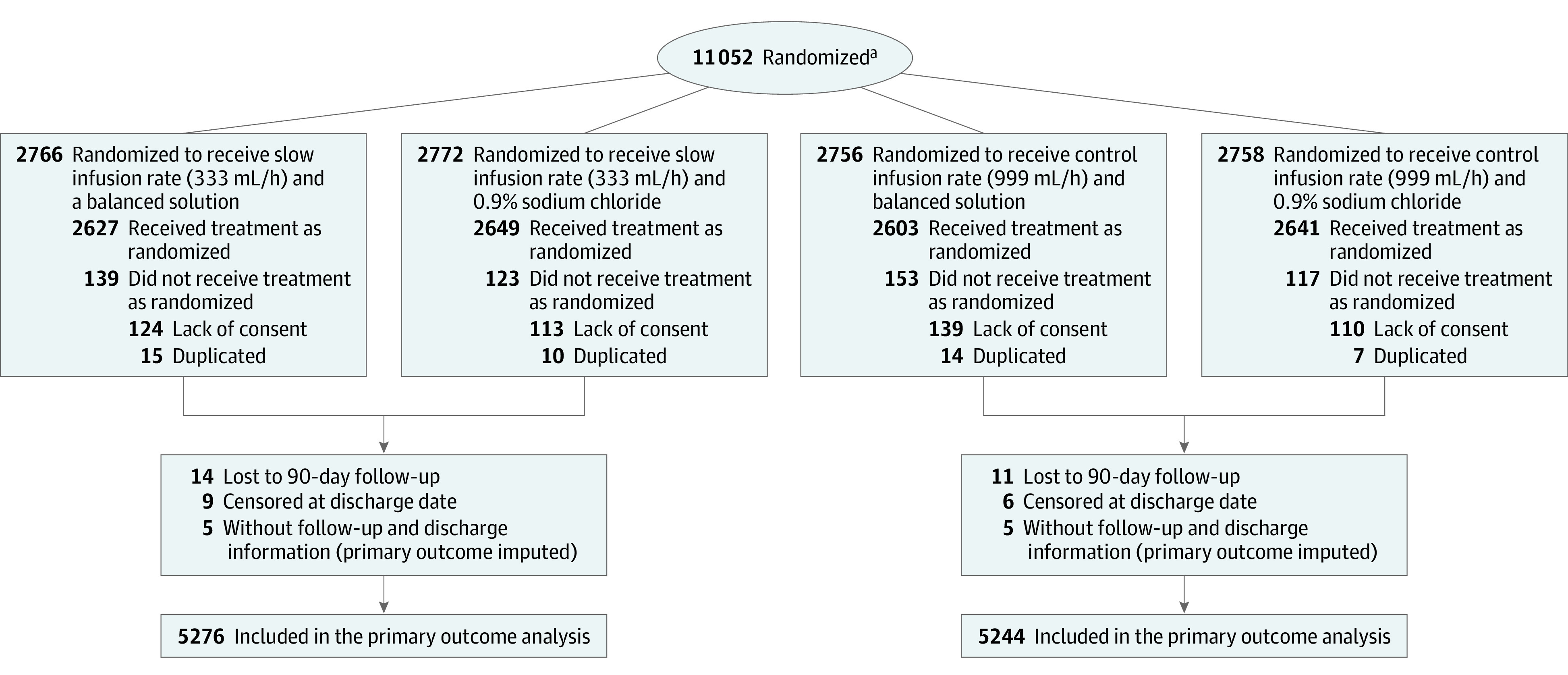
Patients were randomized to receive fluid challenges at 2 different infusion rates by an infusion pump: 999 mL/h (control) or 333 mL/h (slower; Figure 1). Randomization was made using a web-based system maintained by HCor, São Paulo, Brazil. The assessment of fluid infusion rate was unblinded, and patients and physicians were aware of the groups to which they were allocated. The randomization list was generated using random permuted blocks of 12 patients, stratified by center and according to fluid type and infusion rate. Study fluids (Plasma-Lyte or saline) were supplied to enrolling sites at 500-mL blinded bags, labeled using 6 different letters provided by Baxter Hospitalar. Adherence was checked on specific days after enrollment (days 1, 2, 3, and 7).
Figure 1. Flow Chart of the BaSICS Trial of Critically Ill Patients Requiring Fluid Infusions.

aThere was no screening log in the trial; therefore, the number of patients assessed for eligibility are not presented.
Interventions
The control infusion rate was defined to reflect current standard of care (500 mL over approximately 30 minutes; the infusion rate of the control group was set at 999 mL/h because that is usually the upper limit for infusion rate for infusion pumps). The slower infusion rate (333 mL/h) was arbitrarily defined so that it was less than the 25% percentile in the Fluid Challenges in Intensive Care (FENICE) cohort study (500 mL/h),9 but still represented a value that was considered as a bolus (and not maintenance) by clinicians. All fluid challenges performed up to 90 days (or until ICU discharge) after enrollment were requested to be performed according to the assigned infusion rate (eMethods, eFigure 1 and 2 in Supplement 3). Faster infusion rates were allowed at the discretion of the attending physician (either the control infusion of 999 mL/h or other rates) and in the slower infusion group if patients had active bleeding demanding fluid resuscitation or had severe hypotension (systolic blood pressure <80 mm Hg or mean arterial pressure <50 mm Hg). As soon as these conditions resolved, the assigned fluid rate was to be resumed.
Clinical and Laboratory Data
Baseline information included age, sex, admission type (planned, unplanned, both surgical and medical), use of organ support at enrollment, use of fluids in the 24 hours before enrollment, and key laboratory values. Illness severity scores were calculated from user-imputed data and included both Acute Physiology and Chronic Health Evaluation (APACHE II) score13 and the Sequential Organ Failure Assessment (SOFA)14 score. Data on number of fluid bolus used and adherence to the assigned rate were collected at days 1, 2, 3, and 7 after randomization. End point data included 90-day vital status (centrally collected by the coordinating site using phone contacts or, if unavailable, status information from both national deaths database or site records regarding subsequent patient visits to the enrolling site), hospital status at discharge (dead or alive), hospital and ICU length-of-stay, and need for kidney replacement therapy up to 90 days after enrollment. Information on suspected unexpected serious adverse events was also collected.
End Points
The primary end point was 90-day survival. Secondary end points were use of kidney replacement therapy up to 90 days after enrollment; occurrence of acute kidney injury, defined as Kidney Disease: Improving Global Outcomes (KDIGO)15 stages 2 or 3 measured on days 3 and 7, considering only patients without stages 2 or 3 acute kidney injury at baseline; SOFA score (both total score value evaluated as a continuous total and individual component categorized as ≤2 or >2) measured on days 3 and 7; and mechanical ventilation–free days within 28 days. Tertiary end points were hospital and ICU mortality and length-of-stay. Additional definitions are provided in the eMethods section in Supplement 3. No adverse event information was collected besides severe unexpected adverse reactions.
Power Analysis and Sample Size Calculation
Sample size was calculated assuming a 35% mortality within 90 days for the control group and considered 89% power to detect a hazard ratio (HR) for mortality of 0.9 with an α fixed at .05. Absence of interaction between the 2 interventions (fluid type and infusion rate) was assumed.
Statistical Analysis
Three interim analysis were conducted: 1 adverse event analysis at 1000 patients and 3 additional analyses at 25%, 50%, and 75% of the total sample size. Strict safety stopping rules were used (P < .001; Haybittle–Peto boundary16) for 90-day mortality at all interim analyses. There was no plan to interrupt the trial for efficacy or for futility.
Survival at 90 days was evaluated using mixed-effects Cox proportional hazard models (enrolling sites defined as the random variable) and adjusted for age, baseline SOFA score, and admission type (planned admission, unplanned admission with sepsis, and unplanned admission without baseline sepsis). Proportionality of the HR assumption was assessed using the Grambsch and Thernau method.17 Need for kidney replacement therapy was estimated using a mixed Poisson model adjusting for the same variables or, alternatively, estimated in a competing risk model, with death as a competitor and with similar adjustment. Binary end points at days 3 and 7 were tested with mixed generalized linear models; these included occurrences of acute kidney injury defined as KDIGO 2 to 3 and stratified SOFA components. Mechanical ventilation-free days was analyzed using a beta binomial regression. Zero was attributed to all patients who died regardless of how long they remained free from mechanical ventilation within 28 days. Imputation for missing data was made in a single model using a multiple chain equation using age, sex, enrolling site, randomization creatinine values, SOFA score, admission type, use of fluid in the 24 hours before enrollment, presence of heart failure or cirrhosis, traumatic brain injury at enrollment, hypotension at enrollment, mechanical ventilation at enrollment, and the primary end point. Five imputations sets were obtained, and the median of the imputed results (or the most frequent category) were used for analysis. Details can be found in the eMethods section in Supplement 3.
Subgroups for the primary end point were defined a priori according to the following characteristics at baseline: (1) patients with vs without sepsis; (2) patients with KDIGO stages 0 to 1 vs stages 2 to 3 at enrollment; (3) surgical vs nonsurgical patients; (4) patients with vs without traumatic brain injury; (5) patients with APACHE II score of 25 or more vs less than 25 points; and (6) patients who received more than 1.000 mL vs 1.000 mL or less of 0.9% saline in the 24 hours before randomization.
The following post hoc sensitivity analyses were performed: (1) primary end point analysis considering only cases with known primary end point; (2) a stratified analysis of the primary end point according to baseline presence of heart failure; (3) differences in need for vasopressors at days 3 and 7 according to the other factorial group of the trial; (4) a sensitivity exploratory analysis using bayesian networks focusing on changes in cardiovascular SOFA score. More details are provided in the eMethods in Supplement 3.
P values are reported for the primary analyses only; results of secondary, tertiary, and exploratory analyses are reported as estimated effect sizes and 95% CIs. A 2-sided α of .05 was considered statistically significant. Because of the potential for type I error due to multiple comparisons, findings for analyses of secondary end points should be interpreted as exploratory. Analyses were performed using R software, version 4.03.18
Results
Patients
A total of 11 052 patients were randomized from May 29, 2017, to March 2, 2020, at 75 ICUs. Follow-up was concluded on October 29, 2020. A total of 486 patients refused consent after enrollment, and 46 were randomized twice. After excluding these 532 patients, 10 520 were available for analysis (Figure 1, 5276 in slower infusion and 5244 in control infusion group). Baseline features were well balanced between groups. Unplanned admission accounted for 51.2% of patients receiving the slower infusion rate and 52.0% of patients receiving the control infusion rate. A total of 59.9% receiving slower infusion and 61.2% in the control group were either hypotensive or using vasopressors at enrollment. The median SOFA scores were similar between groups (4, interquartile range [IQR]; 2-6 and 4, IQR; 2-7, respectively). Patient characteristics are provided in Table 1; and for all 4 groups in eTable 1 in Supplement 3.
Table 1. Baseline Characteristics of the Included Patientsa.
| Characteristics | No. (%) of patientsb | |
|---|---|---|
| Slower infusion, 333 mL/h (n = 5276)c | Control, 999 mL/h (n = 5244)c | |
| Age, mean (SD), y | 60.8 (17.1) | 61.4 (16.9) |
| Women | 2360 (44.7) | 2295 (43.8) |
| Men | 2196 (55.3) | 2949 (56.2) |
| Admission type to ICU, No./total No. (%) | ||
| No. of missing patients | 16 | 11 |
| Planned admission (elective surgery) | 2569/5250 (48.8) | 2510/5233 (48.0) |
| Unplanned admission | 2691/5260 (51.2) | 2723/5233 (52.0) |
| No. of missing patients | 16 | 11 |
| Emergency department | 1151/5260 (21.9) | 1231/5233 (23.5) |
| Nonelective surgery | 651/5260 (12.4) | 654/5233 (12.5) |
| Ward | 543/5260 (10.3) | 513/5233 (9.8) |
| Transfer from another hospital | 305/5260 (5.8) | 289/5233 (5.5) |
| Transfer from another ICU | 41/5260 (0.8) | 36/5233 (0.7) |
| Illness severity at enrollment | ||
| APACHE II, median (IQR)d | 12 (8-16) | 12 (8-17) |
| No. of patients | ||
| No. of missing patients | 28 | 26 |
| SOFA score, median (IQR)e | 4 (2-6) | 4 (2-7) |
| No. of patients | ||
| No. of missing patients | 28 | 26 |
| KDIGO criteria for acute kidney injury ≥ 1f | 1695/5247 (32.3) | 1753/5216 (33.6) |
| No. of missing patients | 29 | 28 |
| Sepsis | 964/5259 (18.3) | 1017/5233 (19.4) |
| No. of missing patients | 17 | 11 |
| Traumatic brain injury | 254/5260 (4.8) | 232/5233 (4.4) |
| No. of missing patients | 16 | 11 |
| Hypotensiong | 3155/5258 (60.0) | 3201/5233 (61.2) |
| No. of missing patients | 18 | 11 |
| Mechanical ventilation | ||
| No. of missing patients | 16 | 11 |
| Noninvasive for >12 h | 368/5260 (7.0) | 305/5233 (5.8) |
| Invasive | 2294/5260 (43.6) | 2350/5233 (44.9) |
| Serum creatinine, Mean (SD), mg/dL | 1.2 (0.9) | 1.2 (0.9) |
| No. of patients | 5228 | 5206 |
| No. of missing patients | 48 | 38 |
| Creatinine level, mg/dL | ||
| ≤1.5 | 4188/5228 (80.1) | 4113/5206 (79.0) |
| 1.5-2.5 | 698/5228 (13.4) | 741/5206 (14.2) |
| >2.5 | 342/5228 (6.5) | 352/5206 (6.8) |
| Cirrhosis or acute liver failure | 116/5260 (2.2) | 150/5233 (2.9) |
| No. of missing patients | 16 | 11 |
| Heart failure | 542/5260 (10.3) | 594/5233 (11.4) |
| No. of missing patients | 16 | 11 |
| Time from ICU admission to randomization, median (percentiles 2.5%-97.5%), d | 0 (0-1) | 0 (0-1) |
| No. of patients | 5261 | 5233 |
| No. of missing patients | 15 | 11 |
| Administration of any fluid in the 24 h before enrollment | ||
| No. of missing patients | 17 | 11 |
| Received any, No./total No. (%) | 3615/5259 (68.7) | 3545/5233 (67.7) |
| Received >1000 mL, No./total No. (%) | 2395/5259 (45.5) | 2359/5233 (45.1) |
| No. of missing patients | ||
| Volume of any fluids administered within the 24 h before enrollment, median (IQR), mL | 1000 (0-2500) | 1000 (0-2500) |
| No. of patients | 5259 | 5233 |
| No. of missing patients | 17 | 11 |
Abbreviations: APACHE, Acute Physiology and Chronic Health disease Classification System; ICU, intensive care unit; IQR, interquartile range; KDIGO, Kidney Disease: Improving Global Outcomes; SOFA, Sepsis-related Organ Failure Assessment.
SI conversion factor: to convert creatinine from mg/dL to μmol/L, multiply by 88.4.
A table comparing the 4 as-randomized groups is available in eTable 1 (Supplement 3).
Numeric values indicate No. (%) of patients unless otherwise indicated.
Indicates the denominator, unless otherwise specified.
Severity of illness score ranges from 0 to 71, higher scores indicate more severe disease. A score of 12 predicts an in-hospital mortality of 15%.
Severity score ranges from 0 to 24, higher scores indicate more severe disease. A score of 4 predicts an in-hospital mortality of 20%.
An acute kidney injury rating ranges from 0 (no acute kidney injury) to 3 (severe kidney injury).
Defined as mean arterial pressure less than 65 mm Hg or systolic arterial pressure less than 90 mm Hg or use of vasopressors.
Interventions
Most fluid challenges were performed in the assigned rate on measured days, with more than 90% of all fluid challenges on day 1 done at the assigned infusion rate. The mean (SD) volume infused as boluses on day 1 was 1162 mL (916 mL) for slower infusion vs 1252 mL (1009 mL) for control infusion rate. The proportions of fluid boluses at the assigned rate are shown in eTable 2 in Supplement 3; the absolute volume of fluid used as a bolus as well as the proportion of fluid used as a bolus on each measured day are shown in eTable 3 and eFigure 3 in Supplement 3.
Primary End Point
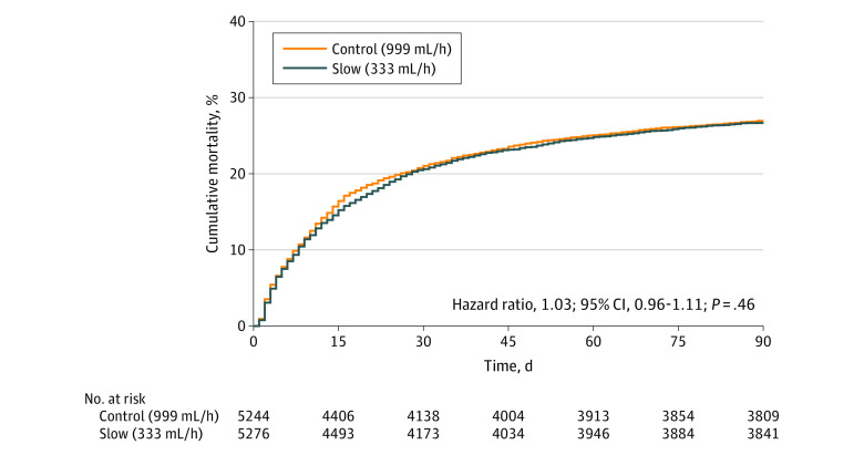
Missing primary end point information for 15 patients was imputed (see Table 2; Figure 2, and eTable 4 in Supplement 3). In the slower infusion group, 1406 of 5276 patients (26.6%) had died by day 90 compared with 1414 of 5244 patients (27.0%) in the control group. Proportionality of hazards assumption was met (P = .25). Adjusted hazard ratio (HR) for 90-day survival was 1.03 (95% CI, 0.96-1.11; P = .46). There was no significant interaction between fluid type and infusion rate (P = .98; eFigure 4 in Supplement 3).
Table 2. Outcomes Comparing Slower (333 mL/h) vs Control (999 mL/h) Infusion Speeda.
| End points | No./total No. (%) | Absolute effect (95% CI) | Relative effect (95% CI) | |
|---|---|---|---|---|
| Slower infusion, 333 mL/h (n = 5276) | Control, 999 mL/h (n = 5244) | |||
| Primary end point | ||||
| 90-Day mortalityb | 1406/5276 (26.6) | 1414/5244 (27) | –0.4 (–2.3 to 1.4) | HR, 1.03 (0.96 to 1.11) |
| P value | .46 | |||
| Secondary end points | ||||
| Acute kidney failure requiring kidney replacement within 90 days | ||||
| Incidence (per 1000 patient-days) | 414/474.84 (0.87) | 445/471.96 (0.94) | –0.07 (–0.17 to 0.04) | RR, 0.95 (0.83 to 1.09) |
| Day 1 | 27/5267 (0.5) | 31/5238 (0.6) | ||
| Day 2 | 122/5226 (2.3) | 130/5190 (2.5) | ||
| Day 3 | 190/5114 (3.7) | 204/5061 (4.0) | ||
| Day 7 | 282/4872 (5.8) | 299/4820 (6.2) | ||
| In hospital (≤1 kidney replacement therapy) | 397/5267 (7.5) | 423/5238 (8.1) | –0.5 (–1.5 to 0.4) | OR, 0.92 (0.80 to 1.06) |
| Day 3 KDIGO scorec | ||||
| ≥2 | 836/3147 (26.6) | 873/3075 (28.4) | –1.5 (–3.6 to 0.6) | OR, 0.93 (0.83 to 1.04) |
| Death or score ≥2 | 840/3147 (26.7) | 876/3075 (28.5) | –1.5 (–3.6 to 0.6) | OR, 0.93 (0.83 to 1.04) |
| Day 7 KDIGO scorec | ||||
| ≥2 | 286/1211 (23.6) | 263/1139 (23.1) | 0.0 (–3.5 to 3.5) | OR, 1.00 (0.82 to 1.22) |
| Death or score ≥2 | 288/1211 (23.8) | 265/1139 (23.3) | 0.0 (–3.5 to 3.5) | OR, 1.00 (0.82 to 1.22) |
| Day 3 SOFAd | ||||
| Total score, median (IQR) | 4 (2 to 6) | 4 (2 to 7) | –0.10 (–0.21 to –0.01) | |
| Score >2 | ||||
| Cardiovascular | 1252/3847 (32.5) | 1338/3788 (35.3) | –2.9 (–5.0 to –0.7) | OR, 0.89 (0.80 to 0.98) |
| Neurological | 662/3847 (17.2) | 628/3788 (16.6) | 0.5 (–0.9 to 1.8) | OR, 1.08 (0.94 to 1.24) |
| Coagulation | 180/3847 (4.7) | 146/3788 (3.9) | 0.8 (–0.1 to 1.6) | OR, 1.31 (1.05 to 1.64) |
| Respiratory | 240/3847 (6.2) | 284/3788 (7.5) | –1.1 (–2.1 to –0.2) | OR, 0.81 (0.68 to 0.97) |
| Hepatic | 41/3847 (1.1) | 52/3788 (1.4) | –0.3 (–0.7 to 0.1) | OR, 0.81 (0.56 to 1.18) |
| Day 7 SOFAd | ||||
| Total score, median (IQR) | 4 (2 to 7) | 4 (2 to 7) | –0.07 (–0.37 to 0.05) | |
| Score >2 | ||||
| Cardiovascular | 403/1600 (25.2) | 426/1525 (27.9) | –2.8 (–5.8 to 0.3) | OR, 0.88 (0.75 to 1.04) |
| Neurological | 467/1600 (29.2) | 440/1525 (28.9) | 0.4 (–2.3 to 3.0) | OR, 1.03 (0.87 to 1.22) |
| Coagulation | 76/1600 (4.8) | 56/1525 (3.7) | 0.8 (–0.4 to 1.9) | OR, 1.37 (0.98 to 1.90) |
| Respiratory | 165/1600 (10.3) | 160/1525 (10.5) | –0.1 (–1.7 to 1.6) | OR, 1.00 (0.79 to 1.25) |
| Hepatic | 26/1600 (1.6) | 26/1525 (1.7) | –0.1 (–0.8 to 0.5) | OR, 1.01 (0.65 to 1.57) |
| Mechanical ventilation–free days within 28 days | 27 (18 to 28) | 27 (17 to 28) | 0.17 (–0.07 to 0.32) | |
| Tertiary end points | ||||
| Death in ICU | 902/5267 (17.1) | 927/5238 (17.7) | –0.1 (–0.8 to 0.5) | OR, 1.00 (0.89 to 1.12) |
| Death in hospital | 1190/5267 (22.6) | 1204/5238 (23.0) | –0.4 (–2.2 to 1.3) | OR, 1.01 (0.91 to 1.12) |
| ICU length of stay, median (IQR), d | 3 (2 to 7) | 3 (2 to 7) | –0.2 (–0.6 to 0.2) | MR, 0.98 (0.93 to 1.03) |
| No. | 5267 | 5238 | ||
| Length of stay, median (IQR), d | 8 (5 to 19) | 9 (5 to 17) | –0.2 (–1.0 to 0.6) | MR, 0.99 (0.94 to 1.04) |
| No. | 5267 | 5238 | ||
Abbreviations: HR, hazard ratio; ICU, intensive care unit; IQR, interquartile range; KDIGO, Kidney Disease: Improving Global Outcomes; MR, mean ratio; OR, odds ratio; RR, rate ratio; SOFA, Sequential Organ Failure Assessment.
Results expressed as No./total No. (%) unless otherwise indicated.
Missing primary end point data for 15 patients were imputed.
Measured at that specific day including only patients who did not have acute kidney injury (stages 2 or3) at baseline. KDIGO ranges from 0 (no acute kidney injury) to 3 (severe kidney injury).
Six organs or systems are assessed, each receiving 0 (no dysfunction) to 4 points (more severe dysfunction). The sum of scores ranges from 0 to 24; higher scores indicate more severe disease: cardiovascular (≥3 is assigned when patient is given >5 µg/kg/min of dopamine or any dose of epinephrine or norepinephrine); neurological (≥3 when the Glasgow Coma Scale is ≤9); coagulation (≥3 when platelet count is ≤49 000 per μL); respiratory (≥3 when the patient is mechanically ventilated with an arterial oxygen partial pressure over inspired oxygen fraction ratio of ≤200); and hepatic (≥3 when serum bilirubin is ≥6 mg/dL [≥102.52 μmol/L]).
Figure 2. Cumulative Incidence of the Primary End Point (90-Day Survival) for Slower vs Control Infusion Rates.
Median observation time was 90 days (interquartile range [IQR], 57-90 days) for the control and 90 days (IQR, 55-90 days) for the slower infusion rate.
Secondary End Points
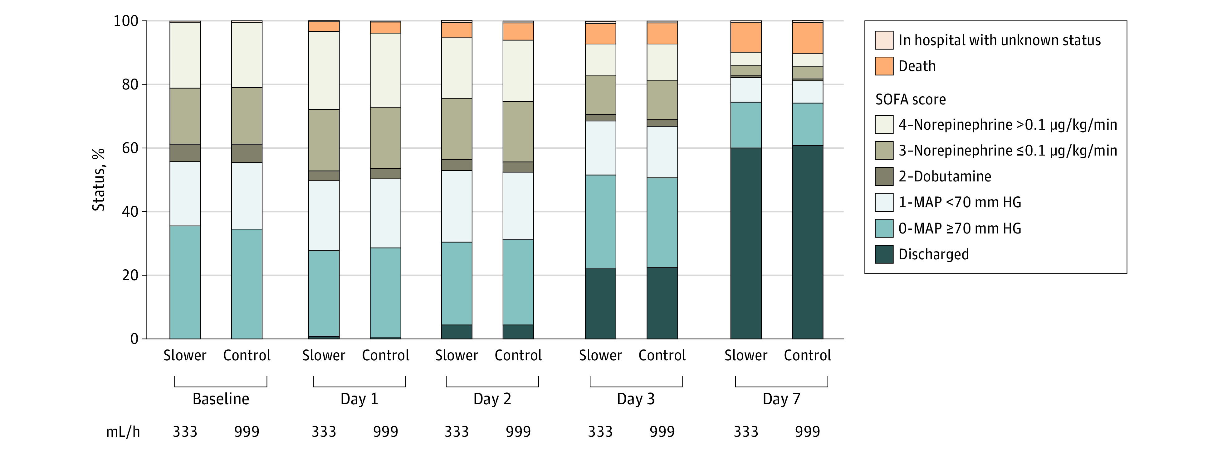
Results for secondary end points are shown in Table 2. Distribution for cardiovascular SOFA scores (range, 0-4), discharge and death on days 1, 2, 3, and 7 are shown in Figure 3. On day 3, patients in the slower infusion group had lower SOFA scores (difference, −0.10; 95% CI –0.21 to –0.01), lower frequency of hemodynamic SOFA score of more than 2 (marking a lower use of vasopressors, 32.5% vs 35.3% for slower vs control infusion rate; odds ratio [OR], 0.89; 95% CI, 0.80 to 0.98) and lower frequency of respiratory SOFA score of more than 2 (that is, a lower frequency of being mechanically ventilated with an arterial oxygen pressure to inspired oxygen fraction ratio less than 200% to 6.2% vs 7.5% for slower and control infusion rates; OR, 0.81; 95% CI, 0.68 to 0.97). However, frequency of coagulation SOFA score of more than 2 was higher on day 3 for the slower infusion group (4.7% vs 3.9%; OR, 1.31; 95% CI, 1.05 to 1.64). None of these differences, however, were sustained on day 7 for patients who remained alive and in the ICU. All other secondary end points were not statistically significant between groups.
Figure 3. Patient Status According to Whether Patients Were Discharged, Dead, or in the Intensive Care Unit (ICU) With Cardiovascular Sequential Organ Failure Score (SOFA).

The cardiovascular SOFA is scored so that patiens are given the highest score that fits their clinical characteristic. The total number in each column for the slow rate group is 5276 patients and 5244 for the control rate.
Tertiary End Points
There was no statistically significant effect of a slower infusion rate vs control infusion rate on ICU and hospital mortality or length-of-stay (Table 2).
Subgroup Analyses
There was no statistically significant difference on the primary end point for any of the a priori defined subgroups (eTable 5 in Supplement 3).
Post Hoc Sensitivity Analyses
The first sensitivity analyses performed included only cases with a known primary end point and provided results consistent with the primary analysis (eTable 6 in Supplement 3). The second sensitivity analysis for the primary end point was stratified according to baseline presence of heart failure (eTable 7 in Supplement 3) for patients with a known diagnosis before enrollment and again provided nonstatistically significant results. The differences in need for vasopressors at days 3 and 7 was further explored according to the other factorial cohort (fluid type; eTable 8 in Supplement 3). This analysis rendered nonstatistically different results (P = .76 and P = .70 for interaction at days 3 and 7, respectively). A sensitivity exploratory analysis using Bayesian networks was performed focusing on changes in cardiovascular SOFA score (eFigure 5 and eTable 9 in Supplement 3). In brief, results mostly confirmed the main analysis. For example, the probability that patients were alive in the ICU without vasopressor or discharged by day 3 regardless of baseline cardiovascular SOFA score was 0.71 for the slower infusion vs 0.69 for the control infusion (resulting in an OR of 1.11; 95% credible interval, 1.01-1.21; that is approximately a 2% increase). This effect was also present specifically for patients who were taking vasopressors at admission, for which the probability of being without vasopressors or discharged alive by day 3 was 0.56 for the slower infusion vs 0.53 for the control infusion rates (OR, 1.14; 95% credible interval, 1.03-1.27). No significant differences were observed at 7 days.
Adverse Events
There were no reported suspected unexpected severe adverse events in either group.
Discussion
In this large, multicenter randomized clinical trial, there was no significant difference in 90-day survival among patients in the ICU requiring fluid challenges who received a slower infusion rate (333 mL/h) compared with a control infusion rate (999 mL/h).
Despite several studies examining composition of fluids used in critically ill patients, little attention is given to other important aspects of fluid therapy, including infusion rate, temperature, among others. These are important targets that are easy to implement and that may be relevant for patient-centered end points. Specifically, previous evidence suggests that fast infusion rates may result in abnormal gas exchange19 and may reduce exercise tolerance,20 among other effects.7 For healthy volunteers, faster fluid infusion may be associated with reduced cardiac output and an increased heart rate.21 The present study aimed to test whether 2 different infusion rates could change the 90-day survival and organ dysfunction profile for critically ill patients. Although a slower infusion rate was not statistically associated with reduction in the primary end point, some secondary end points were statistically different between groups. There was a lower use of vasopressors (hemodynamic SOFA score >2) on day 3 in the slower infusion rate group. In a study by Monge Garciá et al,22 using data from fluid challenges that were performed in patients with sepsis at rates equal to those used in the control group for this study (500 mL in 30 minutes) reported that arterial elastance decreased after fluid loading, possibly suggesting a potential pathway for higher vasopressor requirements in patients receiving faster fluid infusions. These results, however, were of small magnitude (reductions were of an absolute magnitude of close to 2%), were not sustained by day 7, and did not reflect on any patient-centered end points, thereby limiting any strong conclusions. These results are aligned with the more frequent cardiovascular failure observed in the fluid bolus group in the Fluid Expansion as Supportive Therapy (FEAST) trial.4 The respiratory effect observed, which was also punctual, may be related to the plausible hypothesis of reducing the formation of lung edema with slower infusion rates. Conversely, a slower infusion rate might result in more coagulation abnormalities by day 3, which could be related to a higher plasma expansion with slower infusion5,23 or to a slower resuscitation that could result in coagulation abnormalities. Therefore, further studies should explore not only the effects of different infusion rates but also the timing of using faster and slower infusion rates.
Limitations
This study has several limitations. First, the slower infusion rate was defined arbitrarily at 333 mL/h. Second, secondary end points were not adjusted for multiple comparisons and therefore should be considered hypothesis generating. Third, some secondary end points are complicated by competing risks (such as discharge and death); however, in an alternative planned analysis that accounted for these event results were unchanged. Fourth, data were not collected for the reasons for fluid challenges. In addition, there was no record on the immediate effects of fluid challenges on hemodynamic parameters.
Conclusions
Among patients in the intensive care unit requiring fluid challenges, infusing at a slower rate compared with a faster rate did not reduce 90-day mortality. These findings do not support the use of a slower infusion rate.
Trial Protocol
Statistical Analysis Plan
eMethods. Additional trial procedures information
eFigure 1. Study scheme
eFigure 2. Fluid management in BaSICS
eFigure 3. Proportion of fluids used at days 1, 3, and 7 as bolus at assigned (333 versus 999 mL/h), maintenance and dilutions or other rates
eFigure 4. Primary outcome results according to both interventions in BaSICS (infusion rate and fluid type
eTable 1. Baseline characteristics of the included patients of the four groups of the trial
eTable 2. Adhesion to allocated infusion rate according to the day of assessment
eTable 3. Volume of fluid infused at days 1, 2, 3, and 7
eTable 4. Primary outcome model
eTable 5. Subgroup results
eTable 6. Primary outcome for complete analyses
eTable 7. Primary outcome for patients with known heart failure status at enrollment
eTable 8. Hemodynamic SOFA at days 3 and 7 according to infusion rate and fluid type
eFigure 5. Bayesian network
eTable 9. Results for Bayesian network queries
Nonauthor Collaborators. The BaSICS investigators and the Brazilian Research in Intensive Care Network (BRICNet)
Data Sharing Statement
References
- 1.Hernández G, Ospina-Tascón GA, Damiani LP, et al. ; The ANDROMEDA SHOCK Investigators and the Latin America Intensive Care Network (LIVEN) . Effect of a resuscitation strategy targeting peripheral perfusion status vs serum lactate levels on 28-day mortality among patients with septic shock: the ANDROMEDA-SHOCK randomized clinical trial. JAMA. 2019;321(7):654-664. doi: 10.1001/jama.2019.0071 [DOI] [PMC free article] [PubMed] [Google Scholar]
- 2.Dellinger RP, Levy MM, Carlet JM, et al. ; International Surviving Sepsis Campaign Guidelines Committee; American Association of Critical-Care Nurses; American College of Chest Physicians; American College of Emergency Physicians; Canadian Critical Care Society; European Society of Clinical Microbiology and Infectious Diseases; European Society of Intensive Care Medicine; European Respiratory Society; International Sepsis Forum; Japanese Association for Acute Medicine; Japanese Society of Intensive Care Medicine; Society of Critical Care Medicine; Society of Hospital Medicine; Surgical Infection Society; World Federation of Societies of Intensive and Critical Care Medicine . Surviving Sepsis Campaign: international guidelines for management of severe sepsis and septic shock: 2008. Crit Care Med. 2008;36(1):296-327. doi: 10.1097/01.CCM.0000298158.12101.41 [DOI] [PubMed] [Google Scholar]
- 3.Maitland K, Kiguli S, Opoka RO, et al. ; FEAST Trial Group . Mortality after fluid bolus in African children with severe infection. N Engl J Med. 2011;364(26):2483-2495. doi: 10.1056/NEJMoa1101549 [DOI] [PubMed] [Google Scholar]
- 4.Maitland K, George EC, Evans JA, et al. ; FEAST Trial Group . Exploring mechanisms of excess mortality with early fluid resuscitation: insights from the FEAST trial. BMC Med. 2013;11:68. doi: 10.1186/1741-7015-11-68 [DOI] [PMC free article] [PubMed] [Google Scholar]
- 5.Tatara T, Tsunetoh T, Tashiro C. Crystalloid infusion rate during fluid resuscitation from acute haemorrhage. Br J Anaesth. 2007;99(2):212-217. doi: 10.1093/bja/aem165 [DOI] [PubMed] [Google Scholar]
- 6.Hahn RG, Drobin D, Zdolsek J. Distribution of crystalloid fluid changes with the rate of infusion: a population-based study. Acta Anaesthesiol Scand. 2016;60(5):569-578. doi: 10.1111/aas.12686 [DOI] [PubMed] [Google Scholar]
- 7.Muir AL, Flenley DC, Kirby BJ, Sudlow MF, Guyatt AR, Brash HM. Cardiorespiratory effects of rapid saline infusion in normal man. J Appl Physiol. 1975;38(5):786-75. doi: 10.1152/jappl.1975.38.5.786 [DOI] [PubMed] [Google Scholar]
- 8.Robertson HT, Pellegrino R, Pini D, et al. Exercise response after rapid intravenous infusion of saline in healthy humans. J Appl Physiol (1985). 2004;97(2):697-703. doi: 10.1152/japplphysiol.00108.2004 [DOI] [PubMed] [Google Scholar]
- 9.Cecconi M, Hofer C, Teboul JL, et al. ; FENICE Investigators; ESICM Trial Group . Fluid challenges in intensive care: the FENICE study: a global inception cohort study. Intensive Care Med. 2015;41(9):1529-1537. doi: 10.1007/s00134-015-3850-x [DOI] [PMC free article] [PubMed] [Google Scholar]
- 10.Zampieri FG, Azevedo LCP, Corrêa TD, et al. ; BaSICS Investigators and the BRICNet . Study protocol for the Balanced Solution versus Saline in Intensive Care Study (BaSICS): a factorial randomised trial. Crit Care Resusc. 2017;19(2):175-182. [PubMed] [Google Scholar]
- 11.Damiani LP, Cavalcanti AB, Biondi RS, et al. Statistical analysis plan for the Balanced Solution versus Saline in Intensive Care Study (BaSICS). Rev Bras Ter Intensiva. 2020;32(4):493-505. doi: 10.5935/0103-507X.20200081 [DOI] [PMC free article] [PubMed] [Google Scholar]
- 12.Zampieri FG, Machado FR, Biondi RS, et al. ; BaSICS Investigators; Brazilian Research Institute Intensive Care Network (BRICNet) . Effect of intravenous fluid treatment with a balanced solution vs saline solution on mortality in critically ill patients: the BaSICS randomized clinical trial. JAMA. Published online August 10, 2021. doi: 10.1001/jama.2021.11684. [DOI] [PMC free article] [PubMed] [Google Scholar]
- 13.Knaus WA, Draper EA, Wagner DP, Zimmerman JE. APACHE II: a severity of disease classification system. Crit Care Med. 1985;13(10):818-829. doi: 10.1097/00003246-198510000-00009 [DOI] [PubMed] [Google Scholar]
- 14.Vincent JL, Moreno R, Takala J, et al. ; On behalf of the Working Group on Sepsis-Related Problems of the European Society of Intensive Care Medicine . The SOFA (Sepsis-related Organ Failure Assessment) score to describe organ dysfunction/failure. Intensive Care Med. 1996;22(7):707-710. doi: 10.1007/BF01709751 [DOI] [PubMed] [Google Scholar]
- 15.Kidney Disease: Improving Global Outcomes (KDIGO) Acute Kidney Injury Work Group . KDIGO clinical practice guideline for acute kidney injury. Kidney Int Suppl. 2012;2(suppl 1):1-138. Accessed August 3, 2021. https://kdigo.org/wp-content/uploads/2016/10/KDIGO-2012-AKI-Guideline-English.pdf [Google Scholar]
- 16.Haybittle JL. Repeated assessment of results in clinical trials of cancer treatment. Br J Radiol. 1971;44(526):793-797. doi: 10.1259/0007-1285-44-526-793 [DOI] [PubMed] [Google Scholar]
- 17.Grambsch PM, Therneau TM, Proportional hazards tests and diagnostics based on weighted residuals. Biometrika, 1994;81(3): 515–526, doi: 10.1093/biomet/81.3.515 [DOI] [Google Scholar]
- 18.R. Version 4.03. R Foundation for Statistical Computing. 2003. Accessed January 1, 2021. http://www.R-project.org/
- 19.Henderson AC, Sá RC, Barash IA, et al. Rapid intravenous infusion of 20 mL/kg saline alters the distribution of perfusion in healthy supine humans. Respir Physiol Neurobiol. 2012;180(2-3):331-341. doi: 10.1016/j.resp.2011.12.013 [DOI] [PMC free article] [PubMed] [Google Scholar]
- 20.Robertson HT, Pellegrino R, Pini D, et al. Exercise response after rapid intravenous infusion of saline in healthy humans. J Appl Physiol (1985). 2004;97(2):697-703. doi: 10.1152/japplphysiol.00108.2004 [DOI] [PubMed] [Google Scholar]
- 21.Ukor IF, Hilton AK, Bailey MJ, Bellomo R. The haemodynamic effects of bolus versus slower infusion of intravenous crystalloid in healthy volunteers. J Crit Care. 2017;41:254-259. doi: 10.1016/j.jcrc.2017.05.036 [DOI] [PubMed] [Google Scholar]
- 22.Monge García MI, Guijo González P, Gracia Romero M, et al. Effects of fluid administration on arterial load in septic shock patients. Intensive Care Med. 2015;41(7):1247-1255. doi: 10.1007/s00134-015-3898-7 [DOI] [PubMed] [Google Scholar]
- 23.Krzych ŁJ, Czempik PF. Effect of fluid resuscitation with balanced solutions on platelets: in vitro simulation of 20% volume substitution. Cardiol J. 2018;25(2):254-259. doi: 10.5603/CJ.a2017.0054 [DOI] [PubMed] [Google Scholar]
Associated Data
This section collects any data citations, data availability statements, or supplementary materials included in this article.
Supplementary Materials
Trial Protocol
Statistical Analysis Plan
eMethods. Additional trial procedures information
eFigure 1. Study scheme
eFigure 2. Fluid management in BaSICS
eFigure 3. Proportion of fluids used at days 1, 3, and 7 as bolus at assigned (333 versus 999 mL/h), maintenance and dilutions or other rates
eFigure 4. Primary outcome results according to both interventions in BaSICS (infusion rate and fluid type
eTable 1. Baseline characteristics of the included patients of the four groups of the trial
eTable 2. Adhesion to allocated infusion rate according to the day of assessment
eTable 3. Volume of fluid infused at days 1, 2, 3, and 7
eTable 4. Primary outcome model
eTable 5. Subgroup results
eTable 6. Primary outcome for complete analyses
eTable 7. Primary outcome for patients with known heart failure status at enrollment
eTable 8. Hemodynamic SOFA at days 3 and 7 according to infusion rate and fluid type
eFigure 5. Bayesian network
eTable 9. Results for Bayesian network queries
Nonauthor Collaborators. The BaSICS investigators and the Brazilian Research in Intensive Care Network (BRICNet)
Data Sharing Statement


