PLOS ONE logoLink to PLOS ONE
. 2021 Aug 26;16(8):e0256768. doi: 10.1371/journal.pone.0256768

Impact of thermogenesis induced by chronic β3-adrenergic receptor agonist treatment on inflammatory and infectious response during bacteremia in mice

Patrick Munro 1, Samah Rekima 2, Agnès Loubat 2, Christophe Duranton 3, Didier F Pisani 3,*,#, Laurent Boyer 1,*,#
Editor: Fulvio D’Acquisto4
PMCID: PMC8389438  PMID: 34437647

Abstract

White adipocytes store energy differently than brown and brite adipocytes which dissipate energy under the form of heat. Studies have shown that adipocytes are able to respond to bacteria thanks to the presence of Toll-like receptors at their surface. Despite this, little is known about the involvement of each class of adipocytes in the infectious response. We treated mice for one week with a β3-adrenergic receptor agonist to induce activation of brown adipose tissue and brite adipocytes within white adipose tissue. Mice were then injected intraperitoneally with E. coli to generate acute infection. The metabolic, infectious and inflammatory parameters of the mice were analysed during 48 hours after infection. Our results shown that in response to bacteria, thermogenic activity promoted a discrete and local anti-inflammatory environment in white adipose tissue characterized by the increase of the IL-1RA secretion. More generally, activation of brown and brite adipocytes did not modify the host response to infection including no additive effect with fever and an equivalent bacteria clearance and inflammatory response. In conclusion, these results suggest an IL-1RA-mediated immunomodulatory activity of thermogenic adipocytes in response to acute bacterial infection and open a way to characterize their effect along more chronic infection as septicaemia.

1. Introduction

White adipocytes, main constituents of white adipose tissue (WAT) are specialized in the storage and release of energy (carbohydrates, lipids), while brown adipocytes dissipate this energy in the form of heat (thermogenesis) [1]. Brown adipocytes constitute the brown adipose tissue (BAT) but can also be found within the WAT. They are then called beige or brite adipocytes (for "brown in white”) and have an increased thermogenesis capacity in response to prolonged exposure to cold, for instance [2]. In addition to their seminal functions, an essential role in the innate immunity has recently emerged since the characterization of several functional Toll-like receptors (TLRs) in the membrane of white and brown adipocytes [35]. TLRs are transmembrane receptors that play a critical role in the recognition and defense against all types of infectious pathogens, acting in innate, non-specific immunity [6]. TLRs, referred as pattern recognition receptors (PRR), are evolutionarily conserved receptors able to detect a repertoire of pathogen-associated molecular patterns (PAMPS) including constituent of the microbial cell walls such as the lipolysaccharides (LPS) of gram negative bacteria [6,7].

It is well-known that adipocytes respond to LPS stimulation, especially along obesity. During obesity, gut microbiota alteration (dysbiosis) and intestinal barrier disruption lead to an increased permeability to the microbiota degradation products, especially LPS. Obese patient’s plasma displays an increased level of endotoxins (endotoxaemia) carried to organs and tissues, inducing a local activation of TLR-4 displayed by adipocytes and adipose tissue resident macrophages [8]. In response, adipocytes and macrophages secrete inflammatory cytokines (TNFα and IL-1β) and chemokines (MCP-1 and CXCL10) which promote local inflammation and alter tissue homeostasis [9]. Interestingly, it has been shown that brown and white adipocytes in vitro respond to LPS stimulation by modulating differently macrophage inflammatory responses. Conversely to white adipocytes, brown adipocytes reduced macrophage IL-6 secretion and mRNA expression of several inflammatory markers in response to LPS [10]. In this way, we recently demonstrated that after recruitment and activation of brown and brite adipocytes, mice displayed a higher anti-inflammatory response when they are exposed to lipopolysaccharides derived from E. coli bacteria [11]. Especially, these mice secreted high level of interleukin-1 receptor antagonist (IL-1RA) known to counteract interleukin 1β (IL-1β) inflammatory action [11].

In addition to the inflammatory response, LPS alter adipocyte function via TLR-4 activation or through cytokines secreted by macrophages. As an example, LPS induce white adipocyte lipolysis to inhibit adipogenesis [12,13]. A main response of organism to counteract infection is fever. Adipose tissues appear to be involved in this mechanism since WAT secretes leptin, an adipokine essential to pyrexia in rodent by limiting heat loss at the tail level, and BAT make thermogenesis suspected for a long time to participate to pyrexia [14]. Nevertheless, recent evidences have demonstrated that acute (in vitro) and chronic (in vivo) exposure to LPS could inhibit brown adipocytes function [5,15].

WAT is linked to metabolic inflammation but also to local and systemic defence mechanisms against pathogens infection. Indeed, adipocytes are able to respond to bacteria proximity with secretion of antimicrobial peptides as cathelicidin, lipocalin and defensin. First described for dermal adipocytes in response to Staphylococcus aureus infection [16], this characteristic has been extended to adipocytes from intra-abdominal and subcutaneous WAT [17]. Moreover, WAT is able to respond to bacterial infection as previously shown in case of sepsis, a severe infectious situation where adipocytes secrete adipokines known to modulate inflammation [18]. In this situation of sepsis, it was demonstrated that brite adipocytes recruitment was stimulated [19], but might be related to an increased norepinephrine level known to induce the browning of WAT [20].

The antimicrobial response of brown adipocytes has not been yet described despite their ability to respond to endotoxins. Indeed, cold exposure in human, known to induce brown and brite adipocytes activity and recruitment, increases the secretion of inflammatory cytokines in response to LPS and of IL-1RA independently to LPS [21]. These results were in line with our previous observations where we demonstrate the same data in a mice model of recruitment and activation of brown and brite adipocytes followed by an exposition to LPS [11]. Despite it is well established that adipocytes are able to detect and respond to bacteria via TLR-2 and TLR-4, the involvement of the different adipocytes populations in this response is still unknown.

Here, we investigated the in vivo impact of recruitment/activation of brown/brite adipocytes by β-adrenergic receptors agonists on inflammatory response against E. coli infection in mice. We demonstrated that thermogenesis activity did not modify the host response to infection, without additive effect on fever and with an equivalent inflammatory response.

2. Materials and methods

2.1. Reagents

Media and buffer solutions were purchased from Lonza (Ozyme, St-Quentin en Yvelines, France). Other reagents were from Sigma-Aldrich (Saint-Quentin Fallavier, France).

2.2. Animals

The experiments were conducted in accordance with the French and European regulations (2010/63/EU directive) for the care and use of research animals and were approved by the french national experimentation committee (Ministère de l’Enseignement Supérieur, de la Recherche et de l’inovation, N°: APAFIS#18322–2018121809427035_v2). 8-week-old Balb/c male mice from Janvier Laboratory (France) were maintained at housing temperature (22°C) and 12:12-hour light-dark cycles, with ad libitum access to food and water. General health and behavior of mice were monitored daily. None of the mice died or were euthanized before the end of the experiments. All the mice used in the experiments have been included in the final analysis.

Mice were daily treated with β3-adrenergic receptor agonist CL316,243 (1 mg/kg in saline solution, intraperitoneal injection) (Sigma-Aldrich) or with vehicle only. For infection, treatment with CL316,243 was stopped after 7 day, then mice were injected with E. coli (UTI89 strain, 1.71x10⁷ CFU/mouse in phosphate-buffered saline solution (PBS), caudal intra-venous injection) and monitored for 48 hours before sacrifice [22]. At 0, 4, 24 and 48 hours after bacterial infection rectal temperature was recorded with a digital thermometer. For the determination of bacteremia, blood was collected from the tail vein at the indicated times post-infection, serially diluted in sterile PBS and plated on LB plates. The plates were incubated for 16 h at 37°C before counting colonies.

At the end of experiment, mice were sacrificed by cervical dislocation and blood, interscapular brown adipose tissue (iBAT), epididymal (eWAT) and inguinal subcutaneous (scWAT) white adipose tissues were immediately sampled and used for different analyses. General parameters, plasma analysis and expression of adipose tissue molecular markers were evaluated in all the mice (n = 8); secretion of tissue explant and histology analysis were evaluated using whole fat pad or lobe from half of the mice of each group (n = 4).

2.3. Cytokine and metabolic parameters quantification

For blood analysis, freshly prepared plasmas were diluted twice before analysis. For tissue analysis, freshly sampled WAT and BAT were washed in PBS, weighed and incubated in free DMEM (Dulbecco’s modified Eagle medium) for 2 hours at 37°C. Media were kept for various analyses of secreted proteins.

Leptin and cytokines were assayed using “Mouse Leptin Kit” (Meso Scale Discovery, # K152BYC) and “mouse V-PLEX Proinflammatory Panel 1 Kit” (Meso Scale Discovery, # K15048D) respectively, on a QuickPlex SQ 120 apparatus (Meso Scale Discovery) following manufacturer’s instructions. IL-1RA levels were assayed using mouse IL-1RA Elisa kit (#EMIL1RN) from Thermo Fisher Scientific (Courtaboeuf, France). Glycerol and triglycerides determinations were performed using dedicated kit (Free Glycerol reagent and Triglyceride reagent, Sigma Aldrich).

2.4. Histology

Freshly sampled tissues were fixed in 4% paraformaldehyde overnight at RT and then paraffin-embedded. Embedded tissues were cut in 5 μm sections and dried overnight at 37°C. All sections were then deparaffinized in xylene, rehydrated with alcohol, and washed in phosphate-buffered saline (PBS). For histological analysis, the sections were stained with haematoxylin-eosin and mounted in vectamount (Vecto laboratories). For immunohistochemical analysis, antigen retrieval was performed in low pH buffer in a de-cloaking chamber (Dako, S2367). The sections were then permeabilized in PBS with 0.2% Triton X-100 at room temperature for 10 min and blocked in the same buffer containing 3% BSA for 1 hour. The sections were incubated with rat anti-F4/80 antibody (Biorad, clone Cl:A3-1, dilution 1:100) overnight at 4°C. Following a 1-hour incubation with A568-coupled anti-rabbit secondary antibodies, nuclear staining was performed with DAPI, and the sections were mounted in PermaFluor mounting Media (Thermofisher).

2.5. Isolation and analysis of RNA

Total RNA was extracted using a TRI-Reagent kit (Euromedex, Souffelweyersheim, France) according to the manufacturer’s instructions. Tissues were homogenized in TRI-Reagent using a dispersing instrument (ULTRA TURRAX T25). Reverse transcription-polymerase chain reaction (RT-PCR) was performed using M-MLV-RT (Promega). SYBR qPCR premix Ex Taq II from Takara (Ozyme, France) was used for quantitative PCR (qPCR), and assays were run on a StepOne Plus ABI real-time PCR instrument (PerkinElmer Life and Analytical Sciences, Boston). The expression of selected genes was normalized to that of the 36B4 (RPLP0) housekeeping genes and then quantified using the comparative-ΔCt method. Primer sequences are displayed in Table 1.

Table 1. Primers sequences used for qPCR analysis.

Forward Reverse
36b4 (Rplp0) TCCAGGCTTTGGGCATCA CTTTATCAGCTGCACATCACTCAGA
Adiponectin CTTTCCTGCCAGGGGTTC GGAGAGAAAGGAGATGCAGGT
Cd11b (Itgam) TGACCTGGCTTTAGACCCTG ACCTCTGAGCATCCATAGCC
Cd19 (Leu-12) TCCCTGGGTCCTATGGAAAT CTGGTCCTGCCCAAGGTT
Mrc1 TGGATGGATGGGAGCAAAGT GCTGCTGTTATGTCTCTGGC
Perilipin 1 AGCGTGGAGAGTAAGGATGTC CTTCTGGAAGCACTCACAGG
Perilipin 5 CGCTCCATGAGTCAAGCCA CTCAGCTGCCAGGACTGCTA
Tcrβ CTCCACCCAAGGTCTCCTTG GTGGTCAGGGAAGAAGCCC
Ucp1 CACCTTCCCGCTGGACACT CCTGGCCTTCACCTTGGAT

2.6. Statistical analysis

Animal cohort sizes have been determined using G*Power [23]. Data were analyzed using GraphPad Prism 6 software and statistical differences between experimental groups assessed by Kruskal-Wallis multiple comparison test. Differences were considered statistically significant with p<0.05. Data were displayed as scatter plot of independent values and group mean values ± SEM.

3. Results

3.1. Bacterial acute infection does not modify response of mice to CL316,243

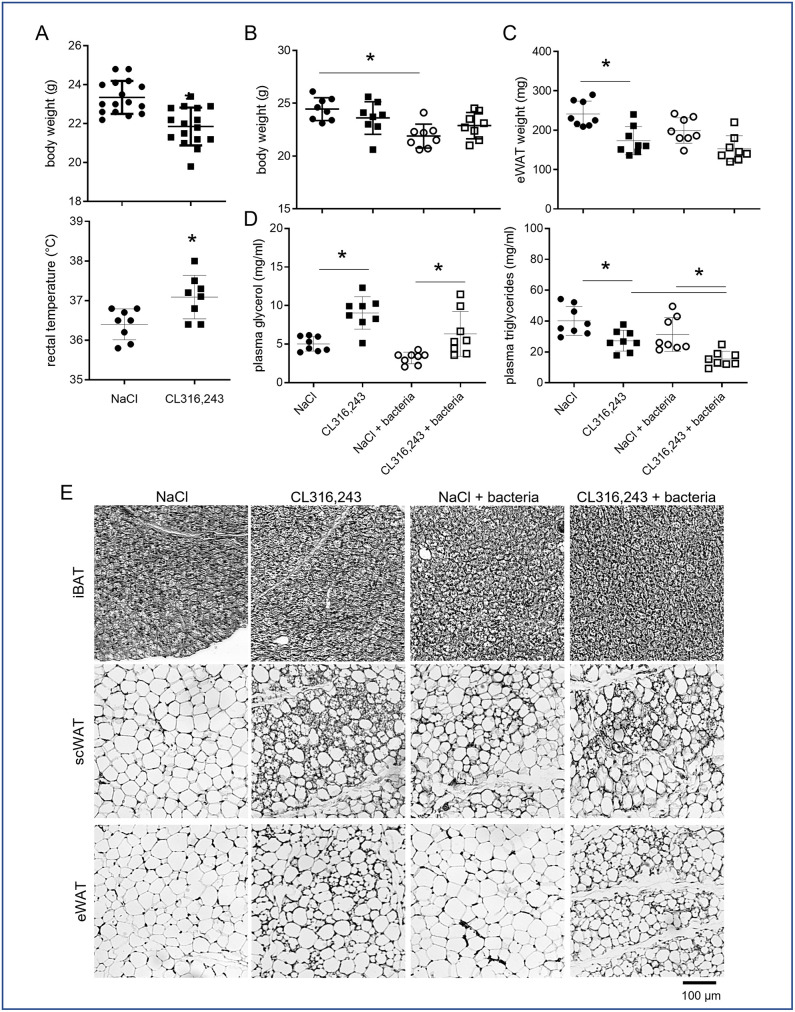
Mice were treated daily for 1 week with the β3-adrenergic receptor agonist CL316,243 (1 mg/kg, intraperitoneal), or vehicle only (NaCl), in order to induce brown adipose tissue (BAT) activation as well as recruitment and activation of brite thermogenic adipocytes within white adipose tissue (WAT). As expected, at the end of the treatment, mice body weight was decreased and rectal temperature increased in CL316,243 group compared to NaCl group (Fig 1A) due to the thermogenic activity of brown and brite adipocytes. At the end of this treatment, half of the mice of each group were infected with E. coli and monitored 48 hours as previously described [22]. While bacteria induced a decrease of body weight in NaCl treated mice, no additive effect was found in CL316,243 treated mice (Fig 1B). Differently, CL316,243 treatment decreased epididymal WAT (eWAT) mass while bacteria did not modify it significantly. Plasma analysis revealed that CL316,243 treated mice displayed higher glycerol level (Fig 1C) and lower triglycerides level (Fig 1D) compared to NaCl group and independently of bacteria treatment. These could be related to an increase fatty acid oxidation in link with thermogenesis. Interestingly, an additive effect was found for triglycerides plasma level in mice treated with CL316,243 and infected with bacteria.

Fig 1. Bacterial infection does not modify metabolic parameters and adipose tissue morphology.

Fig 1

Mice were analysed after 1 week of treatment with CL316,243 daily (1 mg/kg/day) or vehicle only (NaCl), and with or without E. coli infection for 48 hours. (A) Mouse body weights and rectal temperatures at the end of CL316,243 treatment. (B) Mouse body weights and (C) epididymal white adipose tissue (eWAT) weights at the end of infection. (D) Triglycerides and glycerol plasma levels at the end of experiment. (E) Representative picture of four haematoxylin-eosin staining of interscapular BAT (iBAT), sub-cutaneous (scWAT) and eWAT sections. The results are displayed as independent values (dots) and the mean ± SD. n = 16 (A) or 8 (B-D). * p<0.05.

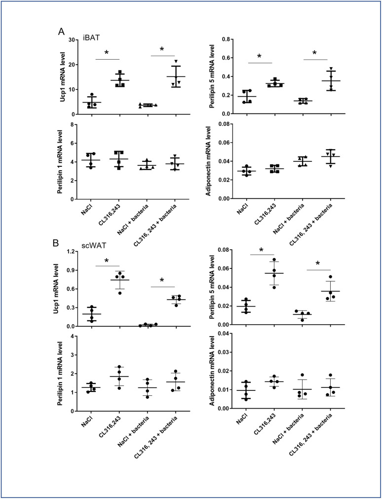
While histological analysis of inter-scapular BAT (iBAT) performed 48 hours after the end of CL316,243 treatment, did not shown major differences between groups (Fig 1E), molecular analysis showed an increased Ucp1 and Perilipin 5 mRNA expressions after CL316,243 treatment and independently of bacteria injection, which is characteristic of activated BAT (Fig 2A). Differently, Perilipin 1 and adiponectin mRNA expression was not affected by treatments (Fig 2A). Sub-cutaneous WAT (scWAT), and for a lower extend eWAT, showed numerous multilocular adipocytes in CL316,243 treated mice, characteristic of brite adipocytes (Fig 1E). This was confirmed by the overexpression of Ucp1 and Perilipin 5 mRNA in scWAT (Fig 2B). Perilipin 1 and adiponectin mRNA expression in scWAT did not change with CL316,243 treatment (Fig 2B). Bacteria did not modify the frequency of brite adipocytes as well as iBAT morphology (Fig 1E) which was corroborated with brown/brite adipocyte markers molecular analysis (Fig 2).

Fig 2. Effect of CL316,243 and bacteria on mRNA of adipocyte markers.

Fig 2

mRNA expression of brown/brite (Ucp1 and perilipin 5) and general adipocyte (perilipin 1 and Adiponectin) markers was analysed by qPCR in iBAT and scWAT from mice that were treated for 1 week with CL316,243 daily (1 mg/kg/day) or vehicle only (NaCl), and then infected or not with E. coli for 48 hours. The results are displayed as independent values (dots) and the mean ± SD. n = 4. * p<0.05.

3.2. Bacterial clearance during bacteremia and pyrexia are independent to leptin systemic level and BAT activation

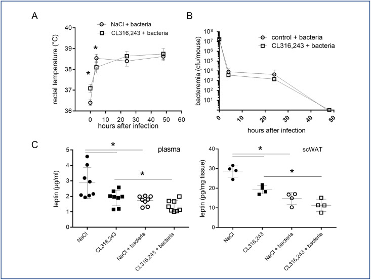
Intraperitoneal injection of bacteria in mice induces in the first 4 hours a pyretic response which was maintained all along the two days of analysis (Fig 3A). Interestingly, even if rectal temperature was different before infection (Fig 1A), the fever measured 4 hours after bacterial infection was equivalent between NaCl- and CL316,243-treated mice. As expected, the bacteremia was decreased at 24 hours and completely resolved after 48 hours, demonstrating bacteria clearance by mice (Fig 3B) [22]. Activation of brown and brite adipocytes did not modify this response to infection (Fig 3B).

Fig 3. Pyrexia, bacteremia and leptin secretion in response to infection.

Fig 3

(A) Rectal temperature and (B) bacteremia of mice treated or not with CL316,243 (1 mg/kg/day; 1 week) were monitored for 48 hours after infection by E. coli. (C) Mice plasma and scWAT explant leptin levels at the end of experiment. The results are displayed as independent values (dots) and the mean ± SD. n = 8 (mice and plasma) or 4 (scWAT explants). * p<0.05.

Leptin is considered as a key mediator of pyrexia especially in mice by limiting body heat loss. Interestingly, we measured a decreased leptinemia as well as leptin secretion by scWAT in mice infected with bacteria as well as in mice treated with CL316,243 (Fig 3C), demonstrating that pyrexia in response to bacteria and thermogenesis are independent to leptin action.

3.3. Systemic and local inflammatory response to bacteremia

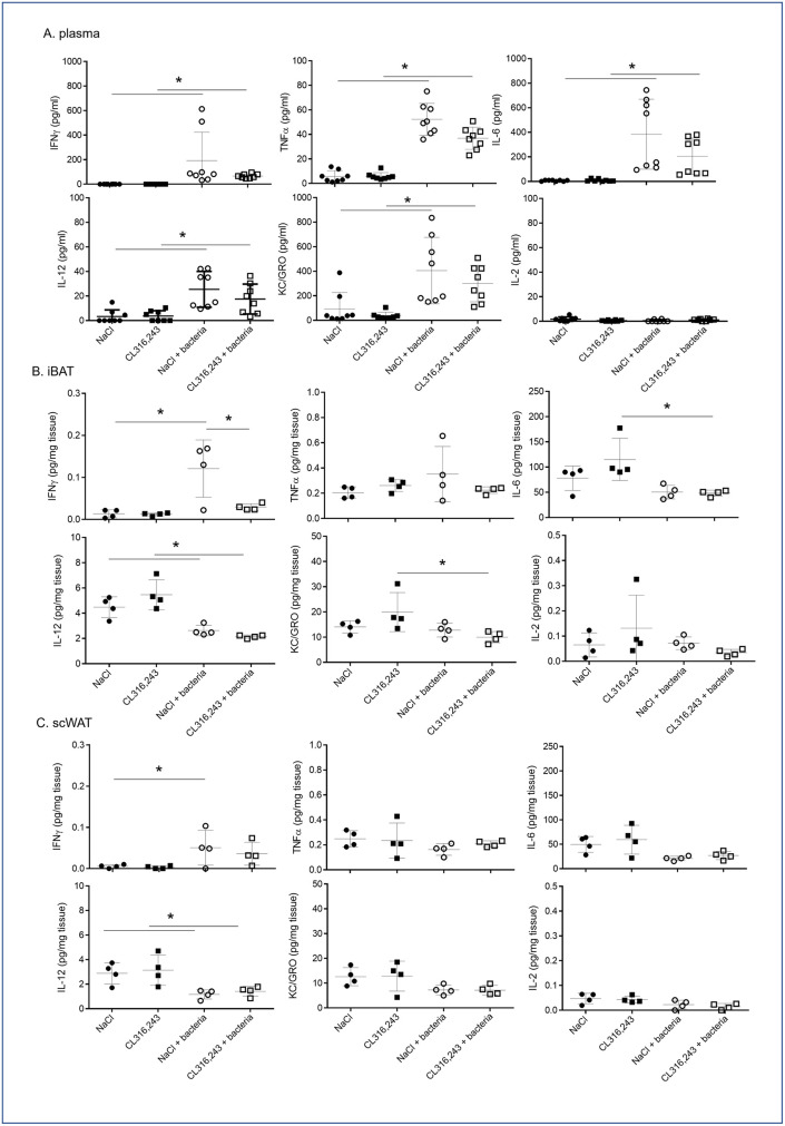
We found that CL316,243 preconditioning did not modify fever and bacteria clearance by mice. Thus, we analyzed cytokine profiling to determine if brown and brite adipocyte activations were modulating the immune response to bacterial infection. As expected, bacterial infection induced a systemic inflammatory response characterized by increased plasmatic levels of TNFα, IL-6, IL-12, IFNγ and KC/GRO (CXCL-1) compared to PBS-treated mice (Fig 4A). We observed that CL316,243 treatment did not modulate basal plasma cytokine levels as well as in response to bacteria, even if a slight decrease was found (Fig 4A). To note, the high variability in plasma quantity for each cytokine (Fig 4A) in response to bacteria could be related to the variability in bacteremia evolution we found between mice (Fig 3B).

Fig 4. Systemic and local inflammatory responses to infection.

Fig 4

IFNγ, TNFα, IL-6, IL-12-p70, KC/GRO (CXCL-1) and IL-2 levels were assessed in the plasma (A) or in the media of iBAT (B) and scWAT (C) explants from mice that were treated for 1 week with CL316,243 daily (1 mg/kg/day) or vehicle only (NaCl), and infected with E. coli for 48 hours. The results are displayed as independent values (dots) and the mean ± SD. n = 8 (plasma) or 4 (explants). * p<0.05.

Interestingly, in iBAT only IFNγ secretion increased in response to bacteria injection (Fig 4B), other cytokines were either unaffected (TNFα, IL-2) or decreased (IL-6, IL-12, KC/GRO) (Fig 4B). In scWAT, most cytokine secretions were unaffected except for IL-12 which was decreased (Fig 4C). In all case, CL316,243 did not modulate local cytokine secretions in response to infection (Fig 4B and 4C).

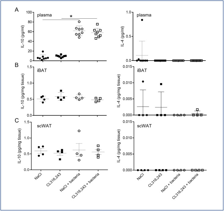
We further analyzed the impact of CL316,243 pretreatment on inflammatory resolving. To this aim we analyzed IL-4 and IL-10 levels in plasma and in secretory media of adipose tissue explant. Unfortunately, IL-4 was never detected and even if IL-10 level increased in plasma of infected mice, it was not modulated by CL316,243 treatment (Fig 5). As for pro-inflammatory cytokines, IL-10 secretion was unaffected by bacteria as well as CL316,243 treatment in iBAT and scWAT (Fig 5).

Fig 5. Systemic and local anti-inflammatory response to bacteria.

Fig 5

IL-4 and IL-10 levels were assessed in plasma (A) and in the media of iBAT (B) and scWAT (C) explants from mice that were treated for 1 week with CL316,243 daily (1 mg/kg/day) or vehicle only (NaCl), and infected with E. coli for 48 hours. The results are displayed as independent values (dots) and the mean ± SD. n = 8 (plasma) or 4 (explant). * p<0.05.

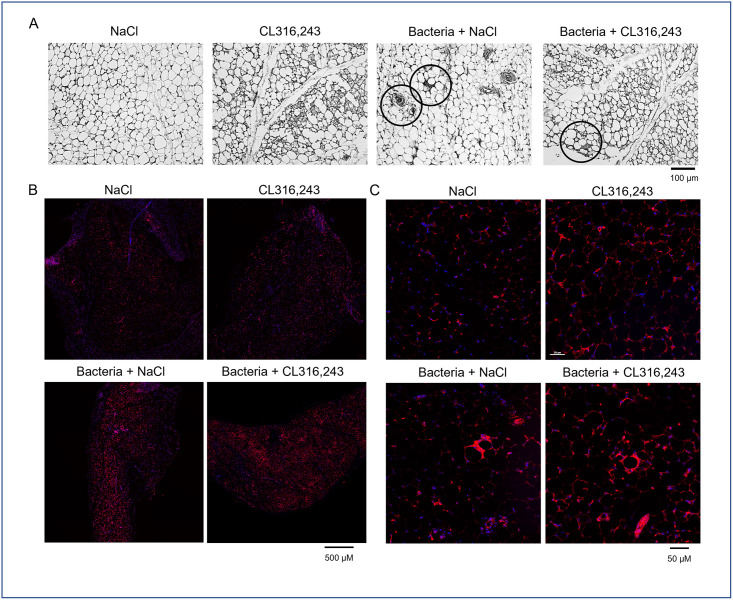
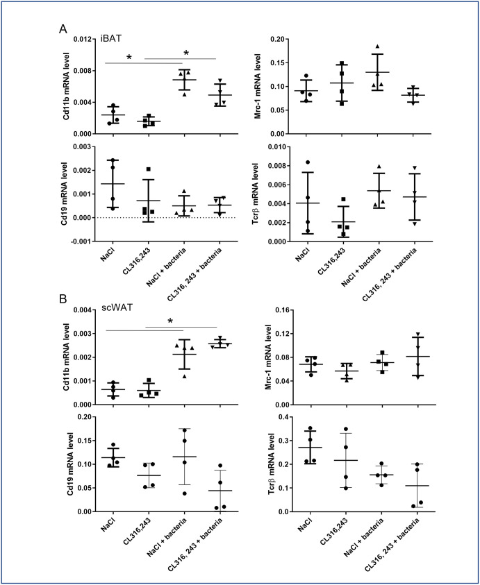
Next, we investigated the immune cells infiltration in the scWAT of infected mice. We found classic figures of inflammation in the scWAT of mice infected by bacteria. The immune cells infiltration in the scWAT was found to be similar in mice pretreated or not with CL316,243, including crown structure and small cell infiltration, potentially related to macrophage and monocyte or lymphocyte presence respectively (Fig 6A). This was confirmed by a F4/80 immunostaining clearly showing macrophage occurrence in adipose tissue (Fig 6B). To improve detection of immune cell content in whole adipose tissues, we have analyzed mRNA expression in iBAT and scWAT of general markers for B lymphocytes (Cd19), T lymphocytes (Tcrβ) and monocytes/macrophages (Cd11b), as well as expression of Mrc-1, a marker of anti-inflammatory macrophages. The results displayed in Fig 7 showed that only Cd11b increase with bacterial infection. Cd19, Tcrβ and Mrc-1 mRNA levels were equivalent between groups. These results corroborated histology results suggesting that only monocytes and inflammatory macrophages have infiltrated adipose tissue of mice in response to bacterial infection. To note, immune cells markers were unaffected by CL316,243 treatment (Fig 7).

Fig 6. Histological characterization of scWAT infiltration by immune cells.

Fig 6

(A) Representative pictures of four haematoxylin-eosin staining of sub-cutaneous white adipose (scWAT) tissue of mice treated or not with CL316,243 (1 mg/kg/day; 1 week) and infected 48 hours with E. coli. Typical figures of immune cell infiltration and crown structure are black circled. (B, C) Low and high magnification of F4/80 immunostaining (in red) in scWAT of the same mice. Scale bars are indicated.

Fig 7. Effect of CL316,243 and bacteria on mRNA of immune cell markers.

Fig 7

mRNA expressions of monocyte/macrophage (Cd11b), anti-inflammatory macrophage (Mrc-1), T (Tcrβ) and B (Cd19) lymphocytes were analysed by qPCR in iBAT and scWAT from mice that were treated for 1 week with CL316,243 daily (1 mg/kg/day) or vehicle only (NaCl), and then infected or not with E. coli for 48 hours. The results are displayed as independent values (dots) and the mean ± SD. n = 4. * p<0.05.

3.4. Systemic and local IL-1β and IL-1RA level in response to bacteria

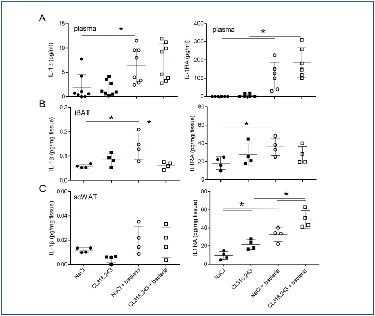
Among inflammatory cytokines, IL-1β is a main actor of pyrexia and anti-bacterial response and is highly secreted by the adipose tissue. Thus, in response to bacterial infection, we found the IL-1β and IL-1RA (IL-1 receptor antagonist) plasma levels increased concomitantly (Fig 8A). Pre-treatment of mice with CL316,243 did not modify this response (Fig 8A). Analysis of IL-1β and IL-1RA secretions in iBAT showed that both increased in infected mice and, interestingly, IL-1β secretion decreased in iBAT activated by CL316,243 treatment (Fig 8B). In contrast, IL-1RA level was equivalent in iBAT of infected mice treated or not with CL316,243 (Fig 8B). The IL-1β secretion by the scWAT was not significantly increased by bacteria infection but we measured an increased IL-1RA secretion. The CL316,243 treatment increased IL-1RA secretion with an additive effect to infection (Fig 8C).

Fig 8. IL-1 pathway response to bacterial infection.

Fig 8

IL-1β (A) and IL-1RA (B) protein levels were assessed in plasma (upper panel) and in the media of iBAT (middle panel) and scWAT (lower panel) explants from mice that were treated for 1 week with CL316,243 daily (1 mg/kg/day) or vehicle only (NaCl), and with or without E. coli infection for 48 hours. The results are displayed as independent values (dots) and the mean ± SD. n = 8 (plasma) or 4 (explant). * p<0.05.

4. Discussion

Adipose tissue is involved in local and systemic response to infection especially due to the expression of Toll-like receptors by adipocytes [3,4]. Indeed, sepsis as well as endotoxaemia directly activate inflammatory response in adipose tissue and alter its homeostasis [5,17,18]. Recent studies have shown that adipose tissue interacts with parasites or bacteria and proposed adipocytes as a potential reservoir [24,25]. In another hand, adipose tissue contains numerous immune cells such as macrophages, eosinophils or lymphocytes that can participate to the resolution of the infection [12]. Recently, it has been demonstrated that mice infected with Yersinia pseudotuberculosis accumulated memory T lymphocytes in adipose tissue, which are able to protect mice from a secondary infection [26].

While various types of adipose tissue exist [27], studies have focused only on white adipose tissue and white adipocytes. In this work, we focused on the impact of brite and brown adipocytes, both displaying a thermogenic function, on the local and systemic inflammation in response to bacterial infection. Indeed, as thermogenic adipocytes have been suspected to be involved in fever and anti-inflammatory response [1], we hypothesized that activating brite and brown adipocytes could lead to a better response against bacteria. Instead of the more physiologically cold exposition which activates numerous pathways in addition to thermogenesis, we have treated mice with a β3-adrenergic agonist leading to a more specific activation of thermogenic pathway. Our results have shown that active thermogenesis did not influence immune response to bacterial infection, including bacteria clearance and cytokines release. Interestingly, we did not find any additivity between pyrexia and thermogenesis. Indeed, mice displaying higher body temperature after CL316,243 treatment did not increase their body temperature after bacteria injection but maintained it to a similar level that the one measured for control infected mice. As bacteria did not modify BAT and scWAT phenotype and function, we can speculate that thermogenesis replaced in part classic pyretic response. In this line, leptin has been described as a major player in pyrexia in mice [28,29]. In our work, we corroborated a decrease in leptin level and secretion with an increase body temperature in response to infection, thus excluding a positive role of leptin in biological response. It would be interesting in the future to reproduce our work using Ucp1-knock out mice known to display lowered thermogenic activity, to clearly delineate the role of thermogenesis in pyretic response to bacterial infection.

Numerous studies have demonstrated an alteration of white and brown adipocytes function due to LPS exposition in vivo and in vitro [4,12,15,17,30]. In contrast to results obtained with LPS, E. coli-triggered acute infection did not seem to modify white and brown adipose tissue function, at least for lipolysis, lipogenesis, thermogenesis and morphology. Another difference exists between LPS and bacteria exposition, while LPS led to increase cytokine secretion in the adipose tissue [10,11,31], bacteria induced a limited reaction despite a significant increase in plasma cytokine levels as well as pro- and anti-inflammatory cytokines. These discrepancies between results found with LPS and E. coli bacteremia ask the question about the amount and the diffusion of E. coli LPS during bacteremia as well as physiological relevance of the LPS triggered sepsis model. Mice display high capacity to eliminate bacteria after 48 to 72 hours [32], a characteristic retrieved in our work, and we can speculate that this fast clearance could preclude the bacteria to target peripheric tissue. This could explain the low inflammatory response found in adipose tissue in term of cytokine release, and the infrequent immune cells infiltration or crown structures. This is a situation completely different from sepsis where bacterial infection was chronic and adipose tissue displayed a sustain inflammatory phenotype [17,33]. Thus, our study supports the hypothesis of an innate immune function of the adipose tissue in chronic rather than in acute infections.

CL316,243 treatment affected slowly the inflammatory response to bacteria of our mice. In control condition, chronic β-adrenergic treatment did not modify cytokine secretion, as well as immune cell content and macrophage phenotype as suggested by molecular analysis. In infected mice, plasma levels of IL-6, IL-12, TNFα, IFNγ and KC/GRO increased and were unaffected by pre-treatment with CL316,243. In adipose tissues, only IFNγ secretion increased after infection and this was blunted by CL316,243 treatment only in BAT. The same profiles were found for IL-1β. IL-6 and KC/GRO secretions by BAT were not affected in infected control mice but decreased in CL316,243 infected mice. Thus, activation of BAT thermogenesis seemed to lead to a lower inflammatory response against bacteria. In addition to these pro-inflammatory cytokines, IL-12 has a central role in the immune response to bacteria [34] and results obtained for its secretion are questionable. Indeed, while IL-12 increased significantly in plasma of our mice as expected, we found a decrease in IL-12 after bacterial infection in adipose tissues independently of CL316,243 treatment.

Anti-inflammatory cytokines secretion by adipose tissue were unaffected by CL316,243 and during bacteraemia. Nevertheless, we showed that bacterial infection increased IL-1RA levels in plasma and BAT independently of CL316,243 treatment and, in contrast, scWAT levels of IL-1RA increased in response to infection and to CL316,243 with an additive effect. IL-1RA, which is highly produced by adipose tissue [35], antagonizes IL-1β cell response by competing for binding to the IL-1 receptor [36]. These results demonstrated that WAT containing brite adipocytes secreted more IL-1RA than WAT and could play an important anti-inflammatory role to control the inflammatory response during bacteremia.

Altogether these results demonstrated that adipose tissues displayed a discreet inflammatory response to acute bacteria exposition during bacteremia with a bare cytokine release and immune cells infiltration. As our study corresponds to an end-point analysis after acute bacteria treatment, we cannot exclude that kinetic analysis with earlier points after infection, as well as multiplicity of infection can modify our conclusions. Nevertheless, it could be interesting to characterize the role of brite adipocytes in case of local or more sustained infections as sepsis, as they released high quantity of IL-1RA in response to bacteria exposition, a cytokine known to counteract IL-1β dependant inflammation.

Supporting information

S1 Dataset

(XLSX)

Acknowledgments

The authors greatly acknowledge the C3M Animal core facility and the IBV histology and cytometry platforms.

Data Availability

All relevant data are within the manuscript and its Supporting information files.

Funding Statement

DFP received a fellowshig from "Société Francophone du Diabète (SFD)/Pierre Fabre Médicament" 2017. https://www.sfdiabete.org/ The funders had no role in the design of the study; in the collection, analyses, or interpretation of data; in the writing of the manuscript; or in the decision to publish the results.

References

  • 1.Cannon B, Nedergaard J. Brown adipose tissue: function and physiological significance. Physiol Rev. 2004;84(1):277–359. doi: 10.1152/physrev.00015.2003 [DOI] [PubMed] [Google Scholar]
  • 2.Cinti S. Transdifferentiation properties of adipocytes in the adipose organ. Am J Physiol Endocrinol Metab. 2009;297(5):E977–86. doi: 10.1152/ajpendo.00183.2009 [DOI] [PubMed] [Google Scholar]
  • 3.Bes-Houtmann S, Roche R, Hoareau L, Gonthier MP, Festy F, Caillens H, et al. Presence of functional TLR2 and TLR4 on human adipocytes. Histochem Cell Biol. 2007;127(2):131–7. doi: 10.1007/s00418-006-0230-1 [DOI] [PubMed] [Google Scholar]
  • 4.Poulain-Godefroy O, Le Bacquer O, Plancq P, Lecoeur C, Pattou F, Fruhbeck G, et al. Inflammatory role of Toll-like receptors in human and murine adipose tissue. Mediators Inflamm. 2010;2010:823486. doi: 10.1155/2010/823486 [DOI] [PMC free article] [PubMed] [Google Scholar]
  • 5.Bae J, Ricciardi CJ, Esposito D, Komarnytsky S, Hu P, Curry BJ, et al. Activation of pattern recognition receptors in brown adipocytes induces inflammation and suppresses uncoupling protein 1 expression and mitochondrial respiration. Am J Physiol Cell Physiol. 2014;306(10):C918–30. doi: 10.1152/ajpcell.00249.2013 [DOI] [PubMed] [Google Scholar]
  • 6.Kawai T, Akira S. The role of pattern-recognition receptors in innate immunity: update on Toll-like receptors. Nat Immunol. 2010;11(5):373–84. doi: 10.1038/ni.1863 [DOI] [PubMed] [Google Scholar]
  • 7.Ibrahim ZA, Armour CL, Phipps S, Sukkar MB. RAGE and TLRs: relatives, friends or neighbours? Mol Immunol. 2013;56(4):739–44. doi: 10.1016/j.molimm.2013.07.008 [DOI] [PubMed] [Google Scholar]
  • 8.Schaffler A, Scholmerich J, Salzberger B. Adipose tissue as an immunological organ: Toll-like receptors, C1q/TNFs and CTRPs. Trends Immunol. 2007;28(9):393–9. doi: 10.1016/j.it.2007.07.003 [DOI] [PubMed] [Google Scholar]
  • 9.Ouchi N, Parker JL, Lugus JJ, Walsh K. Adipokines in inflammation and metabolic disease. Nat Rev Immunol. 2011;11(2):85–97. doi: 10.1038/nri2921 [DOI] [PMC free article] [PubMed] [Google Scholar]
  • 10.Dowal L, Parameswaran P, Phat S, Akella S, Majumdar ID, Ranjan J, et al. Intrinsic Properties of Brown and White Adipocytes Have Differential Effects on Macrophage Inflammatory Responses. Mediators Inflamm. 2017;2017:9067049. doi: 10.1155/2017/9067049 [DOI] [PMC free article] [PubMed] [Google Scholar]
  • 11.Munro P D O.; Rekima S.; Loubat A.; Duranton C.; Boyer L.; Pisani D.F. Modulation of the inflammatory response to LPS by the recruitment and activation of brown and brite adipocytes in mice. AJP Endo Metab. 2020;In Press. doi: 10.1152/ajpendo.00279.2020 [DOI] [PubMed] [Google Scholar]
  • 12.Schaffler A, Scholmerich J. Innate immunity and adipose tissue biology. Trends Immunol. 2010;31(6):228–35. doi: 10.1016/j.it.2010.03.001 [DOI] [PubMed] [Google Scholar]
  • 13.Wellen KE, Hotamisligil GS. Obesity-induced inflammatory changes in adipose tissue. J Clin Invest. 2003;112(12):1785–8. doi: 10.1172/JCI20514 [DOI] [PMC free article] [PubMed] [Google Scholar]
  • 14.Cannon B, Houstek J, Nedergaard J. Brown adipose tissue. More than an effector of thermogenesis? Ann N Y Acad Sci. 1998;856:171–87. doi: 10.1111/j.1749-6632.1998.tb08325.x [DOI] [PubMed] [Google Scholar]
  • 15.Okla M, Wang W, Kang I, Pashaj A, Carr T, Chung S. Activation of Toll-like receptor 4 (TLR4) attenuates adaptive thermogenesis via endoplasmic reticulum stress. J Biol Chem. 2015;290(44):26476–90. doi: 10.1074/jbc.M115.677724 [DOI] [PMC free article] [PubMed] [Google Scholar]
  • 16.Zhang LJ, Guerrero-Juarez CF, Hata T, Bapat SP, Ramos R, Plikus MV, et al. Innate immunity. Dermal adipocytes protect against invasive Staphylococcus aureus skin infection. Science. 2015;347(6217):67–71. doi: 10.1126/science.1260972 [DOI] [PMC free article] [PubMed] [Google Scholar]
  • 17.Schmid A, Karrasch T, Thomalla M, Schlegel J, Salzberger B, Schaffler A, et al. Innate Immunity of Adipose Tissue in Rodent Models of Local and Systemic Staphylococcus aureus Infection. Mediators Inflamm. 2017;2017:5315602. doi: 10.1155/2017/5315602 [DOI] [PMC free article] [PubMed] [Google Scholar]
  • 18.Karampela I, Christodoulatos GS, Dalamaga M. The Role of Adipose Tissue and Adipokines in Sepsis: Inflammatory and Metabolic Considerations, and the Obesity Paradox. Current obesity reports. 2019;8(4):434–57. doi: 10.1007/s13679-019-00360-2 [DOI] [PubMed] [Google Scholar]
  • 19.Ayalon I, Shen H, Williamson L, Stringer K, Zingarelli B, Kaplan JM. Sepsis Induces Adipose Tissue Browning in Nonobese Mice But Not in Obese Mice. Shock. 2018;50(5):557–64. doi: 10.1097/SHK.0000000000001076 [DOI] [PMC free article] [PubMed] [Google Scholar]
  • 20.Miksa M, Wu R, Zhou M, Wang P. Sympathetic excitotoxicity in sepsis: pro-inflammatory priming of macrophages by norepinephrine. Front Biosci. 2005;10:2217–29. doi: 10.2741/1691 [DOI] [PubMed] [Google Scholar]
  • 21.Castellani JW, B IKM, Rhind SG. Cold exposure: human immune responses and intracellular cytokine expression. Med Sci Sports Exerc. 2002;34(12):2013–20. doi: 10.1097/00005768-200212000-00023 [DOI] [PubMed] [Google Scholar]
  • 22.Dufies O, Doye A, Courjon J, Torre C, Michel G, Loubatier C, et al. Escherichia coli Rho GTPase-activating toxin CNF1 mediates NLRP3 inflammasome activation via p21-activated kinases-1/2 during bacteraemia in mice. Nature microbiology. 2021. doi: 10.1038/s41564-020-00832-5 [DOI] [PMC free article] [PubMed] [Google Scholar]
  • 23.Faul F, Erdfelder E, Lang AG, Buchner A. G*Power 3: a flexible statistical power analysis program for the social, behavioral, and biomedical sciences. Behav Res Methods. 2007;39(2):175–91. doi: 10.3758/bf03193146 [DOI] [PubMed] [Google Scholar]
  • 24.Bechah Y, Verneau J, Ben Amara A, Barry AO, Lepolard C, Achard V, et al. Persistence of Coxiella burnetii, the agent of Q fever, in murine adipose tissue. PLoS One. 2014;9(5):e97503. doi: 10.1371/journal.pone.0097503 [DOI] [PMC free article] [PubMed] [Google Scholar]
  • 25.Michel G, Ferrua B, Lang T, Maddugoda MP, Munro P, Pomares C, et al. Luciferase-expressing Leishmania infantum allows the monitoring of amastigote population size, in vivo, ex vivo and in vitro. PLoS Negl Trop Dis. 2011;5(9):e1323. doi: 10.1371/journal.pntd.0001323 [DOI] [PMC free article] [PubMed] [Google Scholar]
  • 26.Han SJ, Glatman Zaretsky A, Andrade-Oliveira V, Collins N, Dzutsev A, Shaik J, et al. White Adipose Tissue Is a Reservoir for Memory T Cells and Promotes Protective Memory Responses to Infection. Immunity. 2017;47(6):1154–68.e6. doi: 10.1016/j.immuni.2017.11.009 [DOI] [PMC free article] [PubMed] [Google Scholar]
  • 27.Cinti S. The adipose organ at a glance. Dis Model Mech. 2012;5(5):588–94. doi: 10.1242/dmm.009662 [DOI] [PMC free article] [PubMed] [Google Scholar]
  • 28.Kaiyala KJ, Ogimoto K, Nelson JT, Muta K, Morton GJ. Physiological role for leptin in the control of thermal conductance. Molecular metabolism. 2016;5(10):892–902. doi: 10.1016/j.molmet.2016.07.005 [DOI] [PMC free article] [PubMed] [Google Scholar]
  • 29.Fischer AW, Hoefig CS, Abreu-Vieira G, de Jong JM, Petrovic N, Mittag J, et al. Leptin Raises Defended Body Temperature without Activating Thermogenesis. Cell reports. 2016;14(7):1621–31. doi: 10.1016/j.celrep.2016.01.041 [DOI] [PubMed] [Google Scholar]
  • 30.Okla M, Zaher W, Alfayez M, Chung S. Inhibitory Effects of Toll-Like Receptor 4, NLRP3 Inflammasome, and Interleukin-1beta on White Adipocyte Browning. Inflammation. 2018;41(2):626–42. doi: 10.1007/s10753-017-0718-y [DOI] [PMC free article] [PubMed] [Google Scholar]
  • 31.Creely SJ, McTernan PG, Kusminski CM, Fisher fM, Da Silva NF, Khanolkar M, et al. Lipopolysaccharide activates an innate immune system response in human adipose tissue in obesity and type 2 diabetes. Am J Physiol Endocrinol Metab. 2007;292(3):E740–7. doi: 10.1152/ajpendo.00302.2006 [DOI] [PubMed] [Google Scholar]
  • 32.Diabate M, Munro P, Garcia E, Jacquel A, Michel G, Obba S, et al. Escherichia coli alpha-hemolysin counteracts the anti-virulence innate immune response triggered by the Rho GTPase activating toxin CNF1 during bacteremia. PLoS Pathog. 2015;11(3):e1004732. doi: 10.1371/journal.ppat.1004732 [DOI] [PMC free article] [PubMed] [Google Scholar]
  • 33.Toledo AG, Golden G, Campos AR, Cuello H, Sorrentino J, Lewis N, et al. Proteomic atlas of organ vasculopathies triggered by Staphylococcus aureus sepsis. Nat Commun. 2019;10(1):4656. doi: 10.1038/s41467-019-12672-x [DOI] [PMC free article] [PubMed] [Google Scholar]
  • 34.Jouanguy E, Doffinger R, Dupuis S, Pallier A, Altare F, Casanova JL. IL-12 and IFN-gamma in host defense against mycobacteria and salmonella in mice and men. Curr Opin Immunol. 1999;11(3):346–51. doi: 10.1016/s0952-7915(99)80055-7 [DOI] [PubMed] [Google Scholar]
  • 35.Juge-Aubry CE, Somm E, Giusti V, Pernin A, Chicheportiche R, Verdumo C, et al. Adipose tissue is a major source of interleukin-1 receptor antagonist: upregulation in obesity and inflammation. Diabetes. 2003;52(5):1104–10. doi: 10.2337/diabetes.52.5.1104 [DOI] [PubMed] [Google Scholar]
  • 36.Seckinger P, Lowenthal JW, Williamson K, Dayer JM, MacDonald HR. A urine inhibitor of interleukin 1 activity that blocks ligand binding. J Immunol. 1987;139(5):1546–9. [PubMed] [Google Scholar]

Decision Letter 0

Fulvio D'Acquisto

28 Jun 2021

PONE-D-21-05608

Impact of thermogenesis induced by chronic β3-adrenergic receptor agonist treatment on inflammatory and infectious response during bacteraemia in mice

PLOS ONE

Dear Dr. Pisani,

Thank you for submitting your manuscript to PLOS ONE. After careful consideration, we feel that it has merit but does not fully meet PLOS ONE’s publication criteria as it currently stands. Therefore, we invite you to submit a revised version of the manuscript that addresses the points raised during the review process.

Please submit your revised manuscript by Aug 08 2021 11:59PM. If you will need more time than this to complete your revisions, please reply to this message or contact the journal office at plosone@plos.org. When you're ready to submit your revision, log on to https://www.editorialmanager.com/pone/ and select the 'Submissions Needing Revision' folder to locate your manuscript file.

Please include the following items when submitting your revised manuscript:

  • A rebuttal letter that responds to each point raised by the academic editor and reviewer(s). You should upload this letter as a separate file labeled 'Response to Reviewers'.

  • A marked-up copy of your manuscript that highlights changes made to the original version. You should upload this as a separate file labeled 'Revised Manuscript with Track Changes'.

  • An unmarked version of your revised paper without tracked changes. You should upload this as a separate file labeled 'Manuscript'.

If you would like to make changes to your financial disclosure, please include your updated statement in your cover letter. Guidelines for resubmitting your figure files are available below the reviewer comments at the end of this letter.

If applicable, we recommend that you deposit your laboratory protocols in protocols.io to enhance the reproducibility of your results. Protocols.io assigns your protocol its own identifier (DOI) so that it can be cited independently in the future. For instructions see: http://journals.plos.org/plosone/s/submission-guidelines#loc-laboratory-protocols. Additionally, PLOS ONE offers an option for publishing peer-reviewed Lab Protocol articles, which describe protocols hosted on protocols.io. Read more information on sharing protocols at https://plos.org/protocols?utm_medium=editorial-email&utm_source=authorletters&utm_campaign=protocols.

We look forward to receiving your revised manuscript.

Kind regards,

Fulvio D'Acquisto, PhD

Academic Editor

PLOS ONE

Journal Requirements:

When submitting your revision, we need you to address these additional requirements.

1. Please ensure that your manuscript meets PLOS ONE's style requirements, including those for file naming. The PLOS ONE style templates can be found at

https://journals.plos.org/plosone/s/file?id=wjVg/PLOSOne_formatting_sample_main_body.pdf and

https://journals.plos.org/plosone/s/file?id=ba62/PLOSOne_formatting_sample_title_authors_affiliations.pdf

2. As part of your revisions, we request additional details pertaining to research animal animal management, health and well-being. Please address the following items: (1)  monitoring parameters (the physical and behavioral criteria used to evaluate health and well-being); (2) unanticipated adverse events (animal sickness stemming from the experiment); (3) rate of moribundity and/or mortality (number of animals that became ill or died at any point during the study; (4) details about humane endpoints; (5) method of euthanasia; (6) any additional steps taken to minimize potential pain and distress."

3. Thank you for including your ethics statement:  "The experiments were conducted in accordance with the French and European regulations (2010/63/EU directive) for the care and use of research animals and were approved by national experimentation committees (MESR No.: APAFIS#18322-2018121809427035 v2). Mice have been sacrified by cervical dislocation.".  

Please amend your current ethics statement to include the full name of the ethics committee that approved your specific study.

For additional information about PLOS ONE submissions requirements for ethics oversight of animal work, please refer to http://journals.plos.org/plosone/s/submission-guidelines#loc-animal-research 

Once you have amended this/these statement(s) in the Methods section of the manuscript, please add the same text to the “Ethics Statement” field of the submission form (via “Edit Submission”).

4. Thank you for stating the following in the Acknowledgments Section of your manuscript:

“This research was funded by INSERM, Region PACA and Société Francophone du Diabète (SFD)/Pierre Fabre Médicament 2017. “

We note that you have provided funding information that is not currently declared in your Funding Statement. However, funding information should not appear in the Acknowledgments section or other areas of your manuscript. We will only publish funding information present in the Funding Statement section of the online submission form.

Please remove any funding-related text from the manuscript and let us know how you would like to update your Funding Statement. Currently, your Funding Statement reads as follows:

“DFP received a fellowshig from "Société Francophone du

Diabète (SFD)/Pierre Fabre Médicament" 2017.

https://www.sfdiabete.org/”

Please include your amended statements within your cover letter; we will change the online submission form on your behalf.

5. In your Data Availability statement, you have not specified where the minimal data set underlying the results described in your manuscript can be found. PLOS defines a study's minimal data set as the underlying data used to reach the conclusions drawn in the manuscript and any additional data required to replicate the reported study findings in their entirety. All PLOS journals require that the minimal data set be made fully available. For more information about our data policy, please see Upon" ext-link-type="uri" xlink:type="simple">http://journals.plos.org/plosone/s/data-availability."Upon re-submitting your revised manuscript, please upload your study’s minimal underlying data set as either Supporting Information files or to a stable, public repository and include the relevant URLs, DOIs, or accession numbers within your revised cover letter. For a list of acceptable repositories, please see http://journals.plos.org/plosone/s/data-availability#loc-recommended-repositories. Any potentially identifying patient information must be fully anonymized.

Important: If there are ethical or legal restrictions to sharing your data publicly, please explain these restrictions in detail. Please see our guidelines for more information on what we consider unacceptable restrictions to publicly sharing data:

http://journals.plos.org/plosone/s/data-availability#loc-unacceptable-data-access-restrictions. Note that it is not acceptable for the authors to be the sole named individuals responsible for ensuring data access.

We will update your Data Availability statement to reflect the information you provide in your cover letter.

Please review your reference list to ensure that it is complete and correct. If you have cited papers that have been retracted, please include the rationale for doing so in the manuscript text, or remove these references and replace them with relevant current references. Any changes to the reference list should be mentioned in the rebuttal letter that accompanies your revised manuscript. If you need to cite a retracted article, indicate the article’s retracted status in the References list and also include a citation and full reference for the retraction notice.

[Note: HTML markup is below. Please do not edit.]

Reviewers' comments:

Reviewer's Responses to Questions

Comments to the Author

1. Is the manuscript technically sound, and do the data support the conclusions?

The manuscript must describe a technically sound piece of scientific research with data that supports the conclusions. Experiments must have been conducted rigorously, with appropriate controls, replication, and sample sizes. The conclusions must be drawn appropriately based on the data presented.

Reviewer #1: Yes

Reviewer #2: Yes

Reviewer #3: Yes

**********

2. Has the statistical analysis been performed appropriately and rigorously?

Reviewer #1: Yes

Reviewer #2: Yes

Reviewer #3: Yes

**********

3. Have the authors made all data underlying the findings in their manuscript fully available?

The PLOS Data policy requires authors to make all data underlying the findings described in their manuscript fully available without restriction, with rare exception (please refer to the Data Availability Statement in the manuscript PDF file). The data should be provided as part of the manuscript or its supporting information, or deposited to a public repository. For example, in addition to summary statistics, the data points behind means, medians and variance measures should be available. If there are restrictions on publicly sharing data—e.g. participant privacy or use of data from a third party—those must be specified.

Reviewer #1: Yes

Reviewer #2: Yes

Reviewer #3: Yes

**********

4. Is the manuscript presented in an intelligible fashion and written in standard English?

PLOS ONE does not copyedit accepted manuscripts, so the language in submitted articles must be clear, correct, and unambiguous. Any typographical or grammatical errors should be corrected at revision, so please note any specific errors here.

Reviewer #1: Yes

Reviewer #2: No

Reviewer #3: Yes

**********

5. Review Comments to the Author

Please use the space provided to explain your answers to the questions above. You may also include additional comments for the author, including concerns about dual publication, research ethics, or publication ethics. (Please upload your review as an attachment if it exceeds 20,000 characters)

Reviewer #1: General information:

The article is well written, the authors analyze the impact of thermogenesis induced by chronic β3-adrenergic receptor agonist treatment on inflammatory and infectious response during bacteremia in mice. In particular, there is a strong focus on the role of pro and anti-inflammatory cytokines. The authors communicate that brown and beige adipocytes activation/recruitment have a very limited impact on the host response to infection.

Strengths:

The whole article is easy to read, and the figures are clear. In addition, the objective of the study as well as the limitations of current knowledge are well highlighted. Finally results and discussion are well organized and present the main finding of the study.

Limitations:

The models used are unfortunately limited, activation of brown adipocytes and recruitment of beige adipocytes is limited to utilization of a single adrenergic agonist and the infection is limited to the utilization of a single dose of a single bacterial strain. In addition to CL316.243 utilization, authors could expose mice to cold which is the main brown/beige fat activator. In addition to this gain of function, a loss of function approach based on utilization of knock out mice (UCP1-KO for example) mice could have been considered.

Minor comments:

• In the second paragraph of the introduction the authors discuss a different response of beige and brown adipocytes after in vitro stimulation with LPS: «Interestingly, it has been shown that brown and white adipocytes respond differently to in vitro LPS stimulation and mediate different inflammatory responses (10)». A sentence explaining what the differences are would be appreciated.

• In the second paragraph of the introduction, it is mentioned that after activation of brown and recruitment of beige adipocytes, mice infected with LPS exhibit a higher pro-inflammatory response characterized by increased secretion of IL-1RA «We recently demonstrated that after recruitment and activation of brown and brite adipocytes, mice displayed a higher pro-inflammatory response when they are exposed to lipopolysaccharides derived from E. coli bacteria (11). Especially, these mice secreted high level of interleukin-1 receptor antagonist (IL-1RA) known to counteract interleukin 1β (IL-1β) inflammatory action (11)». This affirmation seems to be counterintuitive with the data presented in the paper and highlighted in the summary: “our results showed that in response to bacteria, thermogenic activity promoted a discrete and local anti-inflammatory environment in white adipose tissue characterized by the increase of the IL-1RA secretion”. Clarification is required on this matter.

• In Figure 1 and Figure 2, the authors presented an adjusted rectal temperature. However, in the material and method section the rectal temperature measurement and the adjustment are not mentioned. An explanation is required on this matter.

Major comments:

• In Figure 1, effects of CL316.243 a well-known browning inducer and brown adipocyte activator are assessed by measurement of temperature, weight loss, and lipid droplets size. This characterization could be more complete. For example, utilization of qPCR targeting characteristic beige and brown adipocytes genes (in particular the uncoupling protein UCP1) could provide a quantitate evaluation of browning/brown fat activation. In addition to the qPCRs, western blot or imaging (similar to those performed in Figure 5) could assess protein levels. Finally, because CL316.243 is an adrenergic agonist, it is lipolytic inducer. Then the size of lipid droplets is not the ideal parameter to evaluate the activation/recruitment of beige and brown adipocytes.

• In Figure 2B, the initial bacteremia seems to be lower (about 25%) in mice treated with CL316.243 compare to control mice although not significant due to the standard deviations. Thus, if we consider the slope of the graph (which corresponds to the bacteremia reduction rate), it is lower in the treated mice than in the untreated mice. So, should the authors reconsider the conclusion based on the slope? To avoid any doubt, it would be good to have an identical initial bacteremia, and if possible, to increase the number of measurements.

• In Figure 5: the authors use histology to characterize the infiltration of immune cells into subcutaneous adipose tissue. Although informative, these techniques do not provide a global overview of the whole tissue. I think that cytometry utilization after isolation of stromal vascular fraction could provide quantitative data about the different immune populations present in the tissue. In addition, it would be interesting to analyze this immune infiltration after infection in kinetic and in brown adipose tissue.

• Finally, several questions remain unanswered, and it would be interesting to answer or discuss them. Firstly, the role of beige and brown adipocytes has been studied here following a global infection, induced by the systemic injection of E.coli (strain UTI89, 2x10⁷ CFU/ mouse). Do beige and brown adipocytes have an effect in response to local infection, directly in the tissue itself or in adjacent tissues? Secondly, the authors monitored mice during 48 hours after injection. This timing make sense because it is the time necessary for the organism to reduce the bacteremia to 0 (Cf figure 2B). However, this timing being pretty short, it would be interesting to determine the role of these beige and brown adipocytes following a long-term infection. Thirdly, a single dose of E.coli has been used in this publication, it could be interesting to determine if, face to a larger or even lethal infection, brown and beige adipocytes functions would not be exacerbated and thus more obvious. A survival curve in response to a lethal infection perform in CL316.243 treated or untreated mice could be an interesting addition.

Conclusion:

Based on the presented work I think that, although convincing, the data are not, at this stage, complete enough to be published in PLOS One.

Reviewer #2: In this paper, Munro et al investigate the inflammatory response of adipose tissues to bacterial infection in mice that were pre-treated with a B3-adrenergic agonist, inducing activation of brown adipose tissue (BAT) and browning of white adipose tissue (WAT). BAT activation and/or WAT browning were previously shown to prevent or alleviate metabolic disorders related to obesity such as insulin resistance and cardiovascular diseases. Interestingly, as mentioned by the authors, obesity has been associated with endotoxaemia consecutive to alterations in the intestinal barrier. Endotoxaemia results in local adipose tissue inflammation and dysfunction, thereby contributing to obesity-induced metabolic disorders. Hence, it is of importance to determine whether BAT activation and/or WAT browning may improve metabolic profile through modulation of the inflammatory response of adipose tissues.

The findings reported here show no alteration of the inflammatory response in adipose tissues of mice treated with the B3-agonist compared to vehicle, suggesting that activation of adipose thermogenesis does not play a major role in acute infection. Noteworthy, this paper excludes a role for leptin in induction of fever as leptin secretion decreases while body temperature increases in both non-treated and B3-agonist treated mice.

Although the results mostly show no differences between the experimental groups, this work is of interest as it disproves the hypothesis of a role for BAT activation/WAT browning in the response to acute systemic infection. Importantly, Munro et al measured inflammatory cytokines actually secreted by different adipose depots and not only mRNA levels (as usually done in most papers), strengthening the physiological relevance of this study.

Major concerns:

1/ The efficiency of B3-agonist treatment is only assessed by the morphological changes observed in adipose tissues (Figure 1E). As browning is not a homogeneous process within one adipose depot, the authors have to provide molecular data showing upregulation of classical browning markers and thermogenic genes in different adipose depots studied.

2/ The absence of significant differences in cytokine production by adipose tissues between NaCl- and CL-treated animals could be partly due to the heterogeneity of the values obtained within each group. In line with comment 1, correlations between molecular markers of BAT activation or WAT browning and production of cytokines by the different depots could be informative.

3/ Figure 2B: although there are no statistical differences between NaCl- and CL-treated mice, bacteremia seems to be different and/or heterogeneous, especially at the first timepoint. Such difference has to be commented, as well as its potential effects on the different inflammatory parameters measured subsequently.

4/ Adipose tissues comprise numerous cell types including adipocytes and immune cells, both of which could contribute to cytokine secretion. On one hand, cytokine production was measured from BAT and WAT samples; therefore, it cannot be excluded that the absence of differences between NaCl- and CL-treated animals observed at the tissue level actually reflects the contribution of immune cells, which could mask a differential response of the adipocytes at the cellular level. On the second hand, WAT browning has been associated with changes M1/M2 macrophages relative amount, which is expected to result in differences in cytokine production upon B3-agonist treatment. These two points should be discussed.

Minor concerns:

1/ In the materials and methods, it is not clear whether the CL treatment was stopped when bacteria were injected or whether it was prolonged for 2 more days until mice were sacrificed.

2/ Page 8, line 3: authors mention an increase in fatty acid oxidation inferred from a decrease in plasma TG. This is over-interpretation and has to be toned down.

3/ Pages 12-13: results show higher IL-1RA secretion by WAT after CL treatment, not by brite adipocytes (as WAT is not only made of adipocytes).

4/ The whole manuscript has to be re-checked for typos (CL 312,243/CL 316,243,...) and proper English grammar (shown/showed,…).

Reviewer #3: In this study, the authors evaluate the impact of thermogenesis on infectious response during bacteremia in mice. After one week of treatment with a β3-adrenergic receptor agonist (CL316,243), they induce acute bacterial infection with E. coli and they show an IL-1RA-mediated immunomodulatory activity of thermogenic adipocytes. These results are in accordance with a previous study in which the authors demonstrated an IL-1RA anti-inflammatory response to LPS after activation of brown adipose tissue. However, in the present study, the authors do not show an alteration in adipose tissue and a limited pro- and anti- inflammatory cytokines production. These discrepancies between LPS and bacteria infection could be explained by the hypothesis of “an innate immune function of the adipose tissue in chronic rather than in acute infections”. Despite of the limited impact of this work because of some negative results, the use of bacterial infection is more physiological relevant to trigger sepsis than the use of LPS and this study open the way to further works. It highlights the importance to perform a chronic rather than an acute infection to evaluate the impact of adipose tissue on bacterial infection in a physiological context.

Study design is well described and results are clearly analysed and discussed but there are minor concerns regarding some mistake and points that should be clarified:

1/ There are some mistakes in the legend of the figure, notably regarding the number of samples:

• Figure 1: n = 12 -16 or 8 (A) or 6- 8 (B-D)

• Figure 2: n = 6 (mice) or 8 -4 (scWAT explants). The authors forgot to mention n=8 (plasma)

• Figure3: There is a mistake in the legend: “IL-2 levels were assessed in the plasma (A) or in the media of iBAT (AB) and scWAT (BC)”. n = 6 -8 (plasma) or 8 - 4(explants).

• Figure 4: n = 6 (plasma) or 8 -4 (explant).

• Figure 5: The authors have to mention the number of experiments represented by these pictures.

• Figure 6: n = 6- 8 (plasma) or 8 or 4 (explant).

More generally, can the authors explain why the “n” is not the same between plasmas and explants analysis?

2/ Introduction, page 3: there is a mistake, the authors mention that “mice displayed a higher pro-inflammatory response” instead of “anti-inflammatory response”.

3/ Fig 2A: Unlike the authors say that “the fever in response to bacteria is equivalent between NaCl- and CL316,243-treated mice”, this must be confirmed. As the rectal temperature is different before infection, the authors should normalize all the values to the initial value and to comment the increase induced by the infection. It seems evident that during the first 4 hours post infection, the increase is higher in the NaCl group than in the other one.

4/ Fig 3B: The decrease on IL-6, IL-12, KC/GRO seems due to the fact that only 4 mice have been analysed. If we compare with the analyses in plasma, we can observe that for these same cytokines, we distinguish a heterogeneity between two groups of 4 mice. Therefore, if the authors have selected the 4 mice that was lower in plasma cytokines to analyse iBAT, they highlight a decrease which is not very right for all the group samples. It seems that we can observe the same profile in scWAT even if the decrease is no significant. Can the authors explain this point?

The 3.3 and 3.4 title are similar, is-it a mistake?

**********

6. PLOS authors have the option to publish the peer review history of their article (what does this mean?). If published, this will include your full peer review and any attached files.

If you choose “no”, your identity will remain anonymous but your review may still be made public.

Do you want your identity to be public for this peer review? For information about this choice, including consent withdrawal, please see our Privacy Policy.

Reviewer #1: No

Reviewer #2: No

Reviewer #3: Yes: Saint-Laurent Celine

[NOTE: If reviewer comments were submitted as an attachment file, they will be attached to this email and accessible via the submission site. Please log into your account, locate the manuscript record, and check for the action link "View Attachments". If this link does not appear, there are no attachment files.]

While revising your submission, please upload your figure files to the Preflight Analysis and Conversion Engine (PACE) digital diagnostic tool, https://pacev2.apexcovantage.com/. PACE helps ensure that figures meet PLOS requirements. To use PACE, you must first register as a user. Registration is free. Then, login and navigate to the UPLOAD tab, where you will find detailed instructions on how to use the tool. If you encounter any issues or have any questions when using PACE, please email PLOS at figures@plos.org. Please note that Supporting Information files do not need this step.

PLoS One. 2021 Aug 26;16(8):e0256768. doi: 10.1371/journal.pone.0256768.r002

Author response to Decision Letter 0


26 Jul 2021

We want to deeply acknowledge the editor to have accept to manage the editorial process of our article and the three reviewers who have taken time to read our article and for their highly constructive and interesting comments about our work. We hope to have respond to all their interrogations and to propose an improved version of our article.

Reviewer #1:

Limitations:

The models used are unfortunately limited, activation of brown adipocytes and recruitment of beige adipocytes is limited to utilization of a single adrenergic agonist and the infection is limited to the utilization of a single dose of a single bacterial strain. In addition to CL316.243 utilization, authors could expose mice to cold which is the main brown/beige fat activator. In addition to this gain of function, a loss of function approach based on utilization of knock out mice (UCP1-KO for example) mice could have been considered.

We agree with the reviewer about the limitations of our study. We have added in the discussion some words to highlight these especially for cold (p11), Ucp1-ko (p12) and complete the last paragraph by adding “sepsis” (last paragraph).

Minor comments:

• In the second paragraph of the introduction the authors discuss a different response of beige and brown adipocytes after in vitro stimulation with LPS: «Interestingly, it has been shown that brown and white adipocytes respond differently to in vitro LPS stimulation and mediate different inflammatory responses (10)». A sentence explaining what the differences are would be appreciated.

We have added a sentence to explain these results (p3).

• In the second paragraph of the introduction, it is mentioned that after activation of brown and recruitment of beige adipocytes, mice infected with LPS exhibit a higher pro-inflammatory response characterized by increased secretion of IL-1RA «We recently demonstrated that after recruitment and activation of brown and brite adipocytes, mice displayed a higher pro-inflammatory response when they are exposed to lipopolysaccharides derived from E. coli bacteria (11). Especially, these mice secreted high level of interleukin-1 receptor antagonist (IL-1RA) known to counteract interleukin 1β (IL-1β) inflammatory action (11)». This affirmation seems to be counterintuitive with the data presented in the paper and highlighted in the summary: “our results showed that in response to bacteria, thermogenic activity promoted a discrete and local anti-inflammatory environment in white adipose tissue characterized by the increase of the IL-1RA secretion”. Clarification is required on this matter.

We thank the reviewer and we apologize for this mistake which disturb our message. We have corrected “pro-inflammatory” for “anti-inflammatory”.

• In Figure 1 and Figure 2, the authors presented an adjusted rectal temperature. However, in the material and method section the rectal temperature measurement and the adjustment are not mentioned. An explanation is required on this matter.

We have deeply completed the animal analysis part of the Mat Meth as requested by the reviewer.

Major comments:

• In Figure 1, effects of CL316.243 a well-known browning inducer and brown adipocyte activator are assessed by measurement of temperature, weight loss, and lipid droplets size. This characterization could be more complete. For example, utilization of qPCR targeting characteristic beige and brown adipocytes genes (in particular the uncoupling protein UCP1) could provide a quantitate evaluation of browning/brown fat activation. In addition to the qPCRs, western blot or imaging (similar to those performed in Figure 5) could assess protein levels. Finally, because CL316.243 is an adrenergic agonist, it is lipolytic inducer. Then the size of lipid droplets is not the ideal parameter to evaluate the activation/recruitment of beige and brown adipocytes.

We have included accordingly to reviewer’s comment a qPCR analysis of brown (Ucp1, perilipin 5) and general adipocytes (perilipin 1, adiponectin) marker expressions in adipose tissue. These are displayed in the new figure 2 and demonstrated the activation of brown adipose tissue and the recruitment of brite adipocytes within white adipose tissue. We have modified the main text accordingly.

• In Figure 2B, the initial bacteremia seems to be lower (about 25%) in mice treated with CL316.243 compare to control mice although not significant due to the standard deviations. Thus, if we consider the slope of the graph (which corresponds to the bacteremia reduction rate), it is lower in the treated mice than in the untreated mice. So, should the authors reconsider the conclusion based on the slope? To avoid any doubt, it would be good to have an identical initial bacteremia, and if possible, to increase the number of measurements.

Our results of bacteremia demonstrated a lowered but unsignificant cfu/mouse in plasma of mice treated with CL316,243 4 hours and, in a some extend, 24 hours post-infection. This result is not representative of a better clearance of bacteria by mice treated by CL316,243 compared to untreated mice. To clarify this result, we have modified the graph for a more classic representation including the 0 hour point corresponding to the original injected quantity of bacteria by mouse.

These results are consistent with the previous results obtained by our team using the same experimental model (Diabate M et al. (2015) Escherichia coli α-Hemolysin Counteracts the Anti-Virulence Innate Immune Response Triggered by the Rho GTPase Activating Toxin CNF1 during Bacteremia. PLoS Pathog 11(3): e1004732. doi:10.1371/journal.ppat.1004732 ; Dufies O et al. (2021) Escherichia coli Rho GTPase-activating toxin CNF1 mediates NLRP3 inflammasome activation via p21-activated kinases-1/2 during bacteraemia in mice. Nature Microbiology 6: 401-412. doi:10.1038/s41564-020-00832-5).

• In Figure 5: the authors use histology to characterize the infiltration of immune cells into subcutaneous adipose tissue. Although informative, these techniques do not provide a global overview of the whole tissue. I think that cytometry utilization after isolation of stromal vascular fraction could provide quantitative data about the different immune populations present in the tissue. In addition, it would be interesting to analyze this immune infiltration after infection in kinetic and in brown adipose tissue.

To complete our histological approaches, we have analysed mRNA expression of B lymphocyte (Cd19), T lymphocyte (Tcrβ) and macrophages (Cd11b) specific markers. In addition, we have analysed expression of Mrc-1, a marker of M2 macrophage. The results are displayed in the new figure 7 and shown that only Cd11b increase with bacterial infection, and no markers are affected by CL316,243. These demonstrated as expected that only monocytes and inflammatory macrophages have infiltrated adipose tissue of mice in response to bacterial infection. Indeed, lymphocyte T and B are secondarily involved in immune reaction against bacteria. We have modified the main text accordingly.

• Finally, several questions remain unanswered, and it would be interesting to answer or discuss them. Firstly, the role of beige and brown adipocytes has been studied here following a global infection, induced by the systemic injection of E.coli (strain UTI89, 2x10⁷ CFU/ mouse). Do beige and brown adipocytes have an effect in response to local infection, directly in the tissue itself or in adjacent tissues?

To fully answer the reviewer question, we have performed qPCR in order to detect the presence of bacteria within adipose tissue at the end of experiment. We used 2 couple of primers highly specific to the strain we used targeting 2 major toxins of the UTI89 strain CNF1 and HlyA. The CNF1 toxin bacterial RNA has been detected only in one mouse (NaCl-bacteria group) and HLYA toxin in none of them. This indicates that almost no or very low quantity of bacteria are present in the adipose tissue 48 hours after infection, consistent with what we observed for the bacterial load in the blood.

It will be very interesting to perform another study with a sustained infection to overload the immune system and increase the bacterial load in organs, or as suggested by the reviewer by directly injected bacteria within the adipose tissue. We have introduced this point in the discussion section as requested by the reviewer.

Secondly, the authors monitored mice during 48 hours after injection. This timing make sense because it is the time necessary for the organism to reduce the bacteremia to 0 (Cf figure 2B). However, this timing being pretty short, it would be interesting to determine the role of these beige and brown adipocytes following a long-term infection.

We have introduced this point in the discussion section as requested by the reviewer.

Thirdly, a single dose of E.coli has been used in this publication, it could be interesting to determine if, face to a larger or even lethal infection, brown and beige adipocytes functions would not be exacerbated and thus more obvious. A survival curve in response to a lethal infection perform in CL316.243 treated or untreated mice could be an interesting addition.

We are convinced that this study using a model of sepsis will be highly interesting as we discussed it, but in this work, we wanted to decipher the role of brown and brite adipocytes especially in acute infection. We have introduced this point in the discussion section as requested by the reviewer.

Reviewer #2:

Major concerns:

1/ The efficiency of B3-agonist treatment is only assessed by the morphological changes observed in adipose tissues (Figure 1E). As browning is not a homogeneous process within one adipose depot, the authors have to provide molecular data showing upregulation of classical browning markers and thermogenic genes in different adipose depots studied.

We have included accordingly to reviewer’s comment a qPCR analysis of brown (Ucp1, perilipin 5) and general adipocytes (perilipin 1, adiponectin) marker expressions in adipose tissue. These are displayed in the new figure 2 and demonstrated the activation of brown adipose tissue and the recruitment of brite adipocytes within white adipose tissue. We have modified the main text accordingly.

2/ The absence of significant differences in cytokine production by adipose tissues between NaCl- and CL-treated animals could be partly due to the heterogeneity of the values obtained within each group. In line with comment 1, correlations between molecular markers of BAT activation or WAT browning and production of cytokines by the different depots could be informative.

Accordingly with the reviewer’s comment, we have correlated Ucp1 mRNA levels and cytokines production in explants of scWAT and of iBAT of the same mice. As shown below for the reviewer only, no clear correlation appeared between Ucp1 levels and cytokines production in scWAT excepted IL1RA already displayed significatively different in Fig.8. For iBAT, IFNg and IL-1b we found an anticorrelation with Ucp1 level as already shown in Fig.3 and Fig.8 respectively. IL-2 production displayed a tendency to be anti-correlated to Ucp1 levels but unsignificantly.

As this approach did not allow to clearly display new information, we have decided to not include it in our article.

3/ Figure 2B: although there are no statistical differences between NaCl- and CL-treated mice, bacteremia seems to be different and/or heterogeneous, especially at the first timepoint. Such difference has to be commented, as well as its potential effects on the different inflammatory parameters measured subsequently.

Indeed, our results of bacteremia demonstrated a lowered but unsignificant cfu/mouse in plasma of mice treated with CL316,243 4 hours and, in a some extend, 24 hours post-infection. This result is not representative of a better clearance of bacteria by mice treated by CL316,243 compared to untreated mice. To clarify this result, we have modified the graph for a more classic representation with a log10 scale and including the 0 hour point corresponding to the original injected quantity of bacteria by mouse.

These results are consistent with the previous results obtained by our team using the same experimental model (Diabate M et al. (2015) Escherichia coli α-Hemolysin Counteracts the Anti-Virulence Innate Immune Response Triggered by the Rho GTPase Activating Toxin CNF1 during Bacteremia. PLoS Pathog 11(3): e1004732. doi:10.1371/journal.ppat.1004732 ; Dufies O et al. (2021) Escherichia coli Rho GTPase-activating toxin CNF1 mediates NLRP3 inflammasome activation via p21-activated kinases-1/2 during bacteraemia in mice. Nature Microbiology 6: 401-412. doi:10.1038/s41564-020-00832-5).

4/ Adipose tissues comprise numerous cell types including adipocytes and immune cells, both of which could contribute to cytokine secretion. On one hand, cytokine production was measured from BAT and WAT samples; therefore, it cannot be excluded that the absence of differences between NaCl- and CL-treated animals observed at the tissue level actually reflects the contribution of immune cells, which could mask a differential response of the adipocytes at the cellular level.

To complete our histological approaches, we have analysed mRNA expression of B lymphocyte (Cd19), T lymphocyte (Tcrβ) and macrophages (Cd11b) specific markers. In addition, we have analysed expression of Mrc-1, a marker of M2 macrophage. The results are displayed in the new figure 7 and show that only Cd11b increase with bacterial infection, and no markers are affected by CL316,243. These demonstrated as expected that only monocytes and inflammatory macrophages have infiltrated adipose tissue of mice in response to bacterial infection.

We agree that our results displayed whole cytokine production within the tissue and that we are not able to decipher the participation of immune cells and adipocytes to these productions which are not the objective of our work. All along the manuscript we discuss about tissue production and phenotype, without discrimination in the involvement of the different cells constituting adipose tissue. Nevertheless, as we found an increase in IL-1RA production without modification of Mrc-1 expression, we can suppose that IL-1RA is mainly produced by adipocytes as shown in our previous work (Munro et al. AJP 2020). To do not complexify our message we prefer to do not discuss about these points in our manuscript.

On the second hand, WAT browning has been associated with changes M1/M2 macrophages relative amount, which is expected to result in differences in cytokine production upon B3-agonist treatment. These two points should be discussed.

As discussed just before, Cd11b and Mrc-1 mRNA levels were unaffected by CL316,243, suggesting no major modification in M1 and M2 adipose tissue content. We have introduced this point in the discussion section as requested by the reviewer.

Minor concerns:

1/ In the materials and methods, it is not clear whether the CL treatment was stopped when bacteria were injected or whether it was prolonged for 2 more days until mice were sacrificed.

We have clarified this point of the mat met.

2/ Page 8, line 3: authors mention an increase in fatty acid oxidation inferred from a decrease in plasma TG. This is over-interpretation and has to be toned down.

The sentence has been modified to tone down the conclusion.

3/ Pages 12-13: results show higher IL-1RA secretion by WAT after CL treatment, not by brite adipocytes (as WAT is not only made of adipocytes).

Modified accordingly by “WAT containing brite adipocytes…”.

4/ The whole manuscript has to be re-checked for typos (CL 312,243/CL 316,243,...) and proper English grammar (shown/showed,…).

We have rechecked all the manuscript to fix typo errors. 

Reviewer #3:

1/ There are some mistakes in the legend of the figure, notably regarding the number of samples:

We really apologize for these numerous errors in the number of n. We have fixed all errors.

More generally, can the authors explain why the “n” is not the same between plasmas and explants analysis?

Each group includes 8 mice. We have measured general parameters and plasma in all of these (n=8). Then, we have used for half of the mice the right inguinal scWAT and the right lobe of iBAT for histology (n=4) and the lefts for molecular analysis. Finally, for the other half of the mice the right inguinal scWAT and the right lobe of iBAT have been used for explant secretion analysis (n=4) and the lefts for molecular analysis. To be sure to have representative analysis of the tissue, we currently exclude to cut any fat pad in different pieces as they are heterogeneous, especially for browning of scWAT.

We have modified the mat meth to highlight this point.

2/ Introduction, page 3: there is a mistake, the authors mention that “mice displayed a higher pro-inflammatory response” instead of “anti-inflammatory response”.

We thank the reviewer to pinpoint this error. We fixed-it.

3/ Fig 2A: Unlike the authors say that “the fever in response to bacteria is equivalent between NaCl- and CL316,243-treated mice”, this must be confirmed. As the rectal temperature is different before infection, the authors should normalize all the values to the initial value and to comment the increase induced by the infection. It seems evident that during the first 4 hours post infection, the increase is higher in the NaCl group than in the other one.

We agree with the reviewer that only untreated mice displayed an increase in rectal temperature after infection. Nevertheless, pyretic response or fever is the same between the two groups of mice after infection. As discussed in p11, this result indicated that fever and thermogenesis are not additive.

4/ Fig 3B: The decrease on IL-6, IL-12, KC/GRO seems due to the fact that only 4 mice have been analysed. If we compare with the analyses in plasma, we can observe that for these same cytokines, we distinguish a heterogeneity between two groups of 4 mice. Therefore, if the authors have selected the 4 mice that was lower in plasma cytokines to analyse iBAT, they highlight a decrease which is not very right for all the group samples. It seems that we can observe the same profile in scWAT even if the decrease is no significant. Can the authors explain this point?

As mentioned by the reviewer, plasma levels of various cytokines highlight two groups of mice. We have hypothesized that this can reflect a delayed response to bacterial infection. Unfortunately, we have not been able to correlate this to the bacterial load in the blood of mice. Nevertheless, these plasma levels did not influence adipose tissue cytokines secretion. Indeed, first we found for several of these an inverse profile as for IL-6 which increased in plasma after infection and decreased in tissue. Then, we fortunately analysed mice of the two “plasma cytokine profiles” in adipose tissue explant secretion assay. For example, two mice displaying high quantity and 2 mice displaying low quantity of IL-6 in plasma after infection have been used for explant secretion analysis. This is the case for untreated and treated mice.

The 3.3 and 3.4 title are similar, is-it a mistake?

We thank the reviewer and we have modified the 3.4 title.

Attachment

Submitted filename: Response to Reviewers.docx

Decision Letter 1

Fulvio D'Acquisto

16 Aug 2021

Impact of thermogenesis induced by chronic β3-adrenergic receptor agonist treatment on inflammatory and infectious response during bacteremia in mice

PONE-D-21-05608R1

Dear Dr. Pisani,

We’re pleased to inform you that your manuscript has been judged scientifically suitable for publication and will be formally accepted for publication once it meets all outstanding technical requirements.

Within one week, you’ll receive an e-mail detailing the required amendments. When these have been addressed, you’ll receive a formal acceptance letter and your manuscript will be scheduled for publication.

An invoice for payment will follow shortly after the formal acceptance. To ensure an efficient process, please log into Editorial Manager at http://www.editorialmanager.com/pone/, click the 'Update My Information' link at the top of the page, and double check that your user information is up-to-date. If you have any billing related questions, please contact our Author Billing department directly at authorbilling@plos.org.

If your institution or institutions have a press office, please notify them about your upcoming paper to help maximize its impact. If they’ll be preparing press materials, please inform our press team as soon as possible -- no later than 48 hours after receiving the formal acceptance. Your manuscript will remain under strict press embargo until 2 pm Eastern Time on the date of publication. For more information, please contact onepress@plos.org.

Kind regards,

Fulvio D'Acquisto, PhD

Academic Editor

PLOS ONE

Reviewers' comments:

Acceptance letter

Fulvio D'Acquisto

17 Aug 2021

PONE-D-21-05608R1

Impact of thermogenesis induced by chronic β3-adrenergic receptor agonist treatment on inflammatory and infectious response during bacteremia in mice

Dear Dr. Pisani:

I'm pleased to inform you that your manuscript has been deemed suitable for publication in PLOS ONE. Congratulations! Your manuscript is now with our production department.

If your institution or institutions have a press office, please let them know about your upcoming paper now to help maximize its impact. If they'll be preparing press materials, please inform our press team within the next 48 hours. Your manuscript will remain under strict press embargo until 2 pm Eastern Time on the date of publication. For more information please contact onepress@plos.org.

If we can help with anything else, please email us at plosone@plos.org.

Thank you for submitting your work to PLOS ONE and supporting open access.

Kind regards,

PLOS ONE Editorial Office Staff

on behalf of

Professor Fulvio D'Acquisto

Academic Editor

PLOS ONE


Articles from PLoS ONE are provided here courtesy of PLOS

RESOURCES