Abstract
c-myc is a cellular proto-oncogene associated with a variety of human cancers and is strongly implicated in the control of cellular proliferation, programmed cell death, and differentiation. We have previously reported the first isolation of a c-myc-null cell line. Loss of c-Myc causes a profound growth defect manifested by the lengthening of both the G1 and G2 phases of the cell cycle. To gain a clearer understanding of the role of c-Myc in cellular proliferation, we have performed a comprehensive analysis of the components that regulate cell cycle progression. The largest defect observed in c-myc−/− cells is a 12-fold reduction in the activity of cyclin D1-Cdk4 and -Cdk6 complexes during the G0-to-S transition. Downstream events, such as activation of cyclin E-Cdk2 and cyclin A-Cdk2 complexes, are delayed and reduced in magnitude. However, it is clear that c-Myc affects the cell cycle at multiple independent points, because restoration of the Cdk4 and -6 defect does not significantly increase growth rate. In exponentially cycling cells the absence of c-Myc reduces coordinately the activities of all cyclin–cyclin-dependent kinase complexes. An analysis of cyclin-dependent kinase complex regulators revealed increased expression of p27KIP1 and decreased expression of Cdk7 in c-myc−/− cells. We propose that c-Myc functions as a crucial link in the coordinate adjustment of growth rate to environmental conditions.
Although c-myc was one of the first cellular oncogenes to be discovered (8), its biology remains one of the most mysterious. The influence of c-Myc on cell proliferation has been appreciated for a long time (17), but the mechanisms by which it exerts its effects on the cell cycle machinery are poorly understood (85). The generation of a c-myc knockout mouse (21), because of its early embryonic lethality, did not result in significant insights. Unfortunately, all attempts to recover c-myc−/− cells from homozygous knockout embryos have been frustrated by the outgrowth of cells that express one of the other Myc family members, usually N-Myc. To overcome this problem, we used gene targeting to eliminate c-myc expression in a fibroblast cell line shown not to express the other family members (73). The resultant c-myc−/− cells are viable, but their growth rate is reduced threefold, which explains the failure of recovery from knockout embryos.
How does c-myc affect growth rate? A number of genes have been implicated as targets of c-Myc regulation (35, 41). This collection includes both positively and negatively regulated genes; however, the misregulation of this set of genes cannot explain the diverse biological effects of c-Myc, strongly implying that additional target genes remain to be discovered (18). The characterization of c-myc-null cells provides a unique opportunity to validate putative c-myc target genes already described (13), as well as to hunt for new ones.
The c-Myc protein is a transcription factor with basic, helix-loop-helix, and leucine zipper domains (9, 83). High-affinity sequence-specific DNA binding requires the heterodimeric partner Max (10, 56). Studies using Myc and Max proteins with reciprocal complementary mutations in their leucine zippers have shown that heterodimeric complex formation is required for cell cycle progression, apoptosis, and transformation (2, 4). In addition to its role as a transcriptional activator (3, 62, 95), c-Myc has also been shown to participate in repression of transcription (49, 67, 72, 88, 91). Several mechanisms of Myc-dependent transcriptional repression have been proposed (69, 72, 80, 90, 99, 121), and the role of Max in Myc-mediated repression is unclear.
The expression of the c-myc gene is closely correlated with growth, and removal of growth factors at any point in the cell cycle results in its prompt downregulation (22, 117). c-myc expression is absent in quiescent cells but is rapidly induced upon the addition of growth factors (17, 22, 58, 111, 117), and ectopic expression in quiescent cells, under some conditions, can elicit entry into S phase (30, 53, 112). Overexpression of c-Myc in growing cells leads to reduced growth factor requirements and a shortened G1 phase (55), while reduced expression causes lengthening of the cell cycle (108). c-myc has been shown to cooperate with activated ras to promote malignant transformation of primary rodent cells (65).
The transition from G0 to S phase is controlled by a series of sequential regulatory events. The expression of D-type cyclins is an early event that is stimulated by growth factors or other mitogens (76, 105, 118). D-type cyclins bind and activate the cyclin-dependent kinases (Cdks) Cdk4 and Cdk6 (5, 74, 78). In addition to cyclin binding, the activity of Cdks is also regulated by posttranslational modifications and the binding of cyclin-dependent kinase inhibitors (CKIs) (81, 82). The major targets of the cyclin D-Cdk complexes are the retinoblastoma family of proteins Rb, p107, and p130 (6, 7, 57, 77, 119). Phosphorylation of Rb in mid-G1 leads to the release of active forms of the E2F family of transcription factors (15, 29, 42). Targets of E2F identified to date include cyclin E, cyclin A, and many S phase-specific genes, such as thymidine kinase and polymerase α (12, 26, 34, 59, 86, 87, 101). Cyclin E forms an active complex with Cdk2, and this complex, which can also phosphorylate Rb, is necessary for the orderly completion of the G1-to-S phase transition (27, 40, 43, 61, 70).
The CKIs are currently classified in two groups (107). The first group, known as the CIP-KIP family, consists of the p21, p27, and p57 proteins. These inhibitors require preformed cyclin-Cdk complexes for binding and can inhibit all cyclin-Cdk complexes in vitro (39, 66, 92, 93, 120). The second group of inhibitors, known as the INK family, consists of the p15, p16, p18, and p19 proteins. Unlike the CIP-KIP family, these inhibitors are active only on Cdk4 or -6-containing complexes. In addition, binding of the INK proteins to Cdk4 or -6 is independent of cyclins (14, 36, 37, 44, 103). Members of both families of inhibitors have been shown to be important for executing growth arrest signals in response to a variety of signals, such as DNA damage, senescence, contact inhibition, and transforming growth factor β treatment (107).
Despite its clear influence on cell proliferation, the mechanisms by which c-Myc exerts its effects on the cell cycle machinery are not understood. It has been reported that c-Myc can increase the expression levels of cyclins E and A and repress the expression of cyclin D1 (38, 51, 89, 91, 110), but it is likely that the majority of these effects are indirect. Several recent studies have implicated c-Myc in the regulation of cyclin E-Cdk2 complex activity in the absence of any changes in cyclin E or Cdk2 expression (97, 112). Furthermore, c-Myc can prevent growth arrest induced by the overexpression of p27 by sustaining cyclin E-Cdk2 kinase activity (116). To explain these results, it has been suggested that c-Myc induces the expression of a hitherto-unidentified p27-sequestering protein which allows cyclin E-Cdk2 complexes to remain active.
In order to more clearly understand the role of c-Myc in promoting passage through the cell cycle, we have performed a systematic analysis of key regulatory components in c-myc−/− cells. The results presented here indicate that the absence of c-Myc reduces the activity of all cyclin-Cdk complexes and that cell cycle progression is affected at multiple independent points. Cdk7 and p27KIP1 are implicated as downstream effectors of c-Myc.
MATERIALS AND METHODS
Cell lines and culture conditions.
TGR-1 is an hprt− subclone of the Rat-1 cell line (96). HO15.19 is a c-myc-null derivative of TGR-1 constructed by sequential gene targeting (73). HOmyc3 is an HO15.19 derivative which constitutively expresses murine c-Myc from a retroviral promoter. Cultures were grown in Dulbecco’s modified Eagle’s medium supplemented with 10% calf serum (CS) at 37°C in an atmosphere of 5% CO2 (96). To obtain cells in the exponential phase of growth, cultures were passaged under subconfluent (<50%) conditions for at least two passages (minimum of three population doublings per passage). To obtain quiescent cells, confluent cultures were serum starved in medium containing 0.25% CS for 48 h. Cyclin transgenes were introduced in the retrovirus vector LXSH (79) and were packaged in the Ψ2 cell line (71). Following infection of HO15.19 cells, colonies were selected with 150 μg of hygromycin per ml. Colonies were ring cloned and expanded.
Analysis of the G0-to-S transition.
To accurately monitor the G0-to-S transition, a standard time course protocol was established for all experiments. Exponentially growing cells were seeded into the requisite number of dishes and rendered quiescent as described above. Quiescent cultures were rinsed once with prewarmed serum-free medium and stimulated with prewarmed medium containing 10% CS. One sample was harvested immediately before serum addition (the zero time point). Dishes were harvested at various times after serum stimulation. In several experiments, data were normalized to equal cell numbers. The cell number was determined by harvesting duplicate dishes with trypsin and counting in a Coulter Counter.
RNase protection assays.
Total RNA was prepared as described previously (73). Templates for the in vitro synthesis of RNA were synthesized from TGR-1 genomic DNA by PCR, and RNA probes were generated as described previously (73). Probes were specific to sequences in exon 1 of the cyclin D1, D3, and E genes and to sequences in exon 5 of the cyclin A gene. A glyceraldehyde phosphate dehydrogenase (GAPDH) template was synthesized by PCR from a plasmid cDNA clone (108). RNase protection assays were performed with a HybSpeed kit (Ambion). A 2.5- to 5-μg amount of total RNA was used per hybridization reaction. Gels were imaged and quantitated with Fuji BAS-1000 phosphorimager. GAPDH was used as the internal control in all experiments. Data were normalized to cell number by using a GAPDH standard curve (73).
Cdk assays.
Cells at various time points were harvested with trypsin, washed with ice-cold phosphate-buffered saline, and lysed for 2 h at 4°C in a buffer containing 50 mM HEPES (pH 7.5), 150 mM NaCl, 2.5 mM EGTA, 1 mM EDTA, 1 mM dithiothreitol (DTT), 10% glycerol, 0.1% Tween 20, and protease and phosphatase inhibitors as described previously (75). Total protein concentrations were determined by using the Bradford assay (Bio-Rad). Individual kinase assays were initiated with 500 μg of total extract protein for the immunoprecipitation of cyclins D1 and D3 and with 100 μg of total extract protein for the immunoprecipitation of cyclins E, A, and B1 and of Cdk2 and Cdc2. All immunoprecipitations utilized 1 μg of the appropriate antibody and 20 μl of either Gamma-bind Sepharose (Pharmacia) or protein A-agarose (Sigma). Immunoprecipitations were performed for 2 h at 4°C. The beads were washed three times with lysis buffer and twice with kinase buffer (50 mM HEPES [pH 7.5], 10 mM MgCl2, 1 mM DTT, and protease and phosphatase inhibitors). Assays were performed in the presence of 5 μCi of [γ-32P]ATP (6,000 Ci/mmol; NEN Dupont) and 20 μM cold ATP for 30 min at 25°C. One microgram of glutathione S-transferase (GST)–Rb (31) was used as the substrate in Cdk4 and Cdk6 kinase assays, and 2 μg of histone H1 (Boehringer-Mannheim) was used as the substrate in Cdk2 and Cdc2 kinase assays. Following the kinase reaction, samples were boiled in Laemmli sample buffer, separated by sodium dodecyl sulfate-polyacrylamide gel electrophoresis, and blotted onto Immobilon P membranes (Millipore). Phosphorylated proteins were visualized by autoradiography, and quantitation was performed with a Fuji BAS-1000 phosphorimager. The membranes were subsequently used for immunoblot analysis. Cdk-activating kinase reactivation assays were performed on Cdk2 complexes as described previously (16). Cdk2 immunoprecipitates were washed twice in lysis buffer and three times in EB buffer (80 mM β-glycerol phosphate [pH 7.3], 20 mM EGTA, 15 mM MgCl2, 10 mM DTT, 1 mg of bovine serum albumin per ml, and protease inhibitors). Ten nanograms of recombinant yeast Cak1p protein was added in 30 μl of EB supplemented with 1 mM ATP. After 1 h at room temperature, the reaction was stopped by addition of 1 ml of EB buffer. Immunoprecipitates were then washed twice in EB buffer and twice in kinase buffer. The H1 phosphorylation Cdk2 reaction and analysis of the data were performed as described above.
EMSA.
Nuclear extracts were prepared as described previously (97). Probes were generated by annealing synthetic oligonucleotides and labeled in a fill-in reaction with Klenow enzyme. Electrophoretic mobility shift assays (EMSA) were performed with 4 μg of total nuclear protein extract as described previously (51). To visualize free E2F, 0.8% deoxycholate was added to the indicated reaction mixtures, which were then incubated on ice for 20 min. Nonidet P-40 was added to deoxycholate-treated samples to a final concentration of 1.5% prior to electrophoresis.
Antibodies.
The sources of antibodies were as follows: New England Biolabs, phospho-specific mitogen-activated protein kinase (MAPK) (9101S); Oncogene Research, p21 (Ab-5); Pharmingen, Rb (14001A); Santa Cruz, c-Fos (sc-52), cyclin D1 (sc-450), cyclin D3 (sc-182), cyclin E (sc-481), cyclin E (sc-198), cyclin A (sc-596), cyclin B1 (sc-245), Cdk4 (sc-260), Cdk2 (sc-163), Cdk6 (sc-177), p107 (sc-318), p130 (sc-317), E2F-1 (sc-193), E2F-2 (sc-633), E2F-3 (sc-878), E2F-4 (sc-866), E2F-5 (sc-999), p27 (sc-1641), and p16 (sc-1661); Upstate Biotechnology, Cdc2 (06-194) and cyclin D (06-137); and Zymed, Cdk7 (13-8700). The p15 antibody was provided by Charles Sherr. Samples for immunoblotting were prepared by direct rapid lysis in Laemmli sample buffer and analyzed as described previously (38).
RESULTS
Activity of Cdks in growing cells.
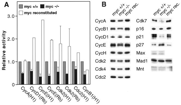
As the first measure of potential cell cycle defects, we determined the activities of all cyclin-Cdk complexes during steady-state exponential growth. Cyclins D1, E, A, and B1, as well as Cdk2 and Cdc2, were immunoprecipitated from cellular extracts, and the kinase activities of the immunoprecipitates were determined by using the appropriate substrates (Fig. 1A). c-myc−/− (HO15.19) cells displayed defects in the range of two- to fivefold in all kinase activities. Reconstitution of c-Myc activity by retrovirus infection (cell line HOmyc3) corrected all of the observed kinase defects and in some cases resulted in a modest increase in activity relative to that in wild-type c-myc+/+ cells. The largest defects were seen in cyclin E-associated activity and total Cdk2 activity. In c-myc−/− cells cyclin E-associated activity was equally defective whether histone H1 or Rb was used as the substrate. These results indicate that the increased cell cycle transition time observed in c-myc−/− cells is caused by a modest but coordinate downregulation of all major cyclin-Cdk complexes.
FIG. 1.
Cdk activities and expression of cell cycle effectors in exponentially growing cells. (A) Cdk activities. Complexes were immunoprecipitated from extracts with the antibodies indicated at the bottom. The substrate used in the kinase assays (histone H1 or Rb) is indicated in parentheses. Cell lines: TGR-1, c-myc+/+; HO15.19, c-myc−/−; HOmyc3, HO15.19 with reconstituted c-Myc expression. All data are normalized to the activity measured in TGR-1 immunoprecipitates. CycA, cyclin A. Error bars indicate standard deviations of a minimum of two independent experiments. (B) Immunoblots of cyclin, Cdk, CKI, and Mad proteins. myc rec., HOmyc3.
To investigate possible causes of reduced Cdk activity, the expression levels of cyclins, Cdks, and CKIs in growing cells were examined by immunoblotting (Fig. 1B). With the exception of cyclin D1, cyclin and Cdk expression levels were slightly but reproducibly reduced in c-myc−/− cells, and expression was restored upon reconstitution of c-Myc activity. Cyclin D1 levels were slightly elevated in c-myc−/− cells. Examination of CKIs showed that p16 expression was unchanged, p21 expression was decreased, and p27 expression was elevated. Finally, expression of Cdk7, the catalytic subunit of the CAK complex, was reduced, while the expression of its regulatory partner (cyclin H) was unchanged. Expression of p21, p27, and Cdk7 has not been previously linked to c-Myc activity. Expression of Max, Mad1, and Mnt was unchanged. Expression of Mad2-Mxi, Mad3, and Mad4 could not be detected.
Activation of immediate-early events.
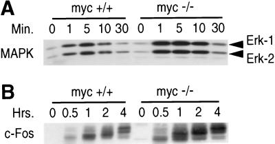
Cell cycle entry of quiescent cells begins with a rapid activation of several signal transduction pathways followed by the activation of immediate-early genes. These events occur prior to the induction of c-myc gene expression and should thus remain unaffected in c-myc-null cells. We examined two representative components of the immediate-early cascade: the activation of the MAPK pathway and the induction of c-fos gene expression (Fig. 2). Cells were made quiescent by a combination of serum deprivation and contact inhibition, and cell cycle reentry was elicited by the addition of fresh whole serum (see Materials and Methods); this basic regimen was followed in all experiments investigating the G0-to-S transition. Both the kinetics and the magnitude of MAPK activation as well as c-fos induction were indistinguishable in c-myc+/+ and c-myc−/− cells. Therefore, loss of c-myc does not cause a generalized defect in signal transduction pathways.
FIG. 2.
Immediate-early serum-induced events. (A) Phosphorylation of Erk-1 and Erk-2 proteins. (B) Induction of c-Fos expression. Both events were analyzed by immunoblotting; in panel A a phospho-specific MAPK antibody was used. In panel B the higher-molecular-weight bands represent phosphorylated forms of the c-Fos protein. Cell lines: TGR-1, c-myc+/+; HO15.19, c-myc−/−. Time points are identified above each lane. Extract from an equal number of cells was loaded in each lane.
Induction of D-type cyclins and activation of Cdk4 and Cdk6.
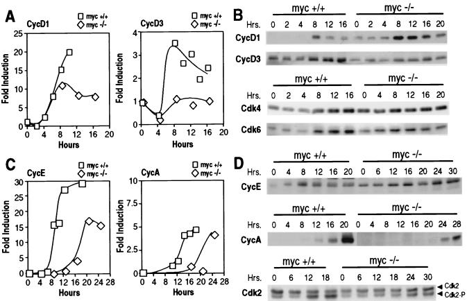
The D-type cyclins are the first cyclins to be induced during the G0-to-S transition. Expression of cyclins D1 and D3 was examined by RNase protection and immunoblotting (Fig. 3A and B). The kinetics of cyclin D1 and D3 mRNA induction were the same in c-myc+/+ and c-myc−/− cell lines. The induction ratios in c-myc+/+ cells were 20- and 3.5-fold for cyclins D1 and D3, respectively. In c-myc−/− cells, there were 2.3- and 2.7-fold reductions in cyclin D1 and D3 mRNA accumulation, respectively. The magnitude of this reduction is the same as previously observed for GAPDH mRNA and 28S rRNA during the same cell cycle interval (13, 73). The induction profile of cyclin D3 protein followed that of the mRNA; however, c-myc−/− cells accumulated somewhat more cyclin D1 protein than c-myc+/+ cells (Fig. 3B). Although the magnitude of this effect was small, it is the opposite of what was observed at the mRNA level. No significant differences in the expression of Cdk4 and Cdk6 proteins were observed between c-myc+/+ and c-myc−/− cells (Fig. 3B). Expression of cyclin D2 was not examined in detail because Rat-1 cells do not express this cyclin (98).
FIG. 3.
Cyclin and Cdk expression during the G0-to-S transition. (A) RNase protection analysis of cyclins D1 (CycD1) and D3. (B) Immunoblot analysis of cyclins D1 and D3, Cdk4, and Cdk6. (C) RNase protection analysis of cyclins E and A. (D) Immunoblot analysis of cyclins E and A and Cdk2. Cell lines: TGR-1, c-myc+/+; HO15.19, c-myc−/−. All data are presented on an equal-cell-number basis. (A and C) The fold induction is expressed relative to the TGR-1 zero time point. (B and D) Time points are identified above each lane, and antibodies are indicated on the left.
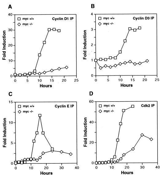
Activities of D-type cyclin-Cdk complexes were determined by direct kinase assays. Cellular extracts were immunoprecipitated with antibodies specific for either cyclin D1 or cyclin D3, and kinase activities were assayed by using a recombinant Rb substrate (Fig. 4A and B). The induction ratios of cyclin-associated kinase activity in c-myc+/+ cells were 30- and 3.0-fold for cyclins D1 and D3, respectively. The induction profiles of kinase activities followed closely those of the cyclin mRNAs, in terms of both the temporal kinetics and the overall magnitude of the response. We estimated that approximately 90% of total Cdk4 and Cdk6 activity was found in cyclin D1 complexes (data not shown). A 12-fold defect in cyclin D1-associated kinase activity was observed in c-myc−/− cells; the defect in cyclin D3-associated kinase activity was 3.4-fold. The dramatic defect in cyclin D1-associated kinase activity was reproducible in four independent experiments.
FIG. 4.
Cdk activities during the G0-to-S transition. (A) Cyclin D1 immunoprecipitates (IP). (B) Cyclin D3 immunoprecipitates. (C) Cyclin E immunoprecipitates. (D) Cdk2 immunoprecipitates. Complexes were immunoprecipitated from extracts containing equal amounts of total protein, and kinase assays were performed with either a GST-Rb substrate (A and B) or a histone H1 substrate (C and D) as described in Materials and Methods. Cell lines: TGR-1, c-myc+/+; HO15.19, c-myc−/−. The fold induction is expressed relative to the TGR-1 zero time point. The kinase assays shown are representative of three independent experiments.
Phosphorylation of the Rb family of proteins.
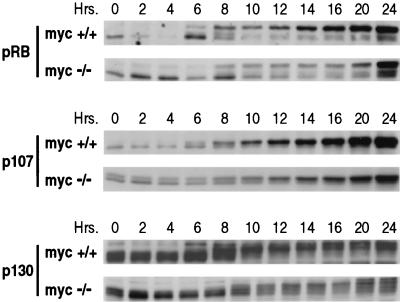
In order to determine whether the observed decrease in cyclin D-associated kinase activity has physiological consequences, the phosphorylation status of Rb and the related p107 and p130 proteins was examined by immunoblotting (Fig. 5). In c-myc+/+ cells, the shift from the faster-migrating, hypophosphorylated forms of all three Rb family proteins to the slower-migrating, hyperphosphorylated forms occurred within a narrow 2-h window between 6 and 8 h after serum stimulation. By 10 h after serum stimulation, all three proteins were found in their hyperphosphorylated forms. In contrast, in c-myc−/− cells, the shift to the hyperphosphorylated forms was delayed, and the time period during which the shift occurred was greatly extended. A complete shift to the hyperphosphorylated migration position was not observed until approximately 20 to 24 h after serum stimulation in the case of Rb, while p130 was not fully shifted even at 24 h. p107 phosphorylation was affected to a lesser extent, but the hypophosphorylated form was still clearly detectable in late G1. The delay in the onset of Rb family protein phosphorylation and the increased time required for full phosphorylation are consistent with the reduced activity of cyclin D-Cdk complexes.
FIG. 5.
Phosphorylation of Rb family members during the G0-to-S transition. Samples were taken at the time points indicated above each lane and analyzed by immunoblotting. Cell lines: TGR-1, c-myc+/+; HO15.19, c-myc−/−. Protein from an equal number of cells was loaded in each lane.
Induction and activation of the E2F family of transcription factors.
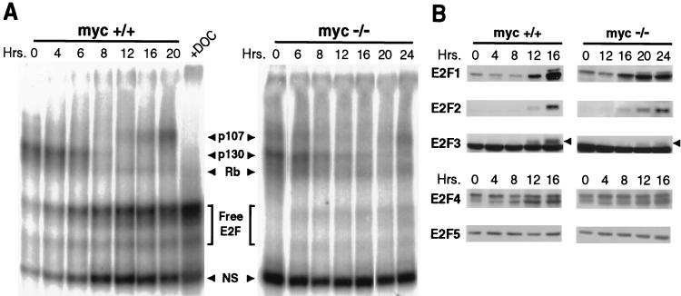
Hypophosphorylated Rb, p107, and p130 bind to and occlude the transactivation potential of E2F transcription factors. Rb binds to E2F-1, E2F-2, E2F-3, and E2F-4, while p107 and p130 bind to E2F-4 and E2F-5 (109). To determine the functional consequences of the persistence of hypophosphorylated Rb proteins at later times during the G0-to-S transition in c-myc−/− cells, we used EMSA to analyze cellular extracts for E2F DNA-binding activity (Fig. 6A). Approximately equivalent amounts of E2F-p130 complexes were present in extracts of quiescent c-myc+/+ and c-myc−/− cells. In c-myc+/+ cells this complex disappeared between 6 and 8 h after serum stimulation. This is the same time period during which p130 was observed to shift from the hypophosphorylated to the hyperphosphorylated form. In c-myc−/− cells the E2F-p130 complex was clearly detectable at the 8-h time point and persisted at lower levels until 16 to 20 h after serum stimulation.
FIG. 6.
E2F DNA-binding activity and protein expression during the G0-to-S transition. (A) E2F DNA-binding activity measured by EMSA. Cell lines: TGR-1, c-myc+/+; HO15.19, c-myc−/−. The times at which samples were collected are identified above the lanes. +DOC, addition of 0.8% deoxycholate to the reaction mixture. The identities of E2F complexes are marked by arrows between the panels and were determined by supershifts with the indicated antibodies. (B) Immunoblot analysis of E2F family members. The antibodies used are indicated on the left.
p107 is known to form S phase-specific complexes with E2F, cyclin A, and Cdk2 (24, 68). The E2F-p107 complex was first detected in c-myc+/+ cells at 12 h and accumulated to appreciable levels by 20 h after serum stimulation. In c-myc−/− cells this complex was not seen until 24 h, and then only at a significantly reduced level. The appearance of the E2F-p107 complex correlated closely with initial S phase entry in both c-myc+/+ and c-myc−/− cells. The E2F-Rb complex was a relatively minor band in extracts from both cell lines; consequently, it is difficult to draw conclusions on differential expression. The most striking feature of the EMSA profiles was the severely reduced free E2F DNA-binding activity in c-myc−/− cells, especially at late times after serum stimulation.
The expression of E2F-4 and E2F-5 is constant throughout the cell cycle, whereas E2F-1, E2F-2, and E2F-3 are induced in mid- to late G1 following serum stimulation (109). E2F activity is known to be important for the correct temporal expression of certain S phase-specific genes, and it also participates in the induction of the E2F-1 and E2F-2 genes by a feedback mechanism (47, 52, 102). Expression of all of the E2F proteins was therefore analyzed by immunoblotting (Fig. 6B). No significant difference in the expression levels of E2F-4 and E2F-5 was observed; however, c-myc−/− cells displayed a significant delay in the induction of E2F-1, E2F-2, and E2F-3.
Induction of cyclins E and A and activation of Cdk2.
Since both the cyclin E and cyclin A genes have been shown to be targets of E2F regulation (12, 34, 86, 101), their expression was examined by RNase protection. In c-myc+/+ cells cyclin E mRNA was induced between 6 and 8 h after serum stimulation, and this induction was delayed by approximately 6 h in c-myc−/− cells (Fig. 3C). The maximum level of cyclin E mRNA accumulation was 1.7-fold lower in c-myc−/− cells. Cyclin A induction was even more severely affected, lagging by approximately 12 h in c-myc−/− cells (Fig. 3C). The expression of both cyclin E and cyclin A proteins was examined by immunoblotting (Fig. 3D), and the induction profiles correlated well with the RNase protection data.
Given the delay in cyclin induction, it was of interest to determine the timing as well as the extent of associated kinase activation. Cyclin E or Cdk2 was immunoprecipitated from cellular extracts, and the associated kinase activity was determined with histone H1 as the substrate (Fig. 4C and D). Cyclin E-associated kinase activity was first detected in c-myc+/+ cells at 12 h after serum addition. The delay in activation in c-myc−/− cells was approximately 6 h, and the maximum level of activity was fourfold lower. Induction of total Cdk2 activity was delayed by approximately 8 h, and the maximum activity was twofold lower.
Immunoblotting did not reveal any clear differences in levels of total Cdk2 protein expression (Fig. 3D); however, changes in the migration positions were observed. In c-myc+/+ cells, more than half of the total Cdk2 protein shifted to a faster-migrating position by 12 h after serum addition, and the ratio of slower- and faster-migrating forms further increased by 18 h. In c-myc−/− cells, however, the appearance of the faster-migrating Cdk2 band was greatly delayed, and the ratio of the two forms remained in favor of the slower-migrating form for as long as 40 h after serum stimulation. It is believed that the shift to higher mobility is due to phosphorylation of Thr 160 of Cdk2 by CAK and that this represents the active form of the enzyme (104). The mobility shift of Cdk2 therefore correlated well with the direct assay of its activity and pointed to a CAK defect as at least one cause of reduced activity.
Regulation of Cdk complex activity by CAK and p27.
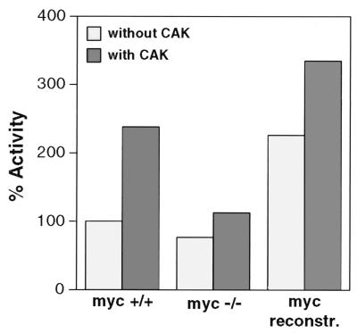
The presence of reduced levels of Cdk7 and increased levels of p27 in c-myc−/− cells provide one possible explanation for the global reduction in Cdk activity. To investigate whether CAK activity may be limiting in cells, Cdk2 complexes were immunoprecipitated from exponentially growing cells, incubated in the presence or absence of active recombinant CAK, and assayed for histone H1 phosphorylation activity (Fig. 7). Cdk2 activity from c-myc+/+ cells was stimulated 2.4-fold by CAK. As previously shown (Fig. 1A), Cdk2 activity was lower in c-myc−/− cells; this activity could also be stimulated by CAK, but to a lesser degree than that in c-myc+/+ cells. Finally, even the elevated Cdk2 activity levels found in c-myc−/− cells with reconstituted c-Myc expression (Fig. 1A) could be further stimulated with CAK. In an independent experiment, Cdk2 complexes immunoprecipitated from cells at various times during the G0-to-S transition (including the time of peak activity) could be further stimulated (threefold) by CAK (data not shown). These results clearly indicate that even in normal (c-myc+/+) cells, CAK activity is not saturating, suggesting that a fraction of fully assembled Cdk complexes are inactive because they lack the activating CAK phosphorylation.
FIG. 7.
Activation of Cdk2 complexes with CAK. Cdk2 complexes were immunoprecipitated from exponentially growing cells with Cdk2 antibodies, incubated with active Cak1p, and assayed for histone H1 phosphorylation activity, as described in Materials and Methods. Cell lines: TGR-1, c-myc+/+; HO15.19, c-myc−/−; HOmyc3, HO15.19 with reconstituted c-Myc expression (myc reconstr.). All data are represented as percentages of the activity measured in TGR-1 immunoprecipitates without CAK addition.
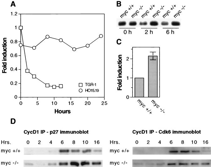
The fact that CAK phosphorylation of Cdk2 complexes from c-myc+/+ and c-myc−/− cells could not increase their activity to the level observed in c-Myc-overexpressing cells indicated the existence of a negative effector whose expression was likewise affected by c-Myc. The expression of p27, and its dependence on c-Myc expression, was therefore carefully examined in exponentially growing cells as well as during the G0-to-S transition. RNase protection during the G0-to-S progression showed a rapid drop in p27 mRNA levels in c-myc+/+ cells, falling 2.5-fold within 2 h and 5-fold within 6 h of serum stimulation (Fig. 8A). In contrast, p27 mRNA levels remained constant in c-myc−/− cells over a period of 24 h. Immunoblotting of p27 protein produced consistent results (Fig. 8B). Under exponential growth conditions, c-myc−/− cells displayed 2.2-fold-elevated p27 mRNA levels (Fig. 8C). This data is consistent with the increased levels of p27 protein observed in c-myc−/− cells under these conditions (Fig. 1B).
FIG. 8.
Expression of p27 and assembly into Cdk4 and -6 complexes. (A) RNase protection analysis during the G0-to-S transition. (B) Immunoblot analysis during the G0-to-S transition. (C) RNase protection analysis in exponentially growing cells. (D) Presence of p27 in cyclin D1-Cdk4 and -Cdk6 complexes during the G0-to-S transition. Complexes were immunoprecipitated (IP) with cyclin D1 antibody from extracts containing equal amounts of total protein and subsequently analyzed by immunoblotting with p27 antibody. Equal loading of lanes was demonstrated by subsequent reblotting with Cdk6 antibody. Cell lines: TGR-1, c-myc+/+; HO15.19, c-myc−/−.
Formation of Cdk complexes was examined by immunoprecipitation with cyclin D1, D3, E, or A antibodies followed by immunoblotting with the corresponding Cdk antibody. No significant differences were detected between c-myc+/+ and c-myc−/− cells in the amounts of any cyclin-Cdk complex (data not shown). However, cyclin D1-Cdk4 and -Cdk6 complexes during the G0-to-S transition contained clearly elevated levels of p27 (Fig. 8D). The expression of CKI proteins in the INK4 family as well as in the CIP-KIP family was also examined by immunoblotting during the G0-to-S transition (data not shown). p15 and p16 were clearly detectable, but no differences in their expression levels were observed. The expression levels of p18, p19, and p57 were too low to be detected by immunoblotting. The levels of p21 protein were lower in c-myc−/− cells; this is consistent with the decreased expression level found in exponentially cycling cells (Fig. 1B).
Ectopic overexpression of cyclins D1, E, and A.
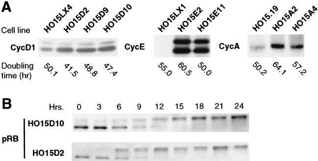
In an attempt to restore normal growth in c-myc−/− cells, we introduced retrovirus vectors expressing human cyclin cDNAs. We isolated numerous clonal cell lines, each expressing a single human cyclin transgene. All clones were screened by immunoblotting for the expression of the exogenous cyclin protein, and cell lines with the highest expression levels were chosen for further analysis (Fig. 9A). In no case did we observe reversion of the slow-growth phenotype. However, overexpression of cyclin D1 restored the kinetics of Rb phosphorylation to normal (Fig. 9B). In one cyclin D1-overexpressing cell line (HO15D2), Rb phosphorylation was actually accelerated relative to that in c-myc+/+ cells, such that the majority of Rb protein was in the hyperphosphorylated form by 6 h after serum stimulation. However, despite the precocious Rb phosphorylation, the growth rate was hardly affected: the doubling times were 50.1 h for the empty vector control (HO15LX4) and 41.5 h for the cyclin D1-overexpressing cells (HO15D2). In this context, it is important to note that the doubling time of parental TGR-1 cells is 17.7 h (73), and it is 18.8 h following reconstitution of c-myc in the knockout cells (HOmyc3). Therefore, c-Myc must affect cell cycle progression at additional points downstream of Cdk4 and -6 activation.
FIG. 9.
Overexpression of cyclins D1, E, and A in c-myc−/− cells. (A) Analysis of protein expression. HO15.19 (c-myc−/−) cells were infected singly with retrovirus vectors, and clonal cell lines were established. Immunoblotting was performed with exponentially growing cells. Cell lines are designated above each lane. HO15LX1 and HO15LX4 are two clones infected with empty LXSH vector. The exponential-phase doubling times for each clone are indicated below the lanes. The antibodies are indicated to the left of each panel. The cyclin D1 and cyclin A antibodies recognize both the human and rodent proteins; the cyclin E antibody is human specific. (B) Phosphorylation of Rb in cyclin D1-overexpressing cells. Cells were rendered quiescent by serum starvation and subsequently stimulated to reenter the cell cycle by serum addition. The zero time point is the time of serum addition. Samples were taken at the time points indicated above each lane and analyzed by immunoblotting. Cell lines and antibodies are indicated on the left. Equal amounts of protein were loaded in each lane.
DISCUSSION
c-Myc is required for the serum-induced activation of Cdk4 and -6 complexes.
The largest defect observed in c-myc−/− cells is a 12-fold reduction in the activation of cyclin D1-Cdk4 and -Cdk6 complexes during the G0-to-S transition. Prior work has demonstrated the influence of c-Myc overexpression on the activity of cyclin D1-Cdk4 (112), cyclin E-Cdk2 (89, 97, 100, 112), and cyclin A-Cdk2 (46, 100) complexes. The effect on Cdk4 and -6 activation during the G0-to-S transition is temporally the earliest cell cycle effect of c-Myc demonstrated to date. The induction of D-type cyclins and the activation of Cdk4 and -6 occurs at 4 to 6 h and 6 to 8 h, respectively, after the stimulation of c-myc+/+ cells with serum. These events thus follow very closely the peak of c-myc expression at 4 h (108). The expression of D-type cyclins is very responsive to growth factor stimulation, which has led to suggestions that cyclin D-Cdk4 and -Cdk6 complexes constitute a regulatory link between the extracellular environment and cell cycle progression (105, 106). Given the strong influence of c-Myc on cell growth, the linkage between c-Myc and Cdk4 and -6 activation is a very intriguing finding. This linkage provides a plausible physiological explanation for the observation that in c-myc−/− cells Cdk4 and -6 activity is affected only modestly during exponential growth, whereas the defect is much more pronounced during the G0-to-S transition. In steady-state growth with optimal serum supplementation, the activity of Cdk4 and -6 varies only minimally during the cell cycle, and c-myc is expressed constitutively at basal levels. In contrast, exit from quiescence requires widespread changes in gene expression as cellular metabolism is adjusted to support active growth; these changes apparently require significant activation of Cdk4 and -6, which is closely preceded by a strong induction of c-myc expression. Basal levels of Cdk4 and -6 activity apparently do not require c-Myc and are sufficient for proliferation, albeit at much reduced levels.
In contrast to the decrease in cyclin D1 mRNA, we reproducibly observed a slight increase in the accumulation of cyclin D1 protein, both in exponentially growing cells and during the G0-to-S transition. The increase in cyclin D1 protein levels was accompanied by a slight increase in the amount of immunoprecipitable cyclin D1-Cdk4 and -Cdk6 complexes. This is in agreement with prior observations that Cdk4 and -6 are present in excess of cyclin D1 and that cyclin D1 is rate limiting for the assembly of the complexes (75, 98). A likely explanation for the discrepancy between the levels of cyclin D1 mRNA and protein is stabilization of the protein to turnover. Cyclin D1 is known to be degraded by the ubiquitin pathway, and its degradation is enhanced by phosphorylation on threonine 286 by glycogen synthase kinase 3β (GSK-3β) (25). It is possible that the inactive cyclin D1-Cdk4 and -Cdk6 complexes that accumulate in c-myc−/− cells are recognized less well by GSK-3β or that the activity of GSK-3β itself is reduced in c-myc−/− cells.
Loss of c-Myc affects the activity of all Cdk complexes.
During the G0-to-S transition, loss of c-Myc activity resulted in the delayed appearance of cyclin E-Cdk2 activity as well as cyclin A-Cdk2 activity. The induction of cyclin E and A gene expression was coordinately delayed, indicating that the delay in Cdk2 activation was caused by a delay in the appearance of its positive effectors, cyclins E and A. The magnitude of induction of Cdk2 activity was also reduced, but not as substantially as the activity of Cdk4 and -6. During exponential growth, loss of c-myc reduced coordinately the activity of all cyclin-Cdk complexes by a factor of two- to fivefold. We know of no other cell cycle regulator with such a pleiotropic phenotype. The emerging picture of the c-myc−/− phenotype has distinct parallels with the shift of yeast from growth on good to poor carbon sources (23). The decrease in yeast growth rate is accompanied by a modest but coordinate downregulation of all cyclin genes; the G1 cyclins CLN1 and CLN2 were most strongly affected (3.81- and 4.42-fold, respectively), while the effects on the remainder of the cyclins (CLN3 and CLB1 to 6) were in the 1.5- to 2-fold range. In c-myc−/− cells under exponential growth conditions, the largest defect was in cyclin E-Cdk2 activity, and this activity was also most sensitive to c-Myc overexpression. It is important to note that c-myc−/− cells do not display a generalized defect in macromolecular synthesis; during steady-state exponential growth, cell volume, total protein, and rRNA content are the same in c-myc+/+ and c-myc−/− cells (73). Furthermore, the expression of numerous housekeeping genes, such as those for GAPDH and the ribosomal protein L32, is not changed.
c-Myc affects cell cycle progression at multiple independent points.
It is often observed that a single primary regulatory defect can elicit pleiotropic phenotypes by a cascade of downstream effects. For example, c-Myc could act primarily at the point of Cdk4 and -6 activation but cause more generalized effects by interfering with the activation of E2F and perhaps additional downstream regulators. The strong defect in Cdk4 and -6 activation during the G0-to-S transition necessitated a direct test of this hypothesis, which was performed by ectopically expressing cyclin D1 by using retrovirus vectors. The rationale for this experiment was provided by observations, made in several laboratories, that cyclins are limiting for the assembly of cyclin-Cdk complexes (75, 98). For example, in normal cells overexpression of cyclins has been observed to increase Cdk kinase activity (75), and cyclin overexpression in yeast could rescue a partial CAK defect both in G1 and in G2 (54, 113). Our results showed that while overexpression of cyclin D1 restored normal Rb phosphorylation, the growth rate in exponential phase was only minimally affected. Likewise, the overexpression of cyclins E and A did not accelerate normal growth. The conclusion that c-Myc affects the cell cycle at multiple points is also supported by the observation that despite extensive, long-term passaging of c-myc−/− cells in several laboratories, revertants to faster growth have never been recovered.
Loss of c-Myc affects the expression of Cdk effectors p27 and Cdk7.
The expression of the CKI p27KIP1 was elevated two- to threefold in c-myc−/− cells; in contrast, the expression of Cdk7, the catalytic subunit of CAK, was reduced by the same magnitude. p27 is a potent inhibitor of Cdk2 (93) and has been linked to cell cycle regulation in response to contact inhibition (92). Knockout studies indicate that p27 is at least partially haplo-insufficient (28, 32, 60, 84); in other words, p27+/− animals as well as cells derived from them display a distinct relaxation of cell cycle controls. The effect of p27 on Cdk4 and -6 complexes is currently not clear. Although p27 binds to these complexes, it has been reported that it does not inhibit their activity (11, 116). However, at least in NIH 3T3 cells, the activity of Cdk4 and -6 complexes may be inhibited by elevated p27 expression (64). We detect significantly increased levels of p27 in cyclin D1-Cdk4 and -Cdk6 complexes in c-myc−/− cells during the G0-to-S transition. Therefore, p27 is a good candidate for a global cell cycle effector whose regulation over a small range of expression could affect cell cycle progression.
The expression of neither Cdk7 nor CAK activity is subject to regulation as part of the intrinsic cell cycle (94, 114). However, both Cdk7 expression and CAK activity have been shown to be downregulated in quiescent cells and to be induced after serum stimulation in NIH 3T3 cells (94). Some cancer-derived cell lines express three- to fivefold-higher levels of Cdk7 than nontransformed cells (114). Recently, Cdk7 was identified as the product of a serum-inducible gene with late kinetics in a cDNA microarray analysis of human fibroblasts (50). We have shown that expression of Cdk7 is reduced in c-myc−/− cells by a factor of two- to threefold. More importantly, Cdk2 complexes immunoprecipitated from c-myc+/+ cells could be activated 2.4-fold by incubation with recombinant CAK. This result indicates that CAK activity is not saturating even in normal (c-myc+/+) cells and that a fraction of fully assembled Cdk complexes are inactive because they lack the activating CAK phosphorylation. Given the fact that CAK acts catalytically on Cdk complexes, even a small change in CAK steady-state levels could translate into physiologically relevant changes in global Cdk activity.
The assembly of any cyclin-Cdk complex was not impaired. The normal accumulation of cyclin D1-Cdk4 and -Cdk6 complexes also suggests that the INK family of inhibitors is not responsible for the low activity of the complexes, since it has been shown that the bindings of cyclin D1 and INK family proteins to Cdk4 and -6 are mutually exclusive (19). In agreement, we did not detect any differences in the steady-state levels of p16 or p15 in c-myc−/− cells. We did detect reduced p21CIP1 expression in c-myc−/− cells. It has been proposed that p21 may act as an assembly factor for cyclin-Cdk complexes (63); however, since the assembly of cyclin-Cdk complexes was not impaired in c-myc−/− cells, the role of p21 needs to be further investigated.
The Cdc25A phosphatase acts to activate Cdks by dephosphorylating the Y17 residue (48, 115). It has been reported that c-Myc regulates the expression of Cdc25A at the transcriptional level (33), but this observation has not been reproduced by others (1, 89, 97, 116). Furthermore, the expression of both Cdc25A mRNA and protein is the same in c-myc+/+ and c-myc−/− cells (13).
Genes regulated by c-Myc.
In all cases studied here, the effects on gene expression in c-myc−/− cells were modest (two- to threefold), and in no case was expression dependent solely on c-Myc. It is very unlikely that all of the changes in gene expression manifested in c-myc−/− cells are direct effects of regulation by c-Myc protein. Some genes, such as those for cyclin D1 (20, 91, 110), p21 (45), and E2F-2 (102), contain E boxes in their regulatory regions and may respond to c-Myc directly. Both p21 and E2F-2 have been shown to respond to regulation by E2F, and thus the defect in their expression may be linked to the generalized defect in E2F activity. The cyclin E and cyclin A genes also respond to activation by E2F and do not contain obvious E boxes. The p27 gene contains an initiator element in its promoter and may thus respond to c-Myc in this manner (69). The murine Cdk7 promoter is TATA-less and GC rich and contains a single consensus E box approximately 150 bp downstream from the start of transcription (108a). The human Cdk7 promoter has the same features, and the E box is perfectly conserved (84a). While this does not establish Cdk7 as a Myc-regulated gene, it certainly makes this possibility worth further investigation.
Perhaps the most enduring mystery of c-Myc has been the identity of its target genes. The availability, for the first time, of c-myc-null cells provides an exciting new framework for addressing this key issue. In this communication we provide the first comprehensive analysis of the changes in gene expression that result from loss of c-Myc function. The complex nature of these changes presents a challenge for the future to differentiate direct and indirect targets of c-Myc, as well as to identify new ones. The study presented here also sheds new light on the biological activities of c-Myc. We propose that c-Myc is a crucial link that functions in the coordinate adjustment of cell cycle progression to environmental conditions.
ACKNOWLEDGMENTS
This work was supported by NIH grant R01-GM-41690 to J.M.S. M.K.M. and A.J.O. were supported in part by predoctoral training grant GM-07601 from the NIH and a postdoctoral fellowship from the Ministerio Educación y Cultura de España, respectively.
We thank K. Davis for excellent technical assistance and A. Bush and M. Cole for numerous discussions and communications of unpublished observations. We gratefully acknowledge M. Ewen for GST-Rb, M. Solomon for GST-Cak1p protein, C. Sherr for p15 antibody, A. Diehl for advice on Cdk4 assays, and K. Zaret for helpful suggestions on the manuscript.
M.K.M. and A.J.O. made equal contributions to this study.
REFERENCES
- 1.Alevizopoulos K, Vlach J, Hennecke S, Amati B. Cyclin E and c-Myc promote cell proliferation in the presence of p16INK4a and of hypophosphorylated retinoblastoma family proteins. EMBO J. 1997;16:5322–5333. doi: 10.1093/emboj/16.17.5322. [DOI] [PMC free article] [PubMed] [Google Scholar]
- 2.Amati B, Brooks M W, Levy N, Littlewood T D, Evan G I, Land H. Oncogenic activity of the c-Myc protein requires dimerization with Max. Cell. 1993;72:233–245. doi: 10.1016/0092-8674(93)90663-b. [DOI] [PubMed] [Google Scholar]
- 3.Amati B, Dalton S, Brooks M W, Littlewood T D, Evan G I, Land H. Transcriptional activation by the human c-Myc oncoprotein in yeast requires interaction with Max. Nature. 1992;359:423–426. doi: 10.1038/359423a0. [DOI] [PubMed] [Google Scholar]
- 4.Amati B, Littlewood T D, Evan G I, Land H. The c-Myc protein induces cell cycle progression and apoptosis through dimerization with Max. EMBO J. 1993;12:5083–5087. doi: 10.1002/j.1460-2075.1993.tb06202.x. [DOI] [PMC free article] [PubMed] [Google Scholar]
- 5.Bates S, Bonetta L, Macallan D, Parry D, Holder A, Dickson C, Peters G. CDK6 (PLSTIRE) and CDK4 (PSKJ3) are a distinct subset of the cyclin-dependent kinases that associate with cyclin D1. Oncogene. 1994;9:71–79. [PubMed] [Google Scholar]
- 6.Beijersbergen R L, Bernards R. Cell cycle regulation by the retinoblastoma family of growth inhibitory proteins. Biochim Biophys Acta. 1996;1287:103–120. doi: 10.1016/0304-419x(96)00002-9. [DOI] [PubMed] [Google Scholar]
- 7.Beijersbergen R L, Carlee L, Kerkhoven R M, Bernards R. Regulation of the retinoblastoma protein-related p107 by G1 cyclin complexes. Genes Dev. 1995;9:1340–1353. doi: 10.1101/gad.9.11.1340. [DOI] [PubMed] [Google Scholar]
- 8.Bishop J M. Cellular oncogenes and retroviruses. Annu Rev Biochem. 1983;52:301–354. doi: 10.1146/annurev.bi.52.070183.001505. [DOI] [PubMed] [Google Scholar]
- 9.Blackwell T K, Kretzner L, Blackwood E M, Eisenman R N, Weintraub H. Sequence-specific DNA binding by the c-Myc protein. Science. 1990;250:1149–1151. doi: 10.1126/science.2251503. [DOI] [PubMed] [Google Scholar]
- 10.Blackwood E M, Eisenman R N. Max: a helix-loop-helix zipper protein that forms a sequence-specific DNA-binding complex with myc. Science. 1991;251:1211–1217. doi: 10.1126/science.2006410. [DOI] [PubMed] [Google Scholar]
- 11.Blain S W, Montalvo E, Massagué J. Differential interaction of the cyclin-dependent kinase (cdk) inhibitor p27Kip1 with cyclin A-cdk2 and cyclin D2-cdk4. J Biol Chem. 1997;272:25863–25872. doi: 10.1074/jbc.272.41.25863. [DOI] [PubMed] [Google Scholar]
- 12.Botz J, Zerfass-Thome K, Spitkovsky D, Delius H, Vogt B, Eilers M, Hatzigeorgiou A, Jansen-Durr P. Cell cycle regulation of the murine cyclin E gene depends on an E2F binding site in the promoter. Mol Cell Biol. 1996;16:3401–3409. doi: 10.1128/mcb.16.7.3401. [DOI] [PMC free article] [PubMed] [Google Scholar]
- 13.Bush A, Mateyak M K, Dugan K, Obaya A J, Adachi S, Sedivy J M, Cole M D. c-myc null cells misregulate cad and gadd45 but not other proposed c-Myc targets. Genes Dev. 1998;12:3797–3802. doi: 10.1101/gad.12.24.3797. [DOI] [PMC free article] [PubMed] [Google Scholar]
- 14.Chan F K M, Zhang J, Chen L, Shapiro D N, Winoto A. Identification of human/mouse p19, a novel cdk4/cdk6 inhibitor with homology to p16INK4. Mol Cell Biol. 1995;15:2682–2688. doi: 10.1128/mcb.15.5.2682. [DOI] [PMC free article] [PubMed] [Google Scholar]
- 15.Chellappan S P, Hiebert S, Mudryj M, Horowitz J M, Nevins J R. The E2F transcription factor is a cellular target for the RB protein. Cell. 1991;65:1053–1061. doi: 10.1016/0092-8674(91)90557-f. [DOI] [PubMed] [Google Scholar]
- 16.Cimowski M J, Laff G M, Solomon M J, Reed S I. KIN28 encodes a C-terminal domain kinase that controls mRNA transcription in Saccharomyces cerevisiae but lacks cyclin-dependent kinase-activating activity. Mol Cell Biol. 1995;15:2983–2992. doi: 10.1128/mcb.15.6.2983. [DOI] [PMC free article] [PubMed] [Google Scholar]
- 17.Cole M D. The myc oncogene: its role in transformation and differentiation. Annu Rev Genet. 1986;20:361–384. doi: 10.1146/annurev.ge.20.120186.002045. [DOI] [PubMed] [Google Scholar]
- 18.Cole M D, McMahon S B. The Myc oncoprotein: a critical evaluation of transactivation and target gene regulation. Oncogene. 1999;18:2916–2924. doi: 10.1038/sj.onc.1202748. [DOI] [PubMed] [Google Scholar]
- 19.Coleman K G, Wautlet B S, Morrisey D, Mulheron J, Sedman S A, Brinkley P, Price S, Webster K R. Identification of Cdk4 sequences involved in cyclin D1 and p16 binding. J Biol Chem. 1997;272:18869–18874. doi: 10.1074/jbc.272.30.18869. [DOI] [PubMed] [Google Scholar]
- 20.Daksis J I, Lu R Y, Facchini L M, Marhin W W, Penn L J Z. Myc induces cyclin D1 expression in the absence of de novo protein synthesis and links mitogen-stimulated signal transduction to the cell cycle. Oncogene. 1994;9:3635–3645. [PubMed] [Google Scholar]
- 21.Davis A C, Wims M, Spotts G D, Hann S R, Bradley A. A null c-myc mutation causes lethality before 10.5 days of gestation in homozygotes and reduced fertility in heterozygous female mice. Genes Dev. 1993;7:671–682. doi: 10.1101/gad.7.4.671. [DOI] [PubMed] [Google Scholar]
- 22.Dean M, Levine R A, Ran W, Kindy M S, Sonenshein G E, Campisi J. Regulation of c-myc transcription and mRNA abundance by serum growth factors and cell contact. J Biol Chem. 1986;261:9161–9166. [PubMed] [Google Scholar]
- 23.DeRisi J L, Iyer V R, Brown P O. Exploring the metabolic and genetic control of gene expression on a genomic scale. Science. 1997;278:680–686. doi: 10.1126/science.278.5338.680. [DOI] [PubMed] [Google Scholar]
- 24.Devoto S H, Mudryj M, Pines J, Hunter T, Nevins J R. A cyclin A-protein kinase complex possesses sequence-specific DNA binding activity: p33cdk2 is a component of the E2F-cyclin A complex. Cell. 1992;68:167–176. doi: 10.1016/0092-8674(92)90215-x. [DOI] [PubMed] [Google Scholar]
- 25.Diehl J A, Cheng M, Roussel M F, Sherr C J. Glycogen synthase kinase-3β regulates cyclin D1 proteolysis and subcellular localization. Genes Dev. 1998;12:3499–3511. doi: 10.1101/gad.12.22.3499. [DOI] [PMC free article] [PubMed] [Google Scholar]
- 26.Dou G-P, Zhao S, Levin A H, Wang J, Helin K, Pardee A B. G1/S-regulated E2F-containing protein complexes bind to the mouse thymidine kinase gene promoter. J Biol Chem. 1994;269:1306–1313. [PubMed] [Google Scholar]
- 27.Dulic V, Lees E, Reed S I. Association of human cyclin E with a periodic G1-S phase protein kinase. Science. 1992;257:1958–1961. doi: 10.1126/science.1329201. [DOI] [PubMed] [Google Scholar]
- 28.Durand B, Fero M L, Roberts J M, Raff M C. p27Kip1 alters the response of cells to mitogen and is part of a cell-intrinsic timer that arrests the cell cycle and initiates differentiation. Curr Biol. 1998;8:431–440. doi: 10.1016/s0960-9822(98)70177-0. [DOI] [PubMed] [Google Scholar]
- 29.Dynlacht B D, Flores O, Lees J A, Harlow E. Differential regulation of E2F transactivation by cyclin/cdk2 complexes. Genes Dev. 1994;8:1772–1786. doi: 10.1101/gad.8.15.1772. [DOI] [PubMed] [Google Scholar]
- 30.Eilers M, Schirm S, Bishop J M. The MYC protein activates transcription of the alpha-prothymosin gene. EMBO J. 1991;10:133–141. doi: 10.1002/j.1460-2075.1991.tb07929.x. [DOI] [PMC free article] [PubMed] [Google Scholar]
- 31.Ewen M E, Sluss H K, Sherr C J, Matsushime H, Kato J, Livingston D M. Functional interactions of the retinoblastoma protein with mammalian D-type cyclins. Cell. 1993;73:487–497. doi: 10.1016/0092-8674(93)90136-e. [DOI] [PubMed] [Google Scholar]
- 32.Fero M L, Rivkin M, Tasch M, Porter P, Carow C E, Firpo E, Polyak K, Tsai L H, Broudy V, Perlmutter R M, Kaushansky K, Roberts J M. A syndrome of multiorgan hyperplasia with features of gigantism, tumorigenesis, and female sterility in p27(Kip1)-deficient mice. Cell. 1996;85:733–744. doi: 10.1016/s0092-8674(00)81239-8. [DOI] [PubMed] [Google Scholar]
- 33.Galaktionov K, Chen X, Beach D. Cdc25 cell-cycle phosphatase as a target of c-myc. Nature. 1996;382:511–517. doi: 10.1038/382511a0. [DOI] [PubMed] [Google Scholar]
- 34.Geng Y, Eaton E N, Picon M, Roberts J M, Lundberg A S, Gifford A, Sardet C, Weinberg R A. Regulation of cyclin E transcription by E2Fs and retinoblastoma protein. Oncogene. 1996;12:1173–1180. [PubMed] [Google Scholar]
- 35.Grandori C, Eisenman R N. Myc target genes. Trends Biochem Sci. 1997;22:177–181. doi: 10.1016/s0968-0004(97)01025-6. [DOI] [PubMed] [Google Scholar]
- 36.Guan K, Jenkins C W, Li Y, Nichols M A, Wu X, O’Keefe C L, Matera A G, Xiong Y. Growth suppression by p18, a p16INK4/MTS1- and p14INK4/MTS2-related CDK6 inhibitor, correlates with wild-type pRb function. Genes Dev. 1994;8:2939–2952. doi: 10.1101/gad.8.24.2939. [DOI] [PubMed] [Google Scholar]
- 37.Hannon G J, Beach D. p15INK4B is a potential effector of cell cycle arrest mediated by TGF-β. Nature. 1994;371:257–261. doi: 10.1038/371257a0. [DOI] [PubMed] [Google Scholar]
- 38.Hanson K D, Shichiri M, Follansbee M R, Sedivy J M. Effects of c-myc expression on cell cycle progression. Mol Cell Biol. 1994;14:5748–5755. doi: 10.1128/mcb.14.9.5748. [DOI] [PMC free article] [PubMed] [Google Scholar]
- 39.Harper J W, Adami G R, Wei N, Keyomarsi K, Elledge S J. The p21 cdk-interacting protein Cip1 is a potent inhibitor of G1 cyclin-dependent kinases. Cell. 1993;75:805–816. doi: 10.1016/0092-8674(93)90499-g. [DOI] [PubMed] [Google Scholar]
- 40.Hatakeyama M, Brill J A, Fink G R, Weinberg R A. Collaboration of G1 cyclins in the functional inactivation of the retinoblastoma protein. Genes Dev. 1994;8:1759–1771. doi: 10.1101/gad.8.15.1759. [DOI] [PubMed] [Google Scholar]
- 41.Henriksson M, Luscher B. Proteins of the Myc network: essential regulators of cell growth and differentiation. Adv Cancer Res. 1996;68:109–182. doi: 10.1016/s0065-230x(08)60353-x. [DOI] [PubMed] [Google Scholar]
- 42.Hiebert S W, Chellappan S P, Horowitz J M, Nevins J R. The interaction of RB with E2F coincides with an inhibition of the transcriptional activity of E2F. Genes Dev. 1992;6:177–185. doi: 10.1101/gad.6.2.177. [DOI] [PubMed] [Google Scholar]
- 43.Hinds P W, Mittnacht S, Dulic V, Arnold A, Reed S I, Weinberg R A. Regulation of retinoblastoma protein functions by ectopic expression of human cyclins. Cell. 1992;70:993–1006. doi: 10.1016/0092-8674(92)90249-c. [DOI] [PubMed] [Google Scholar]
- 44.Hirai H, Roussel M F, Kato J, Ashmun R A, Sherr C J. Novel INK4 proteins, p19 and p18, are specific inhibitors of cyclin D-dependent kinases CDK4 and CDK6. Mol Cell Biol. 1995;15:2672–2681. doi: 10.1128/mcb.15.5.2672. [DOI] [PMC free article] [PubMed] [Google Scholar]
- 45.Hiyama H, Iavarone A, Reeves S A. Regulation of the cdk inhibitor p21 gene during cell cycle progression is under the control of the transcription factor E2F. Oncogene. 1998;16:1513–1523. doi: 10.1038/sj.onc.1201667. [DOI] [PubMed] [Google Scholar]
- 46.Hoang A T, Cohen K J, Barrett J F, Bergstrom D A, Dang C V. Participation of cyclin A in Myc-induced apoptosis. Proc Natl Acad Sci USA. 1994;91:6875–6879. doi: 10.1073/pnas.91.15.6875. [DOI] [PMC free article] [PubMed] [Google Scholar]
- 47.Hsiao K M, McMahon S L, Farnham P J. Multiple DNA elements are required for the growth regulation of the mouse E2F1 promoter. Genes Dev. 1994;8:1526–1537. doi: 10.1101/gad.8.13.1526. [DOI] [PubMed] [Google Scholar]
- 48.Iavarone A, Massagué J. Repression of the Cdk activator Cdc25A and cell-cycle arrest by cytokine TGF-β in cells lacking the Cdk inhibitor p15. Nature. 1997;387:417–421. doi: 10.1038/387417a0. [DOI] [PubMed] [Google Scholar]
- 49.Inghirami G, Grignani F, Sternas L, Lombardi L, Knowles D M, Dalla-Favera R. Down-regulation of LFA-1 adhesion receptors by c-myc oncogene in human B lymphoblastoid cells. Science. 1990;250:682–686. doi: 10.1126/science.2237417. [DOI] [PubMed] [Google Scholar]
- 50.Iyer V R, Eisen M B, Ross D T, Schuler G, Moore T, Lee J C F, Trent J M, Staudt L M, Hudson J, Jr, Boguski M S, Lashkari D, Shalon D, Botstein D, Brown P O. The transcriptional program in the response of human fibroblasts to serum. Science. 1999;283:83–87. doi: 10.1126/science.283.5398.83. [DOI] [PubMed] [Google Scholar]
- 51.Jansen-Durr P, Meichle A, Steiner P, Pagano M, Finke K, Botz J, Wessbecher J, Draetta G, Eilers M. Differential modulation of cyclin gene expression by Myc. Proc Natl Acad Sci USA. 1993;90:3685–3689. doi: 10.1073/pnas.90.8.3685. [DOI] [PMC free article] [PubMed] [Google Scholar]
- 52.Johnson D G, Ohtani K, Nevins J R. Autoregulatory control of E2F1 expression in response to positive and negative regulators of cell cycle progression. Genes Dev. 1994;8:1514–1525. doi: 10.1101/gad.8.13.1514. [DOI] [PubMed] [Google Scholar]
- 53.Kaczmarek L, Hyland J K, Watt R, Rosenberg M, Baserga R. Microinjected c-Myc as a competence factor. Science. 1985;228:1313–1315. doi: 10.1126/science.4001943. [DOI] [PubMed] [Google Scholar]
- 54.Kaldis P, Sutton A, Solomon M J. The Cdk-activating kinase (CAK) from budding yeast. Cell. 1996;86:553–564. doi: 10.1016/s0092-8674(00)80129-4. [DOI] [PubMed] [Google Scholar]
- 55.Karn J, Watson J V, Lowe A D, Green S M, Vedeckis W. Regulation of cell cycle duration by c-myc levels. Oncogene. 1989;4:773–787. [PubMed] [Google Scholar]
- 56.Kato G J, Lee W M F, Chen L, Dang C V. Max: functional domains and interaction with c-Myc. Genes Dev. 1992;6:81–92. doi: 10.1101/gad.6.1.81. [DOI] [PubMed] [Google Scholar]
- 57.Kato J, Matsushime H, Hiebert S W, Ewen M E, Sherr C J. Direct binding of cyclin D to the retinoblastoma gene product (pRb) and pRb phosphorylation by the cyclin D-dependent kinase CDK4. Genes Dev. 1993;7:331–342. doi: 10.1101/gad.7.3.331. [DOI] [PubMed] [Google Scholar]
- 58.Kelly K, Cochran B H, Stiles C D, Leder P. Cell-specific regulation of the c-myc gene by lymphocyte mitogens and platelet-derived growth factor. Cell. 1983;35:603–610. doi: 10.1016/0092-8674(83)90092-2. [DOI] [PubMed] [Google Scholar]
- 59.Kim Y L, Lee A S. Identification of a protein-binding site in the promoter of the human thymidine kinase gene required for the G1-S-regulated transcription. J Biol Chem. 1992;267:2723–2727. [PubMed] [Google Scholar]
- 60.Kiyokawa H, Kineman R D, Manova-Todorova K O, Soares V C, Hoffman E S, Ono M, Khanam D, Hayday A C, Frohman L A, Koff A. Enhanced growth of mice lacking the cyclin-dependent kinase inhibitor function of p27(Kip1) Cell. 1996;85:721–732. doi: 10.1016/s0092-8674(00)81238-6. [DOI] [PubMed] [Google Scholar]
- 61.Koff A, Giordano A, Desai D, Yamashita K, Harper J W, Elledge S, Nishimoto T, Morgan D O, Franza B R, Roberts J M. Formation and activation of a cyclin E-cdk2 complex during the G1 phase of the human cell cycle. Science. 1992;257:1689–1694. doi: 10.1126/science.1388288. [DOI] [PubMed] [Google Scholar]
- 62.Kretzner L, Blackwood E, Eisenman R N. Myc and Max proteins possess distinct transcriptional activities. Nature. 1992;359:426–429. doi: 10.1038/359426a0. [DOI] [PubMed] [Google Scholar]
- 63.LaBaer J, Garrett M D, Stevenson L F, Slingerland J M, Sandhu C, Chou H S, Fattaey A, Harlow E. New functional activities for the p21 family of CDK inhibitors. Genes Dev. 1997;11:847–862. doi: 10.1101/gad.11.7.847. [DOI] [PubMed] [Google Scholar]
- 64.Ladha M H, Lee K Y, Upton T M, Reed M F, Ewen M E. Regulation of exit from quiescence by p27 and cyclin D1-Cdk4. Mol Cell Biol. 1998;18:6605–6615. doi: 10.1128/mcb.18.11.6605. [DOI] [PMC free article] [PubMed] [Google Scholar]
- 65.Land H, Parada L, Weinberg R A. Tumorigenic conversion of primary embryo fibroblasts requires at least two cooperating oncogenes. Nature. 1983;304:596–602. doi: 10.1038/304596a0. [DOI] [PubMed] [Google Scholar]
- 66.Lee M H, Reynisdóttir I, Massagué J. Cloning of p57KIP2, a cyclin-dependent kinase inhibitor with unique domain structure and tissue distribution. Genes Dev. 1995;9:639–649. doi: 10.1101/gad.9.6.639. [DOI] [PubMed] [Google Scholar]
- 67.Lee T C, Li L, Philipson L, Ziff E B. Myc represses transcription of the growth arrest gene gas1. Proc Natl Acad Sci USA. 1997;94:12886–12891. doi: 10.1073/pnas.94.24.12886. [DOI] [PMC free article] [PubMed] [Google Scholar]
- 68.Lees E, Faha B, Dulic V, Reed S I, Harlow E. Cyclin E/cdk2 and cyclin A/cdk2 kinases associate with p107 and E2F in a temporally distinct manner. Genes Dev. 1992;6:1874–1885. doi: 10.1101/gad.6.10.1874. [DOI] [PubMed] [Google Scholar]
- 69.Li L-H, Nerlov C, Prendergast G, MacGregor D, Ziff E B. c-Myc represses transcription in vivo by a novel mechanism dependent on the initiator element and Myc box II. EMBO J. 1994;13:4070–4079. doi: 10.1002/j.1460-2075.1994.tb06724.x. [DOI] [PMC free article] [PubMed] [Google Scholar]
- 70.Lundberg A S, Weinberg R A. Functional inactivation of the retinoblastoma protein requires sequential modification by at least two distinct cyclin-cdk complexes. Mol Cell Biol. 1998;18:753–761. doi: 10.1128/mcb.18.2.753. [DOI] [PMC free article] [PubMed] [Google Scholar]
- 71.Mann R, Mulligan R C, Baltimore D. Construction of a retrovirus packaging mutant and its use to produce helper-free defective retrovirus. Cell. 1983;33:153–159. doi: 10.1016/0092-8674(83)90344-6. [DOI] [PubMed] [Google Scholar]
- 72.Marhin W W, Chen S, Facchini L M, Fornace A J, Jr, Penn L Z. Myc represses the growth arrest gene gadd45. Oncogene. 1997;14:2825–2834. doi: 10.1038/sj.onc.1201138. [DOI] [PubMed] [Google Scholar]
- 73.Mateyak M K, Obaya A J, Adachi S, Sedivy J M. Phenotypes of c-Myc-deficient rat fibroblasts isolated by targeted homologous recombination. Cell Growth Differ. 1997;8:1039–1048. [PubMed] [Google Scholar]
- 74.Matsushime H, Ewen M E, Strom D K, Kato J, Hanks S K, Roussel M F, Sherr C J. Identification and properties of an atypical catalytic subunit (p34PSKJ3/CDK4) for mammalian D-type G1 cyclins. Cell. 1992;71:323–334. doi: 10.1016/0092-8674(92)90360-o. [DOI] [PubMed] [Google Scholar]
- 75.Matsushime H, Quelle D E, Shurtleff S A, Shibuya M, Sherr C J, Kato J V. D-type cyclin-dependent kinase activity in mammalian cells. Mol Cell Biol. 1994;14:2066–2076. doi: 10.1128/mcb.14.3.2066. [DOI] [PMC free article] [PubMed] [Google Scholar]
- 76.Matsushime H, Roussel M F, Ashmun R A, Sherr C J. Colony-stimulating factor 1 regulates novel cyclins during the G1 phase of the cell cycle. Cell. 1991;65:701–713. doi: 10.1016/0092-8674(91)90101-4. [DOI] [PubMed] [Google Scholar]
- 77.Mayol X, Garriga J, Grana X. Cell cycle-dependent phosphorylation of the retinoblastoma-related protein p130. Oncogene. 1995;11:801–808. [PubMed] [Google Scholar]
- 78.Meyerson M, Harlow E. Identification of a G1 kinase activity for Cdk6, a novel cyclin D partner. Mol Cell Biol. 1994;14:2077–2086. doi: 10.1128/mcb.14.3.2077. [DOI] [PMC free article] [PubMed] [Google Scholar]
- 79.Miller A D, Miller D G, Garcia J V, Lynch C M. Use of retroviral vectors for gene transfer and expression. Methods Enzymol. 1993;217:581–599. doi: 10.1016/0076-6879(93)17090-r. [DOI] [PubMed] [Google Scholar]
- 80.Mink S, Mutschler B, Weiskirchen R, Bister K, Klempnauer K-H. A novel function for Myc: inhibition of C/EBP-dependent gene activation. Proc Natl Acad Sci USA. 1996;93:6635–6640. doi: 10.1073/pnas.93.13.6635. [DOI] [PMC free article] [PubMed] [Google Scholar]
- 81.Morgan D O. Cyclin-dependent kinases: engines, clocks, and microprocessors. Annu Rev Cell Dev Biol. 1997;13:261–291. doi: 10.1146/annurev.cellbio.13.1.261. [DOI] [PubMed] [Google Scholar]
- 82.Morgan D O. Principles of CDK regulation. Nature. 1995;374:131–134. doi: 10.1038/374131a0. [DOI] [PubMed] [Google Scholar]
- 83.Murre C, McCaw P S, Baltimore D. A new DNA binding and dimerization motif in immunoglobulin enhancer binding, daughterless, MyoD and Myc proteins. Cell. 1989;56:777–783. doi: 10.1016/0092-8674(89)90682-x. [DOI] [PubMed] [Google Scholar]
- 84.Nakayama K, Ishida N, Shirane M, Inomata A, Inoue T, Shishido N, Horii I, Loh D Y, Nakayama K. Mice lacking p27(Kip1) display increased body size, multiple organ hyperplasia, retinal dysplasia, and pituitary tumors. Cell. 1996;85:707–720. doi: 10.1016/s0092-8674(00)81237-4. [DOI] [PubMed] [Google Scholar]
- 84a.Obaya, A. Unpublished observations.
- 85.Obaya A J, Mateyak M K, Sedivy J M. Mysterious liaisons: the relationship between c-Myc and the cell cycle. Oncogene. 1999;18:2934–2941. doi: 10.1038/sj.onc.1202749. [DOI] [PubMed] [Google Scholar]
- 86.Ohtani K, DeGregori J, Nevins J R. Regulation of the cyclin E gene by transcription factor E2F1. Proc Natl Acad Sci USA. 1995;92:12146–12150. doi: 10.1073/pnas.92.26.12146. [DOI] [PMC free article] [PubMed] [Google Scholar]
- 87.Pearson B E, Nasheuer H-P, Wang T-F. Human DNA polymerase α gene: sequences controlling expression in cycling and serum-stimulated cells. Mol Cell Biol. 1991;11:2081–2095. doi: 10.1128/mcb.11.4.2081. [DOI] [PMC free article] [PubMed] [Google Scholar]
- 88.Penn L J Z, Brooks M W, Laufer E M, Land H. Negative autoregulation of c-myc transcription. EMBO J. 1990;9:1113–1121. doi: 10.1002/j.1460-2075.1990.tb08217.x. [DOI] [PMC free article] [PubMed] [Google Scholar]
- 89.Perez-Roger I, Solomon D L C, Sewing A, Land H. Myc activation of cyclin E/Cdk2 kinase involves induction of cyclin E gene transcription and inhibition of p27Kip1 binding to newly formed complexes. Oncogene. 1997;14:2373–2381. doi: 10.1038/sj.onc.1201197. [DOI] [PubMed] [Google Scholar]
- 90.Peukert K, Staller P, Schneider A, Carmichael G, Hanel F, Eilers M. An alternative pathway for gene regulation by Myc. EMBO J. 1997;16:5672–5686. doi: 10.1093/emboj/16.18.5672. [DOI] [PMC free article] [PubMed] [Google Scholar]
- 91.Philipp A, Schneider A, Vasrik I, Finke K, Xiong Y, Beach D, Alitalo K, Eilers M. Repression of cyclin D1: a novel function of MYC. Mol Cell Biol. 1994;14:4032–4043. doi: 10.1128/mcb.14.6.4032. [DOI] [PMC free article] [PubMed] [Google Scholar]
- 92.Polyak K, Kato J, Solomon M J, Sherr C J, Massagué J, Roberts J M, Koff A. p27Kip1, a cyclin-cdk inhibitor, links transforming growth factor-β and contact inhibition to cell cycle arrest. Genes Dev. 1994;8:9–22. doi: 10.1101/gad.8.1.9. [DOI] [PubMed] [Google Scholar]
- 93.Polyak K, Lee M, Erdjument-Bromage H, Koff A, Roberts J M, Tempst P, Massagué J. Cloning of p27Kip1, a cyclin-dependent kinase inhibitor and a potential mediator of extracellular antimitogenic signals. Cell. 1994;78:59–66. doi: 10.1016/0092-8674(94)90572-x. [DOI] [PubMed] [Google Scholar]
- 94.Poon R Y, Yamashita K, Howell M, Ershler M A, Belyavsky A, Hunt T. Cell cycle regulation of the p34cdc2/p33cdk2-activating kinase p40MO15. J Cell Sci. 1994;107:2789–2799. doi: 10.1242/jcs.107.10.2789. [DOI] [PubMed] [Google Scholar]
- 95.Prendergast G C, Ziff E B. Methylation-sensitive sequence-specific DNA binding by the c-Myc basic region. Science. 1991;251:186–189. doi: 10.1126/science.1987636. [DOI] [PubMed] [Google Scholar]
- 96.Prouty S M, Hanson K D, Boyle A L, Brown J R, Shichiri M, Follansbee M R, Kang W, Sedivy J M. A cell culture model system for genetic analyses of the cell cycle by targeted homologous recombination. Oncogene. 1993;8:899–907. [PubMed] [Google Scholar]
- 97.Pusch O, Bernaschek G, Eilers M, Hengstschlager M. Activation of c-Myc uncouples DNA replication from activation of G1-cyclin-dependent kinases. Oncogene. 1997;15:649–656. doi: 10.1038/sj.onc.1201236. [DOI] [PubMed] [Google Scholar]
- 98.Quelle D E, Ashmun R A, Shurtleff S A, Kato J Y, Bar-Sagi D, Roussel M F, Sherr C J. Overexpression of mouse D-type cyclins accelerates G1 phase in rodent fibroblasts. Genes Dev. 1993;7:1559–1571. doi: 10.1101/gad.7.8.1559. [DOI] [PubMed] [Google Scholar]
- 99.Roy A L, Carruthers C, Gutjahr T, Roeder R G. Direct role for Myc in transcription initiation mediated by interactions with TFII-I. Nature. 1993;365:359–361. doi: 10.1038/365359a0. [DOI] [PubMed] [Google Scholar]
- 100.Rudolph B, Saffrich R, Zwicker J, Henglein B, Müller R, Ansorge W, Eilers M. Activation of cyclin-dependent kinases by Myc mediates induction of cyclin A, but not apoptosis. EMBO J. 1996;15:3065–3076. [PMC free article] [PubMed] [Google Scholar]
- 101.Schulze A, Zerfass K, Spitkovsky D, Middendorp S, Berges J, Helin K, Jansen-Durr P, Henglein B. Cell cycle regulation of the cyclin A gene promoter is mediated by a variant E2F site. Proc Natl Acad Sci USA. 1995;92:11264–11268. doi: 10.1073/pnas.92.24.11264. [DOI] [PMC free article] [PubMed] [Google Scholar]
- 102.Sears R, Ohtani K, Nevins J R. Identification of positively and negatively acting elements regulating expression of the E2F2 gene in response to cell growth signals. Mol Cell Biol. 1997;17:5227–5235. doi: 10.1128/mcb.17.9.5227. [DOI] [PMC free article] [PubMed] [Google Scholar]
- 103.Serrano M, Hannon G J, Beach D. A new regulatory motif in cell cycle control causing specific inhibition of cyclinD/cdk4. Nature. 1993;366:704–707. doi: 10.1038/366704a0. [DOI] [PubMed] [Google Scholar]
- 104.Sheaff R J. Regulation of mammalian cyclin-dependent kinase 2. Methods Enzymol. 1997;283:173–193. doi: 10.1016/s0076-6879(97)83015-7. [DOI] [PubMed] [Google Scholar]
- 105.Sherr C J. D-type cyclins. Trends Biochem Sci. 1995;20:187–190. doi: 10.1016/s0968-0004(00)89005-2. [DOI] [PubMed] [Google Scholar]
- 106.Sherr C J. Mammalian G1 cyclins. Cell. 1993;73:1059–1065. doi: 10.1016/0092-8674(93)90636-5. [DOI] [PubMed] [Google Scholar]
- 107.Sherr C J, Roberts J M. Inhibitors of mammalian G1 cyclin-dependent kinases. Genes Dev. 1995;9:1149–1163. doi: 10.1101/gad.9.10.1149. [DOI] [PubMed] [Google Scholar]
- 108.Shichiri M, Hanson K D, Sedivy J M. Effects of c-myc expression on proliferation, quiescence, and the G0 to G1 transition in nontransformed cells. Cell Growth Differ. 1993;4:93–104. [PubMed] [Google Scholar]
- 108a.Shuttleworth, J. Personal communication.
- 109.Slansky J E, Farnham P J. Introduction to the E2F family: protein structure and gene regulation. Curr Top Microbiol Immunol. 1996;208:1–30. doi: 10.1007/978-3-642-79910-5_1. [DOI] [PubMed] [Google Scholar]
- 110.Solomon D L C, Philipp A, Land H, Eilers M. Expression of cyclin D1 mRNA is not upregulated by Myc in rat fibroblasts. Oncogene. 1995;11:1893–1897. [PubMed] [Google Scholar]
- 111.Spencer C A, Groudine M. Control of c-myc regulation in normal and neoplastic cells. Adv Cancer Res. 1991;56:1–48. doi: 10.1016/s0065-230x(08)60476-5. [DOI] [PubMed] [Google Scholar]
- 112.Steiner P, Philipp A, Lukas J, Godden-Kent D, Pagano M, Mittnacht S, Bartek J, Eilers M. Identification of a Myc-dependent step during the formation of active G1 cyclin-cdk complexes. EMBO J. 1995;14:4814–4826. doi: 10.1002/j.1460-2075.1995.tb00163.x. [DOI] [PMC free article] [PubMed] [Google Scholar]
- 113.Sutton A, Freiman R. The Cak1p protein kinase is required at G1/S and G2/M in the budding yeast cell cycle. Genetics. 1997;147:57–71. doi: 10.1093/genetics/147.1.57. [DOI] [PMC free article] [PubMed] [Google Scholar]
- 114.Tassan J P, Schultz S J, Bartek J, Nigg E A. Cell cycle analysis of the activity, subcellular localization, and subunit composition of human CAK (CDK-activating kinase) J Cell Biol. 1994;127:467–478. doi: 10.1083/jcb.127.2.467. [DOI] [PMC free article] [PubMed] [Google Scholar]
- 115.Terada Y, Tatsuka M, Jinno S, Okayama H. Requirement for tyrosine phosphorylation of Cdk4 in G1 arrest induced by ultraviolet irradiation. Nature. 1995;376:358–362. doi: 10.1038/376358a0. [DOI] [PubMed] [Google Scholar]
- 116.Vlach J, Hennecke S, Alevizopoulos K, Conti D, Amati B. Growth arrest by the cyclin-dependent kinase inhibitor p27Kip1 is abrogated by c-Myc. EMBO J. 1996;15:6595–6604. [PMC free article] [PubMed] [Google Scholar]
- 117.Waters C M, Littlewood T D, Hancock D C, Moore J P, Evan G I. c-myc protein expression in untransformed fibroblasts. Oncogene. 1991;6:797–805. [PubMed] [Google Scholar]
- 118.Won K-A, Xiong Y, Beach D, Gilman M. Growth-regulated expression of D-type cyclin genes in human diploid fibroblasts. Proc Natl Acad Sci USA. 1992;89:9910–9914. doi: 10.1073/pnas.89.20.9910. [DOI] [PMC free article] [PubMed] [Google Scholar]
- 119.Xiao Z-X, Ginsberg D, Ewen M, Livingston D M. Regulation of the retinoblastoma protein-related protein p107 by G1 cyclin-associated kinases. Proc Natl Acad Sci USA. 1996;93:4633–4637. doi: 10.1073/pnas.93.10.4633. [DOI] [PMC free article] [PubMed] [Google Scholar]
- 120.Xiong Y, Hannon G J, Zhang H, Casso D, Kobayashi R, Beach D. p21 is a universal inhibitor of cyclin kinases. Nature. 1993;366:701–704. doi: 10.1038/366701a0. [DOI] [PubMed] [Google Scholar]
- 121.Yang B S, Gilbert J D, Freytag S O. Overexpression of Myc suppresses CCAAT transcription factor/nuclear factor 1-dependent promoters in vivo. Mol Cell Biol. 1993;13:3093–3102. doi: 10.1128/mcb.13.5.3093. [DOI] [PMC free article] [PubMed] [Google Scholar]


