Abstract
Sepsis remains a common cause of death in intensive care units, accounting for approximately 20% of total deaths worldwide. Its pathogenesis is partly attributable to dysregulated inflammatory responses to bacterial endotoxins (such as lipopolysaccharide, LPS), which stimulate innate immune cells to sequentially release early cytokines (such as tumor necrosis factor (TNF) and interferons (IFNs)) and late mediators (such as high-mobility group box 1, HMGB1). Despite difficulties in translating mechanistic insights into effective therapies, an improved understanding of the complex mechanisms underlying the pathogenesis of sepsis is still urgently needed. Here, we review recent progress in elucidating the intricate mechanisms underlying the regulation of HMGB1 release and action, and propose a few potential therapeutic candidates for future clinical investigations.
Keywords: sepsis, pyroptosis, innate immune cells, antibodies, herbal medicine, acute-phase proteins, hemichannel, inflammasome
1. Introduction
Microbial infections and resultant sepsis syndromes are the most common causes of death in intensive care units, accounting for approximately 20% of total deaths worldwide [1]. The pathogenesis of sepsis remains poorly understood, but is partly attributable to immune over-activation or immunosuppression propagated by dysregulated innate immune responses to lethal infections [2,3]. Innate immune cells (such as macrophages, monocytes and neutrophils) constitute a front line of defense against microbial infections by eliminating invading pathogens via phagocytosis, and initiating inflammatory responses via various mediators. Upon detection of microbial products such as bacterial endotoxins (lipopolysaccharide, LPS), circulating neutrophils and monocytes immediately infiltrate into infected tissues [4]. After engulfing and killing pathogens, neutrophils exhaust intracellular enzymes and undergo apoptotic cell death. The cell debris of these apoptotic neutrophils are then removed by tissue macrophages (e.g., Kupffer cells, dendritic cells, and glia cells) [5] terminally differentiated from infiltrated monocytes.
Innate immune cells also carry various pattern recognition receptors (PRRs) to recognize distinct classes of molecules shared by a group of related microbes, which are collectively termed “pathogen-associated molecular pattern molecules” (PAMPs). For instance, Toll-like Receptor 2 (TLR2) [6], TLR4 [7] and TLR9 [8], respectively, serve as PRRs for distinct PAMPs such as peptidoglycans, bacterial endotoxins, and microbial un-methylated CpG-DNAs. The engagement of various PRRs by different PAMPs similarly activates innate immune cells to sequentially release early cytokines (such as tumor necrosis factor (TNF) and interferons (IFNs)) and late-acting pro-inflammatory mediators [such as high-mobility group box 1 (HMGB1) and sequestosome 1 (SQSTM1)] [9,10].
HMGB1 is constitutively expressed by most types of cells to maintain a large “pool” of preformed protein in the nucleus, possibly due to the presence of two lysine-rich nuclear localization sequences (NLS) [11]. It carries two internal repeats of positively charged domains (“HMG boxes” known as “A box” and “B box”) in the N-terminus, and a continuous stretch of negatively charged (aspartic and glutamic acid) residues in the C-terminus. These HMG boxes enable HMGB1 to bind chromosomal DNA, and fulfill its nuclear functions to maintain nucleosomal structure and stability, and regulate gene expression [12,13]. Once released, extracellular HMGB1 can bind many endogenous proteins, thereby modulating divergent innate immune responses to lethal infections [13]. To complement two relevant reviews in this Special Issue [14,15], here we summarize recent progress in elucidating the intricate mechanisms underlying both endogenous regulation and pharmacological modulation of LPS-induced HMGB1 release and action, and propose a few potential therapeutic candidates for future clinical investigations.
2. Role of Cell-Surface PRRs in the Regulation of Early Pro-Inflammatory Cytokines
Innate immune cells employ cell-surface PRRs such as TLR4 [7] to recognize extracellular bacterial endotoxins (e.g., LPS) in conjunction with a LPS-binding protein (LBP) [16] and a cell-surface co-receptor CD14 [17,18]. Upon capturing LPS, LBP interacts with CD14 to deliver it to a cell-surface receptor, TLR4 [7], thereby triggering immediate production of early cytokines (e.g., TNF and IFN-γ) and subsequent release of late-acting mediators (e.g., HMGB1 and SQSTM1) [9,10]. The critical role of CD14 in the regulation of LPS-induced inflammation was evidenced by: (1) an enhanced sensitivity to lethal endotoxemia in CD14-over-expressing mice [19]; (2) a reduced susceptibility to lethal endotoxemia in CD14-deficient mice [20]; and (3) an abolishment of LPS-induced production of early cytokines (e.g., TNF) in CD14-deficient innate immune cells [21,22]. However, we found that the depletion of CD14 expression only partly attenuated the LPS-induced HMGB1 release [22], suggesting a potential involvement of other CD14/TLR4-independet signaling pathways in the regulation of HMGB1 release.
In a murine model of endotoxemia induced by intraperitoneal administration of LPS, HMGB1 was first detected in the circulation eight hours after endotoxemia, and subsequently increased to plateau levels from 16 to 32 hours [9]. This late appearance of circulating HMGB1 paralleled with the onset of animal lethality from endotoxemia, and distinguished itself from TNF and other early proinflammatory cytokines [23]. Lacking a leader peptide sequence, HMGB1 could not be actively secreted through classical endoplasmic reticulum-Golgi exocytotic pathways [9]. Instead, upon post-translational acetylation or phosphorylation [24,25] of the nuclear localization sequence (NLS) [26,27], nuclear HMGB1 is translocated to the cytosol and sequestered into cytoplasmic vesicles [11,24,28,29]. These cytoplasmic HMGB1 vesicles could then be secreted into the extracellular space through pyroptosis, a programmed cell death leading to rapid release of cellular contents such as HMGB1 and SQSTM1 [10].
3. Role of Cytoplasmic PRRs (Caspase-11/4/5/1) in the Regulation of Pyroptosis and HMGB1 Release
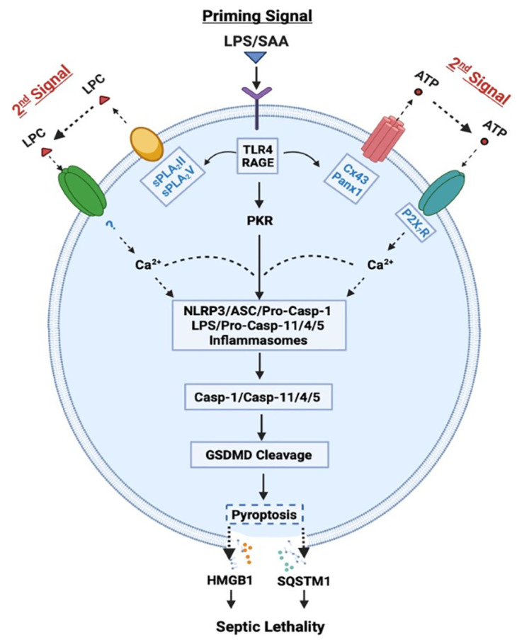
We and others demonstrated that ultra-pure LPS (free from any contaminating bacterial proteins, lipids, or nucleic acids) completely failed to induce HMGB1 release, unless the initial LPS priming was accompanied by a second stimulus (e.g., ATP) [30,31]. However, crude LPS that might carry trace amounts of bacterial proteins, lipids and nucleic acids, triggered a marked HMGB1 release [9]. It is possible that some contaminating bacterial proteins and lipids might enhance endocytosis of LPS, and consequently facilitate its innate recognition by cytoplasmic PRRs such as Casp-11/4/5. Indeed, when LPS was delivered to cytoplasmic Casp-11/4/5 either via CD14/TLR4 receptor-mediated endocytosis or bacteria-derived outer membrane vesicles (OMV) [32], it induced “non-canonical” inflammasome activation via oligomerization and proximity-induced activation of Casp-11/4/5 (Figure 1) [33]. The activated Casp-11/4/5 then catalyzes the cleavage of Gasdermin D (GSDMD) to form cytoplasmic membrane pores that cause immediate ionic gradient loss, osmotic burst and cell membrane rupture, a process aforementioned as “pyroptosis”. For the optimal activation of non-canonical inflammasome, both type I IFN-α/β and type II IFN-γ are needed to up-regulate Casp-11/4/5 [34,35] as well as guanylate-binding proteins [36] responsible for disrupting pathogen-containing vacuoles and releasing LPS. Coincidently, we and others demonstrated that LPS-inducible type I IFN-α/β [37,38] and type II IFN-γ [28] effectively stimulated innate immune cells to release HMGB1 in a time- and dose-dependent fashion.
Figure 1.
Role of Casp-1-mediated canonical and Casp-11/4/5-mediated non-canonical inflammasome activation in LPS- or SAA-induced pyroptosis and HMGB1 release. LPS or SAA may prime innate immune cells to up-regulate the expression of Cx43/Panx1 hemichannels, sPLA2s and interferon-induced double-stranded RNA-activated protein kinase (PKR), thereby eliciting the release of ATP or LPC that may activate P2X7R- or other receptor-mediated Ca2+ signaling. It then induces a feed-forwarding activation of PKR and inflammasome, cleavage of GSDMD, pyroptosis, and subsequent release of late mediators (such as HMGB1 and SQSTM1) of lethal infections.
In contrast, the “canonical” inflammasome activation is characterized by the oligomerization of intracellular “nucleotide-binding oligomerization domain (NOD)-like receptors” (NLRs such as NLRP1, NLRP3, and NLRC4) and the “apoptosis-associated speck-like protein containing a C-terminal caspase recruitment domain” (ASC) adaptor, as well as the recruitment and activation of pro-Casp-1 (Figure 1) [30]. Specifically, the pro-Casp-1 forms a heteromeric protein complex with an ASC adaptor and a NLR receptor, and the resultant protein complex, termed the “inflammasome”, is responsible for cleaving pro-Casp-1 to generate Casp-1, which triggers canonical inflammasome activation and pyroptosis via GSDMD cleavage [30]. Likewise, the optimal activation of canonical inflammasome also depends on a two-step process: (1) a priming signal elicited by extracellular PAMPs (e.g., LPS) to up-regulate NLRP3 expression; and (2) a secondary signal elicited by extracellular damage-associated molecular pattern (DAMPs, e.g., ATP) to induce NLRP3 oligomerization with ASC and pro-Casp-1 (Figure 1). Notably, the cleavage of pannexin-1 (Panx1) hemichannel by Casp-11/4/5 might be needed for releasing ATP and activating the purinergic P2X7 receptor (P2X7R) and inflammasome signalings (Figure 1) [39,40]. Consistently, we found that crude LPS also markedly up-regulated Panx1 expression in macrophages and monocytes, and consequently elevated their hemichannel activities to release ATP [41], supporting a pathogenic role of Panx1 in LPS-induced HMGB1 release and animal lethality [39] (Figure 1).
It is thus possible that following cytoplasmic translocation, HMGB1 could be secreted extracellularly through Casp-1- or Casp-11/4/5-mediated inflammasome activation and pyroptosis (Figure 1). Recent evidence suggested that inflammasome-dependent HMGB1 release could not occur immediately after the formation of GSDMD membrane pores, but became prominent following the rupture of cytoplasmic membranes [42,43]. Consistently, pharmacological inhibition (with a broad-spectrum Caspase inhibitor Z-VAD-FMK) or genetic disruption of key inflammasome components (e.g., Casp-1 or Nlrp3) uniformly blocked the LPS/ATP-induced HMGB1 secretion [30,44]. Likewise, genetic disruption of interferon-induced double-stranded RNA-activated protein kinase (PKR) expression or pharmacological inhibition of its phosphorylation similarly reduced the LPS-induced inflammasome activation [31,45], pyroptosis [31,45], and HMGB1 release [31]. Thus, crude LPS may prime macrophages by simultaneously up-regulating PKR expression and eliciting Panx-1-mediated ATP release, thereby activating P2X7R [46] to induce a feed-forwarding PKR/inflammasome activation, pyroptosis and HMGB1 secretion (Figure 1).
In addition, HMGB1 can also be passively released by somatic cells undergoing cytoplasmic membrane destruction due to accidental mechanical events or regulated processes governed by other caspases or kinases. For instance, circulating levels of HMGB1 were rapidly elevated in critical ill patients with non-penetrating trauma [47,48,49], thereby contributing to trauma-induced dysregulated inflammation, immune paralysis or immunosuppression. Even following viral infections with influenza [50,51] or SARS-CoV-2 [52], proinflammatory cytokines such as TNF and IFN-γ can also induce necroptosis [53,54,55] or PANoptosis [52] via other caspases and kinases such as the Receptor-Interacting Serine/Threonine Kinase 3 (RIPK3) [50,51] and Casp-8 [55]. Thus, various cell death pathways can potentially lead to the passive release of HMGB1 following traumatic injuries or microbial infections. However, the possible roles of HMGB1 and various other cytokines in the pathogenesis of lethal infections such as COVID-19 remain controversial, because there is still a lack of clear association between many cytokine biomarkers and the severity of viral infections [56,57].
4. Pathogenic Role of Extracellular HMGB1 in Dysregulated Inflammation, Immunosuppression, and Immune Paralysis
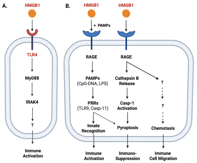
Once released, extracellular HMGB1 can bind various PRRs and PAMPs to orchestrate divergent inflammatory responses. For instance, HMGB1 can bind TLR4 [58,59,60], TLR9 [61], receptor for advanced glycation end products (RAGE) [62], cluster of differentiation 24 (CD24)/Siglec-10 [63], Mac-1 [64], or single-transmembrane-domain proteins (e.g., syndecans) [65]. Due to its relatively higher affinity to TLR4 (KD = 22.0 nM) [66] and lower affinity to RAGE (KD = 97.7-710 nM) [67,68], HMGB1 might first bind TLR4 when it was actively secreted by innate immune cells at relatively lower amounts [69]. Consequently, it could directly activate macrophages [70], neutrophils [71] and endothelial cells [72] to produce various cytokines and chemokines [58,72,73,74,75,76] partly through MyD88-IRAK4-dependent signaling pathways (Figure 2A).
Figure 2.
Role of TLR4 and RAGE in the regulation of HMGB1-mediated divergent inflammatory responses. HMGB1 can bind different PRRs such as TLR4 (Panel A) and RAGE (Panel B) with different affinities, and consequently induce divergent inflammatory responses such as immune cell migration, immune activation, or pyroptosis and resultant immunosuppression.
When HMGB1 was passively released by innate immune and somatic cells at relatively higher levels, it might also bind various microbial PAMPs (e.g., CpG-DNA or LPS) and RAGE [67,77] and consequently promoted RAGE-receptor-mediated endocytosis of these microbial products (Figure 2B) [78]. Upon reaching acidic endosomal and lysosomal compartments near HMGB1′s isoelectric pH, HMGB1 became neutrally charged and set free its cargos (LPS or CpG-DNA) [78], thereby facilitating their recognition by respective PRRs such as TLR9 [61] or Casp-11 [78] to augment inflammatory responses (Figure 2B). Furthermore, the engagement of RAGE with HMGB1 might also induce chemotaxis [79] and the migration of monocytes, dendritic cells [80,81] and neutrophils [64], thereby facilitating the recruitment of innate immune cells to site of the infection to orchestrate inflammatory responses [79] (Figure 2B). Finally, the engagement of HMGB1 with RAGE [67,77] might also induce TLR4 internalization and desensitization to subsequent stimulus (e.g., endotoxin), and might even trigger macrophage pyroptosis [78,82] via a cascade of events including cathepsin B release from ruptured lysosomes followed by pyroptosome formation and Casp-1 activation (Figure 2B).
In neutrophils, HMGB1 can bind TLR4 to promote the formation of neutrophil extracellular traps (NETs), thereby amplifying neutrophil-mediated inflammatory responses [83]. In contrast, the engagement of RAGE by HMGB1 can adversely impair neutrophil NADPH-dependent production of reactive oxidation species (ROS) and associated bacterial killing, contributing to sepsis-induced immune paralysis and immuno-suppression [84,85]. Consistently, the blockade of extracellular HMGB1 activities with neutralizing antibodies even during a late stage of sepsis still restored neutrophil NADPH activity and anti-bacterial capacities [85]. Thus, excessive HMGB1 release contributes to the pathogenesis of lethal infections by posing divergent adverse effects such as immune tolerance [86,87], immune paralysis [84,85,88] and immunosuppression [85,89] (Figure 2B).
5. Positive Regulators of LPS-Induced HMGB1 Release
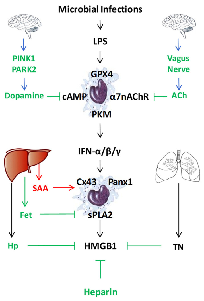
In addition to LPS-inducible type I IFN-α/β [37,38] and type II IFN-γ [28], human serum amyloid A (SAA) also effectively induced HMGB1 release by innate immune cells in a TLR4/RAGE-dependent fashion [90] (Figure 3). Consistent with its capacity in stimulating NLRP3 inflammasome activation [91,92], we observed that SAA also stimulated PKR expression and phosphorylation [90]. Conversely, pharmacological inhibition of PKR inhibited SAA-induced HMGB1 release [90], supporting an important role for PKR phosphorylation, inflammasome activation and pyroptosis in the SAA-induced HMGB1 release (Figure 1). In addition, some LPS-inducible enzymes [such as the 14 kDa type II secretory phospholipase A2 (sPLA2), inducible nitric oxide synthase (iNOS), and pyruvate kinase M2 (PKM2)] were also implicated in the regulation of LPS-induced HMGB1 release (Figure 3) [29,93,94,95,96]. In agreement with these findings, we found that human SAA effectively up-regulated the expression of sPLA2-IIE and sPLA2-V in murine macrophages (Figure 1 and Figure 3) [97], and concurrently induced HMGB1 release [90]. Conversely, the suppression of sPLA2-IIE expression by high density lipoproteins (HDL) also attenuated SAA-induced HMGB1 release, supporting a role of sPLA2 in the regulation of HMGB1 release [97]. It is not yet known whether sPLA2s facilitate HMGB1 release partly by catalyzing the production of lyso-phosphatidylcholine (LPC) and leukotrienes that are capable of activating NLRP3 inflammasome and pyroptosis (Figure 1) [98,99,100].
Figure 3.
Endogenous regulators of LPS-induced HMGB1 release or action. To regulate the LPS-induced HMGB1 release or action, mammals have evolved multiple regulatory mechanisms that include neuro-immune pathways, liver-derived acute-phase proteins (e.g., SAA, Fetuin-A (Fet), Haptoglobin (Hp)), as well as other endogenous proteins (e.g., tetranectin (TN)) or polysaccharides (heparin).
Finally, both crude LPS and human SAA effectively up-regulated the expression of hemichannel molecules such as Panx1 [41] and Connexin 43 (Cx43) [101] in innate immune cells (Figure 1 and Figure 3). The possible role of Cx43 in the regulation of LPS-induced HMGB1 release was supported by our findings that several Cx43 mimetic peptides, the GAP26 and Peptide 5 (ENVCYD), simultaneously attenuated LPS-induced hemichannel activation and HMGB1 release [101]. It was further supported by observation that genetic disruption of macrophage-specific Cx43 expression conferred protection against lethal endotoxemia and sepsis [102]. It is possible that Cx43 hemichannel provides a temporal mode of ATP release [103,104], which then contributes to the LPS-stimulated PKR phosphorylation, inflammasome activation, pyroptosis and HMGB1 secretion (Figure 1 and Figure 3) [41,101]. Intriguingly, recent evidence has suggested that macrophages also form Cx43-containing gap junction with non-immune cells such as cardiomyocytes [105], epithelial [106,107] and endothelial cells [108]. It is possible that innate immune cells may communicate with non-immune cells through Cx43-containing gap junction channels to regulate HMGB1 release and to orchestrate inflammatory responses [109,110]. Interestingly, recent studies have revealed an important role of lipid peroxidation [111] and cAMP immune-metabolism [112] in the regulation of Casp-11-mediated “non-canonical” inflammasome activation and pyroptosis (Figure 3). However, the possible role of these immunometabolism pathways in the regulation of LPS-induced HMGB1 release remains an exciting subject of future investigations.
6. Negative Regulators of the LPS-Induced HMGB1 Release and Action
During evolution, mammals have evolved multiple negative regulatory mechanisms to inhibit HMGB1 release and action. For instance, a local feedback mechanism could be instilled by injured cells via the release of a ubiquitous biogenic molecule, spermine, which inhibited the LPS- and HMGB1-induced release of multiple cytokines and chemokines (e.g., TNF, IL-6, MIP-2, and RANTES) from macrophages and monocytes [70,113,114,115]. Notably, spermine exerted its anti-inflammatory effect in conjunction with a liver-derived negative acute-phase protein, fetuin-A (Figure 3), which served as an opsonin for the cellular uptake of cationic anti-inflammatory molecules such as spermine [116]. In an animal model of lethal endotoxemia, circulating fetuin-A levels were decreased in an anti-parallel fashion when circulating HMGB1 levels were elevated [9,117]. However, supplementation of endotoxemic animals with exogenous fetuin-A resulted in a significant reduction in circulating HMGB1 levels [117]. It is plausible that fetuin-A negatively regulated LPS-induced HMGB1 release partly by facilitating the cellular uptake of cationic anti-inflammatory molecules (spermine), and partly by stimulating macrophages-mediated ingestion and elimination of apoptotic neutrophils [118,119]. This is relevant, because inefficient elimination of apoptotic cells might adversely lead to excessive accumulation of late apoptotic and/or secondary necrotic cells, which may cause passive leakage of HMGB1 and other DAMPs [120].
In addition, recent evidence suggested that the central nervous system could also attenuate peripheral innate immune response through efferent vagus nerve (Figure 3) [121], which could release neurotransmitter such as acetylcholine to inactivate macrophages via nicotinic cholinergic receptors [122]. Indeed, stimulation of the vagus nerve by physical methods (e.g., electrical or mechanical) [123,124] or chemical agonists (such as nicotine and GTS-21) [125,126] conferred protection against lethal endotoxemia partly by attenuating systemic HMGB1 accumulation. Furthermore, mammals have also evolved other neuro-immune pathways by which the PTEN-induced putative kinase 1 (PINK1) and parkin RBR E3 ubiquitin protein ligase (PARK2) counter-regulate lethal systemic inflammation through another neurotransmitter, dopamine (Figure 3) [127], which could turn off systemic inflammation through suppressing NLRP3 inflammasome activation [128].
Finally, emerging evidence has supported a possible role of several endogenous proteins such as thrombomodulin [129], haptoglobin [130], complement factor 1q (C1q) [131], heat shock protein 70 (HSP70) [132,133], vasoactive intestinal peptide [134], urocortin [135], and ghrelin [136] in the regulation of LPS-induced HMGB1 release or cytokine activities. For instance, an endothelial anticoagulant cofactor, thrombomodulin, could bind HMGB1 to prevent its interaction with macrophage cell-surface receptors [137], thereby preventing HMGB1-induced inflammatory responses [129,138]. Similarly, a liver-derived acute-phase protein, haptoglobin (Hp, Figure 3), could capture HMGB1 to trigger CD163-dependent endocytosis of HMGB1/Hp complexes, and induced the production of anti-inflammatory enzymes (heme oxygenase-1) and cytokines (e.g., IL-10) [69,130]. In addition, a component factor 1q (C1q) capable of binding antigen-antibody complexes to initiate the classical complement pathway [15], also interacted with HMGB1 (KD = 200 nM) and formed a tetramolecular complex with RAGE and LAIR-1, resulting in the production of anti-inflammatory cytokines (e.g., IL-10) and pro-resolution lipid mediators [131,139]. Similarly, an anticoagulant polysaccharide, heparin, or other chemically modified (2-O, 3-O desulfated) heparins, could all bind HMGB1 and prevented its interaction with LPS [140] or RAGE receptor [67], thereby inhibiting Casp-11-mediated inflammasome activation and pyroptosis, as well as HMGB1-mediated immunosuppression [141]. Thus, in sharp contrast to exogenous PAMPs (e.g., CpG-DNA and LPS), many endogenous proteins and polysaccharides could bind HMGB1 to tilt the balance towards anti-inflammatory responses via distinct signaling pathways [130,131,137,139,140].
7. Pharmacological Modulation of LPS-Induced HMGB1 Release or Action
Our seminal discovery of HMGB1 as a late mediator of lethal endotoxemia has stimulated extensive interest in search for HMGB1-targeting pharmacological inhibitors ranging from small molecules to large biological agents.
7.1. Small-Molecule Inhibitors
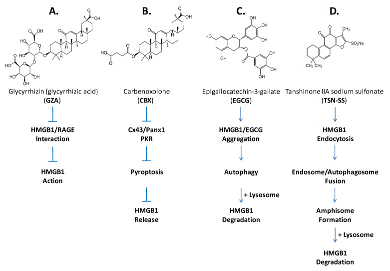
Among many medicinal herbs that we screened for possible HMGB1-inhibiting activities, we found that aqueous extracts of Danggui (Angelica sinensis) [142], Gancao (Radix glycyrrhizae) [143], Green tea (Camellia sinensis) [144], and Danshen (Saliva miltorrhiza) [145] conferred significant protection against lethal endotoxemia partly by inhibiting LPS-induced HMGB1 release via dramatically distinct mechanisms. For instance, a major component of Gancao, glycyrrhizin (GZA), could directly bind HMGB1 [146] to disrupt its engagement with RAGE receptor [147], thereby conferring protection against lethal endotoxemia by inhibiting HMGB1-mediated inflammation (Figure 4) [148]. A chemical derivative of the GZA, carbenoxolone (CBX), however, dose-dependently inhibited the LPS-induced HMGB1 secretion [143] partly by inhibiting the LPS-induced PKR expression and phosphorylation (Figure 4). Given its capacity in inhibiting macrophage Cx43 and Panx1 hemichannel activities [149,150], CBX could also counter-regulate HMGB1 release through inhibiting LPS-induced activation of Cx43 or Panx1 hemichannels in innate immune cells (Figure 4) [143].
Figure 4.
Distinct mechanisms of several pharmacological inhibitors of HMGB1 release or action. Different herbal compounds or derivatives inhibit HMGB1 release or action through distinct mechanisms that include: (A) direct binding to inhibit its engagement with various PRRs; (B) direct binding to induce its aggregation and autophagic degradation; (C) inhibition of key signaling molecules (PKR and hemichannels) involved in inflammasome activation and pyroptosis; and (D) induction of its endocytosis and lysosome-dependent degradation.
A major green tea component, EGCG, prevented the LPS-induced HMGB1 release strategically by destroying it in the cytoplasm via a cellular degradation process—autophagy (Figure 4) [151]. Specifically, EGCG could be trafficked into cytosol to conjugate with cytoplasmic HMGB1 either covalently with the free thiol group of cysteine residues [152] or non-covalently via hydrogen bonding, aromatic stacking or hydrophobic interactions [153]. Consequently, EGCG induced the formation of EGCG–HMGB1 complexes that were eventually engulfed by double-membraned autophagosomes, and subsequently degraded by acidic lysosomal hydrolases [151].
In contrast, a derivative of major ingredient of Danshen, tanshinone IIA sodium sulfonate (TSN-SS, Figure 4), selectively inhibited LPS-induced HMGB1 release without affecting the secretion of other cytokines and chemokines (such as IL-6, IL-12p40/p70, KC, MCP-1, MIP-1α, MIP-2, and TNF) [145]. Unlike EGCG, TSN-SS itself was unable to stimulate autophagic HMGB1 degradation, but instead facilitated the endocytosis of extracellular HMGB1 through clathrin- and caveolin-dependent endocytosis (Figure 4) [154]. Because cytoplasmic HMGB1 could induce autophagy [155,156,157], the TSN-SS-mediated HMGB1 endocytosis may be paralleled with the occurrence of HMGB1-induced autophagy, and eventually converged on a lysosome-dependent final common pathway that eventually leads to HMGB1 degradation (Figure 4). That is, the HMGB1-containing endosomes might fuse with HMGB1-induced autophagosomes to form amphisomes [158,159], and then merge with lysosomes to trigger HMGB1 degradation via a lysosome-dependent pathway (Figure 4) [154]. Given its demonstrated safety in China as a medicine for patients with cardiovascular disorders, and its capacity to inhibit HMGB1 release after LPS stimulation, TSN-SS may be a promising therapeutic agent for inhibiting HMGB1 release in clinical settings [27].
7.2. Development of Antibodies Targeting HMGB1 or Its in-Crime Binding Partners
Neutralizing antibodies against endotoxin [160] or early cytokines (e.g., TNF) [161,162] were protective in an animal model of lethal endotoxemia, but unfortunately failed in clinical trials [163,164,165]. This failure partly reflects the complexity of the underlying lethal infections, and the associated heterogeneity of the patient populations [166]. In contrast to the relatively unified age, body weight, and genetic background of experimental animals in pre-clinical studies, patients recruited in clinical studies usually exhibit intrinsic (genetic and epigenetic) heterogeneity, and harbor various underlying comorbidities that complicate the pathogenesis and progression of clinical sepsis. Nevertheless, the investigation of pathogenic cytokines (such as TNF) has led to the development of anti-TNF therapy for patients with debilitating chronic inflammatory diseases, such as rheumatoid arthritis [167]. Accordingly, we have generated polyclonal and monoclonal antibodies against human HMGB1 and tested their efficacy in animal models of lethal endotoxemia and sepsis induced by a surgical procedure termed cecal ligation and puncture (CLP). In an animal model of lethal endotoxemia, HMGB1-specific polyclonal antibodies were protective in a dose-dependent fashion [9]. In an animal model of CLP-induced sepsis, HMGB1-neutralizing monoclonal antibodies (mAbs) [44,168] conferred significant protection even when the first dose was given 24 h after disease onset [23,169,170,171], establishing HMGB1 as a “late” mediator of experimental sepsis with a relatively wider therapeutic window than that offered by early proinflammatory cytokines.
As aforementioned, many endogenous proteins (such as thrombomodulin, haptoglobin, and C1q) [129,130,131] could physically interact with HMGB1, promoting the search for other HMGB1-binding proteins that might also affect its biological functions. During this process, we noticed that the blood level of a 20 kDa protein was almost completely depleted in patients who died of sepsis. This 20 kDa protein was identified as human tetranectin (TN) by mass spectrometry and immunoblotting assays [172]. Intriguingly, TN selectively inhibited the LPS- and SAA-induced HMGB1 release without affecting the parallel release of other cytokines and chemokines [172], partly because TN could capture extracellular HMGB1 and facilitated the endocytosis of TN/HMGB1 complexes, thereby enhancing HMGB1-induced pyroptosis (Figure 5) [172].
Figure 5.
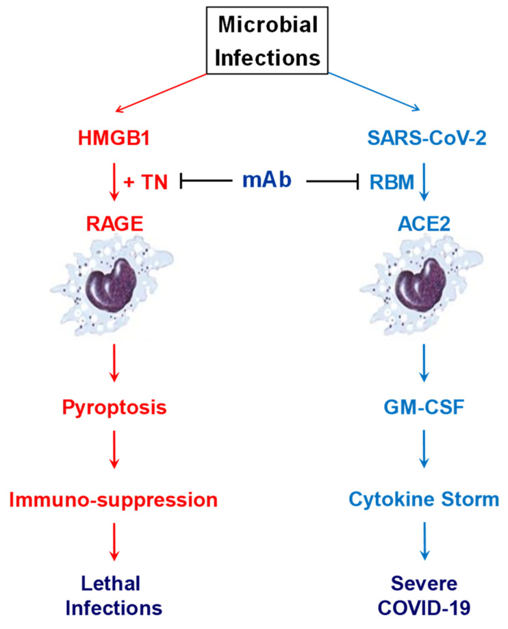
Potential therapeutic effects of cross-reactive monoclonal antibodies against TN and RBM of SARS-CoV-2. Some TN domain (NDALYEYLRQ)-specific mAbs may confer protection against lethal infections partly by preventing harmful TN/HMGB1 interaction that may adversely trigger macrophage pyroptosis and immunosuppression. Some TN-reactive mAbs also cross-reacted with a tyrosine (Y)-rich segment (YNYLYR) in the RBM of SARS-CoV-2, and specifically inhibited RBM-induced production of GM-CSF, a biomarker and mediator of COVID-19. The dual effects of these cross-reactive mAbs in attenuating immuno-suppression and SARS-CoV-2-induced GM-CSF production make them promising therapeutic candidates for treating COVID-19 and other lethal infections.
As discussed earlier, pyroptosis not only allows excessive release of HMGB1 and SQSTM1 that adversely drive a life-threatening dysregulated inflammatory response to lethal infections, but also leads to immune cell depletion and possible immunosuppression that may have compromised the host innate immunity against lethal infections (Figure 5). Accordingly, we have developed a panel of TN-specific mAbs that effectively prevented both harmful HMGB1/TN interaction and resultant macrophage pyroptosis and lethal sepsis [172]. It suggested that TN domain-specific mAbs may confer protection against lethal sepsis partly by preventing harmful TN/HMGB1 interaction that may adversely trigger macrophage pyroptosis and immunosuppression (Figure 5) [173,174]. This antibody strategy has also suggested a possibility to develop therapeutic antibodies against harmless proteins colluding with sepsis mediators [173,174,175].
Surprisingly, we recently discovered that two TN-reactive mAbs capable of rescuing mice from lethal sepsis also cross-reacted with the human ACE2 receptor binding motif (RBM) of SARS-CoV-2 (Figure 5), with an estimated KD of 17.4 and 62.8 nM, respectively [176]. The estimated KD was comparable to that of other SARS-CoV-2 RBD-binding neutralizing antibodies (KD = 14–17 nM) derived from COVID-19 patients [177]. Furthermore, these TN/RBM-reactive mAbs competitively inhibited RBM-ACE2 interactions in vitro [176], and selectively impaired the RBM-induced secretion of the granulocyte macrophage colony-stimulating factor (GM-CSF) [176]. Our findings fully supported the emerging notion that GM-CSF might be a key biomarker for SARS-CoV-2-induced cytokine storm in a subset of COVID-19 patients with more severe pneumonia often escalating to respiratory failure and death [178,179,180,181,182] (Figure 5), although the possible roles of GM-CSF and other cytokines in the pathogenesis COVID-19 remain a subject of ongoing debate [56,57]. Nevertheless, it is possible that these TN/RBM-reactive mAbs might selectively prevent its interaction with a receptor involved in the GM-CSF induction, but did not interfere with its engagement with other pattern recognition receptors responsible for the induction of other cytokines or chemokines [183]. Because HMGB1 was similarly accumulated in patients with COVID-19 [184], these TN/RBM-reactive mAbs might simultaneously block harmful TN/HMGB1 interaction and resultant immunosuppression, and suppress possible SARS-CoV-2/ACE2 interaction to inhibit GM-CSF production in patients with COVID-19 and other lethal infections [71].
8. Future Perspectives
Microbial infections and sepsis remain a major clinical problem that accounts for approximately 20% of total deaths worldwide [1], and annually costs more than $62 billion in the U.S. alone [185]. Despite a robust increase in the understanding of the pathophysiology of sepsis, many antibody-based strategies targeting early cytokines (such as TNF or IL-1) failed in clinical settings. Currently, there is still no effective therapy [173] other than adjunctive use of antibiotics, fluid resuscitation, and supportive care [185]. Thus, it would be beneficial to test the therapeutic efficacy of some promising HMGB1 inhibitors in clinical settings. For instance, a selective HMGB1 inhibitor, TSN-SS, has already been used in China as a medicine for patients with cardiovascular disorders. The dual effects of TSN-SS in attenuating late inflammatory response and improving cardiovascular functions make it a promising therapeutic candidate for treating lethal infections. Similarly, it would also be exciting to test the therapeutic efficacy of TN-specific mAbs that effectively prevented its undesired interaction with pathogenic mediators (HMGB1) and resultant immunosuppression [172]. The discovery of mAbs capable of disrupting TN/HMGB1 interaction and endocytosis and rescuing animals from lethal sepsis has suggested an exciting possibility to develop therapeutic antibodies against harmless proteins colluding with disease mediators [175]. Given the cross-reactivity of several TN-reactive monoclonal antibodies to the RBM of SARS-CoV-2, it will be extremely important to test the efficacy of these TN/RBM-reactive monoclonal antibodies in clinical trials of COVID-19 and other microbial infections.
Author Contributions
All authors have made significant contributions to the work reported, including the conception and interpretation of relevant literature. C.S.Z. and W.W. generated the first drafts incorporated into this review manuscript. X.Q., W.C., X.L. and J.L. provided important contributions. H.W. made significant revisions to the text and summary figures, and finalized the manuscript. All authors have read and agreed to the published version of the manuscript.
Funding
The cited publications from Haichao Wang’s laboratory were supported by the US National Institutes of Health (NIH) grants R01GM063075 and R01AT005076.
Institutional Review Board Statement
Not applicable.
Informed Consent Statement
Not applicable.
Conflicts of Interest
H.W. is a co-inventor of two relevant patent applications entitled “Inhibition of inflammatory cytokine production with tanshinones” and “Hemichannel extracellular-domain specific agents for treating sepsis”. H.W., W.C., and J.L. are co-inventors of a patent application entitled “Tetranectin-targeting monoclonal antibodies to fight against lethal sepsis and other pathologies”. H.W., S.Z., X.Q. and J.L. are co-inventors of a patent application entitled “Use of SARS-CoV-2 receptor binding motif (RBM)-reactive monoclonal antibodies to treat COVID-19”. All other authors declare that they have no competing interests.
Footnotes
Publisher’s Note: MDPI stays neutral with regard to jurisdictional claims in published maps and institutional affiliations.
References
- 1.Rudd K.E., Johnson S.C., Agesa K.M., Shackelford K.A., Tsoi D., Kievlan D.R., Colombara D.V., Ikuta K.S., Kissoon N., Finfer S., et al. Global, regional, and national sepsis incidence and mortality, 1990-2017: Analysis for the Global Burden of Disease Study. Lancet. 2020;395:200–211. doi: 10.1016/S0140-6736(19)32989-7. [DOI] [PMC free article] [PubMed] [Google Scholar]
- 2.Hotchkiss R.S., Coopersmith C.M., McDunn J.E., Ferguson T.A. The sepsis seesaw: Tilting toward immunosuppression. Nat. Med. 2009;15:496–497. doi: 10.1038/nm0509-496. [DOI] [PMC free article] [PubMed] [Google Scholar]
- 3.Tang D., Wang H., Billiar T.R., Kroemer G., Kang R. Emerging mechanisms of immunocoagulation in sepsis and septic shock. Trends. Immunol. 2021;42:508–522. doi: 10.1016/j.it.2021.04.001. [DOI] [PMC free article] [PubMed] [Google Scholar]
- 4.Luster A.D., Alon R., von Andrian U.H. Immune cell migration in inflammation: Present and future therapeutic targets. Nat. Immunol. 2005;6:1182–1190. doi: 10.1038/ni1275. [DOI] [PubMed] [Google Scholar]
- 5.Liles W.C. Immunomodulatory approaches to augment phagocyte-mediated host defense for treatment of infectious diseases. Semin. Respir. Infect. 2001;16:11–17. doi: 10.1053/srin.2001.22724. [DOI] [PubMed] [Google Scholar]
- 6.Brightbill H.D., Libraty D.H., Krutzik S.R., Yang R.B., Belisle J.T., Bleharski J.R., Maitland M., Norgard M.V., Plevy S.E., Smale S.T., et al. Host defense mechanisms triggered by microbial lipoproteins through toll-like receptors. Science. 1999;285:732–736. doi: 10.1126/science.285.5428.732. [DOI] [PubMed] [Google Scholar]
- 7.Poltorak A., He X., Smirnova I., Liu M.Y., Huffel C.V., Du X., Birdwell D., Alejos E., Silva M., Galanos C., et al. Defective LPS signaling in C3H/HeJ and C57BL/10ScCr mice: Mutations in Tlr4 gene. Science. 1998;282:2085–2088. doi: 10.1126/science.282.5396.2085. [DOI] [PubMed] [Google Scholar]
- 8.Hemmi H., Takeuchi O., Kawai T., Kaisho T., Sato S., Sanjo H., Matsumoto M., Hoshino K., Wagner H., Takeda K., et al. A Toll-like receptor recognizes bacterial DNA. Nature. 2000;408:740–745. doi: 10.1038/35047123. [DOI] [PubMed] [Google Scholar]
- 9.Wang H., Bloom O., Zhang M., Vishnubhakat J.M., Ombrellino M., Che J., Frazier A., Yang H., Ivanova S., Borovikova L., et al. HMG-1 as a late mediator of endotoxin lethality in mice. Science. 1999;285:248–251. doi: 10.1126/science.285.5425.248. [DOI] [PubMed] [Google Scholar]
- 10.Zhou B., Liu J., Zeng L., Zhu S., Wang H., Billiar T.R., Kroemer G., Klionsky D.J., Zeh H.J., Jiang J., et al. Extracellular SQSTM1 mediates bacterial septic death in mice through insulin receptor signalling. Nat. Microbiol. 2020;5:1576–1587. doi: 10.1038/s41564-020-00795-7. [DOI] [PMC free article] [PubMed] [Google Scholar]
- 11.Bonaldi T., Talamo F., Scaffidi P., Ferrera D., Porto A., Bachi A., Rubartelli A., Agresti A., Bianchi M.E. Monocytic cells hyperacetylate chromatin protein HMGB1 to redirect it towards secretion. EMBO J. 2003;22:5551–5560. doi: 10.1093/emboj/cdg516. [DOI] [PMC free article] [PubMed] [Google Scholar]
- 12.Bustin M. At the crossroads of necrosis and apoptosis: Signaling to multiple cellular targets by HMGB1. Sci. STKE. 2002;2002:E39. doi: 10.1126/stke.2002.151.pe39. [DOI] [PubMed] [Google Scholar]
- 13.Kang R., Chen R., Zhang Q., Hou W., Wu S., Cao L., Huang J., Yu Y., Fan X.G., Yan Z., et al. HMGB1 in health and disease. Mol. Aspects. Med. 2014;40:1–116. doi: 10.1016/j.mam.2014.05.001. [DOI] [PMC free article] [PubMed] [Google Scholar]
- 14.Ge Y., Huang M., Yao Y.M. The Effect and Regulatory Mechanism of High Mobility Group Box-1 Protein on Immune Cells in Inflammatory Diseases. Cells. 2021;10:1044. doi: 10.3390/cells10051044. [DOI] [PMC free article] [PubMed] [Google Scholar]
- 15.Watanabe H., Son M. The Immune Tolerance Role of the HMGB1-RAGE Axis. Cells. 2021;10:564. doi: 10.3390/cells10030564. [DOI] [PMC free article] [PubMed] [Google Scholar]
- 16.Hailman E., Lichenstein H.S., Wurfel M.M., Miller D.S., Johnson D.A., Kelley M., Busse L.A., Zukowski M.M., Wright S.D. Lipopolysaccharide (LPS)-binding protein accelerates the binding of LPS to CD14. J. Exp. Med. 1994;179:269–277. doi: 10.1084/jem.179.1.269. [DOI] [PMC free article] [PubMed] [Google Scholar]
- 17.Wright S.D., Ramos R.A., Tobias P.S., Ulevitch R.J., Mathison J.C. CD14, a receptor for complexes of lipopolysaccharide (LPS) and LPS binding protein. Science. 1990;249:1431–1433. doi: 10.1126/science.1698311. [DOI] [PubMed] [Google Scholar]
- 18.Frey E.A., Miller D.S., Jahr T.G., Sundan A., Bazil V., Espevik T., Finlay B.B., Wright S.D. Soluble CD14 participates in the response of cells to lipopolysaccharide. J. Exp. Med. 1992;176:1665–1671. doi: 10.1084/jem.176.6.1665. [DOI] [PMC free article] [PubMed] [Google Scholar]
- 19.Ferrero E., Jiao D., Tsuberi B.Z., Tesio L., Rong G.W., Haziot A., Goyert S.M. Transgenic mice expressing human CD14 are hypersensitive to lipopolysaccharide. Proc. Natl. Acad. Sci. USA. 1993;90:2380–2384. doi: 10.1073/pnas.90.6.2380. [DOI] [PMC free article] [PubMed] [Google Scholar]
- 20.Haziot A., Ferrero E., Kontgen F., Hijiya N., Yamamoto S., Silver J., Stewart C.L., Goyert S.M. Resistance to endotoxin shock and reduced dissemination of gram-negative bacteria in CD14-deficient mice. Immunity. 1996;4:407–414. doi: 10.1016/S1074-7613(00)80254-X. [DOI] [PubMed] [Google Scholar]
- 21.Tsan M.F., Clark R.N., Goyert S.M., White J.E. Induction of TNF-alpha and MnSOD by endotoxin: Role of membrane CD14 and Toll-like receptor-4. Am. J. Physiol. Cell. Physiol. 2001;280:C1422–C1430. doi: 10.1152/ajpcell.2001.280.6.C1422. [DOI] [PubMed] [Google Scholar]
- 22.Chen G., Li J., Ochani M., Rendon-Mitchell B., Qiang X., Susarla S., Ulloa L., Yang H., Fan S., Goyert S.M., et al. Bacterial endotoxin stimulates macrophages to release HMGB1 partly through CD14- and TNF-dependent mechanisms. J. Leukoc. Biol. 2004;76:994–1001. doi: 10.1189/jlb.0404242. [DOI] [PubMed] [Google Scholar]
- 23.Wang H., Yang H., Czura C.J., Sama A.E., Tracey K.J. HMGB1 as a Late Mediator of Lethal Systemic Inflammation. Am. J. Respir. Crit. Care. Med. 2001;164:1768–1773. doi: 10.1164/ajrccm.164.10.2106117. [DOI] [PubMed] [Google Scholar]
- 24.Lu B., Antoine D.J., Kwan K., Lundback P., Wahamaa H., Schierbeck H., Robinson M., van Zoelen M.A., Yang H., Li J., et al. JAK/STAT1 signaling promotes HMGB1 hyperacetylation and nuclear translocation. Proc. Natl. Acad. Sci. USA. 2014;111:3068–3073. doi: 10.1073/pnas.1316925111. [DOI] [PMC free article] [PubMed] [Google Scholar]
- 25.Youn J.H., Shin J.S. Nucleocytoplasmic shuttling of HMGB1 is regulated by phosphorylation that redirects it toward secretion. J. Immunol. 2006;177:7889–7897. doi: 10.4049/jimmunol.177.11.7889. [DOI] [PubMed] [Google Scholar]
- 26.Lu B., Wang C., Wang M., Li W., Chen F., Tracey K.J., Wang H. Molecular mechanism and therapeutic modulation of high mobility group box 1 release and action: An updated review. Expert. Rev. Clin. Immunol. 2014;10:713–727. doi: 10.1586/1744666X.2014.909730. [DOI] [PMC free article] [PubMed] [Google Scholar]
- 27.Wang H., Ward M.F., Sama A.E. Targeting HMGB1 in the treatment of sepsis. Expert. Opin. Ther. Targets. 2014;18:257–268. doi: 10.1517/14728222.2014.863876. [DOI] [PMC free article] [PubMed] [Google Scholar]
- 28.Rendon-Mitchell B., Ochani M., Li J., Han J., Wang H., Yang H., Susarla S., Czura C., Mitchell R.A., Chen G., et al. IFN-gamma Induces High Mobility Group Box 1 Protein Release Partly Through a TNF-Dependent Mechanism. J. Immunol. 2003;170:3890–3897. doi: 10.4049/jimmunol.170.7.3890. [DOI] [PubMed] [Google Scholar]
- 29.Gardella S., Andrei C., Ferrera D., Lotti L.V., Torrisi M.R., Bianchi M.E., Rubartelli A. The nuclear protein HMGB1 is secreted by monocytes via a non-classical, vesicle-mediated secretory pathway. EMBO. Rep. 2002;3:955–1001. doi: 10.1093/embo-reports/kvf198. [DOI] [PMC free article] [PubMed] [Google Scholar]
- 30.Lamkanfi M., Sarkar A., Vande W.L., Vitari A.C., Amer A.O., Wewers M.D., Tracey K.J., Kanneganti T.D., Dixit V.M. Inflammasome-dependent release of the alarmin HMGB1 in endotoxemia. J. Immunol. 2010;185:4385–4392. doi: 10.4049/jimmunol.1000803. [DOI] [PMC free article] [PubMed] [Google Scholar]
- 31.Lu B., Nakamura T., Inouye K., Li J., Tang Y., Lundback P., Valdes-Ferrer S.I., Olofsson P.S., Kalb T., Roth J., et al. Novel role of PKR in inflammasome activation and HMGB1 release. Nature. 2012;488:670–674. doi: 10.1038/nature11290. [DOI] [PMC free article] [PubMed] [Google Scholar]
- 32.Vanaja S.K., Russo A.J., Behl B., Banerjee I., Yankova M., Deshmukh S.D., Rathinam V.A.K. Bacterial Outer Membrane Vesicles Mediate Cytosolic Localization of LPS and Caspase-11 Activation. Cell. 2016;165:1106–1119. doi: 10.1016/j.cell.2016.04.015. [DOI] [PMC free article] [PubMed] [Google Scholar]
- 33.Hagar J.A., Powell D.A., Aachoui Y., Ernst R.K., Miao E.A. Cytoplasmic LPS activates caspase-11: Implications in TLR4-independent endotoxic shock. Science. 2013;341:1250–1253. doi: 10.1126/science.1240988. [DOI] [PMC free article] [PubMed] [Google Scholar]
- 34.Rathinam V.A., Vanaja S.K., Waggoner L., Sokolovska A., Becker C., Stuart L.M., Leong J.M., Fitzgerald K.A. TRIF licenses caspase-11-dependent NLRP3 inflammasome activation by gram-negative bacteria. Cell. 2012;150:606–619. doi: 10.1016/j.cell.2012.07.007. [DOI] [PMC free article] [PubMed] [Google Scholar]
- 35.Aachoui Y., Kajiwara Y., Leaf I.A., Mao D., Ting J.P., Coers J., Aderem A., Buxbaum J.D., Miao E.A. Canonical Inflammasomes Drive IFN-γ to Prime Caspase-11 in Defense against a Cytosol-Invasive Bacterium. Cell. Host. Microbe. 2015;18:320–332. doi: 10.1016/j.chom.2015.07.016. [DOI] [PMC free article] [PubMed] [Google Scholar]
- 36.Meunier E., Dick M.S., Dreier R.F., SchÃrmann N., Kenzelmann B.D., Warming S., Roose-Girma M., Bumann D., Kayagaki N., Takeda K., et al. Caspase-11 activation requires lysis of pathogen-containing vacuoles by IFN-induced GTPases. Nature. 2014;509:366–370. doi: 10.1038/nature13157. [DOI] [PubMed] [Google Scholar]
- 37.Kim J.H., Kim S.J., Lee I.S., Lee M.S., Uematsu S., Akira S., Oh K.I. Bacterial endotoxin induces the release of high mobility group box 1 via the IFN-beta signaling pathway. J. Immunol. 2009;182:2458–2466. doi: 10.4049/jimmunol.0801364. [DOI] [PubMed] [Google Scholar]
- 38.Yang X., Cheng X., Tang Y., Qiu X., Wang Z., Fu G., Wu J., Kang H., Wang J., Wang H., et al. The role of type 1 interferons in coagulation induced by gram-negative bacteria. Blood. 2020;135:1087–1100. doi: 10.1182/blood.2019002282. [DOI] [PMC free article] [PubMed] [Google Scholar]
- 39.Yang D., He Y., Munoz-Planillo R., Liu Q., Nunez G. Caspase-11 Requires the Pannexin-1 Channel and the Purinergic P2X7 Pore to Mediate Pyroptosis and Endotoxic Shock. Immunity. 2015;43:923–932. doi: 10.1016/j.immuni.2015.10.009. [DOI] [PMC free article] [PubMed] [Google Scholar]
- 40.Chiu Y.H., Jin X., Medina C.B., Leonhardt S.A., Kiessling V., Bennett B.C., Shu S., Tamm L.K., Yeager M., Ravichandran K.S., et al. A quantized mechanism for activation of pannexin channels. Nat. Commun. 2017;8:14324. doi: 10.1038/ncomms14324. [DOI] [PMC free article] [PubMed] [Google Scholar]
- 41.Chen W., Zhu S., Wang Y., Li J., Qiang X., Zhao X., Yang H., D’Angelo J., Becker L., Wang P., et al. Enhanced Macrophage Pannexin 1 Expression and Hemichannel Activation Exacerbates Lethal Experimental Sepsis. Sci. Rep. 2019;9:160–37232. doi: 10.1038/s41598-018-37232-z. [DOI] [PMC free article] [PubMed] [Google Scholar]
- 42.Volchuk A., Ye A., Chi L., Steinberg B.E., Goldenberg N.M. Indirect regulation of HMGB1 release by gasdermin D. Nat. Commun. 2020;11:4561–18443. doi: 10.1038/s41467-020-18443-3. [DOI] [PMC free article] [PubMed] [Google Scholar]
- 43.Phulphagar K., Kühn L.I., Ebner S., Frauenstein A., Swietlik J.J., Rieckmann J., Meissner F. Proteomics reveals distinct mechanisms regulating the release of cytokines and alarmins during pyroptosis. Cell. Rep. 2021;34:108826. doi: 10.1016/j.celrep.2021.108826. [DOI] [PubMed] [Google Scholar]
- 44.Qin S., Wang H., Yuan R., Li H., Ochani M., Ochani K., Rosas-Ballina M., Czura C.J., Huston J.M., Miller E., et al. Role of HMGB1 in apoptosis-mediated sepsis lethality. J. Exp. Med. 2006;203:1637–1642. doi: 10.1084/jem.20052203. [DOI] [PMC free article] [PubMed] [Google Scholar]
- 45.Hett E.C., Slater L.H., Mark K.G., Kawate T., Monks B.G., Stutz A., Latz E., Hung D.T. Chemical genetics reveals a kinase-independent role for protein kinase R in pyroptosis. Nat. Chem. Biol. 2013;9:398–405. doi: 10.1038/nchembio.1236. [DOI] [PMC free article] [PubMed] [Google Scholar]
- 46.Surprenant A., Rassendren F., Kawashima E., North R.A., Buell G. The cytolytic P2Z receptor for extracellular ATP identified as a P2X receptor (P2X7) Science. 1996;272:735–738. doi: 10.1126/science.272.5262.735. [DOI] [PubMed] [Google Scholar]
- 47.Cohen M.J., Brohi K., Calfee C.S., Rahn P., Chesebro B.B., Christiaans S.C., Carles M., Howard M., Pittet J.F. Early release of high mobility group box nuclear protein 1 after severe trauma in humans: Role of injury severity and tissue hypoperfusion. Crit. Care. 2009;13:R174. doi: 10.1186/cc8152. [DOI] [PMC free article] [PubMed] [Google Scholar]
- 48.Peltz E.D., Moore E.E., Eckels P.C., Damle S.S., Tsuruta Y., Johnson J.L., Sauaia A., Silliman C.C., Banerjee A., Abraham E. HMGB1 is markedly elevated within 6 hours of mechanical trauma in humans. Shock. 2009;32:17–22. doi: 10.1097/SHK.0b013e3181997173. [DOI] [PMC free article] [PubMed] [Google Scholar]
- 49.Huang L.F., Yao Y.M., Dong N., Yu Y., He L.X., Sheng Z.Y. Association of high mobility group box-1 protein levels with sepsis and outcome of severely burned patients. Cytokine. 2011;53:29–34. doi: 10.1016/j.cyto.2010.09.010. [DOI] [PubMed] [Google Scholar]
- 50.Zhang T., Yin C., Boyd D.F., Quarato G., Ingram J.P., Shubina M., Ragan K.B., Ishizuka T., Crawford J.C., Tummers B., et al. Influenza Virus Z-RNAs Induce ZBP1-Mediated Necroptosis. Cell. 2020;180:1115–1129. doi: 10.1016/j.cell.2020.02.050. [DOI] [PMC free article] [PubMed] [Google Scholar]
- 51.Nogusa S., Thapa R.J., Dillon C.P., Liedmann S., Oguin T.H., III, Ingram J.P., Rodriguez D.A., Kosoff R., Sharma S., Sturm O., et al. RIPK3 Activates Parallel Pathways of MLKL-Driven Necroptosis and FADD-Mediated Apoptosis to Protect against Influenza A Virus. Cell Host. Microbe. 2016;20:13–24. doi: 10.1016/j.chom.2016.05.011. [DOI] [PMC free article] [PubMed] [Google Scholar]
- 52.Karki R., Sharma B.R., Tuladhar S., Williams E.P., Zalduondo L., Samir P., Zheng M., Sundaram B., Banoth B., Malireddi R.K.S., et al. Synergism of TNF-α and IFN-γ Triggers Inflammatory Cell Death, Tissue Damage, and Mortality in SARS-CoV-2 Infection and Cytokine Shock Syndromes. Cell. 2021;184:149–168. doi: 10.1016/j.cell.2020.11.025. [DOI] [PMC free article] [PubMed] [Google Scholar]
- 53.Cho Y.S., Challa S., Moquin D., Genga R., Ray T.D., Guildford M., Chan F.K. Phosphorylation-driven assembly of the RIP1-RIP3 complex regulates programmed necrosis and virus-induced inflammation. Cell. 2009;137:1112–1123. doi: 10.1016/j.cell.2009.05.037. [DOI] [PMC free article] [PubMed] [Google Scholar]
- 54.Thapa R.J., Nogusa S., Chen P., Maki J.L., Lerro A., Andrake M., Rall G.F., Degterev A., Balachandran S. Interferon-induced RIP1/RIP3-mediated necrosis requires PKR and is licensed by FADD and caspases. Proc. Natl. Acad. Sci. USA. 2013;110:E3109–E3118. doi: 10.1073/pnas.1301218110. [DOI] [PMC free article] [PubMed] [Google Scholar]
- 55.Gunther C., Martini E., Wittkopf N., Amann K., Weigmann B., Neumann H., Waldner M.J., Hedrick S.M., Tenzer S., Neurath M.F., et al. Caspase-8 regulates TNF-alpha-induced epithelial necroptosis and terminal ileitis. Nature. 2011;477:335–339. doi: 10.1038/nature10400. [DOI] [PMC free article] [PubMed] [Google Scholar]
- 56.Mehta P., Fajgenbaum D.C. Is severe COVID-19 a cytokine storm syndrome: A hyperinflammatory debate. Curr. Opin. Rheumatol. 2021;33:419–430. doi: 10.1097/BOR.0000000000000822. [DOI] [PMC free article] [PubMed] [Google Scholar]
- 57.Sinha P., Matthay M.A., Calfee C.S. Is a “Cytokine Storm” Relevant to COVID-19? JAMA. Intern. Med. 2020;180:1152–1154. doi: 10.1001/jamainternmed.2020.3313. [DOI] [PubMed] [Google Scholar]
- 58.Yu M., Wang H., Ding A., Golenbock D.T., Latz E., Czura C.J., Fenton M.J., Tracey K.J., Yang H. HMGB1 signals through Toll-like Receptor (TLR) 4 and TLR2. Shock. 2006;26:174–179. doi: 10.1097/01.shk.0000225404.51320.82. [DOI] [PubMed] [Google Scholar]
- 59.Ha T., Xia Y., Liu X., Lu C., Liu L., Kelley J., Kalbfleisch J., Kao R.L., Williams D.L., Li C. Glucan phosphate attenuates myocardial HMGB1 translocation in severe sepsis through inhibiting NF-kappaB activation. Am. J. Physiol Heart Circ. Physiol. 2011;301:H848–H855. doi: 10.1152/ajpheart.01007.2010. [DOI] [PMC free article] [PubMed] [Google Scholar]
- 60.Xiang M., Shi X., Li Y., Xu J., Yin L., Xiao G., Scott M.J., Billiar T.R., Wilson M.A., Fan J. Hemorrhagic shock activation of NLRP3 inflammasome in lung endothelial cells. J. Immunol. 2011;187:4809–4817. doi: 10.4049/jimmunol.1102093. [DOI] [PMC free article] [PubMed] [Google Scholar]
- 61.Ivanov S., Dragoi A.M., Wang X., Dallacosta C., Louten J., Musco G., Sitia G., Yap G.S., Wan Y., Biron C.A., et al. A novel role for HMGB1 in TLR9-mediated inflammatory responses to CpG-DNA. Blood. 2007;110:1970–1981. doi: 10.1182/blood-2006-09-044776. [DOI] [PMC free article] [PubMed] [Google Scholar]
- 62.Tian J., Avalos A.M., Mao S.Y., Chen B., Senthil K., Wu H., Parroche P., Drabic S., Golenbock D., Sirois C., et al. Toll-like receptor 9-dependent activation by DNA-containing immune complexes is mediated by HMGB1 and RAGE. Nat. Immunol. 2007;8:487–496. doi: 10.1038/ni1457. [DOI] [PubMed] [Google Scholar]
- 63.Chen G.Y., Tang J., Zheng P., Liu Y. CD24 and Siglec-10 selectively repress tissue damage-induced immune responses. Science. 2009;323:1722–1725. doi: 10.1126/science.1168988. [DOI] [PMC free article] [PubMed] [Google Scholar]
- 64.Orlova V.V., Choi E.Y., Xie C., Chavakis E., Bierhaus A., Ihanus E., Ballantyne C.M., Gahmberg C.G., Bianchi M.E., Nawroth P.P., et al. A novel pathway of HMGB1-mediated inflammatory cell recruitment that requires Mac-1-integrin. EMBO J. 2007;26:1129–1139. doi: 10.1038/sj.emboj.7601552. [DOI] [PMC free article] [PubMed] [Google Scholar]
- 65.Salmivirta M., Rauvala H., Elenius K., Jalkanen M. Neurite growth-promoting protein (amphoterin, p30) binds syndecan. Exp. Cell. Res. 1992;200:444–451. doi: 10.1016/0014-4827(92)90194-D. [DOI] [PubMed] [Google Scholar]
- 66.Yang H., Hreggvidsdottir H.S., Palmblad K., Wang H., Ochani M., Li J., Lu B., Chavan S., Rosas-Ballina M., Al Abed Y., et al. A critical cysteine is required for HMGB1 binding to Toll-like receptor 4 and activation of macrophage cytokine release. Proc. Natl. Acad. Sci. USA. 2010;107:11942–11947. doi: 10.1073/pnas.1003893107. [DOI] [PMC free article] [PubMed] [Google Scholar]
- 67.Ling Y., Yang Z.Y., Yin T., Li L., Yuan W.W., Wu H.S., Wang C.Y. Heparin changes the conformation of high-mobility group protein 1 and decreases its affinity toward receptor for advanced glycation endproducts in vitro. Int. Immunopharmacol. 2011;11:187–193. doi: 10.1016/j.intimp.2010.11.014. [DOI] [PubMed] [Google Scholar]
- 68.Liu R., Mori S., Wake H., Zhang J., Liu K., Izushi Y., Takahashi H.K., Peng B., Nishibori M. Establishment of in vitro binding assay of high mobility group box-1 and S100A12 to receptor for advanced glycation endproducts: Heparin’s effect on binding. Acta Med. Okayama. 2009;63:203–211. doi: 10.18926/AMO/31812. [DOI] [PubMed] [Google Scholar]
- 69.Yang H., Wang H., Andersson U. Targeting Inflammation Driven by HMGB1. Front Immunol. 2020;20:484. doi: 10.3389/fimmu.2020.00484. [DOI] [PMC free article] [PubMed] [Google Scholar]
- 70.Zhu S., Ashok M., Li J., Li W., Yang H., Wang P., Tracey K.J., Sama A.E., Wang H. Spermine protects mice against lethal sepsis partly by attenuating surrogate inflammatory markers. Mol. Med. 2009;15:275–282. doi: 10.2119/molmed.2009.00062. [DOI] [PMC free article] [PubMed] [Google Scholar]
- 71.Fan J., Li Y., Levy R.M., Fan J.J., Hackam D.J., Vodovotz Y., Yang H., Tracey K.J., Billiar T.R., Wilson M.A. Hemorrhagic shock induces NAD(P)H oxidase activation in neutrophils: Role of HMGB1-TLR4 signaling. J. Immunol. 2007;178:6573–6580. doi: 10.4049/jimmunol.178.10.6573. [DOI] [PubMed] [Google Scholar]
- 72.Fiuza C., Bustin M., Talwar S., Tropea M., Gerstenberger E., Shelhamer J.H., Suffredini A.F. Inflammation-promoting activity of HMGB1 on human microvascular endothelial cells. Blood. 2003;101:2652–2660. doi: 10.1182/blood-2002-05-1300. [DOI] [PubMed] [Google Scholar]
- 73.Park J.S., Svetkauskaite D., He Q., Kim J.Y., Strassheim D., Ishizaka A., Abraham E. Involvement of TLR 2 and TLR 4 in cellular activation by high mobility group box 1 protein (HMGB1) J. Biol. Chem. 2004;279:7370–7377. doi: 10.1074/jbc.M306793200. [DOI] [PubMed] [Google Scholar]
- 74.Kokkola R., Andersson A., Mullins G., Ostberg T., Treutiger C.J., Arnold B., Nawroth P., Andersson U., Harris R.A., Harris H.E. RAGE is the Major Receptor for the Proinflammatory Activity of HMGB1 in Rodent Macrophages. Scand. J. Immunol. 2005;61:1–9. doi: 10.1111/j.0300-9475.2005.01534.x. [DOI] [PubMed] [Google Scholar]
- 75.Treutiger C.J., Mullins G.E., Johansson A.S., Rouhiainen A., Rauvala H.M., Erlandsson-Harris H., Andersson U., Yang H., Tracey K.J., Andersson J., et al. High mobility group 1 B-box mediates activation of human endothelium. J. Intern. Med. 2003;254:375–385. doi: 10.1046/j.1365-2796.2003.01204.x. [DOI] [PubMed] [Google Scholar]
- 76.Lv B., Wang H., Tang Y., Fan Z., Xiao X., Chen F. High-mobility group box 1 protein induces tissue factor expression in vascular endothelial cells via activation of NF-kappaB and Egr-1. Thromb. Haemost. 2009;102:352–359. doi: 10.1160/TH08-11-0759. [DOI] [PMC free article] [PubMed] [Google Scholar]
- 77.Hori O., Brett J., Slattery T., Cao R., Zhang J., Chen J.X., Nagashima M., Lundh E.R., Vijay S., Nitecki D. The receptor for advanced glycation end products (RAGE) is a cellular binding site for amphoterin. Mediation of neurite outgrowth and co-expression of rage and amphoterin in the developing nervous system. J. Biol. Chem. 1995;270:25752–25761. doi: 10.1074/jbc.270.43.25752. [DOI] [PubMed] [Google Scholar]
- 78.Deng M., Tang Y., Li W., Wang X., Zhang R., Zhang X., Zhao X., Liu J., Tang C., Liu Z., et al. The Endotoxin Delivery Protein HMGB1 Mediates Caspase-11-Dependent Lethality in Sepsis. Immunity. 2018;49:740–753. doi: 10.1016/j.immuni.2018.08.016. [DOI] [PMC free article] [PubMed] [Google Scholar]
- 79.Degryse B., Bonaldi T., Scaffidi P., Muller S., Resnati M., Sanvito F., Arrigoni G., Bianchi M.E. The high mobility group (HMG) boxes of the nuclear protein HMG1 induce chemotaxis and cytoskeleton reorganization in rat smooth muscle cells. J. Cell. Biol. 2001;152:1197–1206. doi: 10.1083/jcb.152.6.1197. [DOI] [PMC free article] [PubMed] [Google Scholar]
- 80.Yang D., Chen Q., Yang H., Tracey K.J., Bustin M., Oppenheim J.J. High mobility group box-1 protein induces the migration and activation of human dendritic cells and acts as an alarmin. J. Leukoc. Biol. 2007;81:59–66. doi: 10.1189/jlb.0306180. [DOI] [PubMed] [Google Scholar]
- 81.Dumitriu I.E., Bianchi M.E., Bacci M., Manfredi A.A., Rovere-Querini P. The secretion of HMGB1 is required for the migration of maturing dendritic cells. J. Leukoc. Biol. 2007;81:84–91. doi: 10.1189/jlb.0306171. [DOI] [PubMed] [Google Scholar]
- 82.Xu J., Jiang Y., Wang J., Shi X., Liu Q., Liu Z., Li Y., Scott M.J., Xiao G., Li S., et al. Macrophage endocytosis of high-mobility group box 1 triggers pyroptosis. Cell. Death. Differ. 2014;21:1229–1239. doi: 10.1038/cdd.2014.40. [DOI] [PMC free article] [PubMed] [Google Scholar]
- 83.Tadie J.M., Bae H.B., Jiang S., Park D.W., Bell C.P., Yang H., Pittet J.F., Tracey K., Thannickal V.J., Abraham E., et al. HMGB1 promotes neutrophil extracellular trap formation through interactions with Toll-like receptor 4. Am. J. Physiol. Lung. Cell. Mol. Physiol. 2013;304:L342–L349. doi: 10.1152/ajplung.00151.2012. [DOI] [PMC free article] [PubMed] [Google Scholar]
- 84.Tadié J.M., Bae H.B., Banerjee S., Zmijewski J.W., Abraham E. Differential activation of RAGE by HMGB1 modulates neutrophil-associated NADPH oxidase activity and bacterial killing. Am. J. Physiol. Cell. Physiol. 2012;302:C249–C256. doi: 10.1152/ajpcell.00302.2011. [DOI] [PMC free article] [PubMed] [Google Scholar]
- 85.Gregoire M., Tadie J.M., Uhel F., Gacouin A., Piau C., Bone N., Le T.Y., Abraham E., Tarte K., Zmijewski J.W. Frontline Science: HMGB1 induces neutrophil dysfunction in experimental sepsis and in patients who survive septic shock. J. Leukoc. Biol. 2017;101:1281–1287. doi: 10.1189/jlb.5HI0316-128RR. [DOI] [PMC free article] [PubMed] [Google Scholar]
- 86.Robert S.M., Sjodin H., Fink M.P., Aneja R.K. Preconditioning with high mobility group box 1 (HMGB1) induces lipoteichoic acid (LTA) tolerance. J. Immunother. 2010;33:663–671. doi: 10.1097/CJI.0b013e3181dcd111. [DOI] [PMC free article] [PubMed] [Google Scholar]
- 87.Aneja R.K., Tsung A., Sjodin H., Gefter J.V., Delude R.L., Billiar T.R., Fink M.P. Preconditioning with high mobility group box 1 (HMGB1) induces lipopolysaccharide (LPS) tolerance. J. Leukoc. Biol. 2008;84:1326–1334. doi: 10.1189/jlb.0108030. [DOI] [PMC free article] [PubMed] [Google Scholar]
- 88.Patel V.S., Sitapara R.A., Gore A., Phan B., Sharma L., Sampat V., Li J., Yang H., Chavan S.S., Wang H., et al. HMGB1 Mediates Hyperoxia-Induced Impairment of Pseudomonas aeruginosa Clearance and Inflammatory Lung Injury in Mice. Am. J. Respir. Cell Mol. Biol. 2012;48:280–287. doi: 10.1165/rcmb.2012-0279OC. [DOI] [PMC free article] [PubMed] [Google Scholar]
- 89.Wild C.A., Bergmann C., Fritz G., Schuler P., Hoffmann T.K., Lotfi R., Westendorf A., Brandau S., Lang S. HMGB1 conveys immunosuppressive characteristics on regulatory and conventional T cells. Int. Immunol. 2012;24:485–494. doi: 10.1093/intimm/dxs051. [DOI] [PubMed] [Google Scholar]
- 90.Li W., Zhu S., Li J., D’Amore J., D’Angelo J., Yang H., Wang P., Tracey K.J., Wang H. Serum Amyloid A Stimulates PKR Expression and HMGB1 Release Possibly through TLR4/RAGE Receptors. Mol. Med. 2015;21:515–525. doi: 10.2119/molmed.2015.00109. [DOI] [PMC free article] [PubMed] [Google Scholar]
- 91.Niemi K., Teirila L., Lappalainen J., Rajamaki K., Baumann M.H., Oorni K., Wolff H., Kovanen P.T., Matikainen S., Eklund K.K. Serum amyloid A activates the NLRP3 inflammasome via P2X7 receptor and a cathepsin B-sensitive pathway. J. Immunol. 2011;186:6119–6128. doi: 10.4049/jimmunol.1002843. [DOI] [PubMed] [Google Scholar]
- 92.Ather J.L., Ckless K., Martin R., Foley K.L., Suratt B.T., Boyson J.E., Fitzgerald K.A., Flavell R.A., Eisenbarth S.C., Poynter M.E. Serum amyloid A activates the NLRP3 inflammasome and promotes Th17 allergic asthma in mice. J. Immunol. 2011;187:64–73. doi: 10.4049/jimmunol.1100500. [DOI] [PMC free article] [PubMed] [Google Scholar]
- 93.Chen G., Li J., Qiang X., Czura C.J., Ochani M., Ochani K., Ulloa L., Yang H., Tracey K.J., Wang P., et al. Suppression of HMGB1 release by stearoyl lysophosphatidylcholine:an additional mechanism for its therapeutic effects in experimental sepsis. J. Lipid. Res. 2005;46:623–627. doi: 10.1194/jlr.C400018-JLR200. [DOI] [PubMed] [Google Scholar]
- 94.Jiang W., Pisetsky D.S. The Role of IFN-{alpha} and Nitric Oxide in the Release of HMGB1 by RAW 264.7 Cells Stimulated with Polyinosinic-Polycytidylic Acid or Lipopolysaccharide. J. Immunol. 2006;177:3337–3343. doi: 10.4049/jimmunol.177.5.3337. [DOI] [PubMed] [Google Scholar]
- 95.Xie M., Yu Y., Kang R., Zhu S., Yang L., Zeng L., Sun X., Yang M., Billiar T.R., Wang H., et al. PKM2-dependent glycolysis promotes NLRP3 and AIM2 inflammasome activation. Nat. Commun. 2016;7:13280. doi: 10.1038/ncomms13280. [DOI] [PMC free article] [PubMed] [Google Scholar]
- 96.Yang L., Xie M., Yang M., Yu Y., Zhu S., Hou W., Kang R., Lotze M.T., Billiar T.R., Wang H., et al. PKM2 regulates the Warburg effect and promotes HMGB1 release in sepsis. Nat. Commun. 2014;5:4436. doi: 10.1038/ncomms5436. [DOI] [PMC free article] [PubMed] [Google Scholar]
- 97.Zhu S., Wang Y., Chen W., Li W., Wang A., Wong S., Bao G., Li J., Yang H., Tracey K.J., et al. High-Density Lipoprotein (HDL) Counter-Regulates Serum Amyloid A (SAA)-Induced sPLA2-IIE and sPLA2-V Expression in Macrophages. PLoS ONE. 2016;11:e0167468. doi: 10.1371/journal.pone.0167468. [DOI] [PMC free article] [PubMed] [Google Scholar]
- 98.Freeman L., Guo H., David C.N., Brickey W.J., Jha S., Ting J.P. NLR members NLRC4 and NLRP3 mediate sterile inflammasome activation in microglia and astrocytes. J. Exp. Med. 2017;214:1351–1370. doi: 10.1084/jem.20150237. [DOI] [PMC free article] [PubMed] [Google Scholar]
- 99.Salina A.C.G., Brandt S.L., Klopfenstein N., Blackman A., Bazzano J.M.R., Sá-Nunes A., Byers-Glosson N., Brodskyn C., Tavares N.M., Da Silva I.B.S., et al. Leukotriene B(4) licenses inflammasome activation to enhance skin host defense. Proc. Natl. Acad. Sci. USA. 2020;117:30619–30627. doi: 10.1073/pnas.2002732117. [DOI] [PMC free article] [PubMed] [Google Scholar]
- 100.Chaves M.M., Sinflorio D.A., Thorstenberg M.L., Martins M.D.A., Moreira-Souza A.C.A., Rangel T.P., Silva C.L.M., Bellio M., Canetti C., Coutinho-Silva R. Non-canonical NLRP3 inflammasome activation and IL-1β signaling are necessary to L. amazonensis control mediated by P2X7 receptor and leukotriene B4. PLoS. Pathog. 2019;15:e1007887. doi: 10.1371/journal.ppat.1007887. [DOI] [PMC free article] [PubMed] [Google Scholar]
- 101.Li W., Bao G., Chen W., Qiang X., Zhu S., Wang S., He M., Ma G., Ochani M., Al-Abed Y., et al. Connexin 43 Hemichannel as a Novel Mediator of Sterile and Infectious Inflammatory Diseases. Sci. Rep. 2018;8:166–18452. doi: 10.1038/s41598-017-18452-1. [DOI] [PMC free article] [PubMed] [Google Scholar]
- 102.Dosch M., Zindel J., Jebbawi F., Melin N., Sanchez-Taltavull D., Stroka D., Candinas D., Beldi G. Connexin-43-dependent ATP release mediates macrophage activation during sepsis. Elife. 2019;8:e42670. doi: 10.7554/eLife.42670. [DOI] [PMC free article] [PubMed] [Google Scholar]
- 103.Beyer E.C., Steinberg T.H. Evidence that the gap junction protein connexin-43 is the ATP-induced pore of mouse macrophages. J. Biol. Chem. 1991;266:7971–7974. doi: 10.1016/S0021-9258(18)92924-8. [DOI] [PubMed] [Google Scholar]
- 104.Kang J., Kang N., Lovatt D., Torres A., Zhao Z., Lin J., Nedergaard M. Connexin 43 hemichannels are permeable to ATP. J. Neurosci. 2008;28:4702–4711. doi: 10.1523/JNEUROSCI.5048-07.2008. [DOI] [PMC free article] [PubMed] [Google Scholar]
- 105.Hulsmans M., Clauss S., Xiao L., Aguirre A.D., King K.R., Hanley A., Hucker W.J., Wulfers E.M., Seemann G., Courties G., et al. Macrophages Facilitate Electrical Conduction in the Heart. Cell. 2017;169:510–522. doi: 10.1016/j.cell.2017.03.050. [DOI] [PMC free article] [PubMed] [Google Scholar]
- 106.Westphalen K., Gusarova G.A., Islam M.N., Subramanian M., Cohen T.S., Prince A.S., Bhattacharya J. Sessile alveolar macrophages communicate with alveolar epithelium to modulate immunity. Nature. 2014;506:503–506. doi: 10.1038/nature12902. [DOI] [PMC free article] [PubMed] [Google Scholar]
- 107.Al-Ghadban S., Kaissi S., Homaidan F.R., Naim H.Y., El-Sabban M.E. Cross-talk between intestinal epithelial cells and immune cells in inflammatory bowel disease. Sci. Rep. 2016;6:29783. doi: 10.1038/srep29783. [DOI] [PMC free article] [PubMed] [Google Scholar]
- 108.Jara P.I., Boric M.P., Saez J.C. Leukocytes express connexin 43 after activation with lipopolysaccharide and appear to form gap junctions with endothelial cells after ischemia-reperfusion. Proc. Natl. Acad. Sci. USA. 1995;92:7011–7015. doi: 10.1073/pnas.92.15.7011. [DOI] [PMC free article] [PubMed] [Google Scholar]
- 109.Wong C.W., Christen T., Kwak B.R. Connexins in leukocytes: Shuttling messages? Cardiovasc. Res. 2004;62:357–367. doi: 10.1016/j.cardiores.2003.12.015. [DOI] [PubMed] [Google Scholar]
- 110.Scheckenbach K.E., Crespin S., Kwak B.R., Chanson M. Connexin channel-dependent signaling pathways in inflammation. J. Vasc. Res. 2011;48:91–103. doi: 10.1159/000316942. [DOI] [PubMed] [Google Scholar]
- 111.Kang R., Zeng L., Zhu S., Xie Y., Liu J., Wen Q., Cao L., Xie M., Ran Q., Kroemer G., et al. Lipid Peroxidation Drives Gasdermin D-Mediated Pyroptosis in Lethal Polymicrobial Sepsis. Cell Host. Microbe. 2018;24:97–108. doi: 10.1016/j.chom.2018.05.009. [DOI] [PMC free article] [PubMed] [Google Scholar]
- 112.Chen R., Zeng L., Zhu S., Liu J., Zeh H.J., Kroemer G., Wang H., Billiar T.R., Jiang J., Tang D., et al. cAMP metabolism controls caspase-11 inflammasome activation and pyroptosis in sepsis. Sci. Adv. 2019;5:eaav5562. doi: 10.1126/sciadv.aav5562. [DOI] [PMC free article] [PubMed] [Google Scholar]
- 113.Zhang M., Caragine T., Wang H., Cohen P.S., Botchkina G., Soda K., Bianchi M., Ulrich P., Cerami A., Sherry B., et al. Spermine inhibits proinflammatory cytokine synthesis in human mononuclear cells: A counterregulatory mechanism that restrains the immune response. J. Exp. Med. 1997;185:1759–1768. doi: 10.1084/jem.185.10.1759. [DOI] [PMC free article] [PubMed] [Google Scholar]
- 114.Wang H., Zhang M., Bianchi M., Sherry B., Sama A., Tracey K.J. Fetuin (alpha2-HS-glycoprotein) opsonizes cationic macrophagedeactivating molecules. Proc. Natl. Acad. Sci. USA. 1998;95:14429–14434. doi: 10.1073/pnas.95.24.14429. [DOI] [PMC free article] [PubMed] [Google Scholar]
- 115.Zhang M., Wang H., Tracey K.J. Regulation of macrophage activation and inflammation by spermine: A new chapter in an old story. Crit. Care. Med. 2000;28:N60–N66. doi: 10.1097/00003246-200004001-00007. [DOI] [PubMed] [Google Scholar]
- 116.Wang H., Zhang M., Soda K., Sama A., Tracey K.J. Fetuin protects the fetus from TNF [letter] Lancet. 1997;350:861–862. doi: 10.1016/S0140-6736(05)62030-2. [DOI] [PubMed] [Google Scholar]
- 117.Li W., Zhu S., Li J., Huang Y., Zhou R., Fan X., Yang H., Gong X., Eissa N.T., Jahnen-Dechent W., et al. A hepatic protein, fetuin-A, occupies a protective role in lethal systemic inflammation. PLoS. ONE. 2011;6:e16945. doi: 10.1371/journal.pone.0016945. [DOI] [PMC free article] [PubMed] [Google Scholar]
- 118.Lord J.M. A physiological role for alpha2-HS glycoprotein: Stimulation of macrophage uptake of apoptotic cells. Clin. Sci. (Lond.) 2003;105:267–268. doi: 10.1042/CS20030177. [DOI] [PubMed] [Google Scholar]
- 119.Jersmann H.P., Dransfield I., Hart S.P. Fetuin/alpha2-HS glycoprotein enhances phagocytosis of apoptotic cells and macropinocytosis by human macrophages. Clin. Sci. (Lond.) 2003;105:273–278. doi: 10.1042/CS20030126. [DOI] [PubMed] [Google Scholar]
- 120.Bell C.W., Jiang W., Reich C.F., Pisetsky D.S. The Extracellular Release of HMGB1 during Apoptotic Cell Death. Am. J. Physiol. Cell. Physiol. 2006;291:C1318–C1325. doi: 10.1152/ajpcell.00616.2005. [DOI] [PubMed] [Google Scholar]
- 121.Borovikova L.V., Ivanova S., Zhang M., Yang H., Botchkina G.I., Watkins L.R., Wang H., Abumrad N., Eaton J.W., Tracey K.J. Vagus nerve stimulation attenuates the systemic inflammatory response to endotoxin. Nature. 2000;405:458–462. doi: 10.1038/35013070. [DOI] [PubMed] [Google Scholar]
- 122.Wang H., Yu M., Ochani M., Amella C.A., Tanovic M., Susarla S., Li J.H., Wang H., Yang H., Ulloa L., et al. Nicotinic acetylcholine receptor alpha7 subunit is an essential regulator of inflammation. Nature. 2003;421:384–388. doi: 10.1038/nature01339. [DOI] [PubMed] [Google Scholar]
- 123.Huston J.M., Ochani M., Rosas-Ballina M., Liao H., Ochani K., Pavlov V.A., Gallowitsch-Puerta M., Ashok M., Czura C.J., Foxwell B., et al. Splenectomy inactivates the cholinergic antiinflammatory pathway during lethal endotoxemia and polymicrobial sepsis. J. Exp. Med. 2006;203:1623–1628. doi: 10.1084/jem.20052362. [DOI] [PMC free article] [PubMed] [Google Scholar]
- 124.Huston J.M., Gallowitsch-Puerta M., Ochani M., Ochani K., Yuan R., Rosas-Ballina M., Ashok M., Goldstein R.S., Chavan S., Pavlov V.A., et al. Transcutaneous vagus nerve stimulation reduces serum high mobility group box 1 levels and improves survival in murine sepsis. Crit. Care. Med. 2007;35:2762–2768. doi: 10.1097/01.CCM.0000288102.15975.BA. [DOI] [PubMed] [Google Scholar]
- 125.Wang H., Liao H., Ochani M., Justiniani M., Lin X., Yang L., Al Abed Y., Wang H., Metz C., Miller E.J., et al. Cholinergic agonists inhibit HMGB1 release and improve survival in experimental sepsis. Nat. Med. 2004;10:1216–1221. doi: 10.1038/nm1124. [DOI] [PubMed] [Google Scholar]
- 126.Pavlov V.A., Ochani M., Yang L.H., Gallowitsch-Puerta M., Ochani K., Lin X., Levi J., Parrish W.R., Rosas-Ballina M., Czura C.J., et al. Selective alpha7-nicotinic acetylcholine receptor agonist GTS-21 improves survival in murine endotoxemia and severe sepsis. Crit. Care. Med. 2007;35:1139–1144. doi: 10.1097/01.CCM.0000259381.56526.96. [DOI] [PubMed] [Google Scholar]
- 127.Kang R., Zeng L., Xie Y., Yan Z., Zhou B., Cao L., Klionsky D.J., Tracey K.J., Li J., Wang H., et al. A novel PINK1- and PARK2-dependent protective neuroimmune pathway in lethal sepsis. Autophagy. 2016;12:2374–2385. doi: 10.1080/15548627.2016.1239678. [DOI] [PMC free article] [PubMed] [Google Scholar]
- 128.Yan Y., Jiang W., Liu L., Wang X., Ding C., Tian Z., Zhou R. Dopamine controls systemic inflammation through inhibition of NLRP3 inflammasome. Cell. 2015;160:62–73. doi: 10.1016/j.cell.2014.11.047. [DOI] [PubMed] [Google Scholar]
- 129.Abeyama K., Stern D.M., Ito Y., Kawahara K., Yoshimoto Y., Tanaka M., Uchimura T., Ida N., Yamazaki Y., Yamada S., et al. The N-terminal domain of thrombomodulin sequesters high-mobility group-B1 protein, a novel antiinflammatory mechanism. J. Clin. Invest. 2005;115:1267–1274. doi: 10.1172/JCI22782. [DOI] [PMC free article] [PubMed] [Google Scholar]
- 130.Yang H., Wang H., Levine Y.A., Gunasekaran M.K., Wang Y., Addorisio M., Zhu S., Li W., Li J., de Kleijn D.P., et al. Identification of CD163 as an antiinflammatory receptor for HMGB1-haptoglobin complexes. JCI. Insight. 2018;3:e126617. doi: 10.1172/jci.insight.126617. [DOI] [PMC free article] [PubMed] [Google Scholar]
- 131.Son M., Porat A., He M., Suurmond J., Santiago-Schwarz F., Andersson U., Coleman T.R., Volpe B.T., Tracey K.J., Al-Abed Y., et al. C1q and HMGB1 reciprocally regulate human macrophage polarization. Blood. 2016;128:2218–2228. doi: 10.1182/blood-2016-05-719757. [DOI] [PMC free article] [PubMed] [Google Scholar]
- 132.Tang D., Kang R., Xiao W., Jiang L., Liu M., Shi Y., Wang K., Wang H., Xiao X. Nuclear Heat Shock Protein 72 as a Negative Regulator of Oxidative Stress (Hydrogen Peroxide)-Induced HMGB1 Cytoplasmic Translocation and Release. J. Immunol. 2007;178:7376–7384. doi: 10.4049/jimmunol.178.11.7376. [DOI] [PMC free article] [PubMed] [Google Scholar]
- 133.Tang D., Kang R., Xiao W., Wang H., Calderwood S.K., Xiao X. The Anti-inflammatory Effects of Heat Shock Protein 72 Involve Inhibition of High-Mobility-Group Box 1 Release and Proinflammatory Function in Macrophages. J. Immunol. 2007;179:1236–1244. doi: 10.4049/jimmunol.179.2.1236. [DOI] [PMC free article] [PubMed] [Google Scholar]
- 134.Chorny A., Delgado M. Neuropeptides rescue mice from lethal sepsis by down-regulating secretion of the late-acting inflammatory mediator high mobility group box 1. Am. J. Pathol. 2008;172:1297–1307. doi: 10.2353/ajpath.2008.070969. [DOI] [PMC free article] [PubMed] [Google Scholar]
- 135.Chorny A., Anderson P., Gonzalez-Rey E., Delgado M. Ghrelin Protects against Experimental Sepsis by Inhibiting High-Mobility Group Box 1 Release and by Killing Bacteria. J. Immunol. 2008;180:8369–8377. doi: 10.4049/jimmunol.180.12.8369. [DOI] [PubMed] [Google Scholar]
- 136.Wu R., Dong W., Qiang X., Wang H., Blau S.A., Ravikumar T.S., Wang P. Orexigenic hormone ghrelin ameliorates gut barrier dysfunction in sepsis in rats. Crit. Care. Med. 2009;37:2421–2426. doi: 10.1097/CCM.0b013e3181a557a2. [DOI] [PMC free article] [PubMed] [Google Scholar]
- 137.Herzog C., Lorenz A., Gillmann H.J., Chowdhury A., Larmann J., Harendza T., Echtermeyer F., Muller M., Schmitz M., Stypmann J., et al. Thrombomodulin’s lectin-like domain reduces myocardial damage by interfering with HMGB1-mediated TLR2 signalling. Cardiovasc. Res. 2014;101:400–410. doi: 10.1093/cvr/cvt275. [DOI] [PubMed] [Google Scholar]
- 138.Takehara K., Murakami T., Kuwahara-Arai K., Iba T., Nagaoka I., Sakamoto K. Evaluation of the effect of recombinant thrombomodulin on a lipopolysaccharide-induced murine sepsis model. Exp. Ther. Med. 2017;13:2969–2974. doi: 10.3892/etm.2017.4308. [DOI] [PMC free article] [PubMed] [Google Scholar]
- 139.Liu T., Xiang A., Peng T., Doran A.C., Tracey K.J., Barnes B.J., Tabas I., Son M., Diamond B. HMGB1-C1q complexes regulate macrophage function by switching between leukotriene and specialized proresolving mediator biosynthesis. Proc. Natl. Acad. Sci. USA. 2019;116:23254–23263. doi: 10.1073/pnas.1907490116. [DOI] [PMC free article] [PubMed] [Google Scholar]
- 140.Tang Y., Wang X., Li Z., He Z., Yang X., Cheng X., Peng Y., Xue Q., Bai Y., Zhang R., et al. Heparin prevents caspase-11-dependent septic lethality independent of anticoagulant properties. Immunity. 2021;54:454–467. doi: 10.1016/j.immuni.2021.01.007. [DOI] [PubMed] [Google Scholar]
- 141.Wang M., Gauthier A.G., Kennedy T.P., Wang H., Velagapudi U.K., Talele T.T., Lin M., Wu J., Daley L., Yang X., et al. 2-O, 3-O desulfated heparin (ODSH) increases bacterial clearance and attenuates lung injury in cystic fibrosis by restoring HMGB1-compromised macrophage function. Mol. Med. 2021;27:79–00334. doi: 10.1186/s10020-021-00334-y. [DOI] [PMC free article] [PubMed] [Google Scholar]
- 142.Wang H., Li W., Li J., Rendon-Mitchell B., Ochani M., Ashok M., Yang L., Yang H., Tracey K.J., Wang P., et al. The aqueous extract of a popular herbal nutrient supplement, Angelica sinensis, protects mice against lethal endotoxemia and sepsis. J. Nutr. 2006;136:360–365. doi: 10.1093/jn/136.2.360. [DOI] [PMC free article] [PubMed] [Google Scholar]
- 143.Li W., Li J., Sama A.E., Wang H. Carbenoxolone Blocks Endotoxin-Induced Protein Kinase R (PKR) Activation and High Mobility Group Box 1 (HMGB1) Release. Mol. Med. 2013;19:203–211. doi: 10.2119/molmed.2013.00064. [DOI] [PMC free article] [PubMed] [Google Scholar]
- 144.Li W., Ashok M., Li J., Yang H., Sama A.E., Wang H. A Major Ingredient of Green Tea Rescues Mice from Lethal Sepsis Partly by Inhibiting HMGB1. PLoS ONE. 2007;2:e1153. doi: 10.1371/journal.pone.0001153. [DOI] [PMC free article] [PubMed] [Google Scholar]
- 145.Li W., Li J., Ashok M., Wu R., Chen D., Yang L., Yang H., Tracey K.J., Wang P., Sama A.E., et al. A cardiovascular drug rescues mice from lethal sepsis by selectively attenuating a late-acting proinflammatory mediator, high mobility group box 1. J. Immunol. 2007;178:3856–3864. doi: 10.4049/jimmunol.178.6.3856. [DOI] [PMC free article] [PubMed] [Google Scholar]
- 146.Mollica L., De Marchis F., Spitaleri A., Dallacosta C., Pennacchini D., Zamai M., Agresti A., Trisciuoglio L., Musco G., Bianchi M.E. Glycyrrhizin binds to high-mobility group box 1 protein and inhibits its cytokine activities. Chem. Biol. 2007;14:431–441. doi: 10.1016/j.chembiol.2007.03.007. [DOI] [PubMed] [Google Scholar]
- 147.Okuma Y., Liu K., Wake H., Liu R., Nishimura Y., Hui Z., Teshigawara K., Haruma J., Yamamoto Y., Yamamoto H., et al. Glycyrrhizin inhibits traumatic brain injury by reducing HMGB1-RAGE interaction. Neuropharmacology. 2014;85:18–26. doi: 10.1016/j.neuropharm.2014.05.007. [DOI] [PubMed] [Google Scholar]
- 148.Wang W., Zhao F., Fang Y., Li X., Shen L., Cao T., Zhu H. Glycyrrhizin protects against porcine endotoxemia through modulation of systemic inflammatory response. Crit Care. 2013;17:R44. doi: 10.1186/cc12558. [DOI] [PMC free article] [PubMed] [Google Scholar]
- 149.Ma W., Hui H., Pelegrin P., Surprenant A. Pharmacological characterization of pannexin-1 currents expressed in mammalian cells. J. Pharmacol. Exp. Ther. 2009;328:409–418. doi: 10.1124/jpet.108.146365. [DOI] [PMC free article] [PubMed] [Google Scholar]
- 150.Poornima V., Madhupriya M., Kootar S., Sujatha G., Kumar A., Bera A.K. P2X7 receptor-pannexin 1 hemichannel association: Effect of extracellular calcium on membrane permeabilization. J. Mol. Neurosci. 2012;46:585–594. doi: 10.1007/s12031-011-9646-8. [DOI] [PubMed] [Google Scholar]
- 151.Li W., Zhu S., Li J., Assa A., Jundoria A., Xu J., Fan S., Eissa N.T., Tracey K.J., Sama A.E., et al. EGCG stimulates autophagy and reduces cytoplasmic HMGB1 levels in endotoxin-stimulated macrophages. Biochem. Pharmacol. 2011;81:1152–1163. doi: 10.1016/j.bcp.2011.02.015. [DOI] [PMC free article] [PubMed] [Google Scholar]
- 152.Ishii T., Mori T., Tanaka T., Mizuno D., Yamaji R., Kumazawa S., Nakayama T., Akagawa M. Covalent modification of proteins by green tea polyphenol (-)-epigallocatechin-3-gallate through autoxidation. Free Radic. Biol. Med. 2008;45:1384–1394. doi: 10.1016/j.freeradbiomed.2008.07.023. [DOI] [PubMed] [Google Scholar]
- 153.Ehrnhoefer D.E., Bieschke J., Boeddrich A., Herbst M., Masino L., Lurz R., Engemann S., Pastore A., Wanker E.E. EGCG redirects amyloidogenic polypeptides into unstructured, off-pathway oligomers. Nat. Struct. Mol. Biol. 2008;15:558–566. doi: 10.1038/nsmb.1437. [DOI] [PubMed] [Google Scholar]
- 154.Zhang Y., Li W., Zhu S., Jundoria A., Li J., Yang H., Fan S., Wang P., Tracey K.J., Sama A.E., et al. Tanshinone IIA sodium sulfonate facilitates endocytic HMGB1 uptake. Biochem. Pharmacol. 2012;84:1492–1500. doi: 10.1016/j.bcp.2012.09.015. [DOI] [PMC free article] [PubMed] [Google Scholar]
- 155.Tang D., Kang R., Livesey K.M., Cheh C.W., Farkas A., Loughran P., Hoppe G., Bianchi M.E., Tracey K.J., Zeh H.J., III, et al. Endogenous HMGB1 regulates autophagy. J. Cell Biol. 2010;190:881–892. doi: 10.1083/jcb.200911078. [DOI] [PMC free article] [PubMed] [Google Scholar]
- 156.Kang R., Livesey K.M., Zeh H.J., Loze M.T., Tang D. HMGB1: A novel Beclin 1-binding protein active in autophagy. Autophagy. 2010;6:1209–1211. doi: 10.4161/auto.6.8.13651. [DOI] [PubMed] [Google Scholar]
- 157.Yanai H., Matsuda A., An J., Koshiba R., Nishio J., Negishi H., Ikushima H., Onoe T., Ohdan H., Yoshida N., et al. Conditional ablation of HMGB1 in mice reveals its protective function against endotoxemia and bacterial infection. Proc. Natl. Acad. Sci. USA. 2013;110:20699–20704. doi: 10.1073/pnas.1320808110. [DOI] [PMC free article] [PubMed] [Google Scholar]
- 158.Gordon P.B., Seglen P.O. Prelysosomal convergence of autophagic and endocytic pathways. Biochem. Biophys. Res. Commun. 1988;151:40–47. doi: 10.1016/0006-291X(88)90556-6. [DOI] [PubMed] [Google Scholar]
- 159.Klionsky D.J. Autophagy: From phenomenology to molecular understanding in less than a decade. Nat. Rev. Mol. Cell Biol. 2007;8:931–937. doi: 10.1038/nrm2245. [DOI] [PubMed] [Google Scholar]
- 160.Davis C.E., Brown K.R., Douglas H., Tate W.J., III, Braude A.I. Prevention of death from endotoxin with antisera. I. The risk of fatal anaphylaxis to endotoxin. J. Immunol. 1969;102:563–572. [PubMed] [Google Scholar]
- 161.Beutler B., Milsark I.W., Cerami A.C. Passive immunization against cachectin/tumor necrosis factor protects mice from lethal effect of endotoxin. Science. 1985;229:869–871. doi: 10.1126/science.3895437. [DOI] [PubMed] [Google Scholar]
- 162.Tracey K.J., Fong Y., Hesse D.G., Manogue K.R., Lee A.T., Kuo G.C., Lowry S.F., Cerami A. Anti-cachectin/TNF monoclonal antibodies prevent septic shock during lethal bacteraemia. Nature. 1987;330:662–664. doi: 10.1038/330662a0. [DOI] [PubMed] [Google Scholar]
- 163.Ziegler E.J., Fisher C.J., Jr., Sprung C.L., Straube R.C., Sadoff J.C., Foulke G.E., Wortel C.H., Fink M.P., Dellinger R.P., Teng N.N., et al. Treatment of gram-negative bacteremia and septic shock with HA-1A human monoclonal antibody against endotoxin. A randomized, double-blind, placebo-controlled trial. The HA-1A Sepsis Study Group. N. Engl. J. Med. 1991;324:429–436. doi: 10.1056/NEJM199102143240701. [DOI] [PubMed] [Google Scholar]
- 164.Ziegler E.J., McCutchan J.A., Fierer J., Glauser M.P., Sadoff J.C., Douglas H., Braude A.I. Treatment of gram-negative bacteremia and shock with human antiserum to a mutant Escherichia coli. N. Engl. J. Med. 1982;307:1225–1230. doi: 10.1056/NEJM198211113072001. [DOI] [PubMed] [Google Scholar]
- 165.Abraham E., Wunderink R., Silverman H., Perl T.M., Nasraway S., Levy H., Bone R., Wenzel R.P., Balk R., Allred R. Efficacy and safety of monoclonal antibody to human tumor necrosis factor alpha in patients with sepsis syndrome. A randomized, controlled, double-blind, multicenter clinical trial. TNF-alpha MAb Sepsis Study Group. JAMA. 1995;273:934–941. doi: 10.1001/jama.1995.03520360048038. [DOI] [PubMed] [Google Scholar]
- 166.Dellinger R.P., Levy M.M., Carlet J.M., Bion J., Parker M.M., Jaeschke R., Reinhart K., Angus D.C., Brun-Buisson C., Beale R., et al. Surviving Sepsis Campaign: International guidelines for management of severe sepsis and septic shock: 2008. Crit Care Med. 2008;36:296–327. doi: 10.1097/01.CCM.0000298158.12101.41. [DOI] [PubMed] [Google Scholar]
- 167.Feldmann M., Maini R.N. Anti-TNF alpha therapy of rheumatoid arthritis: What have we learned? Annu. Rev. Immunol. 2001;19:163–196. doi: 10.1146/annurev.immunol.19.1.163. [DOI] [PubMed] [Google Scholar]
- 168.Yang H., Ochani M., Li J., Qiang X., Tanovic M., Harris H.E., Susarla S.M., Ulloa L., Wang H., DiRaimo R., et al. Reversing established sepsis with antagonists of endogenous high-mobility group box 1. Proc. Natl. Acad. Sci. USA. 2004;101:296–301. doi: 10.1073/pnas.2434651100. [DOI] [PMC free article] [PubMed] [Google Scholar]
- 169.Wang H., Yang H., Tracey K.J. Extracellular role of HMGB1 in inflammation and sepsis. J Intern. Med. 2004;255:320–331. doi: 10.1111/j.1365-2796.2003.01302.x. [DOI] [PubMed] [Google Scholar]
- 170.Wang H., Zhu S., Zhou R., Li W., Sama A.E. Therapeutic potential of HMGB1-targeting agents in sepsis. Expert. Rev. Mol. Med. 2008;10:e32. doi: 10.1017/S1462399408000884. [DOI] [PMC free article] [PubMed] [Google Scholar]
- 171.Wang H., Ward M.F., Sama A.E. Novel HMGB1-inhibiting therapeutic agents for experimental sepsis. Shock. 2009;32:348–357. doi: 10.1097/SHK.0b013e3181a551bd. [DOI] [PMC free article] [PubMed] [Google Scholar]
- 172.Chen W., Qiang X., Wang Y., Zhu S., Li J., Babaev A., Yang H., Gong J., Becker L., Wang P., et al. Identification of tetranectin-targeting monoclonal antibodies to treat potentially lethal sepsis. Sci. Transl. Med. 2020;12:12–539. doi: 10.1126/scitranslmed.aaz3833. [DOI] [PMC free article] [PubMed] [Google Scholar]
- 173.Paterson C.W., Ford M.L., Coopersmith C.M. Breaking the bond between tetranectin and HMGB1 in sepsis. Sci. Transl. Med. 2020;12:12–539. doi: 10.1126/scitranslmed.abb2575. [DOI] [PMC free article] [PubMed] [Google Scholar]
- 174.Crunkhorn S. Antibody intervention rescues mice from sepsis. Nat. Rev. Drug Discov. 2020;19:385–386. doi: 10.1038/d41573-020-00077-1. [DOI] [PubMed] [Google Scholar]
- 175.Li J., Bao G., Wang H. Time to Develop Therapeutic Antibodies Against Harmless Proteins Colluding with Sepsis Mediators? Immunotargets. Ther. 2020;9:157–166. doi: 10.2147/ITT.S262605. [DOI] [PMC free article] [PubMed] [Google Scholar]
- 176.Qiang X., Zhu S., Li J., Chen W., Yang H., Wang P., Tracey K.J., Wang H. Monoclonal antibodies capable of binding SARS-CoV-2 spike protein receptor-binding motif specifically prevent GM-CSF induction. J. Leukoc. Biol. 2021:10-628RR. doi: 10.1002/JLB.3COVCRA0920-628RR. [DOI] [PMC free article] [PubMed] [Google Scholar]
- 177.Rogers T.F., Zhao F., Huang D., Beutler N., Burns A., He W.T., Limbo O., Smith C., Song G., Woehl J., et al. Isolation of potent SARS-CoV-2 neutralizing antibodies and protection from disease in a small animal model. Science. 2020;369:956–963. doi: 10.1126/science.abc7520. [DOI] [PMC free article] [PubMed] [Google Scholar]
- 178.Hue S., Beldi-Ferchiou A., Bendib I., Surenaud M., Fourati S., Frapard T., Rivoal S., Razazi K., Carteaux G., Delfau-Larue M.H., et al. Uncontrolled Innate and Impaired Adaptive Immune Responses in Patients with COVID-19 ARDS. Am. J. Respir. Crit Care. Med. 2020;202:1509–1519. doi: 10.1164/rccm.202005-1885OC. [DOI] [PMC free article] [PubMed] [Google Scholar]
- 179.Gibellini L., De B.S., Paolini A., Borella R., Boraldi F., Mattioli M., Lo T.D., Fidanza L., Caro-Maldonado A., Meschiari M., et al. Altered bioenergetics and mitochondrial dysfunction of monocytes in patients with COVID-19 pneumonia. EMBO Mol. Med. 2020;19:e13001. doi: 10.15252/emmm.202013001. [DOI] [PMC free article] [PubMed] [Google Scholar]
- 180.Blot M., Bour J.B., Quenot J.P., Bourredjem A., Nguyen M., Guy J., Monier S., Georges M., Large A., Dargent A., et al. The dysregulated innate immune response in severe COVID-19 pneumonia that could drive poorer outcome. J. Transl. Med. 2020;18:457. doi: 10.1186/s12967-020-02646-9. [DOI] [PMC free article] [PubMed] [Google Scholar]
- 181.Thwaites R.S., Sanchez Sevilla U.A., Siggins M.K., Liew F., Russell C.D., Moore S.C., Fairfield C., Carter E., Abrams S., Short C.E., et al. Inflammatory profiles across the spectrum of disease reveal a distinct role for GM-CSF in severe COVID-19. Sci. Immunol. 2021;6:eabg9873. doi: 10.1126/sciimmunol.abg9873. [DOI] [PMC free article] [PubMed] [Google Scholar]
- 182.Zhao Y., Kilian C., Turner J.E., Bosurgi L., Roedl K., Bartsch P., Gnirck A.C., Cortesi F., Schultheiß C., Hellmig M., et al. Clonal expansion and activation of tissue-resident memory-like Th17 cells expressing GM-CSF in the lungs of severe COVID-19 patients. Sci. Immunol. 2021;6:eabf6692. doi: 10.1126/sciimmunol.abf6692. [DOI] [PMC free article] [PubMed] [Google Scholar]
- 183.Li J., Wang P., Tracey K.J., Wang H. Possible inhibition of GM-CSF production by SARS-CoV-2 spike-based vaccines. Mol. Med. 2021;27:49. doi: 10.1186/s10020-021-00313-3. [DOI] [PMC free article] [PubMed] [Google Scholar]
- 184.Chen R., Huang Y., Quan J., Liu J., Wang H., Billiar T.R., Lotze M.T., Zeh H.J., Kang R., Tang D. HMGB1 as a potential biomarker and therapeutic target for severe COVID-19. Heliyon. 2020;6:e05672. doi: 10.1016/j.heliyon.2020.e05672. [DOI] [PMC free article] [PubMed] [Google Scholar]
- 185.Buchman T.G., Simpson S.Q., Sciarretta K.L., Finne K.P., Sowers N., Collier M., Chavan S., Oke I., Pennini M.E., Santhosh A., et al. Sepsis Among Medicare Beneficiaries: 1. The Burdens of Sepsis, 2012–2018. Crit. Care. Med. 2020;48:276–288. doi: 10.1097/CCM.0000000000004224. [DOI] [PMC free article] [PubMed] [Google Scholar]


