Abstract
Background
Molecular biomarkers are promising tools to be routinely used in clinical psychiatry. Among psychiatric diseases, major depression disorder (MDD) has gotten attention due to its growing prevalence and morbidity.
Methods
We tested some peripheral molecular parameters such as serum mature Brain-Derived Neurotrophic Factor (mBDNF), plasma C-Reactive Protein (CRP), serum cortisol (SC), and the salivary Cortisol Awakening Response (CAR), as well as the Pittsburgh sleep quality inventory (PSQI), as part of a multibiomarker panel for potential use in MDD diagnosis and evaluation of disease’s chronicity using regression models, and ROC curve.
Results
For diagnosis model, two groups were analyzed: patients in the first episode of major depression (MD: n = 30) and a healthy control (CG: n = 32). None of those diagnosis models tested had greater power than Hamilton Depression Rating Scale-6. For MDD chronicity, a group of patients with treatment-resistant major depression (TRD: n = 28) was tested across the MD group. The best chronicity model (p < 0.05) that discriminated between MD and TRD included four parameters, namely PSQI, CAR, SC, and mBDNF (AUC ROC = 0.99), with 96% of sensitivity and 93% of specificity.
Conclusion
These results indicate that changes in specific biomarkers (CAR, SC, mBDNF and PSQI) have potential on the evaluation of MDD chronicity, but not for its diagnosis. Therefore, these findings can contribute for further studies aiming the development of a stronger model to be commercially available and used in psychiatry clinical practice.
Introduction
The neurobiology of major depression disorder (MDD) is still a concern among physicians and scientists [1]. MDD is a multifactorial disorder with a complex pathophysiology: neither the biological changes are similar in all patients nor do they evolve with the same intensity [2–4]. Some studies show that patients with a first depressive episode or mild MDD have a greater salivary cortisol awakening response (CAR) and serum cortisol (SC) than healthy volunteers, while they show similar levels of mature brain-derived neurotrophic factor (mBDNF) and C-reactive protein (CRP) [5–7]. In contrast, patients with treatment-resistant depression (TRD) have often shown lower levels of SC and CAR, and higher levels of mBDNF and CRP, when compared to healthy controls [5, 8, 9]. Thus, the pathophysiology of MDD seems to somewhat rely on the chronicity of the disease [5, 10, 11].
Changes in sleep quality are also frequent in MDD. It integrates MDD diagnosis [12–14] and may be a predictor of treatment relapse [15]. While patients with mild MDD show weak sleep changes [5, 16], stronger disruptions are associated with TRD [5, 17, 18]. It is interesting to note that some of those biological changes associated with MDD are also related to sleep disorders [15, 19–21].
Recently, a massive research effort has been made toward the search for MDD biomarkers, which are measurable parameters that can indicate biological states and the response to ongoing treatments [22–24]. Specifically, the suitable use of biomarkers for mental disorders could provide support for a more precise diagnosis and prognosis, as well as for a better identification of clinical evolution [24, 25]. It could also be used as a complementary tool for choosing and monitoring treatments, thus helping to predict the occurrence of remission and relapse [1, 22, 23, 26].
Currently, there is a belief that probably no single biomarker per se can provide enough information to help in MDD diagnosis or support the investigation of its severity [27]. In this sense, a novel paradigm comprising a multimodal biomarker panel emerged in recent research. This panel of multiple biomarkers provides a more complete pathophysiological profile of patients, improving the chances of assisting the clinical practice in a more assertive way [22, 28, 29]. In this search for useful sets of biomarkers, a group of scientists developed the Research Domain Criteria (RDoC). It consists in a large American genomic and neuroscientific project that aims to identify distinct biomarkers for incidence risk, diagnose, and severity of several mental illness [30].
Like RDoC, most part of studies with plural molecular biomarkers panels for MDD are grounded on genetic [31, 32] or metabolomics approaches [33, 34]. There are few studies analyzing neuroendocrine-immune targets as part of a plural biomarker panel [35–37]. In contrast, these targets, such as cortisol and inflammatory cytokines, are the most investigated as single biomarkers of MDD [38–41]. Noteworthy, some of these biomarkers are already measured in routine exams, which facilitate their insertion into a wider panel that could be useful in clinical practice [42].
Therefore, considering the clinical demands for validation of a useful set of molecules and biological processes that can cooperate in MDD and may help in medical practice, in this work we proposed to test some specifics peripheral molecular parameters, such as CAR, SC, mBDNF and CRP, as well as the Pittsburgh sleep quality inventory (PSQI), as part of a multibiomarker panel for diagnosing and evaluating the chronicity of MDD. For this aim, we used regression model and the ROC curves. We hypothesized that the two types of cortisol measures (CAR and SC) can be assumed as a critical component in the proposed model for MDD diagnosis, while a larger panel with all tested molecular parameters and PSQI will have greater accuracy for identification of MDD chronicity.
Methodology
1. Ethical aspects
This is a mathematical study that uses data from a study conducted at Federal University of Rio Grande do Norte (UFRN), between 2014 and 2018 [5]. The sample size of original study was determined by G*Power (version 3.1.9.4) [43]. All participants provided written informed consent and participated in the research voluntarily. The procedures of the study complied with the ethical standards of the relevant national and institutional committees for human experimentation and with the Declaration of Helsinki of 1975, revised in 2008. The study protocol was approved by University Hospital (HUOL) Human Research Ethics Committee (protocol No. 579,479) and UFRN Human Research Ethics Committee (protocol No. 2,628,202). The study was registered at http://clinicaltrials.gov (NCT02914769/U1111-1215-4472). All information used in this study was kept confidential.
2. Participants
The recruitment of participants was performed by advertising on local and social media, as well as via psychiatry referrals. A clinical screening by trained psychiatrists who used the Structured Clinical Interview for Axis I (DSM-IV) and the Hamilton Depression Rating Scale 17 (HAM-D 17) [44] was carried out with all volunteers for attending the inclusion and exclusion criteria.
Major Depression patients (n = 58; 21 men and 37 women). The general exclusion criteria for MDD patients were: present with a current diagnosis of drug abuse or substance-related disorder, schizophrenia, bipolar affective disorder, mania or hypomania and neurological disorder.
After screening the volunteers diagnosed with MDD were clustered into two groups:
Patients in first depressive episode (MD): A group with 30 participants newly diagnosed with MDD (14 men and 16 women), who never used antidepressants and during the study were free of medications with effects on cognition, mood, neurovegetative, immune and endocrine functions.
Patients with treatment-resistant depression (TRD): A group with 28 MDD patients (7 men and 21 women) who did not respond to at least two previous standard antidepressant pharmacotherapies and during the study underwent a 15-day washout period without antidepressants use. The washout is a procedure carried out when changes in antidepressant medication is needed.
Healthy controls (CG: n = 32; 15 men and 17 women). A group of healthy volunteers with similar socio-demographic characteristic of patients, and without diagnosis of physical, sleep, neurological or psychiatric disorders. Along this study they were also free of medications with effects on cognition, mood, neurovegetative, immune and endocrine functions.
For all participants, patients and controls, an additional inclusion criterion was being available to overnight in the University Hospital. In addition, for women, an additional exclusion criterion was not being pregnant or have given birth in last 6 months during the study period.
3. Experimental design
After screening, the volunteers were individually invited to overnight at University (UFRN), in order to collect their saliva at awakening to measure CAR. Therefore, on the following day, around 6:00 am, saliva samples were collected: 1st collection was performed at the volunteer’s awakening (T0); 2nd collection with 30 minutes after awakening (T30) and 3rd collection at 45 minutes after awakening (T45). It was followed by blood collection for dosage of cortisol, CRP and mBDNF. All volunteers were fasting for approximately 8 hours. For more details see Galvão et al. (2021).
4. Biochemical analysis
All biochemical dosages were blindly performed through ELISA technique, in duplicates. Salivary cortisol was measured by direct competitive ELISA using the DRG-SLV 4635 kit. Salivary CAR was calculated as the area under the curve (AUC) of the three saliva points collected at T0, T30 and T45 [45]. For dosage of serum cortisol we used the DRG 1887 kit (direct competitive ELISA). The serum mBDNF was dosed by SK00752-01 Aviscera bioscience ELISA kit (Human, Mouse, Rat sandwich ELISA). CRP was assessed by latex agglutination of EBRAM, which qualitatively indicates the presence or not of inflammation. In this study, the intra and inter-assay coefficients of variation (CV) were respectively 3.97 and 13.01% for serum cortisol, 4.78 and 16.30% for CAR, as well as 6.15 and 21% for mBDNF.
5. Psychometric instruments
The HAM-D 17 that was assessed on screening phase is widely used to MDD diagnosis and to quantify depressive symptoms [46].
The 6-item version of HAM-D (HAM-D 6) is a shorter form of HAMD-17 that has a one-dimensional structure composed by the core of symptoms of depression, such as depressed mood, feeling of guilt, work and activities, motor retardation, psychological anxiety, and somatic symptoms [47–49]. Currently, some mathematical model studies of MDD biomarkers have used the HAM-D 6 in their investigations [10, 50], since its one-dimensional feature is easier to mathematically explorate than the multidimensional HAM-D 17. Therefore, in the regression models explored in this study, the HAM-D 6 was chosen as the standard, with which the predictive value of the potential biomarkers’ models were compared.
The Pittsburgh Sleep Quality Index (PSQI) is a self-reported instrument used to assess sleep quality and sleep disturbances over a 1-month time interval [51, 52]. This tool has an overall score ranging from 0 to 21 points, which can be categorized into good sleep (0–4 points), poor sleep (5–10 points), and sleep disorder (greater than 10 points).
6. Statistical analysis
The groups (MD, CG and TRD) were the categorical independent variables in this study. The molecular parameters (CAR, SC, mBDNF), total PSQI score and HAM-D 6 score were the continuous quantitative dependent variables, and the CRP, a categorical dependent variable (positive/ negative indicator of systemic inflammation). CAR, serum cortisol (SC) and total PSQI were log-transformed to reach Gaussian distribution. To explore the clinical and sociodemographic characteristics between depressive groups, we applied the Mann-Whitney and independent t-test.
First, we used the Boruta random forest-based algorithm to rank sociodemographic characteristics, BMI, and dependent variables with respect to their relevance to discriminate the groups. Those variables scored above the shuffled data (I > 2.98) were categorized as relevant [53] and used to build the regression models to predict the diagnosis and MDD chronicity.
An aim of this study is having as result a model with true clinical value. Thus, on exploration of MDD diagnosis we only tested the dependent variables for CG vs MD, since the clinical diagnosis is potentially more complex to operationalize for subjects with mild than for patients affected by severe symptoms. For models of MDD chronicity, we tested the dependent variables for MD and TRD groups, which in addition to show a significantly difference on the average severity of depressive symptoms (HAM-D 17), had consistent differences in the disease duration, number of episodes, and number of previous treatments.
The regression models were made using the Generalized Linear Mixed Model by glmmTMB package [54]. Each model had from 1 to 6 dependent variables with multiple distinct combinations of molecular biomarkers (CAR, SC, mBDNF and CRP), PSQI and the HAM-D 6 score. Sex and age were controlled in all models, that is, they were used as covariates. The best model must have the lowest Akaike Information Criterion (AICc) value and the delta AICc minor than 2. This selection was performed through the dredge function of the MuMIn package [55].
Then, the Receiver Operating Characteristic Curve (ROC) was applied to test the accuracy of the best regression model [56–59]. The ideal and maximum AUC value of the ROC curve to group discrimination is 1, and values minor than or equal to 0.5 are not significant for group discrimination. It was established that an AUC value equal or larger than 0.8 was necessary for the model to be classified as “good” [60–62]. The model sensitivity was accessed by the probability of the model to show a positive diagnosis in an individual affected by a disease, while specificity through the probability that the model shows a negative result in an individual without the disease [63]. This analysis was performed with the pROC package.
All statistical analyzes were performed using the RStudio program. The significance level considered was p ≤ 0.05 in all tests.
Results
After the screening of 640 volunteers, 58 MDD patients and 32 healthy controls were admitted in this study. The consolidated standards for clinical trial reports (CONSORT) can be found in the supplementary material, S1 Fig. Clinical and sociodemographic characteristics of participants by group (CG, MD, and TRD) are in S1 Table. All groups had a larger proportion of women than men (CG = 53.12%, MD = 53.33% and TRD = 75%). The average age (in years) of groups were: CG μ = 27.06 ± 6.42, MD μ = 24.2 ± 3.84 and TRD μ = 41.57 ± 11.61. All groups had most part of volunteers with low income and undergraduate education. BMI was similar between groups (Mann-Whitney; U = 310 p = 0.08) (S1 Table). The Boruta algorithm showed that from these clinical and sociodemographic characteristics, age is the most relevant for discrimination of MD and CG (I = 5.89) as well as MD and TRD (I = 35.02), therefore it was included as covariate in all predictive models of diagnose and chronicity tested (S2 Fig). As many studies have shown a sex dimorphism in MDD diagnosis in favor of woman [64], and our sample was predominantly women, sex was also included as covariate in all mathematical models analyzed.
The MD group presented in average mild depressive symptoms, while the TRD showed severe symptom levels (HAM-D 17: MD μ = 12.56 ± 0.56, and TRD μ = 21.57 ± 0.99; Mann-Whitney: U = 34.5 p < 0.001). TRD had worse sleep quality than MD (t independent; t = 4.34 p < 0.001). Some comorbidities were diagnosed in TRD participants, such as personality (Histrionic: n = 10/50%; Borderline: n = 9/45%; Schizoid: n = 1/5%) and anxiety disorder (Generalized Anxiety Disorder [GAD]: n = 10/83.33%; Panic Disorder [PD]: n = 5/17.24; Social Phobia [SP]: n = 2/16.67%). However, these comorbidities did not modulate biomarkers’ levels, as shown in Galvão et al (2021); therefore, they were not included as covariates in the tested predictive models.
MDD diagnosis
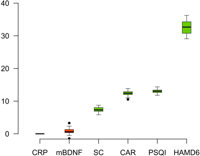
The variables selected by Boruta test for the discrimination of MD (n = 30) and CG (n = 32) were: HAM-D 6 (I = 32.60), PSQI (I = 13.03), CAR (I = 12.32) and SC (I = 7.43) (Fig 1). The mBDNF (I = 0.86) and CRP (I = 0) were not significant to discriminate between these groups.
Fig 1. The random forest-based algorithm (Boruta) of parameter relevance for discrimination between patients in first depressive episode (MD n = 30) and control group (CG n = 32).
Colors: green = relevant (p<0.05); yellow = tentative of relevance; red = no relevant.
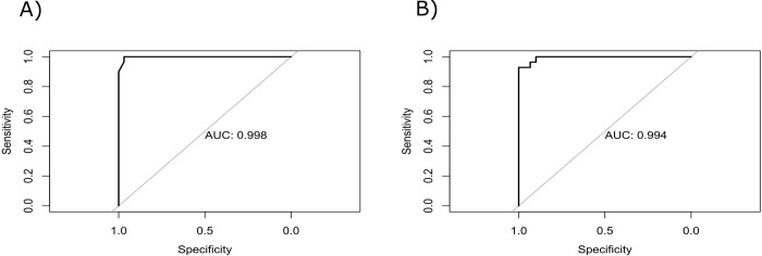
Then, the regression models for MDD diagnosis made of multiple combinations of HAM-D 6, PSQI, CAR and SC resulted in four statistically significant models with ΔAICc < 2 (Table 1). However, from these models, those containing biomarkers were not stronger than HAM-D 6 for diagnosis of de novo patients. Therefore, the best regression model for MDD diagnosis found in this study included only HAM-D 6 (AICc = -37.48, B = 0.06, Z = 16.29, p < 0.001) (Table 1). In our sample, this model showed 100% of sensitivity and 96% of specificity (AUC ROC = 0.99) (Fig 2A).
Table 1. Regression models for major depression disorder diagnosis, possible predictive models of discrimination between patients in first depressive episode (MD: n = 30) and the healthy controls (CG: n = 32).
| Models | AICc | ΔAICc | Β | z-score | p-value |
|---|---|---|---|---|---|
| I | |||||
| HAM-D 6 | -37.48 | 0 | 0.06 | 16.29 | < 0.001 |
| II | |||||
| HAM-D 6 | -36.76 | 0.72 | 0.06 | 16.29 | < 0.001 |
| CAR | 0.07 | 1.29 | 0.19 | ||
| III | |||||
| HAM-D 6 | -36.46 | 1.02 | 0.06 | 16.29 | < 0.001 |
| PSQI | 0.007 | 1.16 | 0.24 | ||
| IV | |||||
| HAM-D 6 | -35.73 | 1.75 | 0.06 | 16.29 | < 0.001 |
| CAR | 0.07 | 1.29 | 0.19 | ||
| PSQI | 0.007 | 1.16 | 0.24 | ||
| V | |||||
| HAM-D 6 | -35.42 | 2.05 | 0.06 | 16.29 | < 0.001 |
| SC | 0.04 | 0.47 | 0.63 | ||
| VI | |||||
| HAM-D 6 | -34.29 | 3.19 | 0.06 | 16.29 | < 0.001 |
| CAR | 0.07 | 1.29 | 0.19 | ||
| SC | 0.04 | 0.47 | 0.63 | ||
| VII | |||||
| HAM-D 6 | -34.27 | 3.20 | 0.06 | 16.29 | < 0.001 |
| SC | 0.04 | 0.47 | 0.63 | ||
| PSQI | 0.007 | 1.16 | 0.24 | ||
| VIII | |||||
| HAM-D 6 | -33.15 | 4.32 | 0.06 | 16.29 | < 0.001 |
| CAR | 0.07 | 1.29 | 0.19 | ||
| SC | 0.04 | 0.47 | 0.63 | ||
| PSQI | 0.007 | 1.16 | 0.24 | ||
Bold result indicates the best regression model with the lowest AICc and ΔAICc< 2. All models showed in this table were statistically significant (p< 0.05) and controlled by age and sex, while four of them had ΔAICc< 2. CAR: cortisol awakening response; SC: serum cortisol; PSQI: Pittsburgh sleep quality index; HAM-D 6: Hamilton Depression Rating Scale with 6 items.
Fig 2.
Area under the curve (AUC) of Receiver Operating Characteristic (ROC) curve for: A) Regression model for diagnosis of patients with first episode of major depression from healthy controls. B) Regression model for major depression disorder chronicity, for discrimination between patients in first depressive episode and patients with treatment-resistant depression.
MDD chronicity
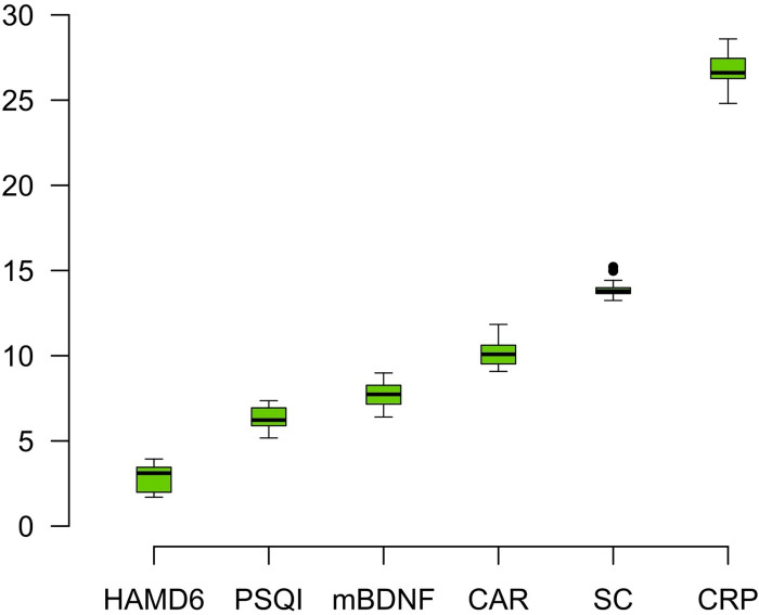
The variables selected by Boruta test for discrimination of MD and TRD (n = 28) were: CRP (I = 26.89), SC (I = 13.96), CAR (I = 10.26), mBDNF (I = 7.96), PSQI (I = 6.28) and HAM-D 6 (I = 2.98) (Fig 3).
Fig 3. The random forest-based algorithm (Boruta) of parameter relevance for discrimination between patients in first depressive episode (MD n = 30) and patients with treatment-resistant depression (TRD n = 28).
Colors: green = relevant (p<0.05); yellow = tentative of relevance; red = no relevant.
The mathematical models for MDD chronicity made of all variables previously select by Boruta algorithm resulted in two statistically significant regression models with ΔAICc < 2 (Table 2). The best regression model for MDD chronicity included (AICc = 32.57): CAR (B < 0.001, Z = 2.70, p = 0.006), SC (B < 0.001, Z = 4.46, p < 0.001), mBDNF (B < 0.001 Z = 2.31 p = 0.02) and PSQI (B < 0.001, Z = 2.06, p = 0.03). The model has 96% of sensitivity and 93% of specificity (AUC ROC = 0.99) (Fig 2B).
Table 2. Regression models for major depression disorder chronicity, possible predictive models of discrimination between patients in first depressive episode (MD: n = 30) and patients with treatment-resistant depression (TRD: n = 28).
| Models | AICc | ΔAICc | Β | z-score | p-value |
|---|---|---|---|---|---|
| I | |||||
| CAR | 32.57 | 0 | < 0.001 | 2.70 | 0.006 |
| SC | < 0.001 | 4.46 | < 0.001 | ||
| mBDNF | < 0.001 | 2.31 | 0.02 | ||
| PSQI | < 0.001 | 2.06 | 0.03 | ||
| II | |||||
| CAR | 34.37 | 1.80 | < 0.001 | 2.70 | 0.006 |
| SC | < 0.001 | 4.46 | < 0.001 | ||
| mBDNF | < 0.001 | 2.31 | 0.02 | ||
| III | |||||
| CAR | 35.31 | 2.74 | < 0.001 | 2.70 | 0.006 |
| SC | < 0.001 | 4.46 | < 0.001 | ||
| PSQI | < 0.001 | 2.06 | 0.03 | ||
| IV | |||||
| SC | 37.73 | 5.16 | < 0.001 | 4.46 | < 0.001 |
| mBDNF | < 0.001 | 2.31 | 0.02 | ||
| PSQI | < 0.001 | 2.06 | 0.03 | ||
| V | |||||
| CAR | 38.06 | 5.49 | < 0.001 | 2.70 | 0.006 |
| SC | < 0.001 | 4.46 | < 0.001 | ||
| VI | |||||
| SC | 39.94 | 7.37 | < 0.001 | 4.46 | < 0.001 |
| PSQI | < 0.001 | 2.06 | 0.03 | ||
| VII | |||||
| SC | 42.39 | 9.82 | < 0.001 | 4.46 | < 0.001 |
| mBDNF | < 0.001 | 2.31 | 0.02 | ||
| VIII | |||||
| SC | 45.82 | 9.82 | < 0.001 | 4.46 | < 0.001 |
| IX | |||||
| CAR | 49.39 | 16.82 | < 0.001 | 2.70 | 0.006 |
| mBDNF | < 0.001 | 2.31 | 0.02 | ||
| PSQI | < 0.001 | 2.06 | 0.03 | ||
| X | |||||
| CAR | 52.19 | 19.62 | < 0.001 | 2.70 | 0.006 |
| mBDNF | < 0.001 | 2.31 | 0.02 | ||
Bold result indicates the best regression model with the lowest AICc and ΔAICc< 2. All models showed in this table were statistically significant (p< 0.05) and controlled by age and sex, while 2 of them were ΔAICc< 2. CAR: cortisol awakening response; SC: total serum cortisol; mBDNF: mature brain-derived neurotrophic factor; PSQI: Pittsburgh sleep quality index.
Discussion
In this work, we searched for mathematical models made of a multibiomarker panel for a potential use in diagnosis and evaluation of MDD chronicity. When we searched for a possible model for MDD diagnosis, those models made of potential biomarkers were not stronger than HAM-D 6 for discrimination of de novo patients from healthy controls. Therefore, the HAM-D 6 still fitted as the best strategy for MDD diagnosis, with 100% of sensibility and 96% of specificity. On the other hand, for MDD chronicity, the best model included a mixed panel made of serum cortisol, salivary cortisol awakening response, serum mature BDNF and the total score of PSQI scale. This panel showed 96% of sensitivity and 93% of specificity to discrimination of TRD from MD patients.
Currently, cortisol is pointed as a good MDD biomarker. However, our model partially contradicts this view since our best diagnosis model doesn’t include this hormone. Although changes in cortisol are frequent in MDD [65, 66], we must consider that some of these changes have small effect sizes and large variance [26, 67, 68], mainly in newly diagnosed patients [5, 6, 69].
However, some studies that used a mathematical prediction model pointed to serum cortisol as a critical biomarker of MDD diagnosis [38, 39]. Nevertheless, it is important to highlight that the sample of those studies did not comprise de novo patients, but participants with distinct MDD severities jointed into a single group [38, 39], in contrast to our sample. Considering that a MDD diagnosis is more complex for subjects with mild than it is for severely impaired patients, our model contemplates a sample with typical characteristics for a first MDD diagnosis and thus can be especially useful for clinical purposes. Therefore, our results indicate that cortisol changes have not pivotal value for being used as a complement tool to define first MDD diagnosis, and the HAM-D 6 seems enough to provide it.
On the other side of investigation, for MDD chronicity, both cortisol measures: salivary cortisol awakening response and serum cortisol, as well as serum mBDNF and sleep quality (PSQI), were part of the best predictive model. Similar to what we have found, an impairment on HPA axis function was associated to severity of depression in a study of mathematical prediction [8].
As we had hypothesized, the levels of serum mBDNF were included in the best model of MDD chronicity. Despite there is not a consensus about BDNF changes in MDD, some studies have suggested its reduction in antidepressant drug-free patients when compared to healthy subjects [5, 70, 71], while others suggest an increased BDNF levels in treated patients that can be partially resulted from previous antidepressant treatments [5, 72, 73]. Therefore, this difference between de novo and treatment-resistant patients with major depression had made this biomarker important in the evaluation of MDD chronicity.
The inclusion of PSQI in the proposed model for MDD chronicity confirms the importance of impairments in sleep quality in the evolution of MDD. Frequently, changes in sleep quality get worse along the course of the disease [46, 74]. Stronger sleep disturbances are related with more severe MDD symptoms and worse treatment response [18]. It is pertinent that the HPA axis and BDNF are often related with sleep disturbances [17, 22, 75, 76].
In contrast to our initial hypothesis, CRP was not part of the model that best fitted for assessing MDD chronicity. While changes in inflammation are often lacking in de novo patients [6, 77], a mild and chronic inflammatory profile is observed in more severe MDD patients [78, 79]. A study that exanimated different molecular biomarkers along MDD chronicity found for both males and females an association in CRP levels and number of MDD episodes [80, 81]. Probably the MDD chronicity model of this study did not include CRP because this biomarker was used here as a qualitative measure, which is lesser sensible than a quantitative value. Therefore, future studies using it as a quantitative data are encouraged.
Therefore, this model of MDD chronicity can be useful for a better understanding of its neurobiology and in the future help in medical decisions about which biological pathway(s) should be targeted to improve treatments. For instance, treatments aiming to regulate cortisol levels might be considered for treatment-resistant patients. The several antidepressants currently used may have distinct actions on the HPA axis. Moreover, the antidepressant treatment duration has a large impact on the modulation of HPA axis as well [82, 83]. Since cortisol is a hormone with multiple roles, its return to homeostatic levels would probably leads to improvements in immune function, neuroplasticity process, and sleep quality [18, 79, 84]. All these are biological processes usually impaired in MDD, most especially in those with severe symptoms [15, 85, 86].
Despite not all patients with major depression progress to TRD—in average 30% of them shows recurrent and/or chronic MDD—this feature is associated to high morbidity and leads to great harm. TRD patients have shown large disability in individual, social, and work fields, and it can ultimately increase suicide risk [23, 64]. For instance, the TRD patients of our sample had about 10 years of MDD, with some volunteers showing until 20 years of disease. Therefore, despite only one-third of MDD patient becoming TRD, studies with this group of patients are important due to its massive damages. Then, a mathematical model like this one could help in understanding the psychobiological ground behind the disorder and in clinical practice.
Moreover, a point that we must highlight in favor of our models is that the studies focusing on biomarkers models for MDD usually did not include psychometric instruments to measure depressive symptoms, such as the HAM-D, as we did [35, 37, 38, 87]. In this sense, it is pointed out that only a model with a robust power, that is larger than the most used psychometrics tools, such as HAM-D, justifies its clinical applicability [87]. Another relevant aspect of our exploratory models was that we controlled both sex and age when analyzing those biomarkers, since many studies have pointed to a possible modulation of molecular biomarkers by these two variables [88–91].
Nevertheless, this study presented some limitations. Our sample has a restricted size and severity levels, and, as a result of the inclusion/exclusion criteria, it may not represent the real profile of the disorder among our population. Moreover, we did not perform a cross-validation analysis using another dataset to confirm our findings and establish a cutoff point for the biomarkers to distinguish them between groups in chronicity model.
Though, our results show the relevance of testing potential biomarkers of MDD in statistical models of adequate prediction [46] and bring a step-forward showing that only for MDD chronicity, and not for diagnosis of de novo patients, some of those biomarkers are somewhat more efficient than the HAM-6, namely: salivary cortisol awakening response and serum cortisol, as well as serum mBDNF and sleep quality (PSQI). Consequently, further studies of cross-validation analysis with larger and heterogeneous populations should be done to verify the proposed model of MDD chronicity and establish the biomarkers’ cutoff, then a robustly validated model could be commercially available to be used in psychiatry clinical practice to assist in charting MDD clinical stages, as well as in choosing the best treatment for patients.
Supporting information
CG: control group, PG: patient group, MD: patients in first depressive episode, TRD: patients with treatment-resistant depression.
(DOCX)
Random forest-based algorithm (Boruta): A) Patients with first episode of major depression (MD, n = 30) and control group (CG, n = 32). B) MD and Patients with treatment-resistant major depression (TRD, n = 28). Colors: green = relevant characteristic; yellow = tentative of relevance; red = no relevant characteristic; blue = randomly shuffled data at a maximum, mean and minimum level.
(TIF)
(DOCX)
Acknowledgments
The authors are thankful to all volunteers for this study and to the Hospital Universitário Onofre Lopes (HUOL) and Federal University of Rio Grande do Norte, Brazil, for institutional support.
Data Availability
All manuscript of files are available from the "Open Science Framework" database (DOI: 10.17605/OSF.IO/2XE7D).
Funding Statement
This study was funded by the Brazilian National Council for Scientific and Technological Development (CNPq, grants #466760/2014 & #479466/2013), and by Coordenação de Aperfeiçoamento de Pessoal de Nível Superior (CAPES) (grants #1677/2012 & #1577/2013) from the Ministry of Science, Technology, Innovations and Communications, and Brazilian Ministry of Education, respectively. ACMG is supported by CAPES (Research Fellowship 88882.344060/2019-01). NLGC is supported by CAPES (Finance Code 001, Research Fellowship 88887.466701/2019-00) and National Science and Technology Institute for Translational Medicine (INCT-TM Fapesp 2014/50891-1; CNPq 465458/2014-9). JS is supported by an NHMRC Clinical Research Fellowship (APP1125000).
References
- 1.Li Z, Ruan M, Chen J, Fang Y. Major Depressive Disorder: Advances in Neuroscience Research and Translational Applications. Neurosci Bull. 2021; 37(6):863–880. doi: 10.1007/s12264-021-00638-3 [DOI] [PMC free article] [PubMed] [Google Scholar]
- 2.American Psychiatric Association. Diagnostic and Statistical Manual of Mental Disorders (DSM-5°). American Psychiatric Publishing; 2014.
- 3.Bremmer MA, Deeg DJ, Beekman AT, Penninx BW, Lips P, Hoogendijk WJ. Major Depression in Late Life Is Associated with Both Hypo- and Hypercortisolemia. Biological Psychiatry. 2007; 62: 479–486. doi: 10.1016/j.biopsych.2006.11.033 [DOI] [PubMed] [Google Scholar]
- 4.Koo P.C, Berger C, Kronenberg G, Bartz J, Wybitul P, Reis O, et al. Combined cognitive, psychomotor and electrophysiological biomarkers in major depressive disorder. European Archives of Psychiatry and Clinical Neuroscience. 2019; 269: 823–832. doi: 10.1007/s00406-018-0952-9 [DOI] [PubMed] [Google Scholar]
- 5.Galvão ACM, Almeida RN, Júnior GMS, Leocadio-Miguel MA, Palhano-Fontes F, de Araujo DB, et al. The Pathophysiology of Major Clinical Depression by the Stages. Frontiers in Psychology. 2021. [DOI] [PMC free article] [PubMed] [Google Scholar]
- 6.Cubała WJ, Landowski J. C-reactive protein and cortisol in drug-naïve patients with short-illness-duration first episode major depressive disorder: possible role of cortisol immunomodulatory action at early stage of the disease. J Affect Disord. 2014; 152–154:534–7. doi: 10.1016/j.jad.2013.10.004 [DOI] [PubMed] [Google Scholar]
- 7.Alenko A, Markos Y, Fikru C, Tadesse E, Gedefaw L. Association of serum cortisol level with severity of depression and improvement in newly diagnosed patients with major depressive disorder in Jimma medical center, Southwest Ethiopia. PLoS ONE 2020; 15(10): e0240668. doi: 10.1371/journal.pone.0240668 [DOI] [PMC free article] [PubMed] [Google Scholar]
- 8.Choi KW, Na EJ, Fava M, Mischoulon D, Cho H, et al. Increased adrenocorticotropic hormone (ACTH) levels predict severity of depression after six months of follow-up in outpatients with major depressive disorder. Psychiatry Research. 2018; 270: 246–252 doi: 10.1016/j.psychres.2018.09.047 [DOI] [PubMed] [Google Scholar]
- 9.Strawbridge R, Hodsoll J, Powell TR, Hotopf M, Hatch SL, Breen G, et al. Inflammatory profiles of severe treatment-resistant depression. Journal of affective disorders. 2019; 246, 42–51. doi: 10.1016/j.jad.2018.12.037 [DOI] [PubMed] [Google Scholar]
- 10.Caldieraro MA, Vares EA, Souza LH, Spanemberg L, Guerra TA, Wollenhaupt-Aguiar B, et al. Illness severity and biomarkers in depression: using a unidimensional rating scale to examine BDNF. Comprehensive Psychiatry. 2017; 75: 46–52. doi: 10.1016/j.comppsych.2017.02.014 [DOI] [PubMed] [Google Scholar]
- 11.Verduijn J, Milaneschi Y, Schoevers RA, van Hemert AM, Beekman AT, Penninx BW. Pathophysiology of major depressive disorder: mechanisms involved in etiology are not associated with disease progression. Translational Psychiatry. 2015; 5: e649–e649. doi: 10.1038/tp.2015.137 [DOI] [PMC free article] [PubMed] [Google Scholar]
- 12.Agargun MY, Kara H, Özer ÖA, Selvi Y, Kiran Ü, Özer B. Clinical importance of nightmare disorder in patients with dissociative disorders. Psychiatry Clin. Neurosci. 2003; 57: 575–579. doi: 10.1046/j.1440-1819.2003.01169.x [DOI] [PubMed] [Google Scholar]
- 13.Chellappa SL, Araujo JF. Qualidade subjetiva do sono em pacientes com transtorno depressivo. Estud. Psicol. 2007b; 12: 269–274. [Google Scholar]
- 14.Sadock BJ, Sadock VA, Ruiz P. Compêndio de Psiquiatria: Ciência do Comportamento e Psiquiatria Clínica. Porto Alegre: Artmed Editora; 2016. doi: 10.1002/jclp.22367 [DOI] [Google Scholar]
- 15.Fang H, Tu S, Sheng J, Shao A. Depression in sleep disturbance: a review on a bidirectional relationship, mechanisms and treatment. Journal of cellular and molecular medicine. 2019; 23(4): 2324–2332. doi: 10.1111/jcmm.14170 [DOI] [PMC free article] [PubMed] [Google Scholar]
- 16.Nutt D, Wilson S, Paterson L. Sleep disorders as core symptoms of depression. Dialogues Clin Neurosci. 2008;10(3):329–336. doi: 10.31887/DCNS.2008.10.3/dnutt [DOI] [PMC free article] [PubMed] [Google Scholar]
- 17.Santiago GTP, de Menezes Galvão AC, de Almeida R, Mota-Rolim SA, Palhano-Fontes F, Maia-de-Oliveira JP, et al. Changes in Cortisol but Not in Brain-Derived Neurotrophic Factor Modulate the Association Between Sleep Disturbances and Major Depression. Frontiers in Behavioral Neuroscience. 2020; 14: 44. doi: 10.3389/fnbeh.2020.00044 [DOI] [PMC free article] [PubMed] [Google Scholar]
- 18.Moos RH, Cronkite RC. Symptom-based predictors of a 10-year chronic course of treated depression. J. Nerv. Ment. Dis. 1999; 187: 360–368. doi: 10.1097/00005053-199906000-00005 [DOI] [PubMed] [Google Scholar]
- 19.Chrousos G, Vgontzas AN, Kritikou I. HPA axis and sleep. MDText.com, Inc., South Dartmouth (MA); 2016. [Google Scholar]
- 20.Schmitt K., Holsboer-Trachsler E., & Eckert A. (2016). BDNF in sleep, insomnia, and sleep deprivation. Annals of medicine, 48(1–2), 42–51. doi: 10.3109/07853890.2015.1131327 [DOI] [PubMed] [Google Scholar]
- 21.Patel S. R., Zhu X., Storfer-Isser A., Mehra R., Jenny N. S., Tracy R., et al. (2009). Sleep duration and biomarkers of inflammation. Sleep, 32(2), 200–204. doi: 10.1093/sleep/32.2.200 [DOI] [PMC free article] [PubMed] [Google Scholar]
- 22.Schmidt HD, Shelton RC, Duman RS. Functional biomarkers of depression: diagnosis, treatment, and pathophysiology. Neuropsychopharmacology. 2011; 36: 2375–2394. doi: 10.1038/npp.2011.151 [DOI] [PMC free article] [PubMed] [Google Scholar]
- 23.Zhou Y, Ren W, Sun Q, Yu KM, Lang X, Li Z, et al. The association of clinical correlates, metabolic parameters, and thyroid hormones with suicide attempts in first-episode and drug-naïve patients with major depressive disorder comorbid with anxiety: a large-scale cross-sectional study. Transl Psychiatry. 2021; 11(1):97. doi: 10.1038/s41398-021-01234-9 [DOI] [PMC free article] [PubMed] [Google Scholar]
- 24.Li Z, Wang Z, Zhang C, Chen J, Su Y, Huang J, et al. Reduced ENA78 levels as novel biomarker for major depressive disorder and venlafaxine efficiency: Result from a prospective longitudinal study. Psychoneuroendocrinology. 2017; 81:113–121. doi: 10.1016/j.psyneuen.2017.03.015 [DOI] [PubMed] [Google Scholar]
- 25.Li Z, Zhang C, Fan J, Yuan C, Huang J, Chen J, et al. Brain-derived neurotrophic factor levels and bipolar disorder in patients in their first depressive episode: 3-year prospective longitudinal study. Br J Psychiatry. 2014; 205(1):29–35. doi: 10.1192/bjp.bp.113.134064 [DOI] [PubMed] [Google Scholar]
- 26.Gururajan A, Clarke G, Dinan TG, Cryan JF. Molecular biomarkers of depression. Neuroscience & Biobehavioral Reviews. 2016; 64: 101–133. doi: 10.1016/j.neubiorev.2016.02.011 [DOI] [PubMed] [Google Scholar]
- 27.Hacimusalar Y, Eşel E. Suggested biomarkers for major depressive disorder. Archives of Neuropsychiatry. 2018; 55: 280–290. doi: 10.5152/npa.2017.19482 [DOI] [PMC free article] [PubMed] [Google Scholar]
- 28.Lakhan SE, Vieira K, Hamlat E. Biomarkers in psychiatry: drawbacks and potential for misuse. International Archives of Medicine. 2010; 3: 1–6. doi: 10.1186/1755-7682-3-1 [DOI] [PMC free article] [PubMed] [Google Scholar]
- 29.McEwen BS. Biomarkers for assessing population and individual health and disease related to stress and adaptation. Metabolism. 2015; 64: S2–S10. doi: 10.1016/j.metabol.2014.10.029 [DOI] [PubMed] [Google Scholar]
- 30.Insel T, Cuthbert B, Garvey M, Heinssen R, Pine DS, Quinn K, et al. Research domain criteria (RDoC): toward a new classification framework for research on mental disorders. The American Journal of Psychiatry. 2010; 167: 7. [DOI] [PubMed] [Google Scholar]
- 31.Le-Niculescu H, Kurian SM, Yehyawi N, Dike C, Patel SD, Edenberg HJ, et al. Identifying blood biomarkers for mood disorders using convergent functional genomics. Molecular Psychiatry. 2009; 14: 156–174. doi: 10.1038/mp.2008.11 [DOI] [PubMed] [Google Scholar]
- 32.Kéri S, Szabó C, Kelemen O. Blood biomarkers of depression track clinical changes during cognitive-behavioral therapy. Journal of Affective Disorders. 2014; 164: 118–122. doi: 10.1016/j.jad.2014.04.030 [DOI] [PubMed] [Google Scholar]
- 33.Martins-de-Souza D. Proteomics, metabolomics, and protein interactomics in the characterization of the molecular features of major depressive disorder. Dialogues in Clinical Neuroscience. 2014; 16: 63–73. doi: 10.31887/DCNS.2014.16.1/dmartins [DOI] [PMC free article] [PubMed] [Google Scholar]
- 34.Duan J, Xie P. The potential for metabolomics in the study and treatment of major depressive disorder and related conditions. Expert Review of Proteomics. 2020; 1–14. doi: 10.1080/14789450.2020.1717951 [DOI] [PubMed] [Google Scholar]
- 35.Papakostas GI, Shelton RC, Kinrys G, Henry ME, Bakow BR, Lipkin SH, et al. Assessment of a multi-assay, serum-based biological diagnostic test for major depressive disorder: a Pilot and Replication Study. Molecular Psychiatry. 2011; 18: 332–339. doi: 10.1038/mp.2011.166 [DOI] [PubMed] [Google Scholar]
- 36.Bilello JA, Thurmond LM, Smith KM, Pi B, Rubin R, Wright SM, et al. MDDScore: confirmation of a blood test to aid in the diagnosis of major depressive disorder. The Journal of Clinical Psychiatry. 2015; 76: 199–206. [DOI] [PubMed] [Google Scholar]
- 37.Liu Y, Yieh L, Yang T, Drinkenburg W, Peeters P, Steckler T, et al. Metabolomic biosignature differentiates melancholic depressive patients from healthy controls. BMC genomics. 2016; 17: 1–17. doi: 10.1186/s12864-015-2294-6 [DOI] [PMC free article] [PubMed] [Google Scholar]
- 38.Xu YY, Ge JF, Liang J, Cao Y, Shan F, Liu Y, et al. Nesfatin-1 and cortisol: potential novel diagnostic biomarkers in moderate and severe depressive disorder. Psychology Research and Behavior Management. 2018; 11: 495–502. doi: 10.2147/PRBM.S183126 [DOI] [PMC free article] [PubMed] [Google Scholar]
- 39.Jia Y, Liu L, Sheng C, Cheng Z, Cui L, Li M, et al. Increased serum levels of cortisol and inflammatory cytokines in people with depression. The Journal of Nervous and Mental Disease, 2019; 207: 271–276. doi: 10.1097/NMD.0000000000000957 [DOI] [PubMed] [Google Scholar]
- 40.van Buel EM, Meddens MJ, Arnoldussen EA, van den Heuvel ER, Bohlmeijer WC, den Boer JA, et al. Major depressive disorder is associated with changes in a cluster of serum and urine biomarkers. Journal of Psychosomatic Research. 2019; 125: 109796. doi: 10.1016/j.jpsychores.2019.109796 [DOI] [PubMed] [Google Scholar]
- 41.Kennis M, Gerritsen L, van Dalen M, Williams A, Cuijpers P, Bockting C. Prospective biomarkers of major depressive disorder: a systematic review and meta-analysis. Molecular Psychiatry. 2020; 25: 321–338. doi: 10.1038/s41380-019-0585-z [DOI] [PMC free article] [PubMed] [Google Scholar]
- 42.Kalia M, Silva JC. Biomarkers of psychiatric diseases: current status and future prospects. Metabolism. 2015; 64: S11–5. doi: 10.1016/j.metabol.2014.10.026 [DOI] [PubMed] [Google Scholar]
- 43.Faul F, Erdfelder E, Buchner A, Lang A-G. Statistical power analyses using G*Power 3.1: Tests for correlation and regression analyses. Behavior Research Methods (2009) 41: 1149–1160. doi: 10.3758/BRM.41.4.1149 [DOI] [PubMed] [Google Scholar]
- 44.Hamilton M. A Rating Scale for Depression. Journal of Neurology Neurosurgery, and Psychiatry. 1960; 23: 56–62. doi: 10.1136/jnnp.23.1.56 [DOI] [PMC free article] [PubMed] [Google Scholar]
- 45.Pruessner JC, Kirschbaum C, Meinlschmid G, Hellhammer DH. Two formulas for computation of the area under the curve represent measures of total hormone concentration versus time-dependent change. Psychoneuroendocrinology. 2003; 28: 916–931. doi: 10.1016/s0306-4530(02)00108-7 [DOI] [PubMed] [Google Scholar]
- 46.Bagby RM., Ryder AG, Schuller DR, Marshall MB. The Hamilton Depression Rating Scale: has the gold standard become a lead weight? American Journal of Psychiatry. 2004; 161(12); 2163–2177. doi: 10.1176/appi.ajp.161.12.2163 [DOI] [PubMed] [Google Scholar]
- 47.Bech P, Fava M, Trivedi MH, Wisniewski SR, Rush AJ. Factor structure and dimensionality of the two depression scales in STAR*D using level 1 datasets. Journal of Affective Disorders. 2011; 132: 396–400. doi: 10.1016/j.jad.2011.03.011 [DOI] [PubMed] [Google Scholar]
- 48.Bech P, Gram LF, Dein E, Jacobsen O, Vitger J, Bolwig TG. Quantitative rating of depressive states: correlation between clinical assessment, Beck’s self-rating scale and Hamilton’s objective rating scale. Acta Psychiatrica Scandinavica 1975; 51(3): 161–170. doi: 10.1111/j.1600-0447.1975.tb00002.x [DOI] [PubMed] [Google Scholar]
- 49.Carrozzino D, Patierno C, Fava GA, Guidi J. The Hamilton Rating Scales for Depression: A critical review of clinimetric properties of different versions. Psychotherapy and Psychosomatics. 2020; 89(3): 133–150. doi: 10.1159/000506879 [DOI] [PubMed] [Google Scholar]
- 50.Alves LPC, Rocha NS. Different cytokine patterns associate with melancholia severity among inpatients with major depressive disorder. Therapeutic advances in Psychopharmacology. 2020; 10: 2045125320937921. [DOI] [PMC free article] [PubMed] [Google Scholar]
- 51.Buysse DJ, Reynolds CF III, Monk TH, Berman SR, Kupfer DJ. The Pittsburgh Sleep Quality Index: a new instrument for psychiatric practice and research. Psychiatry Research. 1989; 28: 193–213. doi: 10.1016/0165-1781(89)90047-4 [DOI] [PubMed] [Google Scholar]
- 52.Bertolazi AN, Fagondes SC, Hoff LS, Dartora EG, da Silva Miozzo IC, de Barba MEF, et al. Validation of the Brazilian Portuguese version of the Pittsburgh sleep quality index. Sleep Medicine. 2011; 12: 70–75. doi: 10.1016/j.sleep.2010.04.020 [DOI] [PubMed] [Google Scholar]
- 53.Kursa MB, Rudnicki WR. Feature selection with the boruta package. Journal of Statistical Software. 2010; 36: 1–13. [Google Scholar]
- 54.Magnusson A, Skaug H, Nielsen A, Berg C, Kristensen K, Maechler M, et al. Package ‘glmmTMB’. R Package Version 0.2. 0. 2017. [Google Scholar]
- 55.Bartón K, Barton MK. (2015). Package ‘MuMIn’. Version, 1; 18. [Google Scholar]
- 56.Martinez EZ, Louzada-Neto F, Pereira BDB. A curva ROC para testes diagnósticos. Cadernos Saúde Coletiva (Rio de Janeiro). 2003; 11: 7–31. [Google Scholar]
- 57.Moraes SAD, Freitas I, Mondini L, Rosas JB. Receiver operating characteristic (ROC) curves to identify birth weight cutoffs to predict overweight in Mexican school children. Jornal de Pediatria. 2009; 85: 42–47. doi: 10.2223/JPED.1858 [DOI] [PubMed] [Google Scholar]
- 58.Freire MA, Figueiro VD, Gomide A, Jansen K, Silva RD, Magalhães PVDS, et al. Escala Hamilton: estudo das características psicométricas em uma amostra do sul do Brasil. Jornal Brasileiro de Psiquiatria. 2014; 63: 281–9. [Google Scholar]
- 59.Cerda J, Cifuentes L. Uso de curvas ROC em investigación clínica: Aspectos teórico-prácticos. Revista Chilena de Infectología. 2012: 29: 138–141. doi: 10.4067/S0716-10182012000200003 [DOI] [PubMed] [Google Scholar]
- 60.Castanho MJ, Barros LC, Vendite LL, Yamakami A. Avaliação de um teste em medicina usando uma curva ROC fuzzy. Biomatematica. 2004; 14: 19–28. [Google Scholar]
- 61.Fávero LPL, Belfiore PP, da Silva FL, Chan BL. Análise de dados: modelagem multivariada para tomada de decisões. Elsevier; 2009. [Google Scholar]
- 62.Perlis RH. Translating biomarkers to clinical practice. Molecular Psychiatry. 2011; 16: 1076–1087. doi: 10.1038/mp.2011.63 [DOI] [PMC free article] [PubMed] [Google Scholar]
- 63.Lopes B, Ramos IC, Ribeiro G, Correa R, Valbon B, Luz A, et al. Biostatistics: fundamental concepts and practical applications. Revista Brasileira de Oftalmologia. 2014; 73: 16–22. [Google Scholar]
- 64.World Health Organization. Depression and other common mental disorders: global 584 health estimates. Geneva (2017).
- 65.Strawbridge R, Young AH, Cleare AJ. Biomarkers for depression: recent insights, current challenges and future prospects. Neuropsychiatric Disease and Treatment. 2017; 13: 1245–1262. doi: 10.2147/NDT.S114542 [DOI] [PMC free article] [PubMed] [Google Scholar]
- 66.Malhi GS, Mann JJ. Seminar Depression. Lancet. 2018; 392: 2299–312. doi: 10.1016/S0140-6736(18)31948-2 [DOI] [PubMed] [Google Scholar]
- 67.Vreeburg SA, Hoogendijk WJ, DeRijk RH, van Dyck R, Smit JH, Zitman FG, et al. Salivary cortisol levels and the 2-year course of depressive and anxiety. Psychoneuroendocrinology. 2013; 38: 1494–1502. doi: 10.1016/j.psyneuen.2012.12.017 [DOI] [PubMed] [Google Scholar]
- 68.Adam EK, Doane LD, Zinbarg RE, Mineka S, Craske MG, Griffith JW. Prospective prediction of major depressive disorder from cortisol awakening responses in adolescence. Psychoneuroendocrinology. 2010; 35(6): 921–931. doi: 10.1016/j.psyneuen.2009.12.007 [DOI] [PMC free article] [PubMed] [Google Scholar]
- 69.Varela YM, De Almeida RN, de Menezes Galvão AC, Junior GMS, de Lima ACL, da Silva NG, et al. Psychophysiological responses to group cognitive-behavioral therapy in depressive patients. Current Psychology. 2021. [Google Scholar]
- 70.Sen S, Duman R, Sanacora G. Serum brain-derived neurotrophic factor, depression, and antidepressant medications: meta-analyses and implications. Biological Psychiatry. 2008; 64: 527–532. doi: 10.1016/j.biopsych.2008.05.005 [DOI] [PMC free article] [PubMed] [Google Scholar]
- 71.Huang TL, Lee CT, Liu YL. Serum brain-derived neurotrophic factor levels in patients with major depression: effects of antidepressants. Journal of Psychiatric Research. 2008; 42: 521–525. doi: 10.1016/j.jpsychires.2007.05.007 [DOI] [PubMed] [Google Scholar]
- 72.Nuernberg GL, Aguiar B, Bristot G, Fleck MP, Rocha NS. Brain-derived neurotrophic factor increase during treatment in severe mental illness inpatients. Translational Psychiatry. 2016; 6: e985–e985. doi: 10.1038/tp.2016.227 [DOI] [PMC free article] [PubMed] [Google Scholar]
- 73.Chauhan VS, Khan SA, Kulhari K. Correlation of brain-derived neurotrophic factor with severity of depression and treatment response. Medical Journal Armed Forces India. 2020. [DOI] [PMC free article] [PubMed] [Google Scholar]
- 74.Ohayon MM, Roth T. Place of chronic insomnia in the course of depressive and anxiety disorders. Journal of Psychiatric Research. 2003; 37: 9–15. doi: 10.1016/s0022-3956(02)00052-3 [DOI] [PubMed] [Google Scholar]
- 75.Giese M, Unternährer E, Hüttig H, Beck J, Brand S, Calabrese P, et al. BDNF: an indicator of insomnia? Molecular Psychiatry. 2014; 19: 151–152. doi: 10.1038/mp.2013.10 [DOI] [PMC free article] [PubMed] [Google Scholar]
- 76.van Dalfsen JH, Markus CR. The influence of sleep on human hypothalamic-pituitary-adrenal (HPA) axis reactivity: a systematic review. Sleep Medicine Reviews. 2018; 39: 187–194. doi: 10.1016/j.smrv.2017.10.002 [DOI] [PubMed] [Google Scholar]
- 77.Zou W, Feng R, Yang Y. Changes in the serum levels of inflammatory cytokines in antidepressant drug-naïve patients with major depression. PLoS one. 2018; 13: e01927267. doi: 10.1371/journal.pone.0197267 [DOI] [PMC free article] [PubMed] [Google Scholar]
- 78.Zalli A, Jovanova O, Hoogendijk WJG, Tiemeier H, Carvalho LA. Low-grade inflammation predicts persistence of depressive symptoms. Psychopharmacology. 2016; 233: 1669–1678. doi: 10.1007/s00213-015-3919-9 [DOI] [PMC free article] [PubMed] [Google Scholar]
- 79.Galvão-Coelho NL, de Menezes Galvão AC, de Almeida RN, Palhano-Fontes F, Campos Braga I, Lobão Soares B, et al. Changes in inflammatory biomarkers are related to the antidepressant effects of Ayahuasca. Journal of Psychopharmacology. 2020; 34: 1125–1133. doi: 10.1177/0269881120936486 [DOI] [PubMed] [Google Scholar]
- 80.Danner M, Kasl SV, Abramson JL, Vaccarino V. Association between depression and elevated C-reactive protein. Psychosomatic Medicine. 2003; 65: 347–356. doi: 10.1097/01.psy.0000041542.29808.01 [DOI] [PubMed] [Google Scholar]
- 81.Pasco JA, Nicholson GC, Williams LJ, Jacka FN, Henry MJ, Kotowicz MA, et al. Association of high-sensitivity C-reactive protein with de novo major depression. The British Journal of Psychiatry. 2010; 197: 372–377. doi: 10.1192/bjp.bp.109.076430 [DOI] [PubMed] [Google Scholar]
- 82.Schüle C, Baghai TC, Eser D, Zwanzger P, Jordan M, Buechs R, et al. Time course of hypothalamic-pituitary-adrenocortical axis activity during treatment with reboxetine and mirtazapine in depressed patients. Psychopharmacology. 2006; 186: 601–611. doi: 10.1007/s00213-006-0382-7 [DOI] [PubMed] [Google Scholar]
- 83.Schüle C. Neuroendocrinological mechanisms of actions of antidepressant drugs. Journal of Neuroendocrinology. 2007; 19: 213–226. doi: 10.1111/j.1365-2826.2006.01516.x [DOI] [PubMed] [Google Scholar]
- 84.Almeida RN, Galvão ACDM, Da Silva FS, Silva EADS, Palhano-Fontes F, Maia-de-Oliveira JP, et al. Modulation of serum brain-derived neurotrophic factor by a single dose of ayahuasca: observation from a randomized controlled trial. Frontiers in Psychology, 2019; 10: 1234. doi: 10.3389/fpsyg.2019.01234 [DOI] [PMC free article] [PubMed] [Google Scholar]
- 85.Lee BH, Kim YK. The roles of BDNF in the pathophysiology of major depression and in antidepressant treatment. Psychiatry Investigation. 2010; 7: 231–235. doi: 10.4306/pi.2010.7.4.231 [DOI] [PMC free article] [PubMed] [Google Scholar]
- 86.Schmidt FM, Schröder T, Kirkby KC, Sander C, Suslow T, Holdt LM, et al. Pro-and anti-inflammatory cytokines, but not CRP, are inversely correlated with severity and symptoms of major depression. Psychiatry Research. 2016; 239: 85–91. doi: 10.1016/j.psychres.2016.02.052 [DOI] [PubMed] [Google Scholar]
- 87.Domenici E, Willé DR, Tozzi F, Prokopenko I, Miller S, McKeown A, et al. Plasma protein biomarkers for depression and schizophrenia by multi analyte profiling of case-control collections. PLoS one. 2010; 5: e9166. doi: 10.1371/journal.pone.0009166 [DOI] [PMC free article] [PubMed] [Google Scholar]
- 88.Halbreich U, Asnis GM, Zumoff B, Nathan RS, Shindledecker R. Effect of age and sex on cortisol secretion in depressives and normals. Psychiatry research. 1984; 13: 221–229. doi: 10.1016/0165-1781(84)90037-4 [DOI] [PubMed] [Google Scholar]
- 89.Heaney JL, Phillips AC, Carroll D. Ageing, depression, anxiety, social support and the diurnal rhythm and awakening response of salivary cortisol. International Journal of Psychophysiology. 2010; 78: 201–208. doi: 10.1016/j.ijpsycho.2010.07.009 [DOI] [PubMed] [Google Scholar]
- 90.Choi SW, Bhang S, Ahn JH. Diurnal variation and gender differences of plasma brain-derived neurotrophic factor in healthy human subjects. Psychiatry Research. 2011; 186: 427–430. doi: 10.1016/j.psychres.2010.07.028 [DOI] [PubMed] [Google Scholar]
- 91.Salk RH, Hyde JS, Abramson LY. Gender differences in depression in representative national samples: meta-analyses of diagnoses and symptoms. Psychological Bulletin. 2017; 143: 783–822. doi: 10.1037/bul0000102 [DOI] [PMC free article] [PubMed] [Google Scholar]
Associated Data
This section collects any data citations, data availability statements, or supplementary materials included in this article.
Supplementary Materials
CG: control group, PG: patient group, MD: patients in first depressive episode, TRD: patients with treatment-resistant depression.
(DOCX)
Random forest-based algorithm (Boruta): A) Patients with first episode of major depression (MD, n = 30) and control group (CG, n = 32). B) MD and Patients with treatment-resistant major depression (TRD, n = 28). Colors: green = relevant characteristic; yellow = tentative of relevance; red = no relevant characteristic; blue = randomly shuffled data at a maximum, mean and minimum level.
(TIF)
(DOCX)
Data Availability Statement
All manuscript of files are available from the "Open Science Framework" database (DOI: 10.17605/OSF.IO/2XE7D).


