Abstract
The recruitment of neutrophils from the microvasculature to the site of injury or infection represents a key event in the inflammatory response. Vitronectin (VN) is a multifunctional macromolecule abundantly present in blood and extracellular matrix. The role of this glycoprotein in the extravasation process of circulating neutrophils remains elusive. Employing advanced in vivo/ex vivo imaging techniques in different mouse models as well as in vitro methods, we uncovered a previously unrecognized function of VN in the transition of dynamic to static intravascular interactions of neutrophils with microvascular endothelial cells. These distinct properties of VN require the heteromerization of this glycoprotein with plasminogen activator inhibitor-1 (PAI- 1) on the activated venular endothelium and subsequent interactions of this protein complex with the scavenger receptor low-density lipoprotein receptor-related protein-1 on intravascularly adhering neutrophils. This induces p38 mitogen-activated protein kinases-dependent intracellular signaling events which, in turn, regulates the proper clustering of the b2 integrin lymphocyte function associated antigen-1 on the surface of these immune cells. As a consequence of this molecular interplay, neutrophils become able to stabilize their adhesion to the microvascular endothelium and, subsequently, to extravasate to the perivascular tissue. Hence, endothelial-bound VN-PAI-1 heteromers stabilize intravascular adhesion of neutrophils by coordinating b2 integrin clustering on the surface of these immune cells, thereby effectively controlling neutrophil trafficking to inflamed tissue. Targeting this protein complex might be beneficial for the prevention and treatment of inflammatory pathologies.
Introduction
Vitronectin (VN) is a multidomain macromolecule synthesized by the liver and found in platelets.1 Upon release into the extracellular space,2 VN becomes capable of establishing interactions with different proteins involved in diverse biological processes: engaging its somatomedin B domain, VN acts as a ligand for the (soluble) urokinase-type plasminogen activator (uPA) receptor or binds to plasminogen activator inhibitor-1 (PAI-1), thereby extending the half life of this protease inhibitor in fibrinolysis. Through its tripeptide arginine, glycine, and aspartate (RGD) sequence, VN can interact with αvb3, αvb5, αIIbb3, and αvb1 integrins which are expressed on the surface of leukocytes and platelets. In addition, several other molecules (e.g., proteoglycans, heparin, collagens, or kininogen) serve as binding partners of VN immobilizing this glycoprotein in the extracellular matrix (ECM), ultimately regulating cell adhesion and migration.3-5 Accordingly, enhanced levels of VN have been detected in various inflammatory pathologies including atherosclerosis, glomerulonephritis, or rheumatic disease.6-10 The functional role of VN under these pathological conditions, however, remains largely unclear.
The recruitment of white blood cells (leukocytes) from the microvasculature to the site of inflammation is a fundamental process in the immune response.11-14 In inflamed tissue, circulating leukocytes become captured and start to roll on the microvascular endothelium in a selectin-dependent manner.15 This triggers the intermediate affinity conformation of b2 integrins on the surface of rolling leukocytes which allows them to further slow down in the bloodstream.16,17 Subsequently, interactions of chemokines presented on the microvascular endothelium with their cognate chemokine receptors on rolling leukocytes as well as of endothelial E-selectin/CD62E with leukocyte P-selectin glycoprotein ligand-1 (PSGL- 1/CD168) are supposed to initiate the full activation of leukocyte integrins ultimately facilitating intravascular adhesion of these immune cells to the endothelial surface. 15,18 After stabilizing their adhesion, leukocytes intravascularly crawl to sites of adherent platelets, from where they finally extravasate to the perivascular tissue and migrate to their target destination.19 Whereas a variety of adhesion and signaling molecules have been characterized to control distinct steps of this highly complex process, the mechanisms underlying the stabilization of leukocyte adhesion to the microvascular endothelium are still poorly understood.
Fibrinolysis is an elementary biological process that maintains blood perfusion by preventing clot formation in the vasculature. Plasmin is the principal effector protease in the fibrinolytic system, which is activated by tissue- plasminogen activator (tPA) and – to a lesser degree – by urokinase-type plasminogen activator (uPA). The activity of these serine proteases is tightly controlled by plasminogen activator inhibitor-1 (PAI-1). Besides these well-known fibrinolytic properties, it has become evident that the components of the fibrinolytic system also considerably contribute to different biological processes such as immune cell trafficking.20-24 With respect to the distinct interactive properties of VN and the substantial involvement of the various binding partners of this glycoprotein in immune cell responses, we hypothesize that VN is critical for leukocyte recruitment to the site of inflammation.
Methods
A detailed description of the methods employed in this study is included in the Online Supplementary Appendix.
All experiments were performed according to German legislation for the protection of animals and approved by the local government authorities (Regierung von Oberbayern).
Six hours after intra-peritoneal injection of the chemokines CXCL1 or CCL2, leukocyte recruitment to the peritoneal cavity was studied by flow cytometry in wild-type (WT) or VN-deficient mice. The single steps of the neutrophil (visualized by fluorescence- labeled anti-Ly-6G monoclonal antibodies) extravasation process were analyzed by in vivo microscopy in the cremaster muscle of anesthetized mice deficient for distinct proteins or receiving different inhibitors/blocking antibodies. Integrin activation/trafficking was assessed in neutrophils from WT mice by flow cytometry and spinning disc confocal microscopy.
Results
Distribution of vitronectin in inflamed tissue
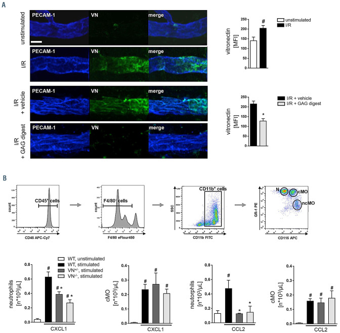
Under a variety of inflammatory conditions, enhanced tissue levels of VN have been observed.6-10 The exact distribution patterns of this glycoprotein in inflamed tissue, however, remained unclear. Employing immunostaining and confocal laser scanning microscopy on tissue whole mounts of the mouse cremaster muscle, VN was barely detected in unstimulated tissue (Figure 1A). In the acute inflammatory response upon sterile (ischemia-reperfusion [I/R]; 30/120 minutes [min]) injury, however, VN was found to be deposited on the luminal surface of postcapillary venules. This microvascular deposition of VN was nearly absent upon enzymatic degradation of glycosaminoglycans (GAG).
Role of vitronectin for myeloid leukocyte trafficking
In order to characterize the role of VN for myeloid leukocyte recruitment to the site of inflammation, we initially used a peritoneal leukocyte trafficking assay. As identified by multi-channel flow cytometry analyses of the peritoneal lavage fluid, 6 hours of intraperitoneal stimulation with the chemokines CXCL1/KC or CCL2/MCP-1 induced a significant increase in numbers of neutrophils (CD45+ CD11b+ Gr-1high CD115low) and classical/ inflammatory monocytes (CD45+ CD11b+ Gr-1high CD115high; Figure 1B), but not of non-classical monocytes (CD45+ CD11b+ Gr-1low CD115high; data not shown) recruited to the peritoneal cavity of WT mice as compared to unstimulated controls. This increase in numbers of neutrophils was almost completely abolished in VN+/- or VN- /- mice, whereas the recruitment of classical/inflammatory monocytes remained unaffected by VN deficiency. In this context, we found that expression of the low density lipoprotein-related receptor protein-1 (LRP-1), which serves as a receptor for the VN binding partner PAI-1,25-27 was higher on the surface of activated murine neutrophils (mean fluorescence intensity 1,023.0±231.4) than on activated classical monocytes (mean fluorescence intensity 473.0±128.8). Moreover, LRP-1 was identified to be expressed heterogeneously in murine neutrophils (Online Supplementary Figure S1).
Role of vitronectin for intravascular interactions of neutrophils
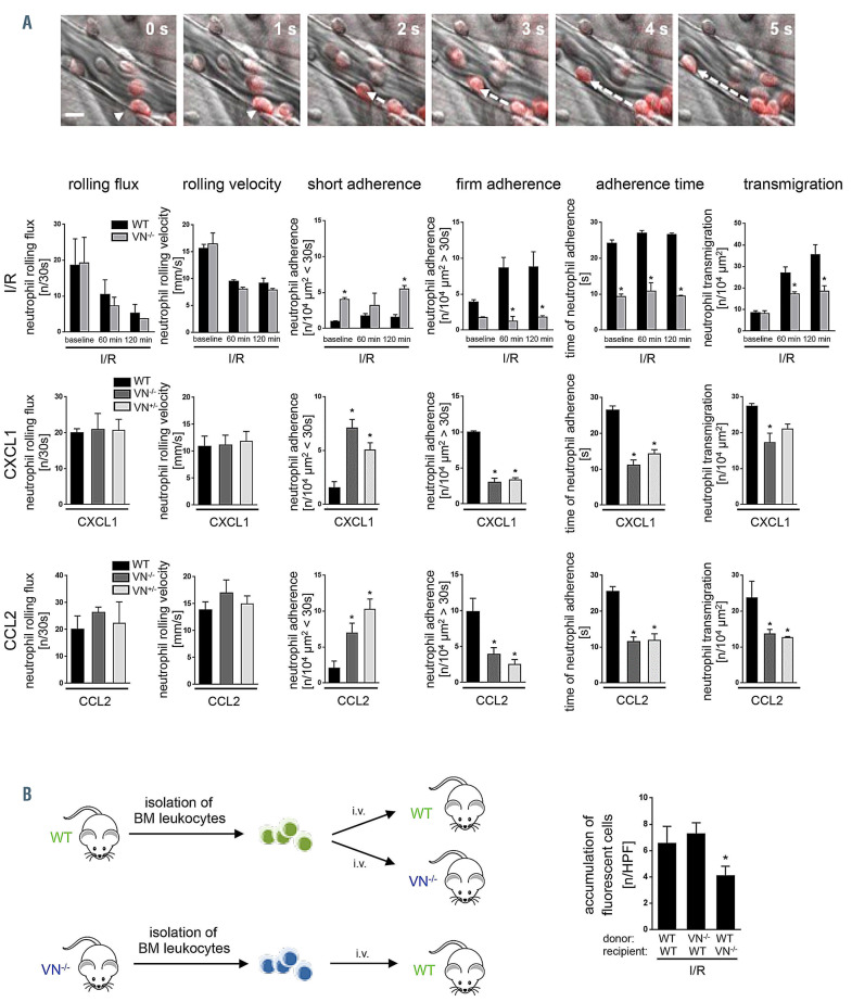
In order to further decipher the role of VN in the extravasation process of neutrophils, we performed multi-channel in vivo microscopy on the mouse cremaster muscle. In these experiments, we observed that Ly-6G+ neutrophils in VN-/- mice are unable to stabilize their adhesion to the endothelial surface of postcapillary venules in inflamed tissue, whereas neutrophils in WT mice properly adhered to the microvascular endothelium (Figure 2A; Online Supplemtary Videos S1 and S2). Accordingly, the quantitative analysis of these events revealed no significant differences in the numbers of rolling neutrophils or in the rolling velocity of neutrophils between WT and VN-/- mice animals upon I/R (30/120 min) or after 6 hours of intrascrotal stimulation with CXCL1/KC or CCL2/MCP-1 (Figure 2A). In contrast, the average number of neutrophils adhering to the vessel wall of postcapillary venules for more than 30 seconds (s) was significantly lower in VN-/- mice than in WT controls, whereas the average number of neutrophils adhering for less than 30 s was significantly higher in VN-/- mice than in WT controls. Consequently, the average adhesion time of a neutrophil to the microvascular endothelium was significantly shorter in VN-/- mice than in WT controls. This defect of neutrophils in VN-/- mice to stabilize intravascular adhesion resulted in a significantly reduced number of extravasated neutrophils. Noteworthy, a minority of neutrophils in VN-/- mice was still able to stabilize their adherence to the microvascular endothelium, which might be explained by the heterogeneous expression levels of the VN receptor LRP-1 (Online Supplementary Figure S1) and/or compensatory mechanisms in a subset of these immune cells. Such alternative mechanisms (which might even include cumulative factors) controlling the stabilization of neutrophil adherence are still unclear and subject of future investigations.
In order to exclude neutrophil-intrinsic effects of VN deficiency on the stabilization of intravascular adhesion of these immune cells, we conducted bone marrow cell transfer experiments (Figure 2B). In the postischemic mouse cremaster muscle (I/R 30/120 min), the number of accumulated fluorescence-labeled neutrophils isolated from VN-/- donor mice and adoptively transferred into WT recipient mice was not significantly altered as compared to the number of accumulated fluorescence-labeled neutrophils isolated from WT donor mice and adoptively transferred into WT mice. In contrast, the number of accumulated fluorescence-labeled neutrophils isolated from WT mice and adoptively transferred into VN-/- recipient mice was significantly reduced as compared to the number of accumulated fluorescence-labeled neutrophils isolated from WT donor mice and adoptively transferred into WT recipient mice. Thus, a neutrophil-intrinsic effect of VN deficiency was not evident.
Figure 1.
Role of vitronectin for leukocyte trafficking to inflamed tissue. (A) Using confocal laser scanning microscopy on tissue whole mounts of the cremaster muscle of wil-type (WT) mice, deposition of vitronectin (VN) (green) in the PECAM-1/CD31+ microvasculature (blue) was analyzed, representative images are shown (scale bar: 20 mm). Panels show quantitative results from relative fluorescence intensity measurements for VN in sham-operated WT mice as well as in WT mice undergoing ischemia-reperfusion (I/R) (30/120 min) and intravenous application of glycosaminoglycans digesting enzymes or vehicle (mean±standard error of the mean [SEM] for n=4 animals per group; #P<0.05 vs. sham; *P<0.05 vs. vehicle). (B) Employing multi-channel flow cytometry, the recruitment of neutrophils and classical monocytes to the peritoneal cavity was analyzed 6 hours after intraperitoneal (i.p.) injection of CXCL1 or CCL2, the gating strategy is shown. Panels show results for lipopolysaccharide- treated WT control mice as well as for WT, VN+/-, or VN-/- mice receiving an i.p. injection of CXCL1 or CCL2 (mean±SEM for n=4 animals per group; #P<0.05 vs. control; *P<0.05 vs. WT). cMO: classical monocytes; ncMO: nonclassical monocytes; N: neutrophils; SSC: side scatter.
Figure 2.
Role of vitronectin for interactions of neutrophils and endothelial cells. (A) Using multi-channel in vivo microscopy on the inflamed mouse cremaster muscle, interactions of Ly-6G+ neutrophils (red) with endothelial cells were analyzed in postcapillary venules, representative still images are shown (scale bar: 20 mm). Panels show quantitative results for rolling flux, rolling velocity, short adhesion, firm adherence, adhesion time, and transmigration of neutrophils in wild-type (WT), vitronectin (VN)+/-, or VN+/+ mice (mean±standard error of the mean [SEM] for n=4 animals per group; *P<0.05 vs. WT). See also Online Supplementary Table S1. (B) Accumulation of calcein AM-labeled bone marrow (BM) leukocytes were quantified in the postischemic cremaster muscle using multi-channel in vivo fluorescence microscopy. Panel shows results for WT recipient mice receiving leukocytes from WT or VN-deficient donors as well as for VN-deficient recipient mice receiving leukocytes from WT donors (mean±SEM for n=5 animals per group; *P<0.05 vs. WT). n: number; s: seconds; HPF: hydroxyphenyl fluorescein; I/R: ischemia-reperfusion.
Role of integrin interaction partners of vitronectin for neutrophil trafficking
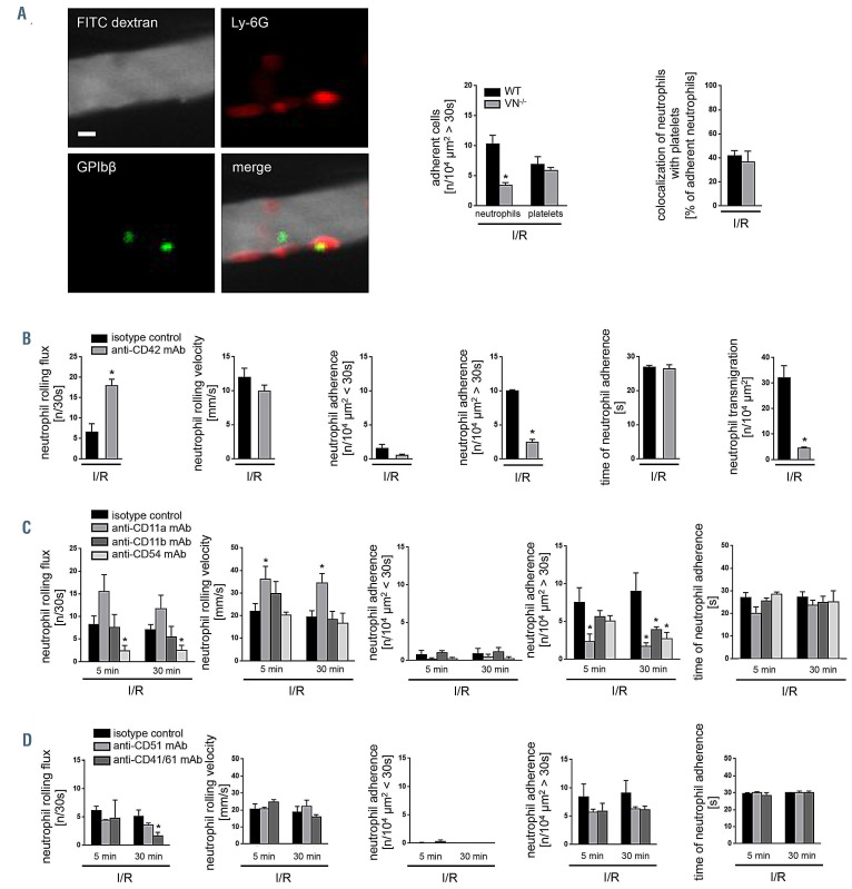
Platelets are known to play a critical role in the extravasation process of leukocytes.11-14 Since αvb3 and αIIbb3 integrins are expressed on the surface of platelets and serve as binding partners of VN, this glycoprotein might stabilize intravascular adhesion of neutrophils by mediating interactions of these cell populations. Using in vivo microscopy on the postischemic mouse cremaster muscle, however, intravascular firm adherence of platelets or interactions of intravascularly adherent platelets and neutrophils did not significantly vary between WT and VN-/- mice (Figure 3A). In contrast, intravascular firm adherence of neutrophils was significantly diminished in VN-/- mice animals as compared to WT animals (Figure 3A). Conversely, antibody- mediated depletion of platelets (by >95%) significantly reduced the number of intravascularly adherent (>30 s) and transmigrated neutrophils in the field of view, but did not significantly change intravascular adhesion times of these immune cells in the microvasculature of postischemic tissue (Figure 3B). These data confirm previous observations documenting that intravascularly adherent neutrophils do not stop crawling in the microvasculature and start their transmigration (which results in less firmly adhered and transmigrated leukocytes quantified in the field of view) until they are captured by intravascularly adherent platelets.19
Figure 3.
Role of platelets and integrins for the stabilization of intravascular adhesion of neutrophils. (A) Interactions of Ly-6G+ neutrophils (red) and GP-Ibb+ platelets (green) were analyzed in postcapillary venules (perfused by FITC dextran; grey) of the postischemic mouse cremaster muscle of wild-type (WT) or vitronectin (VN)-deficient mice by multi-channel in vivo microscopy, representative images from a WT mouse are shown (scale bar: 10 mm). Panels show quantitative results for intravascular adherence of neutrophils or platelets as well as for the colocalization of these two cell populations (mean±standard error of the mean [SEM] for n=3 animals per group; *P<0.05 vs. WT). Further panels show quantitative results for rolling flux, rolling velocity, short adhesion, firm adherence, adhesion time, and transmigration of neutrophils in WT mice receiving platelet-depleting antibodies, blocking antibodies directed against the b2 integrins LFA-1/CD11a and Mac-1/CD11b (B), or ICAM- 1/CD54 (C), or blocking antibodies directed against CD51 integrins and αIIbb3/CD41/CD61 integrins (D; mean±SEM for n=4 animals per group; *P<0.05 vs. isotype control). See also Online Supplementary Table S1. n: numbers, s: seconds; min: minutes; I/R: ischemia-reperfusion; mAb: monoclonal antibody.
In addition to αvb3 and αIIbb3 integrins, VN is able to interact with αvb5, αvb1, αLb2/LFA-1/CD11a, and αMb2/Mac-1/CD11b integrins.3-5 In further in vivo microscopy experiments, we therefore sought to evaluate the effect of these binding partners of VN on the stabilization of neutrophil intravascular adhesion. In the postischemic mouse cremaster muscle, antibody blockade of αv or αIIbb3 integrins did not significantly alter intravascular adhesion of neutrophils to the endothelium. Although antibody blockade of αLb2/LFA-1/CD11a or of αMb2/Mac-1/CD11b as well as of the endothelial b2 interaction partner ICAM-1/CD54 (Figure 3C) significantly reduced numbers of intravascularly firmly adhered (>30 s) neutrophils, average adhesion times of neutrophils in the microvasculature were not significantly changed (Figure 3D). These data indicate that b2 integrins and ICAM- 1/CD54 are already involved in the induction of neutrophil intravascular adherence.
Effect of heteromerization of vitronectin with PAI-1 on neutrophil trafficking
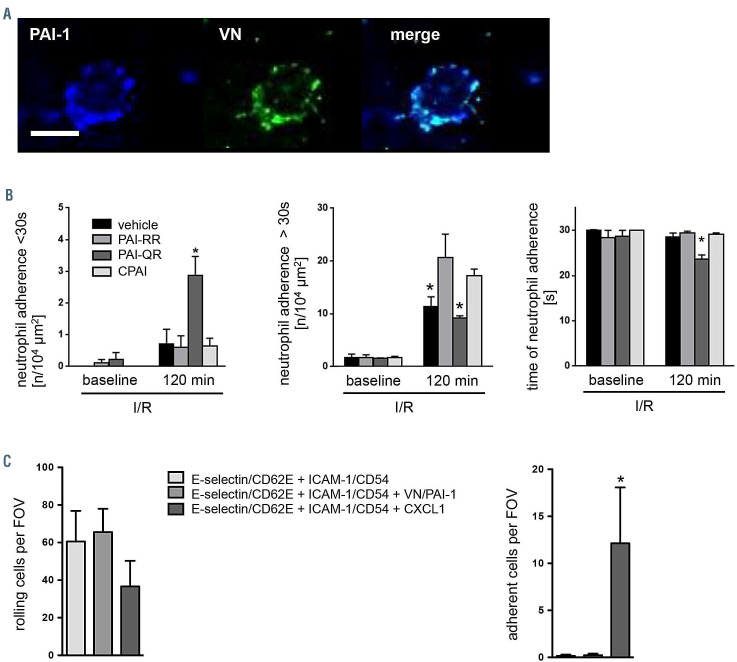
PAI-1 is another binding partner of VN that is also involved in neutrophil trafficking.20-24 We here confirm that murine PAI-1 (Online Supplementary Figure S2) binds to VN, but not to fibronectin (FN) (Online Supplementary Figure S3A), ultimately forming VN-PAI-1 heteromers (Online Supplementary Figure S3B) as evidenced by (sandwich) enzyme-linked immunosorbent assay (ELISA) analyses. Moreover, VN-PAI-1 heteromers were found in the peripheral blood of unstimulated mice whose levels slightly increased upon induction of systemic inflammation (Online Supplementary Figure S4). This increase in circulating VNPAI- 1 heteromers might be due to the release of VN and PAI-1 from the liver, of PAI-1 by the microvascular endothelium, as well as of VN, PAI-1, and pre-formed complexes of the single proteins by activated platelets28 in the acute phase of the inflammatory response. Immunostaining and confocal laser scanning microscopy on cremasteric tissue whole mounts further revealed that VN, PAI-1, and adherent neutrophils co-localize on the postischemic venular endothelium (Figure 4A). In order to evaluate the role of complex formation of VN with PAI-1 for the stabilization of neutrophil intravascular adhesion, we performed in vivo microscopy experiments in the postischemic cremaster muscle of PAI-1-deficient mice which received different PAI-1 mutant proteins (Figure 4B). Substitution of PAI-1-deficient mice with active stable PAI- 1 (CPAI) or a PAI-1 mutant protein lacking anti-protease activity (PAI-RR) resulted in significantly higher numbers of intravascularly firmly adherent (>30 s) neutrophils, significantly lower numbers of intravascularly shortly adhered (<30 s) neutrophils as well as higher average adhesion times of adherent neutrophils as compared to PAI-1- deficient mice substituted by a PAI-1 mutant protein lacking its VN binding domain (PAI-QR). In this context, substitution of PAI-deficient mice with PAI-QR induced short adhesion, but not of firm adherence of neutrophils to the microvascular endothelium as compared to vehicle-treated PAI-1-deficient animals (which might be due to PAI-1 binding to its receptor LRP-129 without prior interaction of PAI- 1 and VN). Further, substitution of VN-/- mice with VN-PAI- 1 protein rescued the adhesion defect arising from VN deficiency (Online Supplementary Figure S5). These results strengthen our concept that the interaction of VN and PAI- 1 is critical for the stabilization of intravascular adhesion of neutrophils.
In autoperfused flow chambers coated with CD62E/Eselectin and ICAM-1/CD54, an additional coating with VNPAI- 1 (Online Supplementary Figure S3) did not significantly alter rolling and adherence of neutrophils, whereas an additional coating with the chemokine CXCL1/KC induced a significant elevation in numbers of adherent neutrophils (Online Supplementary Figure S4C). These data suggest that chemokines, but not VN-PAI-1 heteromers, are critical for the induction of intravascular adherence of neutrophils.
Effect of vitronectin-plasminogen activator inhibitor-1 heteromers on activation of b2 integrins in neutrophils
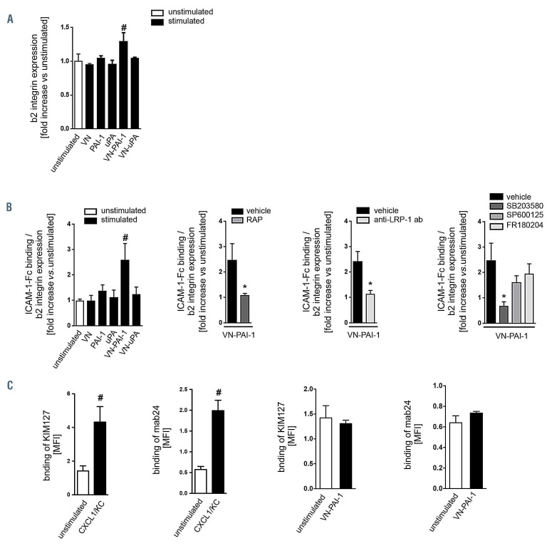
In inflamed tissue, intravascular firm adherence of neutrophils to the microvascular endothelium is facilitated by interactions of endothelial members of the immunoglobulin superfamily (e.g., ICAM-1/CD54) and neutrophil b2 integrins in higher affinity conformation.16,17 Employing multi-channel flow cytometry, exposure to VN-PAI-1, but not to VN, uPA, PAI-1, or VN-uPA significantly enhanced the fluorescence signal for the b2 integrins CD11a/LFA-1 and – to a lesser degree – CD11b/Mac-1 on the surface of neutrophils as compared to unstimulated controls (Online Supplementary Figures S5A and S6).
As a measure of conformational changes of b2 integrins, binding of their interaction partner ICAM-1/CD54 to neutrophils was analyzed in a next step. Similarly to our previous results for expression of b2 integrins, binding of ICAM-1/CD54 to neutrophils was significantly increased upon exposure to VN-PAI-1, but not upon exposure to VN, uPA, PAI-1, or VN-uPA, as compared to unstimulated controls (Figure 5B). Application of receptor associated protein (RAP; blocking members of the LDL receptor family), application of blocking anti-LRP-1 antibodies, or inhibitors of p38 (but not of JNK or ERK1/2) mitogen-activated protein kinases (MAPK) almost completely abolished VN-PAI- 1-elicited ICAM-1/CD54 binding.
In order to specifically evaluate the effect of VN-PAI-1 on conformational changes of b2 integrins, conformationspecific antibodies for the detection of the intermediate (KIM127) or high-affinity conformation (mAb 24) of b2 integrins (which are only available for human integrins) were used (Figure 5C). Upon exposure of VN-PAI-1 to human neutrophils, however, binding of ‘KIM127’ and ‘mAb 24’ was not significantly altered as compared to basal antibody binding in unstimulated controls whereas exposure to the chemokine CXCL1/KC significantly increased ‘KIM127’ and ‘mAb 24’ binding. Collectively, these data suggest that the increased ICAM-1/CD54 binding to neutrophils upon exposure to VN-PAI-1 (Figure 5B) is rather due to enhanced/optimized presentation of b2 integrins on the surface of these immune cells (Figure 5A; Figure 6) than to conformational changes in these adhesion and signaling molecules.
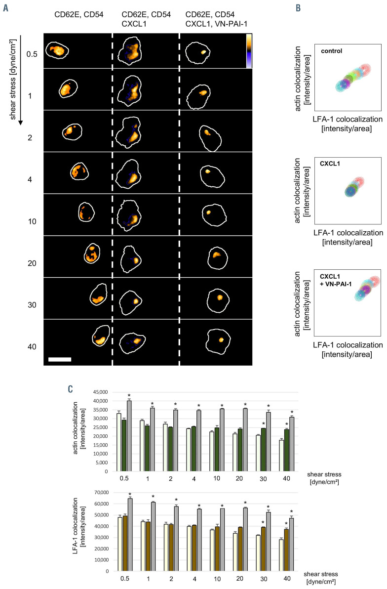
Since integrin clustering is thought to be particularly important in postadhesion strengthening of leukocyteendothelial cell interactions, we employed spinning disc confocal microscopy to study the effect of VN-PAI-1 heteromers on the cell membrane trafficking dynamics of b2 integrins in neutrophils (Figure 6A to C). In these experiments, we found that additional exposure of neutrophils isolated from the peripheral blood of WT mice to VNPAI- 1 heteromers significantly increased the clustering of the b2 integrin CD11a/LFA-1 as compared to exposure to the chemokine CXCL1/KC alone, or to phosphate buffered saline (PBS).
Systemic leukocyte counts and microhemodynamic parameters
In order to assure intergroup comparability in our in vivo microscopy experiments, systemic leukocyte counts and microhemodynamic parameters including blood flow velocity, inner vessel diameter, and wall shear rate were determined in each experiment. No significant differences were detected among experimental groups (Online Supplementary Table S1).
Figure 4.
Effect of vitronectin and plasminogen activator inhibitor-1 heteromers on the stabilization of neutrophil intravascular adhesion. (A) Using confocal laser scanning microscopy on tissue whole mounts of the postischemic cremaster muscle of wild-type (WT) mice, PAI-1 (blue) and vitronectin (VN) (green) deposited on the endothelium of postcapillary venules was detected to co-localize with intravascularly adherent Ly-6G+ neutrophils, representative images are shown (scale bar: 10 mm). (B) Using multi-channel in vivo microscopy on the postischemic mouse cremaster muscle, interactions of Ly-6G+ neutrophils and endothelial cells were analyzed in postcapillary venules. Panels show quantitative results for short adhesion and adhesion time of neutrophils in plasminogen activator inhibitor-1 (PAI-1)-deficient mice receiving vehicle or the PAI-1 mutant proteins PAI-RR (active stable mutant), PAI-QR (unable to bind VN), or CPAI (proteolytically inactive; mean±standard error of the mean [SEM] for n=4 animals per group; *P<0.05 vs. PAI-RR). See also Online Supplementary Table S1. (C) Employing an autoperfused flow chamber assay, intravascular rolling and firm adherence of neutrophils was analyzed in chambers coated with E-selectin/CD62E and ICAM-1/CD54 as well as with or without VN-PAI-1 heteromers or the chemokine CXCL1/KC, panels show quantitative results (mean±SEM for n=7–14 per group; *P<0.05 vs. E-selectin/CD62E + ICAM- 1/CD54 coating). I/R: ischemia-reperfusion; min: muntes, FOV: field of view.
Discussion
The trafficking of circulating leukocytes from the venular microvasculature to the site of injury or infection is a key event in the pathogenesis of inflammatory diseases.11-14 The role of the matricellular protein VN in this fundamental biological process is still unclear. Under homeostatic conditions, VN predominantly circulates in the blood as a monomer. In the acute inflammatory response, however, the binding of activated PAI-1 to VN monomers induces conformational changes in this macromolecule that effectively promote its multimerization. This allows multimeric VN to specifically bind to the surface of endothelial cells and, in turn, to unfold previously cryptic binding sites as evidenced by different in vitro studies.30-32 Accordingly, we found VN to be deposited on the luminal surface of the venular microvasculature in inflamed tissue. Since GAG cover the luminal aspect of microvascular endothelial cells and have previously been reported to interact with VN through its core polypeptide,33 we hypothesized that these carboanhydrates serve as binding partner for VN on the inner vessel wall. Confirming this assumption, enzymatic degradation of GAG almost completely abrogated the deposition of VN on the activated microvascular endothelium, thus suggesting that VN is immobilized in inflamed tissue on the surface of microvascular endothelial cells by endothelial GAG.
In order to evaluate the functional relevance of VN for leukocyte migration to the site of inflammation, we employed a peritoneal leukocyte trafficking assay. In these experiments, neutrophil extravasation to the peritoneal cavity was severely compromised in VN-/- mice as compared to WT controls. Notably, this impairment in neutrophil recruitment reached similar levels in heterozygous and homozygous VN-/- mice suggesting that comparatively large amounts of endothelially deposited VN are required for the induction of neutrophil responses. Moreover, we found that extravasation of classical/inflammatory monocytes remained unaffected by VN deficiency collectively indicating that VN particularly mediates the trafficking of neutrophils to inflamed tissue. This might be explained by higher expression of the scavenger receptor LRP-1 (which serves as a receptor of the VN binding partner PAI-1) on activated neutrophils as compared to activated classical monocytes, hence extending previous observations on the involvement of VN in leukocyte recruitment under inflammatory conditions. 34-38
Figure 5.
Effect of vitronectin and plasminogen activator inhibitor-1 heteromers on activation of neutrophil b2 integrins. (A) Using multi-channel flow cytometry, expression of the b2 integrins LFA-1/CD11a and Mac-1/CD11b was analyzed on the surface of neutrophils isolated from the peripheral blood of wild-type (WT) mice undergoing exposure to vitronectin (VN), plasminogen activator inhibitor-1 (PAI-1), uPA, VN-PAI-1, or VN-uPA, panels show quantitative results (mean±standard error of the mean [SEM] for n=4 per group; #P<0.05 vs. unstimulated). (B) Binding of ICAM-1/CD54-Fc to neutrophils isolated from the peripheral blood of WT mice was analyzed upon exposure to VN, PAI-1, uPA, VN-PAI-1, or VN-uPA, panels show quantitative results (mean±SEM for n=6 per group; #P<0.05 vs. unstimulated). VN-PAI-1-elicited binding of ICAM-1/CD54 to neutrophils isolated from the peripheral blood of WT mice was analyzed after application of receptor associated protein (RAP; blocking receptors of the LDL receptor family), blocking anti-LRP-1 antibodies, or different MAPK inhibitors (mean±SEM for n=4-6 per group; #P<0.05 vs. unstimulated). Binding of conformation- specific antibodies ‚KIM127‘ (intermediate and high affinity conformations of b2 integrins) or ‚mAB 24‘ (high affinity conformation of b2 integrins) to human neutrophils was analyzed after application of human VN-PAI-1 (mean±SEM for n=3 per group). MFI: mean fluorescent intensity.
Figure 6.
Effect of vitronectin and plasminogen activator inhibitor-1 heteromers on clustering of neutrophil b2 integrins. (A) Representative images of LFA-1/CD11a re-distribution and clustering in single neutrophils perfused over surfaces coated with E-selectin/CD62E, ICAM-1/CD54, and CXCL1 or CXCL1/VN-PAI-1 at indicated shear stress levels. LFA-1/CD11a expression is shown in pseudocolors as indicated by the lookup table (scale bar: 10 mm). (B) Scatter plot analysis of co-localized actin sum intensities versus co-localized LFA-1/CD11a sum intensities of single cells at indicated shear stress levels. (C) Quantification of actin co-localization and LFA-1/CD11a co-localization per clustered area at indicated shear stress levels (E-Selectin/CD62E + ICAM-1/CD54 coating: light green/yellow; E-selectin/CD62E + ICAM-1/CD54 + CXCL1 coating: dark green/brown; E-selectin/CD62E + ICAM-1/CD54 + CXCL1 + VN-PAI-1: grey; mean±standard deviation for n=3 independent experiments, n=6 flow chambers/coating, n=40-56 single cells/coating; *P<0.05 vs. E-selectin/CD62E + ICAM-1/CD54 coating).
With respect to the deposition of VN on the surface of postcapillary venules in inflamed tissue, we hypothesized that this glycoprotein contributes to the regulation of intravascular interactions of neutrophils in the extravasation process of these immune cells. Here, we show that VN deficiency does not alter the rolling behavior of neutrophils in inflamed postcapillary venules, suggesting that VN does not influence the ‚selectin-dependent phase‘ of the neutrophil extravasation process. Most interestingly, however, the average adherence time of a neutrophil to the endothelial surface was significantly lower in VN-/- mice than in WT controls. Furthermore, adoptively transferred neutrophils from WT donor mice accumulated less efficiently in the inflamed tissue of VN-/- recipient mice as compared to neutrophils isolated from VN-/- or WT donor mice transferred into WT recipient animals. Hence, our findings clearly demonstrate that endothelially deposited VN stabilizes intravascular adherence of neutrophils on the microvascular endothelium in inflamed tissue. To our knowledge, this is the first description of a protein specifically regulating this critical step in the extravasation cascade of neutrophils. Further, our observations indicate that stable intravascular adherence of neutrophils is prerequisite for (and does not interfere with) the subsequent transmigration of these immune cells into the perivascular space, a process that is facilitated by sequential heterophilic (e.g., between neutrophil LFA-1/CD11a and endothelial ICAM-2/CD102 or JAM-A as well as between neutrophil Mac-1/CD11b and ICAM-2/CD102 or JAM-C) and homophilic (between neutrophil and endothelial PECAM-1/CD31 or CD99) molecular interactions between neutrophils and endothelial cells as well as by endothelial molecules such as VE-cadherin, ESAM, or CD99L2.11-14
In addition to interactions with endothelial cells, the interplay between leukocytes and platelets considerably contributes to the extravasation of leukocytes.39-42 In this context, platelets have recently been demonstrated to guide intravascularly crawling neutrophils and monocytes to their site of transmigration into the interstitial tissue. 19 Since VN is able to bind to platelets through αvb3 and αIIbb3 integrins via its RGD motif43,44 these cellular blood components might also participate in VN-dependent neutrophil trafficking. In further experiments, however, VN deficiency neither altered intravascular adherence of platelets nor interactions of intravascularly adherent platelets and neutrophils. In line with these observations, platelet depletion did also not significantly change intravascular adhesion times of neutrophils in inflamed tissue collectively indicating that platelets do not contribute to the stabilization of neutrophil adherence in the microvasculature.
Besides platelet integrins, integrins expressed on the surface of leukocytes including αvb3, αvb5, αvb1, αLb2, and αMb2 represent potential interaction partners of VN.3-5 Similar to our results for platelet integrins, however, blockade of these integrins did not significantly alter the average time of neutrophils resting on the endothelial surface of postcapillary venules in the inflamed mouse cremaster muscle.
Beyond their established role in fibrinolysis, the components of the fibrinolytic system are increasingly recognized as mediators of immune cell migration.20-24 Recently, PAI-1 has been implicated in leukocyte trafficking to the site of inflammation by regulating intravascular adherence and (subsequent) transmigration of these immune cells.29 Since VN is capable of binding to PAI-1 via its somatomedin B domain (thereby extending the half-life of this protease inhibitor in fibrinolysis,3-5) heteromerization of VN with PAI-1 might promote intravascular adherence of neutrophils to the endothelium in inflamed tissue. In order to prove this hypothesis, we reconstituted PAI-1- deficient animals with different PAI-1 mutant proteins. Substitution with active stable PAI-1 or a PAI-1 mutant protein lacking anti-protease activity completely rescued the adhesion and extravasation defect of neutrophils observed in PAI-1-deficient mice. Substitution of PAI-1- deficient animals with a PAI-1 mutant protein lacking its VN binding domain, however, significantly diminished the average intravascular adhesion time of neutrophils as compared to active stable PAI-1-substituted mice resembling our observations in VN-deficient animals. Conversely, substitution of PAI-1-deficient mice with this non-VN-binding PAI-1 mutant protein induced short, but not firm adherence of neutrophils to the inflamed vessel wall which might be due to VN-independent binding of PAI-1 to its receptor LRP-1.29 Finally, substitution of VNdeficient animals with VN-PAI-1 heteromer protein completely rescued the adhesion defect of neutrophils observed in animals lacking VN. Consequently, complex formation of VN with PAI-1 is needed for the stabilization of intravascular adherence of neutrophils in the acute inflammatory response.
Intravascular firm adherence of leukocytes to microvascular endothelial cells is facilitated by interactions between endothelially expressed members of the immunoglobulin superfamily (e.g., ICAM-1/CD54, VCAM-1/CD106) and leukocyte b2 integrins in higher affinity conformations.11-14 We therefore proposed that VN-PAI-1 heteromers stabilize intravascular adhesion of neutrophils in the inflamed microvasculature by activating neutrophil b2 integrins. In our experiments, endothelial- bound VN and PAI-1 were identified to co-localize with intravascularly adherent neutrophils pointing to interactions of complexes of VN and PAI-1 with adhering neutrophils in the inflamed venular microvasculature. Importantly, however, exposure of neutrophils to the VNPAI- 1 complex (but not to VN, PAI-1, and uPA alone or to the VN-uPA complex) induced a significant increase in the expression of CD11a/LFA-1 and – to a lesser degree – of CD11b/Mac-1 on their cell surface, but did not further promote affinity changes in these b2 integrins. Although the induction of conformational changes in integrins enhances their affinity for their individual binding partners, the formation of multiple bonds to multivalent substrates in a process termed ‘integrin clustering’ is thought to be prerequisite for sustained cell adhesion. In leukocyte adhesion, integrins initially form transient microclusters on the surface of these immune cells that confluent into large focal adhesions. This leads to the establishment of complex clusters of adhesion and signaling molecules that trigger outside-in signaling, thus representing a critical event in post-adhesion strengthening.45-47 Here, we demonstrate that VN-PAI-1 heteromers substantially regulate the clustering of b2 integrins on the surface of neutrophils. With respect to our in vivo microscopy observations, these findings conversely suggest that the stabilization of neutrophil intravascular adherence particularly requires integrin clustering on these immune cells rather than the induction of conformational changes of integrins. Inactive integrins exhibit a bent-closed conformation, not extended (E−) and not high affinity (H−), and are thus unable to bind a ligand. In contrast, fully activated integrins (E+H+) bind to ICAM-1/CD54 expressed on opposing cells in trans (e.g., supporting neutrophil arrest). Integrins transitioning from the resting (E−H−) to the fully activated (E+H+) conformation through E+H− or E-H+ conformations, however, bind to ICAM-1/CD54 on the same cell in cis. In (intravascularly rolling) neutrophils, this transition process prohibits the arrest of these immune cells on the microvascular endothelium.48 Recently, not only E+H+ integrins, but already E-H+ and E+H- integrins were found to form clusters on the surface of neutrophils during cell activation.49 Since endothelially presented VNPAI- 1 heteromers specifically mediate the stabilization of neutrophil adherence on the microvascular endothelium, these protein complexes are thought to promote the binding of clustering integrins in trans.
Importantly, however, we are not able to exclude that binding of VN to PAI-1 concomitantly interferes with the inhibitory action of PAI-1 on tissue plasminogen activator (tPA), a proteolytic enzyme that has also been demonstrated to promote leukocyte trafficking to the site of inflammation.50 Furthermore, previous in vitro studies proposed a mechanism in which uPA cleaves the RGD motif of VN thereby reducing pro-adhesive properties of VN in an uPAR-dependent manner. Binding of PAI-1 to uPA in this protein complex effectively interferes with these events and subsequently restores the pro-adhesive properties of VN.51 In addition, binding of VN to uPAR has been shown to induce integrin signaling in cells by transmitting a mechanical stimulus to the integrin through the plasma membrane ultimately facilitating cell adhesion.52 Since uPAR has recently been demonstrated to be dispensable for the extravasation of neutrophils in the acute inflammatory response (at least in the model systems employed in the present study),53 we cannot clearly state to which extent these uPAR-dependent mechanisms contribute to VN-dependent stabilization of intravascular adherence of neutrophils in the inflamed microvasculature. Along the same line, fibrin(ogen) has been described to facilitate interactions between leukocyte integrins and endothelial ICAM- 1/CD54.54 In this context, however, fibrin(ogen) has been identified to specifically promote the transmigration of neutrophils to postischemic tissue.50
In order to restore homeostasis, VN-PAI-1 heteromers are cleared from circulation through the scavenger receptor LRP-1.25-27 Recently, we have demonstrated that PAI- 1-dependent leukocyte responses in sterile injury are mediated via this receptor protein and subsequent MAPK-dependent intracellular signaling events.29 VNPAI- 1 heteromer-dependent activation of neutrophil b2 integrins might therefore also rely on such molecular processes. In line with this hypothesis, we show that VN-PAI-1 heteromer-elicited surface trafficking of b2 integrins in neutrophils requires LRP-1 and p38 (but not JNK or ERK1/2) MAPK-dependent signaling events. Noteworthy, also selectin- and integrin-triggered signals contribute to the activation of p38 MAPK15 collectively suggesting that these selectin- and integrin-mediated molecular events (already occurring at the initial stages of the leukocyte recruitment process) are not sufficient for the stabilization of intravascular adhesion of neutrophils, but might prepare this critical step in the extravasation cascade of these immune cells.
Figure 7.
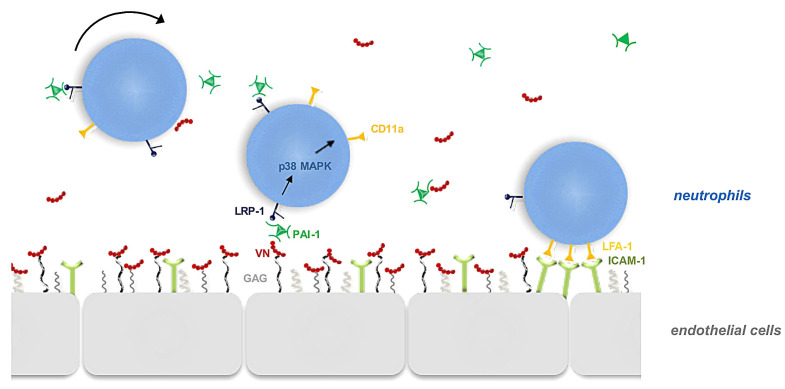
Graphical synopsis. In inflamed tissue, heteromers of vitronectin (VN) and plasminogen activator inhibitor-1 (PAI-1) are deposited on the activated venular endothelium via glycosaminoglycans (GAG). Subsequently, this protein complex interacts with LRP-1 on intravascularly adhering neutrophils and induces p38 MAPKdependent intracellular signaling pathways. As a consequence of these events, b2 integrins form clusters on the surface neutrophils ultimately allowing these immune cells to stabilize their adhesion via endothelial ICAM-1/CD54 in trans.
In conclusion, our experimental data indicate that the heteromerization of VN with PAI-1 on the microvascular endothelium of inflamed tissue represents a critical event for the extravasation process of neutrophils that substantially controls the transition of dynamic to static endothelial interactions of these immune cells. To this end, VNPAI- 1 heteromers promote the surface clustering of b2 integrins on adhering neutrophils, thus enabling effective neutrophil responses (Figure 7). Targeting this process might be beneficial for the prevention and treatment of inflammatory diseases.
Supplementary Material
Acknowledgements
The authors thank Susanne Bierschenk for excellent technical assistance. Data presented in this study are part of the doctoral thesis of MR.
Funding Statement
Funding: This study was supported by Deutsche Forschungsgemeinschaft (DFG), Sonderforschungsbereich (SFB) 914, projects Z03 (to MS), B01 (to MS), and B03 (to CAR).
References
- 1.Seiffert D, Keeton M, Eguchi Y, Sawdey M, Loskutoff DJ. Detection of vitronectin mRNA in tissues and cells of the mouse. Proc Natl Acad Sci USA. 1991;88(21):9402-9406. [DOI] [PMC free article] [PubMed] [Google Scholar]
- 2.Izumi M, Yamada KM, Hayashi M.Vitronectin exists in two structurally and functionally distinct forms in human plasma. Biochim Biophys Acta. 1989;990(2):101-108. [DOI] [PubMed] [Google Scholar]
- 3.Preissner KT, Reuning U.Vitronectin in vascular context: facets of a multitalented matricellular protein. Sem Thromb Hemost. 2011;37(4):408-424. [DOI] [PubMed] [Google Scholar]
- 4.Preissner KT, Jenne D.Structure of vitronectin and its biological role in haemostasis. Thromb Haemost. 1991;66(1):123-132. [PubMed] [Google Scholar]
- 5.Preissner KT, Seiffert D.Role of vitronectin and its receptors in haemostasis and vascular remodeling. Thromb Res. 1998;89(1):1-21. [DOI] [PubMed] [Google Scholar]
- 6.Huang Y, Haraguchi M, Lawrence DA, Border WA, Yu L, Noble NA. A mutant, noninhibitory plasminogen activator inhibitor type 1 decreases matrix accumulation in experimental glomerulonephritis. J Clin Invest. 2003;112(3):379-388. [DOI] [PMC free article] [PubMed] [Google Scholar]
- 7.Martins CO, Demarchi L, Ferreira FM, et al. Rheumatic heart disease and myxomatous degeneration: differences and similarities of valve damage resulting from autoimmune reactions and matrix disorganization. PloS One. 2017;12(1):e0170191. [DOI] [PMC free article] [PubMed] [Google Scholar]
- 8.Stoop AA, Lupu F, Pannekoek H.Colocalization of thrombin, PAI-1, and vitronectin in the atherosclerotic vessel wall: A potential regulatory mechanism of thrombin activity by PAI-1/vitronectin complexes. Arterioscl Thromb Vasc Biol. 2000;20(4):1143-1149. [DOI] [PubMed] [Google Scholar]
- 9.Dufourcq P, Couffinhal T, Alzieu P, et al. Vitronectin is up-regulated after vascular injury and vitronectin blockade prevents neointima formation. Cardiovasc Res. 2002;53(4):952-962. [DOI] [PubMed] [Google Scholar]
- 10.Reheman A, Gross P, Yang H, et al. Vitronectin stabilizes thrombi and vessel occlusion but plays a dual role in platelet aggregation. J Thromb Haemost. 2005;3(5):875-883. [DOI] [PubMed] [Google Scholar]
- 11.Ley K, Laudanna C, Cybulsky MI, Nourshargh S.Getting to the site of inflammation: the leukocyte adhesion cascade updated. Nat Rev Immunol. 2007;7(9):678-689. [DOI] [PubMed] [Google Scholar]
- 12.Nourshargh S, Alon R.Leukocyte migration into inflamed tissues. Immunity. 2014;41(5):694-707. [DOI] [PubMed] [Google Scholar]
- 13.Vestweber D. How leukocytes cross the vascular endothelium. Nat Rev Immunol. 2015;15(11):692-704. [DOI] [PubMed] [Google Scholar]
- 14.Kolaczkowska E, Kubes P.Neutrophil recruitment and function in health and inflammation. Nat Rev Immunol. 2013;13(3):159-175. [DOI] [PubMed] [Google Scholar]
- 15.Zarbock A, Ley K, McEver RP, Hidalgo A.Leukocyte ligands for endothelial selectins: specialized glycoconjugates that mediate rolling and signaling under flow. Blood. 2011;118(26):6743-6751. [DOI] [PMC free article] [PubMed] [Google Scholar]
- 16.Hogg N, Henderson R, Leitinger B, McDowall A, Porter J, Stanley P.Mechanisms contributing to the activity of integrins on leukocytes. Immunol Rev. 2002;186:164-171. [DOI] [PubMed] [Google Scholar]
- 17.Shattil SJ, Kim C, Ginsberg MH. The final steps of integrin activation: the end game. Nat Rev Mol Cell Biol. 2010;11(4):288-300. [DOI] [PMC free article] [PubMed] [Google Scholar]
- 18.Rose DM, Alon R, Ginsberg MH. Integrin modulation and signaling in leukocyte adhesion and migration. Immunol Rev. 2007;218:126-134. [DOI] [PubMed] [Google Scholar]
- 19.Zuchtriegel G, Uhl B, Puhr-Westerheide D, et al. Platelets guide leukocytes to their sites of extravasation. PLoS Biol. 2016;14(5):e1002459. [DOI] [PMC free article] [PubMed] [Google Scholar]
- 20.Del Rosso M, Fibbi G, Pucci M, Margheri F, Serrati S.The plasminogen activation system in inflammation. Front Biosci. 2008;13:4667-4686. [DOI] [PubMed] [Google Scholar]
- 21.Del Rosso M, Margheri F, Serrati S, Chilla A, Laurenzana A, Fibbi G.The urokinase receptor system, a key regulator at the intersection between inflammation, immunity, and coagulation. Curr Pharm Des. 2011;17(19):1924-1943. [DOI] [PubMed] [Google Scholar]
- 22.Reichel CA, Kanse SM, Krombach F.At the interface of fibrinolysis and inflammation: the role of urokinase-type plasminogen activator in the leukocyte extravasation cascade. Trends Cardiovasc Med. 2012;22(7):192-196. [DOI] [PubMed] [Google Scholar]
- 23.Smith HW, Marshall CJ. Regulation of cell signalling by uPAR. Nat Rev Mol Cell Biol. 2010;11(1):23-36. [DOI] [PubMed] [Google Scholar]
- 24.Syrovets T, Lunov O, Simmet T.Plasmin as a proinflammatory cell activator. J Leukocy Biol. 2012;92(3):509-519. [DOI] [PubMed] [Google Scholar]
- 25.Herz J, Strickland DK. LRP: a multifunctional scavenger and signaling receptor. J Clin Invest. 2001;108(6):779-784. [DOI] [PMC free article] [PubMed] [Google Scholar]
- 26.Gliemann J. Receptors of the low density lipoprotein (LDL) receptor family in man. Multiple functions of the large family members via interaction with complex ligands. Biol Chem. 1998;379(8-9):951-964. [PubMed] [Google Scholar]
- 27.Rodenburg KW, Kjoller L, Petersen HH, Andreasen PA. Binding of urokinase-type plasminogen activator-plasminogen activator inhibitor-1 complex to the endocytosis receptors alpha2-macroglobulin receptor/low-density lipoprotein receptorrelated protein and very-low-density lipoprotein receptor involves basic residues in the inhibitor. Biochem J. 1998;329(Pt 1):55-63. [DOI] [PMC free article] [PubMed] [Google Scholar]
- 28.Preissner KT, Holzhuter S, Justus C, Muller-Berghaus G. Identification of and partial characterization of platelet vitronectin: evidence for complex formation with plateletderived plasminogen activator inhibitor-1. Blood. 1989;74(6):1989-1996. [PubMed] [Google Scholar]
- 29.Praetner M, Zuchtriegel G, Holzer M, et al. Plasminogen activator inhibitor-1 promotes neutrophil infiltration and tissue injury on ischemia-reperfusion. Arterioscl Thromb Vasc Biol. 2018;38(4):829-842. [DOI] [PubMed] [Google Scholar]
- 30.Seiffert D, Loskutoff DJ. Type 1 plasminogen activator inhibitor induces multimerization of plasma vitronectin. A suggested mechanism for the generation of the tissue form of vitronectin in vivo. J Biol Chem. 1996;271(47):29644-29651. [DOI] [PubMed] [Google Scholar]
- 31.Volker W, Hess S, Vischer P, Preissner KT. Binding and processing of multimeric vitronectin by vascular endothelial cells. J Histochemi Cytochem. 1993;41(12):1823-1832. [DOI] [PubMed] [Google Scholar]
- 32.Seiffert D, Smith JW. The cell adhesion domain in plasma vitronectin is cryptic. J Biol Chem. 1997;272(21):13705-13710. [DOI] [PubMed] [Google Scholar]
- 33.Leavesley DI, Kashyap AS, Croll T, et al. Vitronectin--master controller or micromanager? IUBMB Life. 2013;65(10):807-818. [DOI] [PubMed] [Google Scholar]
- 34.Tsuruta Y, Park YJ, Siegal GP, Liu G, Abraham E.Involvement of vitronectin in lipopolysaccaride-induced acute lung injury. J Immunol. 2007;179(10):7079-7086. [DOI] [PubMed] [Google Scholar]
- 35.Kanse SM, Matz RL, Preissner KT, Peter K.Promotion of leukocyte adhesion by a novel interaction between vitronectin and the beta2 integrin Mac-1 (alphaMbeta2, CD11b/CD18). Arterioscl Thromb Vasc Biol. 2004;24(12):2251-2256. [DOI] [PubMed] [Google Scholar]
- 36.Frieser M, Hallmann R, Johansson S, Vestweber D, Goodman SL, Sorokin L.Mouse polymorphonuclear granulocyte binding to extracellular matrix molecules involves beta 1 integrins. Eur J Immunol. 1996;26(12):3127-3136. [DOI] [PubMed] [Google Scholar]
- 37.Chavakis T, May AE, Preissner KT, Kanse SM. Molecular mechanisms of zinc-dependent leukocyte adhesion involving the urokinase receptor and beta2-integrins. Blood. 1999;93(9):2976-2983. [PubMed] [Google Scholar]
- 38.Edwards S, Lalor PF, Tuncer C, Adams DH. Vitronectin in human hepatic tumours contributes to the recruitment of lymphocytes in an alpha v beta3-independent manner. Br J Canc. 2006;95(11):1545-1554. [DOI] [PMC free article] [PubMed] [Google Scholar]
- 39.Rossaint J, Zarbock A.Platelets in leucocyte recruitment and function. Cardiovasc Res. 2015;107(3):386-395. [DOI] [PubMed] [Google Scholar]
- 40.Sreeramkumar V, Adrover JM, Ballesteros I, et al. Neutrophils scan for activated platelets to initiate inflammation. Science. 2014;346(6214):1234-1238. [DOI] [PMC free article] [PubMed] [Google Scholar]
- 41.Ed Rainger G, Chimen M, Harrison MJ, et al. The role of platelets in the recruitment of leukocytes during vascular disease. Platelets. 2015;26(6):507-520. [DOI] [PMC free article] [PubMed] [Google Scholar]
- 42.Jenne CN, Kubes P.Platelets in inflammation and infection. Platelets. 2015;26(4):286-292. [DOI] [PubMed] [Google Scholar]
- 43.Ma D, Xu X, An S, et al. A novel family of RGD-containing disintegrins (Tablysin-15) from the salivary gland of the horsefly Tabanus yao targets alphaIIbbeta3 or alphaVbeta3 and inhibits platelet aggregation and angiogenesis. Thromb Haemost. 2011;105(6):1032-1045. [DOI] [PMC free article] [PubMed] [Google Scholar]
- 44.Wright PS, Saudek V, Owen TJ, Harbeson SL, Bitonti AJ. An echistatin C-terminal peptide activates GPIIbIIIa binding to fibrinogen, fibronectin, vitronectin and collagen type I and type IV. Biochem J. 1993;293(Pt 1):263-267. [DOI] [PMC free article] [PubMed] [Google Scholar]
- 45.Ortega-Gomez A, Salvermoser M, Rossaint J, et al. Cathepsin G controls arterial but not venular myeloid cell recruitment. Circulation. 2016;134(16):1176-1188. [DOI] [PMC free article] [PubMed] [Google Scholar]
- 46.Herter J, Zarbock A.Integrin regulation during leukocyte recruitment. J Immunol. 2013;190(9):4451-4457. [DOI] [PubMed] [Google Scholar]
- 47.Iwamoto DV, Calderwood DA. Regulation of integrin-mediated adhesions. Curr Opin Cell Biol. 2015;36:4147. [DOI] [PMC free article] [PubMed] [Google Scholar]
- 48.Fan Z, McArdle S, Marki A, et al. Neutrophil recruitment limited by highaffinity bent beta2 integrin binding ligand in cis. Nat Commun. 2016;7:12658. [DOI] [PMC free article] [PubMed] [Google Scholar]
- 49.Fan Z, Kiosses WB, Sun H, et al. High-affinity bent beta2-integrin molecules in arresting neutrophils face each other through binding to ICAMs In cis. Cell Rep. 2019;26(1):119-130.e5. [DOI] [PMC free article] [PubMed] [Google Scholar]
- 50.Uhl B, Zuchtriegel G, Puhr-Westerheide D, et al. Tissue-type plasminogen activator promotes postischemic neutrophil recruitment via its proteolytic and nonproteolytic properties. Arterioscl Thromb Vasc Biol. 2014;34(7):1495-1504. [DOI] [PubMed] [Google Scholar]
- 51.De Lorenzi V, Sarra Ferraris GM, Madsen JB, Lupia M, Andreasen PA, Sidenius N.Urokinase links plasminogen activation and cell adhesion by cleavage of the RGD motif in vitronectin. EMBO Rep. 2016;17(7):982-998. [DOI] [PMC free article] [PubMed] [Google Scholar]
- 52.Ferraris GM, Schulte C, Buttiglione V, et al. The interaction between uPAR and vitronectin triggers ligand-independent adhesion signalling by integrins. EMBO J. 2014;33(21):2458-2472. [DOI] [PMC free article] [PubMed] [Google Scholar]
- 53.Reichel CA, Uhl B, Lerchenberger M, et al. Urokinase-type plasminogen activator promotes paracellular transmigration of neutrophils via Mac-1, but independently of urokinase-type plasminogen activator receptor. Circulation. 2011;124(17):1848-1859. [DOI] [PubMed] [Google Scholar]
- 54.Languino LR, Plescia J, Duperray A, et al. Fibrinogen mediates leukocyte adhesion to vascular endothelium through an ICAM-1- dependent pathway. Cell. 1993;73(7):1423-1434. [DOI] [PubMed] [Google Scholar]
Associated Data
This section collects any data citations, data availability statements, or supplementary materials included in this article.


