Abstract
Background
The systemic immune-inflammation index (SII) has been demonstrated to be a valid biomarker of a patient's immunological and inflammatory state, with the ability to accurately predict outcomes in a variety of disease conditions. In the absence of comparable studies, we intended to examine the relevance of pretreatment SII in predicting the success rates of temporomandibular joint arthrocentesis (TMJA) at 1-week, 1-month, and 6-month periods, defined as maximum mouth opening (MMO) > 35 mm and VAS ≤ 3.
Methods
A sum of 136 patients with disc displacement without reduction (DDwo-red) who underwent TMJA was included. For each patient, pre-TMJA SII was calculated as; SII = Platelets × neutrophils/lymphocytes. Additionally, baseline MMO and VAS measurements were recorded for each patient. The success criteria of TMJA included MMO > 35 mm and VAS ≤ 3. The optimal pre-TMJA SII cutoff that predicts TMJA success was determined using receiver operating characteristic (ROC) curve analysis. The primary endpoint was the link between the pre-treatment SII and TMJA success (simultaneous achievement of MMO > 35 mm and VAS ≤ 3).
Results
The median pre-TMJA jaw locking duration, maximum mouth opening (MMO), and visual analog score (VAS) were 7 days, 24 mm, and 8, respectively. The overall TMJA success rates were determined as 80.1%, 91.9%, and 69.1% at 1-week, 1-month, and 6-months, respectively. The results of ROC curve analysis exhibited the optimal SII cutoff at 526 (AUC: 67.4%; sensitivity: 66.7%; specificity: 64.2%) that grouped the patients into two subgroups: Group 1: SII ≤ 526 (N = 81) and SII > 526 (N = 55), respectively. Spearman correlation analysis revealed a strong inverse relationship between the pretreatment SII values and the success of TMJA 1-week (rs: − 0.83; P = 0.008) and 1-month, (rs: − 0.89; P = 0.03). Comparative analyses displayed that TMJA success rates at 1-week (87.7% vs. 69.1%; P = 0.008) and 1-month (96.2% vs. 80%; P = 0.03) were significantly higher in the SII ≤ 526 than SII > 526 group, respectively, while the 6-month results favored the SII ≤ 526 group with a trend approaching significance (P = 0.084).
Conclusion
The current study's findings suggested the SII as a unique independent prognostic biomarker that accurately predicts treatment outcomes for up to 6 months.
Trial registration The results of this research were retrospectively registered.
Keywords: Temporomandibular joint arthrocentesis, Systemic immune-inflammation index, Temporomandibular joint disorder, Biomarker, Success
Background
Temporomandibular joint disorders (TMD) are a diverse group of diseases affecting the temporomandibular joint (TMJ), masticatory muscles, and related structures with an overall prevalence rate of 5% to 12% in the general population [1, 2]. Severe pre-auricular and masticatory area pain, joint sounds, and diminished mouth opening capacity embodies the most frequent TMD-related presenting symptoms [3]. The most certain intraarticular cause of TMD is disc displacement with (DDw-red) or without reduction (DDwo-red), which can cause severe joint degeneration and a sharp decline in quality-of-life (QoL) measures related to persistent chronic pain, impaired eating functions, limitation of normal jaw activities, difficulty in falling asleep, and resulting fatigue in DDwo-red patients [2, 4]. Blanco et al. concluded in the largest study ever conducted, with 1220 individuals, that TMD can induce significant pain sensations that impair QoL measurements. Furthermore, Bitiniene et al. showed in a comprehensive systematic review encompassing 12 research published between 2006 and 2016 that psychological and physical problems caused by TMD resulted in diminished patient QoL records [5, 6].
TMJ arthrocentesis (TMJA), with its 70% to 90% long-term success rate, is a relatively easy to perform and highly feasible minimally invasive procedure that efficiently reduces the complaints of DDwo-red patients who are resistant to conservative treatments (self-care measures, occlusal splint, and physical therapy) and drugs [7, 8]. However, literature related to the short-term success of TMJA is scant [9]. The reported prognostic factors for the TMJA procedure usually refer to the patient’s age, disease duration, pain severity, maximum mouth opening (MMO) capacity, and the presence of degenerative changes in the magnetic resonance imaging (MRI) scans. But sadly, emphasizing the compelling need for the identification of novel reliable prognosticators, accessible research results are often conflicting [10].
Another implied cause of TMD is local and chronic systemic inflammation, which manifests with the secretion of various biomarkers in the TMJ synovium, such as cytokines and growth factors [11]. Therefore, TMJA procedure is commonly used to defeat this inflammatory load in the joint cavity, while this procedure additionally promotes disc repair and repositioning by removing the fibrous tissues in DDwo-red patients [7, 8]. In this sense, it is of interest to identify TMD-specific inflammatory cytokines and develop specific treatments for this particular region [10]. For this purpose, Somay and Araz and Kaneyama et al. investigated the influence of pro-inflammatory cytokines and their values in synovial fluid on the success of TMJA, and declared that some inflammation and inflammatory mediators were extremely high in the synovial fluid, which meaningfully impacted the TMJA success rates [12, 13].
SII (Systemic Immune-Inflammation Index), a unique measure of the platelet, neutrophil, and lymphocyte counts, is a biomarker that mirrors the harmony between the patient’s inflammatory and immune status, irrespective of the underlying cause [14]. SII has recently been reported to be effective in predicting the prognosis of many diseases, including osteoporosis, osteoporotic bone fractures, psoriatic arthritis, and Bell's palsy [15–17]. In a clinical study of 23 patients, Somay and Araz investigated the impact of the synovial fluid sIL-1RII, sTNF-αRI and sIL-6R concentrations on the success rates of TMJA procedure [12]. The authors reported that the higher sIL-6R concentrations were associated with significantly reduced TMJA success rates.
Nevertheless, to the best of our knowledge, SII has never been examined for its actual impact on the success rates of the TMJA procedure, despite the accessibility of clinical evidence indicating a considerable detrimental influence of inflammation on the success of the TMJA. Consequently, given that the primary goal of the TMJA procedure is to remove the inflammatory debris together with the presence of the above-mentioned positive clinical data [12], we sought to investigate the prognostic significance of the pre-treatment SII on the short-term success of the TMJA procedure.
Materials and methods
Ethical approval
The present retrospective study was designed, authorized and approved by the institutional review board of Baskent University Medical Faculty (Project no: D-KA19/19) before the compilation of any persistent data, according to the Declaration of Helsinki. The eligible patients provided signed informed consent before the initiation of all dental and medical procedures either themselves or legitimately charged caregivers for acquisition and analysis of the patients’ sociodemographic, dental, and medical records; blood samples, MRI scans, and publication of the outcomes. The results of this research were retrospectively registered and evaluated.
Study population
We conducted a retrospective search of clinical, medical, and radiological records (MRI, Magnetron “Harmony” Siemens, Erlangen, Germany) maintained by the Baskent University Adana Research and Practice Center Dentistry Clinic to identify ≥ 18 years old patients who underwent TMJA for TMD between September 2018 and July 2020. The Diagnostic Criteria for Temporomandibular Joint Disorders (DC/TMD) were used to determine the absence/presence of TMD [2, 18]. To be eligible, all patients had to have a diagnosis of DDwo-red according to these criteria, the Visual Analog Scale (VAS) measured TMJ pain value of ≥ 4, MMO < 35 mm, and available pre-TMJA complete blood count tests. Patients presenting with TMJ pain who had failed two months of conservative treatment (self-care preventions, occlusal splints, physical therapy, and medications) were included. In order to prevent the unintentional biasing effect of baseline immune and inflammatory conditions and drug usage patients who presented with either of the muscle-related pains, systemic inflammatory conditions such as rheumatologic diseases, nephritic disorders, respiratory diseases, viral hepatitis, TMJ ankylosis, proven immune suppressive disorders, chronic inflammatory conditions such as pancreatitis, previous TMJ surgery, history of trauma, and missing natural upper and/or lower central incisors were excluded from the present analysis. Patient complaints, like the presence of pain, restricted mouth opening, masticatory muscle tenderness, jaw deviation during mouth opening, bruxism, were all noted, and also the types of diagnosis indicated by MRI examination were grouped. The jaw locking duration was measured for each patient with limited mouth opening was also recorded, which was defined as the interim delaying the mouth opening and prohibiting eating functions according to the Mandibular Function Impairment Questionnaire [19]. All patients received conservative medications and physical therapy as a norm before the TMJA procedure, with TMJA being offered only for resistant cases.
Preoperative assessments
For each eligible patient, the age, gender, presence of bruxism, jaw deviation during mouth opening, and muscle tenderness were noted before the TMJA procedure. Bruxism was diagnosed when the patient had a history of tooth clenching and grinding occurring ≥ 3 nights per week at the past 6-months [20], the experience of morning stiffness, presence of tooth wear [21], and also the appearance of linea alba in the cheek [22].
The preoperative clinical evaluations incorporated the pain assessments with the VAS [23] and MMO measurements. We used the original version of the VAS assessment tool, which typically consists of a 10-cm line describing the maximum and minimum measurement size values, where the two extremes 0 and 10 represent no pain and unbearable pain, individually. The same competent dentomaxillofacial radiologist (BY) completed the VAS assessments for each patient [24]. Likewise, the MMO was assessed preoperatively and at 1-week, 1-month, and 6-month intervals following the TMJA procedure by the same dentomaxillofacial surgeon (ES). The VAS test was carried out at the same time intervals as the MMO measurements. The 6-month results were specifically obtained to examine the TMJA procedure's short-term efficacy. The treatment was considered successful if the MMO was > 35 mm and the VAS was less ≤ 3 simultaneously [7].
Measurement of maximum mouth opening (MMO)
We have utilized the range of motion scale Therabite® (Atos Medical AB, Hörby, Sweden) to measure MMOs since it enables easy and direct measurement and decreases the risk of procedure-related infection due to its disposable characteristic [25]. Each patient was instructed to open her/his mouth as widely as practically feasible to measure the distance between the top borders of one of the lower central incisors and the lower edge of one of the upper central incisors, with the Therabite® range of motion scale being inserted in the mouth. As a standard, MMO measurements were performed three times per session, with the mean MMO being computed as the average of the three successive measurements.
Arthrocentesis procedure
All patients underwent a single-session TMJA procedure performed by the same oral and maxillofacial surgeon (ES). After measuring the patient's MMO, the preauricular skin and ear regions were prepared by cleaning with a topical antiseptic solution (Povidone-Iodine 10% w/v), and the surgical area was confined with a sterile cover. To block the auriculotemporal nerve, we applied 1–2 ml of a local anesthetic solution [Ultracain® DS Forte 40 mg/ml articaine HCL, 0.0012 mg/ml epinephrine (Sanofi-Aventis, Frankfurt, Germany)] over the preauricular region and into the superior joint space (SJS). As previously described by Nitzan et al., a line was drawn from the most posterior and central point on the tragus to the lateral canthus of the eye by a sterile skin pen [7]. The first entry point was marked 10-mm anterior of the tragus and 2-mm below this line, while the second entry point was 20-mm anterior and 8-mm below this line. Then, a 21‐gauge needle was inserted into the SJS at the glenoid fossa through the first point, and approximately 2–3 mL of Ringer's solution was pumped ten times to expand the SJS. A second 20‐gauge needle was then inserted from the second entry point to enable the free flow of the irrigation solution in the SJS. Lavaging with free outflow under high pressure and with a minimum of 400 ml Ringer's solution was considered regarded as effective irrigation [7, 26]. All patients were advised for a soft diet, hot pad application, and passive stretching exercises for a week following surgery. Analgesics and myorelaxants were prescribed as needed. We standardly prescribed an occlusal splint for at least one month following surgery to prevent bruxism and associated problems.
Systemic immune-inflammation index (SII) assessment
We ran Hu’s original equation to calculate the pre-TMJA SII values for each patient: SII = [Platelets × (Neutrophils/Lymphocytes)], by using the routine complete blood count tests performed on the TMJA day [27].
Statistical analysis
The primary endpoint was the connection between the pretreatment SII values and the TMJA success. Medians and ranges were calculated to describe continuous variables, while categorical variables were expressed with percentage frequency distributions. Chi-square test, Student's t-test, or Spearman correlation analyses and related rs values were employed to compare the patient groups, as indicated. Receiver operating characteristic (ROC) curve analysis was employed to define ideal cutoff(s) that may group the whole study into two distinctive outcome groups, such as the pre-TMJA SII measures. All comparisons were two-tailed, and any P value < 0.05 was considered significant.
Results
Our retrospective database search yielded a sum of 136 patients who had underwent TMJA procedure. As summarized in Table 1, the median age was 34 years (range: 18–59), with a female predominance (77.9%). Pain (47.8%) and difficulty in mouth opening (33.1%) accounted for the majority (80.9%) of the presenting complaints. The median jaw locking duration, pre-TMJA MMO, pre-TMJA VAS were 7 days [95% confidence interval (CI): 2.8–14 days], 24 mm (95% CI: 17.8–28.4 mm), and 8 (95% CI: 6.7–8.7), respectively. Presenting patient and TMD characteristics including the muscle tenderness, deviation during mouth opening, and bruxism were as shown in Table 1. Of note, 50.7% of patients had bilateral DDwo-red (Table 1).
Table 1.
Baseline patient and temporomandibular disorder characteristics for all patients group and per systemic immune-inflammation index groups
| Characteristics | All patients (N = 136) | SII ≤ 526 (N = 81) | SII > 526 (N = 55) | P value |
|---|---|---|---|---|
| Median age, years (range) | 34 (18–59) | 35 (20–59) | 33 (19–58) | 0.81 |
| Gender, N (%) | ||||
| Female | 106 (77.9) | 64 (79.0) | 42 (76.4) | 0.86 |
| Male | 30 (22.1) | 17 (21.0) | 13 (23.6) | |
| Presenting complaints, N (%) | ||||
| Pain | 65 (47.8) | 39 (48.1) | 26 (47.3) | 0.38 |
| Difficulty in mouth opening | 45 (33.1) | 29 (35.8) | 16 (29.1) | |
| Both | 26 (19.1) | 13 (16.1) | 13 (23.6) | |
| Median jaw locking duration, day (95% CI) | 7 (2.8–14) | 8 (4–13) | 6 (3–14) | 0.22 |
| Median pre-TMJA MMO, mm (95% CI) | 24 (17.8–28.4) | 22.6 (18.2–28.1) | 24.7 (19.1–28.4) | 0.34 |
| Median pre-TMJA VAS (95% CI) | 8 (6–9) | 8 (7–9) | 8 (6–9) | 0.94 |
| Muscle tenderness, N (%) | ||||
| Present | 27 (19.9) | 16 (19.8) | 11 (20.0) | 0.91 |
| Absent | 109 (80.1) | 65 (80.2) | 44 (80.0) | |
| Deviation during mouth opening, N (%) | ||||
| Present | 44 (32.4) | 28 (34.6) | 16 (29.1) | 0.52 |
| Absent | 92 (67.6) | 53 (65.4) | 39 (70.9) | |
| MRI findings, N (%) | ||||
| Unilateral DDwo-red | 67 (49.3) | 37 (45.7) | 30 (54.5) | 0.41 |
| Bilateral DDwo-red | 69 (50.7) | 44 (54.3) | 25 (45.5) | |
| Bruxism, N (%) | ||||
| Present | 62 (45.6) | 40 (50.6) | 21 (38.2) | 0.19 |
| Absent | 74 (54.4) | 40 (49.4) | 34 (61.8) | |
CI confidence interval, DDwo-red disc displacement without reduction, mm millimeter, MMO maximum mouth opening, MRI magnetic resonance imaging, SII systemic immune-inflammation index, TMD temporomandibular joint disorder, TMJA temporomandibular joint arthrocentesis, VAS visual analog scale
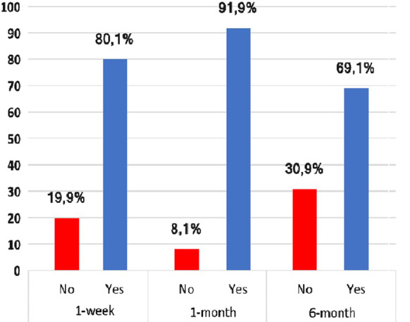
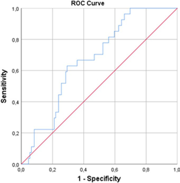
Overall, we found that the 1-week, 1-month, and 6-month TMJA success rates were 80.1% at, 91.9% at 1-month, and 69.1%, respectively, per success criteria defined as MMO > 35 mm and VAS ≤ 3 (Fig. 1). We used ROC curve analysis to reveal the possible connections between the pre-treatment SII levels and TMJA success at 1-week, 1-month, and 6-month intervals. The ROC curve analysis results exhibited the optimal cutoff value at 526 (Area under the curve (AUC): 67.4%; sensitivity: 66.7%; and specificity: 64.2%) for 1-week, 527 (AUC: 66.2%; sensitivity: 65.8%; and specificity: 64.0%), and 524 (AUC: 65.1%; sensitivity: 64.3%; and specificity: 64.1%) for 6-months, respectively. Since the three cutoff values were so close, we utilized the 526 as the common cutoff to separate patients into two groups for all time-dependent TMJA success evaluations (Fig. 2): Group 1: SII ≤ 526 (N = 81) and SII > 526 (N = 55), respectively. Spearman correlation analysis revealed a strong and significant inverse relationship between the pre-treatment SII values and the TMJA success at 1-week and 1-month (rs: − 0.83; P = 0.008, rs: − 0.89; P = 0.03), with an additional trend favoring the SII ≤ 526 group for the TMJA success at 6-month (rs = − 0.42; P = 0.086) evaluations (Table 2). Baseline patients and disease characteristics were almost evenly distributed between the two SII cohorts with no statistically significant differences between them (Table 1). As shown in Table 2, our comparative analyses displayed that the rates of TMJA success for 1-week (87.7% vs. 69.1%; P = 0.008) and 1-month (96.2% vs. 80%; P = 0.03) were significantly higher in the SII ≤ 526 group than its SII > 526 counterparts, respectively. Although the difference between the success rates of the two SII groups could not reach statistical significance at 6-months evaluations, we observed a strong trend approaching statistical significance favoring the SII ≤ 526 over the SII > 526 group (74.1% vs. 60%; P = 0.084).
Fig. 1.

Overall success of TMJA at 1-week, 1-month, and 6-months evaluations
Fig. 2.

Results of ROC analysis evaluating the relationship between the pretreatment SII measures and TMJA success
Table 2.
TMJA success for the per SII status
| TMJA success | All patients (N = 136) | SII ≤ 526 (N = 81) | SII > 526 (N = 55) | P value |
|---|---|---|---|---|
| 1-week, N (%) | ||||
| Yes | 109 (80.1) | 71 (87.7) | 38 (69.1) | 0.008 |
| No | 27 (19.8) | 10 (12.3) | 17 (30.9) | |
| 1-month, N (%) | ||||
| Yes | 122 (89.7) | 78 (96.3) | 44 (80.0) | 0.03 |
| No | 14 (10.3) | 3 (3.7) | 11 (20.0) | |
| 6-month, N (%) | ||||
| Yes | 99 (72.8) | 60 (74.1) | 33 (60.0) | 0.084 |
| No | 43 (27.2) | 21 (25.9) | 22 (40.0) | |
TMJA temporomandibular joint arthrocentesis, SII systemic immune-inflammation index
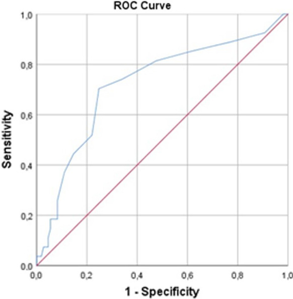
We further sought for the presence of additional relevant cutoff(s) for other covariates which may alter the TMJA success rates significantly in favor of one group (Table 3). Our search with ROC curve analysis revealed a significant cutoff uniquely for the jaw locking duration at a threshold of 7.5 days (AUC: 72.2% sensitivity: 74.1%; and specificity: 66.1%) (Fig. 3). Comparative analyses displayed that the rates of TMJA success were significantly higher in patients presenting with a jaw locking duration < 8 days at 1-week (66% vs. 34%; P < 0.001), 1-month (61.5% vs. 38.5%; P = 0.003), and 6-month (74.2% vs. 25.8%; P < 0.001) group than their ≥ 8 days counterparts.
Table 3.
TMJA success per presenting patient and TMD characteristics
| Characteristic | TMJA success 1-week | P value | TMJA success 1-month | P value | TMJA success 6-month | P value | |||
|---|---|---|---|---|---|---|---|---|---|
| Yes | No | Yes | No | Yes | No | ||||
| Muscle tenderness, N (%) | |||||||||
| Yes | 24 (22.0) | 3 (11.1) | 0.28 | 25 (20.5) | 2 (14.3) | 0.74 | 16 (17.2) | 11 (25.6) | 0.26 |
| No | 85 (78.0) | 24 (88.9) | 97 (79.5) | 12 (85.7) | 77 (82.8) | 32 (74.4) | |||
| Jaw deviation during mouth opening, N (%) | |||||||||
| Yes | 34 (31.2) | 10 (37.0) | 0.65 | 39 (32.0) | 5 (35.7) | 0.77 | 26 (28.0) | 18 (41.9) | 0.12 |
| No | 75 (68.8) | 17 (63.0) | 83 (68.0) | 9 (64.3) | 67 (72.0) | 25 (58.1) | |||
| Types of MRI findings, N (%) | |||||||||
| Unilateral DDwo-red | 51 (46.8) | 16 (59.3) | 0.27 | 60 (49.2) | 7 (50.0) | 0.66 | 45 (48.4) | 22 (51.2) | 0.86 |
| Bilateral DDwo-red | 58 (53.2) | 11 (40.7) | 62 (50.8) | 7 (50.0) | 48 (51.6) | 21 (48.8) | |||
| Bruxism, N (%) | |||||||||
| Yes | 53 (48.6) | 9 (33.3) | 0.20 | 57 (46.7) | 5 (35.7) | 0.57 | 44 (47.3) | 18 (41.9) | 0.58 |
| No | 56 (51.4) | 18 (66.7) | 65 (53.3) | 9 (64.3) | 49 (52.7) | 25 (51.1) | |||
| Jaw locking duration, N (%) | |||||||||
| ≥ 8 | 37 (34.0) | 20 (74.1) | 0.001 | 47 (38.5) | 10 (71.4) | 0.023 | 24 (25.8) | 33 (76.7) | 0.001 |
| < 8 | 72 (66.0) | 7 (25.9) | 75 (61.5) | 4 (28.6) | 69 (74.2) | 10 (23.3) | |||
| Median Pre-TMJA MMO (mm), N (%) | |||||||||
| ≤ 24 | 57 (52.2) | 14 (51.9) | 0.98 | 61 (50.0) | 10 (71.4) | 0.16 | 45 (48.4) | 26 (60.5) | 0.20 |
| > 24 | 52 (47.8) | 13 (48.1) | 61 (50.0) | 4 (28.6) | 48 (51.6) | 17 (39.5) | |||
mm millimeter, MMO maximum mouth opening, MRI magnetic resonance imaging, TMD temporomandibular joint disorder, TMJA temporomandibular joint arthrocentesis, VAS visual analog scale
Fig. 3.

Results of ROC analysis evaluating the relationship between the pretreatment jaw locking duration and TMJA success
Discussion
The results of current retrospective research examining the impact of pre-TMJA values on the success rates of the TMJA procedure discovered that the pre-TMJA SII > 526 was independently associated with significantly lower 1-week (P = 0.008) and 1-month (P = 0.03) TMJA success rates, with an additional trend approaching significance at 6-months (P = 0.084) assessments. Furthermore, the longer presenting jaw locking duration (≥ 8 days; P = 0.001 for 1-week, P = 0.023 for 1-month, and P = 0.001 for 6-months) was found to have a detrimental influence on the short-term TMJA success rates.
The local inflammation of DD is caused by an inflammatory process on the TMJ joint surface and synovial fluid [28]. McCain discovered that synovial hypervascularity was more common and coexisted with exacerbated local inflammation in DDwo-red TMJ patients than in DDw-red TMJ patients [29]. Likewise, Nitzan et al. and Murakami et al. found that the impotence of the condyle to slide during regular mouth opening was linked to inflammatory alterations on the joint surface [7, 30]. Although the precise mechanisms attaching local and systemic inflammation in TMD have yet to be determined, TMD cartilage degeneration has been reported to be caused by expanded local inflammation in individuals with systemic inflammatory diseases such as osteoarthritis, as evidenced by the presence of elevated levels of pro-inflammatory and inflammatory mediators in the TMJ joint space [11]. Because circulating pro-inflammatory mediators such as TNF-α and IL-6 have been shown to influence TMJA success [11, 31, 32], it is prudent to assume that other systemic biomarkers, such as the SII, could also be relevant in predicting TMJA outcomes, given that local inflammation invokes systemic inflammation.
The primary discovery of this study was the demonstration of the SII as a novel indicator for TMJA success, in addition to its known prognostic efficacy in a large variety of diseases [33–35]. Despite the apparent lack of previous results to objectively compare these first outcomes, we can still propose some insightful hypotheses by considering the critical actions of local and systemic inflammation on the success of TMJA, which is exhibited by the immune and inflammatory cell components of the unique SII formula: platelets (PLTs), neutrophils, and lymphocytes. Increased peripheral blood PLT count is considered a powerful indicator of the state of the continuously rising systemic inflammatory response, which can promote occlusion of small blood vessels with subsequent bony ischemia in the jawbones [36]. Neutrophils are the most numerous immune cells in the oral cavity, which assume vital roles in local/systemic immunity and inflammation with their phagocytotic, and reactive oxygen species plus cytokine/chemokine manufacturing and secreting functions [37]. Unlike the inflammatory neutrophils, lymphocytes are immune cells that migrate to the injured region to fight against the causes of inflammation [38]. Hence, as a result of elevated neutrophil and PLT numbers and reduced lymphocyte counts, enhanced systemic inflammation results in a high SII score. Although investigations that directly address this issue are needed, the lower TMJA success rates at all time points in the high SII group may be related to persistent systemic inflammation in this particular patient gathering.
We uncovered an influential link between high pre-TMJA SII values and short-term success of TMJA procedure, especially at 1-week and 1-month, with an extra trend approaching significance at 6-months. This latter result, however, might be attributed to the limited sample size. Furthermore, because SII lost its predictive relevance at 6-months, we assume that the effect of pre-TMJA SII on the success of the procedure was reduced as a result of the time-dependent resolution of the local inflammation. Given these results it is possible that the TMJA treatment might have totally cleared, or at least significantly reduced, the local inflammation and its systemic extension in less than six months of the procedure.
Former research has proposed jaw locking duration as a notable predictor of TMJA success in DDwo-red patients [7, 38, 39]. We likewise distinguished the jaw locking duration as a significant indicator of TMJA success at 1-week, 1-month, and 6-month evaluations. In support, Kaneyama et al. proclaimed that the longer jaw locking durations were associated with the presence of severe synovial inflammation and reduced TMJA success rates [13]. Although the time cutoff of 1-month in Sembronio and colleagues’ study was remarkably longer than our 7-days, the authors reported that the longer jaw locking duration was linked to a significantly reduced TMJA success rate (87.5% vs. 68.0% for > 1-month; P) at 1-year [40]. Although the precise causes are unknown, it is reasonable to deduce that the inflamed synovium and associated local and/or systemic inflammatory mediators may have prolonged jaw locking durations, resulting in decreased TMJA success [32].
Depending on the post-procedural evaluation periods, the overall success rates of the TMJA treatment for TMDs range from 70 to 91.9% [7, 41, 42]. Although the patients were not scheduled on planned intermediate controls, representing the highest of any TMJA success rates ever recorded, Nitzan et al. noted respective success rates of 91% to 95% at 4 to 14 months of follow-up [7]. On the other hand, Murakami [41] and Hosaka [42] reported respective 70% and 79% success rates at 6-month assessments, where our corresponding 69.1% seems to be almost identical to Murakami's 70%. The observation that the 91.9% success rate after 1-month was decreased to 69.1% in this research and 70% in Murakami's study at 6-months of the TMJA suggested a time-dependent reduction in procedural success [41]. Nevertheless, confirming that the TMJA is a legitimate therapeutic strategy for refractory TMDs, our 69.1% success rate at 6-months is still superior to the 55.9% documented for conservative therapies in a previous meta-analysis [43].
The present research is handicapped with several shortcomings. First, because they apply only to single institutional retrospective research with a relatively small cohort size, the observed results should be regarded as just hypothesis-generating. Second, even though the SII was a dynamic systemic biomarker with significant time-dependent variations, our SII measurements were based on a single time-point estimation acquired immediately before the TMJA treatment. Third, we may have lost an opportunity to uncover the complicated processes behind the link between a higher SII value and lower TMJA success rates since we did not assess additional inflammatory markers such as TNF-α, IL-6, and many others. As a result, future studies focusing on these key concerns may give helpful information regarding the real influence of pre-treatment SII values on the TMJA results of TMD patients.
Conclusion
The results of our current retrospective cohort analysis of 136 DDwo-red patients indicate that high measures of pre-TMJA SII are associated with diminished TMJA success rates at 1-week, 1-month, and 6-months time points. Hence, if additional research can approve these results, SII, a novel inflammatory and immune marker that is inexpensive, easy to implement, and calculate, can serve as a reliable predictor of TMJA success in TMD patients.
Acknowledgements
Special thanks to Professor Erkan Topkan, MD for his invaluable assistance in supervising and editing the manuscript.
Abbreviations
- SII
Systemic Immune-Inflammation Index
- TMD
Temporomandibular joint disorders
- TMJ
Temporomandibular joint
- TMJA
TMJ arthrocentesis
- DDw-red
Disc displacement with
- DDwo-red
Disc displacement without reduction
- MMO
Maximum mouth opening
- MRI
Magnetic resonance imaging
- DC/TMD
The Diagnostic Criteria for Temporomandibular Joint Disorders
- VAS
Visual Analog Scale
- SJS
Superior joint space
- ROC
Receiver operating characteristic
- AUC
Area under the curve
- CI
Confidence interval
- PLTs
Platelets
Authors' contributions
ES performed TMJA for all patients; ES and BY conceived the study, participated in the study’s design, and performed clinical examination and statistical analysis. All authors contributed significantly and equal, and all authors approved the final form of the manuscript.
Funding
The authors declare that they have not received any financial support.
Availability of data and materials
Data cannot be shared publicly because the data is owned and saved by Baskent University Medical Faculty. Data are available from the Baskent University Institutional Data Access/Ethics Committee (contact via Baskent University Ethics Committee) for researchers who meet the criteria for access to confidential data: contact address, adanabaskent@baskent.edu.tr.
Declarations
Ethics approval and consent to participate
Before acquiring any information from the patient, the study design has been approved by the Institutional Review Board of the Baskent University School of Medicine and has been in compliance with the Declaration of Helsinki. We ensured that all patients signed an informed consent form before the beginning of the evaluation, either themselves or their legally authorized representatives for acquisition and analysis of the patients’ sociodemographic, dental, and medical records; blood samples, MRI scans, and publication of the outcomes.
Consent for publication
Not applicable.
Competing interests
The authors declare that they have no competing interests.
Footnotes
Publisher's Note
Springer Nature remains neutral with regard to jurisdictional claims in published maps and institutional affiliations.
Contributor Information
Efsun Somay, Email: efsuner@gmail.com.
Busra Yilmaz, Email: uzmdtbusrayilmaz@gmail.com.
References
- 1.Somay E, Yilmaz B. Comparison of clinical and magnetic resonance imagining data of patients with temporomandibular disorders. Niger J Clin Pract. 2020;23:376–380. doi: 10.4103/njcp.njcp_492_19. [DOI] [PubMed] [Google Scholar]
- 2.Schiffman E, Ohrbach R, Truelove E, et al. International RDC/TMD Consortium Network, International association for Dental Research; Orofacial Pain Special Interest Group, International Association for the study of pain: Diagnostic Criteria for Temporomandibular Disorders (DC/TMD) for clinical and research applications: recommendations of the International RDC/TMD Consortium Network and Orofacial Pain Special Interest Group. J Oral Facial Pain Headache. 2014;28:6–27. doi: 10.11607/jop.1151. [DOI] [PMC free article] [PubMed] [Google Scholar]
- 3.Nassif NJ, Al-Salleeh F, Al-Admawi M. The prevalence and treatment needs of symptoms and signs of temporomandibular disorders among young adult males. J Oral Rehabil. 2003;30:944–950. doi: 10.1046/j.1365-2842.2003.01143.x. [DOI] [PubMed] [Google Scholar]
- 4.Ahmad M, Schiffman EL. Temporomandibular joint disorders and orofacial pain. Dent Clin North Am. 2016;60:105–124. doi: 10.1016/j.cden.2015.08.004. [DOI] [PMC free article] [PubMed] [Google Scholar]
- 5.Bitiniene D, Zamaliauskiene R, Kubilius R, Leketas M, Gailius T, Smirnovaite K. Quality of life in patients with temporomandibular disorders. A systematic review. Stomatologija. 2018;20:3–9. [PubMed] [Google Scholar]
- 6.Blanco Aguilera A, Gonzalez Lopez L, Blanco Aguilera E, De la Hoz Aizpurua JL, Rodriguez Torronteras A, Segura Saint-Gerons R, Blanco HA. Relationship between self-reported sleep bruxism and pain in patients with temporomandibular disorders. J Oral Rehabil. 2014;41:564–572. doi: 10.1111/joor.12172. [DOI] [PubMed] [Google Scholar]
- 7.Nitzan DW, Dolwick MF, Martinez GA. Temporomandibular joint arthrocentesis: a simplified treatment for severe, limited mouth opening. J Oral Maxillofac Surg. 1991;49:1163–1167. doi: 10.1016/0278-2391(91)90409-f. [DOI] [PubMed] [Google Scholar]
- 8.Milam SB, Schmitz JP. Molecular biology of temporomandibular joint disorders: proposed mechanisms of disease. J Oral Maxillofac Surg. 1995;53:1448–1454. doi: 10.1016/0278-2391(95)90675-4. [DOI] [PubMed] [Google Scholar]
- 9.Bouloux GF, Chou J, Krishnan D, et al. Is hyaluronic acid or corticosteroid superior to lactated ringer solution in the short term for improving function and quality of life after arthrocentesis? Part 2. J Oral Maxillofac Surg. 2017;75:63–72. doi: 10.1016/j.joms.2016.08.008. [DOI] [PubMed] [Google Scholar]
- 10.Ibi M. Inflammation and temporomandibular joint derangement. Biol Pharm Bull. 2019;42:538–542. doi: 10.1248/bpb.b18-00442. [DOI] [PubMed] [Google Scholar]
- 11.Cevidanes LH, Walker D, Schilling J, et al. 3D osteoarthritic changes in TMJ condylar morphology correlates with specific systemic and local biomarkers of disease. Osteoarthritis Cartilage. 2014;22:1657–1667. doi: 10.1016/j.joca.2014.06.014. [DOI] [PMC free article] [PubMed] [Google Scholar]
- 12.Somay E, Araz K. The evaluation of sIL-1RII, sTNF-αRI and sIL-6R concentrations in synovial fluid in patients with temporomandibular joint derangements and affects on success of arthrocentesis. Int J Sci Res. 2019;8:670–673. [Google Scholar]
- 13.Kaneyama K, Segami N, Sun W, Sato J, Fujimura K. Analysis of tumor necrosis factor-alpha, interleukin-6, interleukin-1beta, soluble tumor necrosis factor receptors I and II, interleukin-6 soluble receptor, interleukin-1 soluble receptor type II, interleukin-1 receptor antagonist, and protein in the synovial fluid of patients with temporomandibular joint disorders. Oral Surg Oral Med Oral Pathol Oral Radiol Endod. 2005;99:276–284. doi: 10.1016/j.tripleo.2004.06.074. [DOI] [PubMed] [Google Scholar]
- 14.Yilmaz A, Mirili C, Bilici M, Tekin SB. A novel predictor in patients with gastrointestinal stromal tumors: systemic immune-inflammation index (SII) J BUON. 2019;24:2127–2135. [PubMed] [Google Scholar]
- 15.Fang H, Zhang H, Wang Z, Zhou Z, Li Y, Lu L. Systemic immune-inflammation index acts as a novel diagnostic biomarker for postmenopausal osteoporosis and could predict the risk of osteoporotic fracture. J Clin Lab Anal. 2020;34:e23016. doi: 10.1002/jcla.23016. [DOI] [PMC free article] [PubMed] [Google Scholar]
- 16.Yorulmaz A, Hayran Y, Akpinar U, Yalcin B. Systemic immune-inflammation index (SII) predicts increased severity in psoriasis and psoriatic arthritis. Curr Health Sci J. 2020;46:352–357. doi: 10.12865/CHSJ.46.04.05. [DOI] [PMC free article] [PubMed] [Google Scholar]
- 17.Kinar A, Ulu S, Bucak A, Kazan E. Can Systemic Immune-Inflammation Index (SII) be a prognostic factor of Bell's palsy patients? Neurol Sci. 2021;42:3197–3201. doi: 10.1007/s10072-020-04921-5. [DOI] [PubMed] [Google Scholar]
- 18.Dworkin SF, LeResche L. Research diagnostic criteria for temporomandibular disorders: review, criteria, examinations and specifications, critique. J Craniomandib Disord. 1992;6:301–355. [PubMed] [Google Scholar]
- 19.Miranda SB, Possebon APDR, Schuster AJ, Marcello-Machado RM, de Rezende PL, Faot F. Relationship between masticatory function impairment and oral health-related quality of life of edentulous patients: an interventional study. J Prosthodont. 2019;28:634–642. doi: 10.1111/jopr.13070. [DOI] [PubMed] [Google Scholar]
- 20.Lee R, Yeo ST, Rogers SN, et al. Randomised feasibility study to compare the use of Therabite® with wooden spatulas to relieve and prevent trismus in patients with cancer of the head and neck. Br J Oral Maxillofac Surg. 2018;56:283–291. doi: 10.1016/j.bjoms.2018.02.012. [DOI] [PMC free article] [PubMed] [Google Scholar]
- 21.Kamstra JI, Roodenburg JL, Beurskens CH, Reintsema H, Dijkstra PU. TheraBite exercises to treat trismus secondary to head and neck cancer. Support Care Cancer. 2013;21:951–957. doi: 10.1007/s00520-012-1610-9. [DOI] [PMC free article] [PubMed] [Google Scholar]
- 22.American Association of Oral and Maxillofacial Surgeons . 1984 criteria for TMJ meniscus surgery. American Association of Oral and Maxillofacial Surgeons; 1984. pp. 1–40. [Google Scholar]
- 23.Emshoff R, Rudisch A. Temporomandibular joint internal derangement and osteoarthrosis: are effusion and bone marrow edema prognostic indicators for arthrocentesis and hydraulic distention? J Oral Maxillofac Surg. 2007;65:66–73. doi: 10.1016/j.joms.2005.11.113. [DOI] [PubMed] [Google Scholar]
- 24.McCormack HM, Horne DJ, Sheather S. Clinical applications of visual analogue scales: a critical review. Psychol Med. 1988;18:1007–1019. doi: 10.1017/s0033291700009934. [DOI] [PubMed] [Google Scholar]
- 25.Bhargava D, Jain M, Deshpande A, Singh A, Jaiswal J. Temporomandibular joint arthrocentesis for internal derangement with disc displacement without reduction. J Maxillofac Oral Surg. 2015;14:454–459. doi: 10.1007/s12663-012-0447-6. [DOI] [PMC free article] [PubMed] [Google Scholar]
- 26.Kaneyama K, Segami N, Nishimura M, Sato J, Fujimura K, Yoshimura H. The ideal lavage volume for removing bradykinin, interleukin-6, and protein from the temporomandibular joint by arthrocentesis. J Oral Maxillofac Surg. 2004;62:657–661. doi: 10.1016/j.joms.2003.08.031. [DOI] [PubMed] [Google Scholar]
- 27.Hu B, Yang XR, Xu Y, et al. Systemic immune-inflammation index predicts prognosis of patients after curative resection for hepatocellular carcinoma. Clin Cancer Res. 2014;20:6212–6222. doi: 10.1158/1078-0432.CCR-14-0442. [DOI] [PubMed] [Google Scholar]
- 28.Dalewski B, Kamińska A, Białkowska K, Jakubowska A, Sobolewska E. Association of estrogen receptor 1 and tumor necrosis factor α polymorphisms with temporomandibular joint anterior disc displacement without reduction. Dis Markers. 2020;2020:6351817. doi: 10.1155/2020/6351817. [DOI] [PMC free article] [PubMed] [Google Scholar]
- 29.McCain JP, de la Rua H, Le Blanc WG. Correlation of clinical, radiographic, and arthroscopic findings in internal derangements of the TMJ. J Oral Maxillofac Surg. 1989;47:913–921. doi: 10.1016/0278-2391(89)90373-x. [DOI] [PubMed] [Google Scholar]
- 30.Murakami KI, Lizuka T, Matsuki M, Ono T. Diagnostic arthroscopy of the TMJ: differential diagnoses in patients with limited jaw opening. Cranio. 1986;4:117–126. doi: 10.1080/08869634.1986.11678136. [DOI] [PubMed] [Google Scholar]
- 31.Kristensen KD, Alstergren P, Stoustrup P, Küseler A, Herlin T, Pedersen TK. Cytokines in healthy temporomandibular joint synovial fluid. J Oral Rehabil. 2014;41:250–256. doi: 10.1111/joor.12146. [DOI] [PubMed] [Google Scholar]
- 32.Gulen H, Ataoglu H, Haliloglu S, Isik K. Proinflammatory cytokines in temporomandibular joint synovial fluid before and after arthrocentesis. Oral Surg Oral Med Oral Pathol Oral Radiol Endod. 2009;107:e1–4. doi: 10.1016/j.tripleo.2009.02.006. [DOI] [PubMed] [Google Scholar]
- 33.Singh N, Baby D, Rajguru JP, Patil PB, Thakkannavar SS, Pujari VB. Inflammation and cancer. Ann Afr Med. 2019;18:121–126. doi: 10.4103/aam.aam_56_18. [DOI] [PMC free article] [PubMed] [Google Scholar]
- 34.Esser N, Legrand-Poels S, Piette J, Scheen AJ, Paquot N. Inflammation as a link between obesity, metabolic syndrome and type 2 diabetes. Diabetes Res Clin Pract. 2014;105:141–150. doi: 10.1016/j.diabres.2014.04.006. [DOI] [PubMed] [Google Scholar]
- 35.Carrizales-Sepúlveda EF, Ordaz-Farías A, Vera-Pineda R, Flores-Ramírez R. Periodontal disease, systemic inflammation and the risk of cardiovascular disease. Heart Lung Circ. 2018;27:1327–1334. doi: 10.1016/j.hlc.2018.05.102. [DOI] [PubMed] [Google Scholar]
- 36.Medara N, Lenzo JC, Walsh KA, Reynolds EC, O'Brien-Simpson NM, Darby IB. Peripheral neutrophil phenotypes during management of periodontitis. J Periodontal Res. 2021;56(1):58–68. doi: 10.1111/jre.12793. [DOI] [PubMed] [Google Scholar]
- 37.Moore C, McLister C, Cardwell C, O'Neill C, Donnelly M, McKenna G. Dental caries following radiotherapy for head and neck cancer: a systematic review. Oral Oncol. 2020;100:104484. doi: 10.1016/j.oraloncology.2019.104484. [DOI] [PubMed] [Google Scholar]
- 38.Talaat W, Ghoneim MM, Elsholkamy M. Single-needle arthrocentesis (Shepard cannula) vs. double-needle arthrocentesis for treating disc displacement without reduction. Cranio. 2016;34:296–302. doi: 10.1080/08869634.2015.1106810. [DOI] [PubMed] [Google Scholar]
- 39.Al-Baghdadi M, Durham J, Araujo-Soares V, Robalino S, Errington L, Steele J. TMJ disc displacement without reduction management: a systematic review. J Dent Res. 2014;93(Suppl 7):S37–S51. doi: 10.1177/0022034514528333. [DOI] [PMC free article] [PubMed] [Google Scholar]
- 40.Sembronio S, Albiero AM, Toro C, Robiony M, Politi M. Is there a role for arthrocentesis in recapturing the displaced disc in patients with closed lock of the temporomandibular joint? Oral Surg Oral Med Oral Pathol Oral Radiol Endod. 2008;105:274–280. doi: 10.1016/j.tripleo.2007.07.003. [DOI] [PubMed] [Google Scholar]
- 41.Murakami K, Hosaka H, Moriya Y, Segami N, Iizuka T. Short-term treatment outcome study for the management of temporomandibular joint closed lock. A comparison of arthrocentesis to nonsurgical therapy and arthroscopic lysis and lavage. Oral Surg Oral Med Oral Pathol Oral Radiol Endod. 1995;80:253–257. doi: 10.1016/s1079-2104(05)80379-8. [DOI] [PubMed] [Google Scholar]
- 42.Hosaka H, Murakami K, Goto K, Iizuka T. Outcome of arthrocentesis for temporomandibular joint with closed lock at 3 years follow-up. Oral Surg Oral Med Oral Pathol Oral Radiol Endod. 1996;82:501–504. doi: 10.1016/s1079-2104(96)80193-4. [DOI] [PubMed] [Google Scholar]
- 43.Al-Moraissi EA, Wolford LM, Ellis E, 3rd, Neff A. The hierarchy of different treatments for arthrogenous temporomandibular disorders: a network meta-analysis of randomized clinical trials. J Craniomaxillofac Surg. 2020;48:9–23. doi: 10.1016/j.jcms.2019.10.004. [DOI] [PubMed] [Google Scholar]
Associated Data
This section collects any data citations, data availability statements, or supplementary materials included in this article.
Data Availability Statement
Data cannot be shared publicly because the data is owned and saved by Baskent University Medical Faculty. Data are available from the Baskent University Institutional Data Access/Ethics Committee (contact via Baskent University Ethics Committee) for researchers who meet the criteria for access to confidential data: contact address, adanabaskent@baskent.edu.tr.


