

ABSTRACT
Stem cell therapy has been used to treat several types of diseases, and it is expected that its therapeutic uses shall increase as novel lines of evidence begin to appear. Furthermore, stem cells have the potential to make new tissues and organs. Thus, some scientists propose that organ transplantation will significantly rely on stem cell technology and organogenesis in the future. Stem cells and its robust potential to differentiate into specific types of cells and regenerate tissues and body organs, have been investigated by numerous clinician scientists and researchers for their therapeutic effects. Degenerative diseases in different organs have been the main target of stem cell therapy. Neurodegenerative diseases such as Alzheimer’s, musculoskeletal diseases such as osteoarthritis, congenital cardiovascular diseases, and blood cell diseases such as leukemia are among the health conditions that have benefited from stem cell therapy advancements. One of the most challenging parts of the process of incorporating stem cells into clinical practice is controlling their division and differentiation potentials. Sometimes, their potential for uncontrolled growth will make these cells tumorigenic. Another caveat in this process is the ability to control the differentiation process. While stem cells can easily differentiate into a wide variety of cells, a paracrine effect controlled activity, being in an appropriate medium will cause abnormal differentiation leading to treatment failure. In this review, we aim to provide an overview of the therapeutic effects of stem cells in diseases of various organ systems. In order to advance this new treatment to its full potential, researchers should focus on establishing methods to control the differentiation process, while policymakers should take an active role in providing adequate facilities and equipment for these projects. Large population clinical trials are a necessary tool that will help build trust in this method. Moreover, improving social awareness about the advantages and adverse effects of stem cell therapy is required to develop a rational demand in the society, and consequently, healthcare systems should consider established stem cell-based therapeutic methods in their treatment algorithms.
Keywords: Stem cell therapy, degenerative diseases, clinical and translational research, pluripotent cells
INTRODUCTION
Stem cells are primal cells that are mutual to all multicellular organisms and can renew themselves through cell divisions, and differentiate into a wide range of specialized cell types [1].One of the main challenges for using therapeutic effects of stem cells is how to direct their differentiation process, decrease the immune rejection, and mitigate the risk of tumors derived from the transplanted stem cells [2]. To tackle these problems some novel technological interventions were proposed, for example, genetic modification of the stem cells, 3-dimensional bioprinting of the stem cells, and the addition of nanocomposites [3]. Numerous papers have been published discussing the ways to get stem cell therapy involved in the current treatment protocols of disease that might benefit from this method. The results are sometimes controversial and confusing though, deeming a comprehensive review of these potentials necessary.
In the present study, we have reviewed the literature reporting the stem cell-based treatments for a range of prevalent diseases in the society. We aim to provide a concise overview of the application of stem cell therapy on diseases of different body systems, including, cardiovascular, neurological, respiratory, gastrointestinal, and genitourinary systems.
Classification of stem cells
The mammalian stem cells can be categorized into five broad categories (Figure 1).
Figure 1.
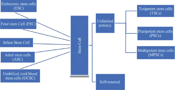
Stem cell potency and classifications.
Embryonic stem cells
Embryonic cell lines are derived from the epiblast tissue of the blastocyst structure, which is aligned in the inner wall of the blastocyst [4-6]. The blastocyst is a structure that forms in the early stages of embryo’s development, approximately during the 4th to 5th day of fertilization in humans. The blastocyst consists of 50-150 cells embryonic stem cells (ESCs) which are pluripotent and can transform to all derivatives of the three primary germ layers, including ectoderm, endoderm, and mesoderm. In other words, they can develop into each of the more than 200 cell types of the adult body, when given the sufficient and necessary stimulation for a specific cell type [4, 7-11]. ESC has the potency to divide in vitro after the administration of proper stimulant agents for differentiation, in which each daughter cell will also remain pluripotent. The pluripotency of ESC -in vitro and in vivo- has been repeatedly demonstrated in previous research [6, 11, 12]. Because of their unique capability of unlimited expansion, ESCs are perceived as a hypothetical source of cells in regenerative medicine and a proposed basis for tissue replacement in several diseases [4-8, 13].
Fetal stem cells
Fetal stem cells (FSCs) are cell lines that are derived from fetal tissues, which have the ability to divide, proliferate, and differentiate into specialized cells. Fetal stem cells can be isolated from fetal hematopoietic stem cells, fetal mesenchymal stem cells, and neural crest stem cells [14]. Studies demonstrated that FSCs have higher pluripotency potentials and lower immunogenicity effects compared to adult stem cells (ASCs) [14].
Infant stem cells
Infant (perinatal) stem cells can be derived from perinatal tissues, including, placenta membranes, amniotic fluid, and umbilical cords [15]. These tissues consist of numerous types of stem cells that possess the characteristics of both ESCs and ASCs [15]. Amniotic fluid stem cells, umbilical cord stem cells (UCSCs), and placenta-derived stem cells can be easily collected at the end of gestation. These stromal cells are considered to be the best candidates for stem cell therapy because they are the most abundant source of hematopoietic stem cells (HSCs) and mesenchymal stem cells (MSCs) [15].
Adult stem cells
ASCs or somatic stem cells are undifferentiated cells that can be found in post-natal adult tissues, which could be unipotent or multipotent [16, 17]. However, sometimes ASCs are known as progenitor cells, due to their less capability of cellular differentiation [8, 16, 17]. These cells are commonly classified into further categories as epidermal stem cells (EDSCs), neural stem cells (NSCs), MSCs, and HSCs [18].
Induced pluripotent stem cells
Induced pluripotent stem cells (iPSCs) are generated from somatic stem cells that have been reprogrammed into ESC-like state. iPSCs have the properties of ESCs, and can differentiate to the three primary germ layers. These cells have several advantages as they are derived from the patients’ own somatic cells leading to lower risks of rejection [18].
Potentials of stem cells
Stem cells have the potential of self-renewal and differentiation as described below (Figure 1).
Self-renewal
Stem cells can undergo multiple cycles of cell division while preserving the undifferentiated state [19, 20].
Unlimited potency
Stem cells also have the capability to differentiate into any mature cell type. This makes stem cells either totipotent, pluripotent, or multipotent [19].
Totipotent stem cells
Totipotent stem cells (TSCs) are the outcome of the fusion of a sperm cell and an oocyte. The fertilized egg initiates a few divisions and forms cells that have totipotent properties. TSCs have the ability to differentiate into embryonic and extraembryonic types of cells [21, 22].
Pluripotent stem cells
Pluripotent stem cells (PSCs) are the descendants of totipotent cells and can differentiate into cells that construct the three germ layers of ectoderm, mesoderm, and endoderm which subsequently give rise to various system organs [21-23].
Multipotent stem cells
Multipotent stem cells (MPSCs) are among the major groups of stem cells which hold similar basic properties of all stem cells [24]. These cells can differentiate into various specific cells leading to particular properties, such as hematopoietic stem cells (HSCs), skeletal myoblasts, MSCs, and endothelial progenitor cells (EPCs) [25-27]. These cells can inhibit or activate a sequence of molecular and cellular pathways resulting in antiapoptotic and anti-inflammatory properties that can contribute to the treatment of various diseases.
Division and differentiation
Stem-cells have two types of cell division in order to remain self-renewed and potent. Firstly, a symmetric division resulting in two identical daughter cells both having the stem-cell potentials. The second type is called asymmetric division whose outcome is one stem-cell with the aforementioned properties and a progenitor cell with less self-renewal potential [9, 10, 28, 29].
Therapeutic effects of the stem cells
Stem cell therapy has been applied in various disorders in which here we review some of the diseases in this regard (Figure 2 and Table 1].
Figure 2.
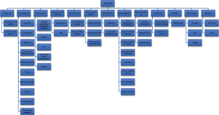
A review of stem cell therapy in various systems.
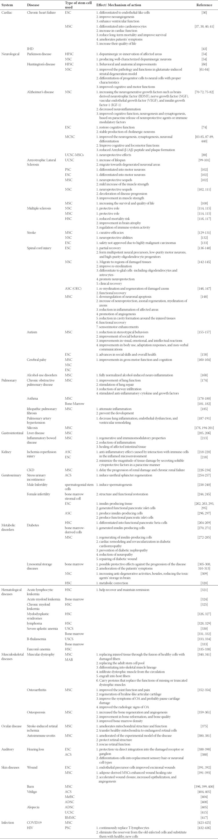
Table 1.
A review of stem cell therapy and effects.
Cardiac diseases
Congestive Heart Failure (CHF) and Ischemic Heart Disease (IHD)
Cardiovascular diseases (CVD) account for about 20% of the burden of noncommunicable diseases (NCD) [30]. These diseases are responsible for nearly one-third of deaths globally, and the prevalence of them are continuing to grow in both developing and developed countries [31]. Current treatments rarely could ameliorate the underlying causes of CVD, which are tissue scarring and cell loss [32].
Rising evidence demonstrated stem-cell therapy could improve heart failure by inducing neovascularization, decreasing cardiomyocyte apoptosis, and forming resident stem-cells [33-35]. Animal studies suggested that ESCs can be differentiated to endothelial-like cells, improve neoangiogenesis, and enhance ventricular function [36]. Besides, transplantation of bone-marrow-derived MSCs for myocardial infarcted animal models showed consistent results, as MSCs differentiated into cardiomyocytes [37, 38].
The first U.S. randomized controlled clinical trial, using a three dimensional guided catheter system to provide the heart with muscular stem-cells, was done in 2009; and after 1 year of follow-up, the results showed significant improvements in New York Heart Association (NYHA) and Minnesota Living with Heart Failure Questionnaire (MLHFQ) scores, besides the increase of ventricular viability [39]. Endomyocardial injection of cardiopoietic stem-cells (MSCs exposed to a cardiogenic cocktail) to the patients with preclinical heart failures showed the same results; as the patients had improvements in NYHA, 6-min walk distance, Quality of Life (QoL) and left ventricular ejection fraction [40]. Furthermore, 6 months follow-up of patients who had done subepicardial transplantation of autologous stem cells into ischemic myocardium demonstrated a major increase in cardiac function, measured by echocardiography, single-photon emission computed tomography, and angiography [41]. The STAR-heart study asserted that intra-coronary bone-marrow-derived MSCs transplantation in patients with chronic heart failure could ameliorate patients’ symptoms, increase their QoL, and improve survival [42]. Stem-cell therapy also can reduce the long-term mortality of patients with CHF and chronic IHD [43]. Moreover, it can reduce incidence of arrhythmias and non-fatal myocardial infarctions in the long-term in these patients [43]. The effects of stem-cell therapy on the rehospitalization and long-term left ventricular ejection fraction of patients with CHF and IHD are controversial, and more studies are needed to find concrete results [43]. To the best of our knowledge, the report of procedural adverse effects and side effects such as cardiac or systemic toxicity induced by stem cells are rare. Thus, the therapy could be perceived as a safe and feasible treatment for patients with CHF and IHD; however, the procedure must be applied cautiously and more clinical evidence is needed for a definite conclusion.
Neurological diseases
Parkinson’s disease
Parkinson’s disease (PD) is a neurodegenerative disease that is common worldwide and clinically demonstrates itself by symptoms of resting tremor, bradykinesia, rigidity, and postural instability [44, 45]. The pathology of the disease is characterized by loss of dopaminergic neurons in the pars compacta of substantia nigra (SNpc) and other areas of the brain, besides development of protein residues in neurons’ cytoplasm (Lewy bodies) [46, 47]. Present therapeutic options for PD patients consisted of deep brain stimulation and dopamine agonists.
Promising evidence has been found regarding neuroprotective and neuroregenerative benefits of stem-cell therapy –while applying with neurotrophic agents. Transplantation of embryonic mesencephalic into the brain of animal models showed improvement of dopamine transmission [48-50].
The proof-of-principle has been achieved by several studies in which evidence was in favor of the hypothesis that the dead dopaminergic neurons can be replenished by stem-cell transplantation in patients with PD [8, 51-53]. Most of the studies have been using human fetal stem-cells (hFSCs) of ventral mesencephalic neurons as the transplantation grafts; which can provide dopaminergic re-innervation of affected areas [54]. Foremost problems of using hFSCs are the low availability and absence of the cell material standardization which causes high inconsistency considering the degree of symptom relief, besides, the occurrence of adverse effects such as graft-induced dyskinesia in some patients [55-57].
Due to the aforementioned issues, the current researches emphasize the adjustment of hFSCs, and also producing well-characterized dopaminergic neurons by using brain-derived NSCs. Stem cell tissues –to be grafted in the striatum- must be analogous to substantia nigra neurons, regenerate the dopamine network in the brain, produce long-term and high symptomatic relief, and have no adverse effects [54]. It is still unclear whether the transplantation of stem-cell-derived neurons can replace dead compartments in PD patients; however; preceding studies on hFSCs provided us with the essential knowledge to establish clinically competitive stem cell therapy in this field.
Huntington’s disease
Huntington’s disease (HD) is an uncommon neurodegenerative disease characterized by undesirable motor and cognitive functions, besides psychiatric syndromes. The disease is considered a genetic disorder that is inherited in an AD (autosomal dominant) fashion and results in cell loss in the striatum and cerebral cortex [58]. Current treatments of HD are mostly based on symptom therapies –antipsychotic and antidepressant drugs- and hardly improve the underlying causes of the disease.
Several studies on animal models have suggested that stem-cell therapy could be used in the case of HD [59]. Behavioral and anatomical improvements have been seen after the transplantation of HFSCs into rodent striatum [60]. Moreover, intravenously injected NSCs have migrated into striatal lesions and improved the pathology and functions in the glutamate-induced striatal degeneration model [61]. The transplantation of adult NSCs in animal models also showed significant improvement of motor function impairments as progenitor cells could differentiate to neural cells with proper characteristics [62].
This accumulating evidence indicates that cell transplantation therapy could be well justified as a treatment of HD. Several studies experimented with the feasibility of cell therapy in this regard and showed promising results. Positron emission tomography (PET) scanning of 3 out of 5 patients who underwent hFSCs transplantation into the striatum demonstrated increased metabolic activity in the region; besides the patients showed improved cognitive and motor functions, through six years of follow-up [63, 64]. The same results were achieved in another study which asserted that 3 patients who underwent striatal transplantation of hFSCs showed increased scores of cognitive measures [65]. A previously done investigation approved the safety of intrastriatal neurotransplantation of hFSCs [66]. However, the main concerns about the use of hFSCs are the high tumorigenic properties of these cells, scarcity of this material, as well as ethical and moral issues.
Alzheimer’s disease
Alzheimer’s disease (AD) is the most common neurodegenerative disease which results in irreversible loss of neurons in the brain, mainly in the hippocampus and cortex [67]. The disease is diagnosed, clinically, by the symptoms of progressive demolition of memory, judgment, decision-making, and orientation [68]. Current AD treatments, such as the prescription of acetylcholinesterase inhibitors to enhance cholinergic function, provide an only partial and temporary alleviation of symptoms. The treatment of AD has been a daily challenge for physicians many years ago because of the preceding fact.
Multiple theoretical approaches have been proposed in order to design stem-cell therapy for AD such as the upregulation of the local NSCs, and stem-cell transplantation [69]. The upregulation of the neurons could be achieved through medicine and gene-therapy by increasing the neurodegenerative growth factors such as brain-derived neurotrophic factor (BDNF), nerve growth factor (NGF), vascular endothelial growth factor (VEGF), and insulin growth factor-1 (IGF-1]; although animal studies showed this approach lacks the potency to repair neurons [70-72].
Investigations on animal models of AD showed ESCs have the potential to restore cognitive function; however, no tangible clinical evidence has been succeeded yet [73]. A recent investigation reported stable production of cholinergic neurons from human ESC, which was able to functionally integrate into hippocampal neuronal circuitry, after transplantation [74]. The transplantation of NSCs has shown more promising outcomes as it decreased the neuroinflammation and improved cognitive function, plus, neurogenesis and synaptogenesis in aged primate models and AD mice replicas; these effects could be because of the paracrine release of neuroprotective agents or immune-modulatory factors, besides, the direct neural differentiation [75-82]. Moreover, investigations have demonstrated that the transplantation of MSCs into the brain of aged mice models have improved the neurogenesis, synaptogenesis, neuronal differentiation, plus, cognitive and locomotor functions, and reduced Amyloid-b (Ab) peptide and plaque formation; it has been also stated the intravenous administration of MSCs could also be effective by crossing the blood-brain barrier and migrating to areas of brain injury [83-89].
Recently, a phase 1 clinical trial showed that human umbilical cord blood-derived mesenchymal stem-cells (hUCB-MSCs) which injected by stereotactic technique into the brain of AD patients could have neuroprotective effects, as patients didn’t show a decline in their cognitive functions; besides, no adverse effects were observed during 24-months follow-up [90].
Given that the rate of neuroregeneration in the adult brain is minimal and that the pathology of AD consists of Ab formation from amyloid precursor protein (APP), Alzhimer’s patients have markedly reduced brain functions. Stem-cell transplantation therapy for AD may not be effective in that environment, where APP metabolism is altered, and it may, conversely, lead to excessive gliogenesis and tumorigenesis. Studies must consider the regulation of APP processing to develop effective stem-cell transplantation protocols for AD patients [91-95].
Amyotrophic Lateral Sclerosis
Amyotrophic Lateral Sclerosis (ALS) is a progressive neurodegenerative disorder that involves degenerations of motor neurons at all stages including the cerebral cortex, brainstem, and spinal cord [96]. The symptoms of the disease consist of the indications of loss of neurons at all levels of the motor system –both upper and lower motor neuron findings- which results in progressive muscular paralysis [96]. The progression of ALS is inevitable, and about 50% of patients die within 3 years of suffering from the disorder [97]. Currently, the condition is perceived as incurable and the treatments provide symptomatic relief in order to increase the QoL of patients. Stem-cell therapy has been under investigation to be used as a cure for the condition and has shown several promising pieces of evidence in this regard. The therapy could restore and preserve the function of both upper and lower motor neurons; besides, newly renovated neurons could be integrated into existing neural circuitries [21, 98].
Preclinical studies and treatment of animal models of ALS with human umbilical cord blood cells demonstrated the increase of lifespan in a dose-dependent way [99, 100]. Moreover, intravenous administration of these cells to the mice models showed stem cells tend to migrate towards degenerated neuronal areas [101]. iPSCs and human-derived ESCs are perceived as the two most powerful types of stem cells that could benefit ALS patients; MSCs and NSCs are also safer products as they do not have the adverse effects of prior cell types such as tumorigenesis [102]. Investigations have suggested that ESCs and iPSCs of ALS patients can be differentiated into motor neurons [103, 104]. MSCs and NSCs have been used in clinical trials and they have shown more neuroprotective sequels rather than neuroregenerative outcomes. Intrathecal and spinal cord injection of MSCs has been used in phase 1 clinical trials which showed a mild increase of the muscle strength with minimal adverse effects [105-107]. Furthermore, autologous-derived stem-cells injected into the frontal cortex of patients with ALS showed the potential of increasing the survival and QoL of these patients [108]. Phase I intraspinal injection of NSCs has also been done in patients with ALS, the disease progression was slowed and no severe adverse effect was observed during the examination [109, 110]. The result of another recent study was also consistent with prior findings as to the microinjections of NSCs into the lumbar cord tract showed an improvement in muscle strength in these patients [111].
Multiple sclerosis
Multiple sclerosis (MS) is an autoimmune, inflammatory, and chronic neurological condition that involves myelinated axons of the central nervous system (CNS) [112]. The definite underlying cause of the disease is yet to be cleared, however, a number of genetic and non-genetic factors have been mentioned to be the cause of the disease [112]. Currently, there is no curative therapy available for the disease and the treatments involve disease-modifying agents such as Beta Interferons, Glatiramer Acetate, Mitoxantrone, Natalizumab, and Fingolimod, besides symptomatic relieving drugs such as Dalfampridine along with a variety of off-label drugs [112]. The disease is a progressive condition and treatments can only partially manage the course of MS.
Previous studies have found that NSCs and MSCs administration may have a protective role against the progression of MS in both rodents and primates [113]. Several pilot studies have suggested that autologous MSC administration may be a feasible and safe method regarding the treatment of MS patients [114, 115]. Moreover, autologous HSC transplantation was also found to be beneficial in patients with progressive MS, as it significantly reduced mortality risk [116]. Multiple phases I/II studies showed that the transplantation of autologous HSC could have promising results such as improvement in brain atrophy that was evaluated by Magnetic Resonance Imaging (MRI), and regulation of immune system activity [117]. It has also been reported that a few MS patients that received allogeneic bone marrow transplantation because of a coincidental hematopoietic disorder had recovered from this immune-mediated disease [117].
Stroke
Stroke disorders are the major cause of morbidity in many areas of the world that could lead to irrecoverable brain tissue damages and functional disabilities. Current therapeutic approaches are mostly focused in order to modify risk factors for stroke such as hypertension, heart diseases, diabetes mellitus, heavy cigarette and alcohol consumption, and hypercholesterolemia [118]. Surgical interventions and intravenous administration of thrombolytic agents, as reperfusion therapies, are only available in the acute stage of the stroke and cannot be done on many occasions. Recently, physicians have suggested that brain function may improve after stroke by the restoration of damaged neuronal cells through stem-cell transplantation.
A variety of precursor cell types such as human bone marrow cells, neuroepithelial stem cell line, NTera-2 cell line have been investigated to renovate brain function after stroke in animal models of stroke which showed improvement of sensorimotor function, spatial memory, neurological severity score, and asymmetrical motor behavior [119-128]. Although, the underlying causes of these improvements are yet to be cleared and there are few indications of neuronal replacement. Autologous somatic stem cells such as MSCs appear to be a reasonable source in this field since the chance of graft rejection is minimal. However, the plasticity of somatic stem cells is not obvious and it is not clear that they can safely evolve into neuronal cells [129]. In this regard, intravenous administration MSCs was done in stroke patients and its safety plus curative efficacies were observed during long-term follow-up [130, 131]. Moreover, studies have suggested that endogenous pools of stem cells, for example, NSCs could also be considered as a source for stem-cell therapy because of their neuroprotective abilities [132]. It is worth mentioning that pieces of evidence from a study on animal models of stroke did not approve the safety of ESCs transplantation in this field, as ESCs produced highly malignant carcinomas at the site of implantation [133].
The curative effects of stem-cell therapy on animal models of stroke made the basis for the treatment to be addressed for use in human subjects. Initial clinical trials showed that stem-cell therapy could be safe, feasible, and therapeutic in patients with stroke; however, more comprehensive is needed to approve the aforementioned issue.
Spinal cord injury
Spinal cord injury (SCI) is a destructive condition that involves both upper and lower motor neurons and could result in paraplegia and quadriplegia. The incidence of traumatic SCI, primarily caused by motor vehicle accidents, is estimated to be between 28 and 55 per million people each year, in the US [134]. The management of the condition consists of both surgical –for example, surgical decompression and spine stabilization- and medical –for instance, steroid therapy- interventions; besides rehabilitation and long-term follow-up of the patients [134]. Recently, several other newly designed approaches have been suggested in this regard such as molecular and cellular therapies. [135]. The cellular therapy of the condition focuses on stem/progenitor cells and transplantation of Schwann cells, peripheral nerve cells, and cells from the olfactory nervous system [135].
The emerging perspective of stem cell therapy as a viable therapeutic tool for the treatment of SCI is based on the preclinical studies which have been performed on animal models and have shown that transplanted progenitor cells can migrate into the injured tissues to improve axonal regeneration and motor function. ESCs have been used for the treatment of paralyzed rats, the paralyzed rodents showed partial recovery from the condition as the transplanted ESC–derived axons restored SCI [136]. Moreover, ESCs were also able to form multipotent neural precursors, low-purity motor neurons, and high-purity oligodendrocyte progenitors [137-141]. NSCs have been proposed as a possible treatment of SCI; because they are already devoted to a neural fate and have a lower potentiality to be neoplastic. Several preclinical investigations demonstrated that transplanted NSCs could reach regions of damaged tissues, improve re-myelination, differentiate to glial cells -including oligodendrocytes and astrocytes-, promote neuroprotection, and cause clinical recovery in animal models [142-145]. Olfactory ensheathing cells (OECs) were also used in rodent models of SCI and caused significant remyelination and regeneration of damaged axons, besides functional recovery [146, 147]. Large production of the prior cells is moderately difficult, and it is not clear whether they can be increased to an adequate amount to be administered in human clinical trials. MSCs have been widely used in the case of SCI and the results were drastically positive regarding the treatment of the condition. Preclinical studies suggested downregulation of neuronal apoptosis, an increase of neuroprotection, axonal regeneration, myelination of axons, reduction in inflammation of affected areas, promotion of angiogenesis, reduction in cavity formation around the injured tissues, besides functional recovery, and sensorimotor enhancements, after the transplantation MSCs in animal models with SCI [148].
NSC transplantation is under investigation at phase I/II clinical trial and is proved to be safe and feasible for perilesional intramedullary injections [145, 149]. The grafting of MSCs in humans also resulted in partial functional and behavioral recovery [148]. However, a recent phase 3 clinical trial showed controversial results as only 2 among the 16 patients showed progress in neurological status after MSCs application (150].
Briefly, animal models of SCI who received stem-cell therapy have demonstrated several promising results, and preclinical investigations have validated scientific principles and approaches. Additionally, multiple signs of progress have been made by a number of studies with practical concerns in this regard.
Autism
Autism and autism spectrum disorders are referred to as neurodevelopmental disorders that are caused by both genetic and environmental factors and are characterized by disruption of social interactions and stereotypic behaviors [151-153]. Current therapeutic interventions for autistic patients include behavioral and psychosocial treatments, besides, psychopharmacological treatments that could not completely resolve the patients’ symptoms [154].
Recently, researchers have conducted several investigations regarding the use of stem cell therapy for autism. Intracerebroventricular transplantation of MSC for animal models of the disease showed promising results, as a reduction in stereotypical behaviors and an improvement of social behaviors were observed during the investigation in animal models who received the transplantation [155].
Consistently with prior results, a phase I/II clinical trial showed that the patients who received the transplantation of human cord blood mononuclear cells and umbilical cord-derived MSCs had improvements in visual, emotional, and intellectual reactions, along with body use, adaptation responses, and non-verbal communications [156]. Autologous bone marrow mononuclear cell transplantation was also found to be useful regarding the treatment of autistic patients [157]. Another pilot study has also demonstrated the beneficial effects of ESC administration for autistic patients, as significant advances in social skills and overall health were observed after the treatment [158]. Moreover, the treatment safety was evaluated and no serious adverse effect was observed during stem cell transplantation in the mentioned patients [156, 158].
Cerebral palsy
Cerebral palsy (CP) is a group of chronic neurological diseases that result in cognitive and motor impairments in children. This may be caused by perinatal infection, asphyxia, and preterm birth, or even postnatal brain injuries [159]. The overall incidence of CP is increasing which could be due to the increased rate of premature infants’ survival [159]. Currently, available treatments for CP include rehabilitation strategies and pharmacological interventions to reduce the symptoms.
Several lines of stem cells such as MSCs, NSCs, and ESCs have been administered to CP patients in previous investigations [160-164]. Stem cell intervention could be a promising alternative treatment for pediatric patients with CP according to the conducted clinical trials. Previous studies demonstrated that stem cell therapy could result in significant improvements in gross motor function and cognition as well [165]. The adverse effects of the stem cell administration in these patients were reported to be at a minimum [165]. However, it has been mentioned the beneficial effects of stem cell interventions in CP patients may be for short-term periods, and in order to achieve comprehensive results, more investigations must be conducted in this regard.
Alcohol-use disorders
Alcohol-use disorders have been reported to be responsible for 1 out of 10 deaths among working-age adults in the US and are also among the leading causes of morbidity and premature mortality worldwide [166]. Also, chronic alcohol consumption is known to be the most common reason for peripheral and central nervous system toxicity [167]. Animal experiments have demonstrated that a single dose of MSC-spheroids can have a significant beneficial effect on the animal model of chronic ethanol intake and relapse-drinking by inhibiting chronic ethanol intake, and subsequently, fully normalized alcohol-induced neuroinflammation [168-171].
Pulmonary diseases
Chronic obstructive pulmonary disease (COPD)
COPD has been reported to be the third-leading cause of mortality in the US [172, 173]. Regardless of recent developments in the management of symptoms with novel pharmaceutical medications and molecules, no effective treatment has yet been obtained in order to attenuate or reverse the disease progression or emphysematous changes.
Meta-analytic studies on animal models of COPD have shown that intra-tracheal instillation or intravenous injection of MSC have satisfactory results, through the significant improvement of lung function and stimulation of lung repair. Furthermore, a considerable reduction of airway infiltration by macrophages, neutrophils, and products of pro-inflammatory cytokines such as IL-6 and IL-1b, but accompanied with substantial stimulated anti-inflammatory cytokine IL-10 and growth factors such as EGF, HGF, TGF-b, and VEGF was detected through MSC administration, suggesting this method as an effective approach in the treatment of animal models of COPD/emphysema, and therefore hold the foundation of further research and application of administering MSC in COPD patients [174].
Asthma
Asthma, affecting around 300 million individuals worldwide, is known to be primarily a chronic inflammatory disease presenting with lifelong conditions with different levels of severity [175, 176]. While the present treatments are effective for the management of asthma, patient’s morbidity, and mortality are still factors requiring attention, which continue attempts in modifying and adjusting therapeutic options are being practiced [177]. There are increasing studies that show the beneficial effects of MSC-derived secretome in the treatment of experimental asthma [178-180]. Moreover, the use of bone marrow-derived mononuclear cells (BMMC) in animal models of severe asthma showed promising results [181, 182].
Idiopathic pulmonary fibrosis
Idiopathic pulmonary fibrosis (IPF) with a prevalence from 4.6 to 16.3/100,000 cases worldwide is characterized by chronic, progressive fibrosing interstitial pneumonia without a known etiology [176, 183]. Despite several therapeutic methods, no significant decrease in mortality rates have been observed [184].
There is evidence that MSCs can prevent the progression of animal model IPF and decrease inflammation, if administered early during the disease, however such evidence during the latent and established fibrosis period of the disease is limited and requires further studies in order to apply in clinical studies [176, 185].
Pulmonary arterial hypertension
Pulmonary arterial hypertension (PAH) is considered a rare and progressive disease which is characterized by the elevation of blood pressure of the lung arteries. While manageable, no effective therapeutic option has yet been approved in order to reduce mortality [186].
Animal studies have demonstrated that administration of MSCs, either intravenous or intratracheal, can decrease factors associated with PAH, including lung inflammation, endothelial dysfunction, and ventricular remodeling [187-191]. Therefore, these pieces of evidence can be the theoretical basis for designing future clinical trial studies.
Silicosis
Silicosis is among the known occupational diseases which cause extensive lung fibrosis and subsequently respiratory failure [192]. Currently, there are no promising clinical treatments to decelerate or halt the progression of the disease [193]. However, MSCs have shown to apply therapeutic properties in lung diseases and represent an alternative treatment for silicosis, particularly adipose-tissue-derived mesenchymal stromal cells [176, 194-201].
Gastrointestinal Diseases
Liver disease
Liver injury can be subsequent to causes such as ischemia, cytotoxic, toxic–metabolic, or other insults causing tissue inflammation and therefore result in innate and adaptive immune responses which in turn can also directly cause additional injury [202]. The currently accepted treatment for end-stage liver disease is liver transplantation, a procedure which has its own challenges. [203, 204]. However, considerable results regarding cell-based therapy using MSCs have been demonstrated in vitro, in vivo, and even in clinical studies for the treatment of liver diseases [205, 206]. Nevertheless, there is still little evidence regarding the efficiency compared to conventional treatments and therefore further studies and clinical trials are justified [207-209].
Inflammatory bowel disease
Inflammatory bowel disease (IBD), which consists of Crohn’s disease (CD) and ulcerative colitis (UC) is assumed to be due to inappropriate response of the host’s immune system to intestinal microbes [210]. Standard treatments for the disease consist of immunosuppressive drugs, such as biologicals, with low or even no response rates [211, 212]. Hence, alternative therapeutic remains an unmet need. Based on reported literature, stem cell transplantation in IBD patients has demonstrated satisfactory therapeutic potential along with a low risk of adverse effects. In this regard, MSCs have immunomodulatory and regenerative properties, which subsequently leads to a decrease in inflammation along with the healing of affected intestinal tissue [213].
Kidney diseases
Acute kidney injury
Acute kidney injury (AKI) is characterized by stimulation of the immune system and elevated oxidative stress which can be caused by ischemia-reperfusion injury (IRI) [214-217]. Stem cell therapy has been proven effective in experimental models of AKI based on decreasing the degree of tissue injury via soluble cytoprotective factors in a paracrine manner, and also based on the anti-inflammatory effect subsequent to the interaction with immune cells in the inflamed microenvironment [218-222]. However, more translational studies from animal models are required to ensure their safety for application in clinical studies [223].
Chronic kidney disease and end stage renal disease
In renal injury, the progression to end-stage renal disease (ESRD) is presently only partially understood due to its complexity [224]. There are controversial reports regarding studies of stem cells in this area. While some studies have reported that MSCs would delay the progression of renal damage and chronic kidney disease (CKD) [225-233], other studies have reported MSCs to be ineffective [229, 230, 234].
Genitourinary diseases
Male Infertility
Infertility is the inability to achieve pregnancy despite one-year attempts, in which the rate of related factors for male infertility alone has been reported from 6.4% to 42.4% in studies [235, 236]. Various treatment methods have been applied such as optimization of sperm production, relief of obstruction, and surgical extraction of sperm [237]. Treatment with stem cells by utilizing spermatogonial stem cells (SSCs) has been evolving in order to induce spermatogenesis from the individuals’ undifferentiated or dysfunctional SSCs [238-240]. However, caution should be warranted in this therapeutic option regarding carcinogenesis and also genetic/epigenetic alterations in offspring which should be fully investigated in humans [241].
Female infertility
The incidence of female infertility ranges from 33 to 41% worldwide [242]. The US reported that in 2002, 7.4% [2.1 million) of married women were infertile [243]. These statistics highlight the notable numbers of females enduring infertility.
Among the many causes of female infertility, reproductive system-related diseases are considered the main causes. The efficacy of bone marrow stromal cells has been demonstrated in several animal model studies regarding the treatment of chemotherapy-induced ovarian failure, indicating structure and functional restoration [244, 245]. Studies have shown restored menstruation in endometrial injury by the implantation of autologous bone marrow stromal cells [246]. Bone marrow stromal cells have also been successful in treating thin endometrium in animal model studies via locating the differentiation site in the tissue and exerting immunomodulatory properties [247]. Furthermore, functional endometrium restoration and improved reproductive outcomes have been detected in patients with Asherman syndrome using bone marrow stromal cells [247-249].
Stress urinary incontinence
Stress urinary incontinence (SUI) is a common urinary system disease affecting more than 200 million people worldwide and notably females, resulting in a serious decrease in QoL with no ideal therapeutic option until now [250-253]. Stem cell therapy has demonstrated promising results, by inducing urethral sphincter regeneration utilizing bone marrow-derived MSCs, muscle-derived stem cells, urine derived stem cells, and adipose-derived stem cells (ADSC) [254-257]. However, administration route and optimal timing of the stem cell requires further evaluation [258-260].
Metabolic disorders
Diabetes
Diabetes is a group of metabolic disorders that are characterized by hyperglycemia which is developed for a defect in insulin production, insulin function, or both. Diabetes mellitus (DM) is considered as one of the most common non-communicable diseases worldwide, with a global growing prevalence [261]. Persistent and uncontrolled hyperglycemia of diabetes may have severe consequences including retinopathy, neuropathy, and nephropathy if remains untreated. Currently approved treatments of DM include lifestyle and dietary modifications, prescription of anti-hyperglycemic agents, insulin, and some alternative medicines. However, these therapeutics could not properly improve the underlying cause of the disease which is the degeneration of pancreatic beta-cells, and in most cases, the disease could not be cured completely [261].
Several investigations have been done regarding the therapeutic use of both ESCs and ASCs in DM and its complications. ESC-derived insulin-producing tissue alleviated diabetes in mice [262, 263]. HSCs were also differentiated into functional pancreatic beta-cells in rodent models [264-269]. In vitro studies have also revealed the possibility of therapeutic administration of bone marrow-derived cells for DM patients, as these cells have generated insulin-producing cells (IPCs) under appropriate circumstances [270, 271]. Similarly, the advantages of MSCs were noticed in multiple studies as they could be beneficial in regenerating of IPCs, cardiac remodeling, and revascularization in diabetic cardiomyopathy, prevention of diabetic nephropathy, reduction of neuropathy, and repairing of the diabetic wound [272-285]. Moreover, other stem-cells such as those that originated from pancreatic duct, pancreatic islet, and exocrine tissue of the pancreas, besides, hepatic progenitor tissue were also investigated in this regard and promising results have been found [286-292]. Several in vivo and in vitro studies have been conducted to produce IPCs and functional pancreatic islet cells from human ESCs and ASCs [293-297]. The result of these experiments was most promising as fully functional beta and islet cells were generated in several laboratories.
In humans, pancreatic islet transplantation was suggested as a therapeutic approach through DM over two decades ago [298]. After the intraportal infusion of pancreatic islets in DM patients, approximately 50% reduction in insulin dependence was observed [299, 300]. Consequently, beta cell replacement therapy was also suggested and experimented as an alternative treatment for diabetic patients [301]. Previous studies have evaluated the efficacy of stem-cell therapy for patients with DM by using various methods such as umbilical cord blood transplantation, umbilical cord MSC therapy, HSCs infusion, and bone marrow MSC transplantation. These studies have suggested that stem-cell therapy could be a promising alternative treatment regarding the DM and its complications [302, 303].
Lysosomal storage diseases
Lysosomal storage diseases (LSDs) is an inherited defect in normal lysosomal functions which are identified by several different pathophysiologies [304]. LSD may have a variety of symptoms according to its pathological severity, including, mental alterations, cognitive retardations, rapid onset disabilities, and death. Several treatment methods have been suggested for these conditions which mostly focus on the replacement of the malfunctioned enzyme that is the underlying cause of the disease [305].
Studies on animal models of LSDs have shown the possible protective effects of bone marrow transplantation (BMT) against the progression of the disease [306-308]. Moreover, animal studies also demonstrated that intracranial injection of NSCs could have beneficial effects in increasing anti-degenerative activities, along with reducing the toxic agents’ storage in rats’ brains [309].
Allogeneic BMT was firstly done over 3 decades ago for patients who suffer from LSDs which showed promising results regarding the amelioration of the patients’ symptoms [305]. Later, this finding was evaluated by more expanded investigations which demonstrated that BMT could alleviate the patients’ symptoms in most cases [310-313]. Regarding the LSDs, BMT was found to be a feasible treatment in Hurler Syndrome, Maroteaux-Lamy Syndrome, Gaucher disease, Metachromatic leukodystrophy [314-319]. Furthermore, HSC transplant has been found to be more effective than enzyme replacement therapy in LSD patients, based on the metabolic correction [320].
Hematological diseases
Acute lymphocytic leukemia
Acute lymphocytic leukemia (ALL) is an aggressive malignancy that the lymphoid line of blood cells tend to be immaturely produced in large numbers. The main therapeutic approaches through this condition are chemotherapy and HSC transplantation that could help the patients to recover and maintain remission [321]. Currently, the main concern in this regard is finding the optimum strategy of the HSC administration [322, 323].
Acute myeloid leukemia
The main characteristics of acute myeloid leukemia (AML) are a rise in the number of immature myeloid cells which could result in several hematopoietic insufficiencies and leukocytosis. Stem cell transplantation has been administered for this condition since over 3 decades ago, however, there is still uncertainty regarding the optimal treatment of AML [324].
Chronic myeloid leukemia
Chronic myeloid leukemia (CML) is another malignancy of bone marrow cell which is characterized by an increased count of white blood cells in peripheral blood. It has been noticed in previous studies that CML is the most common indication of allogeneic HSC transplantation globally [325].
Myelodysplastic syndrome
Myelodysplastic syndromes are a group of bone marrow disorders that the blood cells are not properly matured. The main treatment of this condition is chemotherapy and HSC transplantation [326, 327].
Lymphoma
Another group of blood cancer is lymphoma which originated from the lymphatic system and could affect the whole body. The treatment of lymphoma is commonly chosen according to the type and severity of the disease. Radiation therapy, chemotherapy, immunotherapy, and HSC transplantation are considered as the main therapeutic options in this regard [328, 329].
Severe aplastic anemia
Severe aplastic anemia (SAA) is a condition characterized by the failure of bone marrow to make enough mature blood cells. Stem cell transplantation is a promising therapeutic approach through SAA; as previous investigations mentioned that unrelated cord blood transplantation and allogeneic bone marrow transplantation can be a salvage procedure for SAA patients [330-332].
B-thalassemia major
B-thalassemia is an early-onset and inherited blood disease which is characterized by severe anemia requiring regular blood transfusion. The main treatments of this condition consist of transfusion therapy, chelation therapy, surgical interventions such as splenectomy and cholecystectomy, and stem cell transplantation [333]. Bone marrow transplantation, umbilical cord blood transplantation, and combined transplantation of these two cell lines have been administered for patients with B-thalassemia major which showed satisfactory results [333, 334].
Fanconi’s anemia
Fanconi’s anemia (FA) is a rare genetic blood disorder that is caused by bone marrow failure to produce blood cells. HSC transplantation has been suggested as an effective and exclusive therapeutic intervention for these patients [335-337]. However, HSC transplantation may have some adverse effects which should be managed and reduced, including developing cancerous tissues and graft versus host disease [338].
Musculoskeletal diseases
Muscular dystrophy
Muscular dystrophy (MD) is among the muscle wasting diseases, and is a condition affecting a million individuals worldwide which are the result of mutations in structural proteins and are presented with loss of functional muscle subsequent to muscle fiber injury, deposition of fibrotic tissue, and inflammation [339]. Present therapeutic options are mostly supportive and address the inflammatory response.
The use of stem cell therapy has demonstrated evidence of replacing injured tissue through the fusion of healthy cells with damaged fibers along with replacing the ASC pool for long-term muscle maintenance [340]. Based on the potential of MSCs and mesoangioblasts (MABs) in differentiating into skeletal muscle lineage, Studies on animal models of MD have addressed these cells for the regeneration of injured skeletal muscle. These cells can also infiltrate dystrophic muscle from the circulation, engraft into host fibers, and bring with them proteins that replace the functions of those missing or truncated. Based on the systemic delivery property of these cells, an increase in the feasibility of stem cell therapy has been observed regarding a large number of affected skeletal muscles among muscular dystrophy patients [341].
Osteoarthritis
Osteoarthritis (OA) is the most prevalent joint disease globally, and it has been estimated that about 10% of men and 18% of women with the age of 60 or older suffer from the condition [342]. The pathology of the disease is mostly based on cartilage degradation which could be due to the activation of matrix proteases [343]. In severe cases, the pain and loss of function of the joint may inhibit the patients from doing their own daily activities, and decrease the quality of life (QoL) of the patients. Typically, the treatments of the condition consist of pain management, along with physical and occupational therapies, and joint replacement in end-stage patients [344-348]. Intra-articular injections of several agents such as hyaluronic acid (HA), platelet-rich plasma, hypertonic dextrose prolotherapy, and anabolic cartilaginous mediators have also been part of clinical trials attempting to cure OA [349-351]. The potentials of stem-cells, for instance, the capacity for regeneration of damaged tissues and reversing the chronic degenerative conditions, makes stem-cell therapy as an approach to setback the underlying causes of OA.
A 2-year follow-up, proof-of-concept trial showed that intra-articular injection of MSCs into OA knees improved the joint function and pain without any adverse events by regeneration of hyaline-like articular cartilage [352, 353]. Moreover, a systematic review published in 2018 asserted that stem-cell therapy could improve the symptoms of OA and probably halt cartilage damage [354]. The administration of MSCs has also improved the radiologic signs of OA [354]. The adverse effects after the MSC therapy were rare, and the most important ones were synovial effusion which needed overnight observation, and unstable angina in a case who had multiple risk factors after three months, plus pain and swelling [321, 355-357].
Currently, there are over 500 clinical trials registered on ClinicalTrials.gov, exploring the safety and efficacy of stem cells in order to find a comprehensive protocol to cure OA. Long-term follow-up is needed to evaluate the exact effects of stem-cells therapy in the condition.
Osteoporosis
Osteoporosis is considered the most common skeletal disease which is characterized by decreased bone mineral density and bone structural deformations, which is probably caused by an acceleration in bone resorption by osteoclasts [358]. Current therapeutic interventions for patients with osteoporosis include bone-resorbing drugs which may have several adverse effects.
The application of MSCs has increased bone regeneration and angiogenesis in osteoporotic mice [359]. Osteoporotic mouse models that were treated with a single dose of systemically injected MSCs showed an improvement in bone reformation, and bone quality [360]. A recent meta-analysis on preclinical studies has approved the rationale to conduct further clinical studies regarding the use of stem cell interventions as a treatment of osteoporosis, since the available investigations in animal models mostly showed an improved bone marrow density after the application of stem cells [361]. Furthermore, previous studies have proposed that administration of MSCs could be a promising treatment for osteoporotic patients [362-365]. However, it was also noted that the MSCs have a tendency to differentiate into adipose tissue in these patients; and this problem must be solved in future investigations [362, 366].
Ocular diseases
Stroke-induced retinal ischemia
Among the complications of cerebral ischemia one can name its effect on the retinal, causing cell loss and optic nerve damage [367-369]. Available approved treatments consist of either endovascular thrombectomy or tissue plasminogen activator (tPA) which include limitations such as the increased risk of further damage and short therapeutic window [370-372].
There is rising evidence that MSCs can significantly affect the pathogenesis of the disease [373, 374]. Both in vitro and in vivo treatment with MSCs have shown to transfer healthy mitochondria into injured retinal cells, resulting in the amelioration of mitochondrial function and structure after ischemia [375]. Combinational treatment along with enhancing the timing of stem cell transplantation and delivery route may have additional beneficial effects on recovering cell damage and visual loss and increase functional recovery, which requires additional investigation [376].
Autoimmune Uveitis
Autoimmune uveitis (AU) is among the leading reasons for visual disability. Current treatment options consist of long-term use of corticosteroids and immunosuppressive medicines, which in turn leads to serious side effects and possible progress of cataract and glaucoma; therefore, new and alternative therapeutic options are urgently needed [377, 378]. Recently, MSC-derived exosomes have shown promising therapeutic results in animal models in which periocular injection of MSC-derived exosomes considerably ameliorated experimental autoimmune uveitis, salvaged retinal function and protected retinal based on a clinical and histological basis [379-381]. However, there are still many challenges to be addressed in order to proceed with MSC-derived exosomes to be utilized as an effective therapeutic agent in the treatment of AU.
Auditory diseases
Hearing loss
Hearing loss by affecting more than 360 million people worldwide is among the diseases with a significant impact on the individuals’ physical and mental health [382-386]. The current effective treatment is cochlear implants which offer partial recovery at the expense of loss of residual hearing [387]. Stem cells have demonstrated potential in providing protection through direct integration into the damaged ganglion or receptor followed by differentiation into replacement neuronal cell types or sensory hair [388-390].
Skin diseases
Wound
The cutaneous wound is still considered as a global health care concern and its treatments include topical antibiotics, debridement, and application of grafts. There is growing evidence regarding the promising effect of stem cell therapy in wound healing. Both ESCs and MSCs were found to have wound healing properties. As human ESC-derived endothelial precursor cells improved excisional wounds in rats, and adipose-derived MSCs enhanced wound healing rate in animal models of DM [391, 392]. Furthermore, the administration of MSCs for wounds was found to result in accelerated wound closure, increased epithelization, and angiogenesis [393].
Burns
Burns are considered to be one of the most prevalent public health problems, causing high rates of morbidity and mortality particularly in low- and middle-income countries [394]. Finding a concrete approach in order to heal chemical, electrical, and thermal burns has always been a challenge for physicians. Commonly, surgical interventions (debridement and application of skin grafts) and pharmacological interventions (for example, silver sulfadiazine) are being suggested for patients with burn injuries. However, these therapeutic approaches often fail to prevent uncontrolled consequences of the trauma, such as scar formation and contracture, and neuropathies. Subsequently, researchers started investigating other treatment options, including administration of small interfering RNA, DNA, growth factors, and stem cells for burn injuries [395].
Stem cell therapy could be a promising intervention regarding burn injuries as previous investigations have shown the beneficial effects of EDSCs, MSCs, and ADSCs in wound healing [396]. Critical assessments of preclinical studies have demonstrated that stem cells significantly promote burn healing rate and wound healing time, with hair follicle stem cells (HFSC) having the highest efficacy [395, 397]. The application of stem cells has also resulted in increased angiogenesis and reduced inflammatory markers in burn wounded animals [395]. Besides, intravenous administration of ADSCs was found to be safe and effective in improving skin graft elasticity, in severely burned porcine models [398]. Having said that, several studies suggested that the administration of MSCs could be applied as an alternative treatment for chronic non-healing wounds [399, 400]. The results of clinical trials were also consistent with above-mentioned evidence, such as the vascularization and granulation of the wound, and the patients’ pain levels were considerably improved after administration of MSCs [397]. Moreover, prior clinical trials noticed that stem cell therapy could significantly decrease the scar formation and skin contracture [401]. Overall, it seems that stem cell therapy could improve burn wound healing, however, more clinical studies are needed to determine the safety and efficacy of this approach toward the problem.
Vitiligo
Vitiligo is a depigmenting condition of the skin caused by the loss of melanocytes that globally affects about 2% of the general population [402]. Several hypotheses were asserted to be the pathology of the condition, however, there is no definite explanatory hypothesis in this regard. The most widely accepted hypothesis is that both hereditary and non-hereditary factors, affecting the melanocyte function, can lead to immune-mediated destruction of melanocytes [403]. Currently, therapeutic approaches consist of the use of local and intralesional prescription of corticosteroids, plus psoralen ultraviolet light therapy [402].
The skin is composed of several populations of stem-cells such as EDSCs, HFSCs, and dermal mesenchymal stem cells [404, 405]. Moreover, the outer root sheath of the hair follicles is an undeniable source of melanocytes stem cells (MelSCs) [404]. Subsequently, the idea of reactivating these cells has been the fundamentals of some available treatments, including phototherapy. The MelSCs have been shown to be effective in the recovery of vitiligo, as they can initiate repigmentation, and some therapeutic approaches such as tacrolimus prescription, phototherapy, and dermabrasion help the condition to be cured by reactivation of MelSCs [404]. Recently, autologous transplantation has been used for treatment of vitiligo, whereas, it is relied on MelSCs which are difficult to be cultured and amplified, in vitro [403]. So that it has been suggested that future preclinical and clinical investigations might be needed to consider and investigate other progenitor cells, including, MSCs, ESCs, and iPS [403, 406]. In this case, prior studies have mentioned that MSCs could promote the regimentation process as they regulate the underlying immune response leading to the disease [407]. ADSC were also investigated as supporting graft in animal models that received MelSC transplantation; the co-transplantation of ADSCs and MelSC had more beneficial effects in comparison with monoculture MelSC [408].
Alopecia
Alopecia is one of the most common concerns of many individuals who come for medical consultations, and is caused by reduced hair follicle regenerative functions. The prevalence of the condition is estimated to be high in many regions of the world; for example, it has been calculated that 2% of individuals might suffer from alopecia areata (AA) and androgenetic alopecia (AGA) could be diagnosed in 31% (40-55 years old) to 53% (65-69 years old) men [409, 410]. Considering the psychological impacts of hair loss, clinicians have examined various therapeutic approaches aimed at halting or curing this condition, including, administration of corticosteroids or immunosuppressive agents, topical minoxidil, topical immunotherapy, and low-level laser light therapy [411]. While these therapeutics have been found to be effective to some extent, there’s still a demand for non-surgical curative options, as the patients do not always see that the hair had been significantly restored [412, 413]. Stem cell therapy might be the option that many seek for their hair loss, and be the appropriate treatment for resistant AA and AGA.
It has been found that stem cells could mitigate hair loss by reversing the underlying mechanisms involved in hair loss, regenerating hair follicles, and neogenesis of follicles using stem cell technologies. Having said that, the injection of cultured dermal papilla cells (DPC) in combination with cultured epithelial cells (EC) to animal models resulted in induction of hair growth [414]. Similarly, it has been shown that the addition of ADSCs to the culture of hair cells could enhance the hair follicle viability [415]. Besides, ADSC-conditioned medium was shown to double the proliferation of DPCs [415]. The application of UCSCs was also examined and found to have beneficial effects on the generation of new hair follicles and accelerating their growth in animal models [415]. Additionally, preclinical studies suggested that the administration of stem cell-derived exosomes could be advantageous for hair growth [416].
Currently, the gold standard treatment for the disease is autologous hair transplant, however, this approach also has inadequate usability because of limited material and low viability of cells achieved by the method, that should be improved [415]. In order to address this, various proposals are under investigation such as using computer-controlled stirred suspension bioreactors in order to improve the productivity and uniformity of cultured cells [415]. Furthermore, the administration of BMMCs for the treatment of refractory patchy alopecia was found to be satisfactory [417]. Also, ADSC-conditioned medium combined with a protein solution and growth factors was intradermally administered for patients with alopecia, that led to significant increase in the number of hairs [418]. Correspondingly, other studies showed the patients who had received the conditioned media of ADSCs were considerably benefited from the method, as the mean hair thicknesses and hair rates of these patients were noticeably increased [419, 420]. Another cell line which has been clinically investigated is DPC, that represented promising results, increasing the total density and cumulative diameter of hairs [421].
Infection
Coronavirus disease 19
The novel coronavirus disease of 2019 (COVID-19], which caused a global pandemic during the first half of the twenty-first century, has led many researchers from different biomedical fields to find solutions or treatments to manage the pandemic [422].
Regarding stem cell therapy, MSC administration has been presented among the therapeutic tactics used in the treatment of COVID-19, as it could present immunomodulatory effects [423-425]. Stem cell therapy and especially MSCs should be taken into consideration, either alone or in combination with other treatments, as suitable therapeutic options for the COVID-19 patients [426]. In this regard, a clinical trial showed that the administration of MSCs could significantly improve the symptoms of patients suffering from the disease, particularly, the severely affected patients [424]. In another study, allogenic ADSCs were intravenously administered for the patients under mechanical ventilation, which is found to have therapeutic effects in most cases; as the patients showed clinical, radiologic, and laboratory improvements [427]. Moreover, 3 times intravenous administration of umbilical cord MSCs for a severely ill COVID-19 patient resulted in recovery of vital signs and clinical laboratory data, as well as alleviation of pulmonary inflammation examined by computed tomography (CT) scans [425]. Similar approach, using umbilical cord MSCs, was used in a phase 1 clinical trial that led to complete recovery and discharge of the severely ill patients [428]. It is worth mentioning that exosomes derived from MSCs were also shown to recover the patients’ clinical status, restore the oxygenation capacity, and improve the cytokine storm caused by COVID-19 [429]. It seems that administration of stem cells could be a possible solution for treating the patients suffering from COVID-19, without causing serious side effects. However, clinical studies with higher populations and greater power should be conducted in the future.
Human immunodeficiency virus
Every year, infection with the human immunodeficiency virus (HIV) resulting in acquired immune deficiency syndrome (AIDS) affects around 2 million people worldwide and 40,000 Americans, which leads to lifelong infection, accompanied by several co-infecting diseases in addition to high mortality rates with no effective treatment up to now [430, 431]. Stem cell therapy using PSCs has the potential to continuously replace T-lymphocytes, along with the inevitable occurrence of the graft versus host effect which can subsequently eliminate the reservoir from the old-infected cells and substitute them with healthy, new cells [432-436]. Also, transplantation of stem cells has been reported effective in the management of HIV infected patients [437]. However, the true efficiency of this model is yet to be evaluated in further studies. (Table 1)
Ethical, legal, and social considerations
There are several dilemmas and controversies surrounding stem-cell therapy and research in this regard. For example, ESC investigations are ethically and politically controversial, as it causes the destruction of human embryos. Some individuals believe that an embryo is a person with the identical rights and moral status as adults; they insist on that “human life begins at conception” because of their religious or moral beliefs [438]. From this viewpoint, captivating a blastocyst and detaching the internal cell mass to develop an ESC line is equivalent to murder [438]. Considering these contests, researchers tended to use ESC lines from frozen embryos acquired from couples who underwent infertility treatments and decided to donate their remaining frozen embryos for research purposes. Conversely to ESCs, the use of ASCs and UCSCs is less likely to face ethical challenges because their beneficial effects are already sensible in the treatment of hematological diseases. Researchers must bear in mind that informed and voluntary consent must be obtained prior to obtaining biological materials for stem cell research and clinical trials.
Other ethical issues regarding the field of stem cell therapy are the importance of reducing adverse effects, the matter of overpromising by physicians, adjustment of patient optimism and expectations levels. Moreover, one must be wary of profit making entities trying to pass these novel methods too quickly into the clinic, at the price of not understanding the basic mechanisms.
Policymakers have been discussing several issues around stem-cell therapy such as whether moral and/or religious sights should have a part in strategies, the basis, and level of the association between science and politics, and public engagement and their suitable role in the field; along with the evasion of experts to less restrictive instructions, and probability of falling behind other countries in scientific skills because of restrictive rules [439]. Religious leaders have also asserted their concerns about reproductive cloning, hybrids, and issues of ‘humanness’, and savior siblings.
Wide-ranging demands have been made globally in order to make an international regulation that has a consensus and jurisdictional consistency, regarding stem-cell therapy; hence, an international description of bioethics is needed.
CONCLUSION
Decades of exploration showed that stem cell therapy can be a potential cure for diseases that were once thought to be untreatable. Today, patients are routinely undergoing stem cell transplants for conditions such as leukemia, lymphoma, congenital immune deficiencies, neurodegenerative, and congenital metabolic diseases. While there are numerous publications on the therapeutic uses of stem cells, both in vitro and in vivo, experimental and clinical, finding solid methods to take control of the division and differentiation processes of these cells, as well as methods of administration in different diseases has remained to be a challenge. On the other hand, in many healthcare systems, stem cell therapies are still considered as a research field rather than a therapeutic choice showing that these systems have not still put trust in this method. Hence, future efforts should be focused on removing these challenges both in the clinics and in the administration. A big step towards these goals would be large population clinical trials. If a stem cell-based treatment method can pass a clinical trial, trust will be built in it, and barriers will fall. Another suggestion is to educate society about the benefits and disadvantages of stem cell therapy. Knowledge will bring about demand and demand will motivate the healthcare system to take this therapeutic method into account.
Footnotes
Conflict of interest statement: The authors declare no conflict of interests
Funding: The authors received no financial support for the research and authorship.
REFERENCES
- 1.Becker AJ, McCulloch EA, Till JE. Cytological demonstration of the clonal nature of spleen colonies derived from transplanted mouse marrow cells. 1963 doi: 10.1038/197452a0. [DOI] [PubMed] [Google Scholar]
- 2.Bhagavati S. Stem cell therapy:challenges ahead. Indian J Pediatr. 2015;82(3):286–91. doi: 10.1007/s12098-014-1521-5. [DOI] [PubMed] [Google Scholar]
- 3.Balistreri CR, De Falco E, Bordin A, Maslova O, Koliada A, Vaiserman A. Stem cell therapy:old challenges and new solutions. Mol Biol Rep. 2020;47(4):3117–31. doi: 10.1007/s11033-020-05353-2. [DOI] [PubMed] [Google Scholar]
- 4.Gardner R. Stem cells:potency, plasticity and public perception. Journal of anatomy. 2002;200(3):277–82. doi: 10.1046/j.1469-7580.2002.00029.x. [DOI] [PMC free article] [PubMed] [Google Scholar]
- 5.Song X, Zhu C-H, Doan C, Xie T. Germline stem cells anchored by adherens junctions in the Drosophila ovary niches. Science. 2002;296(5574):1855–7. doi: 10.1126/science.1069871. [DOI] [PubMed] [Google Scholar]
- 6.Takahashi K, Yamanaka S. Induction of pluripotent stem cells from mouse embryonic and adult fibroblast cultures by defined factors. cell. 2006;126(4):663–76. doi: 10.1016/j.cell.2006.07.024. [DOI] [PubMed] [Google Scholar]
- 7.Gahrton G, Björkstrand B. Progress in haematopoietic stem cell transplantation for multiple myeloma. Journal of internal medicine. 2000;248(3):185–201. doi: 10.1046/j.1365-2796.2000.00706.x. [DOI] [PubMed] [Google Scholar]
- 8.Lindvall O. Stem cells for cell therapy in Parkinson's disease. Pharmacological research. 2003;47(4):279–87. doi: 10.1016/s1043-6618(03)00037-9. [DOI] [PubMed] [Google Scholar]
- 9.Goldman SA, Windrem MS. Cell replacement therapy in neurological disease. Philosophical Transactions of the Royal Society B:Biological Sciences. 2006;361(1473):1463–75. doi: 10.1098/rstb.2006.1886. [DOI] [PMC free article] [PubMed] [Google Scholar]
- 10.Shostak S. (Re) defining stem cells. Bioessays. 2006;28(3):301–8. doi: 10.1002/bies.20376. [DOI] [PubMed] [Google Scholar]
- 11.Hedlund E, Pruszak J, Lardaro T, Ludwig W, VInuela A, Kim KS, et al. Embryonic stem cell-derived Pitx3-enhanced green fluorescent protein midbrain dopamine neurons survive enrichment by fluorescence-activated cell sorting and function in an animal model of Parkinson's disease. Stem cells. 2008;26(6):1526–36. doi: 10.1634/stemcells.2007-0996. [DOI] [PMC free article] [PubMed] [Google Scholar]
- 12.Zakrzewski W, Dobrzyński M, Szymonowicz M, Rybak Z. Stem cells:past, present, and future. Stem cell research &therapy. 2019;10(1):1–22. doi: 10.1186/s13287-019-1165-5. [DOI] [PMC free article] [PubMed] [Google Scholar]
- 13.Tuch BE. Stem cells:A clinical update. Australian family physician. 2006;35(9):719. [PubMed] [Google Scholar]
- 14.O'Donoghue K, Fisk NM. Fetal stem cells. Best practice &research Clinical obstetrics &gynaecology. 2004;18(6):853–75. doi: 10.1016/j.bpobgyn.2004.06.010. [DOI] [PubMed] [Google Scholar]
- 15.Abbaspanah B, Momeni M, Ebrahimi M, Mousavi SH. Advances in perinatal stem cells research:a precious cell source for clinical applications. Regenerative medicine. 2018;13(5):595–610. doi: 10.2217/rme-2018-0019. [DOI] [PubMed] [Google Scholar]
- 16.Phinney DG, Prockop DJ. Concise review:mesenchymal stem/multipotent stromal cells:the state of transdifferentiation and modes of tissue repair—current views. Stem cells. 2007;25(11):2896–902. doi: 10.1634/stemcells.2007-0637. [DOI] [PubMed] [Google Scholar]
- 17.Rumman M, Dhawan J, Kassem M. Concise review:quiescence in adult stem cells:biological significance and relevance to tissue regeneration. Stem cells. 2015;33(10):2903–12. doi: 10.1002/stem.2056. [DOI] [PubMed] [Google Scholar]
- 18.Barzegar M, Kaur G, Gavins FNE, Wang Y, Boyer CJ, Alexander JS. Potential therapeutic roles of stem cells in ischemia-reperfusion injury. Stem Cell Research. 2019;37:101421. doi: 10.1016/j.scr.2019.101421. [DOI] [PubMed] [Google Scholar]
- 19.Tsang S, Chen Y, Harris P, Green M. A simple chemical method of opening and filling carbon nanotubes. Nature. 1994;372(6502):159. [Google Scholar]
- 20.Ebbesen T, Ajayan P. Large-scale synthesis of carbon nanotubes. Nature. 1992;358(6383):220. [Google Scholar]
- 21.Nakamura T, Lipton SA. Redox regulation of mitochondrial fission, protein misfolding, synaptic damage, and neuronal cell death:potential implications for Alzheimer's and Parkinson's diseases. Apoptosis. 2010;15(11):1354–63. doi: 10.1007/s10495-010-0476-x. [DOI] [PMC free article] [PubMed] [Google Scholar]
- 22.Oliveira J, Alcyr A, Hodges HM. Alzheimer's disease and neural transplantation as prospective cell therapy. Current Alzheimer Research. 2005;2(1):79–95. doi: 10.2174/1567205052772759. [DOI] [PubMed] [Google Scholar]
- 23.Ramaswamy S, Shannon KM, Kordower JH. Huntington's disease:pathological mechanisms and therapeutic strategies. Cell transplantation. 2007;16(3):301–12. doi: 10.3727/000000007783464687. [DOI] [PubMed] [Google Scholar]
- 24.Mirzaei H, Sahebkar A, Sichani LS, Moridikia A, Nazari S, Sadri Nahand J, et al. Therapeutic application of multipotent stem cells. Journal of Cellular Physiology. 2018;233(4):2815–23. doi: 10.1002/jcp.25990. [DOI] [PubMed] [Google Scholar]
- 25.Clevers H. What is an adult stem cell? Science. 2015;350(6266):1319–20. doi: 10.1126/science.aad7016. [DOI] [PubMed] [Google Scholar]
- 26.Blanpain C, Fuchs E. Plasticity of epithelial stem cells in tissue regeneration. Science. 2014;344(6189) doi: 10.1126/science.1242281. [DOI] [PMC free article] [PubMed] [Google Scholar]
- 27.Trounson A, McDonald C. Stem cell therapies in clinical trials:progress and challenges. Cell stem cell. 2015;17(1):11–22. doi: 10.1016/j.stem.2015.06.007. [DOI] [PubMed] [Google Scholar]
- 28.Bondeson WB. Missouri's Constitutional Amendment No. 2 &stem cell research:some ethical and political considerations. Missouri medicine. 2007;104(1):14–6. [PubMed] [Google Scholar]
- 29.Wattanapanitch M. Recent Updates on Induced Pluripotent Stem Cells in Hematological Disorders. Stem cells international. 2019;2019 doi: 10.1155/2019/5171032. [DOI] [PMC free article] [PubMed] [Google Scholar]
- 30.Yusuf S, Reddy S, Ôunpuu S, Anand S. Global burden of cardiovascular diseases:part I:general considerations, the epidemiologic transition, risk factors, and impact of urbanization. Circulation. 2001;104(22):2746–53. doi: 10.1161/hc4601.099487. [DOI] [PubMed] [Google Scholar]
- 31.Deaton C, Froelicher ES, Wu LH, Ho C, Shishani K, Jaarsma T. The global burden of cardiovascular disease. European Journal of Cardiovascular Nursing. 2011;10(2_suppl):S5–S13. doi: 10.1016/S1474-5151(11)00111-3. [DOI] [PubMed] [Google Scholar]
- 32.Behzad-Behbahani A, Mojiri A, Tabei SZ, Farhadi-Andarabi A, Pouransari R, Yaghobi R, et al. Outcome of hepatitis B and C virus infection on graft function after renal transplantation. Transplant Proc. 2005;37(7):3045–7. doi: 10.1016/j.transproceed.2005.07.039. [DOI] [PubMed] [Google Scholar]
- 33.Wollert KC, Drexler H. Clinical applications of stem cells for the heart. Circulation research. 2005;96(2):151–63. doi: 10.1161/01.RES.0000155333.69009.63. [DOI] [PubMed] [Google Scholar]
- 34.Kocher A, Schuster M, Szabolcs M, Takuma S, Burkhoff D, Wang J, et al. Neovascularization of ischemic myocardium by human bone-marrow–derived angioblasts prevents cardiomyocyte apoptosis, reduces remodeling and improves cardiac function. Nature medicine. 2001;7(4):430–6. doi: 10.1038/86498. [DOI] [PubMed] [Google Scholar]
- 35.Kalka C, Masuda H, Takahashi T, Kalka-Moll WM, Silver M, Kearney M, et al. Transplantation of ex vivo expanded endothelial progenitor cells for therapeutic neovascularization. Proceedings of the National Academy of Sciences. 2000;97(7):3422–7. doi: 10.1073/pnas.070046397. [DOI] [PMC free article] [PubMed] [Google Scholar]
- 36.Li Z, Wu JC, Sheikh AY, Kraft D, Cao F, Xie X, et al. Differentiation, survival, and function of embryonic stem cell derived endothelial cells for ischemic heart disease. Circulation. 2007;116(11 Suppl):I46–54. doi: 10.1161/CIRCULATIONAHA.106.680561. [DOI] [PMC free article] [PubMed] [Google Scholar]
- 37.Orlic D, Kajstura J, Chimenti S, Jakoniuk I, Anderson SM, Li B, et al. Bone marrow cells regenerate infarcted myocardium. Nature. 2001;410(6829):701–5. doi: 10.1038/35070587. [DOI] [PubMed] [Google Scholar]
- 38.Gnecchi M, Danieli P, Cervio E. Mesenchymal stem cell therapy for heart disease. Vascular Pharmacology. 2012;57(1):48–55. doi: 10.1016/j.vph.2012.04.002. [DOI] [PubMed] [Google Scholar]
- 39.Dib N, Dinsmore J, Lababidi Z, White B, Moravec S, Campbell A, et al. One-Year Follow-Up of Feasibility and Safety of the First U.S., Randomized, Controlled Study Using 3-Dimensional Guided Catheter-Based Delivery of Autologous Skeletal Myoblasts for Ischemic Cardiomyopathy (CAuSMIC Study) JACC:Cardiovascular Interventions. 2009;2(1):9–16. doi: 10.1016/j.jcin.2008.11.003. [DOI] [PubMed] [Google Scholar]
- 40.Bartunek J, Behfar A, Dolatabadi D, Vanderheyden M, Ostojic M, Dens J, et al. Cardiopoietic Stem Cell Therapy in Heart Failure. The C-CURE (Cardiopoietic stem Cell therapy in heart failURE) Multicenter Randomized Trial With Lineage-Specified Biologics. 2013;61(23):2329–38. doi: 10.1016/j.jacc.2013.02.071. [DOI] [PubMed] [Google Scholar]
- 41.Patel AN, Geffner L, Vina RF, Saslavsky J, Urschel HC, Kormos R, et al. Surgical treatment for congestive heart failure with autologous adult stem cell transplantation:A prospective randomized study. The Journal of Thoracic and Cardiovascular Surgery. 2005;130(6):1631–8.e2. doi: 10.1016/j.jtcvs.2005.07.056. [DOI] [PubMed] [Google Scholar]
- 42.Strauer BE, Yousef M, Schannwell CM. The acute and long-term effects of intracoronary Stem cell Transplantation in 191 patients with chronic heARt failure:the STAR-heart study. European journal of heart failure. 2010;12(7):721–9. doi: 10.1093/eurjhf/hfq095. [DOI] [PubMed] [Google Scholar]
- 43.Fisher SA, Doree C, Mathur A, Taggart DP, Martin-Rendon E. Stem cell therapy for chronic ischaemic heart disease and congestive heart failure. Cochrane Database of Systematic Reviews. 2016;12 doi: 10.1002/14651858.CD007888.pub3. [DOI] [PMC free article] [PubMed] [Google Scholar]
- 44.Hoehn MM, Yahr MD. Parkinsonism:onset, progression, and mortality. Neurology. 1967;17(5):427-. doi: 10.1212/wnl.17.5.427. [DOI] [PubMed] [Google Scholar]
- 45.Halpern AE, Ramig LO, Matos CE, Petska-Cable JA, Spielman JL, Pogoda JM, et al. Innovative technology for the assisted delivery of intensive voice treatment (LSVT®LOUD) for Parkinson disease. American Journal of Speech-Language Pathology. 2012 doi: 10.1044/1058-0360(2012/11-0125). [DOI] [PubMed] [Google Scholar]
- 46.Pollanen MS, Dickson DW, Bergeron C. Pathology and biology of the Lewy body. Journal of Neuropathology &Experimental Neurology. 1993;52(3):183–91. doi: 10.1097/00005072-199305000-00001. [DOI] [PubMed] [Google Scholar]
- 47.Kuzuhara S, Mori H, Izumiyama N, Yoshimura M, Ihara Y. Lewy bodies are ubiquitinated. Acta neuropathologica. 1988;75(4):345–53. doi: 10.1007/BF00687787. [DOI] [PubMed] [Google Scholar]
- 48.Winkler C, Kirik D, Björklund A, Dunnett SB. Transplantation in the rat model of Parkinson's disease:ectopic versus homotopic graft placement. Progress in brain research. 127:Elsevier. 2000:233–65. doi: 10.1016/s0079-6123(00)27012-x. [DOI] [PubMed] [Google Scholar]
- 49.Brundin P, Duan W, Sauer H. Functional effects of mesencephalic dopamine neurons and adrenal chromaffin cells grafted to the rodent striatum. Functional neural transplantation. 1994;2:9–46. [Google Scholar]
- 50.Annett L. Functional studies of neural grafts in parkinsonian primates. Functional neural transplantation. 1994;2:71–102. [Google Scholar]
- 51.Dunnett SB, Björklund A, Lindvall O. Cell therapy in Parkinson's disease–stop or go? Nature Reviews Neuroscience. 2001;2(5):365. doi: 10.1038/35072572. [DOI] [PubMed] [Google Scholar]
- 52.Politis M, Oertel WH, Wu K, Quinn NP, Pogarell O, Brooks DJ, et al. Graft-induced dyskinesias in Parkinson's disease:High striatal serotonin/dopamine transporter ratio. Movement Disorders. 2011;26(11):1997–2003. doi: 10.1002/mds.23743. [DOI] [PubMed] [Google Scholar]
- 53.Politis M, Wu K, Loane C, Quinn NP, Brooks DJ, Rehncrona S, et al. Serotonergic neurons mediate dyskinesia side effects in Parkinson's patients with neural transplants. Science translational medicine. 2010;2(38):38ra46–38ra46. doi: 10.1126/scitranslmed.3000976. [DOI] [PubMed] [Google Scholar]
- 54.Politis M, Lindvall O. Clinical application of stem cell therapy in Parkinson's disease. BMC Medicine. 2012;10(1):1. doi: 10.1186/1741-7015-10-1. [DOI] [PMC free article] [PubMed] [Google Scholar]
- 55.Hagell P, Piccini P, Björklund A, Brundin P, Rehncrona S, Widner H, et al. Dyskinesias following neural transplantation in Parkinson's disease. Nature neuroscience. 2002;5(7):627. doi: 10.1038/nn863. [DOI] [PubMed] [Google Scholar]
- 56.Freed CR, Greene PE, Breeze RE, Tsai W-Y, DuMouchel W, Kao R, et al. Transplantation of embryonic dopamine neurons for severe Parkinson's disease. New England Journal of Medicine. 2001;344(10):710–9. doi: 10.1056/NEJM200103083441002. [DOI] [PubMed] [Google Scholar]
- 57.Olanow CW, Goetz CG, Kordower JH, Stoessl AJ, Sossi V, Brin MF, et al. A double-blind controlled trial of bilateral fetal nigral transplantation in Parkinson's disease. Annals of neurology. 2003;54(3):403–14. doi: 10.1002/ana.10720. [DOI] [PubMed] [Google Scholar]
- 58.Roos RA. Huntington's disease:a clinical review. Orphanet Journal of Rare Diseases. 2010;5(1):40. doi: 10.1186/1750-1172-5-40. [DOI] [PMC free article] [PubMed] [Google Scholar]
- 59.Dunnett SB, Rosser AE. Stem cell transplantation for Huntington's disease. Experimental Neurology. 2007;203(2):279–92. doi: 10.1016/j.expneurol.2006.11.007. [DOI] [PubMed] [Google Scholar]
- 60.McBride JL, Behrstock SP, Chen E-Y, Jakel RJ, Siegel I, Svendsen CN, et al. Human neural stem cell transplants improve motor function in a rat model of Huntington's disease. Journal of Comparative Neurology. 2004;475(2):211–9. doi: 10.1002/cne.20176. [DOI] [PubMed] [Google Scholar]
- 61.Lee S-T, Chu K, Park J-E, Lee K, Kang L, Kim SU, et al. Intravenous administration of human neural stem cells induces functional recovery in Huntington's disease rat model. Neuroscience Research. 2005;52(3):243–9. doi: 10.1016/j.neures.2005.03.016. [DOI] [PubMed] [Google Scholar]
- 62.Vazey EM, Chen K, Hughes SM, Connor B. Transplanted adult neural progenitor cells survive, differentiate and reduce motor function impairment in a rodent model of Huntington's disease. Experimental Neurology. 2006;199(2):384–96. doi: 10.1016/j.expneurol.2006.01.034. [DOI] [PubMed] [Google Scholar]
- 63.Bachoud-Lévi A-C, Rémy P, Nǵuyen J-P, Brugières P, Lefaucheur J-P, Bourdet C, et al. Motor and cognitive improvements in patients with Huntington's disease after neural transplantation. The Lancet. 2000;356(9246):1975–9. doi: 10.1016/s0140-6736(00)03310-9. [DOI] [PubMed] [Google Scholar]
- 64.Bachoud-Lévi A, Gaura V, Brugières P, Lefaucheur J, Boissé M, Maison P, et al. Persistent benefit of foetal neural transplants in patients with Huntington's disease six years after surgery. Lancet Neurol. 2006;5(4):303–9. doi: 10.1016/S1474-4422(06)70381-7. [DOI] [PubMed] [Google Scholar]
- 65.Philpott LM, Kopyov OV, Lee AJ, Jacques S, Duma CM, Caine S, et al. Neuropsychological Functioning following Fetal Striatal Transplantation in Huntington's Chorea:Three Case Presentations. Cell Transplantation. 1997;6(3):203–12. doi: 10.1177/096368979700600303. [DOI] [PubMed] [Google Scholar]
- 66.Kopyov OV, Jacques S, Lieberman A, Duma CM, Eagle KS. Safety of Intrastriatal Neurotransplantation for Huntington's Disease Patients. Experimental Neurology. 1998;149(1):97–108. doi: 10.1006/exnr.1997.6685. [DOI] [PubMed] [Google Scholar]
- 67.McKhann G, Drachman D, Folstein M, Katzman R, Price D, Stadlan EM. Clinical diagnosis of Alzheimer's disease:Report of the NINCDS-ADRDA Work Group*under the auspices of Department of Health and Human Services Task Force on Alzheimer's Disease. Neurology. 1984;34(7):939-. doi: 10.1212/wnl.34.7.939. [DOI] [PubMed] [Google Scholar]
- 68.Nussbaum RL, Ellis CE. Alzheimer's disease and Parkinson's disease. New england journal of medicine. 2003;348(14):1356–64. doi: 10.1056/NEJM2003ra020003. [DOI] [PubMed] [Google Scholar]
- 69.Duncan T, Valenzuela M. Alzheimer's disease, dementia, and stem cell therapy. Stem Cell Research &Therapy. 2017;8(1):111. doi: 10.1186/s13287-017-0567-5. [DOI] [PMC free article] [PubMed] [Google Scholar]
- 70.Jin K, Zhu Y, Sun Y, Mao XO, Xie L, Greenberg DA. Vascular endothelial growth factor (VEGF) stimulates neurogenesis in vitro and in vivo. Proceedings of the National Academy of Sciences. 2002;99(18):11946–50. doi: 10.1073/pnas.182296499. [DOI] [PMC free article] [PubMed] [Google Scholar]
- 71.Donovan MH, Yazdani U, Norris RD, Games D, German DC, Eisch AJ. Decreased adult hippocampal neurogenesis in the PDAPP mouse model of Alzheimer's disease. Journal of Comparative Neurology. 2006;495(1):70–83. doi: 10.1002/cne.20840. [DOI] [PubMed] [Google Scholar]
- 72.Lopez-Toledano MA, Shelanski ML. Increased neurogenesis in young transgenic mice overexpressing human APP Sw, Ind. Journal of Alzheimer's Disease. 2007;12(3):229–40. doi: 10.3233/jad-2007-12304. [DOI] [PubMed] [Google Scholar]
- 73.Acharya MM, Christie L-A, Lan ML, Donovan PJ, Cotman CW, Fike JR, et al. Rescue of radiation-induced cognitive impairment through cranial transplantation of human embryonic stem cells. Proceedings of the National Academy of Sciences. 2009;106(45):19150–5. doi: 10.1073/pnas.0909293106. [DOI] [PMC free article] [PubMed] [Google Scholar]
- 74.Bissonnette CJ, Lyass L, Bhattacharyya BJ, Belmadani A, Miller RJ, Kessler JA. The controlled generation of functional basal forebrain cholinergic neurons from human embryonic stem cells. Stem Cells. 2011;29(5):802–11. doi: 10.1002/stem.626. [DOI] [PMC free article] [PubMed] [Google Scholar]
- 75.Blurton-Jones M, Kitazawa M, Martinez-Coria H, Castello NA, Müller F-J, Loring JF, et al. Neural stem cells improve cognition via BDNF in a transgenic model of Alzheimer disease. Proceedings of the National Academy of Sciences. 2009;106(32):13594–9. doi: 10.1073/pnas.0901402106. [DOI] [PMC free article] [PubMed] [Google Scholar]
- 76.Kordower JH, Winn SR, Liu Y-T, Mufson EJ, Sladek JR, Hammang JP, et al. The aged monkey basal forebrain:rescue and sprouting of axotomized basal forebrain neurons after grafts of encapsulated cells secreting human nerve growth factor. Proceedings of the National Academy of Sciences. 1994;91(23):10898–902. doi: 10.1073/pnas.91.23.10898. [DOI] [PMC free article] [PubMed] [Google Scholar]
- 77.Park D, Yang Y-H, Bae DK, Lee SH, Yang G, Kyung J, et al. Improvement of cognitive function and physical activity of aging mice by human neural stem cells over-expressing choline acetyltransferase. Neurobiology of Aging. 2013;34(11):2639–46. doi: 10.1016/j.neurobiolaging.2013.04.026. [DOI] [PubMed] [Google Scholar]
- 78.Lee I-S, Jung K, Kim I-S, Lee H, Kim M, Yun S, et al. Human neural stem cells alleviate Alzheimer-like pathology in a mouse model. Molecular neurodegeneration. 2015;10(1):38. doi: 10.1186/s13024-015-0035-6. [DOI] [PMC free article] [PubMed] [Google Scholar]
- 79.Zhang Q, Wu Hh, Wang Y, Gu Gj, Zhang W, Xia R. Neural stem cell transplantation decreases neuroinflammation in a transgenic mouse model of Alzheimer's disease. Journal of neurochemistry. 2016;136(4):815–25. doi: 10.1111/jnc.13413. [DOI] [PubMed] [Google Scholar]
- 80.Lilja AM, Malmsten L, Röjdner J, Voytenko L, Verkhratsky A, Ögren SO, et al. Neural stem cell transplant-induced effect on neurogenesis and cognition in Alzheimer Tg2576 mice is inhibited by concomitant treatment with amyloid-lowering or cholinergic 7 nicotinic receptor drugs. Neural plasticity. 2015;2015 doi: 10.1155/2015/370432. [DOI] [PMC free article] [PubMed] [Google Scholar]
- 81.Ager RR, Davis JL, Agazaryan A, Benavente F, Poon WW, LaFerla FM, et al. Human neural stem cells improve cognition and promote synaptic growth in two complementary transgenic models of Alzheimer's disease and neuronal loss. Hippocampus. 2015;25(7):813–26. doi: 10.1002/hipo.22405. [DOI] [PMC free article] [PubMed] [Google Scholar]
- 82.Xuan A, Luo M, Ji W, Long D. Effects of engrafted neural stem cells in Alzheimer's disease rats. Neuroscience letters. 2009;450(2):167–71. doi: 10.1016/j.neulet.2008.12.001. [DOI] [PubMed] [Google Scholar]
- 83.Park D, Yang G, Bae DK, Lee SH, Yang YH, Kyung J, et al. Human adipose tissue-derived mesenchymal stem cells improve cognitive function and physical activity in ageing mice. Journal of Neuroscience Research. 2013;91(5):660–70. doi: 10.1002/jnr.23182. [DOI] [PubMed] [Google Scholar]
- 84.Zilka N, Zilkova M, Kazmerova Z, Sarissky M, Cigankova V, Novak M. Mesenchymal stem cells rescue the Alzheimer's disease cell model from cell death induced by misfolded truncated tau. Neuroscience. 2011;193:330–7. doi: 10.1016/j.neuroscience.2011.06.088. [DOI] [PubMed] [Google Scholar]
- 85.Lee HJ, Lee JK, Lee H, Carter JE, Chang JW, Oh W, et al. Human umbilical cord blood-derived mesenchymal stem cells improve neuropathology and cognitive impairment in an Alzheimer's disease mouse model through modulation of neuroinflammation. Neurobiology of aging. 2012;33(3):588–602. doi: 10.1016/j.neurobiolaging.2010.03.024. [DOI] [PubMed] [Google Scholar]
- 86.Yun H, Kim H, Park K, Shin J, Kang A, Il Lee K, et al. Placenta-derived mesenchymal stem cells improve memory dysfunction in an Ab 1–42-infused mouse model of Alzheimer's disease. Cell death &disease. 2013;4(12):e958. doi: 10.1038/cddis.2013.490. [DOI] [PMC free article] [PubMed] [Google Scholar]
- 87.Kim K-S, Kim HS, Park J-M, Kim HW, Park M-k, Lee H-S, et al. Long-term immunomodulatory effect of amniotic stem cells in an Alzheimer's disease model. Neurobiology of aging. 2013;34(10):2408–20. doi: 10.1016/j.neurobiolaging.2013.03.029. [DOI] [PubMed] [Google Scholar]
- 88.Oh SH, Kim HN, Park H-J, Shin JY, Lee PH. Mesenchymal stem cells increase hippocampal neurogenesis and neuronal differentiation by enhancing the wnt signaling pathway in an Alzheimer's disease model. Cell transplantation. 2015;24(6):1097–109. doi: 10.3727/096368914X679237. [DOI] [PubMed] [Google Scholar]
- 89.Ra JC, Shin IS, Kim SH, Kang SK, Kang BC, Lee HY, et al. Safety of intravenous infusion of human adipose tissue-derived mesenchymal stem cells in animals and humans. Stem cells and development. 2011;20(8):1297–308. doi: 10.1089/scd.2010.0466. [DOI] [PubMed] [Google Scholar]
- 90.Kim HJ, Seo SW, Chang JW, Lee JI, Kim CH, Chin J, et al. Stereotactic brain injection of human umbilical cord blood mesenchymal stem cells in patients with Alzheimer's disease dementia:a phase 1 clinical trial. Alzheimer's &Dementia:Translational Research &Clinical Interventions. 2015;1(2):95–102. doi: 10.1016/j.trci.2015.06.007. [DOI] [PMC free article] [PubMed] [Google Scholar]
- 91.Wang JM, Singh C, Liu L, Irwin RW, Chen S, Chung EJ, et al. Allopregnanolone reverses neurogenic and cognitive deficits in mouse model of Alzheimer's disease. Proceedings of the National Academy of Sciences. 2010;107(14):6498–503. doi: 10.1073/pnas.1001422107. [DOI] [PMC free article] [PubMed] [Google Scholar]
- 92.Cherubini A, Spoletini I, Péran P, Luccichenti G, Di Paola M, Sancesario G, et al. A multimodal MRI investigation of the subventricular zone in mild cognitive impairment and Alzheimer's disease patients. Neuroscience letters. 2010;469(2):214–8. doi: 10.1016/j.neulet.2009.11.077. [DOI] [PubMed] [Google Scholar]
- 93.Lee JK, Jin HK, Endo S, Schuchman EH, Carter JE, Bae Js. Intracerebral transplantation of bone marrow-derived mesenchymal stem cells reduces amyloid-beta deposition and rescues memory deficits in Alzheimer's disease mice by modulation of immune responses. Stem cells. 2010;28(2):329–43. doi: 10.1002/stem.277. [DOI] [PubMed] [Google Scholar]
- 94.Roy NS, Cleren C, Singh SK, Yang L, Beal MF, Goldman SA. Functional engraftment of human ES cell–derived dopaminergic neurons enriched by coculture with telomerase-immortalized midbrain astrocytes. Nature medicine. 2006;12(11):1259. doi: 10.1038/nm1495. [DOI] [PubMed] [Google Scholar]
- 95.Bergmans BA, Shariati SAM, Habets RL, Verstreken P, Schoonjans L, Müller U, et al. Neurons generated from APP/APLP1/APLP2 triple knockout embryonic stem cells behave normally in vitro and in vivo:lack of evidence for a cell autonomous role of the amyloid precursor protein in neuronal differentiation. Stem Cells. 2010;28(3):399–406. doi: 10.1002/stem.296. [DOI] [PubMed] [Google Scholar]
- 96.Wijesekera LC, Nigel Leigh P. Amyotrophic lateral sclerosis. Orphanet Journal of Rare Diseases. 2009;4(1):3. doi: 10.1186/1750-1172-4-3. [DOI] [PMC free article] [PubMed] [Google Scholar]
- 97.Mitchell JD, Borasio GD. Amyotrophic lateral sclerosis. The Lancet. 2007;369(9578):2031–41. doi: 10.1016/S0140-6736(07)60944-1. [DOI] [PubMed] [Google Scholar]
- 98.Venkataramana NK, Kumar SK, Balaraju S, Radhakrishnan RC, Bansal A, Dixit A, et al. Open-labeled study of unilateral autologous bone-marrow-derived mesenchymal stem cell transplantation in Parkinson's disease. Translational Research. 2010;155(2):62–70. doi: 10.1016/j.trsl.2009.07.006. [DOI] [PubMed] [Google Scholar]
- 99.Chen R, Ende N. The potential for the use of mononuclear cells from human umbilical cord blood in the treatment of amyotrophic lateral sclerosis in SOD1 mice. Journal of medicine. 2000;31(1-2):21–30. [PubMed] [Google Scholar]
- 100.Ende N, Weinstein F, Chen R, Ende M. Human umbilical cord blood effect on sod mice (amyotrophic lateral sclerosis) Life sciences. 2000;67(1):53–9. doi: 10.1016/s0024-3205(00)00602-0. [DOI] [PubMed] [Google Scholar]
- 101.Garbuzova-Davis S, Willing AE, Zigova T, Saporta S, Justen EB, Lane JC, et al. Intravenous administration of human umbilical cord blood cells in a mouse model of amyotrophic lateral sclerosis:distribution, migration, and differentiation. Journal of hematotherapy &stem cell research. 2003;12(3):255–70. doi: 10.1089/152581603322022990. [DOI] [PubMed] [Google Scholar]
- 102.Thomsen GM, Gowing G, Svendsen S, Svendsen CN. The past, present and future of stem cell clinical trials for ALS. Experimental Neurology. 2014;262:127–37. doi: 10.1016/j.expneurol.2014.02.021. [DOI] [PubMed] [Google Scholar]
- 103.Dimos JT, Rodolfa KT, Niakan KK, Weisenthal LM, Mitsumoto H, Chung W, et al. Induced Pluripotent Stem Cells Generated from Patients with ALS Can Be Differentiated into Motor Neurons. Science. 2008;321(5893):1218–21. doi: 10.1126/science.1158799. [DOI] [PubMed] [Google Scholar]
- 104.Di Giorgio FP, Carrasco MA, Siao MC, Maniatis T, Eggan K. Non–cell autonomous effect of glia on motor neurons in an embryonic stem cell–based ALS model. Nature Neuroscience. 2007;10(5):608–14. doi: 10.1038/nn1885. [DOI] [PMC free article] [PubMed] [Google Scholar]
- 105.Janson CG, Ramesh T, During M, Leone P, Heywood J. Human intrathecal transplantation of peripheral blood stem cells in amyotrophic lateral sclerosis. Journal of hematotherapy &stem cell research. 2001;10(6):913–5. doi: 10.1089/152581601317211015. [DOI] [PubMed] [Google Scholar]
- 106.Mazzini L, Fagioli F, Boccaletti R, Mareschi K, Oliveri G, Olivieri C, et al. Stem cell therapy in amyotrophic lateral sclerosis:a methodological approach in humans. Amyotrophic Lateral Sclerosis and Other Motor Neuron Disorders. 2003;4(3):158–61. doi: 10.1080/14660820310014653. [DOI] [PubMed] [Google Scholar]
- 107.Staff NP, Madigan NN, Morris J, Jentoft M, Sorenson EJ, Butler G, et al. Safety of intrathecal autologous adipose-derived mesenchymal stromal cells in patients with ALS. Neurology. 2016;87(21):2230–4. doi: 10.1212/WNL.0000000000003359. [DOI] [PMC free article] [PubMed] [Google Scholar]
- 108.Martinez HR, Gonzalez-Garza MT, Moreno-Cuevas JE, Caro E, Gutierrez-Jimenez E, Segura JJ. Stem-cell transplantation into the frontal motor cortex in amyotrophic lateral sclerosis patients. Cytotherapy. 2009;11(1):26–34. doi: 10.1080/14653240802644651. [DOI] [PubMed] [Google Scholar]
- 109.Feldman EL, Boulis NM, Hur J, Johe K, Rutkove SB, Federici T, et al. Intraspinal neural stem cell transplantation in amyotrophic lateral sclerosis:Phase 1 trial outcomes. Annals of Neurology. 2014;75(3):363–73. doi: 10.1002/ana.24113. [DOI] [PMC free article] [PubMed] [Google Scholar]
- 110.Glass JD, Boulis NM, Johe K, Rutkove SB, Federici T, Polak M, et al. Lumbar Intraspinal Injection of Neural Stem Cells in Patients with Amyotrophic Lateral Sclerosis:Results of a Phase I Trial in 12 Patients. STEM CELLS. 2012;30(6):1144–51. doi: 10.1002/stem.1079. [DOI] [PubMed] [Google Scholar]
- 111.Mazzini L, Gelati M, Profico DC, Sgaravizzi G, Pensi MP, Muzi G, et al. Human neural stem cell transplantation in ALS:initial results from a phase I trial. Journal of translational medicine. 2015;13(1):17. doi: 10.1186/s12967-014-0371-2. [DOI] [PMC free article] [PubMed] [Google Scholar]
- 112.Goldenberg MM. Multiple sclerosis review. Pharmacy and Therapeutics. 2012;37(3):175. [PMC free article] [PubMed] [Google Scholar]
- 113.Martino G, Franklin RJM, Van Evercooren AB, Kerr DA the Stem Cells in Multiple Sclerosis Consensus G. Stem cell transplantation in multiple sclerosis:current status and future prospects. Nature Reviews Neurology. 2010;6(5):247–55. doi: 10.1038/nrneurol.2010.35. [DOI] [PubMed] [Google Scholar]
- 114.Mohyeddin Bonab M, Yazdanbakhsh S, Lotfi J, Alimoghaddom K, Talebian F, Hooshmand F, et al. Does mesenchymal stem cell therapy help multiple sclerosis patients?Report of a pilot study. Iranian journal of immunology:IJI. 2007;4(1):50–7. doi: 10.22034/iji.2007.17180. [DOI] [PubMed] [Google Scholar]
- 115.Karussis D, Karageorgiou C, Vaknin-Dembinsky A, Gowda-Kurkalli B, Gomori JM, Kassis I, et al. Safety and Immunological Effects of Mesenchymal Stem Cell Transplantation in Patients With Multiple Sclerosis and Amyotrophic Lateral Sclerosis. Archives of Neurology. 2010;67(10):1187–94. doi: 10.1001/archneurol.2010.248. [DOI] [PMC free article] [PubMed] [Google Scholar]
- 116.Fassas AS, Passweg JR, Anagnostopoulos A, Kazis A, Kozak T, Havrdova E, et al. Hematopoietic stem cell transplantation for multiple sclerosis. Journal of Neurology. 2002;249(8):1088–97. doi: 10.1007/s00415-002-0800-7. [DOI] [PubMed] [Google Scholar]
- 117.Mancardi G, Saccardi R. Autologous haematopoietic stem-cell transplantation in multiple sclerosis. The Lancet Neurology. 2008;7(7):626–36. doi: 10.1016/S1474-4422(08)70138-8. [DOI] [PubMed] [Google Scholar]
- 118.American College of P. Guidelines for Medical Treatment for Stroke Prevention. Annals of Internal Medicine. 1994;121(1):54–5. doi: 10.7326/0003-4819-121-1-199407010-00009. [DOI] [PubMed] [Google Scholar]
- 119.Modo M, Stroemer RP, Tang E, Patel S, Hodges H. Effects of implantation site of stem cell grafts on behavioral recovery from stroke damage. Stroke. 2002;33(9):2270–8. doi: 10.1161/01.str.0000027693.50675.c5. [DOI] [PubMed] [Google Scholar]
- 120.Chen J, Sanberg PR, Li Y, Wang L, Lu M, Willing AE, et al. Intravenous administration of human umbilical cord blood reduces behavioral deficits after stroke in rats. Stroke. 2001;32(11):2682–8. doi: 10.1161/hs1101.098367. [DOI] [PubMed] [Google Scholar]
- 121.Lu D, Sanberg PR, Mahmood A, Li Y, Wang L, Sanchez-Ramos J, et al. Intravenous administration of human umbilical cord blood reduces neurological deficit in the rat after traumatic brain injury. Cell transplantation. 2002;11(3):275–81. [PubMed] [Google Scholar]
- 122.Li Y, Chen J, Chen X, Wang L, Gautam S, Xu Y, et al. Human marrow stromal cell therapy for stroke in rat:neurotrophins and functional recovery. neurology. 2002;59(4):514–23. doi: 10.1212/wnl.59.4.514. [DOI] [PubMed] [Google Scholar]
- 123.Li Y, Chopp M, Chen J, Wang L, Gautam SC, Xu Y-X, et al. Intrastriatal transplantation of bone marrow nonhematopoietic cells improves functional recovery after stroke in adult mice. Journal of Cerebral Blood Flow &Metabolism. 2000;20(9):1311–9. doi: 10.1097/00004647-200009000-00006. [DOI] [PubMed] [Google Scholar]
- 124.Chen J, Li Y, Wang L, Zhang Z, Lu D, Lu M, et al. Therapeutic benefit of intravenous administration of bone marrow stromal cells after cerebral ischemia in rats. Stroke. 2001;32(4):1005–11. doi: 10.1161/01.str.32.4.1005. [DOI] [PubMed] [Google Scholar]
- 125.Chen J, Zhang ZG, Li Y, Wang L, Xu YX, Gautam SC, et al. Intravenous administration of human bone marrow stromal cells induces angiogenesis in the ischemic boundary zone after stroke in rats. Circulation research. 2003;92(6):692–9. doi: 10.1161/01.RES.0000063425.51108.8D. [DOI] [PubMed] [Google Scholar]
- 126.Park KI, Teng YD, Snyder EY. The injured brain interacts reciprocally with neural stem cells supported by scaffolds to reconstitute lost tissue. Nature biotechnology. 2002;20(11):1111. doi: 10.1038/nbt751. [DOI] [PubMed] [Google Scholar]
- 127.Borlongan CV, Tajima Y, Trojanowski JQ, Lee VM-Y, Sanberg PR. Transplantation of cryopreserved human embryonal carcinoma-derived neurons (NT2N cells) promotes functional recovery in ischemic rats. Experimental neurology. 1998;149(2):310–21. doi: 10.1006/exnr.1997.6730. [DOI] [PubMed] [Google Scholar]
- 128.Zhang H, Vutskits L, Pepper MS, Kiss JZ. VEGF is a chemoattractant for FGF-2–stimulated neural progenitors. The Journal of cell biology. 2003;163(6):1375–84. doi: 10.1083/jcb.200308040. [DOI] [PMC free article] [PubMed] [Google Scholar]
- 129.Carstens MH, Gómez A, Cortés R, Turner E, Pérez C, Ocon M, et al. Non-reconstructable peripheral vascular disease of the lower extremity in ten patients treated with adipose-derived stromal vascular fraction cells. Stem cell research. 2017;18:14–21. doi: 10.1016/j.scr.2016.12.001. [DOI] [PubMed] [Google Scholar]
- 130.Bang OY, Lee JS, Lee PH, Lee G. Autologous mesenchymal stem cell transplantation in stroke patients. Annals of Neurology:Official Journal of the American Neurological Association and the Child Neurology Society. 2005;57(6):874–82. doi: 10.1002/ana.20501. [DOI] [PubMed] [Google Scholar]
- 131.Lee JS, Hong JM, Moon GJ, Lee PH, Ahn YH, Bang OY. A Long-Term Follow-Up Study of Intravenous Autologous Mesenchymal Stem Cell Transplantation in Patients With Ischemic Stroke. STEM CELLS. 2010;28(6):1099–106. doi: 10.1002/stem.430. [DOI] [PubMed] [Google Scholar]
- 132.Haas S, Weidner N, Winkler J. Adult stem cell therapy in stroke. Current opinion in neurology. 2005;18(1):59–64. doi: 10.1097/00019052-200502000-00012. [DOI] [PubMed] [Google Scholar]
- 133.Erdö F, Bührle C, Blunk J, Hoehn M, Xia Y, Fleischmann B, et al. Host-Dependent Tumorigenesis of Embryonic Stem Cell Transplantation in Experimental Stroke. Journal of Cerebral Blood Flow &Metabolism. 2003;23(7):780–5. doi: 10.1097/01.WCB.0000071886.63724.FB. [DOI] [PubMed] [Google Scholar]
- 134.McDonald JW, Sadowsky C. Spinal-cord injury. The Lancet. 2002;359(9304):417–25. doi: 10.1016/S0140-6736(02)07603-1. [DOI] [PubMed] [Google Scholar]
- 135.Thuret S, Moon LD, Gage FH. Therapeutic interventions after spinal cord injury. Nature Reviews Neuroscience. 2006;7(8):628. doi: 10.1038/nrn1955. [DOI] [PubMed] [Google Scholar]
- 136.Deshpande DM, Kim YS, Martinez T, Carmen J, Dike S, Shats I, et al. Recovery from paralysis in adult rats using embryonic stem cells. Annals of neurology. 2006;60(1):32–44. doi: 10.1002/ana.20901. [DOI] [PubMed] [Google Scholar]
- 137.Li X-J, Du Z-W, Zarnowska ED, Pankratz M, Hansen LO, Pearce RA, et al. Specification of motoneurons from human embryonic stem cells. Nature biotechnology. 2005;23(2):215. doi: 10.1038/nbt1063. [DOI] [PubMed] [Google Scholar]
- 138.Carpenter MK, Inokuma MS, Denham J, Mujtaba T, Chiu C-P, Rao MS. Enrichment of neurons and neural precursors from human embryonic stem cells. Experimental neurology. 2001;172(2):383–97. doi: 10.1006/exnr.2001.7832. [DOI] [PubMed] [Google Scholar]
- 139.Reubinoff BE, Itsykson P, Turetsky T, Pera MF, Reinhartz E, Itzik A, et al. Neural progenitors from human embryonic stem cells. Nature biotechnology. 2001;19(12):1134. doi: 10.1038/nbt1201-1134. [DOI] [PubMed] [Google Scholar]
- 140.Keirstead HS, Nistor G, Bernal G, Totoiu M, Cloutier F, Sharp K, et al. Human embryonic stem cell-derived oligodendrocyte progenitor cell transplants remyelinate and restore locomotion after spinal cord injury. Journal of Neuroscience. 2005;25(19):4694–705. doi: 10.1523/JNEUROSCI.0311-05.2005. [DOI] [PMC free article] [PubMed] [Google Scholar]
- 141.Nistor GI, Totoiu MO, Haque N, Carpenter MK, Keirstead HS. Human embryonic stem cells differentiate into oligodendrocytes in high purity and myelinate after spinal cord transplantation. Glia. 2005;49(3):385–96. doi: 10.1002/glia.20127. [DOI] [PubMed] [Google Scholar]
- 142.Einstein O, Karussis D, Grigoriadis N, Mizrachi-Kol R, Reinhartz E, Abramsky O, et al. Intraventricular transplantation of neural precursor cell spheres attenuates acute experimental allergic encephalomyelitis. Molecular and Cellular Neuroscience. 2003;24(4):1074–82. doi: 10.1016/j.mcn.2003.08.009. [DOI] [PubMed] [Google Scholar]
- 143.Pluchino S, Quattrini A, Brambilla E, Gritti A, Salani G, Dina G, et al. Injection of adult neurospheres induces recovery in a chronic model of multiple sclerosis. Nature. 2003;422(6933):688. doi: 10.1038/nature01552. [DOI] [PubMed] [Google Scholar]
- 144.Bulte JW, Ben-Hur T, Miller BR, Mizrachi-Kol R, Einstein O, Reinhartz E, et al. MR microscopy of magnetically labeled neurospheres transplanted into the Lewis EAE rat brain. Magnetic Resonance in Medicine:An Official Journal of the International Society for Magnetic Resonance in Medicine. 2003;50(1):201–5. doi: 10.1002/mrm.10511. [DOI] [PubMed] [Google Scholar]
- 145.Mothe AJ, Tator CH. Review of transplantation of neural stem/progenitor cells for spinal cord injury. International Journal of Developmental Neuroscience. 2013;31(7):701–13. doi: 10.1016/j.ijdevneu.2013.07.004. [DOI] [PubMed] [Google Scholar]
- 146.Richter MW, Roskams AJ. Olfactory ensheathing cell transplantation following spinal cord injury:hype or hope? Experimental neurology. 2008;209(2):353–67. doi: 10.1016/j.expneurol.2007.06.011. [DOI] [PubMed] [Google Scholar]
- 147.Raisman G, Li Y. Repair of neural pathways by olfactory ensheathing cells. Nature Reviews Neuroscience. 2007;8(4):312. doi: 10.1038/nrn2099. [DOI] [PubMed] [Google Scholar]
- 148.Dasari VR, Veeravalli KK, Dinh DH. Mesenchymal stem cells in the treatment of spinal cord injuries:A review. World J Stem Cells. 2014;6(2):120–33. doi: 10.4252/wjsc.v6.i2.120. [DOI] [PMC free article] [PubMed] [Google Scholar]
- 149.Levi AD, Okonkwo DO, Park P, Jenkins AL, 3rd, Kurpad SN, Parr AM, et al. Emerging Safety of Intramedullary Transplantation of Human Neural Stem Cells in Chronic Cervical and Thoracic Spinal Cord Injury. Neurosurgery. 2018;82(4):562–75. doi: 10.1093/neuros/nyx250. [DOI] [PubMed] [Google Scholar]
- 150.Oh SK, Choi KH, Yoo JY, Kim DY, Kim SJ, Jeon SR. A Phase III Clinical Trial Showing Limited Efficacy of Autologous Mesenchymal Stem Cell Therapy for Spinal Cord Injury. Neurosurgery. 2015;78(3):436–47. doi: 10.1227/NEU.0000000000001056. [DOI] [PubMed] [Google Scholar]
- 151.Williams K, Brignell A, Randall M, Silove N, Hazell P. Selective serotonin reuptake inhibitors (SSRIs) for autism spectrum disorders (ASD) Cochrane Database of Systematic Reviews. 2013;8 doi: 10.1002/14651858.CD004677.pub3. [DOI] [PMC free article] [PubMed] [Google Scholar]
- 152.Toro R, Konyukh M, Delorme R, Leblond C, Chaste P, Fauchereau F, et al. Key role for gene dosage and synaptic homeostasis in autism spectrum disorders. Trends in genetics. 2010;26(8):363–72. doi: 10.1016/j.tig.2010.05.007. [DOI] [PubMed] [Google Scholar]
- 153.Siniscalco D, Sapone A, Cirillo A, Giordano C, Maione S, Antonucci N. Autism spectrum disorders:is mesenchymal stem cell personalized therapy the future? BioMed Research International. 2012;2012 doi: 10.1155/2012/480289. [DOI] [PMC free article] [PubMed] [Google Scholar]
- 154.Rogers SJ, Vismara LA. Evidence-based comprehensive treatments for early autism. Journal of Clinical Child &Adolescent Psychology. 2008;37(1):8–38. doi: 10.1080/15374410701817808. [DOI] [PMC free article] [PubMed] [Google Scholar]
- 155.Segal-Gavish H, Karvat G, Barak N, Barzilay R, Ganz J, Edry L, et al. Mesenchymal Stem Cell Transplantation Promotes Neurogenesis and Ameliorates Autism Related Behaviors in BTBR Mice. Autism Research. 2016;9(1):17–32. doi: 10.1002/aur.1530. [DOI] [PubMed] [Google Scholar]
- 156.Lv Y-T, Zhang Y, Liu M, Qiuwaxi J-n-t, Ashwood P, Cho SC, et al. Transplantation of human cord blood mononuclear cells and umbilical cord-derived mesenchymal stem cells in autism. Journal of Translational Medicine. 2013;11(1):196. doi: 10.1186/1479-5876-11-196. [DOI] [PMC free article] [PubMed] [Google Scholar]
- 157.Sharma A, Gokulchandran N, Sane H, Nagrajan A, Paranjape A, Kulkarni P, et al. Autologous bone marrow mononuclear cell therapy for autism:an open label proof of concept study. Stem cells international. 2013;2013 doi: 10.1155/2013/623875. [DOI] [PMC free article] [PubMed] [Google Scholar]
- 158.Bradstreet JJ, Sych N, Antonucci N, Klunnik M, Ivankova O, Matyashchuk I, et al. Efficacy of Fetal Stem Cell Transplantation in Autism Spectrum Disorders:An Open-Labeled Pilot Study. Cell Transplantation. 2014;23(1_suppl):105–12. doi: 10.3727/096368914X684916. [DOI] [PubMed] [Google Scholar]
- 159.Bartley J, Carroll JE. Stem cell therapy forcerebral palsy. Expert opinion on biological therapy. 2003;3(4):541–9. doi: 10.1517/14712598.3.4.541. [DOI] [PubMed] [Google Scholar]
- 160.Shroff G, Gupta A, Barthakur JK. Therapeutic potential of human embryonic stem cell transplantation in patients with cerebral palsy. Journal of Translational Medicine. 2014;12(1):318. doi: 10.1186/s12967-014-0318-7. [DOI] [PMC free article] [PubMed] [Google Scholar]
- 161.Chen G, Wang Y, Xu Z, Fang F, Xu R, Wang Y, et al. Neural stem cell-like cells derived from autologous bone mesenchymal stem cells for the treatment of patients with cerebral palsy. Journal of Translational Medicine. 2013;11(1):21. doi: 10.1186/1479-5876-11-21. [DOI] [PMC free article] [PubMed] [Google Scholar]
- 162.Li M, Yu A, Zhang F, Dai G, Cheng H, Wang X, et al. Treatment of one case of cerebral palsy combined with posterior visual pathway injury using autologous bone marrow mesenchymal stem cells. Journal of Translational Medicine. 2012;10(1):100. doi: 10.1186/1479-5876-10-100. [DOI] [PMC free article] [PubMed] [Google Scholar]
- 163.Purandare C, Shitole D, Belle V, Kedari A, Bora N, Joshi M. Therapeutic potential of autologous stem cell transplantation for cerebral palsy. Case reports in transplantation. 2012;2012 doi: 10.1155/2012/825289. [DOI] [PMC free article] [PubMed] [Google Scholar]
- 164.Ramirez F, Steenblock DA, Payne AG, Darnall L. Umbilical cord stem cell therapy for cerebral palsy. Med Hypotheses Res. 2006;3(2):679–86. [Google Scholar]
- 165.Novak I, Walker K, Hunt RW, Wallace EM, Fahey M, Badawi N. Concise Review:Stem Cell Interventions for People With Cerebral Palsy:Systematic Review With Meta-Analysis. STEM CELLS Translational Medicine. 2016;5(8):1014–25. doi: 10.5966/sctm.2015-0372. [DOI] [PMC free article] [PubMed] [Google Scholar]
- 166.Stahre M, Roeber J, Kanny D, Brewer RD, Zhang X. Peer reviewed:Contribution of excessive alcohol consumption to deaths and years of potential life lost in the United States. Preventing chronic disease. 2014;11 doi: 10.5888/pcd11.130293. [DOI] [PMC free article] [PubMed] [Google Scholar]
- 167.Pfefferbaum A, Sullivan EV, Rosenbloom MJ, Mathalon DH, Lim KO. A controlled study of cortical gray matter and ventricular changes in alcoholic men over a 5-year interval. Archives of general psychiatry. 1998;55(10):905–12. doi: 10.1001/archpsyc.55.10.905. [DOI] [PubMed] [Google Scholar]
- 168.Ezquer F, Morales P, Quintanilla ME, Santapau D, Lespay-Rebolledo C, Ezquer M, et al. Intravenous administration of anti-inflammatory mesenchymal stem cell spheroids reduces chronic alcohol intake and abolishes binge-drinking. Scientific Reports. 2018;8(1):4325. doi: 10.1038/s41598-018-22750-7. [DOI] [PMC free article] [PubMed] [Google Scholar]
- 169.Sari Y, Toalston JE, Rao P, Bell RL. Effects of ceftriaxone on ethanol, nicotine or sucrose intake by alcohol-preferring (P) rats and its association with GLT-1 expression. Neuroscience. 2016;326:117–25. doi: 10.1016/j.neuroscience.2016.04.004. [DOI] [PMC free article] [PubMed] [Google Scholar]
- 170.Gebler A, Zabel O, Seliger B. The immunomodulatory capacity of mesenchymal stem cells. Trends in molecular medicine. 2012;18(2):128–34. doi: 10.1016/j.molmed.2011.10.004. [DOI] [PubMed] [Google Scholar]
- 171.Israel Y, Ezquer F, Quintanilla ME, Morales P, Ezquer M, Herrera-Marschitz M. Intracerebral stem cell administration inhibits relapse-like alcohol drinking in rats. Alcohol and alcoholism. 2017;52(1):1–4. doi: 10.1093/alcalc/agw068. [DOI] [PubMed] [Google Scholar]
- 172.Liu X, Fang Q, Kim HJPO. Preclinical studies of mesenchymal stem cell (MSC) administration in chronic obstructive pulmonary disease (COPD):a systematic review and meta-analysis. 2016;11(6):e0157099. doi: 10.1371/journal.pone.0157099. [DOI] [PMC free article] [PubMed] [Google Scholar]
- 173.Kochanek K, Xu J, Murphy LJNCfHS. Deaths, preliminary data for 2009. Natl Vital Stat Report. 2011;59(4) US Dept of Health and Human Services, CDC. 2011. [PubMed] [Google Scholar]
- 174.Liu X, Fang Q, Kim H. Preclinical studies of mesenchymal stem cell (MSC) administration in chronic obstructive pulmonary disease (COPD):a systematic review and meta-analysis. PLoS One. 2016;11(6):e0157099. doi: 10.1371/journal.pone.0157099. [DOI] [PMC free article] [PubMed] [Google Scholar]
- 175.Nunes C, Pereira AM, Morais-Almeida M. Asthma costs and social impact. Asthma research and practice. 2017;3(1):1. doi: 10.1186/s40733-016-0029-3. [DOI] [PMC free article] [PubMed] [Google Scholar]
- 176.Cruz FF, Rocco PRM. The potential of mesenchymal stem cell therapy for chronic lung disease. Expert Review of Respiratory Medicine. 2020;14(1):31–9. doi: 10.1080/17476348.2020.1679628. [DOI] [PubMed] [Google Scholar]
- 177.Barnes PJ. Current Therapies for Asthma:Promise and Limitations. Chest. 1997;111(2, Supplement):17S–26S. doi: 10.1378/chest.111.2_supplement.17s. [DOI] [PubMed] [Google Scholar]
- 178.de Castro LL, Xisto DG, Kitoko JZ, Cruz FF, Olsen PC, Redondo PAG, et al. Human adipose tissue mesenchymal stromal cells and their extracellular vesicles act differentially on lung mechanics and inflammation in experimental allergic asthma. Stem cell research &therapy. 2017;8(1):1–12. doi: 10.1186/s13287-017-0600-8. [DOI] [PMC free article] [PubMed] [Google Scholar]
- 179.Cruz FF, Borg ZD, Goodwin M, Sokocevic D, Wagner DE, Coffey A, et al. Systemic administration of human bone marrow-derived mesenchymal stromal cell extracellular vesicles ameliorates aspergillus hyphal extract-induced allergic airway inflammation in immunocompetent mice. Stem cells translational medicine. 2015;4(11):1302–16. doi: 10.5966/sctm.2014-0280. [DOI] [PMC free article] [PubMed] [Google Scholar]
- 180.Ionescu LI, Alphonse RS, Arizmendi N, Morgan B, Abel M, Eaton F, et al. Airway delivery of soluble factors from plastic-adherent bone marrow cells prevents murine asthma. American journal of respiratory cell and molecular biology. 2012;46(2):207–16. doi: 10.1165/rcmb.2010-0391OC. [DOI] [PMC free article] [PubMed] [Google Scholar]
- 181.Abreu SC, Antunes MA, de Castro JC, de Oliveira MV, Bandeira E, Ornellas DS, et al. Bone marrow-derived mononuclear cells vs. mesenchymal stromal cells in experimental allergic asthma. Respiratory physiology &neurobiology. 2013;187(2):190–8. doi: 10.1016/j.resp.2013.03.014. [DOI] [PubMed] [Google Scholar]
- 182.Cruz FF, Borg ZD, Goodwin M, Coffey AL, Wagner DE, Rocco PR, et al. CD11b+and Sca-1+cells exert the main beneficial effects of systemically administered bone marrow-derived mononuclear cells in a murine model of mixed Th2/Th17 allergic airway inflammation. Stem cells translational medicine. 2016;5(4):488–99. doi: 10.5966/sctm.2015-0141. [DOI] [PMC free article] [PubMed] [Google Scholar]
- 183.Ragh G. An official ATS/ERS/JRS/ALAT state-ment:idiopathic pulmonary fibrosis:evidence-based guidelines for diagnosis and management. Am J Respir Crit Care Med. 2011;183:788–824. doi: 10.1164/rccm.2009-040GL. [DOI] [PMC free article] [PubMed] [Google Scholar]
- 184.Canestaro WJ, Forrester SH, Raghu G, Ho L, Devine BE. Drug Treatment of Idiopathic Pulmonary Fibrosis:Systematic Review and Network Meta-Analysis. Chest. 2016;149(3):756–66. doi: 10.1016/j.chest.2015.11.013. [DOI] [PubMed] [Google Scholar]
- 185.Srour N, Thébaud B. Mesenchymal stromal cells in animal bleomycin pulmonary fibrosis models:a systematic review. Stem cells translational medicine. 2015;4(12):1500–10. doi: 10.5966/sctm.2015-0121. [DOI] [PMC free article] [PubMed] [Google Scholar]
- 186.Pulido T, Adzerikho I, Channick RN, Delcroix M, Galiè N, Ghofrani H-A, et al. Macitentan and morbidity and mortality in pulmonary arterial hypertension. N engl j med. 2013;369:809–18. doi: 10.1056/NEJMoa1213917. [DOI] [PubMed] [Google Scholar]
- 187.Baber SR, Deng W, Master RG, Bunnell BA, Taylor BK, Murthy SN, et al. Intratracheal mesenchymal stem cell administration attenuates monocrotaline-induced pulmonary hypertension and endothelial dysfunction. American Journal of Physiology-Heart and Circulatory Physiology. 2007;292(2):H1120–H8. doi: 10.1152/ajpheart.00173.2006. [DOI] [PubMed] [Google Scholar]
- 188.Aslam M, Baveja R, Liang OD, Fernandez-Gonzalez A, Lee C, Mitsialis SA, et al. Bone marrow stromal cells attenuate lung injury in a murine model of neonatal chronic lung disease. American journal of respiratory and critical care medicine. 2009;180(11):1122–30. doi: 10.1164/rccm.200902-0242OC. [DOI] [PMC free article] [PubMed] [Google Scholar]
- 189.Spees JL, Whitney MJ, Sullivan DE, Lasky JA, Laboy M, Ylostalo J, et al. Bone marrow progenitor cells contribute to repair and remodeling of the lung and heart in a rat model of progressive pulmonary hypertension. The FASEB Journal. 2008;22(4):1226–36. doi: 10.1096/fj.07-8076com. [DOI] [PubMed] [Google Scholar]
- 190.Kanki-Horimoto S, Horimoto H, Mieno S, Kishida K, Watanabe F, Furuya E, et al. Implantation of mesenchymal stem cells overexpressing endothelial nitric oxide synthase improves right ventricular impairments caused by pulmonary hypertension. Circulation. 2006;114(1_supplement):I–181-I-5. doi: 10.1161/CIRCULATIONAHA.105.001487. [DOI] [PubMed] [Google Scholar]
- 191.Umar S, de Visser YP, Steendijk P, Schutte CI, Laghmani EH, Wagenaar GT, et al. Allogenic stem cell therapy improves right ventricular function by improving lung pathology in rats with pulmonary hypertension. American Journal of Physiology-Heart and Circulatory Physiology. 2009;297(5):H1606–H16. doi: 10.1152/ajpheart.00590.2009. [DOI] [PubMed] [Google Scholar]
- 192.Leung CC, Yu ITS, Chen W. Silicosis. The Lancet. 2012;379(9830):2008–18. doi: 10.1016/S0140-6736(12)60235-9. [DOI] [PubMed] [Google Scholar]
- 193.Banks LTCDE, Houston FS, Jindal SK. Can We Alter the Natural History of Silicosis? CHEST. 2015;148(3):574–6. doi: 10.1378/chest.15-0591. [DOI] [PubMed] [Google Scholar]
- 194.Zhao MM, Cui JZ, Cui Y, Li R, Tian YX, Song SX, et al. Therapeutic effect of exogenous bone marrow-derived mesenchymal stem cell transplantation on silicosis via paracrine mechanisms in rats. Mol Med Rep. 2013;8(3):741–6. doi: 10.3892/mmr.2013.1580. [DOI] [PubMed] [Google Scholar]
- 195.Sueblinvong V, Weiss DJ. Stem cells and cell therapy approaches in lung biology and diseases. Translational Research. 2010;156(3):188–205. doi: 10.1016/j.trsl.2010.06.007. [DOI] [PMC free article] [PubMed] [Google Scholar]
- 196.Lener T, Gimona M, Aigner L, Börger V, Buzas E, Camussi G, et al. Applying extracellular vesicles based therapeutics in clinical trials –an ISEV position paper. Journal of Extracellular Vesicles. 2015;4(1):30087. doi: 10.3402/jev.v4.30087. [DOI] [PMC free article] [PubMed] [Google Scholar]
- 197.Monsel A, Zhu Y-g, Gennai S, Hao Q, Hu S, Rouby J-J, et al. Therapeutic Effects of Human Mesenchymal Stem Cell–derived Microvesicles in Severe Pneumonia in Mice. American Journal of Respiratory and Critical Care Medicine. 2015;192(3):324–36. doi: 10.1164/rccm.201410-1765OC. [DOI] [PMC free article] [PubMed] [Google Scholar]
- 198.Cruz FF, Borg ZD, Goodwin M, Sokocevic D, Wagner DE, Coffey A, et al. Systemic Administration of Human Bone Marrow-Derived Mesenchymal Stromal Cell Extracellular Vesicles Ameliorates Aspergillus Hyphal Extract-Induced Allergic Airway Inflammation in Immunocompetent Mice. STEM CELLS Translational Medicine. 2015;4(11):1302–16. doi: 10.5966/sctm.2014-0280. [DOI] [PMC free article] [PubMed] [Google Scholar]
- 199.Ionescu L, Byrne RN, van Haaften T, Vadivel A, Alphonse RS, Rey-Parra GJ, et al. Stem cell conditioned medium improves acute lung injury in mice:in vivo evidence for stem cell paracrine action. American Journal of Physiology-Lung Cellular and Molecular Physiology. 2012;303(11):L967–L77. doi: 10.1152/ajplung.00144.2011. [DOI] [PMC free article] [PubMed] [Google Scholar]
- 200.Weiss DJ, Bates JHT, Gilbert T, Liles WC, Lutzko C, Rajagopal J, et al. Stem Cells and Cell Therapies in Lung Biology and Diseases:Conference Report. Annals of the American Thoracic Society. 2013;10(5):S25–S44. doi: 10.1513/AnnalsATS.201304-089AW. [DOI] [PMC free article] [PubMed] [Google Scholar]
- 201.Bandeira E, Oliveira H, Silva JD, Menna-Barreto RF, Takyia CM, Suk JS, et al. Therapeutic effects of adipose-tissue-derived mesenchymal stromal cells and their extracellular vesicles in experimental silicosis. Respiratory research. 2018;19(1):1–10. doi: 10.1186/s12931-018-0802-3. [DOI] [PMC free article] [PubMed] [Google Scholar]
- 202.Driscoll J, Patel T. The mesenchymal stem cell secretome as an acellular regenerative therapy for liver disease. Journal of Gastroenterology. 2019;54(9):763–73. doi: 10.1007/s00535-019-01599-1. [DOI] [PMC free article] [PubMed] [Google Scholar]
- 203.Kim WR, Therneau TM, Benson JT, Kremers WK, Rosen CB, Gores GJ, et al. Deaths on the liver transplant waiting list:an analysis of competing risks. Hepatology. 2006;43(2):345–51. doi: 10.1002/hep.21025. [DOI] [PubMed] [Google Scholar]
- 204.Zarrinpar A, Busuttil RW. Liver transplantation:past, present and future. Nature reviews Gastroenterology &hepatology. 2013;10(7):434. doi: 10.1038/nrgastro.2013.88. [DOI] [PubMed] [Google Scholar]
- 205.Owen A, Newsome PN. Mesenchymal stromal cell therapy in liver disease:opportunities and lessons to be learnt? American Journal of Physiology-Gastrointestinal and Liver Physiology. 2015;309(10):G791–G800. doi: 10.1152/ajpgi.00036.2015. [DOI] [PMC free article] [PubMed] [Google Scholar]
- 206.Caplan AI. Mesenchymal stem cells. Journal of orthopaedic research. 1991;9(5):641–50. doi: 10.1002/jor.1100090504. [DOI] [PubMed] [Google Scholar]
- 207.Pan X-N, Zheng L-Q, Lai X-H. Bone marrow-derived mesenchymal stem cell therapy for decompensated liver cirrhosis:a meta-analysis. World Journal of Gastroenterology:WJG. 2014;20(38):14051. doi: 10.3748/wjg.v20.i38.14051. [DOI] [PMC free article] [PubMed] [Google Scholar]
- 208.Mohamadnejad M, Alimoghaddam K, Bagheri M, Ashrafi M, Abdollahzadeh L, Akhlaghpoor S, et al. Randomized placebo-controlled trial of mesenchymal stem cell transplantation in decompensated cirrhosis. Liver International. 2013;33(10):1490–6. doi: 10.1111/liv.12228. [DOI] [PubMed] [Google Scholar]
- 209.Li C, Kong Y, Wang H, Wang S, Yu H, Liu X, et al. Homing of bone marrow mesenchymal stem cells mediated by sphingosine 1-phosphate contributes to liver fibrosis. Journal of hepatology. 2009;50(6):1174–83. doi: 10.1016/j.jhep.2009.01.028. [DOI] [PubMed] [Google Scholar]
- 210.Dave M, Papadakis KA, Faubion WAJGC. Immunology of inflammatory bowel disease and molecular targets for biologics. 2014;43(3):405–24. doi: 10.1016/j.gtc.2014.05.003. [DOI] [PMC free article] [PubMed] [Google Scholar]
- 211.Gisbert JP, Panés JJAJoG. Loss of response and requirement of infliximab dose intensification in Crohn's disease:a review. 2009;104(3):760–7. doi: 10.1038/ajg.2008.88. [DOI] [PubMed] [Google Scholar]
- 212.Billioud V, Sandborn WJ, Peyrin-Biroulet LJAJoG. Loss of response and need for adalimumab dose intensification in Crohn's disease:a systematic review. 2011;106(4):674–84. doi: 10.1038/ajg.2011.60. [DOI] [PubMed] [Google Scholar]
- 213.Dave M, Mehta K, Luther J, Baruah A, Dietz AB, Faubion Jr WAJIbd. Mesenchymal stem cell therapy for inflammatory bowel disease:a systematic review and meta-analysis. 2015;21(11):2696–707. doi: 10.1097/MIB.0000000000000543. [DOI] [PMC free article] [PubMed] [Google Scholar]
- 214.Rabb H, Griffin MD, McKay DB, Swaminathan S, Pickkers P, Rosner MH, et al. Inflammation in AKI:Current Understanding, Key Questions, and Knowledge Gaps. Journal of the American Society of Nephrology:JASN. 2016;27(2):371–9. doi: 10.1681/ASN.2015030261. [DOI] [PMC free article] [PubMed] [Google Scholar]
- 215.Malek M, Nematbakhsh M. Renal ischemia/reperfusion injury;from pathophysiology to treatment. Journal of renal injury prevention. 2015;4(2):20–7. doi: 10.12861/jrip.2015.06. [DOI] [PMC free article] [PubMed] [Google Scholar]
- 216.Mulay SR, Holderied A, Kumar SV, Anders HJ. Targeting Inflammation in So-Called Acute Kidney Injury. Seminars in nephrology. 2016;36(1):17–30. doi: 10.1016/j.semnephrol.2016.01.006. [DOI] [PubMed] [Google Scholar]
- 217.Bonventre JV, Yang L. Cellular pathophysiology of ischemic acute kidney injury. The Journal of clinical investigation. 2011;121(11):4210–21. doi: 10.1172/JCI45161. [DOI] [PMC free article] [PubMed] [Google Scholar]
- 218.Pino CJ, Humes HD. Stem cell technology for the treatment of acute and chronic renal failure. Translational Research. 2010;156(3):161–8. doi: 10.1016/j.trsl.2010.07.005. [DOI] [PMC free article] [PubMed] [Google Scholar]
- 219.Pavyde E, Usas A, Maciulaitis R. Regenerative pharmacology for the treatment of acute kidney injury:Skeletal muscle stem/progenitor cells for renal regeneration? Pharmacological Research. 2016;113:802–7. doi: 10.1016/j.phrs.2016.03.014. [DOI] [PubMed] [Google Scholar]
- 220.Zhao H, Alam A, Soo AP, George AJ, Ma D. Ischemia-reperfusion injury reduces long term renal graft survival:mechanism and beyond. EBioMedicine. 2018;28:31–42. doi: 10.1016/j.ebiom.2018.01.025. [DOI] [PMC free article] [PubMed] [Google Scholar]
- 221.Hu H, Zou C. Mesenchymal Stem Cells in Renal Ischemia-Reperfusion Injury:Biological and Therapeutic Perspectives. Current stem cell research &therapy. 2017;12(3):183–7. doi: 10.2174/1574888X11666161024143640. [DOI] [PubMed] [Google Scholar]
- 222.Aghajani Nargesi A, Lerman LO, Eirin A. Mesenchymal stem cell-derived extracellular vesicles for kidney repair:current status and looming challenges. Stem Cell Res Ther. 2017;8(1):273. doi: 10.1186/s13287-017-0727-7. [DOI] [PMC free article] [PubMed] [Google Scholar]
- 223.Lee K-H, Tseng W-C, Yang C-Y, Tarng D-C. The anti-inflammatory, anti-oxidative, and anti-apoptotic benefits of stem cells in acute ischemic kidney injury. International journal of molecular sciences. 2019;20(14):3529. doi: 10.3390/ijms20143529. [DOI] [PMC free article] [PubMed] [Google Scholar]
- 224.Harris RC, Neilson EGJARM. Toward a unified theory of renal progression. 2006;57:365–80. doi: 10.1146/annurev.med.57.121304.131342. [DOI] [PubMed] [Google Scholar]
- 225.Ninichuk V, Gross O, Segerer S, Hoffmann R, Radomska E, Buchstaller A, et al. Multipotent mesenchymal stem cells reduce interstitial fibrosis but do not delay progression of chronic kidney disease in collagen4A3-deficient mice. 2006;70(1):121–9. doi: 10.1038/sj.ki.5001521. [DOI] [PubMed] [Google Scholar]
- 226.Kunter U, Rong S, Boor P, Eitner F, Müller-Newen G, Djuric Z, et al. Mesenchymal stem cells prevent progressive experimental renal failure but maldifferentiate into glomerular adipocytes. 2007;18(6):1754–64. doi: 10.1681/ASN.2007010044. [DOI] [PubMed] [Google Scholar]
- 227.Kunter U, Rong S, Djuric Z, Boor P, Müller-Newen G, Yu D, et al. Transplanted mesenchymal stem cells accelerate glomerular healing in experimental glomerulonephritis. 2006;17(8):2202–12. doi: 10.1681/ASN.2005080815. [DOI] [PubMed] [Google Scholar]
- 228.Choi S, Park M, Kim J, Hwang S, Park S, Lee YJSc, et al. The role of mesenchymal stem cells in the functional improvement of chronic renal failure. 2009;18(3):521–30. doi: 10.1089/scd.2008.0097. [DOI] [PubMed] [Google Scholar]
- 229.Eliopoulos N, Gagnon RF, Francois M, Galipeau JJJotASoN. Erythropoietin delivery by genetically engineered bone marrow stromal cells for correction of anemia in mice with chronic renal failure. 2006;17(6):1576–84. doi: 10.1681/ASN.2005101035. [DOI] [PubMed] [Google Scholar]
- 230.Prodromidi EI, Poulsom R, Jeffery R, Roufosse CA, Pollard PJ, Pusey CD, et al. Bone marrow-derived cells contribute to podocyte regeneration and amelioration of renal disease in a mouse model of Alport syndrome. 2006;24(11):2448–55. doi: 10.1634/stemcells.2006-0201. [DOI] [PubMed] [Google Scholar]
- 231.Yokoo T, Ohashi T, Shen JS, Sakurai K, Miyazaki Y, Utsunomiya Y, et al. Human mesenchymal stem cells in rodent whole-embryo culture are reprogrammed to contribute to kidney tissues. 2005;102(9):3296–300. doi: 10.1073/pnas.0406878102. [DOI] [PMC free article] [PubMed] [Google Scholar]
- 232.Cavaglieri R, Martini D, Sogayar M, Noronha I, editors. Transplantation proceedings. Elsevier; 2009. Mesenchymal stem cells delivered at the subcapsule of the kidney ameliorate renal disease in the rat remnant kidney model. [DOI] [PubMed] [Google Scholar]
- 233.Semedo P, Correa-Costa M, Antonio Cenedeze M, Maria Avancini Costa Malheiros D, Antonia dos Reis M, Shimizu MH, et al. Mesenchymal stem cells attenuate renal fibrosis through immune modulation and remodeling properties in a rat remnant kidney model. 2009;27(12):3063–73. doi: 10.1002/stem.214. [DOI] [PubMed] [Google Scholar]
- 234.Caldas H, Fernandes I, Gerbi F, Souza A, Baptista M, Ramalho H, et al., editors. Transplantation proceedings. Elsevier; 2008. Effect of whole bone marrow cell infusion in the progression of experimental chronic renal failure. [DOI] [PubMed] [Google Scholar]
- 235.Winters BR, Walsh TJ. The epidemiology of male infertility. Urologic Clinics. 2014;41(1):195–204. doi: 10.1016/j.ucl.2013.08.006. [DOI] [PubMed] [Google Scholar]
- 236.Quallich S. Examining male infertility. Urol Nurs. 2006;26(4):277–88. [PubMed] [Google Scholar]
- 237.Goldstein M, Tanrikut C. Microsurgical management of male infertility. Nature clinical practice Urology. 2006;3(7):381–91. doi: 10.1038/ncpuro0524. [DOI] [PubMed] [Google Scholar]
- 238.Johnson EK, Finlayson C, Rowell EE, Gosiengfiao Y, Pavone ME, Lockart B, et al. Fertility preservation for pediatric patients:current state and future possibilities. The Journal of Urology. 2017;198(1):186–94. doi: 10.1016/j.juro.2016.09.159. [DOI] [PubMed] [Google Scholar]
- 239.Aksglaede L, Juul A. Testicular function and fertility in men with Klinefelter syndrome:a review. Eur J Endocrinol. 2013;168(4):R67–76. doi: 10.1530/EJE-12-0934. [DOI] [PubMed] [Google Scholar]
- 240.Radford JA. Supportive care:Is prevention of sterility possible in men? Annals of oncology. 2000;11:173–4. doi: 10.1093/annonc/11.suppl_3.173. [DOI] [PubMed] [Google Scholar]
- 241.Forbes CM, Flannigan R, Schlegel PN. Spermatogonial stem cell transplantation and male infertility:Current status and future directions. Arab Journal of Urology. 2018;16(1):171–80. doi: 10.1016/j.aju.2017.11.015. [DOI] [PMC free article] [PubMed] [Google Scholar]
- 242.Deroux A, Dumestre-Perard C, Dunand-Faure C, Bouillet L, Hoffmann PJCria, immunology Female infertility and serum auto-antibodies:a systematic review. 2017;53(1):78–86. doi: 10.1007/s12016-016-8586-z. [DOI] [PubMed] [Google Scholar]
- 243.Chandra A, Martinez GM, Mosher WD, Abma JC, Jones J. Fertility, family planning, and reproductive health of US women;data from the 2002 National Survey of Family Growth. 2005 [PubMed] [Google Scholar]
- 244.Abd-Allah SH, Shalaby SM, Pasha HF, Amal S, Raafat N, Shabrawy SM, et al. Mechanistic action of mesenchymal stem cell injection in the treatment of chemically induced ovarian failure in rabbits. 2013;15(1):64–75. doi: 10.1016/j.jcyt.2012.08.001. [DOI] [PubMed] [Google Scholar]
- 245.Badawy A, Sobh MA, Ahdy M, Abdelhafez MSJIjowsh. Bone marrow mesenchymal stem cell repair of cyclophosphamide-induced ovarian insufficiency in a mouse model. 2017;9:441. doi: 10.2147/IJWH.S134074. [DOI] [PMC free article] [PubMed] [Google Scholar]
- 246.Singh N, Mohanty S, Seth T, Shankar M, Bhaskaran S, Dharmendra SJJohrs. Autologous stem cell transplantation in refractory Asherman's syndrome:A novel cell based therapy. 2014;7(2):93. doi: 10.4103/0974-1208.138864. [DOI] [PMC free article] [PubMed] [Google Scholar]
- 247.Gao L, Huang Z, Lin H, Tian Y, Li P, Lin SJRS. Bone marrow mesenchymal stem cells (BMSCs) restore functional endometrium in the rat model for severe Asherman syndrome. 2019;26(3):436–44. doi: 10.1177/1933719118799201. [DOI] [PubMed] [Google Scholar]
- 248.Alawadhi F, Du H, Cakmak H, Taylor HSJPO. Bone marrow-derived stem cell (BMDSC) transplantation improves fertility in a murine model of Asherman's syndrome. 2014;9(5):e96662. doi: 10.1371/journal.pone.0096662. [DOI] [PMC free article] [PubMed] [Google Scholar]
- 249.Wang J, Ju B, Pan C, Gu Y, Zhang Y, Sun L, et al. Application of bone marrow-derived mesenchymal stem cells in the treatment of intrauterine adhesions in rats. 2016;39(4):1553–60. doi: 10.1159/000447857. [DOI] [PubMed] [Google Scholar]
- 250.Lin C-S, Lue TF. Stem cell therapy for stress urinary incontinence:a critical review. Stem cells and development. 2012;21(6):834–43. doi: 10.1089/scd.2011.0621. [DOI] [PMC free article] [PubMed] [Google Scholar]
- 251.Gibbs CF, Johnson II TM, Ouslander JG. Office management of geriatric urinary incontinence. The American journal of medicine. 2007;120(3):211–20. doi: 10.1016/j.amjmed.2006.03.044. [DOI] [PubMed] [Google Scholar]
- 252.Norton P, Brubaker L. Urinary incontinence in women. The Lancet. 2006;367(9504):57–67. doi: 10.1016/S0140-6736(06)67925-7. [DOI] [PubMed] [Google Scholar]
- 253.Zhou S, Zhang K, Atala A, Khoury O, Murphy SV, Zhao W, et al. Stem Cell Therapy for Treatment of Stress Urinary Incontinence:The Current Status and Challenges. Stem Cells International. 2016;2016:7060975. doi: 10.1155/2016/7060975. [DOI] [PMC free article] [PubMed] [Google Scholar]
- 254.Liu G, Pareta RA, Wu R, Shi Y, Zhou X, Liu H, et al. Skeletal myogenic differentiation of urine-derived stem cells and angiogenesis using microbeads loaded with growth factors. Biomaterials. 2013;34(4):1311–26. doi: 10.1016/j.biomaterials.2012.10.038. [DOI] [PMC free article] [PubMed] [Google Scholar]
- 255.Deng K, Lin DL, Hanzlicek B, Balog B, Penn MS, Kiedrowski MJ, et al. Mesenchymal stem cells and their secretome partially restore nerve and urethral function in a dual muscle and nerve injury stress urinary incontinence model. American Journal of Physiology-Renal Physiology. 2015;308(2):F92–F100. doi: 10.1152/ajprenal.00510.2014. [DOI] [PMC free article] [PubMed] [Google Scholar]
- 256.Zhao W, Zhang C, Jin C, Zhang Z, Kong D, Xu W, et al. Periurethral injection of autologous adipose-derived stem cells with controlled-release nerve growth factor for the treatment of stress urinary incontinence in a rat model. European urology. 2011;59(1):155–63. doi: 10.1016/j.eururo.2010.10.038. [DOI] [PubMed] [Google Scholar]
- 257.Carr L, Steele D, Steele S, Wagner D, Pruchnic R, Jankowski R, et al. 1-year follow-up of autologous muscle-derived stem cell injection pilot study to treat stress urinary incontinence. International Urogynecology Journal. 2008;19(6):881–3. doi: 10.1007/s00192-007-0553-z. [DOI] [PubMed] [Google Scholar]
- 258.Fu Q, Song X-F, Liao G-L, Deng C-L, Cui L. Myoblasts differentiated from adipose-derived stem cells to treat stress urinary incontinence. Urology. 2010;75(3):718–23. doi: 10.1016/j.urology.2009.10.003. [DOI] [PubMed] [Google Scholar]
- 259.Shi LB, Cai HX, Chen LK, Wu Y, Zhu SA, Gong XN, et al. Tissue engineered bulking agent with adipose-derived stem cells and silk fibroin microspheres for the treatment of intrinsic urethral sphincter deficiency. Biomaterials. 2014;35(5):1519–30. doi: 10.1016/j.biomaterials.2013.11.025. [DOI] [PubMed] [Google Scholar]
- 260.Corcos J, Loutochin O, Campeau L, Eliopoulos N, Bouchentouf M, Blok B, et al. Bone marrow mesenchymal stromal cell therapy for external urethral sphincter restoration in a rat model of stress urinary incontinence. Neurourology and urodynamics. 2011;30(3):447–55. doi: 10.1002/nau.20998. [DOI] [PubMed] [Google Scholar]
- 261.Hameed I, Masoodi SR, Mir SA, Nabi M, Ghazanfar K, Ganai BA. Type 2 diabetes mellitus:From a metabolic disorder to an inflammatory condition. World J Diabetes. 2015;6(4):598–612. doi: 10.4239/wjd.v6.i4.598. [DOI] [PMC free article] [PubMed] [Google Scholar]
- 262.Hori Y, Rulifson IC, Tsai BC, Heit JJ, Cahoy JD, Kim SK. Growth inhibitors promote differentiation of insulin-producing tissue from embryonic stem cells. Proceedings of the National Academy of Sciences. 2002;99(25):16105–10. doi: 10.1073/pnas.252618999. [DOI] [PMC free article] [PubMed] [Google Scholar]
- 263.Blyszczuk P, Czyz J, Kania G, Wagner M, Roll U, St-Onge L, et al. Expression of Pax4 in embryonic stem cells promotes differentiation of nestin-positive progenitor and insulin-producing cells. Proceedings of the National Academy of Sciences. 2003;100(3):998–1003. doi: 10.1073/pnas.0237371100. [DOI] [PMC free article] [PubMed] [Google Scholar]
- 264.Ianus A, Holz GG, Theise ND, Hussain MA. In vivo derivation of glucose-competent pancreatic endocrine cells from bone marrow without evidence of cell fusion. The Journal of clinical investigation. 2003;111(6):843–50. doi: 10.1172/JCI16502. [DOI] [PMC free article] [PubMed] [Google Scholar]
- 265.Kofman AV, Theise ND, Hussain MA. Paradigms of adult stem cell therapy for type 1 diabetes in mice. transplantation. 2004;9:10. doi: 10.1530/eje.0.1500415. [DOI] [PubMed] [Google Scholar]
- 266.Kodama S, Kühtreiber W, Fujimura S, Dale EA, Faustman DL. Islet regeneration during the reversal of autoimmune diabetes in NOD mice. Science. 2003;302(5648):1223–7. doi: 10.1126/science.1088949. [DOI] [PubMed] [Google Scholar]
- 267.Ryu S, Kodama S, Ryu K, Schoenfeld DA, Faustman DL. Reversal of established autoimmune diabetes by restoration of endogenous b cell function. The Journal of clinical investigation. 2001;108(1):63–72. doi: 10.1172/JCI12335. [DOI] [PMC free article] [PubMed] [Google Scholar]
- 268.Zorina TD, Subbotin VM, Bertera S, Alexander AM, Haluszczak C, Gambrell B, et al. Recovery of the endogenous b cell function in the NOD model of autoimmune diabetes. Stem Cells. 2003;21(4):377–88. doi: 10.1634/stemcells.21-4-377. [DOI] [PubMed] [Google Scholar]
- 269.Hess D, Li L, Martin M, Sakano S, Hill D, Strutt B, et al. Bone marrow–derived stem cells initiate pancreatic regeneration. Nature biotechnology. 2003;21(7):763–70. doi: 10.1038/nbt841. [DOI] [PubMed] [Google Scholar]
- 270.Jahr H, Bretzel R, editors Insulin-positive cells in vitro generated from rat bone marrow stromal cells. Transplantation proceedings. 2003 doi: 10.1016/s0041-1345(03)00747-4. [DOI] [PubMed] [Google Scholar]
- 271.Jiang Y, Jahagirdar BN, Reinhardt RL, Schwartz RE, Keene CD, Ortiz-Gonzalez XR, et al. Pluripotency of mesenchymal stem cells derived from adult marrow. Nature. 2002;418(6893):41–9. doi: 10.1038/nature00870. [DOI] [PubMed] [Google Scholar]
- 272.Yu S, Li C, Hou X-g, Hou W-k, Dong J-j, Lei S, et al. Differentiation of bone marrow-derived mesenchymal stem cells from diabetic patients into insulin-producing cellsin vitro. Chinese medical journal. 2007;120(9):771–6. [PubMed] [Google Scholar]
- 273.Xie Q-P, Huang H, Xu B, Dong X, Gao S-L, Zhang B, et al. Human bone marrow mesenchymal stem cells differentiate into insulin-producing cells upon microenvironmental manipulation in vitro. Differentiation. 2009;77(5):483–91. doi: 10.1016/j.diff.2009.01.001. [DOI] [PubMed] [Google Scholar]
- 274.Volarevic V, Al-Qahtani A, Arsenijevic N, Pajovic S, Lukic ML. Interleukin-1 receptor antagonist (IL-1Ra) and IL-1Ra producing mesenchymal stem cells as modulators of diabetogenesis. Autoimmunity. 2010;43(4):255–63. doi: 10.3109/08916930903305641. [DOI] [PubMed] [Google Scholar]
- 275.Zhang N, Li J, Luo R, Jiang J, Wang J-A. Bone marrow mesenchymal stem cells induce angiogenesis and attenuate the remodeling of diabetic cardiomyopathy. Experimental and clinical endocrinology &diabetes. 2008;116(02):104–11. doi: 10.1055/s-2007-985154. [DOI] [PubMed] [Google Scholar]
- 276.Aziz MTA, El-Asmar MF, Haidara M, Atta HM, Roshdy NK, Rashed LA, et al. Effect of bone marrow-derived mesenchymal stem cells on cardiovascular complications in diabetic rats. Medical Science Monitor. 2008;14(11):BR249–BR55. [PubMed] [Google Scholar]
- 277.Stroedter D, Schmidt T, Bretzel R, Federlin K. Glucose metabolism and left ventricular dysfunction are normalized by insulin and islet transplantation in mild diabetes in the rat. Acta diabetologica. 1995;32(4):235–43. doi: 10.1007/BF00576256. [DOI] [PubMed] [Google Scholar]
- 278.Ezquer FE, Ezquer ME, Parrau DB, Carpio D, Yañez AJ, Conget PA. Systemic administration of multipotent mesenchymal stromal cells reverts hyperglycemia and prevents nephropathy in type 1 diabetic mice. Biology of Blood and Marrow Transplantation. 2008;14(6):631–40. doi: 10.1016/j.bbmt.2008.01.006. [DOI] [PubMed] [Google Scholar]
- 279.Lee RH, Seo MJ, Reger RL, Spees JL, Pulin AA, Olson SD, et al. Multipotent stromal cells from human marrow home to and promote repair of pancreatic islets and renal glomeruli in diabetic NOD/scid mice. Proceedings of the National Academy of Sciences. 2006;103(46):17438–43. doi: 10.1073/pnas.0608249103. [DOI] [PMC free article] [PubMed] [Google Scholar]
- 280.Herrera MB, Bussolati B, Bruno S, Fonsato V, Romanazzi GM, Camussi G. Mesenchymal stem cells contribute to the renal repair of acute tubular epithelial injury. International journal of molecular medicine. 2004;14(6):1035–41. [PubMed] [Google Scholar]
- 281.Shibata T, Naruse K, Kamiya H, Kozakae M, Kondo M, Yasuda Y, et al. Transplantation of bone marrow–derived mesenchymal stem cells improves diabetic polyneuropathy in rats. Diabetes. 2008;57(11):3099–107. doi: 10.2337/db08-0031. [DOI] [PMC free article] [PubMed] [Google Scholar]
- 282.Yang Z, Li K, Yan X, Dong F, Zhao C. Amelioration of diabetic retinopathy by engrafted human adipose-derived mesenchymal stem cells in streptozotocin diabetic rats. Graefe's Archive for Clinical and Experimental Ophthalmology. 2010;248(10):1415–22. doi: 10.1007/s00417-010-1384-z. [DOI] [PubMed] [Google Scholar]
- 283.Kwon DS, Gao X, Liu YB, Dulchavsky DS, Danyluk AL, Bansal M, et al. Treatment with bone marrow-derived stromal cells accelerates wound healing in diabetic rats. International wound journal. 2008;5(3):453–63. doi: 10.1111/j.1742-481X.2007.00408.x. [DOI] [PMC free article] [PubMed] [Google Scholar]
- 284.Wu Y, Chen L, Scott PG, Tredget EE. Mesenchymal stem cells enhance wound healing through differentiation and angiogenesis. Stem cells. 2007;25(10):2648–59. doi: 10.1634/stemcells.2007-0226. [DOI] [PubMed] [Google Scholar]
- 285.Javazon EH, Keswani SG, Badillo AT, Crombleholme TM, Zoltick PW, Radu AP, et al. Enhanced epithelial gap closure and increased angiogenesis in wounds of diabetic mice treated with adult murine bone marrow stromal progenitor cells. Wound Repair and Regeneration. 2007;15(3):350–9. doi: 10.1111/j.1524-475X.2007.00237.x. [DOI] [PubMed] [Google Scholar]
- 286.Meivar-Levy I, Ferber S. New organs from our own tissues:liver-to-pancreas transdifferentiation. Trends in Endocrinology &Metabolism. 2003;14(10):460–6. doi: 10.1016/j.tem.2003.10.006. [DOI] [PubMed] [Google Scholar]
- 287.Zalzman M, Gupta S, Giri RK, Berkovich I, Sappal BS, Karnieli O, et al. Reversal of hyperglycemia in mice by using human expandable insulin-producing cells differentiated from fetal liver progenitor cells. Proceedings of the National Academy of Sciences. 2003;100(12):7253–8. doi: 10.1073/pnas.1136854100. [DOI] [PMC free article] [PubMed] [Google Scholar]
- 288.Lipsett M, Finegood DT. b-cell neogenesis during prolonged hyperglycemia in rats. Diabetes. 2002;51(6):1834–41. doi: 10.2337/diabetes.51.6.1834. [DOI] [PubMed] [Google Scholar]
- 289.Zulewski H, Abraham EJ, Gerlach MJ, Daniel PB, Moritz W, Müller B, et al. Multipotential nestin-positive stem cells isolated from adult pancreatic islets differentiate ex vivo into pancreatic endocrine, exocrine, and hepatic phenotypes. Diabetes. 2001;50(3):521–33. doi: 10.2337/diabetes.50.3.521. [DOI] [PubMed] [Google Scholar]
- 290.Teitelman G. Induction of b-cell Neogenesis by Islet Injury. Diabetes/metabolism reviews. 1996;12(2):91–102. doi: 10.1002/(SICI)1099-0895(199607)12:2<91::AID-DMR166>3.0.CO;2-I. [DOI] [PubMed] [Google Scholar]
- 291.Ramiya VK, Maraist M, Arfors KE, Schatz DA, Peck AB, Cornelius JG. Reversal of insulin-dependent diabetes using islets generated in vitro from pancreatic stem cells. Nature medicine. 2000;6(3):278–82. doi: 10.1038/73128. [DOI] [PubMed] [Google Scholar]
- 292.Bonner-Weir S, Taneja M, Weir GC, Tatarkiewicz K, Song K-H, Sharma A, et al. In vitro cultivation of human islets from expanded ductal tissue. Proceedings of the National Academy of Sciences. 2000;97(14):7999–8004. doi: 10.1073/pnas.97.14.7999. [DOI] [PMC free article] [PubMed] [Google Scholar]
- 293.Kroon E, Martinson LA, Kadoya K, Bang AG, Kelly OG, Eliazer S, et al. Pancreatic endoderm derived from human embryonic stem cells generates glucose-responsive insulin-secreting cells in vivo. Nature Biotechnology. 2008;26(4):443–52. doi: 10.1038/nbt1393. [DOI] [PubMed] [Google Scholar]
- 294.Pagliuca FW, Millman JR, Gürtler M, Segel M, Van Dervort A, Ryu JH, et al. Generation of functional human pancreatic b cells in vitro. Cell. 2014;159(2):428–39. doi: 10.1016/j.cell.2014.09.040. [DOI] [PMC free article] [PubMed] [Google Scholar]
- 295.Rezania A, Bruin JE, Arora P, Rubin A, Batushansky I, Asadi A, et al. Reversal of diabetes with insulin-producing cells derived in vitro from human pluripotent stem cells. Nature biotechnology. 2014;32(11):1121. doi: 10.1038/nbt.3033. [DOI] [PubMed] [Google Scholar]
- 296.Russ HA, Parent AV, Ringler JJ, Hennings TG, Nair GG, Shveygert M, et al. Controlled induction of human pancreatic progenitors produces functional beta-like cells in vitro. The EMBO journal. 2015;34(13):1759–72. doi: 10.15252/embj.201591058. [DOI] [PMC free article] [PubMed] [Google Scholar]
- 297.Zhu S, Russ HA, Wang X, Zhang M, Ma T, Xu T, et al. Human pancreatic beta-like cells converted from fibroblasts. Nature communications. 2016;7(1):1–13. doi: 10.1038/ncomms10080. [DOI] [PMC free article] [PubMed] [Google Scholar]
- 298.Shapiro AJ, Lakey JR, Ryan EA, Korbutt GS, Toth E, Warnock GL, et al. Islet transplantation in seven patients with type 1 diabetes mellitus using a glucocorticoid-free immunosuppressive regimen. New England Journal of Medicine. 2000;343(4):230–8. doi: 10.1056/NEJM200007273430401. [DOI] [PubMed] [Google Scholar]
- 299.Shapiro AJ, Pokrywczynska M, Ricordi C. Clinical pancreatic islet transplantation. Nature Reviews Endocrinology. 2017;13(5):268. doi: 10.1038/nrendo.2016.178. [DOI] [PubMed] [Google Scholar]
- 300.Barton FB, Rickels MR, Alejandro R, Hering BJ, Wease S, Naziruddin B, et al. Improvement in outcomes of clinical islet transplantation:1999–2010. Diabetes care. 2012;35(7):1436–45. doi: 10.2337/dc12-0063. [DOI] [PMC free article] [PubMed] [Google Scholar]
- 301.Sneddon JB, Tang Q, Stock P, Bluestone JA, Roy S, Desai T, et al. Stem Cell Therapies for Treating Diabetes:Progress and Remaining Challenges. Cell Stem Cell. 2018;22(6):810–23. doi: 10.1016/j.stem.2018.05.016. [DOI] [PMC free article] [PubMed] [Google Scholar]
- 302.El-Badawy A, El-Badri N. Clinical efficacy of stem cell therapy for diabetes mellitus:a meta-analysis. PLoS One. 2016;11(4):e0151938. doi: 10.1371/journal.pone.0151938. [DOI] [PMC free article] [PubMed] [Google Scholar]
- 303.Khalilpourfarshbafi M, Hajiaghaalipour F, Selvarajan KK, Adam A. Mesenchymal Stem Cell-Based Therapies against Podocyte Damage in Diabetic Nephropathy. Tissue Engineering and Regenerative Medicine. 2017;14(3):201–10. doi: 10.1007/s13770-017-0026-5. [DOI] [PMC free article] [PubMed] [Google Scholar]
- 304.Parkinson-Lawrence EJ, Shandala T, Prodoehl M, Plew R, Borlace GN, Brooks DA. Lysosomal Storage Disease:Revealing Lysosomal Function and Physiology. Physiology. 2010;25(2):102–15. doi: 10.1152/physiol.00041.2009. [DOI] [PubMed] [Google Scholar]
- 305.Malatack JJ, Consolini DM, Bayever E. The status of hematopoietic stem cell transplantation in lysosomal storage disease. Pediatric Neurology. 2003;29(5):391–403. doi: 10.1016/j.pediatrneurol.2003.09.003. [DOI] [PubMed] [Google Scholar]
- 306.Yeager AM, Brennan S, Tiffany C, Moser HW, Santos GW. Prolonged survival and remyelination after hematopoietic cell transplantation in the twitcher mouse. Science. 1984;225(4666):1052–4. doi: 10.1126/science.6382609. [DOI] [PubMed] [Google Scholar]
- 307.Hoogerbrugge PM, Suzuki K, Poorthuis B, Kobayashi T, Wagemaker G, Van Bekkum DW. Donor-derived cells in the central nervous system of twitcher mice after bone marrow transplantation. Science. 1988;239(4843):1035–8. doi: 10.1126/science.3278379. [DOI] [PubMed] [Google Scholar]
- 308.Matzner U, Hartmann D, Lüllmann-Rauch R, Coenen R, Rothert F, Månsson JE, et al. Bone marrow stem cell-based gene transfer in a mouse model for metachromatic leukodystrophy:effects on visceral and nervous system disease manifestations. Gene Therapy. 2002;9(1):53–63. doi: 10.1038/sj.gt.3301593. [DOI] [PubMed] [Google Scholar]
- 309.Lee J-P, Jeyakumar M, Gonzalez R, Takahashi H, Lee P-J, Baek RC, et al. Stem cells act through multiple mechanisms to benefit mice with neurodegenerative metabolic disease. Nature Medicine. 2007;13(4):439–47. doi: 10.1038/nm1548. [DOI] [PubMed] [Google Scholar]
- 310.Krivit W. Stem cell bone marrow transplantation in patients with metabolic storage diseases. Advances in pediatrics. 2002;49:359–78. [PubMed] [Google Scholar]
- 311.O'Brien J, Miller A, Loverde A, Veath M. Sanfilippo disease type B:enzyme replacement and metabolic correction in cultured fibroblasts. Science. 1973;181(4101):753–5. doi: 10.1126/science.181.4101.753. [DOI] [PubMed] [Google Scholar]
- 312.Krivit W, Hobbs J. A summary of 75 patients with storage disease treated by BMT. Correction of Certain Genetic Diseases by Transplantation London, UK, COGENT. 1989 [Google Scholar]
- 313.Orchard PJ, Blazar BR, Wagner J, Charnas L, Krivit W, Tolar J. Hematopoietic Cell Therapy for Metabolic Disease. The Journal of Pediatrics. 2007;151(4):340–6. doi: 10.1016/j.jpeds.2007.04.054. [DOI] [PubMed] [Google Scholar]
- 314.Krivit W. Maroteaux-Lamy Syndrome:(Mucopolysaccharidosis Type VI) Treatment by Allogeneic Bone Marrow Transplantation in 6 Patients and Potential for Auto-transplantation Bone Marrow Gene Insertion. INTERNATIONAL PEDIATRICS. 1992;7:47-. [Google Scholar]
- 315.Parkman R, Crooks G. Bone marrow transplantation for metabolic diseases. Immunology and Allergy Clinics. 1996;16(2):429–38. [Google Scholar]
- 316.Wraith J. Advances in the treatment of lysosomal storage disease. Developmental medicine and child neurology. 2001;43(9):639–46. doi: 10.1017/s0012162201001165. [DOI] [PubMed] [Google Scholar]
- 317.Bayever E, Philippart M, Nuwer M, Ladisch S, Brill N, Sparkes R, et al. Bone-marrow transplantation for metachromatic leucodystrophy. The Lancet. 1985;326(8453):471–3. doi: 10.1016/s0140-6736(85)90402-7. [DOI] [PubMed] [Google Scholar]
- 318.Krivit W, Lockman L, Watkins PA, Hirsch J, Shapiro EG. The future for treatment by bone marrow transplantation for adrenoleukodystrophy, metachromatic leukodystrophy, globoid cell leukodystrophy and Hurler syndrome. Journal of inherited metabolic disease. 1995;18(4):398–412. doi: 10.1007/BF00710052. [DOI] [PubMed] [Google Scholar]
- 319.Church H, Tylee K, Cooper A, Thornley M, Mercer J, Wraith E, et al. Biochemical monitoring after haemopoietic stem cell transplant for Hurler syndrome (MPSIH):implications for functional outcome after transplant in metabolic disease. Bone Marrow Transplantation. 2007;39(4):207–10. doi: 10.1038/sj.bmt.1705569. [DOI] [PubMed] [Google Scholar]
- 320.Wynn RF, Wraith JE, Mercer J, O'Meara A, Tylee K, Thornley M, et al. Improved Metabolic Correction in Patients with Lysosomal Storage Disease Treated with Hematopoietic Stem Cell Transplant Compared with Enzyme Replacement Therapy. The Journal of Pediatrics. 2009;154(4):609–11. doi: 10.1016/j.jpeds.2008.11.005. [DOI] [PubMed] [Google Scholar]
- 321.Gupta PK, Chullikana A, Rengasamy M, Shetty N, Pandey V, Agarwal V, et al. Efficacy and safety of adult human bone marrow-derived, cultured, pooled, allogeneic mesenchymal stromal cells (Stempeucel®):preclinical and clinical trial in osteoarthritis of the knee joint. Arthritis research &therapy. 2016;18(1):301. doi: 10.1186/s13075-016-1195-7. [DOI] [PMC free article] [PubMed] [Google Scholar]
- 322.Mahmoud HK, Elhaddad AM, Fahmy OA, Samra MA, Abdelfattah RM, El-Nahass YH, et al. Allogeneic hematopoietic stem cell transplantation for non-malignant hematological disorders. Journal of Advanced Research. 2015;6(3):449–58. doi: 10.1016/j.jare.2014.11.001. [DOI] [PMC free article] [PubMed] [Google Scholar]
- 323.Kiehl MG, Kraut L, Schwerdtfeger R, Hertenstein B, Remberger M, Kroeger N, et al. Outcome of allogeneic hematopoietic stem-cell transplantation in adult patients with acute lymphoblastic leukemia:no difference in related compared with unrelated transplant in first complete remission. Journal of Clinical Oncology. 2004;22(14):2816–25. doi: 10.1200/JCO.2004.07.130. [DOI] [PubMed] [Google Scholar]
- 324.Cassileth PA, Harrington DP, Appelbaum FR, Lazarus HM, Rowe JM, Paietta E, et al. Chemotherapy compared with autologous or allogeneic bone marrow transplantation in the management of acute myeloid leukemia in first remission. New England Journal of Medicine. 1998;339(23):1649–56. doi: 10.1056/NEJM199812033392301. [DOI] [PubMed] [Google Scholar]
- 325.Gratwohl A, Heim D. Current role of stem cell transplantation in chronic myeloid leukaemia. Best Practice &Research Clinical Haematology. 2009;22(3):431–43. doi: 10.1016/j.beha.2009.05.002. [DOI] [PubMed] [Google Scholar]
- 326.Kröger N. Allogeneic stem cell transplantation for elderly patients with myelodysplastic syndrome. Blood, The Journal of the American Society of Hematology. 2012;119(24):5632–9. doi: 10.1182/blood-2011-12-380162. [DOI] [PubMed] [Google Scholar]
- 327.Bejar R, Stevenson KE, Caughey B, Lindsley RC, Mar BG, Stojanov P, et al. Somatic mutations predict poor outcome in patients with myelodysplastic syndrome after hematopoietic stem-cell transplantation. J Clin Oncol. 2014;32(25):2691–8. doi: 10.1200/JCO.2013.52.3381. [DOI] [PMC free article] [PubMed] [Google Scholar]
- 328.Lyons JA, Myles J, Pohlman B, Macklis RM, Crowe J, Crownover RL. Treatment and prognosis of primary breast lymphoma:a review of 13 cases. American journal of clinical oncology. 2000;23(4):334–6. doi: 10.1097/00000421-200008000-00003. [DOI] [PubMed] [Google Scholar]
- 329.Kwee TC, Kwee RM, Nievelstein RAJ. Imaging in staging of malignant lymphoma:a systematic review. Blood. 2008;111(2):504–16. doi: 10.1182/blood-2007-07-101899. [DOI] [PubMed] [Google Scholar]
- 330.Yoshimi A, Kojima S, Taniguchi S, Hara J, Matsui T, Takahashi Y, et al. Unrelated Cord Blood Transplantation for Severe Aplastic Anemia. Biology of Blood and Marrow Transplantation. 2008;14(9):1057–63. doi: 10.1016/j.bbmt.2008.07.003. [DOI] [PubMed] [Google Scholar]
- 331.Champlin RE, Perez WS, Passweg JR, Klein JP, Camitta BM, Gluckman E, et al. Bone marrow transplantation for severe aplastic anemia:a randomized controlled study of conditioning regimens. Blood. 2007;109(10):4582–5. doi: 10.1182/blood-2006-10-052308. [DOI] [PMC free article] [PubMed] [Google Scholar]
- 332.Bacigalupo A, Brand R, Oneto R, Bruno B, Sodé G, Passweg J, et al., editors. Treatment of acquired severe aplastic anemia:bone marrow transplantation compared with immunosuppressive therapy-The European Group for Blood and Marrow Transplantation experience. Seminars in hematology;2000:WB Saunders. doi: 10.1016/s0037-1963(00)90031-3. [DOI] [PubMed] [Google Scholar]
- 333.Peng C-T, Chang J-S, Wang L-Y, Chiou S-S, Hsiao C-C, Wang S-C, et al. Update on thalassemia treatment in Taiwan, including bone marrow transplantation, chelation therapy, and cardiomyopathy treatment effects. Hemoglobin. 2009;33(5):304–11. doi: 10.3109/03630260903212969. [DOI] [PubMed] [Google Scholar]
- 334.Goussetis E, Peristeri J, Kitra V, Kattamis A, Petropoulos D, Papassotiriou I, et al. COMBINED UMBILICAL CORD BLOOD AND BONE MARROW TRANSPLANTATION IN THE TREATMENT OF BETA-THALASSEMIA MAJOR. Pediatric hematology and oncology. 2000;17(4):307–14. doi: 10.1080/088800100276299. [DOI] [PubMed] [Google Scholar]
- 335.Peffault de Latour R, Porcher R, Dalle J-H, Aljurf M, Korthof ET, Svahn J, et al. Allogeneic hematopoietic stem cell transplantation in Fanconi anemia:the European Group for Blood and Marrow Transplantation experience. Blood, The Journal of the American Society of Hematology. 2013;122(26):4279–86. doi: 10.1182/blood-2013-01-479733. [DOI] [PubMed] [Google Scholar]
- 336.Pasquini R, Carreras J, Pasquini MC, Camitta BM, Fasth AL, Hale GA, et al. HLA-Matched Sibling Hematopoietic Stem Cell Transplantation for Fanconi Anemia:Comparison of Irradiation and Nonirradiation Containing Conditioning Regimens. Biology of Blood and Marrow Transplantation. 2008;14(10):1141–7. doi: 10.1016/j.bbmt.2008.06.020. [DOI] [PMC free article] [PubMed] [Google Scholar]
- 337.Grewal SS, Kahn JP, MacMillan ML, Ramsay NKC, Wagner JE. Successful hematopoietic stem cell transplantation for Fanconi anemia from an unaffected HLA-genotype–identical sibling selected using preimplantation genetic diagnosis. Blood. 2004;103(3):1147–51. doi: 10.1182/blood-2003-02-0587. [DOI] [PubMed] [Google Scholar]
- 338.Masserot C, Peffault de Latour R, Rocha V, Leblanc T, Rigolet A, Pascal F, et al. Head and neck squamous cell carcinoma in 13 patients with Fanconi anemia after hematopoietic stem cell transplantation. Cancer. 2008;113(12):3315–22. doi: 10.1002/cncr.23954. [DOI] [PubMed] [Google Scholar]
- 339.Emery AEJTL. The muscular dystrophies. 2002;359(9307):687–95. doi: 10.1016/S0140-6736(02)07815-7. [DOI] [PubMed] [Google Scholar]
- 340.Wilschut KJ, Ling VB, Bernstein HSJSctm. Concise review:stem cell therapy for muscular dystrophies. 2012;1(11):833–42. doi: 10.5966/sctm.2012-0071. [DOI] [PMC free article] [PubMed] [Google Scholar]
- 341.Berry SEJSctm. Concise review:mesoangioblast and mesenchymal stem cell therapy for muscular dystrophy:progress, challenges, and future directions. 2015;4(1):91–8. doi: 10.5966/sctm.2014-0060. [DOI] [PMC free article] [PubMed] [Google Scholar]
- 342.Woolf AD, Pfleger B. Burden of major musculoskeletal conditions. Bulletin of the World Health Organization. 2003;81:646–56. [PMC free article] [PubMed] [Google Scholar]
- 343.Glyn-Jones S, Palmer AJR, Agricola R, Price AJ, Vincent TL, Weinans H, et al. Osteoarthritis. The Lancet. 2015;386(9991):376–87. doi: 10.1016/S0140-6736(14)60802-3. [DOI] [PubMed] [Google Scholar]
- 344.Bijlsma JW, Berenbaum F, Lafeber FP. Osteoarthritis:an update with relevance for clinical practice. The Lancet. 2011;377(9783):2115–26. doi: 10.1016/S0140-6736(11)60243-2. [DOI] [PubMed] [Google Scholar]
- 345.Pivec R, Johnson AJ, Mears SC, Mont MA. Hip arthroplasty. The Lancet. 2012;380(9855):1768–77. doi: 10.1016/S0140-6736(12)60607-2. [DOI] [PubMed] [Google Scholar]
- 346.Carr AJ, Robertsson O, Graves S, Price AJ, Arden NK, Judge A, et al. Knee replacement. The Lancet. 2012;379(9823):1331–40. doi: 10.1016/S0140-6736(11)60752-6. [DOI] [PubMed] [Google Scholar]
- 347.Zhang W, Nuki G, Moskowitz R, Abramson S, Altman RD, Arden N, et al. OARSI recommendations for the management of hip and knee osteoarthritis:part III:Changes in evidence following systematic cumulative update of research published through January 2009. Osteoarthritis and cartilage. 2010;18(4):476–99. doi: 10.1016/j.joca.2010.01.013. [DOI] [PubMed] [Google Scholar]
- 348.Nelson AE, Allen KD, Golightly YM, Goode AP, Jordan JM. Seminars in arthritis and rheumatism. Elsevier; 2014. A systematic review of recommendations and guidelines for the management of osteoarthritis:the chronic osteoarthritis management initiative of the US bone and joint initiative. [DOI] [PubMed] [Google Scholar]
- 349.Patel S, Dhillon MS, Aggarwal S, Marwaha N, Jain A. Treatment with platelet-rich plasma is more effective than placebo for knee osteoarthritis:a prospective, double-blind, randomized trial. The American journal of sports medicine. 2013;41(2):356–64. doi: 10.1177/0363546512471299. [DOI] [PubMed] [Google Scholar]
- 350.Rabago D, Patterson JJ, Mundt M, Kijowski R, Grettie J, Segal NA, et al. Dextrose prolotherapy for knee osteoarthritis:a randomized controlled trial. The Annals of Family Medicine. 2013;11(3):229–37. doi: 10.1370/afm.1504. [DOI] [PMC free article] [PubMed] [Google Scholar]
- 351.Wang C-T, Lin J, Chang C-J, Lin Y-T, Hou S-M. Therapeutic effects of hyaluronic acid on osteoarthritis of the knee:a meta-analysis of randomized controlled trials. JBJS. 2004;86(3):538–45. doi: 10.2106/00004623-200403000-00012. [DOI] [PubMed] [Google Scholar]
- 352.Jo CH, Chai JW, Jeong EC, Oh S, Shin JS, Shim H, et al. Intra-articular injection of mesenchymal stem cells for the treatment of osteoarthritis of the knee:a 2-year follow-up study. The American journal of sports medicine. 2017;45(12):2774–83. doi: 10.1177/0363546517716641. [DOI] [PubMed] [Google Scholar]
- 353.Jo CH, Lee YG, Shin WH, Kim H, Chai JW, Jeong EC, et al. Intra-articular injection of mesenchymal stem cells for the treatment of osteoarthritis of the knee:a proof-of-concept clinical trial. Stem cells. 2014;32(5):1254–66. doi: 10.1002/stem.1634. [DOI] [PubMed] [Google Scholar]
- 354.Jevotovsky DS, Alfonso AR, Einhorn TA, Chiu ES. Osteoarthritis and stem cell therapy in humans:a systematic review. Osteoarthritis and Cartilage. 2018;26(6):711–29. doi: 10.1016/j.joca.2018.02.906. [DOI] [PubMed] [Google Scholar]
- 355.Pers Y-M, Rackwitz L, Ferreira R, Pullig O, Delfour C, Barry F, et al. Adipose mesenchymal stromal cell-based therapy for severe osteoarthritis of the knee:A phase I dose-escalation trial. Stem cells translational medicine. 2016;5(7):847–56. doi: 10.5966/sctm.2015-0245. [DOI] [PMC free article] [PubMed] [Google Scholar]
- 356.Peeters C, Leijs MJ, Reijman M, van Osch G, Bos P. Safety of intra-articular cell-therapy with culture-expanded stem cells in humans:a systematic literature review. Osteoarthritis and cartilage. 2013;21(10):1465–73. doi: 10.1016/j.joca.2013.06.025. [DOI] [PubMed] [Google Scholar]
- 357.Centeno CJ, Al-Sayegh H, Freeman MD, Smith J, Murrell WD, Bubnov R. A multi-center analysis of adverse events among two thousand, three hundred and seventy two adult patients undergoing adult autologous stem cell therapy for orthopaedic conditions. International orthopaedics. 2016;40(8):1755–65. doi: 10.1007/s00264-016-3162-y. [DOI] [PubMed] [Google Scholar]
- 358.Yamaza T, Miura Y, Bi Y, Liu Y, Akiyama K, Sonoyama W, et al. Pharmacologic stem cell based intervention as a new approach to osteoporosis treatment in rodents. PloS one. 2008;3(7):e2615–e. doi: 10.1371/journal.pone.0002615. [DOI] [PMC free article] [PubMed] [Google Scholar]
- 359.Qi X, Zhang J, Yuan H, Xu Z, Li Q, Niu X, et al. Exosomes Secreted by Human-Induced Pluripotent Stem Cell-Derived Mesenchymal Stem Cells Repair Critical-Sized Bone Defects through Enhanced Angiogenesis and Osteogenesis in Osteoporotic Rats. International journal of biological sciences. 2016;12(7):836–49. doi: 10.7150/ijbs.14809. [DOI] [PMC free article] [PubMed] [Google Scholar]
- 360.Kiernan J, Hu S, Grynpas MD, Davies JE, Stanford WL. Systemic Mesenchymal Stromal Cell Transplantation Prevents Functional Bone Loss in a Mouse Model of Age-Related Osteoporosis. STEM CELLS Translational Medicine. 2016;5(5):683–93. doi: 10.5966/sctm.2015-0231. [DOI] [PMC free article] [PubMed] [Google Scholar]
- 361.Li F, Zhou C, Xu L, Tao S, Zhao J, Gu Q. Effect of Stem Cell Therapy on Bone Mineral Density:A Meta-Analysis of Preclinical Studies in Animal Models of Osteoporosis. PloS one. 2016;11(2):e0149400–e. doi: 10.1371/journal.pone.0149400. [DOI] [PMC free article] [PubMed] [Google Scholar]
- 362.Antebi B, Pelled G, Gazit D. Stem Cell Therapy for Osteoporosis. Current Osteoporosis Reports. 2014;12(1):41–7. doi: 10.1007/s11914-013-0184-x. [DOI] [PubMed] [Google Scholar]
- 363.Clines GA. Prospects for osteoprogenitor stem cells in fracture repair and osteoporosis. Curr Opin Organ Transplant. 2010;15(1):73–8. doi: 10.1097/MOT.0b013e328333d52c. [DOI] [PMC free article] [PubMed] [Google Scholar]
- 364.Kiernan J, Davies JE, Stanford WL. Concise Review:Musculoskeletal Stem Cells to Treat Age-Related Osteoporosis. STEM CELLS Translational Medicine. 2017;6(10):1930–9. doi: 10.1002/sctm.17-0054. [DOI] [PMC free article] [PubMed] [Google Scholar]
- 365.Phetfong J, Sanvoranart T, Nartprayut K, Nimsanor N, Seenprachawong K, Prachayasittikul V, et al. Osteoporosis:the current status of mesenchymal stem cell-based therapy. Cellular &Molecular Biology Letters. 2016;21(1):12. doi: 10.1186/s11658-016-0013-1. [DOI] [PMC free article] [PubMed] [Google Scholar]
- 366.Rodríguez JP, Astudillo P, Ríos S, Pino AM. Involvement of adipogenic potential of human bone marrow mesenchymal stem cells (MSCs) in osteoporosis. Current stem cell research &therapy. 2008;3(3):208–18. doi: 10.2174/157488808785740325. [DOI] [PubMed] [Google Scholar]
- 367.Osborne NN, Casson RJ, Wood JP, Chidlow G, Graham M, Melena J. Retinal ischemia:mechanisms of damage and potential therapeutic strategies. Progress in retinal and eye research. 2004;23(1):91–147. doi: 10.1016/j.preteyeres.2003.12.001. [DOI] [PubMed] [Google Scholar]
- 368.Brown SM, Vasudevan A. Acute retinal arterial ischemia:an emergency often ignored. American journal of ophthalmology. 2014;158(6):1353. doi: 10.1016/j.ajo.2014.08.033. [DOI] [PubMed] [Google Scholar]
- 369.Lee J, Kim SW, Lee SC, Kwon OW, Kim YD, Byeon SH. Co-occurrence of acute retinal artery occlusion and acute ischemic stroke:diffusion-weighted magnetic resonance imaging study. American journal of ophthalmology. 2014;157(6):1231–8. doi: 10.1016/j.ajo.2014.01.033. [DOI] [PubMed] [Google Scholar]
- 370.Kaesmacher J, Kaesmacher M, Maegerlein C, Zimmer C, Gersing AS, Wunderlich S, et al. Hemorrhagic Transformations after Thrombectomy:Risk Factors and Clinical Relevance. Cerebrovascular diseases (Basel, Switzerland) 2017;43(5-6):294–304. doi: 10.1159/000460265. [DOI] [PubMed] [Google Scholar]
- 371.Gumbinger C, Reuter B, Stock C, Sauer T, Wiethölter H, Bruder I, et al. Time to treatment with recombinant tissue plasminogen activator and outcome of stroke in clinical practice:retrospective analysis of hospital quality assurance data with comparison with results from randomised clinical trials. BMJ (Clinical research ed) 2014;348:g3429. doi: 10.1136/bmj.g3429. [DOI] [PMC free article] [PubMed] [Google Scholar]
- 372.Saver JL, Goyal M, van der Lugt A, Menon BK, Majoie CB, Dippel DW, et al. Time to Treatment With Endovascular Thrombectomy and Outcomes From Ischemic Stroke:A Meta-analysis. Jama. 2016;316(12):1279–88. doi: 10.1001/jama.2016.13647. [DOI] [PubMed] [Google Scholar]
- 373.Chen XL, Zhang GP, Guo SL, Ding JQ, Lin JJ, Yang Q, et al. Mfn2-Mediated Preservation of Mitochondrial Function Contributes to the Protective Effects of BHAPI in Response to Ischemia. Journal of molecular neuroscience:MN. 2017;63(3-4):267–74. doi: 10.1007/s12031-017-0976-z. [DOI] [PubMed] [Google Scholar]
- 374.Shi Y, Yi C, Li X, Wang J, Zhou F, Chen X. Overexpression of Mitofusin2 decreased the reactive astrocytes proliferation in vitro induced by oxygen-glucose deprivation/reoxygenation. Neurosci Lett. 2017;639:68–73. doi: 10.1016/j.neulet.2016.12.052. [DOI] [PubMed] [Google Scholar]
- 375.Nguyen H, Lee JY, Sanberg PR, Napoli E, Borlongan CV. Eye Opener in Stroke. Stroke. 2019;50(8):2197–206. doi: 10.1161/STROKEAHA.119.025249. [DOI] [PMC free article] [PubMed] [Google Scholar]
- 376.Kingsbury C, Heyck M, Bonsack B, Lee J-Y, Borlongan CV. Stroke gets in your eyes:stroke-induced retinal ischemia and the potential of stem cell therapy. Neural Regen Res. 2020;15(6):1014–8. doi: 10.4103/1673-5374.270293. [DOI] [PMC free article] [PubMed] [Google Scholar]
- 377.Kulkarni P. Uveitis and immunosuppressive drugs. Journal of ocular pharmacology and therapeutics. 2001;17(2):181–7. doi: 10.1089/10807680151125537. [DOI] [PubMed] [Google Scholar]
- 378.Prete M, Dammacco R, Fatone MC, Racanelli V. Autoimmune uveitis:clinical, pathogenetic, and therapeutic features. Clinical and experimental medicine. 2016;16(2):125–36. doi: 10.1007/s10238-015-0345-6. [DOI] [PubMed] [Google Scholar]
- 379.Harrell CR, Markovic BS, Fellabaum C, Arsenijevic A, Djonov V, Arsenijevic N, et al. Therapeutic potential of mesenchymal stem cell-derived exosomes in the treatment of eye diseases. Cell Biology and Translational Medicine, Volume 2:Springer. 2018:47–57. doi: 10.1007/5584_2018_219. [DOI] [PubMed] [Google Scholar]
- 380.Bai L, Shao H, Wang H, Zhang Z, Su C, Dong L, et al. Effects of mesenchymal stem cell-derived exosomes on experimental autoimmune uveitis. Scientific reports. 2017;7(1):1–11. doi: 10.1038/s41598-017-04559-y. [DOI] [PMC free article] [PubMed] [Google Scholar]
- 381.Shigemoto-Kuroda T, Oh JY, Kim D-k, Jeong HJ, Park SY, Lee HJ, et al. MSC-derived extracellular vesicles attenuate immune responses in two autoimmune murine models:type 1 diabetes and uveoretinitis. Stem cell reports. 2017;8(5):1214–25. doi: 10.1016/j.stemcr.2017.04.008. [DOI] [PMC free article] [PubMed] [Google Scholar]
- 382.Contrera KJ, Betz J, Deal JA, Choi JS, Ayonayon HN, Harris T, et al. Association of hearing impairment and emotional vitality in older adults. Journals of Gerontology Series B:Psychological Sciences and Social Sciences. 2016;71(3):400–4. doi: 10.1093/geronb/gbw005. [DOI] [PMC free article] [PubMed] [Google Scholar]
- 383.Roland L, Fischer C, Tran K, Rachakonda T, Kallogjeri D, Lieu JE. Quality of life in children with hearing impairment:systematic review and meta-analysis. Otolaryngology–Head and Neck Surgery. 2016;155(2):208–19. doi: 10.1177/0194599816640485. [DOI] [PMC free article] [PubMed] [Google Scholar]
- 384.Homans NC, Metselaar RM, Dingemanse JG, van der Schroeff MP, Brocaar MP, Wieringa MH, et al. Prevalence of age-related hearing loss, including sex differences, in older adults in a large cohort study. The Laryngoscope. 2017;127(3):725–30. doi: 10.1002/lary.26150. [DOI] [PubMed] [Google Scholar]
- 385.Goman AM, Reed NS, Lin FR. Addressing estimated hearing loss in adults in 2060. JAMA Otolaryngology–Head &Neck Surgery. 2017;143(7):733–4. doi: 10.1001/jamaoto.2016.4642. [DOI] [PMC free article] [PubMed] [Google Scholar]
- 386.Organization WH. Multi-country assessment of national capacity to provide hearing care, 2013. 2019 [Google Scholar]
- 387.Mittal R, Nguyen D, Patel AP, Debs LH, Mittal J, Yan D, et al. Recent Advancements in the Regeneration of Auditory Hair Cells and Hearing Restoration. Frontiers in Molecular Neuroscience. 2017;10(236) doi: 10.3389/fnmol.2017.00236. [DOI] [PMC free article] [PubMed] [Google Scholar]
- 388.Géléoc GS, Holt JR. Sound strategies for hearing restoration. Science. 2014;344(6184) doi: 10.1126/science.1241062. [DOI] [PMC free article] [PubMed] [Google Scholar]
- 389.Koehler KR, Mikosz AM, Molosh AI, Patel D, Hashino E. Generation of inner ear sensory epithelia from pluripotent stem cells in 3D culture. Nature. 2013;500(7461):217–21. doi: 10.1038/nature12298. [DOI] [PMC free article] [PubMed] [Google Scholar]
- 390.DeJonge RE, Liu X-P, Deig CR, Heller S, Koehler KR, Hashino E. Modulation of Wnt signaling enhances inner ear organoid development in 3D culture. PloS one. 2016;11(9):e0162508. doi: 10.1371/journal.pone.0162508. [DOI] [PMC free article] [PubMed] [Google Scholar]
- 391.Maharlooei MK, Bagheri M, Solhjou Z, Jahromi BM, Akrami M, Rohani L, et al. Adipose tissue derived mesenchymal stem cell (AD-MSC) promotes skin wound healing in diabetic rats. Diabetes Research and Clinical Practice. 2011;93(2):228–34. doi: 10.1016/j.diabres.2011.04.018. [DOI] [PubMed] [Google Scholar]
- 392.Lee MJ, Kim J, Lee KI, Shin JM, Chae JI, Chung HM. Enhancement of wound healing by secretory factors of endothelial precursor cells derived from human embryonic stem cells. Cytotherapy. 2011;13(2):165–78. doi: 10.3109/14653249.2010.512632. [DOI] [PubMed] [Google Scholar]
- 393.Hocking AM, Gibran NS. Mesenchymal stem cells:Paracrine signaling and differentiation during cutaneous wound repair. Experimental Cell Research. 2010;316(14):2213–9. doi: 10.1016/j.yexcr.2010.05.009. [DOI] [PMC free article] [PubMed] [Google Scholar]
- 394.Smolle C, Cambiaso-Daniel J, Forbes AA, Wurzer P, Hundeshagen G, Branski LK, et al. Recent trends in burn epidemiology worldwide:A systematic review. Burns. 2017;43(2):249–57. doi: 10.1016/j.burns.2016.08.013. [DOI] [PMC free article] [PubMed] [Google Scholar]
- 395.Li Y, Xia WD, Van der Merwe L, Dai WT, Lin C. Efficacy of stem cell therapy for burn wounds:a systematic review and meta-analysis of preclinical studies. Stem Cell Res Ther. 2020;11(1):322. doi: 10.1186/s13287-020-01839-9. [DOI] [PMC free article] [PubMed] [Google Scholar]
- 396.Huang L, Burd A. An update review of stem cell applications in burns and wound care. Indian Journal of Plastic Surgery. 2012;45(02):229–36. doi: 10.4103/0970-0358.101285. [DOI] [PMC free article] [PubMed] [Google Scholar]
- 397.Ahmadi AR, Chicco M, Huang J, Qi L, Burdick J, Williams GM, et al. Stem cells in burn wound healing:A systematic review of the literature. Burns. 2019;45(5):1014–23. doi: 10.1016/j.burns.2018.10.017. [DOI] [PubMed] [Google Scholar]
- 398.Foubert P, Liu M, Anderson S, Rajoria R, Gutierrez D, Zafra D, et al. Preclinical assessment of safety and efficacy of intravenous delivery of autologous adipose-derived regenerative cells (ADRCs) in the treatment of severe thermal burns using a porcine model. Burns. 2018;44(6):1531–42. doi: 10.1016/j.burns.2018.05.006. [DOI] [PubMed] [Google Scholar]
- 399.Hanson SE, Bentz ML, Hematti P. Mesenchymal stem cell therapy for nonhealing cutaneous wounds. Plastic and reconstructive surgery. 2010;125(2):510–6. doi: 10.1097/PRS.0b013e3181c722bb. [DOI] [PMC free article] [PubMed] [Google Scholar]
- 400.Branski LK, Gauglitz GG, Herndon DN, Jeschke MG. A review of gene and stem cell therapy in cutaneous wound healing. Burns. 2009;35(2):171–80. doi: 10.1016/j.burns.2008.03.009. [DOI] [PMC free article] [PubMed] [Google Scholar]
- 401.Henriksen JL, Sørensen NB, Fink T, Zachar V, Porsborg SR. Systematic Review of Stem-Cell-Based Therapy of Burn Wounds:Lessons Learned from Animal and Clinical Studies. Cells. 2020;9(12) doi: 10.3390/cells9122545. [DOI] [PMC free article] [PubMed] [Google Scholar]
- 402.Halder RM, Young CM. NEW AND EMERGING THERAPIES FOR VITILIGO. Dermatologic Clinics. 2000;18(1):79–89. doi: 10.1016/s0733-8635(05)70149-2. [DOI] [PubMed] [Google Scholar]
- 403.Birlea SA, Costin GE, Roop DR, Norris DA. Trends in Regenerative Medicine:Repigmentation in Vitiligo Through Melanocyte Stem Cell Mobilization. Med Res Rev. 2017;37(4):907–35. doi: 10.1002/med.21426. [DOI] [PMC free article] [PubMed] [Google Scholar]
- 404.Owczarczyk-Saczonek A, Placek W, Maksymowicz W. The importance and use of stem cells in dermatology. Dermatology Review/Przegląd Dermatologiczny. 2016;103(4):309–15. [Google Scholar]
- 405.Owczarczyk-Saczonek A, Wociór A, Placek W, Maksymowicz W, Wojtkiewicz J. The use of adipose-derived stem cells in selected skin diseases (Vitiligo, Alopecia, and Nonhealing Wounds) Stem cells international. 2017;2017 doi: 10.1155/2017/4740709. [DOI] [PMC free article] [PubMed] [Google Scholar]
- 406.Lee JH, Fisher DE. Melanocyte stem cells as potential therapeutics in skin disorders. Expert opinion on biological therapy. 2014;14(11):1569–79. doi: 10.1517/14712598.2014.935331. [DOI] [PMC free article] [PubMed] [Google Scholar]
- 407.Esquivel D, Mishra R, Srivastava A. Stem Cell Therapy Offers a Possible Safe and Promising Alternative Approach for Treating Vitiligo:A Review. Curr Pharm Des. 2020;26(37):4815–21. doi: 10.2174/1381612826666200730221446. [DOI] [PubMed] [Google Scholar]
- 408.Lim W-S, Kim C-H, Kim J-Y, Do B-R, Kim EJ, Lee A-Y. Adipose-derived stem cells improve efficacy of melanocyte transplantation in animal skin. Biomolecules &therapeutics. 2014;22(4):328. doi: 10.4062/biomolther.2014.065. [DOI] [PMC free article] [PubMed] [Google Scholar]
- 409.Villasante Fricke AC, Miteva M. Epidemiology and burden of alopecia areata:a systematic review. Clin Cosmet Investig Dermatol. 2015;8:397–403. doi: 10.2147/CCID.S53985. [DOI] [PMC free article] [PubMed] [Google Scholar]
- 410.Severi G, Sinclair R, Hopper JL, English DR, McCredie MR, Boyle P, et al. Androgenetic alopecia in men aged 40-69 years:prevalence and risk factors. Br J Dermatol. 2003;149(6):1207–13. doi: 10.1111/j.1365-2133.2003.05565.x. [DOI] [PubMed] [Google Scholar]
- 411.Adil A, Godwin M. The effectiveness of treatments for androgenetic alopecia:A systematic review and meta-analysis. J Am Acad Dermatol. 2017;77(1):136–41.e5. doi: 10.1016/j.jaad.2017.02.054. [DOI] [PubMed] [Google Scholar]
- 412.Gupta AK, Bamimore MA, Foley KA. Efficacy of non-surgical treatments for androgenetic alopecia in men and women:a systematic review with network meta-analyses, and an assessment of evidence quality. J Dermatolog Treat. 2020:1–11. doi: 10.1080/09546634.2020.1749547. [DOI] [PubMed] [Google Scholar]
- 413.Hosking AM, Juhasz M, Atanaskova Mesinkovska N. Complementary and Alternative Treatments for Alopecia:A Comprehensive Review. Skin Appendage Disord. 2019;5(2):72–89. doi: 10.1159/000492035. [DOI] [PMC free article] [PubMed] [Google Scholar]
- 414.Nilforoushzadeh M, Rahimi Jameh E, Jaffary F, Abolhasani E, Keshtmand G, Zarkob H, et al. Hair Follicle Generation by Injections of Adult Human Follicular Epithelial and Dermal Papilla Cells into Nude Mice. Cell J. 2017;19(2):259–68. doi: 10.22074/cellj.2016.3916. [DOI] [PMC free article] [PubMed] [Google Scholar]
- 415.Owczarczyk-Saczonek A, Krajewska-Włodarczyk M, Kruszewska A, Banasiak Ł, Placek W, Maksymowicz W, et al. Therapeutic Potential of Stem Cells in Follicle Regeneration. Stem Cells Int. 2018;2018:1049641. doi: 10.1155/2018/1049641. [DOI] [PMC free article] [PubMed] [Google Scholar]
- 416.Egger A, Tomic-Canic M, Tosti A. Advances in Stem Cell-Based Therapy for Hair Loss. CellR4 Repair Replace Regen Reprogram. 2020;8 [PMC free article] [PubMed] [Google Scholar]
- 417.Elmaadawi IH, Mohamed BM, Ibrahim ZAS, Abdou SM, El Attar YA, Youssef A, et al. Stem cell therapy as a novel therapeutic intervention for resistant cases of alopecia areata and androgenetic alopecia. J Dermatolog Treat. 2018;29(5):431–40. doi: 10.1080/09546634.2016.1227419. [DOI] [PubMed] [Google Scholar]
- 418.Fukuoka H, Suga H. Hair Regeneration Treatment Using Adipose-Derived Stem Cell Conditioned Medium:Follow-up With Trichograms. Eplasty. 2015;15:e10. [PMC free article] [PubMed] [Google Scholar]
- 419.Narita K, Fukuoka H, Sekiyama T, Suga H, Harii K. Sequential Scalp Assessment in Hair Regeneration Therapy Using an Adipose-Derived Stem Cell-Conditioned Medium. Dermatol Surg. 2020;46(6):819–25. doi: 10.1097/DSS.0000000000002128. [DOI] [PubMed] [Google Scholar]
- 420.Shin H, Ryu HH, Kwon O, Park BS, Jo SJ. Clinical use of conditioned media of adipose tissue-derived stem cells in female pattern hair loss:a retrospective case series study. Int J Dermatol. 2015;54(6):730–5. doi: 10.1111/ijd.12650. [DOI] [PubMed] [Google Scholar]
- 421.Tsuboi R, Niiyama S, Irisawa R, Harada K, Nakazawa Y, Kishimoto J. Autologous cell-based therapy for male and female pattern hair loss using dermal sheath cup cells:A randomized placebo-controlled double-blinded dose-finding clinical study. J Am Acad Dermatol. 2020;83(1):109–16. doi: 10.1016/j.jaad.2020.02.033. [DOI] [PubMed] [Google Scholar]
- 422.Wu F, Zhao S, Yu B, Chen Y-M, Wang W, Song Z-G, et al. A new coronavirus associated with human respiratory disease in China. Nature. 2020;579(7798):265–9. doi: 10.1038/s41586-020-2008-3. [DOI] [PMC free article] [PubMed] [Google Scholar]
- 423.Metcalfe SM. Mesenchymal stem cells and management of COVID-19 pneumonia. Medicine in drug discovery. 2020:100019. doi: 10.1016/j.medidd.2020.100019. [DOI] [PMC free article] [PubMed] [Google Scholar]
- 424.Leng Z, Zhu R, Hou W, Feng Y, Yang Y, Han Q, et al. Transplantation of ACE2-mesenchymal stem cells improves the outcome of patients with COVID-19 pneumonia. Aging and disease. 2020;11(2):216. doi: 10.14336/AD.2020.0228. [DOI] [PMC free article] [PubMed] [Google Scholar]
- 425.Liang B, Chen J, Li T, Wu H, Yang W, Li Y, et al. Clinical remission of a critically ill COVID-19 patient treated by human umbilical cord mesenchymal stem cells. ChinaXiv. 2020;2:v1. doi: 10.1097/MD.0000000000021429. [DOI] [PMC free article] [PubMed] [Google Scholar]
- 426.Golchin A, Seyedjafari E, Ardeshirylajimi A. Mesenchymal stem cell therapy for COVID-19:present or future. Stem cell reviews and reports. 2020:1–7. doi: 10.1007/s12015-020-09973-w. [DOI] [PMC free article] [PubMed] [Google Scholar]
- 427.Sánchez-Guijo F, García-Arranz M, López-Parra M, Monedero P, Mata-Martínez C, Santos A, et al. Adipose-derived mesenchymal stromal cells for the treatment of patients with severe SARS-CoV-2 pneumonia requiring mechanical ventilation. A proof of concept study. EClinicalMedicine. 2020;25:100454. doi: 10.1016/j.eclinm.2020.100454. [DOI] [PMC free article] [PubMed] [Google Scholar]
- 428.Meng F, Xu R, Wang S, Xu Z, Zhang C, Li Y, et al. Human umbilical cord-derived mesenchymal stem cell therapy in patients with COVID-19:a phase 1 clinical trial. Signal Transduct Target Ther. 2020;5(1):172. doi: 10.1038/s41392-020-00286-5. [DOI] [PMC free article] [PubMed] [Google Scholar]
- 429.Sengupta V, Sengupta S, Lazo A, Woods P, Nolan A, Bremer N. Exosomes Derived from Bone Marrow Mesenchymal Stem Cells as Treatment for Severe COVID-19. Stem Cells Dev. 2020;29(12):747–54. doi: 10.1089/scd.2020.0080. [DOI] [PMC free article] [PubMed] [Google Scholar]
- 430.Shahriarirad R, Erfani A, Rastegarian M, Zeighami A, Arefkhah N, Ghorbani F, et al. Seroprevalence of anti-hepatitis E antibodies and antigens among HIV-infected patients in Fars Province, southern Iran. Virology Journal. 2020;17(1):1–5. doi: 10.1186/s12985-020-01384-0. [DOI] [PMC free article] [PubMed] [Google Scholar]
- 431.Riddell J, Amico KR, Mayer KH. HIV preexposure prophylaxis:a review. Jama. 2018;319(12):1261–8. doi: 10.1001/jama.2018.1917. [DOI] [PubMed] [Google Scholar]
- 432.Henrich TJ, Hanhauser E, Marty FM, Sirignano MN, Keating S, Lee T-H, et al. Antiretroviral-free HIV-1 remission and viral rebound after allogeneic stem cell transplantation:report of 2 cases. Annals of internal medicine. 2014;161(5):319–27. doi: 10.7326/M14-1027. [DOI] [PMC free article] [PubMed] [Google Scholar]
- 433.Holt N, Wang J, Kim K, Friedman G, Wang X, Taupin V, et al. Human hematopoietic stem/progenitor cells modified by zinc-finger nucleases targeted to CCR5 control HIV-1 in vivo. Nature biotechnology. 2010;28(8):839–47. doi: 10.1038/nbt.1663. [DOI] [PMC free article] [PubMed] [Google Scholar]
- 434.Serrano D, Miralles P, Balsalobre P, Kwon M, Rodriguez-Macias G, Gayoso J, et al. Graft-versus-tumor effect after allogeneic stem cell transplantation in HIV-positive patients with high-risk hematologic malignancies. AIDS research and human retroviruses. 2013;29(10):1340–5. doi: 10.1089/aid.2013.0001. [DOI] [PMC free article] [PubMed] [Google Scholar]
- 435.Patel S, Lam S, Cruz CR, Wright K, Cochran C, Ambinder RF, et al. Functionally active HIV-specific T cells that target gag and Nef can be expanded from virus-naive donors and target a range of viral epitopes:implications for a cure strategy after allogeneic hematopoietic stem cell transplantation. Biology of Blood and Marrow Transplantation. 2016;22(3):536–41. doi: 10.1016/j.bbmt.2015.12.007. [DOI] [PMC free article] [PubMed] [Google Scholar]
- 436.Hütter G. Stem cell transplantation in strategies for curing HIV/AIDS. AIDS Research and Therapy. 2016;13(1):31. doi: 10.1186/s12981-016-0114-y. [DOI] [PMC free article] [PubMed] [Google Scholar]
- 437.Hütter G, Nowak D, Mossner M, Ganepola S, Müßig A, Allers K, et al. Long-term control of HIV by CCR5 Delta32/Delta32 stem-cell transplantation. 2009;360(7):692–8. doi: 10.1056/NEJMoa0802905. [DOI] [PubMed] [Google Scholar]
- 438.Lo B, Parham L. Ethical Issues in Stem Cell Research. Endocrine Reviews. 2009;30(3):204–13. doi: 10.1210/er.2008-0031. [DOI] [PMC free article] [PubMed] [Google Scholar]
- 439.Zarzeczny A, Caulfield T. Emerging ethical, legal and social issues associated with stem cell research &and the current role of the moral status of the embryo. Stem Cell Reviews and Reports. 2009;5(2):96–101. doi: 10.1007/s12015-009-9062-4. [DOI] [PubMed] [Google Scholar]
- 440.Yang H, Xie ZH, Wei LF, Yang HN, Yang SN, Zhu ZY, et al. Human umbilical cord mesenchymal stem cell-derived neuron-like cells rescue memory deficits and reduce amyloid-beta deposition in an AbPP/PS1 transgenic mouse model. Stem cell research &therapy. 2013;4(4):76. doi: 10.1186/scrt227. [DOI] [PMC free article] [PubMed] [Google Scholar]