Abstract
Background
Cryopreservation of ovarian tissue is a powerful technique for preserving female fertility, as it can restore fertility and endocrine function. To increase the longevity of the transplant and decrease the risk of reimplantation of neoplastic cells, several studies have been carried out with culture of ovarian tissue. The aim of this study was to compare a conventional (2D) culture with an alginate matrix three-dimensional (3D) model for ovarian tissue culture.
Results
The ovarian tissue culture within the alginate matrix (3D) was similar to 2D culture, regarding follicular density and cell apoptosis in follicles and stroma. The proliferation rate remained stable in both models for follicles, but for stromal cell proliferation it decreased only in 3D culture (p = 0.001). At 24 h of culture, cytotoxicity was lower in the 3D model (p = 0.006). As culture time increased, cytotoxicity seemed similar. Degradation of the tissue was suggested by the histological score analysis of tissue morphology after 72 h of culture. Tissue injury was greater (p = 0.01) in 3D culture due to higher interstitial oedema (p = 0.017) and tissue necrosis (p = 0.035).
Conclusion
According to our results, 3D culture of ovarian tissue has no advantage over 2Dculture; it is more time consuming and difficult to perform and has worse reproducibility.
Keywords: Alginate, Ovary, Fertility preservation, Tissue culture techniques
Background
Over the past decade, cancer incidence has been stable in women, with an overall decline in cancer mortality [1, 2]. This improvement in cancer survival is closely related to the availability of oncological treatments with higher levels of effectiveness, resulting in the focus on issues related to quality of life and long-term survival [3]. Treatments for cancer can be potentially gonadotoxic by directly affecting not only the ovarian follicle pool, but also the ovarian stroma or the blood supply [4]. Thus, premature ovarian failure may arise in patients of reproductive age and, consequently, infertility. Several approaches can be proposed for cancer patients, such as immature/mature oocyte cryopreservation, embryo cryopreservation and ovarian tissue cryopreservation (OTC) [4].
Fertility preservation through OTC is a powerful technique for preserving female reproductive potential; it can preserve thousands of ovarian follicles at once and simultaneously restore endocrine function and fertility, thus allowing spontaneous conception [5, 6] and is the only option for prepubertal girls and women who cannot delay the start of oncological treatments [7]. According to the American Society for Reproductive Medicine (ASRM), this approach is no longer considered experimental [6]. However, there are some concerns about the graft’s survival after transplantation and the potential risk of reimplantation of tumour cells [5, 6]. Therefore, many studies are underway to overcome these limitations and new and experimental techniques have been developed, such as in vitro maturation [8–10].
The ovarian follicles are the morpho-functional unit in the ovary and are composed of an oocyte surrounded by somatic granulosa and theca cells. Basal folliculogenesis is a complex process, involving a dialogue between the oocyte and its closely surrounding cells, through autocrine and paracrine bidirectional signalling [11, 12]. Due to this cross talk, the maintenance of the follicle’s three-dimensional nature during the whole growth process is crucial for the correct acquisition of developmental competence [11]. However, research on in vitro follicle culture has shifted from two dimensional (2D) toward the use of three-dimensional (3D) structures [11]. The use of a matrix maintains the follicle’s 3D architecture and mimics in vivo conditions, with variable access to oxygen and nutrients [11, 13]. This contributes to bridging the gap between conventional cell culture and animal models [14].
Historically, ovarian research is focused on folliculogenesis, but recently the ovarian stroma has become an exciting new field of research, holding critical keys to understanding complex ovarian dynamics [15]. Activation of primordial follicles involves signalling pathways that reach the follicle through microvascularization present in the ovarian cortex [12]. Additionally, stromal tissue also plays an important role for the continued growth of follicles; a vascularized theca differentiates by recruitment of progenitor cells present in the ovarian cortex immediately adjacent to the follicle [12, 16]. Therefore, the culture of fragments of ovarian tissue maintains the integrity and three-dimensional structure of the follicles supporting stromal tissue [16].
By culturing organs in pieces, several cultures from a single organ can be generated, increasing the number of experiments from a single animal [17]. Culture of these tissue types in alginate hydrogels provides mechanical support in order to maintain the three dimensional architecture of the tissue, while the gel itself does not interact with the cells [17].
Alginate offers several advantages over other types of matrices. It is a biocompatible and bioactive natural matrix that floats in standard cell culture medium and provides mechanical support for growing tissues [17, 18].
The aim of this study was to compare a conventional 2D culture with an alginate matrix scaffold for ovarian tissue culture optimization.
Results
Tissue morphology and viability
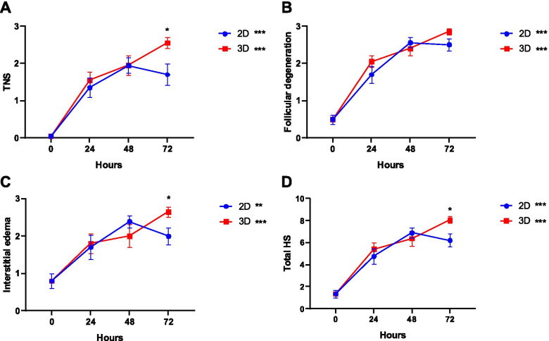
The comparison of histopathologic scores is shown in Fig. 1. During the study period, degeneration of tissue morphology was observed, with a significant increase in the score of all parameters.
Fig. 1.
Histopathologic score (HS) results regarding tissue necrosis score (TNS) (A), follicular degeneration (B), interstitial oedema (C) and total HS. A linear regression was performed for 2D and 3D groups and their corresponding slopes show an increase over time. Additionally, a comparison at 72 h was made using the t-test, which shows statistical differences. Data presented as mean ± SEM. *p < 0.05; **p < 0.01; ***p < 0.001
According to Fig. 1A, the increase of tissue necrosis score (TNS) is larger in the 3D culture (p < 0.001, B = 0.033, R2adj = 0.663) than for 2D (p < 0.001, B = 0.023, R2adj = 0.381). At 72 h of culture, specimens of the ovary cultured within the alginate matrix (3D) showed higher scores for tissue necrosis (2D = 1.700 ± 0.291 vs. 3D = 2.550 ± 0.138, p = 0.017).
The proportion of the degenerated follicles increased over time and was very similar in the two models (2D, p < 0.001, B = 0.028, R2adj = 0.617; 3D, p < 0.001, B = 0.031, R2adj = 0.696; Fig. 1B).
For the score of interstitial oedema (Fig. 1C), a greater increase in the 3D culture was observed (2D, p = 0.001, B = 0.018, R2adj = 0.253; 3D, p < 0.001, B = 0.024, R2adj = 0.428). At 72 h of culture, interstitial oedema was more evident in the 3D group compared to 2D (2D = 2.000 ± 0.224 vs. 3D = 2.650 ± 0.130, p = 0.035).
The total histopathologic score (HS), which is the total score of three ovarian injury parameters, increased in both groups but more sharply in 3D culture (2D, p < 0.001, B = 0.069, R2adj = 0.477; 3D, p < 0.001, B = 0.088, R2adj = 0.657; Fig. 1D). Also, after 72 h of culture, the total score was significantly higher in 3D culture than 2D culture (2D = 6.200 ± 0.569 vs. 3D = 8.050 ± 0.302, p = 0.01).
During the study period, there were no changes in the amount of lactate dehydrogenase (LDH) released into the culture medium (Fig. 2). The amount of LDH released was lower in culture with the alginate matrix than in conventional culture (1.16 ± 0.10 vs. 1.66 ± 0.11, p = 0.006), after 24 h. However, no differences were found at 48 and 72 h of culture.
Fig. 2.
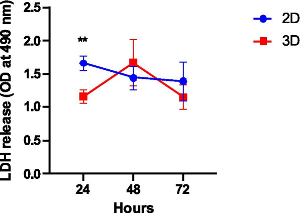
Cytotoxicity analysed through the release of lactate dehydrogenase (LDH) into culture supernatants. Data presented as mean of optical density (OD) ± SEM. A comparison at 24 h was made using the t-test, which shows statistical differences. **p < 0.01
Follicular analysis
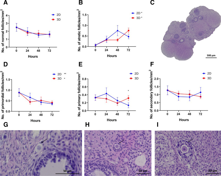
From the histological evaluation, we observed that density of morphologically normal follicles was maintained during the 72 h of culture (Fig. 3A). However, the density of atretic follicles increased significantly in both groups (2D, p = 0.027, B = 0.007, R2adj = 0.102; 3D, p < 0.001, B = 0.009, R2adj = 0.412; Fig. 3B).
Fig. 3.
Follicular analysis. Representative images of ovarian tissue (C scale bar: 500 μm) and morphological normal primordial (G), primary (H) and secondary (I) follicles (arrow, scale bar: 50 μm). Density of follicles classified as normal (A) and degenerated (B) according to oocyte and granulosa cell morphology. Density of primordial (D), primary (E) and secondary (F) follicles. A linear regression was performed for 2D and 3D groups and their corresponding slopes show an increase over time in atretic follicles and a decrease in primordial follicles. Additionally, a comparison between 2D and 3D at 48 and 72 h was made using the t-test, which shows statistical differences for the density of primary follicles at 72 h. Original magnification: A × 100; B × 400; H and I × 200. Data presented as mean ± SEM. *p < 0.05; **p < 0.01; ***p < 0.001
Regarding follicular classification, there was a significant decrease of primordial follicles in both types of culture (2D, p = 0.009, B = − 0.008, R2adj = 0.169; 3D, p = 0.036, B = − 0.006, R2adj = 0.11; Fig. 3D and G). The density of primary and secondary follicles remained the same during the 72 h of culture (Fig. 3E, F, H and I, respectively). At 72 h of culture, the density of primary follicles was significantly higher in the samples cultured with the alginate matrix (2D = 0.138 ± 0.064 vs. 3D = 0.318 ± 0.063 follicles/mm2, p = 0.035; Fig. 3E).
Proliferation and apoptosis in ovarian tissue
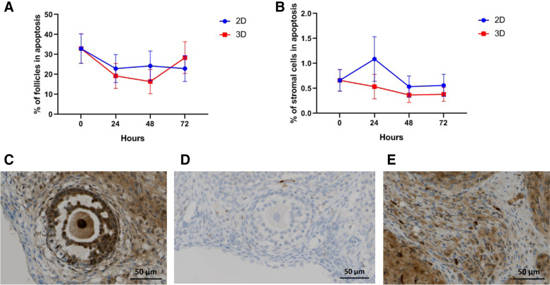
The levels of apoptosis assessed by the percentage of caspase-3 positive follicles and stromal cells remained stable during the culture period; no significant differences were found (Fig. 4).
Fig. 4.
Tissue apoptosis evaluated through caspase-3 staining in follicles (A) and stromal cells (B). Representative images of AC-3 staining in a secondary follicle (C) and stromal cells (E). A negative secondary follicle is represented in D. Original magnification × 200 and scale bar 50 μm (C, D and E). Data presented as mean ± SEM
The percentage of proliferative follicles, assessed with Ki67 staining, remained stable during the culture period in both groups (Fig. 5A, C and D). However, the evaluation of proliferation in stromal cells revealed a significant decrease in the 3D culture (p = 0.001, B = − 0.003, R2adj = 0.220; Fig. 5B and E).
Fig. 5.
Tissue proliferation evaluated through Ki67 staining in follicles (A) and stromal cells (B). Representative images of Ki67 staining in a secondary follicle (C) and stromal cells (E). A negative primary follicle (arrow) is represented in D. A linear regression was performed for 2D and 3D groups and their corresponding slopes show a decrease over time in stromal cell proliferation for 3D group. Original magnification × 200 (C, E) and × 400 (D), scale bar 50 μm. Data presented as mean ± SEM. ***p < 0.001
Discussion
Cryopreservation of ovarian tissue has been considered a reliable possibility for preservation of fertility in young women at risk of iatrogenic premature ovarian failure due to oncological treatments [19]. This is the only option for prepubertal girls and women who cannot delay the beginning of chemotherapy or cannot undergo an ovarian stimulation protocol for oocyte or embryo cryopreservation [19, 20].
The cryopreservation of ovarian tissue began in the 1950s; in the following years, several improvements were accomplished in the post-thaw viability of rat and mouse ovarian tissue. But only in the 1990s, after the successful delivery of a live birth from a frozen-thawed mouse primordial follicle [21], was this technique envisaged as an option for preservation of fertility [22].
The possibility of achieving complete oocyte development in vitro, in primates and most domestic species, has boosted research in this field. Different options were evaluated in order to overcome technical difficulties and an in vitro system was developed for culturing small pieces of ovarian cortex [20, 23]. The cortical fragments are the main reservoir of dormant primordial follicles in the postnatal ovary, which determines the woman’s fertile lifespan [20, 24].
Many culture approaches have been developed for different animal models [25]; however, the optimal model has not been achieved. Further efforts are needed in order to establish a reliable model for tissue culture [23, 26]. Higuchi et al. described the in situ ovarian tissue culture as an innovative and promising methodology to grow follicles in vitro due to its easy and safe handling, and more importantly the possibility of reconstituting the ovarian microenvironment or manipulating some conditions to induce follicle growth [27, 28].
Our work aimed to evaluate some drawbacks concerning the culture of ovarian tissue in a 3D model. In this model, OT was cultured in an alginate matrix, which has been widely used for the in vitro culture of isolated follicles in mice [29, 30] and rats [31]. The biochemical properties of this matrix make it a good candidate for 3D models. It has gained recognition for: its physicochemical and biological properties, such as its ability to make hydrogels under physiological conditions, providing little disruption to the tissue when encapsulated; the fact that the dissolution of gels for cell/tissue retrieval is easy to control; and its transparency for microscopic evaluation [17]. The physical features of the alginate hydrogel pore network allow the diffusion of nutrients and waste materials, namely the bidirectional diffusion of hormones and other proteins that are essential for tissue survival and also follicular development [32, 33]. Also, it is not of animal origin, which makes it biocompatible [33].
The histopathological evaluation of ovarian tissue (Fig. 1) elicited a global deterioration of tissue viability, and this decrease is time dependent. Tissue deterioration was observed in both models, with no differences between 2D and 3D within the first 48 h. This is in accordance with other previously published works that report no difference in cell viability in the first days of culture in the 2D and 3D models [34]. However, after 72 h there is a significant decrease in tissue viability in the 3D model. A decrease in tissue viability when the culture time is prolonged was previously described and a possible explanation for this is that the organ structure might retain the cellular waste produced and possibly induce a shortfall in the supply of oxygen and nutrients [35]. In the 3D model, the architecture is complex, with the tissue presenting three layers: (i) an outer layer with a high proliferation rate, (ii) a middle layer with senescent cells and (iii) a hypoxic core; this organization affects the supply of oxygen and nutrients, retaining the cellular waste produced [35, 36].
To measure cell death, LDH assay was performed in culture medium; this assay is based on the ability of LDH to convert pyruvate to lactate while consuming NADH [37]. In our data, we demonstrate that it is possible to detect and quantify LDH in the medium in both models, as previously described [37]. In the first 24 h, the 3D model presents a low cytotoxicity when compared with conventional culture. At 48 h and 72 h hours of culture, no difference was observed in the amount of LDH released in culture medium for both models. This effect may occur due to LDH retention in the alginate matrix. Although alginate matrix has higher porosity, allowing the diffusion of small substrates (molecular weight (MW) < 20 kDa) at the same speed as water [38], some proteins with higher MW, such as albumin (MW = 69 kDa), have a lower diffusion. Our results suggest that LDH (140 kDa) [39] may be retained longer in the spheroid structure. The diffusion resistance of alginate matrix was described for the first time, in 2006, in HepG2 cells encapsulated in alginate beads [37].
In order to establish a more detailed histopathological evaluation, we developed a score, based on the evaluation of three histologic parameters: TNS, interstitial oedema and follicular cell degeneration [40–42]. TNS and interstitial oedema are closely related parameters. Necrosis is characterized as an unprogrammed cell death process, which begins with cell swelling and results in cell membrane rupture and release of cell cytoplasmic content into the extracellular space [43], creating an extracellular movement of fluids. Our data show an increase in TNS and in interstitial oedema over experimental time in both models. However, after 72 h the increase in both parameters was greater in 3D culture. In the interior of the 3D structure, the bioavailability of oxygen and nutrients may be limited, affecting cell survival over time and conditioning higher level of necrosis and release of intracellular content when compared to more superficial cells [35]. In our characterization of tissue degeneration in the in vitro culture models, we detected an abundant level of follicular degeneration (Fig. 1B), and no differences were observed in the 2D model versus the 3D. A more detailed analysis reveals an increase in follicular atresia (Fig. 3B) accompanied by a decrease in primordial follicles (Fig. 3D). Our results are in line with previous work, which showed a time-related increase of follicular atresia in culture [44].
The biocompatibility of alginate matrix was intensively investigated using in vitro and in vivo models, and it was described as not inducing an immune response [45], first because of the absence of cell receptors to recognize alginate and second due to the high-purity commercialized alginate. This compound is extracted from brown algae, and may contain various impurities such as heavy metals, endotoxins, proteins, and polyphenolic compounds [46]; however, multi-step purification ensures a high level of purity in the alginate. Despite the safety of this compound, its widespread use has been challenged by some drawbacks concerning the molecular size, charges and viscosity of alginate, different culture models, different implantation sites and different animal models employed [45].
A different proliferation rate is observed when 2D and 3D models are compared. As shown in Fig. 5B, the proliferation rate of stromal cells is decreased in the 3D model. The difference in the proliferation rates between 2D and 3D models was previously described; cell lines cultured in the 3D system showed a reduced proliferation when compared with the 2D model, which proliferate at an unnaturally rapid rate [14]; these effects are dependent on the 3D model employed, and more specifically are matrix dependent, and also dependent on specific cells properties [14, 36]. In the field of fertility preservation, this effect was also reported in follicular in vitro culture; some authors have described that the physical properties of alginate matrices can limit the growth and development of earlier secondary follicles [17, 28, 30]. However, this topic is very controversial, and it is continuously under review. Different reasons have been suggested to explain these findings. In the context of our work, the rigidity of alginate matrix may be a possible explanation for the decreased proliferation rate [27]. Softer matrices with lower alginate concentrations [30] or the combination of different elements in the matrix to allow more space and the diffusion of macromolecules responsible for the tissue survival and growth were proposed [28].
Additionally, we evaluated cellular death using a caspase-3 antibody as a marker for programmed cell death. Caspase-3 is a well characterized protease which plays an effective role in apoptosis [47, 48]. The caspase-3 expression was already studied in granulosa [49] and theca cells [50] and also in oocytes [44]. In our study, apoptosis was evaluated in stromal cells and follicles, with the level of apoptosis in stromal cells and follicles being stable during the experimental time with no difference between conditions (2D vs. 3D model) (Fig. 4). However, it is important to keep in mind that apoptosis is a central process in ovarian function and development and occurs from foetal life on; it mainly affects the oocyte, until adult life when it affects the granulosa cells in growing follicles [47, 51].
In this study, we performed a well-structured histopathological analysis and the major strengths of our study are the sample size and the duplicate and blind analysis of results. However, some points could be addressed in future research to step up the 3D model, namely the study of the tissue-matrix interactions and culture medium supplementation to decrease follicular atresia.
Conclusions
In conclusion, the ovarian tissue culture within an alginate matrix was similar to 2D culture, regarding follicular density, follicular cell proliferation and cell apoptosis in follicles and stroma. In 3D culture, greater levels of tissue injury and oedema and lower stromal cell proliferation were seen. Therefore, there is no clear advantage in the 3D culture of ovarian tissue, as it is more time-consuming, difficult to perform and less reproductible.
Methods
Ethical statement
The present study was approved by the Ethics Committee for Animal Experimentation (ORBEA Authorization number 11060495/23-11-2016) of the Faculty of Medicine of the University of Coimbra and performed according to European Guidelines and Portuguese Law. The Guide for the Care and Use of Laboratory Animals of the National Institutes of Health was followed to take care of the animals and the ARRIVE guidelines to perform and report the experimental protocols.
Study design
The experimental scheme is shown in Fig. 6. Briefly, ovarian tissue (OT) preparation, cryopreservation, 2D system or encapsulated model procedure and timescale for the experiment are shown.
Fig. 6.
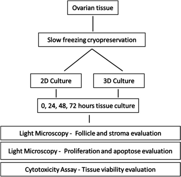
Schematic representation of the study. The figure shows the experimental design of the comparative study of in vitro culture system for ovarian tissue – 2D versus 3D
Experimental animals
The animal facility of the Faculty of Medicine of the University of Coimbra provided 12 female Rowett nude rats (RNU, homozygous) aged 8-10 weeks and with an average weight of 200 g. Housing, under a 12-h light/dark cycle, was in individually ventilated cages, with access to standard diet and filtered water ad libitum. Animals were distributed in a randomized manner into the study groups described below.
Ovaries collection
Surgical procedures were performed under inhalation anaesthesia with sevoflurane (5%) and with subcutaneous analgesia with carprofen (5 mg/kg, making a total of 0.2 mL per animal).
For bilateral ovariectomy, animals were placed in a supine position and the abdominal wall was shaved, cleaned and sterilized with povidone-iodine solution. A longitudinal median laparotomy was performed with a 2–3 cm incision in the lower area of the abdomen. The ovaries were identified and removed with the ligation of the vascular pedicle [52, 53]. After ensuring adequate haemostasis, the abdominal cavity was closed in layers.
After ovariectomy, the ovaries were placed in cold Dulbecco’s phosphatase-buffered solution (DPBS) (Biological Industries, Sartorius, Gottingen, Germany) supplemented with 10% foetal bovine serum (FBS) (Biological Industries, Sartorius, Gottingen, Germany) and each ovary was cut in 2 hemi-ovaries (3 × 2 × 1 mm). The fragments were maintained for 30 min in cold DPBS + 10% FBS before cryopreservation.
Cryopreservation and thawing protocols
The cryopreservation protocol consisted of a slow freezing and a rapid thawing method, adapted from Rosendahl M et al. [54]. The fragments were placed in plastic cryovials (Nunc, Thermo Fisher) containing 1.5 mL of freezing media consisting of 1.5 M ethylene glycol (Sigma-Aldrich, St. Louis, Missouri, USA), 0.1% sucrose (Merck, Darmstadt, Germany) and 10 mg/mL human albumin serum (HAS) (Grifols, Barcelona, Spain), and maintained in an ice bath. The cryovials were transferred to a rolling system for 30 min at 4 °C to allow the cryoprotectant to enter the tissue. Then, cooling in a programmable freezer (Planner cryo 10 Series 2 Freezer) followed the subsequent protocol. The starting temperature was 0 °C, and it was slowly reduced to − 9 °C at a rate of − 2 °C/min. After a 5-min holding time at − 9 °C, manual seeding was performed, after then the cryovials were cooled to − 40 °C, at a rate of − 0.3 °C/min, and the final step, a rapid decrease to − 140 °C (− 10 °C/min). When the programme was completed, cryovials were transferred into liquid nitrogen tank and stored for 1 week until thawing.
On the day of the experiment, ovarian fragments were thawed. The vials were air-warmed for 30 s and then immersed in a 37 °C water bath for 5 min. The freezing media was removed at room temperature by stepwise dilution of freezing media in sequential thawing media stabilized at room temperature. Three culture dishes were filled with thawing medium I (0.75 M ethylene glycol + 0.25 M sucrose in PBS + 10 mg/mL HAS), medium II (0.25 M Sucrose in PBS+ 10 mg/mL HAS), and medium III (PBS + 10 mg/mL HAS). Ovarian fragments were transferred into thawing medium I using sterile forceps and stirred for 10 min at room temperature. The same procedure was performed for thawing media II and III. Thawed tissue was transferred to PBS before culture.
Ovary culture
Hemi-ovaries (3 × 2 × 1 mm) were cultured in the growth media with and without encapsulation in an alginate matrix scaffold. The conventional culture was defined as 2D and the use of the scaffold as 3D. A 1.5% (w/v) solution of sodium alginate (Sigma-Aldrich, St. Louis, Missouri, USA) was prepared by mixing into sterile DPBS and heating to 37 °C. To encapsulate the ovarian fragments, the agarose (Invitrogen, California, USA) ring protocol was used, adapted from Henry, 2015 [55]. Agarose rings were filled with a layer of alginate matrix, fragments were placed in the ring and they were covered with matrix. Cross-linking solution (50 mM CaCl2 + 140 mM NaCl) was added, allowing the solution to solidify into a gel around the ovarian organoid.
The gel-organoid was then placed in the growth media to be cultured for 24 h, 48 h and 72 h. The growth media consisted of α-MEM (22561-021, ThermoFisher, Waltham, Massachusetts, USA), 10% FBS and 1/1000 penicillin/streptomycin (15140-122, Gibco, ThermoFisher, Waltham, Massachusetts, USA).
Tissue morphology and viability
The ovaries were fixed in 4% formaldehyde (Panreac Quimica Sau, Barcelona, Spain), embedded in paraffin, and sectioned serially at 5 μm. Three sections per transplant were stained with haematoxylin and eosin (HE) for morphological analysis. The images were acquired on the Axioscan Z1 (Carl Zeiss), with a Plan-Apochromat 10x/0.8 lens, and photographed with the Zen 2 program blue edition (Carl Zeiss Microscopy GmbH, 2011). Histological analysis was performed blindly by a researcher, at two different points in time, using the Image J software.
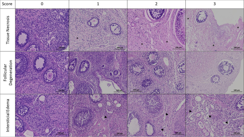
The criteria for ovarian tissue viability was adapted from criteria previously described for ovarian tissue injury after in vivo experiments of ischemia/reperfusion [40–42, 56]. As shown in Fig. 7, histopathological examination of the tissue damage was performed in terms of three visual parameters: interstitial oedema, follicular cell degeneration and percentage of tissue in necrosis (TNS).
Fig. 7.
Histopathologic score (HS) represented in haematoxylin and eosin-stained ovarian sections. The images show areas of tissue necrosis (*), follicular degeneration and interstitial oedema (▼). A scoring system where none = 0, mild = +, moderate = ++ and severe = +++ was applied. Original magnification × 100 and scale bar 100 μm
The follicles were histologically classified as degenerated when they included cells with pyknotic nucleus, shrunken ooplasm and disorganized granulosa cells. Follicular degeneration score was calculated as a proportion of the degenerated follicles to the total number of follicles [56]. Oedema is analysed by the subjective quantity of intercellular liquid. The extent of overall necrosis within each ovary was quantified by a visual assessment of the percentage of area in necrosis (TNS) for each specimen [40]. Necrotic cells typically present cytoplasmic hypereosinophilia, cellular swelling, blebbing, and loss of microvilli [57]. Each parameter was scored using a scale ranging from 0 to 3 (0, none; 1, mild or < 33%; 2, moderate or 33–66%; 3, severe or > 66%) [41]. Total scores were calculated according to these parameters [41]. Ovary sections were analyzed double blind.
Follicular analysis
Follicles were quantified manually and, to avoid double counting, only follicles with a visible nucleus were taken into account. Only morphologically normal follicles were taken into consideration for quantitative analysis. Follicles were then classified according to their maturity as primordial (constituted by a single layer of flattened granulosa cells), primary (they present a single layer of cuboid granulosa cells) and secondary (composed by two or more layers of granulosa cells around the oocyte) [58]. The pre-antral and antral follicles were grouped with the secondary follicles. Follicle atresia was assessed with morphologic criteria, such as irregular shape, granulosa cell pyknosis, cytoplasmic contraction, presence of vacuoles and ooplasm eosinophilia [58]. Follicular densities were calculated as the number of follicles per area (number/mm2), after measuring the tissue by manual surrounding of the surface (Fig. 3C) [59].
Proliferation and apoptosis evaluation
Immunohistochemistry assay was performed with the cell proliferation biomarker rabbit polyclonal antibody Ki-67 (diluted 1:300; PA5-19462, Thermofisher, Waltham, Massachusetts, USA) and cell apoptosis rabbit polyclonal antibody caspase-3 (diluted 1:100; AHP2286, Bio-Rad Laboratories, Hercules, California, USA). Antigen retrieval was performed with CC1, pH 8 EDTA/Tris-based buffer (Ventana Medical Systems, Tucson, Arizona, USA), followed by primary antibody incubation according to the manufacturer’s procedures and the dilutions previously mentioned. Detection of immunostaining was performed with OptiView DAB IHC Detection Kit (Ventana Medical Systems, Tucson, Arizona, USA) in accordance with the manufacturer’s recommendations, by the detection of DAB chromogenic substrate precipitated. All the immunostained sections were then nuclear counterstained with haematoxylin, dehydrated in a graded series of ethanol, cleared in xylene and mounted using a synthetic mounting medium.
After the immunohistochemistry protocol, in which the incubation with the cell proliferation indicator (Ki-67) and cell apoptosis (caspase-3), the images were acquired on the Axioscan Z1 (Carl Zeiss), with a Plan-Apochromat 10x/0.8 lens and photographed with the aid of the Zen 2 blue edition program (Carl Zeiss Microscopy GmbH, 2011). The results analysis was double and blinded.
Follicles were classified as positive or negative for Ki67 and caspase-3 (Figs. 4C, D and 5C, D). For this, positivity was considered when staining was observed in the oocyte and/or in at least one granulosa cell [60, 61]. Regarding the stroma, the quantification of the area marked with each antibody was performed using the Image J program (Fiji version, 1.8.0, USA). Five zones of each sample were randomly selected through the application of a rectangular grid and the results subsequently presented as an average (Figs. 4E and 5E).
Cytotoxicity evaluation
To evaluate the tissue viability, the lactate dehydrogenase (LDH) released in the culture medium from damaged cells was measured using CytoTox96® non-radioactive cytotoxicity assay (Promega G1780, Madison, Wisconsin, USA) [62–64]. The assay was performed according to the manufacturer’s protocol. Briefly, after 24 h, 48 h and 72 h of culture, the supernatant of each condition was collected and stored at − 20 °C. On the day of LDH assay, the CytoTox96® reagent was prepared mixing the buffer assay with the substrate mix. To test the effects of 2D vs. 3D culture, 50 μL of culture medium was transferred to a 96 multi-well, flat, clear-bottom plate and mixed with 50 μL CytoTox96® reagent and incubated for 30 min, protected from light. After that, 50 μL of stop solution was added to each well, and the optical density at 490 nm was measured using an EnSpire microplate reader. The absorbance values from the conditioned media supernatant were normalized to mean absorbance values calculated from control samples.
Statistical analyses
The number of ovaries to be used was estimated using the G*Power software version 3.1.9.4 (Kiel, Germany). A comparison of results between the seven described groups was considered, with a type I error of 0.05 and a statistical power of 0.90, for an effect size of 0.60. In this way, a total sample size of 66 fragments (9.4 per group) was estimated, with an effective power of 0.901. Therefore, 10 hemi-ovaries were used per group.
Statistical analyses were performed using the SPSS version 22.0 (IBM, Armonk, New York, USA). To compare the 2D and 3D groups, simple linear regression over time was performed using a bootstrapping strategy (1000 samples). The regression coefficient (B), which represents the slope, was reported. A positive B-value means an increase over time, while a negative one means a decrease. For example: p < 0.001, B = 0.033, R2adj = 0.663, means that per hour there is a significant increase of 0.033 (B) in the analysed variable and the linear model explains 66% (R2adj) of the variation over time. Normal distribution was then evaluated to compare the two study groups (2D vs. 3D), which visually showed a reasonable difference. Normally distributed variables were compared by means of the Student t-test and non-normally by means of a Mann-Whitney test. A p-value of less than 0.05 was considered statistically significant. The results are expressed as mean ± standard error of the mean (SEM).
Acknowledgments
The authors acknowledge to the Microscopy Imaging Center of Coimbra, at the Center for Neuroscience and Cell Biology (MICC-CNC), for their technical assistance.
Abbreviations
- 2D
Two-dimensional
- 3D
Three-dimensional
- ASRM
American Society for Reproductive Medicine
- DPBS
Dulbecco’s phosphatase-buffered solution
- FBS
Foetal bovine serum
- HAS
Human albumin serum
- HE
Haematoxylin and eosin
- HS
Histopathologic score
- LDH
Lactate dehydrogenase
- OT
Ovarian tissue
- OTC
Ovarian tissue cryopreservation
- SEM
Standard error of the mean
- TNS
Tissue necrosis score
Authors’ contributions
Conceptualization, A.S.P. and S.R.; methodology, A.S.P., S.R., M.L., F.S. and F.C.; validation, M.L., F.L. and T.A.S.; formal analysis, A.S.P.; investigation, A.S.P. and S.R.; resources, M.L., F.L. and T.A.S.; writing—original draft preparation, A.S.P. and S.R.; writing—review and editing, M.L., F.B. and T.A.S.; supervision, F.L. and T.A.S.; funding acquisition, A.S.P., F.L. and T.A.S. All authors have read and agreed to the published version of the manuscript.
Funding
The study was funded by Centro Hospitalar e Universitário de Coimbra, Coimbra (CHUC), Sociedade Portuguesa de Ginecologia (SPG), Merck® and National Funds via FCT (Foundation for Science and Technology) through the Strategic Project UID/NEU/04539/2019, UIDB/04539/2020 and UIDP/04539/2020 (CIBB), and by COMPETE-FEDER (POCI-01-0145-FEDER-007440).
Availability of data and materials
Not applicable.
Declarations
Ethics approval and consent to participate
The present study was approved by the Ethics Committee for Animal Experimentation (ORBEA Authorization number 11060495/23-11-2016) of the Faculty of Medicine of the University of Coimbra and performed according to the European Guidelines and Portuguese Law.
Consent for publication
Not applicable.
Competing interests
None.
Footnotes
Publisher’s Note
Springer Nature remains neutral with regard to jurisdictional claims in published maps and institutional affiliations.
References
- 1.Siegel RL, Miller KD, Jemal A. Cancer statistics, 2020. CA Cancer J Clin. 2020;70(1):7–30. doi: 10.3322/caac.21590. [DOI] [PubMed] [Google Scholar]
- 2.Arnold M, Rutherford MJ, Bardot A, Ferlay J, Andersson TML, Myklebust TÅ, et al. Progress in cancer survival, mortality, and incidence in seven high-income countries 1995–2014 (ICBP SURVMARK-2): a population-based study. Lancet Oncol. 2019;20(11):1493–1505. doi: 10.1016/S1470-2045(19)30456-5. [DOI] [PMC free article] [PubMed] [Google Scholar]
- 3.Akasha AM, Woodruff TK. The ovary. 3. Elsevier Inc.; 2019. Oncofertility: preservation of ovarian function after a cancer diagnosis; pp. 501–508. [Google Scholar]
- 4.Anderson RA, Amant F, Braat D, D’Angelo A, Chuva de Sousa Lopes SM, Demeestere I, et al. ESHRE guideline: female fertility preservation. Hum Reprod Open. 2020;2020(4):1–17. doi: 10.1093/hropen/hoaa052. [DOI] [PMC free article] [PubMed] [Google Scholar]
- 5.Duncan FE, Feinberg E, Brannigan RE, Edmonds M, Ataman L, Woodruff TK. Yen & Jaffe’s reproductive endocrinology: physiology, pathophysiology, and clinical management: eighth edition. 8. Elsevier Inc.; 2019. Fertility preservation; pp. 857–886.e6. [Google Scholar]
- 6.Practice Commitee of the American Society for Reproductive Medicine Fertility preservation in patients undergoing gonadotoxic therapy or gonadectomy: a committee opinion. Fertil Steril. 2019;112(6):1022–1033. doi: 10.1016/j.fertnstert.2019.09.013. [DOI] [PubMed] [Google Scholar]
- 7.Donnez J, Dolmans MM, Demylle D, Jadoul P, Pirard C, Squifflet J, et al. Livebirth after orthotopic transplantation of cryopreserved ovarian tissue. Obstet Gynecol. 2005;105(1):214. doi: 10.1016/S0140-6736(04)17222-X. [DOI] [PubMed] [Google Scholar]
- 8.Chen J, Todorov P, Isachenko E, Rahimi G, Mallmann P, Isachenko V. Construction and cryopreservation of an artificial ovary in cancer patients as an element of cancer therapy and a promising approach to fertility restoration. Hum Fertil. 2021;1:1–21. doi: 10.1080/14647273.2021.1885756. [DOI] [PubMed] [Google Scholar]
- 9.Lee S, Ozkavukcu S, Ku SY. Current and future perspectives for improving ovarian tissue cryopreservation and transplantation outcomes for cancer patients. Reprod Sci. 2021;28(6):1746–1758. doi: 10.1007/s43032-021-00517-2. [DOI] [PMC free article] [PubMed] [Google Scholar]
- 10.Terren C, Nisolle M, Munaut C. Pharmacological inhibition of the PI3K / PTEN / Akt and mTOR signalling pathways limits follicle activation induced by ovarian cryopreservation and in vitro culture. J Ovarian Res. 2021:1–15. 10.1186/s13048-021-00846-5. [DOI] [PMC free article] [PubMed]
- 11.Belli M, Vigone G, Merico V, Redi CA, Zuccotti M, Garagna S. Towards a 3D culture of mouse ovarian follicles. Int J Dev Biol. 2012;56(10–12):931–937. doi: 10.1387/ijdb.120175mz. [DOI] [PubMed] [Google Scholar]
- 12.Monniaux D, Cadoret V, Clément F, Dalbies-Tran R, Elis S, Fabre S, et al. Folliculogenesis. In: Huhtaniemi I, Martini L, editors. Encyclopedia of Endocrine Diseases (Second Edition), vol. 2. Oxford: Academic Press; 2019. p. 377–98. 10.1016/B978-0-12-801238-3.64550-6.
- 13.Kapałczyńska M, Kolenda T, Przybyła W, Zajączkowska M, Teresiak A, Filas V, et al. State of the art paper 2D and 3D cell cultures – a comparison of different types of cancer cell cultures. Arch Med Sci. 2018;14(4):910–919. doi: 10.5114/aoms.2016.63743. [DOI] [PMC free article] [PubMed] [Google Scholar]
- 14.Jensen C, Teng Y. Is it time to start transitioning from 2D to 3D cell culture? Front Mol Biosci. 2020;7(March):1–15. doi: 10.3389/fmolb.2020.00033. [DOI] [PMC free article] [PubMed] [Google Scholar]
- 15.Kinnear HM, Tomaszewski CE, Chang FL, Moravek MB, Xu M, Padmanabhan V, et al. The ovarian stroma as a new frontier. Reproduction. 2020;160(3):R25–R39. doi: 10.1530/REP-19-0501. [DOI] [PMC free article] [PubMed] [Google Scholar]
- 16.Scott J, Carlsson IB, Bavister BD, Hovatta O. Human ovarian tissue cultures: extracellular matrix composition , coating density and tissue dimensions. Reprod Biomed Online. 2004;9(3):287–293. doi: 10.1016/S1472-6483(10)62143-8. [DOI] [PubMed] [Google Scholar]
- 17.King SM, Quartuccio S, Hilliard TS, Inoue K, Burdette JE. Alginate hydrogels for three-dimensional organ culture of ovaries and oviducts. J Vis Exp. 2011;52(June):4–9. doi: 10.3791/2804. [DOI] [PMC free article] [PubMed] [Google Scholar]
- 18.Jackson KS, Inoue K, Davis DA, Hilliard TS, Burdette JE. Three-dimensional ovarian organ culture as a tool to study normal ovarian surface epithelial wound repair. Endocrinology. 2009;150(8):3921–3926. doi: 10.1210/en.2008-1674. [DOI] [PMC free article] [PubMed] [Google Scholar]
- 19.Rivas Leonel EC, Lucci CM, Amorim CA. Cryopreservation of human ovarian tissue: a review. Transfus Med Hemother. 2019;46(3):173–181. doi: 10.1159/000499054. [DOI] [PMC free article] [PubMed] [Google Scholar]
- 20.Yin H, Kristensen SG, Jiang H, Rasmussen A, Andersen CY. Survival and growth of isolated pre-antral follicles from human ovarian medulla tissue during long-term 3D culture. Hum Reprod. 2016;31(7):1531–1539. doi: 10.1093/humrep/dew049. [DOI] [PubMed] [Google Scholar]
- 21.Gunasena KT, Villines PM, Critser ES, Critser JK. Live births after autologous transplant of cryopreserved mouse ovaries. Hum Reprod. 1997;12(1):101–106. doi: 10.1093/humrep/12.1.101. [DOI] [PubMed] [Google Scholar]
- 22.Eppig JJ, O’Brien MJ. Development in vitro of mouse oocytes from primordial follicles. Biol Reprod. 1996;54(1):197–207. doi: 10.1095/biolreprod54.1.197. [DOI] [PubMed] [Google Scholar]
- 23.Smitz J, Dolmans MM, Donnez J, Fortune JE, Hovatta O, Jewgenow K, et al. Current achievements and future research directions in ovarian tissue culture, in vitro follicle development and transplantation: implications for fertility preservation. Hum Reprod Update. 2010;16(4):395–414. doi: 10.1093/humupd/dmp056. [DOI] [PMC free article] [PubMed] [Google Scholar]
- 24.Wagner M, Yoshihara M, Douagi I, Damdimopoulos A, Panula S, Petropoulos S, et al. Single-cell analysis of human ovarian cortex identifies distinct cell populations but no oogonial stem cells. Nat Commun. 2020;11(1):1–15. doi: 10.1038/s41467-020-14936-3. [DOI] [PMC free article] [PubMed] [Google Scholar]
- 25.Vanacker J, Amorim CA. Alginate: a versatile biomaterial to encapsulate isolated ovarian follicles. Ann Biomed Eng. 2017;45(7):1633–1649. doi: 10.1007/s10439-017-1816-6. [DOI] [PubMed] [Google Scholar]
- 26.Desai N, Alex A, AbdelHafez F, Calabro A, Goldfarb J, Fleischman A, et al. Three-dimensional in vitro follicle growth: overview of culture models, biomaterials, design parameters and future directions. Reprod Biol Endocrinol. 2010;8(1):119. doi: 10.1186/1477-7827-8-119. [DOI] [PMC free article] [PubMed] [Google Scholar]
- 27.Fathi R, Valojerdi MR, Ebrahimi B, Eivazkhani F, Akbarpour M, Tahaei LS, et al. Fertility preservation in cancer patients: in vivo and in vitro options. Cell J. 2017;19(10):173–183. doi: 10.22074/cellj.2016.4880. [DOI] [PMC free article] [PubMed] [Google Scholar]
- 28.Higuchi CM, Maeda Y, Horiuchi T, Yamazaki Y. A simplified method for three-dimensional ( 3-D) ovarian tissue culture yielding oocytes competent to produce full-term offspring in mice. PLoS One. 2015;10(11). 10.1371/journal.pone.0143114. [DOI] [PMC free article] [PubMed]
- 29.Pangas SA, Saudye H, Shea LD, Woodruff TK. Novel approach for the three-dimensional culture of granulosa cell-oocyte complexes. Tissue Eng. 2003;9(5):1013–1021. doi: 10.1089/107632703322495655. [DOI] [PubMed] [Google Scholar]
- 30.Xu M, West E, Shea LD, Woodruff TK. Identification of a stage-specific permissive in vitro culture environment for follicle growth and oocyte development. Biol Reprod. 2006;75(6):916–923. doi: 10.1095/biolreprod.106.054833. [DOI] [PubMed] [Google Scholar]
- 31.Heise M, Koepsel R, Russell AJ, McGee EA. Calcium alginate microencapsulation of ovarian follicles impacts FSH delivery and follicle morphology. Reprod Biol Endocrinol. 2005;3:1–8. doi: 10.1186/1477-7827-3-47. [DOI] [PMC free article] [PubMed] [Google Scholar]
- 32.Amorim CA, Van Langendonckt A, David A, Dolmans MM, Donnez J. Survival of human pre-antral follicles after cryopreservation of ovarian tissue, follicular isolation and in vitro culture in a calcium alginate matrix. Hum Reprod. 2009;24(1):92–99. doi: 10.1093/humrep/den343. [DOI] [PubMed] [Google Scholar]
- 33.Andersen T, Auk-Emblem P, Dornish M. 3D cell culture in alginate hydrogels. Microarrays. 2015;4(2):133–161. doi: 10.3390/microarrays4020133. [DOI] [PMC free article] [PubMed] [Google Scholar]
- 34.Gurski L, Jha A, Zhang C, Jia X, Farach-Carson M. Hyaluronic acid-based hydrogels as 3D matrices for in vitro evaluation of chemotherapeutic drugs using poorly adherent prostate cancer cells. Biomaterials. 2009;30(30):6076–6085. doi: 10.1016/j.biomaterials.2009.07.054. [DOI] [PMC free article] [PubMed] [Google Scholar]
- 35.Hoarau-Véchot J, Rafii A, Touboul C, Pasquier J. Halfway between 2D and animal models: are 3D cultures the ideal tool to study cancer-microenvironment interactions? Int J Mol Sci. 2018;19(1):181. doi: 10.3390/ijms19010181. [DOI] [PMC free article] [PubMed] [Google Scholar]
- 36.Edmondson R, Broglie JJ, Adcock AF, Yang L. Three-dimensional cell culture systems and their applications in drug discovery and cell-based biosensors. Assay Drug Dev Technol. 2014;12(4):207–218. doi: 10.1089/adt.2014.573. [DOI] [PMC free article] [PubMed] [Google Scholar]
- 37.Khattak SF, Spatara M, Roberts L, Roberts SC. Application of colorimetric assays to assess viability, growth and metabolism of hydrogel-encapsulated cells. Biotechnol Lett. 2006;28(17):1361–1370. doi: 10.1007/s10529-006-9104-9. [DOI] [PubMed] [Google Scholar]
- 38.Brito IR, Lima IMT, Xu M, Shea LD. Three-dimensional systems for in vitro follicular culture : overview of alginate-based matrices. Reprod Fertil Dev. 2014;26:915–930. doi: 10.1071/RD12401. [DOI] [PMC free article] [PubMed] [Google Scholar]
- 39.Javed MH, Azimuddin SMI, Hussain AN, Ahmed A, Ishaq M. Purification and characterization of lactate dehydrogenase from Varanus liver. Exp Mol Med. 1997;29(1):25–30. doi: 10.1038/emm.1997.4. [DOI] [Google Scholar]
- 40.Asfour V, Polson A, Varma R, Menon P. Evaluating ovarian preservation after ovarian torsion using the ovarian preservation score and tissue necrosis score. Clin Obstet Gynecol Reprod Med. 2015;1(2):34–39. doi: 10.15761/COGRM.1000110. [DOI] [Google Scholar]
- 41.Guven S, Muci E, Unsal MA, Yulug E, Alver A, Duman MK, et al. The effects of carbon dioxide pneumoperitoneum on ovarian blood flow, oxidative stress markers, and morphology during laparoscopy: a rabbit model. Fertil Steril. 2010;93(4):1327–1332. doi: 10.1016/j.fertnstert.2008.10.053. [DOI] [PubMed] [Google Scholar]
- 42.Eser A, Hizli D, Haltas H, Namuslu M, Kosus A, Kosus N, et al. Effects of curcumin on ovarian ischemia-reperfusion injury in a rat model. Biomed Rep. 2015;3(6):807–813. doi: 10.3892/br.2015.515. [DOI] [PMC free article] [PubMed] [Google Scholar]
- 43.Lee SY, Ju MK, Jeon HM, Jeong EK, Lee YJ, Kim CH, et al. Regulation of tumor progression by programmed necrosis. Oxidative Med Cell Longev. 2018;2018:3537471. doi: 10.1155/2018/3537471. [DOI] [PMC free article] [PubMed] [Google Scholar]
- 44.Otala M, Erkkilä K, Tuuri T, Sjöberg J, Suomalainen L, Suikkari AM, et al. Cell death and its suppression in human ovarian tissue culture. Mol Hum Reprod. 2002;8(3):228–236. doi: 10.1093/molehr/8.3.228. [DOI] [PubMed] [Google Scholar]
- 45.Orive G, Ponce S, Hernández RM, Gascón AR, Igartua M, Pedraz JL. Biocompatibility of microcapsules for cell immobilization elaborated with different type of alginates. Biomaterials. 2002;23(18):3825–3831. doi: 10.1016/S0142-9612(02)00118-7. [DOI] [PubMed] [Google Scholar]
- 46.Lee KY, Mooney DJ. Alginate: properties and biomedical applications. Prog Polym Sci. 2012;37(1):106–126. doi: 10.1016/j.progpolymsci.2011.06.003. [DOI] [PMC free article] [PubMed] [Google Scholar]
- 47.Hussein MR. Apoptosis in the ovary: molecular mechanisms. Hum Reprod Update. 2005;11(2):161–177. doi: 10.1093/humupd/dmi001. [DOI] [PubMed] [Google Scholar]
- 48.Cohen GM. Caspases: executioners of apoptosis. Biochem J. 1997;326:1–16. doi: 10.1042/bj3260001. [DOI] [PMC free article] [PubMed] [Google Scholar]
- 49.Izawa M, Nguyen PH, Kim HH, Yeh J. Expression of the apoptosis-related genes, caspase-1, caspase-3, DNA fragmentation factor, and apoptotic protease activating factor-1, in human granulosa cells. Fertil Steril. 1998;70(3):549–552. doi: 10.1016/S0015-0282(98)00194-0. [DOI] [PubMed] [Google Scholar]
- 50.Krajewska M, Wang HG, Krajewski S, Zapata JM, Shabaik A, Gascoyne R, et al. Immunohistochemical analysis of in vivo patterns of expression of CPP32 (Caspase-3), a cell death protease. Cancer Res. 1997;57(8):1605–1613. [PubMed] [Google Scholar]
- 51.Milenkovic M, Diaz-Garcia C, Wallin A, Brännström M. Viability and function of the cryopreserved whole rat ovary: comparison between slow-freezing and vitrification. Fertil Steril. 2012;97(5):1176–1182. doi: 10.1016/j.fertnstert.2012.01.123. [DOI] [PubMed] [Google Scholar]
- 52.Di Loreto VE, Pera LI, Rigalli A. In: Experimental surgical models in the laboratory rat. 1. Rigalli A, Di Loreto V, editors. 2009. pp. 149–151. [Google Scholar]
- 53.Khajuria DK, Razdan R, Mahapatra DR. Descrição de um novo método de ooforectomia em ratas. Rev Bras Reum. 2012;52(3):462–470. [PubMed] [Google Scholar]
- 54.Rosendahl M, Schmidt KT, Ernst E, Rasmussen PE, Loft A, Byskov AG, et al. Cryopreservation of ovarian tissue for a decade in Denmark: a view of the technique. Reprod Biomed Online. 2011;22(2):162–171. doi: 10.1016/j.rbmo.2010.10.015. [DOI] [PubMed] [Google Scholar]
- 55.Henry L, Labied S, Fransolet M, Kirschvink N, Blacher S, Noel A, et al. Isoform 165 of vascular endothelial growth factor in collagen matrix improves ovine cryopreserved ovarian tissue revascularisation after xenotransplantation in mice. Reprod Biol Endocrinol. 2015;13:1–10. doi: 10.1186/s12958-015-0015-2. [DOI] [PMC free article] [PubMed] [Google Scholar]
- 56.Yildirim N, Yigitturk G, Sahingoz Yildirim AG, Akdemir A, Ilgen O, Yeniel O, et al. Octreotide protects ovary against ischemia-reperfusion injury in rats: evaluation of histological and biochemical parameters. J Obstet Gynaecol Res. 2015;41(10):1591–1597. doi: 10.1111/jog.12770. [DOI] [PubMed] [Google Scholar]
- 57.Brown K, Awan NA, Le PH, Wilson AM. Histology, Cell Death. In: StatPearls [Internet]. Treasure Island (FL): StatPearls Publishing; 2021. Available at: http://www.ncbi.nlm.nih.gov/books/NBK526045/. [cited April 24, 2021]
- 58.Gougeon A. Regulation of ovarian follicular development in primates: facts and hypotheses. Endocr Rev. 1996;17(2):121–155. doi: 10.1210/edrv-17-2-121. [DOI] [PubMed] [Google Scholar]
- 59.Fransolet M, Labied S, Henry L, Masereel MC, Rozet E, Kirschvink N, et al. Strategies for using the sheep ovarian cortex as a model in reproductive medicine. PLoS One. 2014;9(3):1–7. doi: 10.1371/journal.pone.0091073. [DOI] [PMC free article] [PubMed] [Google Scholar]
- 60.Luyckx V, Scalercio S, Jadoul P, Amorim CA, Soares M, Donnez J, et al. Evaluation of cryopreserved ovarian tissue from prepubertal patients after long-term xenografting and exogenous stimulation. Fertil Steril. 2013;100(5):1350–1357. doi: 10.1016/j.fertnstert.2013.07.202. [DOI] [PubMed] [Google Scholar]
- 61.Dolmans MM, Binda MM, Jacobs S, Dehoux JP, Squifflet JL, Ambroise J, et al. Impact of the cryopreservation technique and vascular bed on ovarian tissue transplantation in cynomolgus monkeys. J Assist Reprod Genet. 2015;32(8):1251–1262. doi: 10.1007/s10815-015-0542-y. [DOI] [PMC free article] [PubMed] [Google Scholar]
- 62.Korzeniewski C, Callewaert DM. An enzyme-release assay for natural cytotoxicity. J Immunol Methods. 1983;64(3):313–320. doi: 10.1016/0022-1759(83)90438-6. [DOI] [PubMed] [Google Scholar]
- 63.De Michele F, Poels J, Weerens L, Petit C, Evrard Z, Ambroise J, et al. Preserved seminiferous tubule integrity with spermatogonial survival and induction of Sertoli and Leydig cell maturation after long-term organotypic culture of prepubertal human testicular tissue. Hum Reprod. 2017;32(1):32–45. doi: 10.1093/humrep/dew300. [DOI] [PubMed] [Google Scholar]
- 64.Keshaw H, Forbes A, Day RM. Release of angiogenic growth factors from cells encapsulated in alginate beads with bioactive glass. Biomaterials. 2005;26(19):4171–4179. doi: 10.1016/j.biomaterials.2004.10.021. [DOI] [PubMed] [Google Scholar]
Associated Data
This section collects any data citations, data availability statements, or supplementary materials included in this article.
Data Availability Statement
Not applicable.


