

Abstract
Background
Fibrodysplasia Ossificans Progressiva (FOP) is a rare autosomal dominant disease characterized by congenital malformation of the great toes and progressive heterotopic ossification of soft tissues leading to cumulative disability. The genetic cause of FOP are mutations in the ACVR1 gene that encodes a type I receptor of Bone Morphogenetic Proteins. The most recurrent mutation in FOP patients is R206H affecting the Glycine‐Serine rich domain and causing the hyper‐activation of the receptor and the responsivity to the non‐canonical ligand, Activin A.
In the present study, we described a 3‐years old child with early and highly suggestive clinical features of FOP who was found negative for the recurrent p.R206H substitution.
Methods
Molecular screening of the whole ACVR1 coding sequence and functional characterization in transfection‐based assays.
Results and Conclusions
We identified a novel, de novo variant in the fifth ACVR1 coding exon (NM_001111067.4:c.772A>T; NP_001104537.1:p.(R258W)). This substitution, never reported in association with FOP, affects a conserved arginine residue in the kinase domain of the protein. In silico analysis predicted the pathogenicity of this substitution, demonstrated by in vitro assays showing that the p.R258W ACVR1 mutated receptor acquires the ability to transduce the aberrant Activin A‐mediated signaling, as observed for the gene variants associated with FOP.
Keywords: Activin A, ACVR1, BMP signaling, Fibrodysplasia Ossificans Progressiva, p.R258W
FOP is a rare genetic disease representing the most severe and disabling condition due to heterotopic ossification associated with gain‐of‐function mutations of the ACVR1 gene. We describe here a little patient with an early clinical presentation of the disease carrying a novel substituton of the ACVR1 causative gene.
1. INTRODUCTION
Fibrodysplasia Ossificans Progressiva (FOP; OMIM #135100) is an extremely rare autosomal dominant disease in which heterotopic bone forms in muscle and soft tissue, leading to joint dysfunction and severe disability. The incidence of FOP is 1 of 2 million and there is no regional, racial, or sexual predisposition (Baujat et al., 2017; Qi et al., 2017).
Classical presentation of FOP is characterized by a specific, congenital big toe malformation (hallux valgus deformity) and apparently spontaneous occurring of acute phases of the disease called flare‐ups, with inflammatory soft tissue swelling, followed by progressive and disabling heterotopic endochondral ossification (Kaliya‐Perumal et al., 2020; Qi et al., 2017). Flare‐ups and heterotopic ossification usually manifest in the first decade of life in 95% cases (Baujat et al., 2017). Acute phases caused by the underlying inflammation in the ligaments, tendons, or skeletal muscle occur upon pro‐inflammatory insults such as muscle fatigue, tissue damage, intramuscular injections, or viral illness or apparently without any recognizable trigger. They are generally sporadic and unpredictable, in terms of occurrence, duration, and outcome: not all the flare‐ups end up with bone neoformation, on the other hand a spontaneous, creeping ossification in absence of acute phases is also observed (Pignolo et al., 2016). Over time, owing to the repeated flare‐ups at different sites, progressive and cumulative ossification of soft tissues occurs, leading to the debilitating effects of FOP (Kaliya‐Perumal et al., 2020). Some patients have atypical FOP signs, or so‐called FOP plus forms: tibial osteochondromas, spinal malformations and broad femoral neck, thumb malformations, cognitive impairment, and diffuse scalp thinning (Kaplan et al., 2009; Qi et al., 2017).
In 2006 the R206H mutation in the ACVR1 (OMIM #102576, also known as ALK2) gene encoding a bone morphogenetic protein (BMP) type I receptor was described as the molecular cause of FOP (Shore et al., 2006). The most common R206H substitution affects the intracellular glycine‐serine (GS) domain of the ACVR1 receptor. Atypical missense mutations (p.L196P, p.R202I, p.Q207E, p.R258G/S, p.G328R/W/E, p.G356D, and p.R375P) in the GS or protein kinase domains of ACVR1 have been also identified in some FOP patients (Kaliya‐Perumal et al., 2020). De novo heterozygous mutations are found in most cases, but there are familial cases with autosomal inheritance reported. Usually, FOP diagnosis is made on a clinical basis by the observation of the presence of pathognomonic features such as typical toe deformation and ossification foci and is confirmed by DNA sequence analysis of the ACVR1 gene.
2. MATERIALS AND METHODS
2.1. ACVR1 screening
Peripheral blood samples were collected upon informed consent administration. Genomic DNA was extracted using the QIAamp DNA Mini kit (Qiagen) according to the manufacturer's instructions and quantified with the Nanodrop 2000 (Thermo Scientific).
Genomic DNA fragments corresponding to the nine coding exons and flanking intronic sequences of ACVR1 (RefSeq NM_001111067.4; RefSeq NP_001104537.1), were amplified by polymerase chain reaction (PCR; the primer sequences are listed in Table S1) using the GoTaq Master mix (Promega) with reactions carried out in a GeneAmp PCR System 2720 Thermocycler (Applied Biosystems).
PCR products were cleaned up by Exo/SAP‐IT (Thermofisher Scientific) digestion and then used for direct sequencing. Reactions were set up with a Big Dye Terminator Cycle Sequencing Kit according to the provided protocol, run on a 3130xl Genetic Analyzer (Applied Biosystems) and sequences analyzed by the Sequencer 4.7 software. Sequence variations were described according to the Human Genome Variation Society guidelines (http://www.hgvs.org).
2.2. In silico analysis
In order to estimate the impact of the identified variant on ACVR1 receptor function, we performed an in silico analysis by using the Ensembl Variant Effect Predictor (VEP) toolset (available at https://grch37.ensembl.org/Tools/VEP) [McLaren et al., 2016].
2.3. Expression plasmids
Complementary DNA (cDNA) for human, wild‐type ACVR1 (Table S1 for oligonucleotides used to generate the product) was obtained by RT‐PCR and then subcloned into the expression vector pCMV‐3Tag‐8 (pCMV_ACVR1) without the native STOP codon, to be in frame with the 3xFLAG tag provided by the vector.
ACVR1 variants p.R206H, p.R258S, and p.R258W were introduced by mutagenesis, by using the QuikChange Lightning Site‐Directed Mutagenesis kit (Agilent) according to the provided protocol. All the obtained clones were checked by Sanger sequencing. The generation of the pGL4.17 vector containing the BMP‐Responsive Element (BRE) upstream the Luciferase reporter gene (indicated as pGL4.17‐BRE) was previously described by our group (Cappato et al., 2016).
2.4. Cell culture, transient transfection, and treatments
U2OS cells were already available originally obtained from ATCC (HTB‐96). Cells were routinely cultured in a complete medium consisting of Dulbecco's Modified Eagle's Medium (DMEM), containing 10% fetal bovine serum (FBS, Gibco). Depletion medium was prepared by reducing FBS at 0.5% or by replacing it with 0.1% BSA (Bovine Serum Albumin, Sigma‐Aldrich, Merk). Cells were maintained at 37℃ in a humidified atmosphere with 5% CO2.
Transient transfections were performed in 96‐well plates, by seeding 5 × 10⁴/well U2OS cells directly in the transfection mix, composed by 30 ng of the pGL4.17‐BRE, 40 ng of pCMV_ACVR1 constructs and 2.5 ng of pGL4.73[hRluc/SV40] to normalize toward transfection efficiency, using the Lipofectamine 2000 reagent protocol (Invitrogen, Life Technologies). After 4 hr, cells were washed and incubated for additional 16 hr in a fresh medium with 0.5% FBS, and 33 nM ActA or 50 ng/ml BMP2. Next day cells were lysed and processed to evaluate the Luciferase activities with the Dual Luciferase Reporter Assay (Promega) according to the manufacturer's instruction.
ID1 gene expression and P‐SMAD1/5/9 proteins levels were evaluated by seeding 3 × 10⁵ U2OS in 6‐well plates for transfection with 2 µg of pCMV_ACVR1 constructs using the Lipofectamine 2000 reagent (Invitrogen, Life Technologies). After 48 hr, cells were incubated over‐night in a depletion medium containing 0.5% FBS, then activated in a medium containing 0.1% BSA for 3h with 33nM Activin A or 50 ng/ml BMP2 to evaluate ID1 gene expression and 45 min with 33 nM ActA or 100 ng/ml BMP2 to verify the P‐SMAD1/5/9 phosphorylation state.
2.5. RNA extraction and quantitative PCR (qPCR)
For expression studies, treated and untreated cells were harvested and total RNA was isolated by using the RNeasy Mini Kit (Qiagen), according to the provided protocol. RNA was quantified with Nanodrop Spectrophotometer (Thermo Scientific), and first‐strand cDNA was synthesized by the iSCript cDNA Synthesis Kit (BioRad) from 500 ng of total RNA.
Expression of endogenous ID1 gene was evaluated through qPCR using specific Syber Green strategy (see Table S1 for Oligo sequences, Tib Molbiol Srl). Samples were measured in duplicate and the results were normalized on the geometric mean of B2M and HPRT genes. qPCR was run on the IQ5 instrument from BioRad and data analysis was performed using the ΔCt method: ratio reference/target = 2ΔCt (Cappato et al., 2019; Livak & Schmittgen, 2001).
2.6. Western blot
For detection of the P‐SMAD1/5/9 protein level, cells were washed twice with PBS and lysed in 1xRIPA buffer (50 mM Tris HCl pH 7.5, 150 mM NaCl, 1% Nonidet P‐40, 1% Sodium Deoxycholic, 0.1% SDS), containing phosphatase and protease inhibitors (PhosSTOP cocktail and Complete tablets, Roche), NaF 50 mM and Na3VO4 1 mM. Protein concentration was determined by the PierceTM BCA Protein Assay Kit (Thermo Scientific) according to the manufacturer's protocol and 15 µg of total lysates run onto precasted 4%–15% Midi Criterion TGX‐gels 18W (BioRad). Proteins were transferred onto Nitrocellulose membrane (BioRad) and probed with the indicated primary antibody at 4℃ overnight. After incubation with HRP‐conjugated secondary antibodies, protein bands were revealed by chemiluminescence with the Amersham ECL Detection Reagents (GE Healthcare) and detected with the Uvitec instrument (Cambridge, UK). Densitometric analysis of the western blot signals was performed by using the Uvitec software. Primary antibodies were diluted as follows: P‐SMAD1/5/9 1:2000 (#13820, SMAD1 1:2000 #9743, Cell Signaling); anti‐FLAG M2 monoclonal antibody 1:3000 (#F1804, Sigma‐Aldrich, Merk); anti‐GAPDH 1:20000 (#MAB374, Millipore).
2.7. Statistical analysis
Luciferase reporter gene assays were performed independently three times in triplicate (n = 9). Experiments to evaluate gene expression by qPCR were performed in duplicate from three independent RNA extractions. The analysis of P‐SMAD proteins was carried out on three independent experiments. The Unpair t test was applied to verify statistical significance of the observed variations (https://www.graphpad.com/quickcalcs/ttest1.cfm). Significant differences were given as p < .05*, p < .01**, p < .001***.
3. RESULTS AND DISCUSSION
3.1. Patient description
The proband is a 3‐year‐old Lithuanian boy referred to our examination by the orthopedic surgeon. He is the first child of a non‐related Lithuanian couple. The pregnancy was uneventful until the 33rd week of gestation when preeclampsia for the mother was diagnosed. The patient was born after emergency Cesarean section. Birth weight was 1580 g (fifth percentile). He was admitted to the newborn intensive care unit due to respiratory distress. Newborn period was otherwise normal.
Soon after birth, hallux deformation of the feet and mild plagiocephaly were noticed. Conventional cytogenetic analysis was performed and revealed a normal male karyotype 46, XY.
The patient was referred to orthopedic surgeon at 4 months of age (2 months of corrected age) and congenital hip dysplasia was diagnosed. Treatment with the Frejka pillow started and continued for 3 months. The X‐ray of the feet showed bilateral hallux valgus with accessory rudimentary phalanges (Figure 1a and b). His growth and development were normal and appropriate for age and prematurity. The first lumps appeared at 7 months of age when non‐painful flexible masses were noticed bottom to the lambdoid suture on the occiput. The lump formation progressed, and further examinations were performed at 11 months of age. There were hard palpable masses on the occiput with limited motions of the head. Neurologic examination was normal. Ultrasound of the masses showed enlarged muscle and thickened periosteum. MRI of the head region revealed infiltration of fascia in the site of the lumps (fasciitis; Figure 1c). Endocrinology workup showed no pathologic changes. The next follow‐up of the patient was done at 20 months of age and para‐spinal, hard subcutaneous lesions of the back were noticed (Figure 1d). The motion of the neck was limited. No new lumps appeared in 1‐year period after the last examination.
FIGURE 1.

Clinical features of the proband. (a,b) Bilateral malformation of the big toes. (c) MRI of the head region showing infiltration of fascia in the site of the lumps (fasciitis). (d) Hard lesions indicating the presence of para‐vertebral heterotopic ossifications of the back
3.2. Identification and characterization of a novel variant of the ACVR1 gene
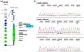
The clinical presentation of the proband was highly suggestive of FOP, a rare genetic condition associated with mutations of the ACVR1 gene (Figure 2a). As a first approach, molecular analysis was focused on the search of the recurrent ACVR1 p.R206H substitution with a negative result. Therefore, molecular screening was extended to the whole coding sequence of the gene with identification of the heterozygous c.772A>T (NM_001111067.4) variant in the fifth coding exon, and consequent change of the Arginine 258 by a Tryptophan (NP_001104537.1: p.(R258W); Figure 2b). The mutation was absent in the healthy parents suggesting a likely de novo origin.
FIGURE 2.

Novel variant of the ACVR1 gene associated with Fibrodysplasia Ossificans Progressiva. (a) Schematic representation of the ACVR1 gene and protein. ACVR1 gene consists of two untranslated (UTR) and nine coding exons. Mutations of the gene associated with FOP are indicated. EC, extra‐cellular domain; TM, trans‐membrane domain; GS, Glycine‐Serine rich domain; KD, kinase domain. (b) Electropherograms showing the region of the fifth ACVR1 coding exon containing the heterozygous c.772A>T (RefSeq NM_001111067.4) in the proband (Fop85.1) and absent in his parents (Fop85.2 and Fop85.3)
This variant is not reported in genomic databases and has never been described in association with FOP. However, it affects a highly conserved residue of the tyrosine kinase domain of the protein (Bocciardi et al., 2009). Noteworthy, other disease variants affecting the p.R258 have been already reported: p.R258S (Bocciardi et al., 2009; Eresen Yazıcıoğlu et al., 2013; Morales‐Piga et al., 2012; Ratbi et al., 2010) and p.R258G (Kaplan et al., 2015) associated with rare cases of FOP; p.R258G and p.R258M were found in tumor tissues (somatic mutations) from Diffuse Intrinsic Pontine Gliomas (DIPG), Brainstem gliomas, midline astrocytomas, and stomach adenocarcinoma (Buczkowicz et al., 2014; Chang et al., 2016; Taylor et al., 2014).
The change of a positively charged, hydrophilic residue such the Arginine by a Tryptophan with a large aromatic side chain is likely to impact the receptor function. A deleterious impact is also predicted through the sequence analysis performed by the Ensemble Variant Effect Predictor toolset which provides access to a wide collection of genomic annotations and tools suitable to predict the effect of a genetic variant (McLaren et al., 2016). A summary of this analysis is shown in Table 1, where the effect predicted for the novel substitution is compared to that observed for the other R258 variants associated with FOP.
TABLE 1.
Results of the comparative in silico analysis of the variants affecting the R258 residue associated with FOP
| CDS position | Residue | SIFT | PolyPhen | ||||||
|---|---|---|---|---|---|---|---|---|---|
| NM_001111067.4 | NP_001104537.1 | Codons | dbSNP | Prediction | Score | Prediction | Score | CADD_PHRED | CADD_RAW |
| c.774G>C | p.R258S | agG/agC | rs121912680 | Deleterious | 0 | Probably_damaging | 0.978 | 25.4 | 3.69050 |
| c.774G>T | p.R258S | agG/agT | rs121912680 | Deleterious | 0 | Probably_damaging | 0.978 | 25.4 | 3.70276 |
| c.773G>T | p.R258M | aGg/aTg | rs1057519875 | Deleterious | 0 | Probably_damaging | 0.997 | 28.0 | 4.12562 |
| c.772A>G | p.R258G | Agg/Ggg | rs863224846 | Deleterious | 0 | Probably_damaging | 0.998 | 27.9 | 4.12266 |
| c.772A>T | p.R258W | Agg/Tgg | Deleterious | 0 | Probably_damaging | 1 | 29.2 | 4.25469 | |
The novel R258W substitution is highlighted. A SIFT score is a normalized probability of observing the new amino acid at that position, and ranges from 0 to 1. A variant scored with a value of between 0 and 0.05 is predicted to affect protein function. A scaled CADD score (PRHED) of 20 means that a variant is amongst the top 1% of deleterious variants in the human genome. Negative Raw CADD values refer to variants likely “to be observed”; positive values suggest that variant is “simulated”, thus likely to be “not observed” and deleterious, with likelihood increasing with the score.
Abbreviations: CADD, combined annotation‐dependent depletion; CDS, coding sequence; PolyPhen, polymorphism phenotyping; SIFT, sorting intolerant from tolerant.
ACVR1 mutation associated with FOP causes an aberrant SMAD1/5/9 signaling and, most importantly, confers to the mutated receptor the ability to respond to a non‐canonical ligand, the Activin A (ActA), thus triggering the ectopic bone formation (Hatsell et al., 2015; Hino et al., 2015). As BMPs, ActA belongs to the TGFB family and is secreted by several cell types including immune cells (Kaplan et al., 2016).
In physiological conditions, ActA does not show osteogenic properties, can bind wild‐type ACVR1 without transducing any intra‐cellular signaling, rather negatively affecting the BMP cascade (Hatsell et al., 2015; Lees‐Shepard et al., 2018). On the contrary, the mutated receptor triggers the SMAD1/5/9 cascade upon ActA binding (Hatsell et al., 2015; Hino et al., 2015). The acquisition of this neo‐function has been demonstrated for the recurrent R206H mutation as well as for other substitutions associated with FOP, comprising variants of the R258 residue (Hino et al., 2015; Machiya et al., 2018).
We thus performed functional characterization of p.R258W. U2OS cells were transiently transfected with both the wild‐type and mutated ACVR1 cDNAs, then treated with ActA and BMP2. As shown in Figure 3, the novel variant perceives ActA as an agonist similarly to what observed for p.R206H and p.R258S, used as positive controls, whereas wild‐type expressing cells are insensitive to the treatment. This effect can be observed as an increase in the activity of a BMP‐responsive reporter gene, called BRE‐Luc (Figure 3a); as an up‐regulation of the ID1 target gene expression (Figure 3b) and as an increase of the phosphorylation state of the SMAD1/5/9 mediators (Figure 3c and d). These data indicate that, as already reported for the other mutations associated with FOP, also the p.R258W carrying ACVR1 can transduce an aberrant intra‐cellular signaling due to the altered, mutation‐induced, perception of the binding with ActA. As expected, both wild‐type and mutated ACVR1 constructs can elicit a response to BMP2.
FIGURE 3.

The novel p.R258W variant perceives Activin A as an agonist. U2OS cells were transiently transfected with the empty pCMV expression vector (EV) or carrying the wild‐type (WT), R206H, R258S, and R258W ACVR1 cDNAs, then evaluated in basal condition or upon Activin A or BMP2 treatment. A and B, ActA increased the BRE‐Luc activity (a) and the expression of the ID1 target gene (b) in cells expressing mutated cDNA but not in cells transfected with the WT nor with the EV. Histograms represent the fold activation compared to the activity measured in cells transfected with wild‐type ACVR1 construct in basal condition set as 1. (c) ActA induced phosphorylation of SMAD1/5/9 (p‐SMAD) in U2OS cells transiently expressing mutant R206H, R258S and the R258W ACVR1 cDNAs but not in cells transfected with the WT form, or the EV. A representative image of the Western blot (WB) analysis is shown. (d) Densitometric analysis of relative p‐SMAD1/5/9 phosphorylation levels corrected over total SMAD1. The histograms represent the densitometric mean values (±SD) obtained in three different WB experiments. As expected, in all the presented experiments, cells expressing both the WT and mutated ACVR1 cDNAs can transduce signaling upon BMP2 treatment. UN, untreated cells (white bars); ActA, cells treated with Activin A (grey bars); BMP2, cells treated with BMP2 (black bars). Results are the mean ± SD. n = 3–4 (A and B), n = 3 (D) *p < .05; **p < .01; ***p < .001 by Unpair t test compared with untreated cells transfected with WT ACVR1 cDNA
4. CONCLUSION
We described here the case of a little child with an early clinical presentation of FOP and carrier of a novel variant of the ACVR1 gene. FOP is one of the most severe genetic diseases of heterotopic ossification. Early clinical diagnosis is mandatory for the management of the patient to avoid any procedure that might be worthless and harmful. Molecular analysis with the identification of a causative mutation of the ACVR1 gene is crucial to confirm or exclude the condition, thus providing the patient and his family with the proper counseling about the nature, the course, and the management of the disease. Furthermore, it is worth to remind that FOP is an autosomal dominant condition and although most cases are sporadic and due to de novo mutations, evidence of germline mosaicism has been reported (Janoff et al., 1996) and this may be associated with a higher risk of recurrence in successive pregnancies.
So far, there is no etiologic treatment for FOP. However, many efforts are currently ongoing to develop specific therapies targeting the receptor activity and the downstream aberrant pathway or targeting cellular components and/or processes that are important in modifying the local environment promoting bone neoformation. The study of these basic mechanisms is opening the door to the development of targeted therapies (for a revision see Cappato et al., 2018; Katagiri et al., 2018) and clinical trials are presently ongoing with a RARγ agonist derivative, Palovarotene, with a humanized anti‐ActA antibody and others may be active in the near future (please refer to the ClinicalTrials.gov site for a comprehensive and updated list).
Therefore, FOP patients are facing a new era with the opportunity of participating in interventional clinical studies and, hopefully, to have access to a possible therapy in the next future. The mandatory prerequisite to benefit from these opportunities is a clinical diagnosis confirmed by the molecular characterization to define the presence of a causative mutation of the ACVR1 gene.
CONFLICT OF INTEREST
The authors declare no conflicts of interest.
AUTHOR CONTRIBUTIONS
SC, functional studies, data analysis, and manuscript writing; RT, clinical diagnosis and description, patient's clinical management, manuscript writing; JG, medical consultation, manuscript revision; FZ critical reading of the manuscript; RB, conceptualization, molecular diagnosis, manuscript writing, and revision.
ETHICAL CONSIDERATIONS
This study was performed with a written, informed consent of the proband's parents.
Supporting information
Table S1
ACKNOWLEDGMENTS
We gratefully thank the patients and the FOP Italia Association of patients and their families for funding and for continuous support to our work. This work was also supported by the "Cinque per mille" and "Ricerca corrente" (Italian Ministry of Health) and developed within the framework of the DINOGMI, Department of Excellence of MIUR 2018‐2022 (legge 232 del 2016).
Cappato, S. , Traberg, R. , Gintautiene, J. , Zara, F. , & Bocciardi, R. (2021). A case of Fibrodysplasia Ossificans Progressiva associated with a novel variant of the ACVR1 gene. Molecular Genetics & Genomic Medicine, 9, e1774. 10.1002/mgg3.1774
Serena Cappato and Rasa Traberg contributed equally to this work.
REFERENCES
- Baujat, G. , Choquet, R. , Bouée, S. , Jeanbat, V. , Courouve, L. , Ruel, A. , Michot, C. , Le Quan Sang, K.‐H. , Lapidus, D. , Messiaen, C. , Landais, P. , & Cormier‐Daire, V. (2017). Prevalence of fibrodysplasia ossificans progressiva (FOP) in France: An estimate based on a record linkage of two national databases. Orphanet Journal of Rare Diseases, 12(1), 123. 10.1186/s13023-017-0674-5 [DOI] [PMC free article] [PubMed] [Google Scholar]
- Bocciardi, R. , Bordo, D. , Di Duca, M. , Di Rocco, M. , & Ravazzolo, R. (2009). Mutational analysis of the ACVR1 gene in Italian patients affected with fibrodysplasia ossificans progressiva: Confirmations and advancements. European Journal of Human Genetics, 17(3), 311–318. 10.1038/ejhg.2008.178 [DOI] [PMC free article] [PubMed] [Google Scholar]
- Buczkowicz, P. , Hoeman, C. , Rakopoulos, P. , Pajovic, S. , Letourneau, L. , Dzamba, M. , Morrison, A. , Lewis, P. , Bouffet, E. , Bartels, U. , Zuccaro, J. , Agnihotri, S. , Ryall, S. , Barszczyk, M. , Chornenkyy, Y. , Bourgey, M. , Bourque, G. , Montpetit, A. , Cordero, F. , … Hawkins, C. (2014). Genomic analysis of diffuse intrinsic pontine gliomas identifies three molecular subgroups and recurrent activating ACVR1 mutations. Nature Genetics, 46(5), 451–456. 10.1038/ng.2936 [DOI] [PMC free article] [PubMed] [Google Scholar]
- Cappato, S. , Giacopelli, F. , Ravazzolo, R. , & Bocciardi, R. (2018). The horizon of a therapy for rare genetic diseases: A “Druggable” future for Fibrodysplasia Ossificans Progressiva. International Journal of Molecular Sciences, 19(4), 989. 10.3390/ijms19040989 [DOI] [PMC free article] [PubMed] [Google Scholar]
- Cappato, S. , Giacopelli, F. , Tonachini, L. , Ravazzolo, R. , & Bocciardi, R. (2019). Identification of reference genes for quantitative PCR during C3H10T1/2 chondrogenic differentiation. Molecular Biology Reports, 46(3), 3477–3485. 10.1007/s11033-019-04713-x [DOI] [PMC free article] [PubMed] [Google Scholar]
- Cappato, S. , Tonachini, L. , Giacopelli, F. , Tirone, M. , Galietta, L. J. , Sormani, M. , Giovenzana, A. , Spinelli, A. E. , Canciani, B. , Brunelli, S. , Ravazzolo, R. , & Bocciardi, R. (2016). High‐throughput screening for modulators of ACVR1 transcription: Discovery of potential therapeutics for fibrodysplasia ossificans progressiva. Disease Models & Mechanisms, 9(6), 685–696. 10.1242/dmm.023929 [DOI] [PMC free article] [PubMed] [Google Scholar]
- Chang, M. T. , Asthana, S. , Gao, S. P. , Lee, B. H. , Chapman, J. S. , Kandoth, C. , Gao, J. , Socci, N. D. , Solit, D. B. , Olshen, A. B. , Schultz, N. , & Taylor, B. S. (2016). Identifying recurrent mutations in cancer reveals widespread lineage diversity and mutational specificity. Nature Biotechnology, 34(2), 155–163. 10.1038/nbt.3391 [DOI] [PMC free article] [PubMed] [Google Scholar]
- Eresen Yazıcıoğlu, C. , Karatosun, V. , Kızıldağ, S. , Ozsoylu, D. , & Kavukçu, S. (2013). ACVR1 gene mutations in four Turkish patients diagnosed as fibrodysplasia ossificans progressiva. Gene, 515(2), 444–446. 10.1016/j.gene.2012.12.005 [DOI] [PubMed] [Google Scholar]
- Hatsell, S. J. , Idone, V. , Wolken, D. M. , Huang, L. , Kim, H. J. , Wang, L. , Wen, X. , Nannuru, K. C. , Jimenez, J. , Xie, L. , Das, N. , Makhoul, G. , Chernomorsky, R. , D’Ambrosio, D. , Corpina, R. A. , Schoenherr, C. J. , Feeley, K. , Yu, P. B. , Yancopoulos, G. D. , … Economides, A. N. (2015). ACVR1R206H receptor mutation causes fibrodysplasia ossificans progressiva by imparting responsiveness to activin A. Science Translational Medicine, 7(303), 303ra137. 10.1126/scitranslmed.aac4358 [DOI] [PMC free article] [PubMed] [Google Scholar]
- Hino, K. , Ikeya, M. , Horigome, K. , Matsumoto, Y. , Ebise, H. , Nishio, M. , Sekiguchi, K. , Shibata, M. , Nagata, S. , Matsuda, S. , & Toguchida, J. (2015). Neofunction of ACVR1 in fibrodysplasia ossificans progressiva. Proceedings of the National Academy of Sciences of the United States of America, 112(50), 15438–15443. 10.1073/pnas.1510540112 [DOI] [PMC free article] [PubMed] [Google Scholar]
- Janoff, H. B. , Muenke, M. , Johnson, L. O. , Rosenberg, A. , Shore, E. M. , Okereke, E. , Zasloff, M. , & Kaplan, F. S. (1996). Fibrodysplasia ossificans progressiva in two half‐sisters: Evidence for maternal mosaicism. American Journal of Medical Genetics, 61(4), 320–324. [DOI] [PubMed] [Google Scholar]
- Kaliya‐Perumal, A.‐K. , Carney, T. J. , & Ingham, P. W. (2020). Fibrodysplasia ossificans progressiva: Current concepts from bench to bedside. Disease Models & Mechanisms, 13(9), 6441. 10.1242/dmm.046441 [DOI] [PMC free article] [PubMed] [Google Scholar]
- Kaplan, F. S. , Kobori, J. A. , Orellana, C. , Calvo, I. , Rosello, M. , Martinez, F. , Lopez, B. , Xu, M. , Pignolo, R. J. , Shore, E. M. , & Groppe, J. C. (2015). Multi‐system involvement in a severe variant of fibrodysplasia ossificans progressiva (ACVR1 c.772G>A; R258G): A report of two patients. American Journal of Medical Genetics, Part A, 167A(10), 2265–2271. 10.1002/ajmg.a.37205 [DOI] [PMC free article] [PubMed] [Google Scholar]
- Kaplan, F. S. , Pignolo, R. J. , & Shore, E. M. (2016). Granting immunity to FOP and catching heterotopic ossification in the Act. Seminars in Cell & Developmental Biology, 49, 30–36. 10.1016/j.semcdb.2015.12.013 [DOI] [PMC free article] [PubMed] [Google Scholar]
- Kaplan, F. S. , Xu, M. , Seemann, P. , Connor, J. M. , Glaser, D. L. , Carroll, L. , Delai, P. , Fastnacht‐Urban, E. , Forman, S. J. , Gillessen‐Kaesbach, G. , Hoover‐Fong, J. , Köster, B. , Pauli, R. M. , Reardon, W. , Zaidi, S.‐A. , Zasloff, M. , Morhart, R. , Mundlos, S. , Groppe, J. , & Shore, E. M. (2009). Classic and atypical fibrodysplasia ossificans progressiva (FOP) phenotypes are caused by mutations in the bone morphogenetic protein (BMP) type I receptor ACVR1. Human Mutation, 30(3), 379–390. 10.1002/humu.20868 [DOI] [PMC free article] [PubMed] [Google Scholar]
- Katagiri, T. , Tsukamoto, S. , & Kuratani, M. (2018). Heterotopic bone induction via BMP signaling: Potential therapeutic targets for fibrodysplasia ossificans progressiva. Bone, 109, 241–250. 10.1016/j.bone.2017.07.024 [DOI] [PubMed] [Google Scholar]
- Lees‐Shepard, J. B. , Yamamoto, M. , Biswas, A. A. , Stoessel, S. J. , Nicholas, S. E. , Cogswell, C. A. , Devarakonda, P. M. , Schneider, M. J. Jr , Cummins, S. M. , Legendre, N. P. , Yamamoto, S. , Kaartinen, V. , Hunter, J. W. , & Goldhamer, D. J. (2018). Activin‐dependent signaling in fibro/adipogenic progenitors causes fibrodysplasia ossificans progressiva. Nature Communications, 9(1), 471. 10.1038/s41467-018-02872-2 [DOI] [PMC free article] [PubMed] [Google Scholar]
- Livak, K. J. , & Schmittgen, T. D. (2001). Analysis of relative gene expression data using real‐time quantitative PCR and the 2(‐Delta Delta C(T)) method. Methods, 25(4), 402–408. 10.1006/meth.2001.1262 [DOI] [PubMed] [Google Scholar]
- Machiya, A. , Tsukamoto, S. , Ohte, S. , Kuratani, M. , Fujimoto, M. , Kumagai, K. , Osawa, K. , Suda, N. , Bullock, A. N. , & Katagiri, T. (2018). Effects of FKBP12 and type II BMP receptors on signal transduction by ALK2 activating mutations associated with genetic disorders. Bone, 111, 101–108. 10.1016/j.bone.2018.03.015 [DOI] [PubMed] [Google Scholar]
- McLaren, W. , Gil, L. , Hunt, S. E. , Riat, H. S. , Ritchie, G. R. S. , Thormann, A. , Flicek, P. , & Cunningham, F. (2016). The ensembl variant effect predictor. Genome Biology, 17(1), 122. 10.1186/s13059-016-0974-4 [DOI] [PMC free article] [PubMed] [Google Scholar]
- Morales‐Piga, A. , Bachiller‐Corral, J. , Trujillo‐Tiebas, M. J. , Villaverde‐Hueso, A. , Gamir‐Gamir, M. L. , Alonso‐Ferreira, V. , Vázquez‐Díaz, M. , Posada de la Paz, M. , & Ayuso‐García, C. (2012). Fibrodysplasia ossificans progressiva in Spain: Epidemiological, clinical, and genetic aspects. Bone, 51(4), 748–755. 10.1016/j.bone.2012.07.002 [DOI] [PubMed] [Google Scholar]
- Pignolo, R. J. , Bedford‐Gay, C. , Liljesthröm, M. , Durbin‐Johnson, B. P. , Shore, E. M. , Rocke, D. M. , & Kaplan, F. S. (2016). The natural history of flare‐ups in Fibrodysplasia Ossificans Progressiva (FOP): A comprehensive global assessment. Journal of Bone and Mineral Research, 31(3), 650–656. 10.1002/jbmr.2728 [DOI] [PMC free article] [PubMed] [Google Scholar]
- Qi, Z. , Luan, J. , Zhou, X. , Cui, Y. , & Han, J. (2017). Fibrodysplasia ossificans progressiva: Basic understanding and experimental models. Intractable & Rare Diseases Research, 6(4), 242–248. 10.5582/irdr.2017.01055 [DOI] [PMC free article] [PubMed] [Google Scholar]
- Ratbi, I. , Bocciardi, R. , Regragui, A. , Ravazzolo, R. , & Sefiani, A. (2010). Rarely occurring mutation of ACVR1 gene in Moroccan patient with fibrodysplasia ossificans progressiva. Clinical Rheumatology, 29(1), 119–121. 10.1007/s10067-009-1283-z [DOI] [PubMed] [Google Scholar]
- Shore, E. M. , Xu, M. , Feldman, G. J. , Fenstermacher, D. A. , Cho, T.‐J. , Choi, I. H. , Connor, J. M. , Delai, P. , Glaser, D. L. , LeMerrer, M. , Morhart, R. , Rogers, J. G. , Smith, R. , Triffitt, J. T. , Urtizberea, J. A. , Zasloff, M. , Brown, M. A. , & Kaplan, F. S. (2006). A recurrent mutation in the BMP type I receptor ACVR1 causes inherited and sporadic fibrodysplasia ossificans progressiva. Nature Genetics, 38(5), 525–527. 10.1038/ng1783 [DOI] [PubMed] [Google Scholar]
- Taylor, K. R. , Vinci, M. , Bullock, A. N. , & Jones, C. (2014). ACVR1 mutations in DIPG: Lessons learned from FOP. Cancer Research, 74(17), 4565–4570. 10.1158/0008-5472.CAN-14-1298 [DOI] [PMC free article] [PubMed] [Google Scholar]
Associated Data
This section collects any data citations, data availability statements, or supplementary materials included in this article.
Supplementary Materials
Table S1