Supplemental Digital Content is available in the text.
Background.
Perfusate lactate clearance (LC) is considered one of the useful indicators of liver viability assessment during normothermic machine perfusion (NMP); however, the applicable scope and potential mechanisms of LC remain poorly defined in the setting of liver donation after circulatory death.
Methods.
The ex situ NMP of end-ischemic human livers was performed using the OrganOx Metra device. We further studied the extracellular signal-regulated kinases (phospho-extracellular signal-regulated kinase1/2 [pERK1/2]) pathway and several clinical parameters of these livers with successful LC (sLC, n = 5) compared with non-sLC (nLC, n = 5) in the perfusate (<2.2 mmol/L at 2 h, n = 5, rapid retrieval without normothermic regional perfusion).
Results.
We found the pERK1/2 level was substantially higher in the nLC livers than in the sLC livers (n = 5) at 2- and 6-h NMP (P = 0.035 and P = 0.006, respectively). Immunostaining showed that upregulation of pERK1/2 was in both the hepatocytes and cholangiocytes in the nLC livers. Successful LC was associated with a marginally higher glycogen restoration than nLC at 2 h NMP (n = 5, P = 0.065). Furthermore, bile lactate levels in all sLC livers were cleared into the normal range at 6 h NMP, whereas in the nLC group, only 2 livers had lower bile lactate levels, and the other livers had rising bile lactate levels in comparison with the corresponding perfusate lactate levels. The necrosis scores were higher in the nLC than in the sLC livers (n = 5) at 0- and 6-h NMP (P = 0.047 and P = 0.053, respectively).
Conclusions.
The dual LC in perfusate and bile can be helpful in evaluating the hypoxic injury of hepatocytes and cholangiocytes during the NMP of donation after circulatory death in liver donors.
INTRODUCTION
Donor shortages have been a consistent challenge in organ transplantation, and there has been an increased focus on not only expanding the current donor pool through the use of marginal donors or extended criteria donations (ECDs), such as steatotic livers or donation after circulatory death (DCD), but also on attempting to resuscitate declined grafts for transplantation. In the United States, over 12 000 patients are awaiting liver transplantation, with recent data reporting a waitlist mortality of 13.2 per 100 waitlist years1; however, organ utilization has substantially decreased in the last 15 y, with almost 700 (8.9%) procured livers being discarded annually because of concerns about poor organ quality.2-4 Alarmingly, it has been predicted that the use of donor livers will fall to 44% by the year 2030, resulting in over 2000 fewer liver transplants.5 The demand for liver grafts has driven the broader use of ECDs; however, ECDs are strongly associated with an increased risk of primary nonfunction or delayed failure.6
Although normothermic machine perfusion (NMP) of liver grafts has been considered as an alternative strategy to standard cold storage to lessen ischemia-reperfusion injury (IRI), only recently has this been shown to be clinically feasible. This technology is now approved for routine clinical use in Europe, and phase-3 testing is underway in the United States.7 Another unique advantage of NMP over static preservation is that drugs or specific gene therapies can be administered directly to the liver during NMP. This window of opportunity for delivery of therapeutics may significantly impact posttransplant survival.8 Encouraging data applying NMP techniques to discarded livers have emerged supporting its use over static cold storage techniques, with the added advantage of maintaining physiological states in the former to allow for real-time functional testing.9-11 Recently, it has been reported that NMP can increase the transplant yield of donor livers,12 and previous studies have shown that up to 71% of discarded livers can be subsequently transplanted.13 In clinical practice, identifying reliable biomarkers of injury and clinically relevant viability assessments to determine posttransplantation outcomes is being increasingly implemented in machine perfusion protocols.14 NMP allows assessing the functional performance of the liver, and several biomarkers have been proposed to determine optimal clinical and metabolic liver responses during ex vivo NMP, including perfusate lactate clearance (LC), the maintenance of a stable perfusate pH value, glucose utilization, bile production, and bile pH value. Perfusate LC has emerged as a relatively reliable indicator for liver viability.7,15,16 In the current literature, bile production and pH have been used to measure biliary function following machine perfusion.17,18 In a study of 12 human extended criteria donor livers declined for transplantation, the high bile output group demonstrated more favorable markers in terms of functional performance, biochemical analyses, histological responses, and displaying fewer signs of hepatic necrosis and venous congestion.19 In a later study of 6 human livers undergoing 6 h of NMP by the same group, biliary pH >7.48 combined with biliary bicarbonate >18 mmol/L and biliary glucose <16 mmol/L were associated with satisfactory bile duct viability and appeared to preclude the development of posttransplant cholangiopathy.18
Hypoxia-inducible factors (HIFs) belong to a small family of heterodimeric transcriptional activators that orchestrate the cellular responses to hypoxia.20 In the setting of DCD liver donors, It has been recognized that the expression HIFs can be upregulated in response to the prolonged warm ischemia time (WIT),21 suggesting that HIFs could be an ideal marker to assess liver donor function during NMP. Several findings have indicated that extracellular signal-regulated kinase 1/2 (ERK1/2) can also serve as an additional transmitter of the hypoxic signal because hypoxia has been shown to activate ERK1/2 in cell lines.22-24 Moreover, several studies have demonstrated that the ERK1/2 pathway had been activated in hepatic IRI25,26 and that hepatic apoptosis, necrosis, inflammation, and autophagy were mitigated following inhibition of the ERK1/2 pathway.26-29
To explore the prognostic value of LC in liver viability assessment during NMP, we studied hepatic ERK1/2 phosphorylation, necrosis, apoptosis, steatosis, and cluster of differentiation 3 (CD3)–positive cell infiltration, as well as perfusate and bile chemistry with or without successful LC (sLC).
MATERIALS AND METHODS
Liver Inclusion and Exclusion Criteria
This study was approved by our institutional review board. Ten livers donated after circulatory death (DCD, n = 9) or donated after brain death (DBD, n = 1) were registered in DonorNet with written consent for research but subsequently declined for transplant by all centers were used in this study. These livers donors were rapid retrieval without normothermic regional perfusion. Donor criteria for inclusion in the study were as follows: (1) donor age >6 y, (2) donor serum bilirubin <10 mg/dL, (3) functional WIT (fWIT) <40 min (from donor systolic blood pressure below 70 mm Hg to the initiation of aortic perfusion), (4) rapid recovery donors with research consent for liver procurement, with fWIT <40 min, (5) suboptimal liver graft perfusion reported by procuring surgeons, and (6) liver cold ischemia time <8 h for DBD or 6 h for DCD. Livers with the following findings during procurement were excluded from the study: (1) cirrhosis or (2) advanced fibrosis.
NMP Device, Perfusate, and Sampling
All livers were flushed with histidine-tryptophan-ketoglutarate solution after cessation of circulation during procurement. Cholecystectomy was performed during procurement. After a cold flush with histidine-tryptophan-ketoglutarate at the time of donor recovery, perfusion cannulas were placed in the portal vein, hepatic artery, and subhepatic inferior vena cava, respectively, whereas the suprahepatic inferior vena cava was closed via suturing. The common bile duct was cannulated with a 12F drainage tube for bile collection. The cannulated liver underwent final flushing with 500 mL of 5% albumin solution to prevent air trapping. The OrganOx Metra device was calibrated and initialized per the manufacturer’s instructions for use. Five hundred milliliters of 5% albumin solution and 3 units of fresh-packed red blood cells were sequentially introduced into device circulation to constitute the perfusate. Followed by injection of antibiotic (cefuroxime 750 mg), heparin (10 000 IU), and calcium gluconate bolus, 4 syringes (50 mL) containing sodium taurocholate, heparin, insulin, and epoprostenol solutions were placed in microinfusion pumps and connected to device circulation. After priming the device, a centrifugal pump was switched on, and circulation was initiated before attaching the organ. Once the perfusate temperature reached 36 °C, boluses of 5 mL 8.4% sodium bicarbonate injection were given in at least 10-min intervals to adjust the perfusate to a pH 7.3 or above, after which the device would be ready for the liver. Circulation was temporarily stopped, and the liver was connected to the OrganOx Metra device; after the confirmation of liver connection and absence of air trapped inside the circulation, circulation was initiated again, and device was closely monitored until the liver reached homogenous perfusion and presented soft, warm parenchyma; bleeding sites were repaired by suturing before the liver compartment was closed, and then the liver was subjected to at least 4 to 6 h of NMP. All liver manipulations were performed with aseptic techniques. Perfusate pH adjustment after commencement of liver perfusion was performed with a 5 mL bolus of 8.4% sodium bicarbonate injection to maintain perfusate pH 7.3 when necessary. During device operation, hepatic arterial (HA) and venous pressure, portal and venous flow rates, perfusate pH, perfusate Pco2, and Po2 data were logged and stored inside the onboard data storage. Both wedge and needle liver biopsies of the right lobe as well as perfusate samples were periodically taken at these time points during NMP: A baseline biopsy was taken at the back table, and baseline perfusate was taken within 3 min of NMP initiation after the liver was connected. Biopsy and arterial perfusate samples were taken when NMP reached 15 min and were taken hourly from 1 to 6 h. Bile (0.5 mL), if present, was collected at each hourly mark when perfusate was collected. Total bile production was quantified by collecting bile in a measuring tube. Perfusate samples were immediately subjected to a blood-gas analyzer (GEM Premier 4000), and glucose and lactate readings were obtained. If perfusate glucose measured <180 mg/dL, Clinimix E 5/20 would be connected to the device circulation and administered at the rate of 2 mL/min to provide nutrition. Chemistry analyses were performed on a chemistry analyzer (Piccolo Xpress) using venous perfusate samples.
Hematoxylin and Eosin Staining and Periodic Acid–Schiff (PAS) Staining
The liver samples were fixed using 10% neutral formalin, paraffin-embedded or fast-frozen using liquid nitrogen, and cut with a thickness of 5 to 10 µm. Hematoxylin and eosin staining was performed at the anatomic and molecular pathology core lab of our institute using a standard protocol. A liver pathologist (K.B.) did the histology interpretation in a blinded fashion. Periodic acid–Schiff staining was performed according to the manufacture’s instruction (Sigma 395B). Briefly, the frozen liver sections were immersed in periodic acid solution for 5 min at room temperature (23 °C). Then, the slides were rinsed in several changes of distilled water and incubated with Schiff’s reagent for 15 min at room temperature. The slides were washed in running tap water for 5 min, counterstained with hematoxylin solution for 90 seconds, rinsed in running tap water, dehydrated, and mounted with xylene-based mounting media. The hematoxylin and eosin and PAS images were taken by the Olympus BX61 microscope and CellSens Dimension 1.18 software.
Western Blotting and Immunofluorescence Staining
The Western blotting/densitometry analysis and immunofluorescence staining were done as described previously.30 The following primary and secondary antibodies were used to perform immunoblotting or immunostaining: phospho-ERK1/2 (CST No. 9102), total-ERK1/2 (CST No. 9101), caspase-3 (CST No. 9661), and GAPDH (CST No. 3683); horseradish peroxidase–conjugated goat antirabbit (7074, CST) or goat antimouse immunoglobulins (7076, CST); CD3 (Abcam No. ab16669), Hep par-1 (Novus, NBP2-45272), cytokeratin-19 (CST No. 4558), and p62 (BD No. 610832). The immunofluorescence photomicrographs were taken with the Olympus BX61 microscope, and images were captured by CellSens Dimension 1.18 software.
Data and Statistical Analysis
Numeric data are presented as mean ± SEM, and categorical data were shown in percentages. GraphPad Prism 7 software (San Diego, CA) was utilized to plot the graphs. A Student t test was applied to detect the differences between study groups. The P values <0.05 were considered significant.
RESULTS
Demographics of Liver Donors, NMP Setting, and Real-Time Parameters Recording
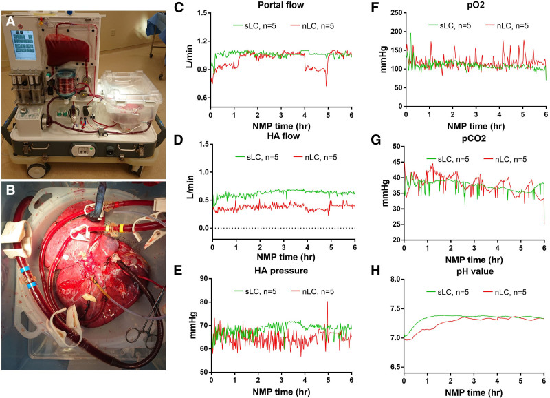
Of the 10 discarded livers included in our study, the mean donor age was 33.8 ± 14.3, with 60% male donors, 90% Caucasian donors, and 10% African American donors; 90% of livers were DCD, and 10% of livers were DBD; the mean body mass index (kg/m2) was 28.3 ± 8.6. The mean WIT of DCD livers was 29.4 ± 11.8 min. The average time of DCD livers with systolic blood pressure <70 mm Hg (fWIT) was 17.8 ± 6.3 min, and with Sao2, <80% were 27.0 ± 12.4 min; the average time from declaration of death to cross-clamp was 10.0 ± 3.2 min, and mean cold ischemia time was 309.8 ± 94.6 min. The average volume of sodium bicarbonate for pH maintenance was 38.0 ± 12.3 mL (Table S1, SDC, http://links.lww.com/TXD/A380). Figure 1 shows the NMP device with a perfused liver on the system. The portal flow, HA pressure, HA flow, perfusate Po2, Pco2, and pH value were presented as average per minute (Figure 1C–H).
FIGURE 1.
NMP setting and real-time parameter recording. A and B, The OrganOx device and liver during NMP; portal flow (C); HA pressure (D); HA flow (E); perfusate Po2 (F); Pco2 (G); and pH value (H) according to lactate clearance (green: 2 h lactate <2.5, sLC; red: 2 h lactate ≥2.5, nLC). HA, hepatic arterial; nLC, nonsuccessful lactate clearance; NMP, normothermic machine perfusion; sLC, successful lactate clearance.
Lactate Dehydrogenase and ERK Phosphorylation in the Livers With or Without sLC
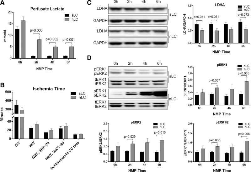
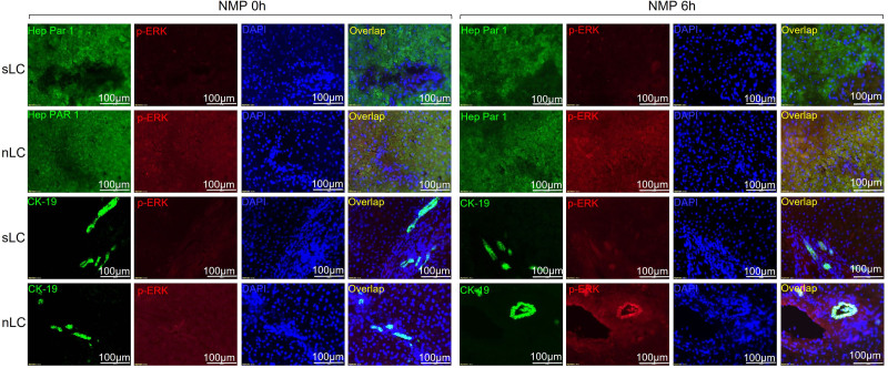
To explore the biological and molecular significance of perfusate LC, we used a cutoff value of 2.2 mmol/L at second-hour NMP to define the sLC (≤2.2 mmol/L) and non-sLC (nLC, >2.2 mmol/L). This lactate cutoff value has been used as a measure of preserved liver function on NMP in several previous studies.7,11,13 Of the 10 NMP livers in our study, 5 livers achieved sLC at secondhour NMP. As shown in Figure 2A, the livers with sLC had significantly lower perfusate lactate levels at second-, fourth-, and sixth-hour NMP than the livers with nLC (P = 0.003, P = 0.002, or P = 0.001, respectively); however, no significant differences in warm, cold, or functional ischemia times were found between sLC and nLC livers (Figure 2B). The hepatic lactate dehydrogenase A (LDHA) levels in livers with sLC were marginally higher at 0, 2, and 6 h than those in nLC livers (Figure 2C; P = 0.051, P = 0.031, or P = 0.073, respectively). As shown in Figure 2D, the phosphorylations ERK1, ERK2, and ERK1/2 were significantly increased in the livers with nLC at 6 h of NMP compared with those in the livers with sLC (P = 0.006, P = 0.035, or P = 0.010, respectively). To localize the expression of phosphorylated ERK, the hepatocytes and cholangiocytes were labeled with Hep Par-1 and cytokeratin-19, respectively, and double immunofluorescence staining with phospho-ERK1/2 was performed. An increased ERK phosphorylation was found mainly in the hepatocyte and cholangiocyte in livers with nLC when compared with the livers with sLC (Figure 3). In summary, the LC was well correlated to the phosphorylation of ERK in hepatocytes and cholangiocytes.
FIGURE 2.
Lactate clearance and ERK phosphorylation in livers during NMP with sLC or nLC. Perfusate lactate levels (A); CIT, WIT, and fWIT (B); Western blotting of hepatic lactate dehydrogenase (C); and ERK1/2 phosphorylation (D). n = 5, P < 0.05 is considered as significant. CC, cross-clamp; CIT, cold ischemia time; ERK, extracellular signal-regulated kinase; fWIT, functional warm ischemia time; LDHA, lactate dehydrogenase A; nLC, nonsuccessful lactate clearance; NMP, normothermic machine perfusion; pERK, phospho-ERK; Sao2, oxygen saturation; SBP, systolic blood pressure; sLC, successful lactate clearance; tERK, total-ERK; WIT, warm ischemia time.
FIGURE 3.
Double immunofluorescence staining of p-ERK in livers with sLC and nLC at 0 h and 6 h NMP. The p-ERK signal is represented in red; the hepatocytes (labeled with hepatocyte specific antigen (Hep PAR-1) and cholangiocytes (labeled with CK-19) are represented in green; the nuclei are represented in blue (DAPI). CK-19, cytokeratin-19; DAPI, 4′,6-diamidino-2-phenylindole; p-ERK, phosphorylated extracellular signal-regulated kinase; nLC, nonsuccessful lactate clearance; NMP, normothermic machine perfusion; sLC, successful lactate clearance.
Hepatocellular Function and Glycogen Content in the Livers With or Without sLC
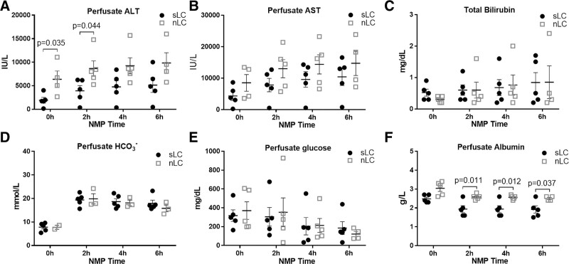
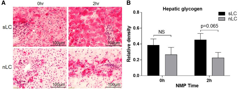
To assess liver damage and functional status, we measured perfusate liver enzymes (alanine aminotransferase [ALT], aspartate aminotransferase), bilirubin, albumin, glucose, and liver glycogen content using PAS staining. As shown in Figure 4A, ALT levels were significantly lower in the perfusate with sLC at 0 and 2 h than in the perfusate without sLC (P = 0.035 or P = 0.044, respectively). No significant change was found with regard to perfusate aspartate aminotransferase, bilirubin, bicarbonate, and glucose (Figure 4B–D). Albumin levels were remarkably decreased in the perfusate with sLC at 2, 4, and 6 h when compared with nLC (Figure 4E; P = 0.011, P = 0.012, or P = 0.037, respectively). Furthermore, we also found that the glycogen content was marginally higher in the livers with sLC at 2 h than those with nLC (Figure 5A and 5B; P = 0.065). Briefly, LC was associated with the hepatocyte function and injury.
FIGURE 4.
Hepatocellular function parameters during liver NMP with perfusate sLC or nLC. The perfusate ALT and AST (A and B), total bilirubin (C), and bicarbonate (D), albumin (E), glucose (F). P < 0.05 is considered as significant. ALT, alanine aminotransferase; AST, aspartate aminotransferase; nLC, nonsuccessful lactate clearance; NMP, normothermic machine perfusion; sLC, successful lactate clearance.
FIGURE 5.
The PAS staining and quantitative analysis of hepatic glycogen content. The representative PAS images (A) and the average density of glycogen content (B) in livers with sLC and nLC at 0 and 2 h NMP. nLC, nonsuccessful lactate clearance; NMP, normothermic machine perfusion; NS, not significant; PAS, periodic acid–Schiff; sLC, successful lactate clearance.
Cholangiocyte Function of the Livers With or Without sLC
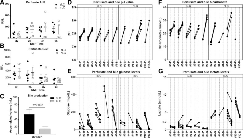
To evaluate biliary function and damage, we studied the perfusate alkaline phosphatase (ALP) and gamma-glutamyl transferase. No significant differences in perfusate ALP and gamma-glutamyl transferase were found between sLC and nLC livers (Figure 6A and B). There were 9 livers out of 10 that produced bile in our study. As shown in Figure 6C, the volume of bile production by livers with sLC was significantly higher than that by livers with nLC (P = 0.032). Moreover, we compared the changes in pH value and bicarbonate, glucose, and lactate levels between perfusate and bile. Regardless of the perfusate LC, all 9 livers producing bile appeared to have an increased bile pH value and increased bicarbonate levels compared with perfusate levels (Figure 6D–F), as well as decreased levels of bile glucose (Figure 6E). Interestingly, all of the livers with sLC in perfusate had decreasing bile lactate levels at all NMP time points except the initial time points in the livers numbered 5, 6, and 7l however, among nLC livers, only 1 (number 8) had a bile lactate level lower than 2.2 mmol/L at the end of NMP despite a decreasing trend that was seen in 2 livers (numbers 8 and 10). In contrast, another 2 livers (numbers 2 and 4) with nLC had rising bile lactate levels compared with the corresponding perfusate levels (Figure 6G), which were consistent with the changing trends of perfusate ALP levels (Figure 6A). In short, we found that the bile lactate levels were associated with cholangiocyte function in the setting of liver NMP.
FIGURE 6.
Cholangiocyte function parameters during liver NMP with perfusate sLC or nLC. The perfusate ALP and GGT (A and B), bile production (C), changes of perfusate and bile pH value (D), glucose (E), and bicarbonate (F), and bile lactate levels (G). P < 0.05 is considered as significant. ALP, alkaline phosphatase; GGT, gamma-glutamyl transferase; B, bile; nLC, nonsuccessful lactate clearance; NMP, normothermic machine perfusion; P, perfusate; sLC, successful lactate clearance.
Necrosis, Apoptosis, and Inflammatory Cell Infiltration of the Livers With or Without sLC
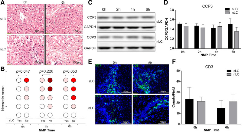
In the histology analysis, the hepatic necrosis and score were reported as no necrosis (0), single-cell necrosis (1), up to 30% necrosis (2), 30% to 60% necrosis (3), and >60% necrosis (4) by a pathologist (K.B.). We found that the livers with sLC had marginally lower necrosis scores at 0- and 6-h of NMP in comparison with livers with nLC (P = 0.047 and P = 00.053, respectively), but the LC was not associated with hepatic steatosis (Figure 7A and B). Cleaved caspase-3 levels (Figure 7C and D) and CD3-positive cell infiltration (Figure 7E and F) were also comparable between sLC and nLC livers. These results suggest that the role of LC in the assessment of necrosis, apoptosis, steatosis, and inflammatory cell infiltration was limited; further studies are required to evaluate these aspects in addition to LC. There was no significant difference in p62 levels between sLC and nLC livers (Figure S1, SDC, http://links.lww.com/TXD/A380).
FIGURE 7.
Hepatic necrosis, apoptosis, and CD3 positive cell infiltration with perfusate sLC or nLC. Hematoxylin and eosin staining (A) and necrosis scores with a circle that is blank as no necrosis, pink as single cell to 30% necrosis, bright red as 30%–60% necrosis, and dark red as >60% necrosis (B); the representative Western blotting band of CCP-3 (C) and densitometry analysis (D); and representative immunofluorescence staining of CD3 cells in the livers (E) and counting (F). P < 0.05 is considered as significant. CD3, cluster of differentiation 3; CCP-3, cleaved caspase-3; nLC, nonsuccessful lactate clearance; NMP, normothermic machine perfusion; sLC, successful lactate clearance.
DISCUSSION
LC has been widely accepted as a critical marker to assess liver viability during NMP; however, its regulatory mechanisms and the scope of application remain poorly defined. Our data suggest dual LC in the perfusate and bile can be used to evaluate the severity of hypoxia-induced hepatocyte and cholangiocyte dysfunction that correlate to ERK1/2 pathway activation but not cold or warm time criteria currently used in the clinical practice. Furthermore, LC does not appear to be associated with hepatic apoptosis, steatosis, and inflammatory cell infiltration during liver NMP.
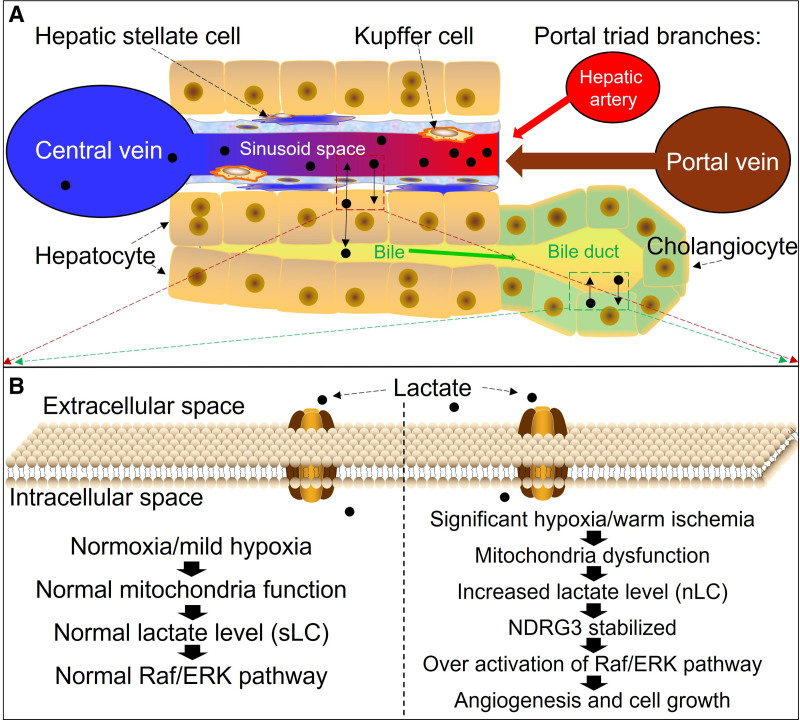
Lactate traditionally has been used as a marker of sepsis and imminent mortality in critical care.31,32 Lactatemia can subsequently develop in tissue hypoxia, and earlier reports have revealed an essential correlation between LC and improved outcomes.31,33,34 In this context, the role of the liver is paramount and is responsible for removing 50% to 70% of circulating serum lactate.35 As such, many have suggested LC to be a reliable surrogate marker of hypoxic injury and downstream hepatocyte functionality.36,37 The lactate level rises inevitably in the procured liver organ because of reduced blood flow and oxygen delivery, which can activate the ERK pathway in a lactate-dependent manner.38 The N-Myc downstream-regulated gene 3 protein, which is degraded in a prolyl-4-hydroxylase 2/VHL-dependent manner in normoxia, is stabilized by binding to lactate accumulating under hypoxia and subsequently binds c-rapidly accelerated fibrosarcoma to mediate the hypoxia-induced activation of the rapidly-accelerated-fibrosarcoma–ERK pathway, promoting angiogenesis and cell growth.38 It has been shown that hypoxia-activated ERK1 was able to directly phosphorylate the carboxyl-terminal domain of HIF-1K.23 An ERK inhibitor (PD98059), as well as a negative mutant of ERK1 dominant in a hypoxic culturing model of human microvascular endothelial cells-1, diminished the transcriptional activity HIF-1.23,39 Additionally, in the presence of oxygen, HIF-2α is modified by HIF-specific prolyl-4-hydroxylases, leading to proteasomal degradation mediated in part by the Von Hippel Lindau tumor suppressor protein.40 An in vitro study suggested that HIF-2α was phosphorylated by ERK1/2 serine residue 672 in a hypoxia environment with only 1% of oxygen, and a mutation of this site to an alanine residue or an ERK1/2 inhibitor decreased HIF-2 transcriptional activity and displaced HIF-2α to the cytoplasm without changing its protein expression levels.41 Furthermore, our data also showed that livers with sLC had higher hepatic LDHA levels, which is a crucial enzyme to mediate the conversion between lactate and pyruvate, and the LDHA level could be increased in a hypoxia environment.42 One of the possible regulatory mechanisms for this is that the LDHA promoter contains a core sequence 5′-RCGTG-3′ that can be recognized by HIF-1, thus promoting LDHA transcription.43 These previous studies support our findings that phosphorylation of ERK1/2 in hepatocytes and cholangiocytes was inversely correlated with LC in the perfusate and bile accordingly, suggesting that the dual LC in the perfusate and bile can be a useful hypoxic marker to assess the function of hepatocytes and cholangiocytes during liver NMP (Figure 8).
FIGURE 8.
The lactate metabolism in hepatic cells and ERK activation during ischemia-reperfusion injury. A, The proportional structure of a liver lobule is represented. B, The molecular events regarding lactate clearance and ERK pathway during normoxia and hypoxia. Raf, rapidly accelerated fibrosarcoma; ERK, extracellular signal-regulated kinase; NDRG3, N-Myc downstream-regulated gene 3 protein; nLC, nonsuccessful lactate clearance; sLC, successful lactate clearance.
Interestingly, our data also suggest that sLC at 2 h of NMP was associated with less hepatocellular injury, as evidenced by lower levels of perfusate ALT and less hepatic necrosis, improved glucose utilization and hepatic glycogen restoration, albumin uptake, and bile production, but did not correlate with hepatic apoptosis, steatosis, or CD3-positive cell infiltration. One of the notable differences in our study is that the 9 out of 10 livers produced bile and almost all of the livers showed a good capacity to alkalize the bile and glucose reuptake, which is in contrast with the bile pH value and bicarbonate criteria reported in previous studies.18,19 These discrepancies may be due to our relatively small sample size or an overall better quality of the discarded livers included in our study. An obvious shortfall of our study is that no liver with successful dual LC was transplanted; therefore, the long-term prognosis is not available. This does not affect the clinical significance of our study, as we used the same standard NMP system (OrganOx Metra) as previous clinical studies with transplantation.7 The major takeaway in our study is that we clarified the regulatory mechanism of perfusate LC as a hypoxic hepatocellular marker closely linking to the ERK1/2 phosphorylation and extended its use to evaluate cholangiocyte hypoxia status.
Besides the relatively small numbers, there are several other limitations in our present study. Further studies are required to explore the possible mechanisms between the ERK activation and LC status, as well as the possible downstream targets. More NMP cases with liver transplantation are also desired to evaluate the significance of bile LC in predicting biliary complications. Another issue is that the livers enrolled in our study were DCD donors with rapid retrieval without normothermic regional perfusion, which might limit the applicability of our results only to DCD organs without normothermic regional perfusion. Furthermore, we do not have direct evidence in this study to demonstrate that the cholangiocyte could actively uptake or release lactate, despite the fact that our results alluded it was possible, which also can be supported by the fact that biliary cells can actively exchange metabolites with the bile.44
In conclusion, we found that the dual LC in perfusate and bile could be helpful in evaluating the severity of hypoxic IRI of hepatocytes and cholangiocytes during NMP. Additionally, histology studies may be required to provide further insight into steatosis, inflammatory infiltration, and apoptosis in NMP of discarded human livers.
ACKNOWLEDGMENTS
This project was funded Mid-America Transplant Foundation and the Barnes-Jewish Hospital Foundation Project Award, Transplant Research.
Supplementary Material
Footnotes
These authors contributed equally.
Published online 17 November, 2021.
This project was funded in part by the Mid-America Transplant Foundation and the Barnes-Jewish Hospital Foundation Project Award, Transplant Research.
W.C.C. is a founder of Pathfinder Therapeutics and an advisory board of Novartis Pharmaceutical. The other authors declare no conflicts of interest.
M.X. participated in research design, writing of the article, performance of the research, and data analysis. F.Z. participated in performance of the research and data analysis. O.A. participated in writing of the article and performance of the research. L.V.R. participated in training on the use of the OrganOx device, in remote monitoring, in the consultation of liver status during normothermic machine perfusion (NMP), and in data retrieval from the device. J.-K.S. analyzed tissue for biomarkers of autophagy-related study. Y.Z. participated in performance of the research and data collection. G.A.U. participated in research design and data analysis. K.B. participated in data analysis. B.W. participated in research design and data analysis. J.-S.K. participated in research design and data analysis. Y.L. and W.C.C. participated in research design, data analysis, and writing of the article.
Supplemental digital content (SDC) is available for this article. Direct URL citations appear in the printed text, and links to the digital files are provided in the HTML text of this article on the journal’s Web site (www.transplantationdirect.com).
References
- 1.Kwong A, Kim WR, Lake JR, et al. OPTN/SRTR 2018 annual data report: liver. Am J Transplant. 2020;20 (Suppl s1):193–299. [DOI] [PubMed] [Google Scholar]
- 2.Israni AK, Zaun D, Rosendale JD, et al. OPTN/SRTR 2017 annual data report: deceased organ donation. Am J Transplant. 2019;19 (Suppl 2):485–516. [DOI] [PubMed] [Google Scholar]
- 3.Israni AK, Zaun D, Rosendale JD, et al. OPTN/SRTR 2016 annual data report: deceased organ donation. Am J Transplant. 2018;18 (Suppl 1):434–463. [DOI] [PubMed] [Google Scholar]
- 4.Israni AK, Zaun D, Hadley N, et al. OPTN/SRTR 2018 annual data report: deceased organ donation. Am J Transplant. 2020;20 (Suppl s1):509–541. [DOI] [PubMed] [Google Scholar]
- 5.Orman ES, Mayorga ME, Wheeler SB, et al. Declining liver graft quality threatens the future of liver transplantation in the United States. Liver Transpl. 2015;21:1040–1050. [DOI] [PMC free article] [PubMed] [Google Scholar]
- 6.Marcon F, Schlegel A, Bartlett DC, et al. Utilization of declined liver grafts yields comparable transplant outcomes and previous decline should not be a deterrent to graft use. Transplantation. 2018;102:e211–e218. [DOI] [PubMed] [Google Scholar]
- 7.Nasralla D, Coussios CC, Mergental H, et al. ; Consortium for Organ Preservation in Europe. A randomized trial of normothermic preservation in liver transplantation. Nature. 2018;557:50–56. [DOI] [PubMed] [Google Scholar]
- 8.Griffiths C, Scott WE, Ali S, et al. Maximising organs for donation: the potential for ex-situ normothermic machine perfusion. QJM. 2020. In Press. doi:10.1093/qjmed/hcz321 [DOI] [PubMed] [Google Scholar]
- 9.Vogel T, Brockmann JG, Quaglia A, et al. The 24-hour normothermic machine perfusion of discarded human liver grafts. Liver Transpl. 2017;23:207–220. [DOI] [PubMed] [Google Scholar]
- 10.Perera T, Mergental H, Stephenson B, et al. First human liver transplantation using a marginal allograft resuscitated by normothermic machine perfusion. Liver Transpl. 2016;22:120–124. [DOI] [PubMed] [Google Scholar]
- 11.Watson CJ, Kosmoliaptsis V, Randle LV, et al. Preimplant normothermic liver perfusion of a suboptimal liver donated after circulatory death. Am J Transplant. 2016;16:353–357. [DOI] [PubMed] [Google Scholar]
- 12.MacConmara M, Hanish SI, Hwang CS, et al. Making every liver count: increased transplant yield of donor livers through normothermic machine perfusion. Ann Surg. 2020;272:397–401. [DOI] [PubMed] [Google Scholar]
- 13.Mergental H, Laing RW, Kirkham AJ, et al. Transplantation of discarded livers following viability testing with normothermic machine perfusion. Nat Commun. 2020;11:2939. [DOI] [PMC free article] [PubMed] [Google Scholar]
- 14.Bhogal RH, Mirza DF, Afford SC, et al. Biomarkers of liver injury during transplantation in an era of machine perfusion. Int J Mol Sci. 2020;21:E1578. [DOI] [PMC free article] [PubMed] [Google Scholar]
- 15.Ravikumar R, Jassem W, Mergental H, et al. Liver transplantation after ex vivo normothermic machine preservation: a phase 1 (first-in-man) clinical trial. Am J Transplant. 2016;16:1779–1787. [DOI] [PubMed] [Google Scholar]
- 16.Vogel T, Brockmann JG, Coussios C, et al. The role of normothermic extracorporeal perfusion in minimizing ischemia reperfusion injury. Transplant Rev (Orlando). 2012;26:156–162. [DOI] [PubMed] [Google Scholar]
- 17.Laing RW, Mergental H, Yap C, et al. Viability testing and transplantation of marginal livers (VITTAL) using normothermic machine perfusion: study protocol for an open-label, non-randomised, prospective, single-arm trial. BMJ Open. 2017;7:e017733. [DOI] [PMC free article] [PubMed] [Google Scholar]
- 18.Matton APM, de Vries Y, Burlage LC, et al. Biliary bicarbonate, pH, and glucose are suitable biomarkers of biliary viability during ex situ normothermic machine perfusion of human donor livers. Transplantation. 2019;103:1405–1413. [DOI] [PMC free article] [PubMed] [Google Scholar]
- 19.Sutton ME, op den Dries S, Karimian N, et al. Criteria for viability assessment of discarded human donor livers during ex vivo normothermic machine perfusion. PLoS One. 2014;9:e110642. [DOI] [PMC free article] [PubMed] [Google Scholar]
- 20.Rankin EB, Giaccia AJ. Hypoxic control of metastasis. Science. 2016;352:175–180. [DOI] [PMC free article] [PubMed] [Google Scholar]
- 21.Wilson GK, Tennant DA, McKeating JA. Hypoxia inducible factors in liver disease and hepatocellular carcinoma: current understanding and future directions. J Hepatol. 2014;61:1397–1406. [DOI] [PubMed] [Google Scholar]
- 22.Salceda S, Beck I, Srinivas V, et al. Complex role of protein phosphorylation in gene activation by hypoxia. Kidney Int. 1997;51:556–559. [DOI] [PubMed] [Google Scholar]
- 23.Minet E, Arnould T, Michel G, et al. ERK activation upon hypoxia: involvement in HIF-1 activation. FEBS Lett. 2000;468:53–58. [DOI] [PubMed] [Google Scholar]
- 24.Conrad PW, Freeman TL, Beitner-Johnson D, et al. EPAS1 trans-activation during hypoxia requires p42/p44 MAPK. J Biol Chem. 1999;274:33709–33713. [DOI] [PubMed] [Google Scholar]
- 25.Li J, Wang F, Xia Y, et al. Astaxanthin pretreatment attenuates hepatic ischemia reperfusion-induced apoptosis and autophagy via the ROS/MAPK pathway in mice. Mar Drugs. 2015;13:3368–3387. [DOI] [PMC free article] [PubMed] [Google Scholar]
- 26.Feng J, Zhang Q, Mo W, et al. Salidroside pretreatment attenuates apoptosis and autophagy during hepatic ischemia-reperfusion injury by inhibiting the mitogen-activated protein kinase pathway in mice. Drug Des Devel Ther. 2017;11:1989–2006. [DOI] [PMC free article] [PubMed] [Google Scholar]
- 27.Xu S, Niu P, Chen K, et al. The liver protection of propylene glycol alginate sodium sulfate preconditioning against ischemia reperfusion injury: focusing MAPK pathway activity. Sci Rep. 2017;7:15175. [DOI] [PMC free article] [PubMed] [Google Scholar]
- 28.Li S, Takahara T, Fujino M, et al. Astaxanthin prevents ischemia-reperfusion injury of the steatotic liver in mice. PLoS One. 2017;12:e0187810. [DOI] [PMC free article] [PubMed] [Google Scholar]
- 29.Hong JM, Kim SJ, Lee SM. Role of necroptosis in autophagy signaling during hepatic ischemia and reperfusion. Toxicol Appl Pharmacol. 2016;308:1–10. [DOI] [PubMed] [Google Scholar]
- 30.Xu M, Garcia-Aroz S, Banan B, et al. Enhanced immunosuppression improves early allograft function in a porcine kidney transplant model of donation after circulatory death. Am J Transplant. 2019;19:713–723. [DOI] [PubMed] [Google Scholar]
- 31.Haas SA, Lange T, Saugel B, et al. Severe hyperlactatemia, lactate clearance and mortality in unselected critically ill patients. Intensive Care Med. 2016;42:202–210. [DOI] [PubMed] [Google Scholar]
- 32.Zhang Z, Xu X. Lactate clearance is a useful biomarker for the prediction of all-cause mortality in critically ill patients: a systematic review and meta-analysis*. Crit Care Med. 2014;42:2118–2125. [DOI] [PubMed] [Google Scholar]
- 33.Vincent JL, Dufaye P, Berré J, et al. Serial lactate determinations during circulatory shock. Crit Care Med. 1983;11:449–451. [DOI] [PubMed] [Google Scholar]
- 34.Nguyen HB, Rivers EP, Knoblich BP, et al. Early lactate clearance is associated with improved outcome in severe sepsis and septic shock. Crit Care Med. 2004;32:1637–1642. [DOI] [PubMed] [Google Scholar]
- 35.Kim DG, Lee JY, Jung YB, et al. Clinical significance of lactate clearance for the development of early allograft dysfunction and short-term prognosis in deceased donor liver transplantation. Clin Transplant. 2017;31:1–7. doi:10.1111/ctr.13136 [DOI] [PubMed] [Google Scholar]
- 36.Shin YH, Yu SK, Kwon CH, et al. The comparison of the perioperative changes in lactate and prothrombin time between deceased versus living donor liver transplantation. Transplant Proc. 2010;42:4151–4153. [DOI] [PubMed] [Google Scholar]
- 37.Orii R, Sugawara Y, Hayashida M, et al. Peri-operative blood lactate levels in recipients of living-related liver transplantation. Transplantation. 2000;69:2124–2127. [DOI] [PubMed] [Google Scholar]
- 38.Lee DC, Sohn HA, Park ZY, et al. A lactate-induced response to hypoxia. Cell. 2015;161:595–609. [DOI] [PubMed] [Google Scholar]
- 39.Mottet D, Michel G, Renard P, et al. Role of ERK and calcium in the hypoxia-induced activation of HIF-1. J Cell Physiol. 2003;194:30–44. [DOI] [PubMed] [Google Scholar]
- 40.Ohh M, Park CW, Ivan M, et al. Ubiquitination of hypoxia-inducible factor requires direct binding to the beta-domain of the von Hippel-Lindau protein. Nat Cell Biol. 2000;2:423–427. [DOI] [PubMed] [Google Scholar]
- 41.Gkotinakou IM, Befani C, Simos G, et al. ERK1/2 phosphorylates HIF-2α and regulates its activity by controlling its CRM1-dependent nuclear shuttling. J Cell Sci. 2019;132:jcs225698. [DOI] [PubMed] [Google Scholar]
- 42.Semenza GL. Oxygen-dependent regulation of mitochondrial respiration by hypoxia-inducible factor 1. Biochem J. 2007;405:1–9. [DOI] [PubMed] [Google Scholar]
- 43.Semenza GL, Jiang BH, Leung SW, et al. Hypoxia response elements in the aldolase A, enolase 1, and lactate dehydrogenase A gene promoters contain essential binding sites for hypoxia-inducible factor 1. J Biol Chem. 1996;271:32529–32537. [DOI] [PubMed] [Google Scholar]
- 44.Banales JM, Huebert RC, Karlsen T, et al. Cholangiocyte pathobiology. Nat Rev Gastroenterol Hepatol. 2019;16:269–281. [DOI] [PMC free article] [PubMed] [Google Scholar]
Associated Data
This section collects any data citations, data availability statements, or supplementary materials included in this article.


