Abstract
Although the whole world is currently observing the global battle against COVID-19, it should not be underestimated that in the next 30 years, approximately 10 million people per year could be exposed to infections caused by multi-drug resistant bacteria. As new antibiotics come under pressure from unpredictable resistance patterns and relegation to last-line therapy, immediate action is needed to establish a radically different approach to countering resistant microorganisms. Among the most widely explored alternative methods for combating bacterial infections are metal complexes and nanoparticles, often in combination with light, but strategies using monoclonal antibodies and bacteriophages are increasingly gaining acceptance. Photodynamic inactivation (PDI) uses light and a dye termed a photosensitizer (PS) in the presence of oxygen to generate reactive oxygen species (ROS) in the field of illumination that eventually kill microorganisms. Over the past few years, hundreds of photomaterials have been investigated, seeking ideal strategies based either on single molecules (e.g., tetrapyrroles, metal complexes) or in combination with various delivery systems. The present work describes some of the most recent advances of PDI, focusing on the design of suitable photosensitizers, their formulations, and their potential to inactivate bacteria, viruses, and fungi. Particular attention is focused on the compounds and materials developed in our laboratories that are capable of killing in the exponential growth phase (up to seven logarithmic units) of bacteria without loss of efficacy or resistance, while being completely safe for human cells. Prospectively, PDI using these photomaterials could potentially cure infected wounds and oral infections caused by various multidrug-resistant bacteria. It is also possible to treat the surfaces of medical equipment with the materials described, in order to disinfect them with light, and reduce the risk of nosocomial infections.
Keywords: Antibiotics, Bacteriochlorins, Delivery systems, Metal complexes, Reactive oxygen species (ROS), Porphyrins, Photochemical mechanism, Photodynamic inactivation (PDI), Photosensitizers
1. Introduction
The global pandemic of COVID-19 has been raging during the last 2 years, but medical experts warn another health crisis is looming—antibiotic resistance of bacteria and development of “superbugs.” Since the penicillin discovery, antibiotics have been named the “silver bullets” of medicine. However, less than a century following, the future impact of antibiotics is decreasing at a pace that no one expected, with more microbes out-smarting and out-evolving these “miracle” medicines. Unfortunately, bacteria resistance is still in progress and, consequently, if this problem remains unresolved, it could kill an estimated 10 million people each year by 2050.1, 2, 3 Thus, the current situation requires rapid intervention and the main challenge of modern medicine is to develop a novel, innovative antimicrobial treatment that will overcome bacterial resistance mechanisms.
Antimicrobial resistance (AMR) of bacteria, driven by antibiotic consumption, is a global problem and a major threat to public health. Globalization and the spread of long-distance travel have contributed to the spread of pathogens on an unprecedented scale.4 In parallel, the genes responsible for resistance have also become widespread. This has led to the emergence of superbugs - microorganisms that are very difficult, if not impossible, to eradicate with existing drugs. These include methicillin-resistant Staphylococcus aureus (MRSA) and extremely resistant Mycobacterium tuberculosis (TB). Another urgent problem is the growing hospital infections (nosocomial) associated with medical devices such as ventilator-associated pneumonia (VAP), central-catheter bloodstream infection, and catheter-associated urinary tract infection, accounting for approx. 26% of nosocomial infections, followed by surgical-site infections of approximately 22%.5 It is also worth noting that projections of global antibiotic consumption in the near future, assuming no policy changes, look unlikely to be optimistic. It has been estimated that global antibiotic consumption will increase from 42 billion defined daily doses (DDD) in 2015 to as much as 128 billion DDD in 2030.6 This is consistent with the increase in the number of infections resistant to antibiotics. The demand for antibiotics effective against multidrug-resistant microorganisms is not reflected in the pipelines of pharmaceutical industries. Marketed antibiotics are very inexpensive, so it is difficult to predict the emergence of antibiotic resistance for a newly approved antibiotic. The global market increased human mobility and facilitated access to medicines to accelerate the onset of resistance. At least 700,000 people currently die each year worldwide from untreatable infections. Moreover, it has been estimated that by 2050 drug-resistant strains of TB, malaria, HIV and several bacterial infections could claim 10 million lives annually. This will come at an economic cost of $100 trillion from global gross domestic product (GDP) over the next 35 years.7 There is no doubt, therefore, that all these problems discussed above force both scientists and politicians to urgently investigate and promote alternative methods of combating bacterial infections. This chapter describes some state-of-the-art approaches to this problem, particularly the employment of (light-activated) metal complexes and nanoparticles or monoclonal antibodies and bacteriophages. Our strategy to control infections is to combine a non-toxic photosensitizer with visible light, which in the presence of oxygen leads to the formation of reactive oxygen species (ROS) that are cytotoxic but have a very small (nanometer size) diffusion radius and can overcome multi-drug resistance. This approach is known as photodynamic inactivation of microorganisms (PDI) and, as the main topic of this chapter, has been described most extensively. We have mainly focused on exploring the physicochemical and pharmacological properties of new photosensitizing drugs/materials and elucidating the unique mechanisms of PDI, which make this method an alternative to the current treatments of multidrug-resistant pathogens. Furthermore, strategies that combine multiple approaches to increase antimicrobial efficacy will be presented.
2. Resistance of microorganisms to antibiotics
2.1. Antibiotics
Antibiotics are naturally occurring compounds produced by microorganisms (and their semi-synthetic and synthetic derivatives) to destroy (bactericidal effect) or inhibit the growth (bacteriostatic effect) of other microorganisms. There are several classes of antibiotics according to their targets. The most significant among them are: (i) inhibitors of the cell wall biosynthesis, (ii) proteins, and (iii) nucleic acids.8 The cell wall provides the shape and appropriate rigidity to bacteria and protects them from adverse effects of the external environment (Fig. 1 ).
Fig. 1.
Scheme showing the composition of bacterial cell wall structure: left—Gram-positive bacteria possessing a thick peptidoglycan layer combined with teichoic acid molecules and no external lipid membrane; right—Gram-negative bacteria with a thin peptidoglycan layer and an external lipid membrane that contains porin proteins.
Its main component is peptidoglycan (PGN), a heteropolymer consisting of a sugar molecules, composed of N-acetylglucosamine and N-acetylmuramic acid and a protein molecule that forms cross-links between the sugar chains. This unique structure of PGN imparts rigidity and mechanical strength to the cell wall. Biosynthesis of PGN is a multi-step process involving about 30 different enzymes. Many of these have no counterparts in the human body, making them attractive targets for antibiotics.8, 9, 10 Antibiotics bind covalently to the enzyme active site, blocking its activity and ultimately reducing the availability of the PGN precursor.11 A PGN monomer is transported across the cytoplasmic membrane to the outside of the cell, becoming a target for glycopeptide antibiotics.12 β-lactam derived antibiotics, including penicillins, cephalosporins and carbapenems, inhibit transpeptidases activity. Thus, they contribute to the weakening of the cell wall and, consequently, the inhibition of bacterial growth and often to bacterial death.9, 13 Antibiotics that inhibit protein biosynthesis act by blocking ribosomes. This class includes aminoglycosides, tetracyclines, macrolides, and lincosamides.14, 15, 16 Antibiotics that affect nucleic acid biosynthesis are inhibitors of enzymes involved in these processes. They inhibit the action of DNA gyrase and topoisomerase IV—enzymes crucial in DNA replication,17 whereas rifamycin binds to bacterial RNA polymerase, disrupting the transcription process.18 The last class includes polymyxins-antibiotics that are not directly involved in DNA replication, transcription, translation, and cell wall synthesis. They bind to lipopolysaccharide (LPS), a component of the outer membrane of Gram-negative bacteria, destabilizing the membrane and increasing its permeability, ultimately leading to cell death.19 This class also includes gramicidin, which impairs cell membrane function by generating defects within it.20
Antibiotics, as compounds produced by living organisms, have been present in the environment long before the appearance of humans, and some of the defense mechanisms of bacteria against them have evolved as far back as ancient times.21 These mechanisms allow bacteria to share ecological niches with organisms that secrete bactericidal substances. The progression of bacterial resistance to new antibiotics continues due to increasing environmental pressures resulting from the careless, unnecessary, and excessive use of antibiotics.21 There are two main routes by which bacteria acquire resistance: mutations and horizontal gene transfer. In the first case, there are accidental changes in genes related to the antibiotic uptake from the environment or the protein synthesis that is its biological target. In the second situation, the bacteria acquire resistance through the incorporation of host DNA by one of the following mechanisms: (i) transformation (uptake of free DNA present in the environment), (ii) transduction (acquisition of host DNA via bacteriophage), and (iii) conjugation (exchange of mobile genetic elements, e.g. plasmids between two cells in direct contact).21 Acquired genes can induce resistance through four main mechanisms: (i) modification or synthesis of a novel biological target, (ii) enzymatic inactivation of the antibiotic, (iii) decreasing bacterial envelope permeability, and (iv) increasing active efflux of the antibiotic from the cell by proteins known as efflux pumps.8
2.2. Mechanisms of resistance
2.2.1. Structural modification of a biological target: synthesis of novel molecular entities
Modification of the biological target represents a mechanism of resistance to β-lactam and glycopeptide-type antibiotics. Resistance of some bacterial strains, including methicillin-resistant S. aureus, to β-lactams, results from an acquired gene encoding a modified version of the transpeptidase designated PBP2a. Kinetic studies indicate that the binding rate constant of PBP2a to β-lactams is reduced by 3–4 orders of magnitude compared to other transpeptidases.22 This significant decrease is attributed to the structure of PBP2a, which exhibits significant conformational changes before covalent binding to a β-lactam can occur.23 Glycopeptides inhibit bacterial cell wall biosynthesis by binding to the D-alanine-D-alanine terminal fragment of the peptidoglycan precursor. The resistance of most bacteria to antibiotics in this group may be due to their acquisition of genes encoding enzymes whose concerted activity leads to the formation of a peptidoglycan precursor with a terminal fragment in the form of D-alanine-D-lactate.24 Glycopeptides show a much lower affinity for such a modified peptidoglycan precursor than for its standard form.12
2.2.2. Enzymatic inactivation of antibiotics
Inactivation of antibiotics usually occurs by enzymatically catalyzed hydrolysis, group transfer, or redox reactions. These result either in modification of the drug molecule or in its complete decomposition.25 Antibiotics containing ester, amide, or epoxy groups in their structure are susceptible to inactivation by hydrolysis which can easily occur both inside and outside the bacterial cell if the bacterium releases enzymes into the external environment, as the only necessary co-substrate for this reaction is a water molecule.26
2.2.3. Reduction in permeability of bacterial membranes
This mechanism is particularly important for Gram-negative bacteria and is one of the reasons for the increased resistance to many antibiotics of Gram-negative species compared to Gram-positive ones. This is due to the differences in the structure of their outer shell: gram-negative bacteria are additionally surrounded by an outer membrane. It is composed of phospholipids, ensuring its hydrophobic character, hindering the diffusion of hydrophilic moieties, LPS, which determines the membrane impermeability for many hydrophobic compounds, and proteins involved in the transport of molecules.27, 28 The general structures of the Gram-positive and Gram-negative bacteria cell walls are presented in Fig. 1. Although the outer membrane of Gram-negative bacteria is itself a good barrier to many xenobiotics, in some resistant strains, an additional reduction of its permeability is observed. It is achieved by reducing the number of porins, changing their structure, or modifying LPS. Porins are protein channels in the cell membrane loaded with water molecules allowing diffusion of hydrophilic substances. Antibiotics such as β-lactams, chloramphenicol or fluoroquinolones only penetrate the outer membrane of Gram-negative bacteria. Therefore, a decrease in the number of porins, a reduction in the diameter of their channels, or other modifications of their structure can limit the penetration of specific antibiotics into the cell, leading to a decrease in bacterial susceptibility. LPS is also important for the proper functioning of the outer membrane of Gram-negative bacteria. Its presence in the outer membrane of Gram-negative bacteria is mainly responsible for its impermeability to hydrophobic antibiotics. Several characteristic features of the LPS structure contribute to this phenomenon. First, there are saturated fatty acid residues in its structure, which are responsible for its gel-like structure and low fluidity, making the diffusion of small molecules much more difficult. Second, O-antigen is endowed with a strong negative charge. Third, there are numerous cross-links within the central region of LPS. Phosphate groups and divalent cations are involved, further contributing to reducing bacterial membrane permeability to hydrophobic substances.28 In addition, LPS can undergo modifications that contribute to bacterial resistance to specific groups of antibiotics, such as polymyxins, which are the last line of defense for infections with strains exhibiting multidrug resistance. LPS is the target of polymyxins. Its most common modifications are changes in lipid A, which lead to a reduction of its negative charge. As a result, the positively charged polymyxins bind more weakly with it decreasing their effectiveness. Other common changes in LPS structure include deacylation, hydroxylation, and palmitoylation.29, 30
2.2.4. The activity of efflux pumps
Efflux pumps are membrane proteins responsible for transporting substances across the cell membrane. They are found in almost all prokaryotic and eukaryotic organisms. They are involved in various processes, such as maintaining an appropriate potential and pH gradient across the cell membrane, intercellular signaling, processes associated with microbial virulence, and removal of unwanted metabolites and toxic substances from the cell. Thus, they contribute to the maintenance of cell homeostasis.31, 32, 33 The activity of efflux pumps is one of the reasons for bacterial resistance to certain antibiotics and bactericides. This occurs when the substance structurally resembles the pump's natural substrate or when the selectivity of the pump is modest (multidrug resistance, MDR pumps). Chromosomal DNA usually encodes pumps with a broader spectrum of substrates. In contrast, mobile genetic elements, such as plasmids, typically contain genes encoding pumps with greater substrate selectivity.32, 33 Efflux pumps contribute to antibiotic resistance according to three fundamental mechanisms: natural, acquired, and phenotypic. Natural resistance results from the constitutive expression of pumps. Inhibition of their expression in bacteria considered sensitive to a given antibiotic leads to the development of hypersensitivity. Higher levels of bacteria resistance can be acquired through horizontal gene transfer and mutations, leading to overexpression of chromosomally encoded pumps. Phenotypic resistance, on the other hand, is based on transient overexpression of pump-encoding genes triggered by specific external conditions or the presence of an appropriate inducer.33 Various pumps differ in their selectivity, structure, source of energy used, and occurrence. There are five basic families of efflux pumps: SMR (Small Multidrug Resistance), MATE (Multi Antimicrobial Extrusion), MFS (Major Facilitator Superfamily), RND (Resistance Nodulation and cell Division), and ABC (ATP Binding Cassette). The ABC family transporters utilize energy derived from ATP hydrolysis. On the other hand, pumps belonging to the other four families are second-order transporters—they utilize a proton gradient.31, 33 The structure of efflux pumps is diverse. They can be constructed from one or several subunits. In Gram-positive bacteria, pumps built from a single polypeptide chain are predominant. In contrast, in Gram-negative bacteria, the pumps consist of three subunits: an inner membrane protein, a periplasmic linker protein, and an outer membrane protein (Fig. 1). Most transporters of the RND family and representatives of the ABC, MFS, and MATE families contain such a structure. This type of pump contributes to the increased resistance of Gram-negative bacteria to antibiotics more than single-subunit transporters (belonging mainly to the SMR and MFS families).31
2.2.5. Biofilm formation
In addition to the antibiotic resistance mechanisms described above, biofilm formation is highly important for the survival of bacteria. It is an aggregation of bacterial cells attached to the substrate and immersed in the matrix they form. It comprises polysaccharides, proteins, lipids, and DNA collectively referred to as extracellular polymeric substances (EPS).34 Biofilm is the most common form of bacteria, found on various biotic and abiotic surfaces, including pipelines, drinking water distribution systems, and medical devices. It poses a severe threat to human health and life. It is assumed that about 65–80% of infections are related to biofilm.35 These include hospital-acquired pneumonia, infections associated with the insertion of non-sterile implants, surgical wound and burn infections, chronic urinary tract infections, sinus infections, middle ear infections, and periodontitis.11, 13 Biofilm formation is a survival strategy for bacteria in adverse conditions. Immersed cells in the matrix are protected from factors such as: temperature and pH extremes, high salinity, dehydration, and UV radiation. The matrix also provides them with a reservoir of oxygen and nutrients.34, 36, 37 In addition, these unique physical properties protect it from mechanical removals (Fig. 2 ).
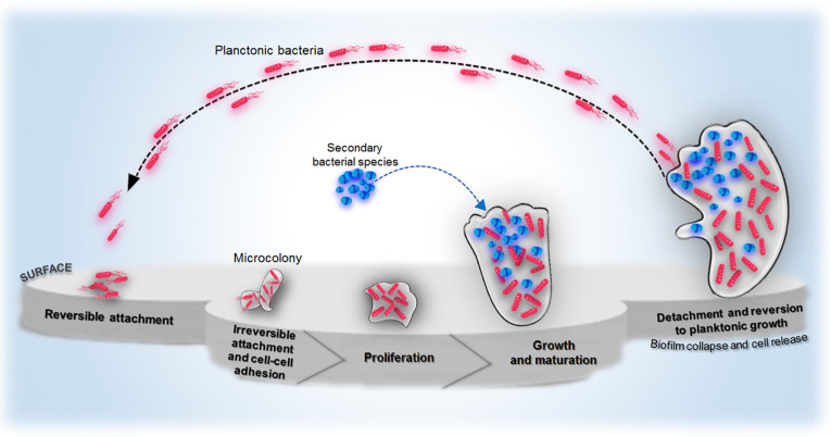
Fig. 2.
Schematic representation of steps leading to bacterial biofilm growth.
Adapted and modified from Pinto, A. M.; Cerqueira, M. A.; Bañobre-Lópes, M.; Pastrana, L. M.; Sillankorva, S. Viruses2020,12 (2), 235 and Maunders, E.; Welch, M. FEMS Microbiol. Lett.2017,364 (13).
Bacteria in a biofilm can be up to 1000 times less sensitive to a given antibiotic than their counterparts of the same species found in the planktonic form.38 It is recognized that there are some specific properties of the biofilm responsible for antibiotic treatment failures and chronic and recurrent inflammation. The complete removal of biofilm from its occupied surface represents a major challenge for modern medicine. Biofilm resistance to antibiotics is an extremely complex phenomenon. Many factors contribute to its overall condition: the previously described mechanisms common to both the biofilm and the planktonic form, specific properties of the matrix surrounding the cells, the high heterogeneity of the biofilm, and changes in gene expression profile and metabolism compared to the planktonic form.39, 40 The matrix provides some structure to the biofilm and to a certain extent, protects the cells within it from antibiotic access.41 It provides a physical and chemical barrier that impedes the diffusion of drugs deep into the biofilm, and components of the matrix may even bind some antibiotics. However, it applies only to a few types of antibiotics (e.g., aminoglycosides), and its effectiveness depends on several factors such as species and strain of bacteria, the composition of the matrix, age, and biofilm growth conditions.39, 40, 42 However, limited matrix permeability is not considered the leading cause of biofilm resistance.40 Instead, it is worth emphasizing the role of the interaction of the matrix components with the host immune system. Staphylococcus bacteria are able to produce some extracellular polymeric substances (EPS), most notably polysaccharide intercellular adhesin (PIA). The presence of PIA may protect the biofilm from the host immune response. Numerous studies suggest that PIA can reduce the susceptibility of the biofilm to phagocytosis by macrophages, reducing granulocyte activation, and decreasing cytokine secretion.43 It has been reported that another exopolysaccharide, alginate, produced by P. aeruginosa, may also provide protection against phagocytosis.39 However, the main reason for biofilm persistence and resistance lies elsewhere. Its formation is not a simple combination of matrix and cells. It represents an extremely dynamic and heterogeneous structure.41 The key factor of this heterogeneity is the oxygen and nutrient gradient. As the biofilm becomes deeper, their content decreases because they are consumed by cells that lie close to the surface before they can reach the deepest layers of the biofilm. Differences in access to oxygen and nutrients are the cause of physiological cell heterogeneity in biofilms.39, 40, 44 Cells situated near the surface are different from those deep within the biofilm. This is due to changes in gene expression in response to stress, such as hypoxia. These changes contribute to a decrease in the metabolic activity of the cells and their transition to a stationary-like state. The so-called persisters, representing up to 1% of all biofilm cells, are most responsible for recurrent, difficult-to-control infections.45
These are dormant, non-dividing cells in which metabolic processes have been significantly or completely inhibited. Due to the arrest of certain metabolic pathways, antibiotics lose their target. For this reason, surviving cells can persist through antibiotic therapy. However, once the therapy is completed, they regain metabolic activity and proliferate, leading to population recovery and recurrence of the infection.46, 47
3. Alternative methods for controlling bacterial infections
3.1. Small-molecule metal complexes
One common approach to combat bacterial resistance to antibiotics is the use of small-molecule metal complexes. The antibacterial activity of metals has been known for a long time and was used long before the discovery of microbes. Specific metals can bind to biomolecules present in the cell. This leads to changes in the structure of these molecules and, consequently, to their dysfunction. Some metals also bind to cell membranes, compromising their integrity and affecting their membrane potential. In addition, redox-active metals catalyze the Fenton reaction, which results in the formation of reactive oxygen species (ROS) capable of oxidizing proteins, lipids, and nucleic acids.25, 48, 49 Metal complexes can simultaneously exhibit several of the mechanisms of action mentioned above, and additionally combine them with the ligands' activity.50, 51, 52 Due to the synergism of action of the antibiotic and desired metal ions, complexes obtained in this way often show enhanced antimicrobial activity compared to the antibiotic alone. This effect is mainly observed with resistant bacterial strains.25 The second approach is to synthesize completely new metal complexes and to test their antimicrobial activity. Silver, copper, ruthenium, and iron complexes are particularly interesting in this aspect.25
3.2. Antibacterial oligonucleotides
The mode of antibacterial oligonucleotides action is based on inhibiting the expression of specific bacterial genes. These may be genes essential for bacterial survival, as well as those related to virulence or antibiotic resistance. The latter approach may be applicable in combined therapy with an appropriate antibiotic.53 Oligonucleotides with antibacterial activity include antisense nucleotides (ASOs) and short interfering RNA (siRNA). The sequence of both ASOs and siRNA is complementary to the bacterial mRNA. Hybridization of the oligonucleotide to the mRNA inhibits the translation process. This occurs by degrading the mRNA or blocking its binding to the ribosome.54, 55 The most significant challenge to the therapeutic success of antibacterial oligonucleotides is their delivery to the site of action, which is the interior of the bacterial cell.56 To avoid premature excretion by the kidney and degradation by nucleases present in the blood, nucleotides must be chemically modified. Additionally, due to their large size, oligonucleotides are unable to penetrate the bacterial cell wall. This problem can be solved by combining them with peptides capable of penetrating the wall.53 Moreover, the concern may be whether the APOs or siRNAs used will also interact with the host mRNA. Nevertheless, since they target specific bacterial genes, the effect on human gene expression should be minimal.55 Furthermore, the use of bioanalytical screening allows the identification and elimination of molecules that have a high risk of unwanted cross-reactivity.57 The development of antimicrobial oligonucleotides is still in its early stages as none of these compounds have yet been approved for therapeutic use by the Food and Drug Administration (FDA). Yet there is no doubt that therapies based on silencing bacterial genes have considerable potential. Blocking gene expression may produce faster and longer-lasting effects than conventional therapies. However, the development of such an approach requires a large initial investment to understand the pharmacological profile of oligonucleotides and methods to modify them. Anyhow, costs are likely to decrease significantly as the pharmacological and toxicological profile of oligonucleotides within a particular class is very similar.
3.3. Monoclonal antibodies
Monoclonal antibody therapy has its origins in the use of serum as the treatment of choice for numerous infections, including tetanus, scarlet fever, and pneumonia, in the early 20th century. It involved the passive introduction of animal serum into the patient's body. Humanized or fully human antibodies characterized by increased selectivity and reduced toxicity are used.58 The mechanism of antibacterial activity of antibodies varies depending on the antibody class, the type of targeted molecule, and the role it plays in the pathogenesis process. The direct action of antibodies is based on their binding to proteins and polysaccharides present on the surface of the bacterial cell and inducing a host immune response. On the other hand, the indirect action is based on binding to virulence factors secreted by bacteria (exotoxins, proteases, and signaling molecules, among others). After binding to antibodies, these factors are neutralized—they lose their ability to bind to elements of the host organism. Thus, the pathogenesis process is inhibited, but the organism itself must fight the infection in parallel.59, 60, 61 Currently, all FDA-approved monoclonal antibodies for antimicrobial therapy have been shown to neutralize virulence factors. The advantage of this strategy is that virulence factors are highly conserved and essential to pathogenesis.53 There are also antibodies that are completely independent of the host immune system and exhibit direct bactericidal activity.62
3.4. Nanoparticles
The use of metal nanoparticles and metal oxides is still considered one of the most promising strategies to combat resistant microbes. The probability of microbes developing resistance to these types of pharmaceuticals is low since they exhibit various mechanisms of action simultaneously, such as cell wall damage, generation of ROS, indirect effects on respiratory chain inhibition, transcription and translation, and disruption of nutrient uptake from the environment. The antibacterial and antifungal properties of nanoparticles are mainly influenced by their size and distribution, shape, and morphology.25 Nanoparticles generally exhibit more favorable morphological, catalytic, optical, and magnetic properties than their micro-sized counterparts. Moreover, their high surface area to volume ratio allows for increased contact with the pathogen surface. Additionally, the surface of nanoparticles can be easily functionalized with polymers, peptides, antibodies, etc. Thus, their biocompatibility, selectivity, and colloidal stability can be enhanced, and they can acquire an additional mechanism of antibacterial action.63, 64 Nanoparticles can also serve as carriers for other antibacterial drugs and photosensitizers.25
3.5. Bacteriophages
Bacteriophages are viruses that selectively attack bacteria. They bind to the surface receptors of bacterial cells and then insert their own genetic material into the cells. New viruses-copies of the virus that attacked the cell are assembled from the elements synthesized in this way. The newly formed virions leave the cell and can attack other cells, starting another infection cycle. Thus, bacteriophages are self-replicating pharmaceuticals.65 Most bacteriophages are characterized by their selectivity toward the bacterial strains attacked. This can be considered as both an advantage and a disadvantage. It may allow selective attack of pathogenic bacteria while not harming the natural microflora.66 However, there are currently no effective immediate methods to assess the affinity of a bacteriophage for a bacterial strain isolated from the patient's body. Thus, it is often necessary to deliver a mixture of different bacteriophages.65, 67
3.6. Photodynamic inactivation of microorganisms (PDI)
Photodynamic inactivation of microorganisms (PDI), also referred to as antimicrobial photodynamic therapy (aPDT), photodynamic antimicrobial chemotherapy (PACT) or photodynamic disinfection (PDDI), is a method of destroying microorganisms by inducing oxidative stress in their cells.68, 69, 70, 71, 72 It is used to treat localized infections, as well as disinfection and sterilization.73 The mechanism of action of PDI is based on the generation of reactive oxygen species (ROS), including singlet oxygen, which trigger a cascade of oxidation reactions within the microbial cell. The main molecules targeted by ROS are cell membrane proteins and lipids, as well as other cell wall components.74, 75 Such oxidative stress-induced damage is often irreversible and then ultimately leads to cell death.73, 76, 77 The cascade of oxidative reactions can also lead to the inactivation of bacterial enzymes such as NADH dehydrogenase, lactate dehydrogenase, ATPase, and succinate dehydrogenase. This results in cell cycle inhibition and the death of the microorganism (Fig. 3 ).75
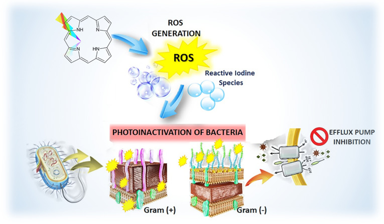
Fig. 3.
Mechanisms of action of photodynamic inactivation (PDI) of resistant microorganism including efflux pump inhibition.
The diversity of photogenerated ROS and their high reactivity toward multiple biomolecules therefore makes PDI a multi-target approach with a non-specific mechanism of action, so that the development of bacterial resistance to this treatment regimen is highly unlikely. However, it should be recognized that bacterial cells are equipped with scavenger enzymes and various antioxidantsthat combat oxidative stress. These include, in particular, superoxide dismutase, catalase, cysteine and glutathione.78 Nevertheless, bacteria have not yet developed an adequate defense system against singlet oxygen. An additional advantage is a possibility of modifying the molecular pathways of bacteria, e.g., by blocking proteins belonging to the efflux pumps system (Fig. 3). Thus, the effectiveness of PDI is influenced by the selection of the appropriate structure of the photosensitizer, its dose, as well as the light dose, so that the required ROS are generated at the right time in the right place with a sufficiently high efficiency.
3.6.1. Mechanisms of PDI
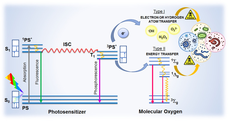
There are three individually non-toxic elements involved in PDI: a chemical compound called a photosensitizer, light from the visible or near-infrared (NIR) range of electromagnetic radiation, and molecular oxygen present within or around the bacterial cells. When a photon with energy tailored to this absorption band is absorbed, the PS is excited to one of the excited electronic states presented in Fig. 4 . Among the processes illustrated in a Jablonski Diagram, intersystem crossing (ISC) is the most important transition in PDI, even if it is forbidden according to selection rules.79 The occurrence of such a phenomenon is possible due to spin-orbital coupling. It occurs predominantly in the presence of a heavy atom (the so-called heavy atom effect).80 The photosensitizer in the triplet excited state can transfer an electron/hydrogen atom (type I mechanism) or energy (type II mechanism) to the molecular oxygen in its ground state.73, 79 The photosensitizer then returns to the singlet ground state, while the oxygen molecule undergoes excitations and transformations to various ROS.80 The transitions and possible reactions described above are illustrated in Fig. 4.
Fig. 4.
Jablonski Diagram and mechanisms of ROS generation as crucial processes and reactions in photoinactivation of microorganisms.
3.6.1.1. Type I mechanism
This mechanism involves the transfer of an electron (or hydrogen atom) to the π*2p orbital of an oxygen molecule, resulting in the formation of an oxygen-centered radical and a cascade of subsequent redox reactions. The electron transfer to the oxygen molecule can occur directly (Eq. 1) from the excited photosensitizer or indirectly with the participation of a reducing agent (e.g., NADH, Eqs. 2 and 3). In both cases, a superoxide ion is formed.
| (1) |
| (2) |
| (3) |
O2 •– can be further converted to a perhydroxyl radical (Eq. 4). Both ROS undergo a disproportionation reaction, resulting in H2O2 production (Eqs. 5 and 6). The disproportionation reaction of the superoxide ion is catalyzed by superoxide dismutase.79
| (4) |
| (5) |
| (6) |
Hydrogen peroxide has a much longer half-life than other ROS and, unlike others, can cross biological membranes, causing damage to other compartments of the cell.81 H2O2 can undergo several reactions (Eqs. (7), (8), (9)) to produce highly reactive hydroxyl radicals. The particularly important one is the so-called Fenton reaction which occurs in the presence of Fe2 + ions (Eq. 9). In the cellular environment, the Fe3 + ion can be reduced back to Fe2 + by the hydroxyl anion radical (Eq. 10).79 The combination of these two reactions is referred to as the Haber-Weiss reaction.82
| (7) |
| (8) |
| (9) |
| (10) |
The hydroxyl radical formed by the aforementioned reactions readily oxidizes biologically important molecules, especially proteins, lipids, and carbohydrates. In addition, it can inactivate naturally occurring antioxidants, such as tocopherol.79
3.6.1.2. Type II mechanism
According to the following equations, this mechanism is driven by the direct transfer of energy from the excited photosensitizer in the triplet excited state to the oxygen molecule.
| (11) |
| (12) |
| (13) |
The presence of an unfilled π*2p orbital in the singlet oxygen molecule is responsible for its reactivity toward electron-rich compounds. When exposed to 1O2, lipids and the amino acids (tryptophan, tyrosine, histidine, methionine, cysteine, and cystine) are oxidized. These reactions are responsible for the cytotoxic effects of singlet oxygen.79, 83 The mechanisms described above can occur simultaneously, and the contribution of each depends on several factors such as the type of photosensitizer, its electronic structure, and photophysical properties, as well as the concentration of oxygen in the reaction medium.84 Nevertheless, increasing evidence, including our work, suggests that mechanism II is more relevant for effective PDI because bacteria have not yet developed resistance to singlet oxygen.
3.6.2. Light excitation
Since ancient times, sun and light have always been associated with health, wellness, and vitality. The use of sunlight to treat, among others, tuberculosis was popular already in the 19th century. However, the 20th century brought the development of various therapeutic lamps, as well as other light sources. Before the invention of antibiotics, light, especially from ultraviolet radiation, was widely used to eradicate several types of microorganisms. In clinical practice, phototherapy refers not only to therapeutic, but also prophylactic and diagnostic applications of various ranges of electromagnetic radiation—from ultraviolet (UV) through visible, to near-infrared (NIR) light.85
Light is also a fundamental part of photodynamic therapy whereby it is most frequently associated with the concept of the “phototherapeutic window.” It refers to photons of relatively low energy in the visible/near-infrared range corresponding to wavelengths in the 630–850 nm range.73, 86, 87, 88 This energy is high enough for efficient ROS generation but not as high to be harmful to the body. More importantly, photons in this range can penetrate much deeper into tissue than those in the blue range. Moreover, the light of wavelengths below 630 nm is absorbed by several endogenous chromophores, including hemoglobin and melanins.79 This is actually more important in the anticancer approach, and the treatment of localized superficial infections usually does not require high penetration depth. Therefore, higher energy light such as blue light can be used in some of the applications discussed.89
Nevertheless, the use of longer wavelengths may be crucial for chronic infections that, in many cases, involve built-up microbial biofilms.90 Bacterial biofilms are not only more difficult to destroy than planktonic forms, but also bacteria in a biofilm form are much more prone to become resistant to antimicrobial agents.91 This reinforced strength of biofilm-growing bacteria against antimicrobials is related to differences in molecular mechanisms compared with the planktonic counterparts, e.g., horizontal gene transfer, genetic diversity, and alterations in gene expression while occurring only in a biofilm state. Moreover, drug diffusion is slowed down by the higher viscosity of the biofilm matrix due to the EPS network that is a 3D structure surrounding the bacteria within the biofilm and acts as a physically rugged barrier to protect the biofilm. The presence of the negatively charged EPS may protect the bacteria from the photodynamic action of positively charged photosensitizers (no sufficient attachment, no uptake to induce a photodynamic reaction after light activation). In this regard, the photodynamic inactivation procedure against biofilms generally requires longer preincubation times (up to 24 h), higher concentrations (up to 25 times), and light exposure times (up to 30 min) to reach relevant phototoxicity.
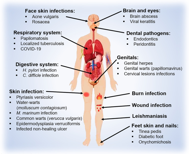
As already mentioned, PDT is used to treat localized infections, as well as disinfection and sterilization.73 The PDI procedure leads to the generation of high amounts of ROS on the site of infection due to local irradiation. PDI can be repeated at least 25 times with the same microorganism (under subtherapeutic conditions to allow the survivors to grow back again) without significant loss of phototoxicity.92 There are several potential milieus, in which PDI can replace or complement conventional antibacterial therapies for bacterial and fungal, viral, and parasitic infections. The easiest targets are: skin, oral and periodontal infections. Further development will allow for the treatment of infections where endoluminal illumination is possible, including nasal, ear, throat, lung and urinary tract infections (Fig. 5 ). It has been employed to treat acne vulgaris, biofilms associated with chronic periodontitis, burn infections, nasal decolonization of MRSA, surgical wound infections or infected wounds (e.g., venous, pressure, or diabetic ulcers).72 PDI has proven to be an effective therapeutic strategy in the treatment of fungal (Candida albicans), viral (Condylomata acuminata, Molluscum contagiosum) and protozoan (Leishmania) diseases.93
Fig. 5.
Schematic illustration of sites of infection that are or could potentially be treated with PDI along with the characteristic types of microorganisms.
The light sources employed in PDI should be suitably adapted to the areas to be illuminated. Nowadays there are more and more solutions in the form of flexible fiber optics that can adapt to different anatomical structures of the body, allowing uniform illumination of large areas. The cost of light sources, recently considered a limitation, is becoming lower due to the availability of LEDs characterized by relatively narrow emission bands and fluence rates of tens of mW/cm2. In most preclinical protocols, this is sufficient to deliver light doses up to ca. 10 J/cm2 within a few minutes of illumination.
3.6.3. Photosensitizers for PDI
The effectiveness of PDI depends, among others, on the selection of a suitable photosensitizer. A promising photosensitizer (PS) is characterized by high purity, stability, low toxicity in the dark, and high molar absorption coefficient in the visible or near-infrared light range.79 Another important aspect to be considered when selecting a PS for PDI is to ensure the appropriate interaction of the PS with the bacterial cell. The PS can: (i) bind/interact with the bacterial cell membrane, (ii) penetrate the bacterial cell, and (iii) affect the bacterial cell without direct contact if 1O2 is generated with high efficiency.94
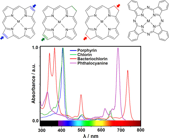
The mechanism of PS binding to the bacterial cell membrane differs for Gram-positive and Gram-negative bacteria. This is due to the differences in cell wall structure described before. The cell wall of Gram-positive bacteria, made up of a thick layer of porous peptidoglycan, is relatively easy for PSs to cross, making it easy for them to reach the cell membrane, which is their site of action. The situation is quite different in the case of Gram-negative bacteria (Fig. 1). Gram-negative bacteria are equipped with an additional membrane, a physical and functional barrier for PSs, making it difficult to reach the cell membrane. Thus, many PSs show greater efficacy in inactivating Gram-positive than Gram-negative bacteria. Positively charged PSs generally lead to the more effective inactivation of Gram-negative bacteria.89, 94 They bind to the negatively-charged phosphate groups of the outer membrane and, when excited, contribute to damage to membrane-building lipids and proteins, including membrane-associated enzymes.95 Negatively charged and neutral PSs also bind to the outer membrane of Gram-negative bacteria but do not inactivate them effectively. Gram-positive bacteria are susceptible to both negatively and positively charged PSs. Neutral compounds may also show particularly good results.89, 94, 96 Photosensitizers can also penetrate bacterial cells. The hydrophilicity of the PS often determines the ability to penetrate the bacterial cells. The cell wall of Gram-positive bacteria is hydrophilic due to its carbohydrate and amino acid content. Thus, it impedes the penetration of hydrophobic compounds into the cell.94 There are also reports of successful microbial inactivation when the PS has not penetrated the cell or is bound to the cell surface. This is possible if the PS generates sufficiently large amounts of singlet oxygen. Its lifetime in biological systems averages 0.04 μs97, roughly corresponding to an action radius of 0.02 μm. Thus, if PS is located at or near this distance from a bacterial cell, there is the opportunity to damage its external, essential structures.94 In recent years, many new photosensitizers with promising properties have been studied.88, 98, 99, 100, 101, 102, 103, 104 Synthetic macrocyclic compounds such as porphyrins, chlorins, bacteriochlorins, and phthalocyanines are of greatest interest.73, 86 Fig. 6 .
Fig. 6.
The general structures of porphyrins, chlorins, bacteriochlorins, and phthalocyanines—the macrocyclic photosensitizers that are promising candidates for PDI, and their electronic absorption spectra.
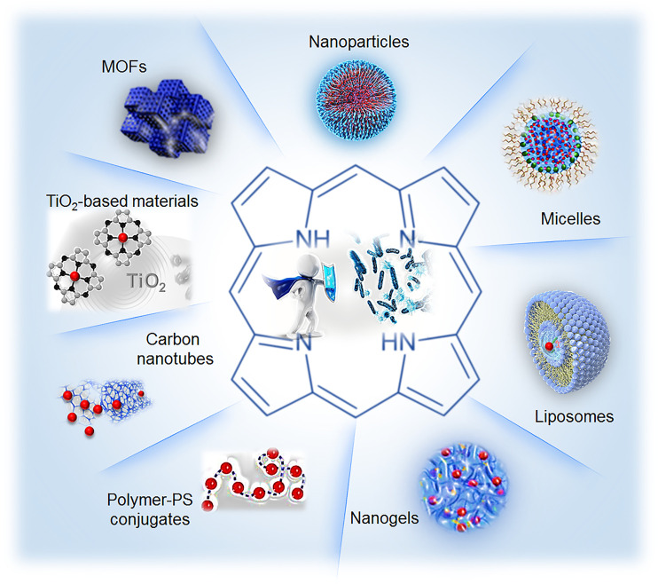
Porphyrins are a class of heterocyclic aromatic compounds constituted by four subunits of pyrrole type linked by methynic bridges. Synthetic meso-aryl-substituted porphyrins are particularly versatile starting materials to design new PSs as either ionic or nonionic moieties can be equally positioned on the periphery of the tetrapyrrole ring, thus modulating the photosensitizer polar character.73, 101, 103, 105 In order to obtain PSs with significantly longer absorption wavelength for optimal penetration in tissue, reduction of a single pyrrole double bond on the porphyrin periphery affords the chlorin core, and further reduction of a second pyrrole double bond on the chlorin periphery gives the bacteriochlorin derivative (Fig. 6). Therefore, both classes of molecules possess electronic absorption at longer wavelengths (λmax = 650–670 nm for chlorins and λmax = 730–800 nm for bacteriochlorins) than porphyrins and yet remain efficient ROS generators.79, 86, 106, 107, 108 Phthalocyanines also exhibit several features making them potentially good photosensitizers for PDI.109, 110 They are characterized by high molar absorption coefficients within the phototherapeutic window, low toxicity in the dark, high photo- and thermal stability, and the ability to generate singlet oxygen efficiently due to the high quantum yield of the triplet state and its relatively long lifetime. The properties of phthalocyanine derivatives depend largely on the nature of the coordinated metal ion.111 Moreover, the introduction of specific functional groups into their structure at the axial and peripheral positions allows control of their solubility in biological media.112 Unsubstituted phthalocyanines are characterized by very high hydrophobicity, resulting in poor solubility in the physiological environment and the tendency to form aggregates. The introduction of hydrophilic groups into their structure, e.g., carboxylic or tertbutoxysulfonyl groups, reduces this phenomenon, at least partially.113, 114, 115 However, this approach is sometimes insufficient. Then it becomes necessary to use drug delivery systems, such as liposomes, micelles, microemulsions or nanocrystalline TiO2, because aggregation of photosensitizer adversely affects the efficiency of ROS generation (Fig. 7 ).114, 116, 117
Fig. 7.
Types of drug delivery systems and materials that can be used to improve photosensitizer' pharmacological optical and photosensitizing properties.
3.6.4. Tetrapyrrolic derivatives as antibacterial photoagents—a proof of concept examples
The scientific interests of our research group are broadly focused on the use of modified halogenated (metallo)tetrapyrroles for both photodynamic therapy of cancer (PDT) and PDI. Recently, we have developed the library of synthetic halogenated porphyrins and their derivatives.118, 119 The preparation of these compounds consists of several steps: (i) the synthesis of appropriate porphyrins by a modified nitrobenzene method that involves the condensation of pyrrole with the desired aromatic aldehyde in the presence of acetic acid and nitrobenzene; (ii) chlorosulfonation to the corresponding chlorosulfophenylporphyrins; (iii) hydrolysis or nucleophilic attack with amines to yield either hydrophilic sulfonated or amphiphilic sulfonamide halogenated porphyrins.99, 119, 120, 121 The key advantages are: efficiency, simplicity, low environmental impact and the ability to provide a library of multi-gram pure, stable and versatile UV–Vis/NIR absorbing dyes with favorable physicochemical properties.118, 120, 121, 122, 123, 124, 125 In addition to the effect on the photophysical properties of these porphyrin derivatives, the introduction of halogen atoms into the structure also plays an important role in pharmacological properties. The following possible modification of the new halogenated tetrapyrroles involves functionalizing with various peripheral groups and coordinating with certain metal ions (i.e., Zn2 +, Pd2 +).73, 103 Substitution of polarity-tunable groups with higher electron-receptor properties allows control of the hydrophobicity of molecules and increases their stability by the effect of steric protection. It affects its interaction with biological membranes and biologically important molecules, leading to a remarkable increase in the photodynamic efficacy of such photosensitizers.98, 100, 106, 126, 127, 128
The unprecedented success of our collaboration with the group of Professor Arnaut and Professsor Mariette M. Pereira from the University of Coimbra (Portugal) resulted in the development of Redaporfin (NCT02070432).87, 129, 130, 131, 132, 133 During the last decade, we have also concentrated our scientific efforts on the design of effective tetrapyrrolic-based photosensitizers for antimicrobial PDT. We mainly focus on antibiotic resistance, which contributes to one of the leading healthcare problems in a clinically vicious cycle related to the overuse of antibiotics and the development of multidrug resistant microbes. The use of higher doses of antibiotics to fight infections also leads to their increased toxicity to the human body. A consequence of this is that emerging superbugs could develop the ability to resist commonly prescribed medications. As a further consequence, new antibiotic development is not as successful. Therefore, developing novel alternative antibacterial strategies such as photodynamic inactivation still remains a top challenge for scientists worldwide.
The photosensitizers commonly used in antimicrobial therapy are phenothiazine dyes (toluidine blue O, methylene blue) that absorb radiation in the 600–700 nm range. In an aqueous solution, phenothiazines have a positive charge that provides the desired affinity for bacterial cell walls. The FDA has approved them for the therapy of periodontal diseases (at a concentration of 0.01% (0.1 mg/mL).95, 134, 135 A group of our tetrapyrrolic photosensitizers for PDI, appropriately modified, could also be targeted against G(+), G(−) bacteria (groups with a positive or negative charge), and fungi (sulfonamide substituents) and to show selectivity toward microorganisms cells in comparison to normal skin cells.73, 86 For instance, by investigating a series of modified tetraphenylporphyrin derivatives in the context of PDI, we have shown that the substitution of halogen atoms (—F or —Cl) into the peripheral phenyl rings increases the ISC leading to enhanced photophysical properties. Moreover, the further introduction of sulfonic or sulfonamide groups provides water solubility that moderates their possible interactions with biological membranes. All of these substituents also prevent PS from aggregation and increase their photostability. It was reported that the sulfonated porphyrin derivatives (e.g., 5,10,15,20-tetrakis(2,6-difluorosulfonylophenyl)porphyrin [F2POH] and 5,10,15,20-tetrakis(2,6-dichlorosulfonylophenyl)porphyrin [Cl2POH]) as well as sulfonamide derivatives (e.g., 5,10,15,20-tetrakis(2,6-dichloro-3-N-ethylsulfamoylphenyl)porphyrin [Cl2PEt]) combine properties mentioned above with low dark cytotoxicity and efficient accumulation in both cancer and microbial cells. However, the photobiological properties in vitro (i.e., cellular uptake, ROS generation, and photodynamic efficacy) of hydrophilic F2POH were significantly increased after its encapsulation in Pluronic L121 micelles.89 PDI experiments showed that halogenated porphyrins investigated by us indicate various antimicrobial activity after a short incubation time and 10 J/cm2 blue light irradiation. As expected, due to the differences in the microbial cell wall structure, the complete eradication of Gram-negative E. coli was more difficult to achieve than that for the Gram-positive species. The PDI susceptibility of C. albicans was intermediate between both types of microorganisms. Sulfonic acid derivatives were shown to inactivate effectively planktonic S. aureus related to the negative molecular charge of these PS derived from the peripheral sulfonic groups (–SO3H). On the other hand, only the sulfonamide conjugate (Cl2PEt) indicated the synergistic effect of chemo- and photodynamic inactivation and was toxic to fungal yeast in the dark (ca. one order of magnitude extra killing). Thus, these results clearly show that the molecular design of porphyrin-based PSs, including their substitution patterns and formulation, may improve PDI efficacy toward each species of microorganisms.89
In recent years, PDI with porphyrin derivatives has been proposed as an alternative treatment for localized infections in response to the ever-growing problem of antibiotic resistance.25 Among the various molecular and biochemical mechanisms of antibiotic resistance, active efflux of antibiotics in bacteria plays an important role in both intrinsic and acquired multidrug resistance of clinical relevance. It also interplays with other resistance mechanisms, such as the membrane permeability barrier, enzymatic inactivation/modification of drugs, and/or antibiotic target changes/protection, in significantly increasing the levels and profiles of resistance. Thus, in our work, we also reported that PDI could be more effective by combining the PS with an efflux pump inhibition (EPI).28, 31, 136
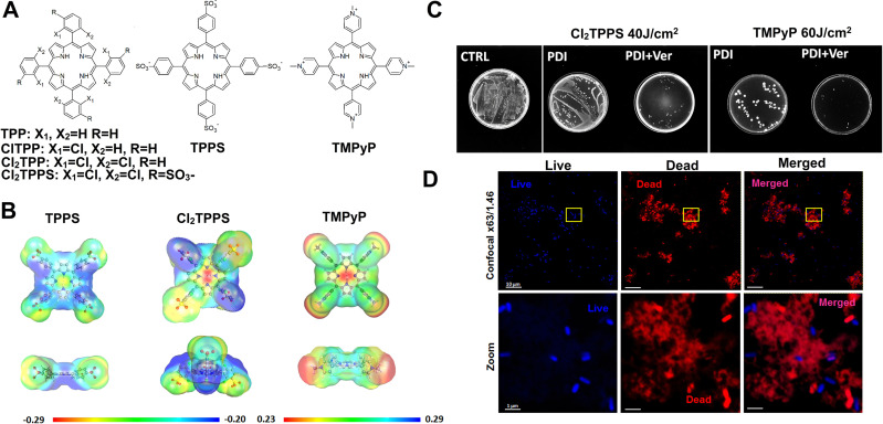
The current efforts to make the “ideal photosensitizer” focus on binding and permeability through the bacterial membrane, its accumulation at the cytoplasmic membrane, and possessing high quantum efficiency for generating singlet oxygen or other ROS. Therefore, PSs used in PDI may include cationic, neutral, and anionic meso-tetraphenylporphyrins with targeted antimicrobial properties. The presence of a negatively or positively charged group alters the physicochemical properties of PSs, making them more amphiphilic and interfering with the balance between hydrophilic and hydrophobic moieties with modulation of cell membranes permeability and, overall, the PDI effect. Our recent work demonstrated that negatively charged sulfonyl porphyrins (TPPS and Cl2TPPS) had increased cellular uptake in Gram-positive bacteria. In contrast, positively charged TMPyP is required for proper electrostatic adhesion to, or penetration of Gram-negative bacterial cell walls.137 (Fig. 8A and B). Moreover, the introduction of halogen atoms stabilizes the porphyrin macrocycle and increases activity due to the high singlet oxygen quantum yield (Φ△ = 0.95). This study shows that the most appropriate PDI photosensitizers should be charged, water-soluble, and photostable. Nevertheless, an affinity for the microbial cell wall is an essential factor affecting PDI performance. Low diffusion of the PS limits the efficacy of PDI against the fungal yeast into the cytoplasm due to their cell walls, which also contain β-glucan. Thus, the positively charged PS or the addition of substances that can significantly increase the permeability of the outer membrane and may enhance the inactivation efficiency.
Fig. 8.
Porphyrin-mediated PDI—the effect of charge and lipophilicity on their biological activity in vitro: (A) the chemical structures of tetraphenylporphyrin derivatives with various charge; (B) electronic density maps of the photosensitizers from the total self-consistent field density mapped with the electrostatic potential; (C) the effect of the efflux pump inhibition on high-dose porphyrin-mediated PDI with and without verapamil (Ver) addition; (D) the antibacterial effect of PDI confirmed by confocal microscopy images of the non-treated PDI-treated E. coli.130
The other challenge for effective PDI is the ability to overcome antibiotic resistance. Many studies have revealed that a PDI + EPI combination may result in (i) increase in the PS cellular uptake that often is excreted by the efflux system; (ii) reduced antibiotic/PS resistance; (iii) diminish the resistance mechanism derived from efflux pumps overexpression and (iv) decrease the development of more resistant superbugs. Therefore, we combined porphyrin-based PDI with verapamil (a well-known efflux pump inhibitor) (Fig. 8).
We indicated that the combination of PDI with verapamil reduced the Gram-negative bacteria survival—for E. coli, by even three orders of magnitude of extra killing. For S. aureus, the verapamil addition did not influence the PDI efficacy, and the bactericidal effect (six orders of magnitude reduction, > 99.9999% killed bacteria) was achieved after each PDI mode. Nevertheless, the investigated PSs did not yield significant activity against E. coli after PDI with low light doses (up to 20 J/cm2). Thus, KI was used, leading to strong potentiation of TPPS-, Cl2TPPS-, and TMPyP-mediated PDI (three to four orders of magnitude of extra killing), suggesting the contribution of hypoiodite and other reactive iodine species to the PDI mechanism. Due to the high stability of halogenated PS, the PDI might also be performed in higher light doses (up to 120 J/cm2), but satisfactory results were obtained after using 60 J/cm2. Thus, these data suggest that for high light-dose-PDI, its combination with EPI may enhance the inactivation. However, for low light-dose-PDI, the KI addition with the generation of reactive iodide species gives more promising results.137
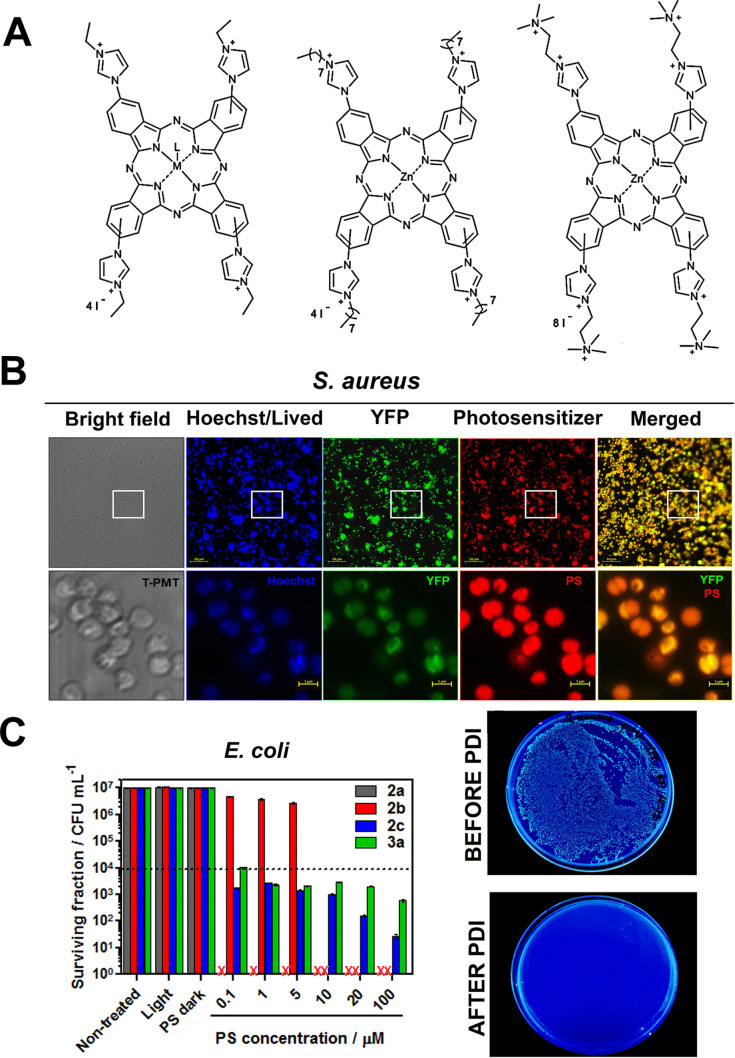
Keeping in mind the importance of the molecular charge of a PS, we also described the synthesis of a family of cationic tetra-imidazolyl phthalocyanine-based photosensitizers with varying structural features including the size of the cationized alkylic chain, degree of cationization, and the type of central coordinating metal (Fig. 9 ).109
Fig. 9.
Metallophthalocyanines as promising antimicrobial agents for PDI: (A) the structures of modified imidazolium metallophthalocyanines and their biological activity: (B) cellular accumulation in bacteria investigated by confocal microscopy imaging and (C) bacteria survival curves and Petri dishes images before and after PDI.102
The influence of these structural modifications on their biological performance was assessed based upon their spectroscopic and photophysical properties, as well as ROS generation. The antimicrobial activity tests (PDI with white light) reveal the remarkable differences in efficacy between tested metallophthalocyanines. Some examples were highly active, especially in killing Gram-negative bacteria (E. coli, P. aeruginosa) and fungi (C. albicans). Among others, the Zn(II) tetra-ethyl cationic phthalocyanine derivative showed PDI activity against tested Gram-negative bacteria and fungi, with six orders of magnitude reduction in viability of those species at PS concentrations as low as 100 nM and 1 μM, respectively.109 The cationic PS evaluated in this study showed low toxicity toward human keratinocytes cells (HaCaT) at concentrations effective in killing Gram-negative bacteria, which raises the potential for in vivo utility for developing treatments of localized infections.
3.6.5. Porphyrin-based hybrid materials
The next-generation of nanomaterials that can be activated with visible light represent an exciting new step in progress toward PDI. Furthermore, the surface chemistry of the nanomaterials also plays a crucial role in PS uptake, which can be modulated through the addition of different targeted substituents. While there is significant research on PSs accumulation in mammalian cells, the cellular uptake pathways in bacteria and fungi are less well studied. Cellular uptake of metal nanomaterials can occur when the materials are sufficiently small to cross the cellular membrane. In the case of mammalian cells, it has been suggested that particles below 100 nm are most efficient for cellular uptake. Moreover, some metal oxides and semiconductors (e.g., ZnO, TiO2) may be functionalized with tetrapyrroles toward better antimicrobial activity. This group of hybrid materials was found to be very stable in the biological environment and active in the PDI against a broad spectrum of microbes. Especially, the physicochemical properties of TiO2 ensure the effective interaction between its surface and porphyrin derivatives. It results in a suitable absorption profile and the possibility of using visible light to photoinactivate the bacteria more efficiently than employing the corresponding porphyrin alone.25, 73, 138, 139 TiO2 has been successfully used in heterogeneous photocatalysis. It owes its popularity mainly to high photocatalytic activity, high chemical and photochemical stability, low toxicity, and low cost.140 The possibility of using TiO2 for photocatalytic water and air purification is being intensively studied. Photocatalysis, based on the oxidation of pollutants by ROS generated by excited TiO2, is classified as one of the so-called advanced oxidation technologies (AOTs), which are an alternative to the commonly used water treatment methods.115 The properties of TiO2 mentioned above, its biocompatibility and unique surface properties as well as effective ROS generation, make it attractive as a nanomaterial also for biomedical applications, including PDI.116 However, a factor that significantly limits the clinical application of TiO2 is its large energy gap (3.2 eV). Its absorption, therefore, does not fall within the phototherapeutic window. One strategy to cope with this problem is to impregnate TiO2 with organic dyes, including porphyrins and phthalocyanines.141 Due to their redox properties, phthalocyanines are very well suited for sensitizing broadband semiconductors.115 The process of excitation of TiO2 modified with organic dyes proceeds as follows: upon irradiation, the dye (e.g., porphyrin) is first excited and e −/dye + pairs are generated, followed by electron transfer from the dye to the conduction band of TiO2.117 The excited semiconductor reacts with molecular oxygen, water, and hydroxyl ions in the immediate environment, resulting in ROS generation and eventually leading to oxidative stress in cells.115 The fabrication of a dye-TiO2 hybrid material also carries other benefits. It may help overcome the limitations that diminish the widespread use of phthalocyanines in clinical practice: their high hydrophobicity, low solubility in biological media, and low penetration into cells.141 The synergistic action of dyes and TiO2 may also help to enhance the photodynamic effect and produce more effective photomaterials than the compound alone.142 In addition, the fluorescent properties of porphyrins and phthalocyanines may allow the use of such hybrid materials in bioimaging.126, 140, 143, 144
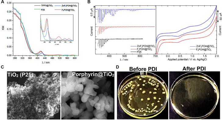
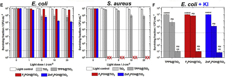
Unfortunately, the bandgap of bulky TiO2 lies in the UV range (3.2 eV for anatase and 3.0 eV for rutile, respectively), which may be intrinsically harmful and carcinogenic. Among the various dyes, particular attention should be paid to transition metal complexes with macrocyclic ligands (mainly metallophthalocyanines and metalloporphyrins) that are capable of injecting electrons from their elctronic excited states into the conduction band of titanium dioxide.142 Such hybrid TiO2-based materials may be obtained via impregnation of the semiconductor surface with the sulfonic porphyrins due to the presence of -SO3H groups, which interact directly with the surface of TiO2 (P25), Fig. 10 .138 The photophysical properties (derived from, e.g., spectroscopic data and photocurrents measurements) confirmed the electron transfer from the excited PS molecule to the valence band (VB) of TiO2. It has been shown that the modification of TiO2 with halogenated (metallo)porphyrins—difluorinated sulfonic derivative F2POH and its complex ZnF2POH moved the absorption to the visible part of the electromagnetic radiation (> 400 nm) and consequently improved the photoproperties and photoactivity mediated by these hybrid materials. They indicated high antimicrobial activity, which can be further potentiated by KI addition. The most important feature of their increased PDI efficacy toward bacteria derived from their ability to generate various ROS (singlet oxygen and oxygen-centered radicals) and generation of reactive iodide species upon visible light irradiation, because only materials able to act via multiple mechanisms prove the best efficiency against drug-resistant microorganisms.138, 139
Fig. 10.
Antimicrobial activity of (metallo)porphyrin@TiO2 hybrid materials: (A) diffuse reflectance spectra in the representation of the Kubelka-Munk function of bare P25 and surface modified TiO2 materials; (B) photocurrent generation of TiO2 and TiO2 with adsorbed (metallo)porphyrins and their cyclic voltammograms; (C) SEM images of porphyrin modified TiO2 surface; (D) petri dishes before and after PDI; (E) photodynamic inactivation of E.coli and S. aureus mediated by TiO2-based hybrid materials under visible light irradiation and (F) the potentiation effect of KI on the phototoxicity of TiO2-based hybrid materials against E. coli.131
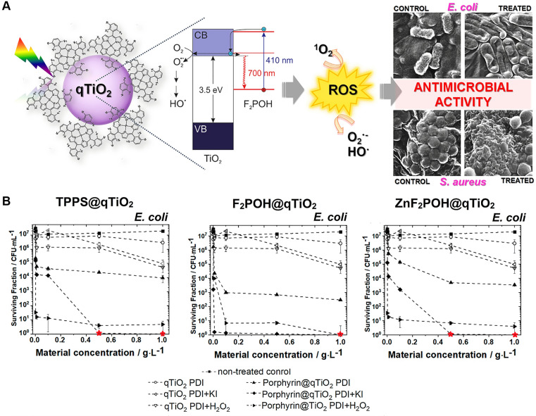
Porphyrins and metalloporphyrins, mainly with sulfonyl groups that can act as anchors, thus improving the interaction between the semiconducting support and the sensitizer, were also successfully used to sensitize nanostructured qTiO2. Well designed nanomaterials can provide better affinity toward microbial cells over host cells, enhance ROS formation, and overcome antibiotic-resistance mechanisms. Highly active surface-modified nanoparticles were synthesized and fully characterized in our laboratories. The morphology and crystal structure of the (metallo)porphyrin@qTiO2 materials obtained were examined by several techniques, including absorption and fluorescence spectroscopies as well as scanning electron microscopy (SEM) imaging (Fig. 11 ).139
Fig. 11.
The antibacterial activity of porphyrin@qTiO2 nanomaterials: (A) Proposed mechanism of the TiO2 photosensitization, and possible pathway of ROS generation after excitation of porphyrin@qTiO2 nanomaterials with their antimicrobial activity. (B) Photodynamic inactivation of E. coli and S. aureus mediated by qTiO2-based hybrid materials (porphyrin@qTiO2) and KI and H2O2 PDI potentiation after 2 h incubation and irradiated by 420 ± 20 nm LED light.132
These 20–70 nm-sized, modified TiO2 nanoparticles were found to be effective ROS generators under blue light irradiation (420 ± 20 nm). Their PDI potency against Gram-negative and Gram-positive bacteria was also investigated and revealed that the PDI with porphyrin@qTiO2 nanoparticles (1 g/L) and irradiation with a light dose of 10 J/cm2 led to reduced bacteria survival up to three logarithmic units for E. coli and up to four logarithmic units for S. aureus, respectively. A more pronounced antibacterial effect was observed when PDI was potentiated with H2O2 or KI, leading to complete eradication of tested species, even with lower concentrations of nanomaterials (starting from 0.1 g/L). Imaging of bacteria morphology after PDI suggested distinct mechanisms of cell destruction, which depends on the type of generated ROS and/or reactive iodine species. These data indicate that TiO2-based materials modified with sulfonated porphyrins are promising photoagents that may be useful in several biomedical strategies, especially in PDI. Taken together, all of the studies we performed clearly show that hybrid porphyrin-based nanomaterials represent bioinorganic photoactive agents with proven biological activity. Notably, the in vitro antibacterial efficacy makes them worth further investigation in vivo for the treatment of localized infections and wound-healing therapy.
4. Summary and future perspectives
The era of antibiotics is inevitably coming to an end and even mild infections can be potentially deadly in the future. According to the WHO report, results obtained for E. coli, Klebsiella pneumoniae and S. aureus showed that the proportion resistance to commonly used antibacterial drugs exceeded 50% in many settings.65, 145, 146 The most common cause of surgical wound infections is S. aureus a Gram-positive bacterium that can be a part of the normal flora on the skin. Strains of S. aureus resistant to penicillins, including ampicillin and amoxicillin, have acquired a gene (mecA) that codes for a novel penicillin-binding protein with low affinity for β-lactams and thus, leads to the resistance. These strains are termed methicillin-resistant S. aureus (MRSA) and are resistant to all penicillins. The future perspectives are rather daunting, due to the low chances of developing new antibacterial drugs as a result of the difficulty of finding alternative mechanisms of action for the antibiotics. The number of infections by multidrug-resistant bacteria in Europe was 400,000 in 2007, and there were 25,000 attributable deaths.147 Unless proper action is taken, this number can reach 10 million by 2050. Antibiotic resistance is exacerbated in bacteria biofilms where gene mutation frequency is significantly increased compared to planktonically growing isogenic bacteria. However, antibiotic resistance mechanisms are not sufficient to explain most cases of antibiotic-resistant biofilm infection. The same bacteria can become up to 1000 times more resistant to antibiotics when grown in biofilm.39 The biofilm matrix reduces the bioavailability of antibiotics by physical and chemical processes. The penetration of the antibiotics is impaired, leading to diffusion coefficients that are a factor of 2–3 lower than in pure water, and the chemistry of the biofilm microenvironment alters the growth of the bacteria and the antibiotic potency. The reduced bioavailability of the antibiotics and the diversity of the microenvironment enable a timely adaptive response of some biofilm cells to an antimicrobial challenge. Finally, biofilms favor the occurrence of subpopulations of cells that entered a highly protected, spore-like state—the persisters.148 Thus, there is an urgent need to find alternatives to deal with microbial infections. Ideally, such alternatives should minimize the risk of developing new resistance mechanisms, spare healthy tissue, and stimulate the host immune system to deal with the infection. The conventional treatment consists in the administration of topical and systemic antibiotics for long periods of time and may be responsible for the increased microbial strains resistant to available drugs. Therefore, it is important to develop a treatment regimen specifically targeting pathogens without causing any side effects on the host tissue.
The compounds that are used in PDT are characterized by strong absorption in the phototherapeutic window, long triplet state lifetime, high ROS generation quantum yields, no toxicity in the dark, and favorable pharmacokinetics.70, 149, 150, 151, 152 The most frequently studied photosensitizers for PDT are porphyrins, chlorins, bacteriochlorins or phthalocyanines—organic compounds generally characterized by a high molecular weight. The ideal photosensitizer/material for PDI of microorganisms should rather have a low molecular weight/small size, a strong light absorption in the visible and appropriate lipophilicity to facilitate the diffusion of non-aggregated molecules in the biofilms. Additionally, the ideal PS for PDI may require the presence of positive charges to inactivate Gram-negative bacteria. Biofilms may reduce the amount of oxygen available for PDI, but this can be compensated with PSs with long triplet lifetimes and high rates of triplet-state quenching by molecular oxygen. Finally, the photostability of the PS is critical to the success of efficient photodynamic treatment. It is therefore important to find the appropriate balance between the potential toxicity of the material itself, photostability and efficiency of ROS generation when developing photosensitizers/materials for PDI application. This brings up another difference between anticancer and antitumor photodynamic treatments. Our ongoing studies have revealed that in order to achieve adequate therapeutic success manifested not only by the destruction of the primary tumor but also by controlling distant metastases, Mechanism I seems to be more efficient, because such photogenerated oxygen-centered radicals are well-known inflammatory mediators and guarantee an adequate immune response. Whereas the opposite is true for PDI and Mechanism II is more preferred, for the simple reason that bacteria have not yet developed resistance to the main product of photoinduced energy transfer—singlet oxygen. Looking ahead, it may be possible in the near future to apply PDI using the compounds and materials described in this paper to treat infections of Gram-positive and Gram-negative multidrug-resistant bacteria in relevant animal models of infected burns and ulcers. Compared to other strategies mentioned in this paper, PDI is safe, cheap, and can be repeated hundreds of times without significant loss of efficacy. We believe that with the current development of nanotechnology and the availability of light sources, PDI can become the first line of treatment for localized infections. Moreover, PDI can reduce antibiotic use and make hospital infections more manageable through the disinfection of surface-modified medical devices.
Acknowledgments
This research was funded by the National Science Center (NCN), Poland, Sonata Bis grant number 2016/22/E/NZ7/00420 given to J.M.D.
References
- 1.Lesho E.P., Laguio-Vila M. Vol. 94. Elsevier; 2019. Mayo Clinic Proceedings; pp. 1040–1047. [DOI] [PubMed] [Google Scholar]
- 2.Tagliabue A., Rappuoli R. Front. Immunol. 2018;9:1068. doi: 10.3389/fimmu.2018.01068. [DOI] [PMC free article] [PubMed] [Google Scholar]
- 3.Abadi A.T.B., Rizvanov A.A., Haertlé T., Blatt N.L. BioNanoScience. 2019;9(4):778–788. [Google Scholar]
- 4.Bryan-Wilson J. Artforum Int. 2016;54(10):113–114. [Google Scholar]
- 5.Kollef M.H., Hamilton C.W., Ernst F.R. Infect. Control Hosp. Epidemiol. 2012;33:250–256. doi: 10.1086/664049. [DOI] [PubMed] [Google Scholar]
- 6.Klein E.Y., van Boeckel T.P., Martinez E.M., Pant S., Gandra S., Levin S.A., Laxminarayan R. Proc. Natl. Acad. Sci. U. S. A. 2018;115:E3463–E3470. doi: 10.1073/pnas.1717295115. [DOI] [PMC free article] [PubMed] [Google Scholar]
- 7.O'Niell, J. M, Infection Prevention, Control and Surveillance: Limiting the Development and Spread of Drug Resistance, Government and the Wellcome Trust, London, 2016.
- 8.Perichon B., Courvalin P., Stratton C. Elsevier, 2009. The Desk Encyclopedia of Microbiology. [Google Scholar]
- 9.Lima L.M., da Silva B.N.M., Barbosa G., Barreiro E.J. Eur. J. Med. Chem. 2020 doi: 10.1016/j.ejmech.2020.112829. [DOI] [PubMed] [Google Scholar]
- 10.Sobhanifar S., King D.T., Strynadka N.C. Curr. Opin. Struct. Biol. 2013;23(5):695–703. doi: 10.1016/j.sbi.2013.07.008. [DOI] [PubMed] [Google Scholar]
- 11.Falagas M.E., Athanasaki F., Voulgaris G.L., Triarides N.A., Vardakas K.Z. Int. J. Antimicrob. Agents. 2019;53(1):22–28. doi: 10.1016/j.ijantimicag.2018.09.013. [DOI] [PubMed] [Google Scholar]
- 12.Allen N.E., Nicas T.I. FEMS Microbiol. Rev. 2003;26(5):511–532. doi: 10.1111/j.1574-6976.2003.tb00628.x. [DOI] [PubMed] [Google Scholar]
- 13.Rice L.B. Vol. 87. Elsevier; 2012. Mayo Clinic Proceedings; pp. 198–208. [DOI] [PMC free article] [PubMed] [Google Scholar]
- 14.Tenson T., Lovmar M., Ehrenberg M. J. Mol. Biol. 2003;330(5):1005–1014. doi: 10.1016/s0022-2836(03)00662-4. [DOI] [PubMed] [Google Scholar]
- 15.Samanta, I.; Bandyopadhyay, S. Antimicrobial Resistance in Agriculture, Academic Press, 2019.
- 16.Krause K.M., Serio A.W., Kane T.R., Connolly L.E. Cold Spring Harb. Perspect. Med. 2016;6(6) doi: 10.1101/cshperspect.a027029. [DOI] [PMC free article] [PubMed] [Google Scholar]
- 17.Koteva, K.; Cox, G.; Kelso, J. K.; Surette, M. D.; Zubyk, H. L.; Ejim, L.; Stogios, P.; Savchenko, A.; Sørensen, D.; Wright, G. D. Cell Chem. Biol. 2018, 25 (4), 403–412. e405. [DOI] [PubMed]
- 18.Rodríguez-Martínez J.M., Cano M.E., Velasco C., Martínez-Martínez L., Pascual A. J. Infect. Chemother. 2011;17(2):149–182. doi: 10.1007/s10156-010-0120-2. [DOI] [PubMed] [Google Scholar]
- 19.Bakthavatchalam Y.D., Pragasam A.K., Biswas I., Veeraraghavan B. J. Glob. Antimicrob. Resist. 2018;12:124–136. doi: 10.1016/j.jgar.2017.09.011. [DOI] [PubMed] [Google Scholar]
- 20.Ashrafuzzaman M., Andersen O., McElhaney R. Biochim. Biophys. Acta (BBA)-Biomembr. 2008;1778(12):2814–2822. doi: 10.1016/j.bbamem.2008.08.017. [DOI] [PMC free article] [PubMed] [Google Scholar]
- 21.Wright G.D. Nat. Rev. Microbiol. 2007;5(3):175–186. doi: 10.1038/nrmicro1614. [DOI] [PubMed] [Google Scholar]
- 22.Fuda C., Suvorov M., Vakulenko S.B., Mobashery S. J. Biol. Chem. 2004;279(39):40802–40806. doi: 10.1074/jbc.M403589200. [DOI] [PubMed] [Google Scholar]
- 23.Gordon E., Mouz N., Duee E., Dideberg O. J. Mol. Biol. 2000;299(2):477–485. doi: 10.1006/jmbi.2000.3740. [DOI] [PubMed] [Google Scholar]
- 24.Lambert P.A. Adv. Drug Deliv. Rev. 2005;57(10):1471–1485. doi: 10.1016/j.addr.2005.04.003. [DOI] [PubMed] [Google Scholar]
- 25.Regiel-Futyra A., Dąbrowski J.M., Mazuryk O., Śpiewak K., Kyzioł A., Pucelik B., Brindell M., Stochel G. Coord. Chem. Rev. 2017;351:76–117. [Google Scholar]
- 26.Wright G.D. Adv. Drug Deliv. Rev. 2005;57(10):1451–1470. doi: 10.1016/j.addr.2005.04.002. [DOI] [PubMed] [Google Scholar]
- 27.Poole K. J. Appl. Microbiol. 2002;92:55S–64S. [PubMed] [Google Scholar]
- 28.Kumar A., Schweizer H.P. Adv. Drug Deliv. Rev. 2005;57(10):1486–1513. doi: 10.1016/j.addr.2005.04.004. [DOI] [PubMed] [Google Scholar]
- 29.Olaitan A.O., Morand S., Rolain J.-M. Front. Microbiol. 2014;5:643. doi: 10.3389/fmicb.2014.00643. [DOI] [PMC free article] [PubMed] [Google Scholar]
- 30.Velkov T., Thompson P.E., Nation R.L., Li J. J. Med. Chem. 2010;53(5):1898–1916. doi: 10.1021/jm900999h. [DOI] [PMC free article] [PubMed] [Google Scholar]
- 31.Auda I.G., Salman I.M.A., Odah J.G. Gene Rep. 2020;20 [Google Scholar]
- 32.Schindler B.D., Kaatz G.W. Drug Resist. Updat. 2016;27:1–13. doi: 10.1016/j.drup.2016.04.003. [DOI] [PubMed] [Google Scholar]
- 33.Hernando-Amado S., Blanco P., Alcalde-Rico M., Corona F., Reales-Calderón J.A., Sánchez M.B., Martínez J.L. Drug Resist. Updat. 2016;28:13–27. doi: 10.1016/j.drup.2016.06.007. [DOI] [PubMed] [Google Scholar]
- 34.Gędas A., Olszewska M.A. Elsevier; 2020. Recent Trends in Biofilm Science and Technology; pp. 1–21. [Google Scholar]
- 35.Van Acker H., Van Dijck P., Coenye T. Trends Microbiol. 2014;22(6):326–333. doi: 10.1016/j.tim.2014.02.001. [DOI] [PubMed] [Google Scholar]
- 36.Pinto A.M., Cerqueira M.A., Bañobre-Lópes M., Pastrana L.M., Sillankorva S. Viruses. 2020;12(2):235. doi: 10.3390/v12020235. [DOI] [PMC free article] [PubMed] [Google Scholar]
- 37.Maunders E., Welch M. FEMS Microbiol. Lett. 2017;364(13):1–10. doi: 10.1093/femsle/fnx120. [DOI] [PMC free article] [PubMed] [Google Scholar]
- 38.Gebreyohannes G., Nyerere A., Bii C., Sbhatu D.B. Heliyon. 2019;5(8) doi: 10.1016/j.heliyon.2019.e02192. [DOI] [PMC free article] [PubMed] [Google Scholar]
- 39.Mah T.-F. Future Microbiol. 2012;7(9):1061–1072. doi: 10.2217/fmb.12.76. [DOI] [PubMed] [Google Scholar]
- 40.Hall C.W., Mah T.-F. FEMS Microbiol. Rev. 2017;41(3):276–301. doi: 10.1093/femsre/fux010. [DOI] [PubMed] [Google Scholar]
- 41.Bi Y., Xia G., Shi C., Wan J., Liu L., Chen Y., Wu Y., Zhang W., Zhou M., He H. Fundamen. Res. 2021 [Google Scholar]
- 42.Proctor R.A., Von Eiff C., Kahl B.C., Becker K., McNamara P., Herrmann M., Peters G. Nat. Rev. Microbiol. 2006;4(4):295–305. doi: 10.1038/nrmicro1384. [DOI] [PubMed] [Google Scholar]
- 43.Nguyen H.T., Nguyen T.H., Otto M. Comput. Struct. Biotechnol. J. 2020 doi: 10.1016/j.csbj.2023.03.012. [DOI] [PMC free article] [PubMed] [Google Scholar]
- 44.Crabbé A., Jensen P.Ø., Bjarnsholt T., Coenye T. Trends Microbiol. 2019;27(10):850–863. doi: 10.1016/j.tim.2019.05.003. [DOI] [PubMed] [Google Scholar]
- 45.Yan J., Bassler B.L. Cell Host Microbe. 2019;26(1):15–21. doi: 10.1016/j.chom.2019.06.002. [DOI] [PMC free article] [PubMed] [Google Scholar]
- 46.Ranieri M.R., Whitchurch C.B., Burrows L.L. Curr. Opin. Microbiol. 2018;45:164–169. doi: 10.1016/j.mib.2018.07.006. [DOI] [PubMed] [Google Scholar]
- 47.Parastan R., Kargar M., Solhjoo K., Kafilzadeh F. J. Glob. Antimicrob. Resist. 2020;22:379–385. doi: 10.1016/j.jgar.2020.02.025. [DOI] [PubMed] [Google Scholar]
- 48.Lemire J.A., Harrison J.J., Turner R.J. Nat. Rev. Microbiol. 2013;11(6):371–384. doi: 10.1038/nrmicro3028. [DOI] [PubMed] [Google Scholar]
- 49.Kuncewicz J., Dąbrowski J.M., Kyzioł A., Brindell M., Łabuz P., Mazuryk O., Macyk W., Stochel G. Coord. Chem. Rev. 2019;398 [Google Scholar]
- 50.Kong B., Joshi T., Belousoff M.J., Tor Y., Graham B., Spiccia L. J. Inorg. Biochem. 2016;162:334–342. doi: 10.1016/j.jinorgbio.2015.11.029. [DOI] [PubMed] [Google Scholar]
- 51.Guerra W., de Andrade Azevedo E., de Souza Monteiro A.R., Bucciarelli-Rodriguez M., Chartone-Souza E., Nascimento A.M.A., Fontes A.P.S., Le Moyec L., Pereira-Maia E.C. J. Inorg. Biochem. 2005;99(12):2348–2354. doi: 10.1016/j.jinorgbio.2005.09.001. [DOI] [PubMed] [Google Scholar]
- 52.Drevenšek P., Košmrlj J., Giester G., Skauge T., Sletten E., Sepčić K., Turel I. J. Inorg. Biochem. 2006;100(11):1755–1763. doi: 10.1016/j.jinorgbio.2006.06.011. [DOI] [PubMed] [Google Scholar]
- 53.Streicher L.M. J. Glob. Antimicrob. Resist. 2021;14:285–295. doi: 10.1016/j.jgar.2020.12.025. [DOI] [PubMed] [Google Scholar]
- 54.Watts J.K., Corey D.R. J. Pathol. 2012;226(2):365–379. doi: 10.1002/path.2993. [DOI] [PMC free article] [PubMed] [Google Scholar]
- 55.Chi X., Gatti P., Papoian T. Drug Discov. Today. 2017;22(5):823–833. doi: 10.1016/j.drudis.2017.01.013. [DOI] [PubMed] [Google Scholar]
- 56.Xue X.-Y., Mao X.-G., Zhou Y., Chen Z., Hu Y., Hou Z., Li M.-K., Meng J.-R., Luo X.-X. Nanomed. Nanotechnol. Biol. Med. 2018;14(3):745–758. doi: 10.1016/j.nano.2017.12.026. [DOI] [PubMed] [Google Scholar]
- 57.Frazier K.S. Toxicol. Pathol. 2015;43(1):78–89. doi: 10.1177/0192623314551840. [DOI] [PubMed] [Google Scholar]
- 58.Casadevall A., Scharff M.D. Antimicrob. Agents Chemother. 1994;38(8):1695–1702. doi: 10.1128/aac.38.8.1695. [DOI] [PMC free article] [PubMed] [Google Scholar]
- 59.Pelfrene E., Mura M., Sanches A.C., Cavaleri M. Clin. Microbiol. Infect. 2019;25(1):60–64. doi: 10.1016/j.cmi.2018.04.024. [DOI] [PMC free article] [PubMed] [Google Scholar]
- 60.Bebbington C., Yarranton G. Curr. Opin. Biotechnol. 2008;19(6):613–619. doi: 10.1016/j.copbio.2008.10.002. [DOI] [PubMed] [Google Scholar]
- 61.Hansel T.T., Kropshofer H., Singer T., Mitchell J.A., George A.J. Nat. Rev. Drug Discov. 2010;9(4):325–338. doi: 10.1038/nrd3003. [DOI] [PubMed] [Google Scholar]
- 62.Wang-Lin S.X., Balthasar J.P. Antibodies. 2018;7(1):5. doi: 10.3390/antib7010005. [DOI] [PMC free article] [PubMed] [Google Scholar]
- 63.Kango S., Kalia S., Celli A., Njuguna J., Habibi Y., Kumar R. Prog. Polym. Sci. 2013;38(8):1232–1261. [Google Scholar]
- 64.Sperling R.A., Parak W.J. Philos. Trans. Roy. Soc. A Math. Phys. Eng. Sci. 1915;2010(368):1333–1383. doi: 10.1098/rsta.2009.0273. [DOI] [PubMed] [Google Scholar]
- 65.Ghosh C., Sarkar P., Issa R., Haldar J. Trends Microbiol. 2019;27(4):323–338. doi: 10.1016/j.tim.2018.12.010. [DOI] [PubMed] [Google Scholar]
- 66.Clark J.R., March J.B. Trends Biotechnol. 2006;24(5):212–218. doi: 10.1016/j.tibtech.2006.03.003. [DOI] [PubMed] [Google Scholar]
- 67.Abedon S.T., Kuhl S.J., Blasdel B.G., Kutter E.M. Bacteriophage. 2011;1(2):66–85. doi: 10.4161/bact.1.2.15845. [DOI] [PMC free article] [PubMed] [Google Scholar]
- 68.Tunçel A., Öztürk İ., Ince M., Ocakoglu K., Hoşgör-Limoncu M., Yurt F. J. Porphyrins Phthalocyanines. 2019;23(01n02):206–212. [Google Scholar]
- 69.Pérez-Laguna V., Gilaberte Y., Millán-Lou M.I., Agut M., Nonell S., Rezusta A., Hamblin M.R. Photochem. Photobiol. Sci. 2019;18(5):1020–1029. doi: 10.1039/c8pp00534f. [DOI] [PMC free article] [PubMed] [Google Scholar]
- 70.Hamblin M.R., Hasan T. Photochem. Photobiol. Sci. 2004;3(5):436–450. doi: 10.1039/b311900a. [DOI] [PMC free article] [PubMed] [Google Scholar]
- 71.Sabino C., Garcez A., Núñez S., Ribeiro M., Hamblin M. Lasers Med. Sci. 2015;30(6):1657–1665. doi: 10.1007/s10103-014-1629-x. [DOI] [PMC free article] [PubMed] [Google Scholar]
- 72.Kawczyk-Krupka A., Pucelik B., Międzybrodzka A., Sieroń A.R., Dąbrowski J.M. Photodiagn. Photodyn. Ther. 2018;23:132–143. doi: 10.1016/j.pdpdt.2018.05.001. [DOI] [PubMed] [Google Scholar]
- 73.Dąbrowski J.M., Pucelik B., Regiel-Futyra A., Brindell M., Mazuryk O., Kyzioł A., Stochel G., Macyk W., Arnaut L.G. Coord. Chem. Rev. 2016;325:67–101. [Google Scholar]
- 74.Alves E., Esteves A.C., Correia A., Cunha A., Faustino M.A., Neves M.G., Almeida A. Photochem. Photobiol. Sci. 2015;14(6):1169–1178. doi: 10.1039/c4pp00194j. [DOI] [PubMed] [Google Scholar]
- 75.Koch G. CHIMIA Int. J. Chem. 2017;71(10):643(1). [PubMed] [Google Scholar]
- 76.Tavares A., Dias S.R., Carvalho C.M., Faustino M.A., Tomé J.P., Neves M.G., Tome A.C., Cavaleiro J.A., Cunha Â., Gomes N.C. Photochem. Photobiol. Sci. 2011;10(10):1659–1669. doi: 10.1039/c1pp05097d. [DOI] [PubMed] [Google Scholar]
- 77.Lopes D., Melo T., Santos N., Rosa L., Alves E., Gomes M.C., Cunha Â., Neves M.G., Faustino M.A., Domingues M.R.M. J. Photochem. Photobiol. B Biol. 2014;141:145–153. doi: 10.1016/j.jphotobiol.2014.08.024. [DOI] [PubMed] [Google Scholar]
- 78.Mitoraj D., Jańczyk A., Strus M., Kisch H., Stochel G., Heczko P.B., Macyk W. Photochem. Photobiol. Sci. 2007;6(6):642–648. doi: 10.1039/b617043a. [DOI] [PubMed] [Google Scholar]
- 79.Dąbrowski J.M. Adv. Inorg. Chem. 2017;70:343–394. [Google Scholar]
- 80.Whittaker, A.; Mount, A.; Heal, M.; Galus, M. Wydawnictwo Naukowe PWN, 2012.
- 81.Oszajca M., Brindell M., Orzeł Ł., Dąbrowski J.M., Śpiewak K., Łabuz P., Pacia M., Stochel-Gaudyn A., Macyk W., van Eldik R. Coord. Chem. Rev. 2016;327:143–165. [Google Scholar]
- 82.Kehrer J.P. Toxicology. 2000;149(1):43–50. doi: 10.1016/s0300-483x(00)00231-6. [DOI] [PubMed] [Google Scholar]
- 83.DeRosa M.C., Crutchley R.J. Coord. Chem. Rev. 2002;233:351–371. [Google Scholar]
- 84.Agostinis P., Berg K., Cengel K.A., Foster T.H., Girotti A.W., Gollnick S.O., Hahn S.M., Hamblin M.R., Juzeniene A., Kessel D. CA Cancer J. Clin. 2011;61(4):250–281. doi: 10.3322/caac.20114. [DOI] [PMC free article] [PubMed] [Google Scholar]
- 85.Mead, M. N., Benefits of Sunlight: A Bright Spot for Human Health, Secondary National Institute of Environmental Health Sciences: 2008. [DOI] [PMC free article] [PubMed]
- 86.Pucelik B., Sułek A., Dąbrowski J.M. Coord. Chem. Rev. 2020;416 [Google Scholar]
- 87.Pucelik B., Arnaut L.G., Stochel G.Y., Dabrowski J.M. ACS Appl. Mater. Interfaces. 2016;8(34):22039–22055. doi: 10.1021/acsami.6b07031. [DOI] [PubMed] [Google Scholar]
- 88.Rocha L.B., Gomes-da-Silva L.C., Dąbrowski J.M., Arnaut L.G. Eur. J. Cancer. 2015;51(13):1822–1830. doi: 10.1016/j.ejca.2015.06.002. [DOI] [PubMed] [Google Scholar]
- 89.Pucelik B., Paczyński R., Dubin G., Pereira M.M., Arnaut L.G., Dąbrowski J.M. PLoS One. 2017;12(10) doi: 10.1371/journal.pone.0185984. [DOI] [PMC free article] [PubMed] [Google Scholar]
- 90.Vinagreiro C.S., Zangirolami A., Schaberle F.A., Nunes S.C., Blanco K.C., Inada N.M., da Silva G.J., Pais A.A., Bagnato V.S., Arnaut L.G. ACS Infect. Dis. 2020;6(6):1517–1526. doi: 10.1021/acsinfecdis.9b00379. [DOI] [PubMed] [Google Scholar]
- 91.Davies D. Nat. Rev. Drug Discov. 2003;2(2):114–122. doi: 10.1038/nrd1008. [DOI] [PubMed] [Google Scholar]
- 92.Pedigo L.A., Gibbs A.J., Scott R.J., Street C.N. Proc. SPIE. 2009;7380:73803H. [Google Scholar]
- 93.Biel M.A. Adv. Exp. Med. Biol. 2015;831:119–136. doi: 10.1007/978-3-319-09782-4_8. [DOI] [PubMed] [Google Scholar]
- 94.Nagata J.Y., Hioka N., Kimura E., Batistela V.R., Terada R.S.S., Graciano A.X., Baesso M.L., Hayacibara M.F. Photodiagn. Photodyn. Ther. 2012;9(2):122–131. doi: 10.1016/j.pdpdt.2011.11.006. [DOI] [PubMed] [Google Scholar]
- 95.Usacheva M.N., Teichert M.C., Biel M.A. Lasers Surg. Med. Official J. Am. Soc. Laser Med. Surg. 2001;29(2):165–173. doi: 10.1002/lsm.1105. [DOI] [PubMed] [Google Scholar]
- 96.Wood S., Metcalf D., Devine D., Robinson C. J. Antimicrob. Chemother. 2006;57(4):680–684. doi: 10.1093/jac/dkl021. [DOI] [PubMed] [Google Scholar]
- 97.Ochsner M. J. Photochem. Photobiol. B Biol. 1997;39(1):1–18. doi: 10.1016/s1011-1344(96)07428-3. [DOI] [PubMed] [Google Scholar]
- 98.Pucelik B., Arnaut L.G., Dąbrowski J.M. J. Clin. Med. 2020;9(1):8. doi: 10.3390/jcm9010008. [DOI] [PMC free article] [PubMed] [Google Scholar]
- 99.Pereira M.M., Monteiro C.J., Simões A.V., Pinto S.M., Arnaut L.G., Sá G.F., Silva E.F., Rocha L.B., Simões S., Formosinho S.J. J. Porphyrins Phthalocyanines. 2009;13(04n05):567–573. [Google Scholar]
- 100.Pucelik B., Sułek A., Drozd A., Stochel G., Pereira M.M., Pinto S., Arnaut L.G., Dąbrowski J.M. Int. J. Mol. Sci. 2020;21(8):2786. doi: 10.3390/ijms21082786. [DOI] [PMC free article] [PubMed] [Google Scholar]
- 101.Arnaut L.G., Pereira M.M., Dąbrowski J.M., Silva E.F., Schaberle F.A., Abreu A.R., Rocha L.B., Barsan M.M., Urbańska K., Stochel G. Chemistry – A Eur. J. Dermatol. 2014;20(18):5346–5357. doi: 10.1002/chem.201304202. [DOI] [PubMed] [Google Scholar]
- 102.Dąbrowski J.M., Pereira M.M., Arnaut L.G., Monteiro C.J., Peixoto A.F., Karocki A., Urbańska K., Stochel G. Photochem. Photobiol. 2007;83(4):897–903. doi: 10.1111/j.1751-1097.2007.00073.x. [DOI] [PubMed] [Google Scholar]
- 103.Dąbrowski J.M., Pucelik B., Pereira M.M., Arnaut L.G., Stochel G. J. Coord. Chem. 2015;68(17–18):3116–3134. [Google Scholar]
- 104.Luz A.F., Pucelik B., Pereira M.M., Dąbrowski J.M., Arnaut L.G. Lasers Surg. Med. 2018;50(5):451–459. doi: 10.1002/lsm.22931. [DOI] [PubMed] [Google Scholar]
- 105.Arnaut L.G. Adv. Inorg. Chem. 2011;63:187–233. [Google Scholar]
- 106.Silva E.F., Schaberle F.A., Monteiro C.J., Dąbrowski J.M., Arnaut L.G. Photochem. Photobiol. Sci. 2013;12(7):1187–1192. doi: 10.1039/c3pp25419d. [DOI] [PubMed] [Google Scholar]
- 107.Dabrowski J.M., Arnaut L.G., Pereira M.M., Monteiro C., Urbanska K., Simões S., Stochel G. ChemMedChem. 2010;5(10):1770–1780. doi: 10.1002/cmdc.201000223. [DOI] [PubMed] [Google Scholar]
- 108.Dąbrowski J.M., Krzykawska M., Arnaut L.G., Pereira M.M., Monteiro C.J., Simões S., Urbańska K., Stochel G. ChemMedChem. 2011;6(9):1715–1726. doi: 10.1002/cmdc.201100186. [DOI] [PubMed] [Google Scholar]
- 109.Aroso R.T., Calvete M.J., Pucelik B., Dubin G., Arnaut L.G., Pereira M.M., Dąbrowski J.M. Eur. J. Med. Chem. 2019;184 doi: 10.1016/j.ejmech.2019.111740. [DOI] [PubMed] [Google Scholar]
- 110.Mete E., Kabay N., Dumoulin F., Ahsen V., Tuncel Kostakoğlu S., Ergin Ç. Biotech. Histochem. 2021;96(4):311–314. doi: 10.1080/10520295.2020.1854855. [DOI] [PubMed] [Google Scholar]
- 111.Lo P.-C., Rodríguez-Morgade M.S., Pandey R.K., Ng D.K., Torres T., Dumoulin F. Chem. Soc. Rev. 2020;49(4):1041–1056. doi: 10.1039/c9cs00129h. [DOI] [PubMed] [Google Scholar]
- 112.Topal S.Z., İşci Ü., Kumru U., Atilla D., Gürek A.G., Hirel C., Durmuş M., Tommasino J.-B., Luneau D., Berber S. Dalton Trans. 2014;43(18):6897–6908. doi: 10.1039/c3dt53410c. [DOI] [PubMed] [Google Scholar]
- 113.Tunç G., Albakour M., Ahsen V., Gürek A.E.G. Vol. 3. World Scientific; 2019. Porphyrin Science by Women; pp. 698–707. [Google Scholar]
- 114.Santos K.L.M., Barros R.M., da Silva Lima D.P., Nunes A.M.A., Sato M.R., Faccio R., de Lima Damasceno B.P.G., Junior J.A.O. Photodiagn. Photodyn. Ther. 2020 doi: 10.1016/j.pdpdt.2020.102032. [DOI] [PubMed] [Google Scholar]
- 115.Güzel E., Şişman İ., Gül A., Kocak M.B. J. Porphyrins Phthalocyanines. 2019;23(03):279–286. [Google Scholar]
- 116.Mantareva, V.; Eneva, I.; Kussovski, V.; Borisova, E.; Angelov, I. In 18th International School on Quantum Electronics: Laser Physics and Applications, 2015; International Society for Optics and Photonics: Vol. 9447, p 94470W.
- 117.Yurt F., Ince M., Colak S.G., Ocakoglu K., Er O., Soylu H.M., Gunduz C., Avci C.B., Kurt C.C. Int. J. Pharm. 2017;524(1–2):467–474. doi: 10.1016/j.ijpharm.2017.03.050. [DOI] [PubMed] [Google Scholar]
- 118.Simoes A.V., Adamowicz A., Dąbrowski J.M., Calvete M.J., Abreu A.R., Stochel G., Arnaut L.G., Pereira M.M. Tetrahedron. 2012;68(42):8767–8772. [Google Scholar]
- 119.Pinto S.M., Henriques C.A., Tomé V.A., Vinagreiro C.S., Calvete M.J., Dąbrowski J.M., Piñeiro M., Arnaut L.G., Pereira M.M. J. Porphyrins Phthalocyanines. 2016;20(01n04):45–60. [Google Scholar]
- 120.Pereira M.M., Monteiro C.J., Simoes A.V., Pinto S.M., Abreu A.R., Sá G.F., Silva E.F., Rocha L.B., Dąbrowski J.M., Formosinho S.J. Tetrahedron. 2010;66(49):9545–9551. [Google Scholar]
- 121.Pereira M.M., Abreu A.R., Goncalves N.P., Calvete M.J., Simões A.V., Monteiro C.J., Arnaut L.G., Eusebio M.E., Canotilho J. Green Chem. 2012;14(6):1666–1672. [Google Scholar]
- 122.Dąbrowski J.M., Urbanska K., Arnaut L.G., Pereira M.M., Abreu A.R., Simões S., Stochel G. ChemMedChem. 2011;6(3):465–475. doi: 10.1002/cmdc.201000524. [DOI] [PubMed] [Google Scholar]
- 123.Saavedra R., Rocha L.B., Dąbrowski J.M., Arnaut L.G. ChemMedChem. 2014;9(2):390–398. doi: 10.1002/cmdc.201300449. [DOI] [PubMed] [Google Scholar]
- 124.Gonçalves N.P., Simões A.V., Abreu A.R., Abrunhosa A.J., Dąbrowski J.M., Pereira M.M. J. Porphyrins Phthalocyanines. 2015;19(08):946–955. [Google Scholar]
- 125.Dąbrowski J.M., Arnaut L.G., Pereira M.M., Urbańska K., Simões S., Stochel G., Cortes L. Free Radic. Biol. Med. 2012;52(7):1188–1200. doi: 10.1016/j.freeradbiomed.2011.12.027. [DOI] [PubMed] [Google Scholar]
- 126.Pucelik B., Gürol I., Ahsen V., Dumoulin F., Dąbrowski J.M. Eur. J. Med. Chem. 2016;124:284–298. doi: 10.1016/j.ejmech.2016.08.035. [DOI] [PubMed] [Google Scholar]
- 127.Silva, E. F.; Serpa, C.; Dąbrowski, J. M.; Monteiro, C. J.; Formosinho, S. J.; Stochel, G.; Urbanska, K.; Simões, S.; Pereira, M. M.; Arnaut, L. G. Chemistry–A Eur. J. Dermatol. 2010, 16 (30), 9273–9286. [DOI] [PubMed]
- 128.Soares H.T., Campos J., Gomes-da-Silva L.C., Schaberle F.A., Dabrowski J.M., Arnaut L.G. Chembiochem. 2016;17(9):836–842. doi: 10.1002/cbic.201500573. [DOI] [PubMed] [Google Scholar]
- 129.Santos L.L., Oliveira J., Monteiro E., Santos J., Sarmento C. Case Rep. Oncol. 2018;11(3):769–776. doi: 10.1159/000493423. [DOI] [PMC free article] [PubMed] [Google Scholar]
- 130.Pucelik B., Sułek A., Barzowska A., Dąbrowski J.M. Cancer Lett. 2020;492:116–135. doi: 10.1016/j.canlet.2020.07.007. [DOI] [PubMed] [Google Scholar]
- 131.Krzykawska-Serda M., Dąbrowski J.M., Arnaut L.G., Szczygieł M., Urbańska K., Stochel G., Elas M. Free Radic. Biol. Med. 2014;73:239–251. doi: 10.1016/j.freeradbiomed.2014.05.003. [DOI] [PubMed] [Google Scholar]
- 132.Karwicka M., Pucelik B., Gonet M., Elas M., Dąbrowski J.M. Sci. Rep. 2019;9(1):1–15. doi: 10.1038/s41598-019-49064-6. [DOI] [PMC free article] [PubMed] [Google Scholar]
- 133.Rocha L.B., Schaberle F., Dąbrowski J.M., Simões S., Arnaut L.G. Int. J. Mol. Sci. 2015;16(12):29236–29249. doi: 10.3390/ijms161226162. [DOI] [PMC free article] [PubMed] [Google Scholar]
- 134.Huang Y.-Y., Wintner A., Seed P.C., Brauns T., Gelfand J.A., Hamblin M.R. Sci. Rep. 2018;8(1):1–9. doi: 10.1038/s41598-018-25365-0. [DOI] [PMC free article] [PubMed] [Google Scholar]
- 135.Shen X., Dong L., He X., Zhao C., Zhang W., Li X., Lu Y. Photodiagn. Photodyn. Ther. 2020;32 doi: 10.1016/j.pdpdt.2020.102051. [DOI] [PubMed] [Google Scholar]
- 136.de Aguiar Coletti T.M.S.F., De Freitas L.M., Almeida A.M.F., Fontana C.R. Photomed. Laser Surg. 2017;35(7):378–385. doi: 10.1089/pho.2016.4246. [DOI] [PubMed] [Google Scholar]
- 137.Sułek A., Pucelik B., Kobielusz M., Barzowska A., Dąbrowski J.M. Int. J. Mol. Sci. 2020;21(22):8716. doi: 10.3390/ijms21228716. [DOI] [PMC free article] [PubMed] [Google Scholar]
- 138.Sułek A., Pucelik B., Kuncewicz J., Dubin G., Dąbrowski J.M. Catal. Today. 2019;335:538–549. [Google Scholar]
- 139.Sułek A., Pucelik B., Kobielusz M., Łabuz P., Dubin G., Dąbrowski J.M. Catalysts. 2019;9(10):821. [Google Scholar]
- 140.França M.D., Santos L.M., Silva T.A., Borges K.A., Silva V.M., Patrocinio A.O., Trovó A.G., Machado A.E. J. Braz. Chem. Soc. 2016;27:1094–1102. [Google Scholar]
- 141.Flak D., Yate L., Nowaczyk G., Jurga S. Mater. Sci. Eng. C. 2017;78:1072–1085. doi: 10.1016/j.msec.2017.04.107. [DOI] [PubMed] [Google Scholar]
- 142.Dąbrowski J.M., Pucelik B., Pereira M.M., Arnaut L.G., Macyk W., Stochel G. RSC Adv. 2015;5(113):93252–93261. [Google Scholar]
- 143.Lobo A.C., Silva A.D., Tome V.A., Pinto S.M., Silva E.F., Calvete M.J., Gomes C.M., Pereira M.M., Arnaut L.G. J. Med. Chem. 2016;59(10):4688–4696. doi: 10.1021/acs.jmedchem.6b00054. [DOI] [PubMed] [Google Scholar]
- 144.Iqbal Z., Chen J., Chen Z., Huang M. Curr. Drug Metab. 2015;16(9):816–832. doi: 10.2174/1389200217666151120165404. [DOI] [PubMed] [Google Scholar]
- 145.Aslam B., Wang W., Arshad M.I., Khurshid M., Muzammil S., Rasool M.H., Nisar M.A., Alvi R.F., Aslam M.A., Qamar M.U. Infect. Drug Resist. 2018;11:1645. doi: 10.2147/IDR.S173867. [DOI] [PMC free article] [PubMed] [Google Scholar]
- 146.Mehellou, Y.; Willcox, B. E., A Two-Pronged Attack on Antibiotic-Resistant Microbes, Secondary Nature Publishing Group: 2021. [DOI] [PubMed]
- 147.Bush K., Courvalin P., Dantas G., Davies J., Eisenstein B., Huovinen P., Jacoby G.A., Kishony R., Kreiswirth B.N., Kutter E. Nat. Rev. Microbiol. 2011;9(12):894–896. doi: 10.1038/nrmicro2693. [DOI] [PMC free article] [PubMed] [Google Scholar]
- 148.Stewart P.S. Int. J. Med. Microbiol. 2002;292(2):107–113. doi: 10.1078/1438-4221-00196. [DOI] [PubMed] [Google Scholar]
- 149.Hamblin M.R. Curr. Opin. Microbiol. 2016;33:67–73. doi: 10.1016/j.mib.2016.06.008. [DOI] [PMC free article] [PubMed] [Google Scholar]
- 150.Wainwright M., Maisch T., Nonell S., Plaetzer K., Almeida A., Tegos G.P., Hamblin M.R. Lancet Infect. Dis. 2017;17(2):e49–e55. doi: 10.1016/S1473-3099(16)30268-7. [DOI] [PMC free article] [PubMed] [Google Scholar]
- 151.Demidova T., Hamblin M. Int. J. Immunopathol. Pharmacol. 2004;17(3):245–254. doi: 10.1177/039463200401700304. [DOI] [PMC free article] [PubMed] [Google Scholar]
- 152.Mikula P., Kalhotka L., Jancula D., Zezulka S., Korinkova R., Cerny J., Marsalek B., Toman P. J. Photochem. Photobiol. B Biol. 2014;138:230–239. doi: 10.1016/j.jphotobiol.2014.04.014. [DOI] [PubMed] [Google Scholar]


