Abstract
Purpose
Children with Down's syndrome (DS) are known to have poorer visual acuity than neurotypical children. One report has shown that children with DS and nystagmus also have poor acuity when compared to typical children with nystagmus. What has not been established is the extent of any acuity deficit due to nystagmus and whether nystagmus affects refractive error within a population with DS.
Methods
Clinical records from the Cardiff University Down's Syndrome Vision Research Unit were examined retrospectively. Binocular visual acuity and refraction data were available for 50 children who had DS and nystagmus and 176 children who had DS but no nystagmus. Data were compared between the two groups and with published data for neurotypical children with nystagmus.
Results
The study confirms the deficit in acuity in DS, compared to neurotypical children, of approximately 0.2 logMAR and shows a deficit attributable to nystagmus of a further 0.2 logMAR beyond the first year of life. Children with both DS and nystagmus clearly have a significant additional impairment. Children with DS have a wide range of refractive errors, but nystagmus increases the likelihood of myopia. Prevalence and axis direction of astigmatism, on the other hand, appear unaffected by nystagmus.
Conclusions
Nystagmus confers an additional visual impairment on children with DS and must be recognized as such by families and educators. Children with both DS and nystagmus clearly need targeted support.
Keywords: nystagmus, children's vision, Down's syndrome, refractive error, visual acuity
Infantile nystagmus (IN) is one of the most frequently seen ocular disorders in children with Down's syndrome (DS) and is estimated to occur in 15% to 30% of the population.1,2 Among neurotypical children, IN is associated with poorer visual acuity (e.g., Fu et al.3) and a wide spectrum of refractive errors associated with failure of emmetropization.4
There are little published data on visual acuity (VA) in children with DS and nystagmus (DSN). A study by Felius et al.5 investigated the VA deficit of 16 children with DSN between the age of 10 months and 14 years using Teller cards. The VA reported was poorer than that of neurotypical children with nystagmus, but no comparison was made with children with DS and no nystagmus. It has been widely reported that children with DS have poorer visual acuity than the neurotypical norm.6–11 However, in these studies, children with nystagmus or any other visually impairing condition are often excluded.9–11
The prevalence of refractive errors has been reported to be much higher in both children and adults with DS compared to the typical population.12,13 Refractive errors in infants with DS are similar to those of typical infants, but emmetropization does not occur,14 so the prevalence and degree of refractive errors increase and remain high.15 To date, there are no published data concerning the refractive status of children with DSN exclusively. Therefore, information on the visual and refractive status of these children is very limited.
The aim of this study was to determine whether there are any differences in the distribution and development of VA and refractive error among children with DSN compared to those of children with DS by analyzing, retrospectively, the clinical records of children participating in the Cardiff University Down's Syndrome Vision Research Unit (CDSVRU) studies.
Methods
In total, 258 clinical records of children in the CDSVRU between 1992 and 2017 were examined retrospectively. The recruitment procedures were explained in detail in Zahidi et al.11 Inclusion criterion was a diagnosis of trisomy 21, and there were no exclusion criteria. Qualified optometrists conducted optometric assessments at the children's home, at school, or in the clinic at the School of Optometry and Vision Sciences, Cardiff University.
The method of visual acuity measurement varied, depending on the child's age and cognitive ability, but was confined to preferential looking at 38 cm with Teller Acuity cards (Precision Vision, Woodstock, Illinois, USA)16 or at 50 cm with the Cardiff Acuity Test,17 or at 3 m with the Kay Picture LogMAR test (singles or crowded)18 or Keeler LogMAR Crowded test.19 Depending on the child's cooperation, VA was measured binocularly first and then monocularly. Only binocular data were included in this analysis. Children who had been prescribed eyeglasses wore their corrections during VA measurement. This longitudinal study obtained continual and ongoing approval from NHS Ethics in Wales (National Institute for Social Care and Health Research Ethics Service 08/MRE09/46, amendment 5, July 7, 2016). Study information was given to parents, and written consent was obtained from the parents of all participants involved. This study was conducted in accordance with the Declaration of Helsinki.
Refraction was performed using the Mohindra technique in a completely dark room or light-proof tent using a dim retinoscope light following the procedure outlined by Elliot.20 This technique has been shown to obtain results not significantly different from cycloplegic refraction in children with DS.14 Refractive error was recorded in sphere, minus cylinder (cyl) form, and axis. Significant refractive error was defined as spherical equivalent refractive error (SER) of <−0.50 diopter (D) (myopia) or ≥+2.50D (hypermetropia).21 Significant astigmatism was defined as <−0.50 Diopter cyl (DC). Data for the right eye (RE) were used for analysis for all participants except for those with anisometropia (a difference in SER ≥1.00D between right and left eyes) when the data of the least ametropic eye was used.22 Axis of astigmatism was recorded as with the rule (WTR: minus cyl axis 180° ± 15°), against the rule (minus cyl axis 90° ± 15°), or oblique (minus cyl axis greater than ±15° from the horizontal and vertical meridian).21
Children with either manifest or latent nystagmus during two or more visits were identified and grouped into the DSN group, with the remainder in the non-nystagmus group (DS). Nystagmus in all children was assessed clinically by observation, including cover test and ocular motility. In addition, parents of two children in the DSN group provided a report from their child's ophthalmologist, and letters were sent to the ophthalmologists of a further 18 children in the DSN group who joined the study from 2016, requesting confirmation of a diagnosis of nystagmus. Nine responses with the details of the participant's diagnosis were received. Therefore, information on the diagnosis of nystagmus was available for 11 children. Eye movement recording was carried out on 28 children with DSN to investigate the characteristics of nystagmus; these data will be described in a separate publication.
Records from 32 children were excluded from analysis for the following reasons: (1) there were no visits in which binocular acuity data were obtained (n = 8), (2) the age when entering the study was over 12 years (n = 13), (3) ocular condition such as cataract (n = 2), and (4) not fully corrected during visual acuity measurement (n = 9).
To prevent any bias, the database was inspected without names (codes were used), acuity, or refractive error data. The children were allocated to seven age groups to enable meaningful comparison of the findings with that of typically developing children with nystagmus3,4,23,24: 1 to 11.9 months, 12 to 23.9 months, 2 to 3.9 years, 4 to 5.9 years, 6 to 7.9 years, 8 to 9.9 years, and 10 to 11.9 years. Participants were limited to inclusion in one age group only.
Statistical analysis was performed using the IBM SPSS version 23 statistical package (SPSS, Inc., Chicago, IL, USA). Descriptive analysis was performed on both VA and refractive error data to determine the mean, standard deviation (SD), median, 95% confidence intervals, and frequency of each age group for both the DSN and DS groups. The distribution of binocular VA, SER, and astigmatism data for each group of children at each age group was tested for normality using the Shapiro–Wilk test. Data that were normally distributed (P > 0.05) were analyzed using parametric statistical tests; otherwise, non-parametric statistical tests were used.
Results
After exclusions, 226 children were included in the cross-sectional study, which consisted of children with DSN (n = 50) and DS (n = 176). Of these, 91 (40%) were female and 135 (60%) were male. Note that the acuity of 159 children in the DS group has already been reported.11 The database was updated for the current analysis and age groups modified. The distribution of children in each age group is presented in Table 1.
Table 1.
Distribution and Mean ± SD Age of Children With DS and Nystagmus (DSN) and Without Nystagmus (DS) in Each Age Group
| Age | |||||||
|---|---|---|---|---|---|---|---|
| Characteristic | 1–11.9 Months | 12–23.9 Months | 2–3.9 Years | 4–5.9 Years | 6–7.9 Years | 8–9.9 Years | 10–11.9 Years |
| DSN | |||||||
| Number | 8 | 9 | 9 | 9 | 5 | 5 | 5 |
| Age, mean ± SD, y | 0.59 ± 0.25 | 1.48 ± 0.30 | 2.88 ± 0.47 | 5.15 ± 0.56 | 6.92 ± 0.46 | 8.44 ± 0.35 | 11.11 ± 0.79 |
| DS | |||||||
| Number | 38 | 35 | 25 | 23 | 23 | 14 | 18 |
| Age, mean ± SD, y | 0.56 ± 0.23 | 1.45 ± 0.28 | 2.78 ± 0.58 | 4.95 ± 1.05 | 6.81 ± 0.69 | 8.86 ± 1.21 | 10.53 ± 0.47 |
Diagnosis of Nystagmus
On the basis of the responses of the ophthalmologists, five children with DSN were diagnosed with INS, two with latent nystagmus and two of unknown type. Only one child with DSN (out of nine) had a thorough investigation of nystagmus, which consisted of electroretinogram, visual evoked potential, and orthoptic assessment. The nystagmus of all five children with DSN who were diagnosed with INS was reported to be “associated with the DS” and no other ocular conditions. Although there are no reports of children with DSN presenting with any ocular abnormalities, equally, there are no reports of specific investigations performed to confirm those claims. The following is a typical quote from a referral letter:
“Horizontal nystagmus is a reasonably common association of Down's syndrome, so I have not advised any investigation” (ophthalmologist of P82 2018, response letter).
Visual Acuity
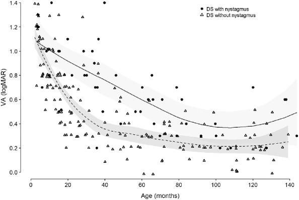
The mean binocular acuity (BVA) of children with DS and DSN is shown for age groups in Figure 1. In both groups, because the older age groups had fewer participants (only five in the case of DSN; see Table 1), the acuity results showing an increase should be treated with caution. BVA ranged from 0.2 and 1.4 logMAR for the children in the DSN group and 0.0 and 1.4 logMAR for the children in the DS group. In the first year of life, the median BVA of the children in the DSN group was 0.1 logMAR poorer than that of children in the DS group and worsened to approximately 0.2 logMAR (two lines) poorer beyond the first year of life. Analysis of covariance (ANCOVA) performed on all data with age as a covariant showed a significant difference (F = 28.42, P < 0.05), with the DSN group having poorer BVA than the DS group.
Figure 1.

Distribution of binocular VA with age of both groups of children with DS, with and without nystagmus. Solid and dashed lines: Best-fit line for BVA of children with DS and nystagmus and DS without nystagmus, respectively. Shaded areas indicate 95% confidence intervals.
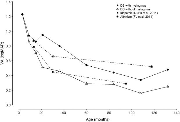
The mean BVA of neurotypical children with idiopathic IN and IN associated with albinism reported by Fu et al.3 was plotted in Figure 2 along with the mean BVA of children with DS with and without nystagmus from the present study. The BVA of children with DSN appears more similar to that of neurotypical children with albinism than neurotypical children with IN. The progression of BVA with age of children in the DS group is not markedly different from that of typically developing children with idiopathic IN.
Figure 2.

Mean binocular VA of children with DS, with and without nystagmus, compared to published data of idiopathic IN and IN associated with albinism (Fu et al.3).
SER
Seven (14%) and 18 (10.2%) of the children in the DSN and DS groups, respectively, were anisometropic. Data of the left eye were used for five children in the DSN group and eight children in DS group; otherwise, RE data were used. SER data of both groups of children were normally distributed (P > 0.05) for all age groups except those 12 to 23.9 months and 10 to 11.9 years in the DSN group and children 12 to 23.9 months in the DS group (P < 0.05 in all cases). SER was between −12.00 D and +7.75 D in the DSN group and between −10.00 D and +10.38 D in the DS group.
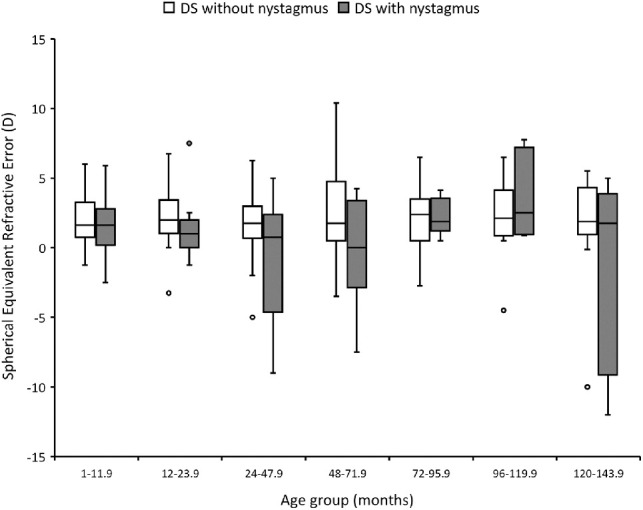
Figure 3 shows the median SER of both groups of children for each age group. Data for children under 1 year were removed because refractive error is likely to change in early infancy.25 Although the data were normally distributed, medians and interquartile ranges were used to enable comparisons of the results with those reported by Al-Bagdady et al.,22 whose data were not normally distributed. Children in the DSN group showed more variability in the SER compared to the DS group. Regression analysis showed no significant change in SER with age for both groups of children (DSN, P = 0.936; DS, P = 0.889). ANCOVA showed a significant difference in the SER between children in the DSN and DS group when age was taken into account (F = 8.30, P < 0.05).
Figure 3.

SER for each age group of children with DSN and DS. Box represents median and interquartile range (IQR). Whiskers represent minimum and maximum values excluding outliers. Circles represent outliers.
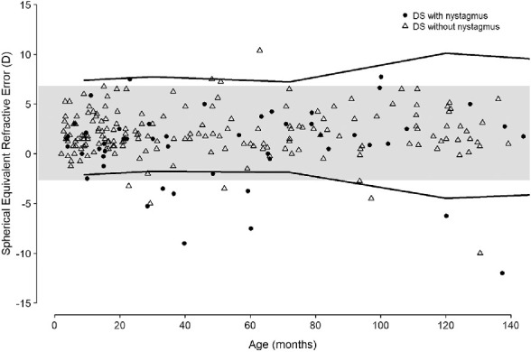
The SER distribution of children in the DSN group was plotted alongside SER data of typically developing children with idiopathic infantile nystagmus (IIN) reported by Healey et al.4 As shown in Figure 4, only eight (16%) of the children in the DSN group fell outside the 95% confidence limits of the SER of children with DS and IIN; seven of these were more myopic and only one more hypermetropic.
Figure 4.

SER for both groups of children with DS, with and without nystagmus, alongside. Shaded area depicts 95% confidence interval of overall mean SER of children with DS without nystagmus. Solid lines depict upper and lower limit of mean SER in children with idiopathic infantile nystagmus at different age groups published by Healy et al4.
Table 2 shows the frequency of type of refractive error for each group of children with DS. Children in the DSN group showed a significantly higher prevalence of myopia (40.7%) than the DS group (11.2%) (χ2 = 13.790, P < 0.05).
Table 2.
Frequency of Refractive Error Type in Each Group of Children With DS
| Frequency of Refractive Error Type, No. (%) | |||
|---|---|---|---|
| Characteristic | Myopia | Hyperopia | Emmetropia |
| DSN (n = 50) | 12 (24.0) | 14 (32.0) | 24 (48.0) |
| DS (n = 176) | 12 (6.82) | 62 (35.23) | 102 (57.95) |
Astigmatism
Mean ± SD astigmatism was −0.76 ± 0.62 DC for the children in the DSN group and −0.74 ± 0.81 DC for the children in the DS group, which were not significantly different (ANCOVA F = 0.16, P = 0.68), with age as a covariant. Twenty-seven (54%) children in the DSN group and 102 (58%) children in the DS group had significant astigmatism, a difference that was also not significant (χ2 = 1.65, P = 0.69). Furthermore, when the children were divided into three age groups, including infancy (up to 23.9 months), early childhood (2–5.9 years), and later childhood (6–11.9 years), the prevalence of significant astigmatism increased (50%–67.3% for DS and 47.1%–73.3% for DSN), but the difference between DS and DSN was not significant at any age (χ2 = 0.48, 1.21, and 0.20 and P = 0.83, 0.27, and 0.65, respectively). Moreover, there was no significant difference in the axis of astigmatism between children with DS and DSN (χ2 = 1.46, P = 0.48). The most common type of astigmatism seen in both groups of children was WTR (DS = 61.76% and DSN = 74.07%), followed by oblique astigmatism (DS = 19.61% and DSN = 18.51%). The average age of children with WTR astigmatism in the DS and DSN groups was 3.49 years and 5.04 years, respectively. The average age of children with oblique astigmatism in both the DS and DSN groups was 6.38 years and 7.28 years, respectively.
Discussion
To our knowledge, this retrospective study is the first to specifically describe the visual and refractive status of children with DSN, who comprise over 20% of our study cohort. The current analysis shows that children with DSN have significantly poorer VA compared to that of children with DS. A recent retrospective analysis of VA data by Zahidi et al.11 confirms that children with DS have poorer corrected acuity compared to typically developing children by approximately 0.2 logMAR. The additional deficit in the median acuity of children with DSN was a further 0.2 logMAR. Felius et al.5 also reported a VA deficit of 0.4 logMAR in children with DSN compared to typical norms and suggested that nystagmus is not the sole cause of poor vision in children with DSN. The current study confirms this, as children with DS and no nystagmus appear to have VA on par with neurotypical children with nystagmus. The difference in VA between neurotypical children and those with DS is not yet fully explained.
Self et al.26 emphasize the importance of a full clinical examination (possibly including electrophysiology) to differentiate between idiopathic IN and nystagmus with an underlying cause. Since children with DS presumably also carry a risk for an underlying cause for their nystagmus, it is clear that a full investigation is called for. Our small sample (nine children) suggests that some ophthalmologists might assume that an additional category of nystagmus exclusive to DS exists, and no other cause of the condition can apply to children with DS.
The VA deficit of children with IN has been associated with the onset of binocular visual deprivation and the change in the nystagmus waveform.27 Typically developing children with IN usually have a triangular waveform during infancy, which transforms into pendular and then to jerk waveforms.28–30 This change in the waveform type results in, or perhaps is a consequence of, changes in the foveation strategy, which has an impact on the visual performance in IN.27 Reinecke et al.28 speculate that children with IN adopt new foveation strategies as they try to focus on the objects that interest them, hence producing the observed change in the nystagmus waveform type. Therefore, the onset of change from pendular to jerk nystagmus is crucial to the visual development of children with nystagmus. Longitudinal data from infancy were available for only four children with DSN, so it is difficult to estimate the timing of the onset of nystagmus for this group of children. Longitudinal eye movement recording data would be needed to determine any changes in the nystagmus waveforms.
One of the limitations of the current study is that VA was measured with current eyeglasses and not necessarily with the children's best correction, as testing acuity while wearing a trial frame can distract the children, perhaps affecting their performance during the test. This would apply, of course, to both groups. Although there could have been some change in refractive error since their last clinical correction, this is likely to be small because the children were seen at regular intervals. The visual acuity of the typically developing children with INS in the study by Fu et al.3 was also measured using “habitual optical correction” as well as with age-appropriate tests. Children with DS have been reported to have poorer VA than the expected norm,6,8–10 despite refractive errors being corrected. None of the reported studies measured acuity with the children's best correction. In the current study, more than 85% of VA in DSN fell below the 95% confidence limits of typically developing children with INS and children in the DS group.
Neurotypical children with IN have been shown to have unconventional refractive development.4 Refractive error of children with DSN differs significantly from that of children with DS who do not have nystagmus over the age of 1 year. Although both groups of children were hypermetropic since early infancy, children with DSN were more likely to be myopic compared to the DS group and showed a larger variability in refractive error. Previous studies of children with DS have shown an association between congenital heart defect and nystagmus,31,32 as well as between congenital heart defect and myopia.31 Therefore, it may not be surprising that children with DSN are more myopic than those who do not have nystagmus.
Astigmatism is common in children with DS21,22 and, in this study, equally common in children with DSN. Weiss et al.33 reported a prevalence of 66% in their small cohort of 18 children with DSN. The pattern of the development of astigmatism in children with DS has been reported to differ significantly from typically developing children with no ocular conditions.21,22 In both DS and DSN groups, significant astigmatism increased with age, as expected. Children with DS present with WTR astigmatism from an early age, which then develops into oblique astigmatism later during their childhood,22 and this pattern was also observed in the group of children with DSN. Weiss et al.33 also recorded WTR and oblique astigmatism in their group of children with DSN (one child's data had no axis recorded). On the other hand, neurotypical children with IIN have been reported to have WTR astigmatism throughout their childhood.23,34 Our analysis showed that nystagmus had no effect on the prevalence or type of astigmatism in children with DS.
Conclusions
The results of this study have shown that children with DSN have poorer VA than children with DS, in a similar manner to neurotypical children with nystagmus having poorer acuity than children without nystagmus. However, it is quite clear that there is a significant baseline deficit in acuity attributable to DS and an additional deficit associated with nystagmus. There was no difference in astigmatism between children with DSN and DS, which contrasts with typically developing children who have a higher prevalence of astigmatism compared to their counterparts without nystagmus. Finally, the findings of this study show that myopia is associated with nystagmus in children with DS.
It is clear that children with DSN have an increased visual impairment. It is essential, therefore, that nystagmus in DS receives the same level of attention as it does among typical children (i.e., with definitive diagnosis, appropriate advice for parents, and targeted educational support for children).
Acknowledgments
The authors thank the past members of the Down's Syndrome Vision Research Unit for their contribution to the data collection: Val Pakeman, Mary Cregg, Ruth Stewart, Mohammed Al-Bagdady, Stephanie Campbell, and Valldeflors Vinuela-Navarro. They also thank Rhod Woodhouse for statistical advice and Paul Artes for his help with coding in R. Most important thanks go to the children and their families for their unfailing support for our studies.
Members of the research group who contributed to the data collection have been funded over the years by the Down's Syndrome Association, Medical Research Council, Mencap City Foundation, National Lotteries Charity Board with Mencap, PPP Healthcare Medical Trust, National Eye Research Centre, College of Optometrists, Action Medical Research and Garfield Weston Foundation, and the Malaysian Government Postgraduate Scholarship (Majlis Amanah Rakyat, MARA).
The views expressed in the submitted article are the authors’ own and not an official position of the institution or funder.
Disclosure: A.A.A. Zahidi, None; L. McIlreavy, None; J.T. Erichsen, None; J.M. Woodhouse, None
References
- 1. Wagner RS, Caputo AR, Reynolds DR.. Nystgmus in Down syndrome. Ophthalmology. 1990; 97(11): 1439–1444. [DOI] [PubMed] [Google Scholar]
- 2. Averbuch-Heller L, Dell'Osso LF, Jacobs JB, Remler BF. Latent and congenital nystagmus in Down syndrome. J Neuroophthalmol. 1999; 19(3): 166–172. [PubMed] [Google Scholar]
- 3. Fu VL, Bilonick RA, Felius J, Hertle RW, Birch EE.. Visual acuity development of children with infantile nystagmus syndrome. Invest Ophthalmol Vis Sci. 2011; 52(3): 1404–1411. [DOI] [PMC free article] [PubMed] [Google Scholar]
- 4. Healey N, McClelland JF, Saunders KJ, Jackson AJ.. Longitudinal study of spherical refractive error in infantile nystagmus syndrome. Ophthalmic Physiol Opt. 2014; 34(3): 369–375. [DOI] [PubMed] [Google Scholar]
- 5. Felius J, Beauchamp CL, Stager DR.. Visual acuity deficits in children with nystagmus and Down syndrome. Am J Ophthalmol. 2014; 157(2): 458–463. [DOI] [PubMed] [Google Scholar]
- 6. Courage ML, Adams RJ, Reyno S, Kwa PG.. Visual acuity in infants and children with Down syndrome. Dev Med Child Neurol. 1994; 36(7): 586–593. [DOI] [PubMed] [Google Scholar]
- 7. Woodhouse JM, Pakeman VH, Saunders KJ, et al.. Visual acuity and accommodation in infants and young children with Down's syndrome. J Intellect Disabil Res. 1996; 40(pt 1): 49–55. [DOI] [PubMed] [Google Scholar]
- 8. John FM, Bromham NR, Woodhouse JM, Candy TR.. Spatial vision deficits in infants and children with Down syndrome. Invest Ophthalmol Vis Sci. 2004; 45(5): 1566–1572. [DOI] [PubMed] [Google Scholar]
- 9. Little J-A, Woodhouse JM, Lauritzen JS, Saunders KJ.. The impact of optical factors on resolution acuity in children with Down syndrome. Invest Ophthalmol Vis Sci. 2007; 48(9): 3995–4001. [DOI] [PubMed] [Google Scholar]
- 10. Tomita K. Visual characteristics of children with Down syndrome. Jpn J Ophthalmol. 2017; 61: 271–279. [DOI] [PubMed] [Google Scholar]
- 11. Zahidi AA, Vinuela-Navarro V, Woodhouse JM.. Different visual development: norms for visual acuity in children with Down's syndrome. Clin Exp Optom. 2018; 101(4): 535–540. [DOI] [PubMed] [Google Scholar]
- 12. Turner S, Sloper P, Cunningham C, Knussen C.. Health problems in children with Down's syndrome. Child Care Health Dev. 1990; 16(2): 83–97. [DOI] [PubMed] [Google Scholar]
- 13. Woodhouse JM, Meades JS, Leat SJ, Saunders KJ.. Reduced accommodation in children with Down syndrome. Invest Ophthalmol Vis Sci. 1993; 34(7): 2382–2387. [PubMed] [Google Scholar]
- 14. Woodhouse JM, Pakeman VH, Cregg M, et al.. Refractive errors in young children with Down syndrome. Optom Vis Sci. 1997; 74(10): 844–851. [DOI] [PubMed] [Google Scholar]
- 15. Haugen OH, Høvding G, Lundström I.. Refractive development in children with Down's syndrome: a population based, longitudinal study. Br J Ophthalmol. 2001; 85(6): 714–719. [DOI] [PMC free article] [PubMed] [Google Scholar]
- 16. McDonald MA, Dobson V, Sebris SL.. The acuity card procedure: A rapid test of infant acuity. Invest Ophthalmol Vis Sci. 1985; 26(8): 1158–1162. [PubMed] [Google Scholar]
- 17. Adoh TO, Woodhouse JM.. The Cardiff acuity test used for measuring visual acuity development in toddlers. Vision Res. 2003; 34(4): 555–560. [DOI] [PubMed] [Google Scholar]
- 18. Kay H. New method of assessing visual acuity with pictures. Br J Ophthalmol. 1983; 62(2): 131–133. [DOI] [PMC free article] [PubMed] [Google Scholar]
- 19. McGraw P V, Winn B.. Glasgow Acuity Cards: a new test for the measurement of letter acuity in children. Ophthalmic Physiol Opt. 1993; 13(4): 400–404. [DOI] [PubMed] [Google Scholar]
- 20. Elliot DB. Clinical Procedures in Primary Eye Care. 4th ed. Philadelphia, PA: Elsevier Saunders; 2014. [Google Scholar]
- 21. Little J-A, Woodhouse JM, Saunders KJ.. Corneal power and astigmatism in Down syndrome. Optom Vis Sci. 2009; 86(6): 748–754. [DOI] [PubMed] [Google Scholar]
- 22. Al-Bagdady M, Murphy PJ, Woodhouse JM.. Development and distribution of refractive error in children with Down's syndrome. Br J Ophthalmol. 2011; 95(8): 1091–1097. [DOI] [PubMed] [Google Scholar]
- 23. Wang J, Wyatt LM, Felius J, et al.. Onset and progression of with-the-rule astigmatism in children with infantile nystagmus syndrome. Invest Ophthalmol Vis Sci. 2010; 51(1): 594–601. [DOI] [PMC free article] [PubMed] [Google Scholar]
- 24. Felius J, Fu VLN, Birch EE, Hertle RW, Jost RM, Subramanian V.. Quantifying nystagmus in infants and young children: relation between foveation and visual acuity deficit. Invest Ophthalmol Vis Sci. 2011; 52(12): 8724–8731. [DOI] [PubMed] [Google Scholar]
- 25. Saunders KJ, Cotlier E, Weinreb R, Saunders KJ.. Early refractive development in humans. Surv Ophthalmol. 1995; 40(3): 207–216. [DOI] [PubMed] [Google Scholar]
- 26. Self JE, Dunn MJ, Erichsen JT, et al.. Management of nystagmus in children: a review of the literature and current practice in UK specialist services. Eye. 2020; 34(9): 1515–1534. [DOI] [PMC free article] [PubMed] [Google Scholar]
- 27. Felius J, Muhanna Z.. Visual deprivation and foveation characteristics both underlie visual acuity deficits in idiopathic infantile nystagmus. Invest Ophthalmol Vis Sci. 2013; 54(5): 3520–3525. [DOI] [PubMed] [Google Scholar]
- 28. Reinecke RD, Guo S, Goldstein HP.. Waveform evolution in infantile nystagmus: an electro-oculo-graphic study of 35 cases. Binocul Vis. 1988; 3(4): 191–202. [Google Scholar]
- 29. Gottlob I. Infantile nystagmus: development documented by eye movement recordings. Invest Ophthalmol Vis Sci. 1997; 38(3): 767–773. [PubMed] [Google Scholar]
- 30. Maldonado VK, Hertle RW.. Clinical and ocular motor analysis of congenital nystagmus in the first six months of life. Invest Ophthalmol Vis Sci. 2001; 42(4): S164–S164. [Google Scholar]
- 31. Bromham NR, Woodhouse JM, Cregg M, Webb E, Fraser WI.. Heart defects and ocular anomalies in children with Down's syndrome. Br J Ophthalmol. 2002; 86(12): 1367–1368. [DOI] [PMC free article] [PubMed] [Google Scholar]
- 32. Kranjc B. Ocular abnormalities and systemic disease in Down syndrome. Strabismus. 2012; 20(2): 74–77. [DOI] [PubMed] [Google Scholar]
- 33. Weiss AH, Kelly JP, Phillips JO.. Infantile nystagmus and abnormalities of conjugate eye movements in Down syndrome. Invest Ophthalmol Vis Sci. 2016; 57(3): 1301–1309. [DOI] [PubMed] [Google Scholar]
- 34. Jethani J, Prakash K, Vijayalakshmi P, Parija S.. Changes in astigmatism in children with congenital nystagmus. Graefes Arch Clin Exp Ophthalmol. 2006; 244(8): 938–943. [DOI] [PubMed] [Google Scholar]


