



Abstract
Background
Brown adipose tissue (BAT) has been primarily researched as a potential target for mitigating obesity. However, the physiological significance of BAT in relation to cachexia remains poorly understood. The objective of this study was to investigate the putative contribution of BAT on different components of energy metabolism in emphysematous chronic obstructive pulmonary disease (COPD) patients.
Methods
Twenty COPD patients (mean ± SD age 62 ± 6, 50% female, median [range] BMI 22.4 [15.1–32.5] kg/m2 and 85% low FFMI) were studied. Basal metabolic rate (BMR) was assessed by ventilated hood, total daily energy expenditure (TDEE) by doubly labelled water and physical activity by triaxial accelerometry. BMR was adjusted for fat‐free mass (FFM) as assessed by deuterium dilution. Analysis of BAT and WAT was conducted in a subset of ten patients and six age‐matched, gender‐matched and BMI‐matched healthy controls. BAT glucose uptake was assessed by means of cold‐stimulated integrated [18F]FDG positron‐emission tomography and magnetic resonance imaging. WAT was collected from subcutaneous abdominal biopsies to analyse metabolic and inflammatory gene expression levels. Lung function was assessed by spirometry and body plethysmography and systemic inflammation by high sensitivity C‐reactive protein.
Results
Mean TDEE was 2209 ± 394 kcal/day, and mean BMR was 1449 ± 214 kcal/day corresponding to 120% of predicted. FFM‐adjusted BMR did not correlate with lung function or C‐reactive protein. Upon cooling, energy expenditure increased, resulting in a non‐shivering thermogenesis of (median [range]) 20.1% [3.3–41.3] in patients and controls. Mean BAT glucose uptake was comparable between COPD and controls (1.5 [0.1–6.2] vs. 1.1 [0.7–3.9]). In addition, no correlation was found between BMR adjusted for FFM and BAT activity or between cold‐induced non‐shivering energy expenditure and BAT activity. Gene expression levels of the brown adipocyte or beige markers were also comparable between the groups. No (serious) adverse events were reported.
Conclusions
Although COPD patients were hypermetabolic at rest, no correlation was found between BMR or TDEE and BAT activity. Furthermore, both BAT activity and gene expression levels of the brown adipocyte or beige markers were comparable between COPD patients and controls.
Keywords: Brown adipose tissue, COPD, Energy metabolism, Cachexia, Muscle wasting
Introduction
Chronic obstructive pulmonary disease (COPD) is an airway and lung disease with persistent airflow obstruction, which is the result of small airway remodelling and loss of elastic recoil. 1 Disturbances in whole body and cellular energy metabolism are common in COPD and contribute to weight loss and muscle wasting in particular in the emphysematous phenotype. 2 , 4
Total daily energy expenditure (TDEE) can be divided in obligatory and facultative thermogenesis. Obligatory thermogenesis consists of basal metabolic rate (BMR) and facultative thermogenesis consists of diet‐induced thermogenesis and activity‐induced energy expenditure (AEE). 5 AEE has been shown to be elevated due to decreased mechanical efficiency 6 , 7 but may also be (relatively) decreased in patients with very low physical activity levels. 3 Diet‐induced thermogenesis was shown to be normal in previous research. 8 , 9 Increased BMR relative to predictive values has been consistently reported in COPD 2 and found more aggravated among weight losing patients 2 , 3 and among those with emphysema. 10 , 11 Hypermetabolic patients do not necessarily exhibit an elevated TDEE. 12 Systemic inflammation has been proposed as cause of elevated BMR in cachexia induced by cancer 13 or COPD. 14 Another contributing factor to hypermetabolism in COPD might be the workload of breathing 15 due to lung hyperinflation. Nevertheless, we recently showed that decreasing hyperinflation with 25% by endoscopic lung volume reduction in patients with emphysema did not decrease BMR (unpublished own results, submitted).
Activation of brown adipose tissue (BAT) or browning of white adipose tissue (WAT) has been proposed as putative trigger for hypermetabolism in cachexia but to date little evidence from human research is available. 16 , 17 BAT tissue is characterized by abundance of mitochondria and by heat production via the process of proton leakage over the mitochondrial membrane mediated by uncoupling protein 1. 18 Compared with BAT‐negative healthy young lean subjects, those with BAT had a higher cold‐induced energy expenditure, with an average difference of 370 kcal/day. 19 In healthy young, lean men, cold induction increased energy expenditure by, on average, 17%. 20 This increase in energy expenditure upon cold in the absence of shivering (i.e. non‐shivering thermogenesis) implies that BAT thermogenesis may be a significant component of facultative thermogenesis and thereby plays a role in regulation of body weight as suggested by an inverse association between cold‐induced BAT activity and body mass index in adults. 21 , 22 , 23
Brown adipose tissue has been primarily researched as a potential target for mitigating obesity. 24 Yet few research attempts have been made to explore the role of BAT in relation to cachexia as observed in patients with COPD, and no clinical studies are available. There is some indirect evidence pointing towards a possible role for BAT as driver of hypermetabolism in the emphysematous COPD subtype. Patients with advanced emphysema have high plasma levels of norepinephrine at rest, 25 which is a potential browning factor. 26 Furthermore, whole body glucose production 27 and skeletal muscle glycolysis 28 , 29 are increased in COPD patients, which result in increased plasma lactate levels, even at rest. 30 Lactate in turn is able to control expression of uncoupling protein 1 (UCP1), which also promotes browning. 31 We therefore hypothesized that BAT or browning of WAT contributes to energy expenditure in emphysematous COPD patients. Because limited studies are available regarding TDEE and the different components in mixed COPD phenotypes, we first measured energy expenditure in emphysematous COPD patients and related its components to airflow obstruction and hyperinflation. We then conducted a controlled clinical experiment in a subgroup to assess presence of BAT by cold‐induced BAT activity and BAT and beige gene expression markers in WAT.
Methods
Participants
Whole body energy metabolism and its components were assessed in 20 Caucasian COPD patients with emphysema. For diagnosis of COPD, patients underwent spirometry and body plethysmography. The contribution of BAT and WAT was studied in a subgroup of 10 patients and a control group, matched for ageing, male gender and adiposity, because these factors are known to negatively correlate with BAT activity. 23 Furthermore, BAT volumes may differ between ethnicities. 32 In addition, because cigarette smoke is acknowledged as the most important risk factor for COPD and might stimulate BAT activity 33 and control subjects may not be diagnosed with COPD yet, non‐smoking controls were included.
Patients were recruited from Maastricht University Medical Centre (MUMC+), and the healthy controls were recruited via advertisements in local newspapers. The Ethics Committee of MUMC+ approved the study protocol, and all participants provided written informed consent. Procedures were conducted according to the principles of the Declaration of Helsinki. The study was registered at ClinicalTrial.gov (NCT02500004).
Exclusion criteria for the COPD as well as the healthy controls were diabetes mellitus, severe clotting disorder, active malignancy, claustrophobia, long‐term oxygen therapy, history of radiotherapeutic radiation in the neck and/or upper chest, cervical or thoracic sympathectomy and the presence of magnetic resonance imaging (MRI) contraindications, such as pacemaker, cochlear implant and vascular clips. In addition, subjects using medication that influences the sympathetic nerve system, including ß‐blockers, α‐blockers, central antihypertensive drugs, certain anti‐depression drugs (MAO inhibitors and tricyclic anti‐depressives), reserpine, cocaine, calcium channel blockers and certain tranquillizers (fenothiazines) were also not eligible. Furthermore, patients with pancreatic cancer were included, with similar exclusion criteria as mentioned above.
The original intention was to compare the COPD patients not only with healthy controls but also with matched lung cancer and pancreatic cancer patients. However, the BAT protocol was too strenuous, regarding eligibility criteria as well as methodology used, for most of the cachectic cancer patients. We were able to measure only two pancreatic cancer patients and no lung cancer patients before study closure. In addition, one COPD patient suffered from a severe panic attack immediately before initiation of the scan. Due to this panic attack, we were unable to scan this patient.
Study design
The overall study design is shown in Figure 1. All participants completed the 2 week study period focusing on whole body energy metabolism. A subset underwent the protocol focusing on the contribution of BAT and subcutaneous adipose tissue to energy metabolism. This protocol included an individualized cooling protocol, 34 [18F]FDG positron‐emission tomography (PET) and magnetic resonance imaging (MRI) scanning in order to measure BAT activity and a subcutaneous fat biopsy obtained after an overnight fast.
Figure 1.
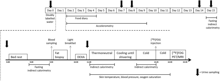
Schematic illustration of the study protocol. MRI, magnetic resonance imaging; PET, positron‐emission tomography.
Resting metabolic rate
Basal metabolic rate was measured by indirect calorimetry using a ventilated hood (EZCAL; Maastricht Instruments, Maastricht, the Netherlands). Patients were in a fasting state for at least 10 h and were requested to abstain from heavy exercise, inhalation medication and smoking for 24 h. Prior to the measurement, the subjects had a period of 30 min bed rest during which they were lying on bed in supine position. After stabilization, BMR was recorded during a period of 30 min. BMR was assessed during bed rest at room temperature. BMR was calculated from oxygen consumption (VO2) and carbon dioxide (VCO2) production using the abbreviated Weir formula. 35 BMR was also predicted using the equation from Slinde et al. 4
Doubly labelled water
Total daily energy expenditure was determined by the doubly labelled water technique over 2 weeks according to the Maastricht protocol. 36 In the evening, prior to dosing, a urine sample was collected for determination of background isotope enrichment. Each patient received a weighted oral dose of water labelled with ~ 7% deuterium and 10% oxygen‐18. The given dose was calculated based on the subjects' total body water, which was estimated based on body mass index, age and gender. 37 Subjects received a dose of 2.4 g/L total body water resulting in an initial excess enrichment of approximately 150 ppm for deuterium and 200 ppm for oxygen‐18. The basic principle of DLW is that after equilibration with total body water, deuterium and oxygen‐18 are eliminated from the body at a different rate. Deuterium is eliminated from the body as water, whereas oxygen‐18 is eliminated as water and carbon dioxide. The difference in elimination rates is hence a measure of CO2 production. After ingestion of doubly labelled water, urine samples were collected at standardized time points (in the evening of Days 1, 7 & 14 and in the morning of Days 8 and 15). CO2 production was calculated by the linear regression from the difference between elimination constants of deuterium and oxygen‐18. TDEE was calculated from CO2 production assuming an RQ of 0.85, corresponding to the RQ of an average Western diet.
Energy expenditure for activities was calculated by (0.9*TDEE)‐BMR, assuming a diet‐induced thermogenesis of 10% of TDEE. 5
Body composition
Body height was determined to the nearest 0.5 cm while subjects were standing barefoot. Weight was assessed with a beam scale to the nearest 0.1 kg while subjects were standing barefoot and in light clothing. FFM was calculated from total body water assessment using the deuterium dilution technique assuming a hydration fraction of FFM of 73%. 36
Physical activity
Actigraph GTX3 accelerometers (Actigraph, Pensacola, FL, USA) were used to assess physical activity. This activity monitor has been validated against activity related energy expenditure measured by doubly labelled water in patients with different stages of COPD. 38 The triaxial accelerometers were attached to the lower back with an elastic belt and worn for seven consecutive days. Subjects were instructed to wear the accelerometer during the time they were not asleep, except when showering or bathing. Only days with ≥8 h of wear time were accepted as valid days.
Dietary intake
Energy intake was recorded by a food diary for two week days and one weekend day to judge if subjects were in a stable energy balance.
Individualized cooling and positron‐emission tomography/magnetic resonance imaging scanning
For individualized cooling, subjects were wrapped in a water‐perfused suit. During the cooling procedure, skin temperature, blood pressure, heart rate and oxygen saturation were monitored. Skin temperatures were measured continually at 14 positions by means of iButtons (type DS1291H, Dallas Maxim Semiconductor Corp., USA) 39 and on three additional positions (supraclavicular, underarm and fingertip). Blood pressure and heart rate were measured via an automated blood pressure monitor (Omron Healthcare Inc, Field Court Lake Forest, USA), and oxygen saturation was measured via pulse oximetry (Nellcor Puritan Bennett NPB‐40, Pleasanton, CA, USA).
For the first 45 min, subjects were kept at thermoneutral conditions with water temperature of the water‐perfused suit at 34°C. Following this thermoneutral condition, a gradual step‐wise decrease in temperature followed (water temperature was lowered with 4°C every 15 min) until shivering occurred. After the onset of shivering, subjects were warmed up for 5 min after which the temperature was set slightly above shivering level for 30 min. At thermoneutral condition and during cold exposure, indirect calorimetry was performed to measure energy expenditure. Subsequently, on average, 150 MBq of 18F‐FDG was injected intravenously while mild cold stimulation remained for 45 min after injection. At 60 min p.i., a hybrid PET/MRI scan (Siemens Healthcare GmbH, Erlangen, Germany, software version VB20P) was acquired.
Positron‐emission tomography/magnetic resonance imaging analysis
Positron‐emission tomography/magnetic resonance imaging scans were analysed using the PMOD software (Version 3.7, PMOD Technologies, Zurich, Switzerland). For assessment of BAT activity, first volumes of interest (VOIs) were drawn, which totally encompassed supraclavicular adipose tissue. Inside this selected VOI of supraclavicular adipose tissue, a sphere with radius 6.2 mm was deposited on the location where the average standardized uptake value (SUV) value was maximal. Additionally, VOIs were placed in the liver, several skeletal muscle groups (biceps and triceps brachii, deltoid, and erector spinae), subcutaneous adipose tissue and visceral adipose tissue. For each VOI and sphere, the measured activity concentration was corrected for radioactive decay, total administered activity and body weight, resulting in the mean SUV, SUVmean.
Blood analysis
Venous blood was obtained after an overnight fast for analysis of C‐reactive protein (CRP), glucose, insulin, thyroid stimulating hormone, free fatty acids, cholesterol, triglycerides, high‐density and low‐density lipoprotein. Homeostatic model assessment to estimate insulin resistance was calculated. Analysis was performed in the central diagnostic laboratory of Maastricht University Medical Centre.
Fat biopsy
A subcutaneous fat biopsy was obtained para‐umbilically during fasting conditions through needle biopsy before initiation of cooling procedure. This biopsy was snap‐frozen in liquid nitrogen and stored at −80°C until analysing gene expression by qPCR. Refer to the supporting information for a detailed description of the analysis and list of primers (Supporting Information, Table S1).
Statistics
Descriptive statistics of demographic and clinical variables were obtained. Means (±SD) are provided for continuous normally distributed variables, median [range] for continuous not‐normally distributed variables and percentages are shown for categorical variables. Between group differences were tested with an independent t‐test, Mann–Whitney U test or χ 2 test. All analyses were performed using SPSS statistical software (SPSS Statistics for Windows, Version 24.0, IBM, Armonk, NY). Results with two‐sided P values (<0.05) were considered statistically significant.
Results
Baseline characteristics of participants
Ten male and ten female COPD patients with emphysema were enrolled. Baseline characteristics are depicted in Table 1. The COPD population was characterized by moderate to severe airflow obstruction and low diffusion capacity (FEV1: 47 ± 20% of predicted and DLCO: 38 ± 9% predicted). Median body mass index (BMI) (23.4 [15.1–35.4]) was within the normal range but median [range] FFM index was below normal (FFMI male patients 15.7 kg/m2 [14.2–18.2], female patients 13.5 kg/m2 [12.7–17.6]), and 85% was depleted according to current definition (FFMI ≤ 17 kg/m2 for male patients or ≤15 kg/m2 for female patients 40 ).
Table 1.
Baseline characteristics of COPD patients (N = 20) and healthy controls (N = 6)
| COPD | Controls | |
|---|---|---|
| General | ||
| Male/Female, N | 10/10 | 4/2 |
| Age (years) | 62 ± 6 | 60 ± 6 |
| Pack years | 36 ± 17 | 4 ± 8 |
| Smoking status, N | ||
| Never | 0 | 4 |
| Current | 4 | 0 |
| Former | 16 | 2 |
| Body composition | ||
| Weight (kg) | 67 ± 15 | 70.8 ± 3.8 |
| BMI (kg/m2) | 22.4 [15.1–32.5] | 23.1 [21.2–30.8] |
| FFM (kg) | 44.7 [31.7–57.4] | 50.5 [44.6–63.8] |
| FFMI (kg/m2) | ||
| Male | 15.7 [14.2–18.2] | 16.9 [15.3–19.7] |
| Female | 13.5 [12.7–17.6] | 18.0 [16.2–19.8] |
| Lung function | ||
| FEV1, % predicted | 36.0 ± 15.4 | Not applicable |
| FVC, % predicted | 82.2 ± 18.3 | Not applicable |
| FEV1/FVC | 39.2 ± 15.4 | Not applicable |
| RV, % predicted | 209.7 ± 61.2 | Not applicable |
| TLC, % predicted | 128.7 ± 23.7 | Not applicable |
| RV/TLC | 1.6 ± 0.3 | Not applicable |
| DLCO, % predicted | 38.0 ± 9.4 | Not applicable |
BMI, body mass index; COPD, chronic obstructive pulmonary disease; DLCO, diffusion capacity of lung for carbon monoxide; FEV1, forced expiratory volume in 1 s; FFM, fat‐free mass; FFMI, fat‐free mass index; FVC, forced vital capacity; RV, residual volume; TLC, total lung capacity.
Data are represented as mean ± SD or median [range].
A subset of ten COPD patients (five male and five female patients) and six controls (BMI 23.1 [21.2–30.8] kg/m2, FFMI male patients 16.9 kg/m2 [15.33–19.7] and female patients 18.0 kg/m2 [16.20–19.8]) underwent individualized cooling and PET/MRI scanning in order to measure BAT activity. Insulin levels and fasting glucose levels were comparable between COPD patients and controls. Groups were comparable regarding thyroid function and lipid profile. CRP levels on average were low but tended to be higher in the COPD patients (2.5 [2.0–21.0] vs. 0.0 [0.0–3.0] mg/L) (Table S2).
Participants are hypermetabolic at rest
Mean TDEE was 2209 ± 394 kcal/day in the COPD patients and 2631 ± 265 kcal/day in healthy controls (P = 0.022) (Table 2, Figure 2). TDEE was not significantly different from dietary intake. AEE amounted to 538 ± 258 kcal/day (23% of TDEE) in COPD and 812 ± 255 (32% of TDEE) in healthy controls, indicating a low physical activity level in the COPD group. Indeed, median (range) steps per day were 3097 [468–8792] and 8519 [6517–13 518] for respectively COPD patients and healthy controls. Patients compared with controls spend a larger part of the day in sedentary state (70.2% of the wear time [56.5–90.6] vs. 51.3% of wear time [41.7–66.8]) (Figure 2). TDEE was highly correlated to AEE (r = 0.80, P < 0.001).
Table 2.
Components of energy balance of healthy controls (N = 6) and COPD patients (N = 20)
| COPD (N = 20) | Controls (N = 6) | P value | |
|---|---|---|---|
| TDEE (kcal/day) | 2209 ± 394 | 2631 ± 265 | 0.022 |
| BMR measured (kcal/day) | 1449 ± 214 | 1555 ± 109 | 0.260 |
| BMR measured corrected for FFM (kcal/day/kg) | 34 ± 3 | 30 ± 4 | 0.053 |
| BMR predicted (kcal/day) | 1216 ± 142 | 1415 ± 115 | 0.004 |
| AEE (kcal/day) | 538 ± 258 | 812 ± 255 | 0.031 |
AEE, activity‐induced energy expenditure; BMR, basal metabolic rate; TDEE, total daily energy expenditure.
Data are represented as mean ± SD.
Figure 2.
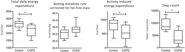
Components of energy balance of healthy controls (N = 6) and COPD patients (N = 20). COPD, chronic obstructive pulmonary disease.
In COPD, the mean BMR of 1449 ± 214 kcal/day corresponded to 120% of predicted. Seventeen patients were characterized as hypermetabolic based on measured BMR > 110% of predicted. No significant correlations could be found between adjusted BMR and FEV1% predicted (r = −0.22, P = 0.366) and between adjusted BMR and RV % predicted (r = 0.04, P = 0.888). Furthermore, adjusted BMR was not correlated to CRP (r = −0.18, P = 0.621).
Hypermetabolism is not associated with brown adipose tissue activation
Upon cooling in both COPD patients and controls, mean skin temperature decreased from 31.9 ± 2.3 to 27.6 ± 3.3°C and supraclavicular skin temperature decreased from 35.3 ± 0.9 to 30.4 ± 5.2°C. The cooling protocol increased energy expenditure (4.5 [3.1–5.0] vs. 5.1 [4.1–7.0] kJ/min) (P < 0.001), resulting in an increment of average non‐shivering thermogenesis of 20.1% (3.3–41.3). In both COPD and healthy controls, this increase was significant (1516 [1041–1873] vs. 1644 [1376–2187] kcal/day; P < 0.001 and 1564 [1387–1685] vs. 1822 [1686–2380] kcal/day; P = 0.013).
Blood pressure and heart rate increased upon cold exposure, while oxygen saturation remained unchanged. This increment in blood pressure and heart rate was not different between groups, indicating a comparable cold stress reaction.
Mean SUV of the region of BAT was comparable between COPD and controls (1.5 g/mL [0.1–6.2] vs. 1.1 g/mL [0.7–3.9]). Furthermore, cold‐induced [18F]FDG uptake in skeletal muscle, subcutaneous adipose tissue and visceral adipose tissue were neither different (Figure 3). In addition, no correlations were found between FFM‐adjusted BMR and BAT activity (r = −0.06, P = 0.881) and between non‐shivering thermogenesis and SUV uptake in skeletal muscles (r = 0.011, P = 0.970). Additionally, no significant relation was found between non‐shivering thermogenesis and BAT activity (r = −0.044, P = 0.877).
Figure 3.
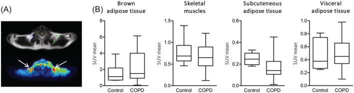
(A) Area of brown adipose tissue on MRI and PET. (B) Mean uptake of [18F]FDG in brown adipose tissue and other tissues during cold exposure. COPD, chronic obstructive pulmonary disease; SUV, standardized uptake value.
Gene expression levels of brown adipose tissue or beige markers in white fat are not upregulated
In subcutaneous adipose tissue, UCP1 and ZIC1 mRNA, which are considered as an important BAT and beige markers, 41 were both undetectable in most subjects. Furthermore, we did not detect differences in mRNA expression of the brown adipocyte marker Cidea or beige markers TMEM26, SHOX2, TNFRSF9 and CD137 between COPD patients and controls (Figure 4).
Figure 4.
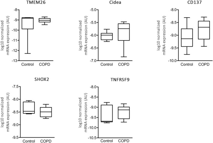
Adipose tissue gene expression levels of brown adipose tissue and beige markers. AU, arbitrary units; COPD, chronic obstructive pulmonary disease.
Hypoxia marker GLUT‐1 mRNA expression level was higher in COPD compared with controls and HIF1 alpha tended to be more expressed in COPD than in control subjects. Gene expression levels of markers of inflammation, macrophages, glycolysis, lipolysis and adipokines were not differentially expressed (Table S1).
Role of brown adipose tissue in cachectic cancer patients: a case report
A 61‐year‐old male patient (BMI 23.9 kg/m2) and a 70‐year‐old female patient (BMI 23.8 kg/m2) diagnosed with pancreatic cancer were both enrolled in one arm of the current study that was closed due to recruitment difficulties (refer to Methods section for explanation). The male patient underwent a pancreaticoduodenectomy, and because of a relapse, he started with chemotherapy which comprised oxaliplatin, folinezuur, irinotecan and fluorouracil. The female patient was diagnosed with pancreatic cancer with pulmonary and lymph node metastasis, and she was treated with gemcitabine. Both were cachectic at time of enrolment with a weight loss of more than 5% of body weight in the previous 6 months, and both were hypermetabolic at rest (mean BMR was 120% of predicted). Despite a cold‐induced increased RMR from 4.5 to 5.3 kJ/min, no BAT activity was found. In line with the results from COPD patients and controls, no up‐regulation of BAT and beige markers were found in subcutaneous fat.
Discussion
Activation of BAT or browning of WAT has been proposed as putative trigger for hypermetabolism and cachexia in wasting disorders including COPD. This is the first study that investigated the contribution of BAT on hypermetabolism in patients with emphysema in comparison with appropriately matched healthy controls. In contrast to our hypothesis, emphysematous patients did not exhibit enhanced BAT activity or altered gene expression of BAT markers in WAT compared with age‐matched, gender‐matched and BMI‐matched healthy non‐smoking controls.
Consistent with previous publications, 2 , 10 we found that BMR was elevated among those with COPD. Although, upon cooling energy expenditure increased with an average non‐shivering thermogenesis of 20.1 [3.3–41.3] %, no relation between BMR and BAT activity was found. This is in line with some, 24 , 42 , 43 but not all, 44 , 45 previous studies using healthy young subjects. Absence of a relation between BMR and BAT activity and between non‐shivering thermogenesis and BAT activity could be due to the low overall BAT activity in the current study population (also certainly related in part to the age). The SUV value of BAT was 1.5 [0.1–6.2], which was considerably lower than reported by others 19 , 46 , 47 who found SUV values in the range of 2–18 among 20‐ to 30‐year‐old healthy study subjects. The relatively low metabolic rate of BAT depots may not be entirely unexpected, as BAT is inversely correlated with age, and BAT is barely detected in healthy subjects above 60 years old. 48 Nevertheless, one autopsy study 49 and one retrospective imaging study 50 reported the presence of BAT activity among elderly with cancer, suggesting that independent of age, disease might be a trigger for BAT activity. However, in those studies, 49 , 50 no data are presented considering the stage of cancer. Furthermore, regarding the available imaging study, the results might be biased by the retrospective design, which did not allow to control environmental temperatures. In male patients, 2.8% of scans demonstrated BAT activity compared with 7.2% of scans in female patients. 50
Although, we found no differences in CRP levels between COPD patients and healthy controls in this study, previous studies showed enhanced systemic inflammation using other sensitive markers in hypermetabolic COPD patients, 51 , 52 which has been proposed as BAT activator. 16 , 53 Furthermore, previous studies 54 , 55 , 56 repeatedly showed an elevated whole body protein turnover in COPD patients, and we also showed in muscle biopsies 57 , 58 elevated muscle protein turnover signalling, which are energy‐consuming processes.
In the studied patients with clinically stable COPD, systemic inflammation assessed by CRP was low and not associated with BMR. Additionally, no altered gene expression of inflammation or macrophage markers was observed compared with healthy controls. In this study also, two pancreatic cancer patients were enrolled. In line with the results in COPD patients, no BAT activity was found.
Interestingly, despite that no BAT activity was detected, the non‐shivering thermogenesis was relatively high in the whole study cohort. Non‐shivering thermogenesis reported in BAT‐negative healthy young subjects was on average 4% compared with 30% in BAT positive subjects. 19 Others reported average non‐shivering thermogenesis of 12% in BAT positive subjects, 45 , 59 , 60 which is remarkably lower compared with our results. No correlation between non‐shivering thermogenesis and SUV of skeletal muscles was found, arguing that the elevated energy expenditure upon cold could not be attributed to involuntary muscle contractions. Furthermore, some studies indicate a role of insulin resistance that point towards either impaired glucose uptake by BAT 60 or reduced BAT activity. 61 Participants in the current cohort were not diagnosed with insulin resistance and were not using antidiabetic medicines. Nevertheless, insulin sensitivity was not measured in the current study. Another explanation for the relatively high non‐shivering thermogenesis might be that there is an effect of diet‐induced energy expenditure. Three hours before cold‐induced energy expenditure was assessed, participants had a light breakfast. In general, after 3 h, most of the thermic effect of a meal dissolved. 62 Although individual differences might exist, this cannot fully explain the high non‐shivering thermogenesis.
This study is not without limitations. The major limitation in this study was the smaller number of enrolled subjects than intended for multiple group comparison (10 subjects per subgroup, in total 50 subjects). Accrual of COPD patients was hampered by the exclusion of subjects using medication that influences the sympathetic nerve system, including ß‐blockers and calcium channel blockers. Partly due to shared risk factors such as smoking, cardiovascular diseases commonly coincide COPD. 63 Therefore, ß‐blockers and calcium channel blockers are widely used by COPD patients. Another factor contributing to low accrual was the use of MRI instead of computed tomography, because a significant part of the COPD patients who were approached to participate suffered from claustrophobia. This might not be totally unexpected because the patients already suffered from dyspnoea, which might be reinforced by MRI scanning. In addition, it appeared impossible to include lung cancer patients, and only two pancreatic cancer patients were enrolled because the extensive study methodologies turned out to be too severe in this critically ill population. 64 , 65 , 66 Furthermore, subcutaneous fat biopsies were obtained para‐umbilically, which is not the primary site of brown adipose tissue. This might partially explain that no brown fat markers were found in our biopsies; nevertheless, in line, no FDG uptake was found in the neck region, supporting our evidence that BAT was not activated in this population.
Nevertheless, the consistency of our findings in the hypermetabolic COPD patients and confirmed in the two cancer cases do not support a role for BAT activation as trigger of disease‐induced hypermetabolism and cachexia.
Conflict of interest
Dr Wierts declared ‘Since 2019 an agreement between GE and the MUMC+ is in place to facilitate site visits for GE Healthcare for PET/CT and SPECT/CT systems’. All other authors declare that they have no relevant conflict of interest.
Funding
The study was funded by MUMC 2020 project “Cachexia”.
Supporting information
Data S1. Supporting information
Acknowledgements
We thank Boudewijn Brans for his contribution to writing of the ethics protocol. The authors certify that they comply with the ethical guidelines for authorship and publishing of the Journal of Cachexia, Sarcopenia and Muscle. 67
Sanders K. J. C., Wierts R., van Marken Lichtenbelt W. D., de Vos‐Geelen J., Plasqui G., Kelders M. C. J. M., Schrauwen‐Hinderling V. B., Bucerius J., Dingemans A.‐M. C., Mottaghy F. M., and Schols A. M. W. J. (2022) Brown adipose tissue activation is not related to hypermetabolism in emphysematous chronic obstructive pulmonary disease patients, Journal of Cachexia, Sarcopenia and Muscle, 13, 1329–1338, 10.1002/jcsm.12881
References
- 1. Vogelmeier CF, Criner GJ, Martinez FJ, Anzueto A, Barnes PJ, Bourbeau J, et al. Global strategy for the diagnosis, management, and prevention of chronic obstructive lung disease 2017 report. GOLD executive summary. Am J Respir Crit Care Med 2017;195:557–582. [DOI] [PubMed] [Google Scholar]
- 2. Schols AM, Soeters PB, Mostert R, Saris WH, Wouters EF. Energy balance in chronic obstructive pulmonary disease. Am Rev Respir Dis 1991;143:1248–1252. [DOI] [PubMed] [Google Scholar]
- 3. Hugli O, Schutz Y, Fitting JW. The daily energy expenditure in stable chronic obstructive pulmonary disease. Am J Respir Crit Care Med 1996;153:294–300. [DOI] [PubMed] [Google Scholar]
- 4. Slinde F, Ellegard L, Gronberg AM, Larsson S, Rossander‐Hulthen L. Total energy expenditure in underweight patients with severe chronic obstructive pulmonary disease living at home. Clin Nutr 2003;22:159–165. [DOI] [PubMed] [Google Scholar]
- 5. van Marken Lichtenbelt WD, Schrauwen P. Implications of nonshivering thermogenesis for energy balance regulation in humans. Am J Physiol Regul Integr Comp Physiol 2011;301:R285–R296. [DOI] [PubMed] [Google Scholar]
- 6. Baarends EM, Schols AM, Akkermans MA, Wouters EF. Decreased mechanical efficiency in clinically stable patients with COPD. Thorax 1997;52:981–986. [DOI] [PMC free article] [PubMed] [Google Scholar]
- 7. Baarends EM, Schols AM, Pannemans DL, Westerterp KR, Wouters EF. Total free living energy expenditure in patients with severe chronic obstructive pulmonary disease. Am J Respir Crit Care Med 1997;155:549–554. [DOI] [PubMed] [Google Scholar]
- 8. Hugli O, Frascarolo P, Schutz Y, Jequier E, Leuenberger P, Fitting JW. Diet‐induced thermogenesis in chronic obstructive pulmonary disease. Am Rev Respir Dis 1993;148:1479–1483. [DOI] [PubMed] [Google Scholar]
- 9. Dore MF, Laaban JP, Orvoen‐Frija E, Kouchakji B, Joubert M, Rochemaure J. Role of the thermic effect of food in malnutrition of patients with chronic obstructive pulmonary disease. Am J Respir Crit Care Med 1997;155:1535–1540. [DOI] [PubMed] [Google Scholar]
- 10. Cohen RI, Marzouk K, Berkoski P, O'Donnell CP, Polotsky VY, Scharf SM. Body composition and resting energy expenditure in clinically stable, non‐weight‐losing patients with severe emphysema. Chest 2003;124:1365–1372. [DOI] [PubMed] [Google Scholar]
- 11. Green JH, Muers MF. The thermic effect of food in underweight patients with emphysematous chronic obstructive pulmonary disease. Eur Respir J 1991;4:813–819. [PubMed] [Google Scholar]
- 12. Baarends EM, Schols AM, Westerterp KR, Wouters EF. Total daily energy expenditure relative to resting energy expenditure in clinically stable patients with COPD. Thorax 1997;52:780–785. [DOI] [PMC free article] [PubMed] [Google Scholar]
- 13. Straub RH, Cutolo M, Buttgereit F, Pongratz G. Energy regulation and neuroendocrine‐immune control in chronic inflammatory diseases. J Intern Med 2010;267:543–560. [DOI] [PubMed] [Google Scholar]
- 14. Broekhuizen R, Wouters EF, Creutzberg EC, Schols AM. Raised CRP levels mark metabolic and functional impairment in advanced COPD. Thorax 2006;61:17–22. [DOI] [PMC free article] [PubMed] [Google Scholar]
- 15. Donahoe M, Rogers RM, Wilson DO, Pennock BE. Oxygen consumption of the respiratory muscles in normal and in malnourished patients with chronic obstructive pulmonary disease. Am Rev Respir Dis 1989;140:385–391. [DOI] [PubMed] [Google Scholar]
- 16. Petruzzelli M, Schweiger M, Schreiber R, Campos‐Olivas R, Tsoli M, Allen J, et al. A switch from white to brown fat increases energy expenditure in cancer‐associated cachexia. Cell Metab 2014;20:433–447. [DOI] [PubMed] [Google Scholar]
- 17. de Vos‐Geelen J, Fearon KC, Schols AM. The energy balance in cancer cachexia revisited. Curr Opin Clin Nutr Metab Care 2014;17:509–514. [DOI] [PubMed] [Google Scholar]
- 18. Heaton GM, Wagenvoord RJ, Kemp A Jr, Nicholls DG. Brown‐adipose‐tissue mitochondria: photoaffinity labelling of the regulatory site of energy dissipation. Eur J Biochem 1978;82:515–521. [DOI] [PubMed] [Google Scholar]
- 19. Yoneshiro T, Aita S, Matsushita M, Kameya T, Nakada K, Kawai Y, et al. Brown adipose tissue, whole‐body energy expenditure, and thermogenesis in healthy adult men. Obesity (Silver Spring) 2011;19:13–16. [DOI] [PubMed] [Google Scholar]
- 20. Claessens‐van Ooijen AM, Westerterp KR, Wouters L, Schoffelen PF, van Steenhoven AA, van Marken Lichtenbelt WD. Heat production and body temperature during cooling and rewarming in overweight and lean men. Obesity (Silver Spring) 2006;14:1914–1920. [DOI] [PubMed] [Google Scholar]
- 21. Cypess AM, Lehman S, Williams G, Tal I, Rodman D, Goldfine AB, et al. Identification and importance of brown adipose tissue in adult humans. N Engl J Med 2009;360:1509–1517. [DOI] [PMC free article] [PubMed] [Google Scholar]
- 22. Wang Q, Zhang M, Xu M, Gu W, Xi Y, Qi L, et al. Brown adipose tissue activation is inversely related to central obesity and metabolic parameters in adult human. PLoS ONE 2015;10:e0123795. [DOI] [PMC free article] [PubMed] [Google Scholar]
- 23. Lee P, Greenfield JR, Ho KK, Fulham MJ. A critical appraisal of the prevalence and metabolic significance of brown adipose tissue in adult humans. Am J Physiol Endocrinol Metab 2010;299:E601–E606. [DOI] [PubMed] [Google Scholar]
- 24. van Marken Lichtenbelt WD, Vanhommerig JW, Smulders NM, Drossaerts JM, Kemerink GJ, Bouvy ND, et al. Cold‐activated brown adipose tissue in healthy men. N Engl J Med 2009;360:1500–1508. [DOI] [PubMed] [Google Scholar]
- 25. Hofford JM, Milakofsky L, Vogel WH, Sacher RS, Savage GJ, Pell S. The nutritional status in advanced emphysema associated with chronic bronchitis. A study of amino acid and catecholamine levels. Am Rev Respir Dis 1990;141:902–908. [DOI] [PubMed] [Google Scholar]
- 26. Atgie C, D'Allaire F, Bukowiecki LJ. Role of beta1‐ and beta3‐adrenoceptors in the regulation of lipolysis and thermogenesis in rat brown adipocytes. Am J Physiol 1997;273:C1136–C1142. [DOI] [PubMed] [Google Scholar]
- 27. Franssen FM, Sauerwein HP, Ackermans MT, Rutten EP, Wouters EF, Schols AM. Increased postabsorptive and exercise‐induced whole‐body glucose production in patients with chronic obstructive pulmonary disease. Metabolism 2011;60:957–964. [DOI] [PubMed] [Google Scholar]
- 28. Green HJ, Burnett ME, D'Arsigny CL, O'Donnell DE, Ouyang J, Webb KA. Altered metabolic and transporter characteristics of vastus lateralis in chronic obstructive pulmonary disease. J Appl Physiol (1985) 2008;105:879–886. [DOI] [PubMed] [Google Scholar]
- 29. Green HJ, Bombardier E, Burnett M, Iqbal S, D'Arsigny CL, O'Donnell DE, et al. Organization of metabolic pathways in vastus lateralis of patients with chronic obstructive pulmonary disease. Am J Physiol Regul Integr Comp Physiol 2008;295:R935–R941. [DOI] [PubMed] [Google Scholar]
- 30. Engelen MP, Schols AM, Does JD, Gosker HR, Deutz NE, Wouters EF. Exercise‐induced lactate increase in relation to muscle substrates in patients with chronic obstructive pulmonary disease. Am J Respir Crit Care Med 2000;162:1697–1704. [DOI] [PubMed] [Google Scholar]
- 31. Carriere A, Jeanson Y, Berger‐Muller S, Andre M, Chenouard V, Arnaud E, et al. Browning of white adipose cells by intermediate metabolites: an adaptive mechanism to alleviate redox pressure. Diabetes 2014;63:3253–3265. [DOI] [PubMed] [Google Scholar]
- 32. Bakker LE, Boon MR, van der Linden RA, Arias‐Bouda LP, van Klinken JB, Smit F, et al. Brown adipose tissue volume in healthy lean south Asian adults compared with white Caucasians: a prospective, case‐controlled observational study. Lancet Diabetes Endocrinol 2014;2:210–217. [DOI] [PubMed] [Google Scholar]
- 33. Martinez de Morentin PB, Whittle AJ, Ferno J, Nogueiras R, Dieguez C, Vidal‐Puig A, et al. Nicotine induces negative energy balance through hypothalamic AMP‐activated protein kinase. Diabetes 2012;61:807–817. [DOI] [PMC free article] [PubMed] [Google Scholar]
- 34. van der Lans AA, Wierts R, Vosselman MJ, Schrauwen P, Brans B, van Marken Lichtenbelt WD. Cold‐activated brown adipose tissue in human adults: methodological issues. Am J Physiol Regul Integr Comp Physiol 2014;307:R103–R113. [DOI] [PubMed] [Google Scholar]
- 35. Weir JB. New methods for calculating metabolic rate with special reference to protein metabolism. 1949. Nutrition 1990;6:213–221. [PubMed] [Google Scholar]
- 36. Westerterp KR, Wouters L, van Marken Lichtenbelt WD. The Maastricht protocol for the measurement of body composition and energy expenditure with labeled water. Obes Res 1995;3:49–57. [DOI] [PubMed] [Google Scholar]
- 37. Deurenberg P, Weststrate JA, Seidell JC. Body mass index as a measure of body fatness: age‐ and sex‐specific prediction formulas. Br J Nutr 1991;65:105–114. [DOI] [PubMed] [Google Scholar]
- 38. Rabinovich RA, Louvaris Z, Raste Y, Langer D, Van Remoortel H, Giavedoni S, et al. Validity of physical activity monitors during daily life in patients with COPD. Eur Respir J 2013;42:1205–1215. [DOI] [PubMed] [Google Scholar]
- 39. van Marken Lichtenbelt WD, Daanen HA, Wouters L, Fronczek R, Raymann RJ, Severens NM, et al. Evaluation of wireless determination of skin temperature using iButtons. Physiol Behav 2006;88:489–497. [DOI] [PubMed] [Google Scholar]
- 40. Vestbo J, Prescott E, Almdal T, Dahl M, Nordestgaard BG, Andersen T, et al. Body mass, fat‐free body mass, and prognosis in patients with chronic obstructive pulmonary disease from a random population sample: findings from the Copenhagen City Heart Study. Am J Respir Crit Care Med 2006;173:79–83. [DOI] [PubMed] [Google Scholar]
- 41. Klingenberg M, Huang SG. Structure and function of the uncoupling protein from brown adipose tissue. Biochim Biophys Acta 1999;1415:271–296. [DOI] [PubMed] [Google Scholar]
- 42. Ouellet V, Labbe SM, Blondin DP, Phoenix S, Guerin B, Haman F, et al. Brown adipose tissue oxidative metabolism contributes to energy expenditure during acute cold exposure in humans. J Clin Invest 2012;122:545–552. [DOI] [PMC free article] [PubMed] [Google Scholar]
- 43. Muzik O, Mangner TJ, Leonard WR, Kumar A, Janisse J, Granneman JG. 15O PET measurement of blood flow and oxygen consumption in cold‐activated human brown fat. J Nucl Med 2013;54:523–531. [DOI] [PMC free article] [PubMed] [Google Scholar]
- 44. Yoneshiro T, Aita S, Matsushita M, Kayahara T, Kameya T, Kawai Y, et al. Recruited brown adipose tissue as an antiobesity agent in humans. J Clin Invest 2013;123:3404–3408. [DOI] [PMC free article] [PubMed] [Google Scholar]
- 45. van der Lans AA, Hoeks J, Brans B, Vijgen GH, Visser MG, Vosselman MJ, et al. Cold acclimation recruits human brown fat and increases nonshivering thermogenesis. J Clin Invest 2013;123:3395–3403. [DOI] [PMC free article] [PubMed] [Google Scholar]
- 46. Vosselman MJ, Hoeks J, Brans B, Pallubinsky H, Nascimento EB, van der Lans AA, et al. Low brown adipose tissue activity in endurance‐trained compared with lean sedentary men. Int J Obes (Lond) 2015;39:1696–1702. [DOI] [PubMed] [Google Scholar]
- 47. van der Lans AA, Vosselman MJ, Hanssen MJ, Brans B, van Marken Lichtenbelt WD. Supraclavicular skin temperature and BAT activity in lean healthy adults. J Physiol Sci 2016;66:77–83. [DOI] [PMC free article] [PubMed] [Google Scholar]
- 48. Yoneshiro T, Aita S, Matsushita M, Okamatsu‐Ogura Y, Kameya T, Kawai Y, et al. Age‐related decrease in cold‐activated brown adipose tissue and accumulation of body fat in healthy humans. Obesity (Silver Spring) 2011;19:1755–1760. [DOI] [PubMed] [Google Scholar]
- 49. Shellock FG, Riedinger MS, Fishbein MC. Brown adipose tissue in cancer patients: possible cause of cancer‐induced cachexia. J Cancer Res Clin Oncol 1986;111:82–85. [DOI] [PMC free article] [PubMed] [Google Scholar]
- 50. Au‐Yong IT, Thorn N, Ganatra R, Perkins AC, Symonds ME. Brown adipose tissue and seasonal variation in humans. Diabetes 2009;58:2583–2587. [DOI] [PMC free article] [PubMed] [Google Scholar]
- 51. Schols AM, Buurman WA, Staal van den Brekel AJ, Dentener MA, Wouters EF. Evidence for a relation between metabolic derangements and increased levels of inflammatory mediators in a subgroup of patients with chronic obstructive pulmonary disease. Thorax 1996;51:819–824. [DOI] [PMC free article] [PubMed] [Google Scholar]
- 52. Nguyen LT, Bedu M, Caillaud D, Beaufrère B, Beaujon G, Vasson M, et al. Increased resting energy expenditure is related to plasma TNF‐alpha concentration in stable COPD patients. Clin Nutr 1999;18:269–274. [DOI] [PubMed] [Google Scholar]
- 53. Tsoli M, Moore M, Burg D, Painter A, Taylor R, Lockie SH, et al. Activation of thermogenesis in brown adipose tissue and dysregulated lipid metabolism associated with cancer cachexia in mice. Cancer Res 2012;72:4372–4382. [DOI] [PubMed] [Google Scholar]
- 54. Engelen MP, Deutz NE, Wouters EF, Schols AM. Enhanced levels of whole‐body protein turnover in patients with chronic obstructive pulmonary disease. Am J Respir Crit Care Med 2000;162:1488–1492. [DOI] [PubMed] [Google Scholar]
- 55. Engelen MP, Deutz NE, Mostert R, Wouters EF, Schols AM. Response of whole‐body protein and urea turnover to exercise differs between patients with chronic obstructive pulmonary disease with and without emphysema. Am J Clin Nutr 2003;77:868–874. [DOI] [PubMed] [Google Scholar]
- 56. Morrison WL, Gibson JN, Scrimgeour C, Rennie MJ. Muscle wasting in emphysema. Clin Sci (Lond) 1988;75:415–420. [DOI] [PubMed] [Google Scholar]
- 57. Agusti AG, Sauleda J, Miralles C, Gomez C, Togores B, Sala E, et al. Skeletal muscle apoptosis and weight loss in chronic obstructive pulmonary disease. Am J Respir Crit Care Med 2002;166:485–489. [DOI] [PubMed] [Google Scholar]
- 58. Debigare R, Maltais F, Cote CH, Michaud A, Caron MA, Mofarrahi M, et al. Profiling of mRNA expression in quadriceps of patients with COPD and muscle wasting. COPD 2008;5:75–84. [DOI] [PubMed] [Google Scholar]
- 59. Broeders EP, Vijgen GH, Havekes B, Bouvy ND, Mottaghy FM, Kars M, et al. Thyroid hormone activates brown adipose tissue and increases non‐shivering thermogenesis—a cohort study in a group of thyroid carcinoma patients. PLoS ONE 2016;11:e0145049. [DOI] [PMC free article] [PubMed] [Google Scholar]
- 60. Hanssen MJ, Wierts R, Hoeks J, Gemmink A, Brans B, Mottaghy FM, et al. Glucose uptake in human brown adipose tissue is impaired upon fasting‐induced insulin resistance. Diabetologia 2015;58:586–595. [DOI] [PubMed] [Google Scholar]
- 61. Lean ME, Murgatroyd PR, Rothnie I, Reid IW, Harvey R. Metabolic and thyroidal responses to mild cold are abnormal in obese diabetic women. Clin Endocrinol (Oxf) 1988;28:665–673. [DOI] [PubMed] [Google Scholar]
- 62. Reed GW , Hill JO. Measuring the thermic effect of food. Am J Clin Nutr 1996;63:164–169. [DOI] [PubMed] [Google Scholar]
- 63. Mullerova H, Agusti A, Erqou S, Mapel DW. Cardiovascular comorbidity in COPD: systematic literature review. Chest 2013;144:1163–1178. [DOI] [PubMed] [Google Scholar]
- 64. Vosselman MJ, van der Lans AA, Brans B, Wierts R, van Baak MA, Schrauwen P, et al. Systemic beta‐adrenergic stimulation of thermogenesis is not accompanied by brown adipose tissue activity in humans. Diabetes 2012;61:3106–3113. [DOI] [PMC free article] [PubMed] [Google Scholar]
- 65. Cypess AM, Chen YC, Sze C, Wang K, English J, Chan O, et al. Cold but not sympathomimetics activates human brown adipose tissue in vivo. Proc Natl Acad Sci U S A 2012;109:10001–10005. [DOI] [PMC free article] [PubMed] [Google Scholar]
- 66. Cypess AM, Weiner LS, Roberts‐Toler C, Franquet Elia E, Kessler SH, Kahn PA, et al. Activation of human brown adipose tissue by a beta3‐adrenergic receptor agonist. Cell Metab 2015;21:33–38. [DOI] [PMC free article] [PubMed] [Google Scholar]
- 67. von Haehling S, Morley JE, Coats AJS, Anker SD. Ethical guidelines for publishing in the Journal of Cachexia, Sarcopenia and Muscle: update 2019. J Cachexia Sarcopenia Muscle 2019;10:1143–1145. [DOI] [PMC free article] [PubMed] [Google Scholar]
Associated Data
This section collects any data citations, data availability statements, or supplementary materials included in this article.
Supplementary Materials
Data S1. Supporting information