

Abstract
Tuberculosis (TB) and non-small cell lung cancer (NSCLC) are two major contributors to mortality and morbidity worldwide. In this regard, TB and NSCLC have similar symptoms, and TB has symptoms that are identical to malignancy; therefore, sometimes it is mistakenly diagnosed as lung cancer. Moreover, patients with active pulmonary TB are at a higher risk of dying due to lung cancer. In addition, several signaling pathways involved in TB and NSCLC have been identified. Also, the miRNAs are biological molecules shown to play essential roles in the above-mentioned diseases through targeting the signaling pathways’ genes. Most of the pathways affected by miRNAs are immune responses such as autophagy and apoptosis in TB and NSCLC, respectively. Several studies have separately investigated the expression of miRNAs profile in patients with NSCLC and infectious TB. In this critical review, we attempted to gather common miRNAs between TB and NSCLC and to explain the involved-pathways, which are affected by miRNAs in both TB and NSCLC. Results of this critical review show that the expressions of miR-155, miR-146a, miR-125b, miR-30a, miR-29a, and miR-Let7 have significantly changed in TB and NSCLC. The data suggest that miRNAs expression may provide a new method for screening or differential diagnosis of NSCLC and TB.
Keywords: Tuberculosis, Non-small cell lung cancer, miRNA, Expression
INTRODUCTION
Tuberculosis (TB) disease is considered as a major global public health threat (1, 2). Generally, TB affects the lungs, but the other parts of the body can also be affected. Accordingly, TB is caused by Mycobacterium tuberculosis (Mtb), which is a notorious intracellular pathogen resulting in millions of global deaths annually (3). Moreover, TB is one of the most important infectious diseases throughout history worldwide (4). So far, about one-third of the world's population has been infected by Mtb, and with respect to the reports, it accounted for about 1.6 million deaths in 2017 (5).
Lung cancer is a malignant tumor and uncontrolled cell growth of lung tissue, responsible for the highest rate of cancer-related mortality worldwide (6). Based on differences in histology, lung cancer is divided into small and non-small cell lung cancers (approximately 85%) (7, 8). In this regard, 25% of deaths due to cancers are related to non-small cell lung cancer (NSCLC) (9).
Micro-Ribonucleic Acids (miRNAs) are small, non-coding RNAs with a length of about 18–25 nucleotides (10, 11). These small molecules can regulate mRNA expression at the translational level or post-transcriptional level (12, 13). Correspondingly, several experimental studies demonstrated that miRNAs participate in a wide range of physiological and pathological cellular processes such as cellular proliferation, metastasis, differentiation, and apoptosis through having interactions with target genes (14–20). In addition, miRNAs, as novel regulatory elements encoded within the human genome, are potentially oncomiRs or miR suppressors (21). Also, microRNAs were shown to play essential roles during the progress or suppression of human tumor development in various tissues (15, 22, 23). The role of miRNAs in human cancers is shown in Figure 1.
Figure 1.
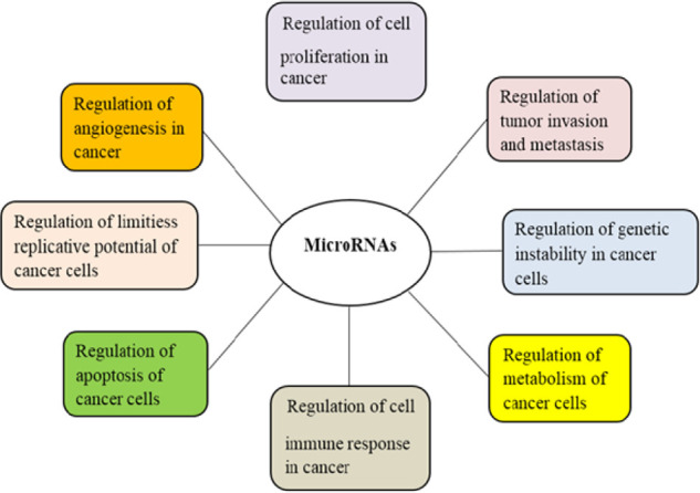
The role of microRNAs in human cancers
Although there have been relatively many studies conducted on the role of microRNAs in TB and NSCLC, no comprehensive study has been performed on the common microRNAs as well as the role of these microRNAs in these two diseases, so far. So, in this study, the common microRNAs between TB and NSCLC and their target genes, which can be used as serum markers for differential diagnosis of TB and NSCLC, were introduced.
This article research was performed in the PubMed and Scopus databases to find the involved-miRNAs in NSCLC and TB separately. Afterward, one thousand sixty-three research papers were found published from 2000 to 2019. The research papers were then studied and the associated miRNAs with NSCLC and TB were separately extracted. Therefore, the common miRNAs between TB and NSCLC were found and one hundred two articles were reviewed by the authors. In all of the chapters, the alternation of miRNAs levels was mentioned, and the pathways and the genes affected by miRNAs were also explained.
The association between TB and NSCLC
Lung cancer is one of the most important causes of death worldwide (24). As many pathologists and clinical doctors have verified it, the connection between lung tuberculosis and lung or bronchial carcinoma certainly exists. Having TB prior to lung cancer increases mortality rates, so it is significantly correlated with mortality due to lung cancer, especially adenocarcinoma. Notably, it is much higher among smokers compared to non-smokers (25). Also, Human Papillomavirus (HPV), Immunodeficiency virus (HIV), and Hepatitis C increase the risk of cancers (26). TB has several symptoms similar to malignancy, so that, sometimes it can be mistakenly diagnosed as lung cancer (27). Moreover, some people after a relatively long period with delaying treatment and diagnosis and sometimes misdiagnosis of lung cancer and even taking anti-cancer drugs, are diagnosed with TB or as people with a history of TB who had lung cancer. A study in 2013, examined the relationship between TB and lung cancer, and it has been shown that TB is directly linked to lung cancer and its mortality, and this stance is intensified in smokers with TB. Also, Leung et al. demonstrated that mortality and morbidity due to lung cancer are associated with TB (28). In a population cohort study in 2011, the incidence of lung cancers between the two cohorts (subjects free from cancers and patients with newly diagnosed TB) was compared and the associated hazard of developing lung cancer was measured. Accordingly, this study demonstrated that the risk of lung cancer is higher in individuals with TB compared to normal subjects (29). Additionally, in countries where lung cancer is highly prevalent, TB patients are often misdiagnosed with the delayed treatment start and unnecessary diagnostic procedures (30). In a research paper Abd-El-Fattah et al. (31) performed microarray and qRT-PCR and revealed that miR-21, miR-155, miR-182, and miR-197 are higher in the serum of lung cancer patients; however, miR-197 is higher in TB patients compared with healthy controls.
MicroRNAs in NSCLC
MicroRNAs are small molecules in the blood that play essential roles in the promotion or inhibition of some cancers like NSCLC (32, 33). The studies have demonstrated that microRNAs improve or inhibit the development of tumors by targeting the genes in biological pathways of cells (9). Also, microRNAs regulate the target mRNA translation into functional protein through binding to the 3′-untranslated region of target mRNAs (7, 34). In a research by Yan et al. (35), qRT-PCR technique was used and the correlation between the expressions of miR-99a and miR-224 was then analyzed in the serum of the NSCLC patients with clinicopathological features. Results showed that miR-224 is associated with NSCLC pathological stage, lymph node metastasis, and pathological grade; whereas miR-99 was shown to be associated with NSCLC pathological stage, lymph node metastasis, and tissue differentiation. Furthermore, miR-141, miR-200b, miR-193b, miR-200c, and miR-106b were the other NSCLC-related miRNAs overexpressed in NSCLC serum. In addition, Nadal et al. (36) analyzed the pathways upon the validation target genes and demonstrated that these miRNAs are associated with the pathways related to lung cancer biology such as MAPK, PI3K-AKT, p53, and neurotrophin signaling pathways. In another research performed by Jeon et al. (37) in situ hybridization, it was shown that that the expressions of miR-224 and miR-520c are higher in metastasized lung tissues compared to primary tumors. Also, it was demonstrated that miR-224 and miR-520c enhance the metastatic potential of NSCLC by suppressing the endogenous Tumor Suppressor Candidate 3 (TUSC3) protein in A549 and H460 cells.
MicroRNAs in TB
Several miRNAs have been investigated to play a role in TB. In this regard, it was indicated that, the miRNAs affect the TB-related processes such as autophagy through regulating multiple target genes and mRNA translation. Notably, autophagy is a process occurring in macrophages by Mtb, which plays a role in the activation of the innate and adaptive immune systems against intracellular bacteria. Also, one of the discovered TB-related miRNAs was miR-33. An in vitro study was conducted by Ouimet et al. (38) and showed that miR-33 is overexpressed in macrophages by Mtb and repress genes encoding key products in the autophagy pathway. Tu et al. (39) discovered miR-423-5p as another involved-miRNA in autophagy, which showed a higher expression in the serum of patients with TB compared to healthy controls. Moreover, miR-423-5p was discovered to target VPS33A gene to inhibit the autophagosome-lysosome fusion in macrophages. Also, miR-27a was identified to be involved in autophagy process. Accordingly, the level of this miRNA was shown to be upregulated in macrophages. In addition, it was shown that, miR-27a could inhibit the autophagosome formation as well as promoting the intracellular survival of Mycobacterium TB (40). The results of the previous studies provided evidence that miRNAs and their target genes can be used as serum markers for the diagnosis and treatment of TB and NSCLC.
The common microRNAs between TB and NSCLC
In this chapter, some of the common involved-microRNAs in TB and NSCLC were found which have been studied in the current papers (Table 1). Therefore, the expression alterations, gene targets, and the physiological and pathological mechanisms of some common microRNAs between TB and NSCLC were reviewed in this review paper. Herein, we attempted to focus on miRNAs that have been studied more and affect their gene targets and pathways, which have been discovered in the current papers. As a result of each part of the common miRNAs between TB and NSCLC (miR-155, miR-146a, miR-29a, miR-30a, miR125b, and Let-7), it can be said that miRNAs and their target genes can be used as therapeutic and diagnostic biomarkers in both TB and NSCLC, which may provide a new method for differential diagnosis of NSCLC and TB. The expression alterations, the gene targets, and the function of miRNAs are summarized in table 1, table 2, and table 3, respectively.
Table 1.
The common miRNAs between TB and NSCLC
Table 2.
The comparison of miRNAs expression in TB and non-small cell lung cancer
| Common miRNAs between TB and NSCLC | Genomic location | Expression in NSCLC | Reference | Expression in TB | Reference |
|---|---|---|---|---|---|
| miRNA-155 | 21q21.3 | High | (53) | High | (54) |
| miRNA-146a | 5q33.3 | High | (43) | Low | (44) |
| miRNA-125b | 11q24.1 | Low | (61) | Low | (62) |
| miRNA-30a | 6q13 | Low | (50) | High | (51) |
| miRNA-29a | 7q32.3 | Low | (59) | Low | (60) |
| miRNA-Let-7 | 9q22.32 | Low | (41) | Low | (42) |
Table 3.
The target genes of microRNAs in TB and NSCLC
| Common miRNAs between TB and NSCLC | The target genes in NSCLC | References | The target genes in TB | References |
|---|---|---|---|---|
| miRNA-155 | SOCS1 | (53) | FOXO3 | (72) |
| SOCS6 | (69) | ATG3 | (73) | |
| PTEN | SHIP1 | (62) | ||
| miRNA-146a | PDCD4 | |||
| JNK2 | (79) | COX-2 | (97) | |
| miRNA-125b | IRS2 | (80) | FLAP | |
| KLC2 | (88) | TNF | (62) | |
| MMP-13 | (61) | |||
| miRNA-30a | MYBL2 | (50) | ATG5 | (51) |
| BCL11A | (89) | beclin-1 | ||
| SNAI1 | (90) | |||
| AEG-1 | (91) | |||
| Snail | ||||
| Vimentin | ||||
| miRNA-29a | IGF1R | (92) | ||
| CDC42 | (98) | Interleukin 17 | (60) | |
| MTSS1 | (99) | |||
| miRNA-Let-7 | LASP1 | (59) | ||
| LIN28A | (95) | TNFAIP3 | (42) | |
| LIN28B |
miR-155
Notably, miR-155 is one of the miRNAs, which was shown to be involved in both NSCLC and TB diseases. Moreover, the role of miR-155 in TB and NSCLC diseases was through targeting the genes in biological signaling pathways (63, 64). Also, the expression of miR-155 was shown to be increased in TB and NSCLC; (53, 54) therefore, the results of the previous studies showed that miR-155 can be used as a prognostic biomarker (65). In this regard, Yang et al. (66) showed that, by the evaluation of miR-155, the recurrence and poor survival in NSCLC can be predicted. In another study performed by Donnem et al. (67) in situ hybridization, the prognostic impact of miR-155 in NSCLC was evaluated. Furthermore, they showed that the prognostic impact of miR-155 is significantly associated with the histological subtype and nodal status in NSCLC. Also, Wang et al. (68) performed a meta-analysis and calculated a pooled hazard ratio as well as analyzing the sensitivity. Moreover, they showed that the high expression levels of miR-155 are significantly correlated with the worst NSCLC survival. On the other hand, in several studies, the pathways and the genes have been discovered, which are affected by miRNAs in TB and NSCLC. Accordingly, Xue et al. (53) revealed that miR-155 targets SOCS1, SOCS6, and PTEN genes. Thus, it promotes the development of NSCLC by downregulation of the above-mentioned genes. Also, in a study, the researchers showed the oncogenic act of miR-155 in NSCLC through targeting the PDCD4 gene and negatively regulating it (69). Additionally, in the previous studies performed on the role of miR-155 in TB, it was identified as a diagnostic biomarker that might present some potential signatures for the diagnosis of TB (70). Wagh et al. (71) demonstrated that the mycobacterium leads to the improvement of miR-155 expression alternation in TB patients (72). Autophagy was also described as a pathway, which causes the activation of the immune system against pathogens such as Mtb. Moreover, Etna et al. (73) identified the role of miR-155 in the autophagy and also discovered that the ATG3 gene is inhibited by miR-155 in autophagosomes. The apoptosis is another pathway that can be modulated by Mtb in the cells (74). Huang et al. (72) discovered that miR-155 targets the FOXO3 in the apoptosis pathway in monocytes, which results in the inhibition of apoptosis. Additionally, inactive TB, the long non-coding PCED1B-AS1 was shown to bind to miR-155 and to block it, which results in the regulation of the macrophage apoptosis and autophagy (75). In another study, miR-155 was shown to provide an effective adaptive immune response by promoting the survival and function of Mtb-specific T cells (76).
miR-146a
In addition, miR-146a was found to be altered in both TB and NSCLC. Accordingly, its expression was increased in NSCLC and decreased in TB (43, 44). In a research performed on the expression pattern of miR-146a in NSCLC patients, Wang et al. (43) performed qRT-PCR and observed that the expression of miR-146a has increased in NSCLC patients compared to the healthy controls. Therefore, it was identified as a diagnostic biomarker in NSCLC (43, 77). The results of several types of research have also shown that, in NSCLC, miR-146a plays essential roles in cell processes such as migration, apoptosis, and growth (78). Pang et al. (79) demonstrated that, when the JNK2 gene is targeted by miR146a, it increases the cisplatin sensitivity of NSCLC cells. In another study, Park et al. (80) performed a microarray and qRT-PCR and finally showed that miR-146a inhibits epithelial-mesenchymal transition in NSCLC cells by repressing the expressions of Insulin Receptor Substrate 2 (IRS2). Also, the results of another study showed that miR-146a plays a role in the development of the acquired drug resistance to the Cisplatin (DDP) in NSCLC cells by downregulating the cyclin J (81). Moreover, Zhang et al. (82) showed that miR-146a is a genetic factor that may be closely related to the susceptibility to pulmonary TB. Other scientists’ studies performed in this regard recruited pulmonary TB patients with healthy controls. They concluded that genetic polymorphisms of miR-146a have significant roles in the risk of TB (83).
miR-125b
Notably, miR-125b is another miRNA that was shown to be downregulated in both TB and NSCLC, which represents as another diagnostic biomarker (61, 62, 84). Li et al. performed real-time PCR and western blot on human NSCLC cells isolated from surgical tissues Moreover, they identified tumor protein 53-induced nuclear protein 1(TP53INP1) as a target of miR-125b. Thus, miR-125b promotes tumor metastasis by targeting this gene (85). Additionally, it was shown that the gene expression pattern alterations of miR-125b results in the regulation of apoptosis signaling pathways such as the PI3K/Akt/GSK3beta and Wnt/beta-catenin (86). Also, Wang et al. (87) in their study revealed that miR-125b promotes the tumor invasion via the activation of the PI3K/AKT signaling pathway. Another study showed that, in NSCLC, miR-125b inhibits the Kinesin-1 light chain-2, which acts as a proto-oncogene (88). Another function of miR-125b is through the regulation of MMP-13. Yu et al. (61) revealed that the reduced expression levels of miR-125b lead to the increased MMP-13 expression levels, followed by the inhibition of invasive capabilities of cancer cells. Also, the effects of miR-125b expression on TB were assessed in some studies. Such studies showed that TNF is a target of miR-125b, which destabilizes the transcript of this gene (62).
miR-30a
The lower expression of miR-30a in NSCLC patients compared to healthy controls was revealed in a study by Geng et al. (50) and higher expression of miR-30a in TB patients compared to healthy controls was revealed by Chen et al.(51). Notably, BCL11A is a target of miR-30a in NSCLC. MicroRNA-30a was shown to induce the increased expression levels of BCL11A in NSCLC tissues (89). Also, the inhibitory role of miR-30a in invasion and metastasis of NSCLC cell lines has been reported to be through targeting SNAI1 (90). Additionally, the AEG-1, Snail, and Vimentin genes were investigated by Liu et al. (91), by binding to 3'- UTR of miR-30a and promoting the metastasis of NSCLC tumor in A549 cells. Wen et al. (92) used a luciferase reporter assay and demonstrated that miR-30a could suppress NSCLC cell proliferation through targeting IGF1R, which is in PI3K/AKT signaling pathway. Chen et al. (51) discovered the role of miR-30a in the immune response against Mtb in human macrophages through the inhibition of the autophagy-induced by Mtb.
miR-29a
Hu et al. performed in vitro study and showed that miR-29a expression level is lower in NSCLC cells compared to the healthy controls. Moreover, they identified LASP1 as a target gene of miR-29a and then explained that the overexpression of miR-29a decreases the growth of A549 cells in nude mice by targeting LASP1 (59). Additionally, several studies were conducted to indicate the role of miR-29a in TB. T cells were involved in the protection of the Mtb infected individuals from developing TB. Kleinsteuber et al. showed lower expressions of miR-29a and miR-21 in TB patients. They observed no correlation between the expression of miR-29a and Interferon-gamma; however, a significant correlation was observed between the IL-17 positive T-cell clone’s activation and miR-29a expression (60). Also, Afum-Adjei Awuah et al. (93) conducted a similar research, which demonstrated no correlation between the expression of miR-29a and Interferon-gamma.
miR-let-7
The expression level of miR-let-7 was shown to be lower in NSCLC patients compared to the healthy controls (41), which played a tumor suppressor role in NSCLC (94). Moreover, Yin et al. (95) demonstrated that the down-regulation of miR-let-7, up-regulation of LIN28A, and LIN28B expression resulted in the regulation of the single-cell proliferative capability of NSCLC cells. Thus, resistance to irradiation or cisplatin has been promoted. Also, miR-let-7 was shown to be altered in TB (96), and involved in immune response (42). Results showed that miR-let-7 is downregulated in Mtb-infected macrophages. Researchers indicated that miR-let-7 targets the TNFAIP3, as an inhibitor of the NF-κB pathway, which consequently results in the modulation of the immune response to Mtb infection of cytokines, including TNF and IL-1β (Table 3,4).
Table 4.
The functions of each microRNAs in TB and NSCLC
| MicroRNA | Function in NSCLC | Function in TB |
|---|---|---|
| miR-125b | Promotes tumor metastasis | Enhances TNF production |
| Regulates apoptosis | ||
| Promotes tumor invasion | ||
| miR-146a | Inhibits the migration | Involves in regulating inflammatory responses |
| Induces apoptosis | ||
| Suppresses cell growth | ||
| miR-155 | Has oncogenic act | Has a role in autophagy |
| miR-30a | Increases cell apoptosis | Inhibits the autophagy |
| Induces cell cycle arrest | ||
| Attenuates tumor growth | ||
| Suppresses cell proliferation | ||
| miR-29a | Decreases the growth of A549 cells | Regulates T-cell clones activation |
| miR-Let-7 | Tumor suppressor | Modulates the immune response to Mtb |
CONCLUSION
Several studies have been performed on the association between TB and lung cancer, and it was shown that TB is directly linked to lung cancer and its mortality. The risk of being affected by lung cancer is increased in patients with TB and having TB before lung cancer increases mortality rates. Additionally, TB has some symptoms similar to malignancy; therefore, sometimes it is mistakenly diagnosed as lung cancer. So, the identification of biological markers is necessary for differential diagnosis of TB and NSCLC (25, 27–29, 31, 100–102).
MicroRNAs are small molecules in blood of people, which play essential roles in biological pathways, through targeting the related genes. Several studies have been conducted on investigating the TB and NSCLC-related miRNAs, which finally resulted in identifying several miRNAs that play essential roles in signaling pathways of these two diseases. According to the results of this review, among all the miRNAs that were separately identified in TB and NSCLC, some of them were found to be common in both diseases. Additionally, the common microRNAs between TB and NSCLC have been shown to act through the same pathways in TB and NSCLC. Therefore, most of the pathways affected by miRNAs are immune responses such as autophagy in TB and apoptosis in NSCLC.
With a glimpse of studies on the assessment of miRNAs among TB and NSCLC patients, it is obvious that these biological molecules can be used as diagnostic and therapeutic biomarkers for differential diagnosis, prognosis, screening, treatment strategies, and clinical management of these two diseases. The expression pattern of common miRNAs between TB and NSCLC should also be evaluated and then compared at the experimental level in a greater population that may be used as blood markers. So, it can prevent delayed diagnosis, delayed treatment, misdiagnosis, and getting lung cancer after TB. As well as the expression pattern of miRNA, target genes can also be evaluated to identify the action mechanisms of miRNAs.
Acknowledgments
We thank our colleagues from the Department of Mycobacteriology and Pulmonary Research, Microbiology Research Center, Pasteur Institute of Iran.
Abbreviation
- SOCS1:
Suppressor Of Cytokine Signaling;
- PTEN:
Phosphatase and Tensin Homolog;
- PDCD4:
Programmed Cell Death 4;
- JNK2:
c-Jun N-terminal Kinase 2;
- IRS2:
Insulin Receptor Substrate 2;
- KLC2:
Kinesin Light Chain 2;
- MMP13:
Matrix Metallopeptidase 13;
- MYBL2:
MYB Proto-Oncogene Like 2;
- BCL11A:
BAF Chromatin Remodeling Complex Subunit BCL11A;
- SNAI1:
Snail Family Transcriptional Repressor 1;
- AEG-1:
Astrocyte Elevated Gene-1;
- IGF1R:
Insulin-Like Growth Factor 1 Receptor;
- CDC42:
Cell Division Cycle 42;
- MTSS1:
MTSS I-BAR Domain Containing 1;
- LASP1:
LIM And SH3 Protein 1;
- LIN28A:
Lin-28 Homolog A;
- FOXO3:
Forkhead Box O3;
- ATG3:
Autophagy Related 3;
- COX-2:
Cyclooxygenase-2;
- FLAP:
Five-Lipoxygenase Activating Protein;
- TNF:
Tumor Necrosis Factor;
- ATG5:
Autophagy Related 5;
- TNFAIP3:
TNF Alpha Induced Protein 3.
Footnotes
Conflict of interest
The authors declare that there is no conflict of interest.
Funding
The authors have no funding source.
REFERENCES
- 1.Farhat MR, Freschi L, Calderon R, Ioerger T, Snyder M, Meehan CJ, et al. GWAS for quantitative resistance phenotypes in Mycobacterium tuberculosis reveals resistance genes and regulatory regions. Nat Commun 2019;10(1):2128. [DOI] [PMC free article] [PubMed] [Google Scholar]
- 2.Awoniyi DO, Baumann R, Chegou NN, Kriel B, Jacobs R, Kidd M, et al. Detection of a combination of serum IgG and IgA antibodies against selected mycobacterial targets provides promising diagnostic signatures for active TB. Oncotarget 2017;8(23):37525–37537. [DOI] [PMC free article] [PubMed] [Google Scholar]
- 3.Liu Y, Xie Z, Zhou X, Li W, Zhang H, He ZG. NapM enhances the survival of Mycobacterium tuberculosis under stress and in macrophages. Commun Biol 2019;2:65. [DOI] [PMC free article] [PubMed] [Google Scholar]
- 4.Li J, Sun L, Xu F, Xiao J, Jiao W, Qi H, et al. Characterization of plasma proteins in children of different Mycobacterium tuberculosis infection status using label-free quantitative proteomics. Oncotarget 2017;8(61):103290–103301. [DOI] [PMC free article] [PubMed] [Google Scholar]
- 5.Mustafa AD, Kalyanasundram J, Sabidi S, Song AA, Abdullah M, Abdul Rahim R, et al. Recovery of recombinant Mycobacterium tuberculosis antigens fused with cell wall-anchoring motif (LysM) from inclusion bodies using nondenaturing reagent (N-laurylsarcosine). BMC Biotechnol 2019;19(1):27. [DOI] [PMC free article] [PubMed] [Google Scholar]
- 6.Finkelstein DM, Ettinger DS, Ruckdeschel JC. Long-term survivors in metastatic non-small-cell lung cancer: an Eastern Cooperative Oncology Group Study. J Clin Oncol 1986;4(5):702–9. [DOI] [PubMed] [Google Scholar]
- 7.Abolfathi H, Sheikhpour M, Shahraeini SS, Khatami S, Nojoumi SA. Studies in lung cancer cytokine proteomics: a review. Expert Rev Proteomics 2021;18(1):49–64. [DOI] [PubMed] [Google Scholar]
- 8.Cheriyan VT, Alsaab H, Sekhar S, Venkatesh J, Mondal A, Vhora I, et al. A CARP-1 functional mimetic compound is synergistic with BRAF-targeting in non-small cell lung cancers. Oncotarget 2018;9(51):29680–29697. [DOI] [PMC free article] [PubMed] [Google Scholar]
- 9.Yang Y, Li H, Liu Y, Chi C, Ni J, Lin X. MiR-4319 hinders YAP expression to restrain non-small cell lung cancer growth through regulation of LIN28-mediated RFX5 stability. Biomed Pharmacother 2019;115:108956. [DOI] [PubMed] [Google Scholar]
- 10.Lee RC, Feinbaum RL, Ambros V. The C. elegans heterochronic gene lin-4 encodes small RNAs with antisense complementarity to lin-14. Cell 1993;75(5):843–54. [DOI] [PubMed] [Google Scholar]
- 11.Wightman B, Ha I, Ruvkun G. Posttranscriptional regulation of the heterochronic gene lin-14 by lin-4 mediates temporal pattern formation in C. elegans. Cell 1993;75(5):855–62. [DOI] [PubMed] [Google Scholar]
- 12.Jebessa E, Ouyang H, Abdalla BA, Li Z, Abdullahi AY, Liu Q, et al. Characterization of miRNA and their target gene during chicken embryo skeletal muscle development. Oncotarget 2017;9(25):17309–17324. [DOI] [PMC free article] [PubMed] [Google Scholar]
- 13.Alharris E, Singh NP, Nagarkatti PS, Nagarkatti M. Role of miRNA in the regulation of cannabidiol-mediated apoptosis in neuroblastoma cells. Oncotarget 2019;10(1):45–59. [DOI] [PMC free article] [PubMed] [Google Scholar]
- 14.Berezikov E, Guryev V, van de Belt J, Wienholds E, Plasterk RH, Cuppen E. Phylogenetic shadowing and computational identification of human microRNA genes. Cell 2005;120(1):21–4. [DOI] [PubMed] [Google Scholar]
- 15.Esquela-Kerscher A, Slack FJ. Oncomirs - microRNAs with a role in cancer. Nat Rev Cancer 2006;6(4):259–69. [DOI] [PubMed] [Google Scholar]
- 16.Kent OA, Mendell JT. A small piece in the cancer puzzle: microRNAs as tumor suppressors and oncogenes. Oncogene 2006;25(46):6188–96. [DOI] [PubMed] [Google Scholar]
- 17.Lee YS, Dutta A. MicroRNAs in cancer. Annu Rev Pathol 2009;4199–227 [DOI] [PMC free article] [PubMed] [Google Scholar]
- 18.Williams AE. Functional aspects of animal microRNAs. Cell Mol Life Sci 2008;65(4):545–62. [DOI] [PMC free article] [PubMed] [Google Scholar]
- 19.Sugita BM, Pereira SR, de Almeida RC, Gill M, Mahajan A, Duttargi A, et al. Integrated copy number and miRNA expression analysis in triple negative breast cancer of Latin American patients. Oncotarget 2019;10(58):6184–6203. [DOI] [PMC free article] [PubMed] [Google Scholar]
- 20.Lee JY, Yun SJ, Jeong P, Piao XM, Kim YH, Kim J, et al. Identification of differentially expressed miRNAs and miRNA-targeted genes in bladder cancer. Oncotarget 2018;9(45):27656–27666. [DOI] [PMC free article] [PubMed] [Google Scholar]
- 21.Hoballa MH, Soltani BM, Mowla SJ, Sheikhpour M, Kay M. Identification of a novel intergenic miRNA located between the human DDC and COBL genes with a potential function in cell cycle arrest. Mol Cell Biochem 2018;444(1–2):179–186. [DOI] [PubMed] [Google Scholar]
- 22.Stahlhut Espinosa CE, Slack FJ. The role of microRNAs in cancer. Yale J Biol Med 2006;79(3–4):131–40. [PMC free article] [PubMed] [Google Scholar]
- 23.Zhang B, Pan X, Cobb GP, Anderson TA. microRNAs as oncogenes and tumor suppressors. Dev Biol 2007;302(1):1–12. [DOI] [PubMed] [Google Scholar]
- 24.Silva DR, Valentini DF, Jr, Müller AM, de Almeida CP, Dalcin Pde T. Pulmonary tuberculosis and lung cancer: simultaneous and sequential occurrence. J Bras Pneumol 2013;39(4):484–9. [DOI] [PMC free article] [PubMed] [Google Scholar]
- 25.Cukic V. The Association Between Lung Carcinoma and Tuberculosis. Med Arch 2017;71(3):212–214. [DOI] [PMC free article] [PubMed] [Google Scholar]
- 26.de Martel C, Shiels MS, Franceschi S, Simard EP, Vignat J, et al. Cancers attributable to infections among adults with HIV in the United States. AIDS 2015;29(16):2173–81. [DOI] [PMC free article] [PubMed] [Google Scholar]
- 27.Bhatt M, Kant S, Bhaskar R. Pulmonary tuberculosis as differential diagnosis of lung cancer. South Asian J Cancer 2012;1(1):36–42. [DOI] [PMC free article] [PubMed] [Google Scholar]
- 28.Leung CC, Hui L, Lee RS, Lam TH, Yew WW, Hui DS, et al. Tuberculosis is associated with increased lung cancer mortality. Int J Tuberc Lung Dis 2013;17(5):687–92. [DOI] [PubMed] [Google Scholar]
- 29.Yu YH, Liao CC, Hsu WH, Chen HJ, Liao WC, Muo CH, et al. Increased lung cancer risk among patients with pulmonary tuberculosis: a population cohort study. J Thorac Oncol 2011;6(1):32–7. [DOI] [PubMed] [Google Scholar]
- 30.Hammen I. Tuberculosis mimicking lung cancer. Respir Med Case Rep 2015. 10;16:45–7. [DOI] [PMC free article] [PubMed] [Google Scholar]
- 31.Abd-El-Fattah AA, Sadik NA, Shaker OG, Aboulftouh ML. Differential microRNAs expression in serum of patients with lung cancer, pulmonary tuberculosis, and pneumonia. Cell Biochem Biophys 2013;67(3):875–84. [DOI] [PubMed] [Google Scholar]
- 32.Zhang WC, Chin TM, Yang H, Nga ME, Lunny DP, Lim EK, et al. Tumour-initiating cell-specific miR-1246 and miR-1290 expression converge to promote non-small cell lung cancer progression. Nat Commun 2016;7:11702. [DOI] [PMC free article] [PubMed] [Google Scholar]
- 33.Li C, Yin Y, Liu X, Xi X, Xue W, Qu Y. Non-small cell lung cancer associated microRNA expression signature: integrated bioinformatics analysis, validation and clinical significance. Oncotarget 2017;8(15):24564–24578. [DOI] [PMC free article] [PubMed] [Google Scholar]
- 34.Hirono T, Jingushi K, Nagata T, Sato M, Minami K, Aoki M, et al. MicroRNA-130b functions as an oncomiRNA in non-small cell lung cancer by targeting tissue inhibitor of metalloproteinase-2. Sci Rep 2019;9(1):6956. [DOI] [PMC free article] [PubMed] [Google Scholar]
- 35.Yan HZ, Wang W, Du X, Jiang XD, Lin CY, Guo JL, et al. The expression and clinical significance of miRNA-99a and miRNA-224 in non-small cell lung cancer. Eur Rev Med Pharmacol Sci 2019;23(4):1545–1552. [DOI] [PubMed] [Google Scholar]
- 36.Nadal E, Truini A, Nakata A, Lin J, Reddy RM, Chang AC, et al. A Novel Serum 4-microRNA Signature for Lung Cancer Detection. Sci Rep 2015;5:12464. [DOI] [PMC free article] [PubMed] [Google Scholar]
- 37.Jeon YJ, Kim T, Park D, Nuovo GJ, Rhee S, Joshi P, et al. miRNA-mediated TUSC3 deficiency enhances UPR and ERAD to promote metastatic potential of NSCLC. Nat Commun 2018;9(1):5110. [DOI] [PMC free article] [PubMed] [Google Scholar]
- 38.Ouimet M, Koster S, Sakowski E, Ramkhelawon B, van Solingen C, Oldebeken S, et al. Mycobacterium tuberculosis induces the miR-33 locus to reprogram autophagy and host lipid metabolism. Nat Immunol 2016;17(6):677–86. [DOI] [PMC free article] [PubMed] [Google Scholar]
- 39.Tu H, Yang S, Jiang T, Wei L, Shi L, Liu C, et al. Elevated pulmonary tuberculosis biomarker miR-423-5p plays critical role in the occurrence of active TB by inhibiting autophagosome-lysosome fusion. Emerg Microbes Infect 2019;8(1):448–460. [DOI] [PMC free article] [PubMed] [Google Scholar]
- 40.Liu F, Chen J, Wang P, Li H, Zhou Y, Liu H, et al. MicroRNA-27a controls the intracellular survival of Mycobacterium tuberculosis by regulating calcium-associated autophagy. Nat Commun 2018;9(1):4295. [DOI] [PMC free article] [PubMed] [Google Scholar]
- 41.Xia XM, Jin WY, Shi RZ, Zhang YF, Chen J. Clinical significance and the correlation of expression between Let-7 and K-ras in non-small cell lung cancer. Oncol Lett 2010;1(6):1045–1047. [DOI] [PMC free article] [PubMed] [Google Scholar]
- 42.Kumar M, Sahu SK, Kumar R, Subuddhi A, Maji RK, Jana K, et al. MicroRNA let-7 modulates the immune response to Mycobacterium tuberculosis infection via control of A20, an inhibitor of the NF-κB pathway. Cell Host Microbe 2015;17(3):345–356. [DOI] [PubMed] [Google Scholar]
- 43.Wang RJ, Zheng YH, Wang P, Zhang JZ. Serum miR-125a-5p, miR-145 and miR-146a as diagnostic biomarkers in non-small cell lung cancer. Int J Clin Exp Pathol 2015;8(1):765–71. [PMC free article] [PubMed] [Google Scholar]
- 44.Malardo T, Gardinassi LG, Moreira BP, Padilha É, Lorenzi JC, Soares LS, et al. MicroRNA expression signatures in lungs of mice infected with Mycobacterium tuberculosis. Tuberculosis (Edinb) 2016;101151–159 [DOI] [PubMed] [Google Scholar]
- 45.Zeng Z, Zhao G, Rao C, Hua G, Yang M, Miao X, et al. Knockdown of lncRNA ZFAS1-suppressed non-small cell lung cancer progression via targeting the miR-150-5p/HMGA2 signaling. J Cell Biochem 2019. [DOI] [PubMed] [Google Scholar]
- 46.Wang Y, Xu YM, Zou YQ, Lin J, Huang B, Liu J, et al. Identification of differential expressed PE exosomal miRNA in lung adenocarcinoma, tuberculosis, and other benign lesions. Medicine (Baltimore) 2017;96(44):e8361. [DOI] [PMC free article] [PubMed] [Google Scholar]
- 47.Dong J, Zhang Z, Gu T, Xu SF, Dong LX, Li X, et al. The role of microRNA-21 in predicting brain metastases from non-small cell lung cancer. Onco Targets Ther 2016;10185–194 [DOI] [PMC free article] [PubMed] [Google Scholar]
- 48.Xia H, Xiu M, Gao J, Jing H. LncRNA PLAC 2 downregulated miR-21 in non-small cell lung cancer and predicted survival. BMC Pulm Med 2019;19(1):172. [DOI] [PMC free article] [PubMed] [Google Scholar]
- 49.Zhao Z, Hao J, Li X, Chen Y, Qi X. MiR-21-5p regulates mycobacterial survival and inflammatory responses by targeting Bcl-2 and TLR4 in Mycobacterium tuberculosis-infected macrophages. FEBS Lett 2019;593(12):1326–1335. [DOI] [PubMed] [Google Scholar]
- 50.Geng GJ, Yang YT, Jiang J, Yu XY, Fa XE. MicroRNA-30a suppresses non-small-cell lung cancer by targeting Myb-related protein B. Exp Ther Med 2018;15(2):1633–1639. [DOI] [PMC free article] [PubMed] [Google Scholar]
- 51.Chen Z, Wang T, Liu Z, Zhang G, Wang J, Feng S, et al. Inhibition of Autophagy by MiR-30A Induced by Mycobacteria tuberculosis as a Possible Mechanism of Immune Escape in Human Macrophages. Jpn J Infect Dis 2015;68(5):420–4. [DOI] [PubMed] [Google Scholar]
- 52.Zhang Y, Zhang Y, Yin Y, Li S. Detection of circulating exosomal miR-17-5p serves as a novel non-invasive diagnostic marker for non-small cell lung cancer patients. Pathol Res Pract 2019;215(8):152466. [DOI] [PubMed] [Google Scholar]
- 53.Xue X, Liu Y, Wang Y, Meng M, Wang K, Zang X, Zhao S, Sun X, Cui L, Pan L, Liu S. MiR-21 and MiR-155 promote non-small cell lung cancer progression by downregulating SOCS1, SOCS6, and PTEN. Oncotarget 2016;7(51):84508–84519. [DOI] [PMC free article] [PubMed] [Google Scholar]
- 54.Zhang C, Xi X, Wang Q, Jiao J, Zhang L, Zhao H, et al. The association between serum miR-155 and natural killer cells from tuberculosis patients. Int J Clin Exp Med 2015;8(6):9168–72. [PMC free article] [PubMed] [Google Scholar]
- 55.Xie Y, Zhao F, Zhang P, Duan P, Shen Y. miR-29b inhibits non-small cell lung cancer progression by targeting STRN4. Hum Cell 2020;33(1):220–231. [DOI] [PubMed] [Google Scholar]
- 56.Yi Z, Gao K, Li R, Fu Y. Changed immune and miRNA response in RAW264.7 cells infected with cell wall deficient mycobacterium tuberculosis. Int J Mol Med 2018;41(5):2885–2892. [DOI] [PubMed] [Google Scholar]
- 57.Huang H, Huang J, Yao J, Li N, Yang Z. miR-125a regulates HAS1 and inhibits the proliferation, invasion and metastasis by targeting STAT3 in non-small cell lung cancer cells. J Cell Biochem 2020;121(5–6):3197–3207. [DOI] [PubMed] [Google Scholar]
- 58.Niu W, Sun B, Li M, Cui J, Huang J, Zhang L. TLR-4/microRNA-125a/NF-κB signaling modulates the immune response to Mycobacterium tuberculosis infection. Cell Cycle 2018;17(15):1931–1945. [DOI] [PMC free article] [PubMed] [Google Scholar]
- 59.Hu Z, Cui Y, Zhou Y, Zhou K, Qiao X, Li C, et al. MicroRNA-29a plays a suppressive role in non-small cell lung cancer cells via targeting LASP1. Onco Targets Ther 2016;96999–7009 [DOI] [PMC free article] [PubMed] [Google Scholar] [Retracted]
- 60.Kleinsteuber K, Heesch K, Schattling S, Kohns M, Sander-Jülch C, Walzl G, et al. Decreased expression of miR-21, miR-26a, miR-29a, and miR-142-3p in CD4+ T cells and peripheral blood from tuberculosis patients. PLoS One 2013;8(4):e61609. [DOI] [PMC free article] [PubMed] [Google Scholar]
- 61.Yu X, Wei F, Yu J, Zhao H, Jia L, Ye Y, et al. Matrix metalloproteinase 13: a potential intermediate between low expression of microRNA-125b and increasing metastatic potential of non-small cell lung cancer. Cancer Genet 2015;208(3):76–84. [DOI] [PubMed] [Google Scholar]
- 62.Rajaram MV, Ni B, Morris JD, Brooks MN, Carlson TK, Bakthavachalu B, et al. Mycobacterium tuberculosis lipomannan blocks TNF biosynthesis by regulating macrophage MAPK-activated protein kinase 2 (MK2) and microRNA miR-125b. Proc Natl Acad Sci U S A 2011;108(42):17408–13. [DOI] [PMC free article] [PubMed] [Google Scholar]
- 63.Abolfathi H, Sheikhpour M, Soltani BM, Fahimi H. The comparison and evaluation of the miR-16, miR-155 and miR-146a expression pattern in the blood of TB and NSCLC patients: A Research paper. Gene Reports 2021;22:100967. [Google Scholar]
- 64.Wu KL, Tsai YM, Lien CT, Kuo PL, Hung AJ. The Roles of MicroRNA in Lung Cancer. Int J Mol Sci 2019;20(7):1611. [DOI] [PMC free article] [PubMed] [Google Scholar]
- 65.Xu TP, Zhu CH, Zhang J, Xia R, Wu FL, Han L, Shen H, Liu LX, Shu YQ. MicroRNA-155 expression has prognostic value in patients with non-small cell lung cancer and digestive system carcinomas. Asian Pac J Cancer Prev 2013;14(12):7085–90. [DOI] [PubMed] [Google Scholar]
- 66.Yang M, Shen H, Qiu C, Ni Y, Wang L, Dong W, et al. High expression of miR-21 and miR-155 predicts recurrence and unfavourable survival in non-small cell lung cancer. Eur J Cancer 2013;49(3):604–15. [DOI] [PubMed] [Google Scholar]
- 67.Donnem T, Eklo K, Berg T, Sorbye SW, Lonvik K, Al-Saad S, Al-Shibli K, et al. Prognostic impact of MiR-155 in non-small cell lung cancer evaluated by in situ hybridization. J Transl Med 2011;9:6. [DOI] [PMC free article] [PubMed] [Google Scholar]
- 68.Wang Y, Li J, Tong L, Zhang J, Zhai A, Xu K, et al. The prognostic value of miR-21 and miR-155 in non-small-cell lung cancer: a meta-analysis. Jpn J Clin Oncol 2013;43(8):813–20. [DOI] [PubMed] [Google Scholar]
- 69.Liu F, Song D, Wu Y, Liu X, Zhu J, Tang Y. MiR-155 inhibits proliferation and invasion by directly targeting PDCD4 in non-small cell lung cancer. Thorac Cancer 2017;8(6):613–619. [DOI] [PMC free article] [PubMed] [Google Scholar]
- 70.Zheng ML, Zhou NK, Luo CH. MiRNA-155 and miRNA-132 as potential diagnostic biomarkers for pulmonary tuberculosis: A preliminary study. Microb Pathog 2016;10078–83 [DOI] [PubMed] [Google Scholar]
- 71.Wagh V, Urhekar A, Modi D. Levels of microRNA miR-16 and miR-155 are altered in serum of patients with tuberculosis and associate with responses to therapy. Tuberculosis (Edinb) 2017;10224–30 [DOI] [PubMed] [Google Scholar]
- 72.Huang J, Jiao J, Xu W, Zhao H, Zhang C, Shi Y, et al. MiR-155 is upregulated in patients with active tuberculosis and inhibits apoptosis of monocytes by targeting FOXO3. Mol Med Rep 2015;12(5):7102–8. [DOI] [PubMed] [Google Scholar]
- 73.Etna MP, Sinigaglia A, Grassi A, Giacomini E, Romagnoli A, Pardini M, et al. Mycobacterium tuberculosis-induced miR-155 subverts autophagy by targeting ATG3 in human dendritic cells. PLoS Pathog 2018;14(1):e1006790. [DOI] [PMC free article] [PubMed] [Google Scholar]
- 74.Lin J, Chang Q, Dai X, Liu D, Jiang Y, Dai Y. Early secreted antigenic target of 6-kDa of Mycobacterium tuberculosis promotes caspase-9/caspase-3-mediated apoptosis in macrophages. Mol Cell Biochem 2019;457(1–2):179–189. [DOI] [PubMed] [Google Scholar]
- 75.Li M, Cui J, Niu W, Huang J, Feng T, Sun B, et al. Long non-coding PCED1B-AS1 regulates macrophage apoptosis and autophagy by sponging miR-155 in active tuberculosis. Biochem Biophys Res Commun 2019. Feb 12;509(3):803–809. [DOI] [PubMed] [Google Scholar]
- 76.Rothchild AC, Sissons JR, Shafiani S, Plaisier C, Min D, Mai D, et al. MiR-155-regulated molecular network orchestrates cell fate in the innate and adaptive immune response to Mycobacterium tuberculosis. Proc Natl Acad Sci U S A 2016;113(41):E6172–E6181. [DOI] [PMC free article] [PubMed] [Google Scholar]
- 77.Wu C, Cao Y, He Z, He J, Hu C, Duan H, et al. Serum levels of miR-19b and miR-146a as prognostic biomarkers for non-small cell lung cancer. Tohoku J Exp Med 2014;232(2):85–95. [DOI] [PubMed] [Google Scholar]
- 78.Chen G, Umelo IA, Lv S, Teugels E, Fostier K, Kronenberger P, et al. miR-146a inhibits cell growth, cell migration and induces apoptosis in non-small cell lung cancer cells. PLoS One 2013;8(3):e60317. [DOI] [PMC free article] [PubMed] [Google Scholar]
- 79.Pang L, Lu J, Huang J, Xu C, Li H, Yuan G, et al. Upregulation of miR-146a increases cisplatin sensitivity of the non-small cell lung cancer A549 cell line by targeting JNK-2. Oncol Lett 2017;14(6):7745–7752. [DOI] [PMC free article] [PubMed] [Google Scholar]
- 80.Park DH, Jeon HS, Lee SY, Choi YY, Lee HW, Yoon S, et al. MicroRNA-146a inhibits epithelial mesenchymal transition in non-small cell lung cancer by targeting insulin receptor substrate 2. Int J Oncol 2015;47(4):1545–53. [DOI] [PubMed] [Google Scholar]
- 81.Shi L, Xu Z, Wu G, Chen X, Huang Y, Wang Y, et al. Up-regulation of miR-146a increases the sensitivity of non-small cell lung cancer to DDP by downregulating cyclin. J BMC Cancer 2017;17(1):138. [DOI] [PMC free article] [PubMed] [Google Scholar]
- 82.Zhang X, Li Y, Li X, Zhang W, Pan Z, Wu F, et al. Association of the miR-146a, miR-149, miR-196a2 and miR-499 polymorphisms with susceptibility to pulmonary tuberculosis in the Chinese Uygur, Kazak and Southern Han populations. BMC Infect Dis 2015;15:41. [DOI] [PMC free article] [PubMed] [Google Scholar]
- 83.Wang M, Xu G, Lü L, Xu K, Chen Y, Pan H, et al. Genetic polymorphisms of IL-17A, IL-17F, TLR4 and miR-146a in association with the risk of pulmonary tuberculosis. Sci Rep 2016;6:28586. [DOI] [PMC free article] [PubMed] [Google Scholar]
- 84.Shahsavani M, Baghbani-Arani F, Sheikhpour M. The expression profile evaluation of Mir-125b in tuberculosis and non-small cell lung cancer patients. Clinical Cancer Investigation Journal 2021;10(2):60. [Google Scholar]
- 85.Li Q, Han Y, Wang C, Shan S, Wang Y, Zhang J, et al. MicroRNA-125b promotes tumor metastasis through targeting tumor protein 53-induced nuclear protein 1 in patients with non-small-cell lung cancer. Cancer Cell Int 2015;15:84. [DOI] [PMC free article] [PubMed] [Google Scholar]
- 86.Wang Y, Zhao M, Liu J, Sun Z, Ni J, Liu H. miRNA-125b regulates apoptosis of human non-small cell lung cancer via the PI3K/Akt/GSK3β signaling pathway. Oncol Rep 2017;38(3):1715–1723. [DOI] [PubMed] [Google Scholar]
- 87.Wang HH, Wang YC, Wu DW, Hung CS, Chen CY, Lee H. Targeting insulin-like growth factor-binding protein-3 by microRNA-125b promotes tumor invasion and poor outcomes in non-small-cell lung cancer. Tumour Biol 2017;39(4):1010428317694316. [DOI] [PubMed] [Google Scholar]
- 88.Wang M, Zhu X, Sha Z, Li N, Li D, Chen L. High expression of kinesin light chain-2, a novel target of miR-125b, is associated with poor clinical outcome of elderly non-small-cell lung cancer patients. Br J Cancer 2015;112(5):874–82. [DOI] [PMC free article] [PubMed] [Google Scholar]
- 89.Jiang BY, Zhang XC, Su J, Meng W, Yang XN, Yang JJ, et al. BCL11A overexpression predicts survival and relapse in non-small cell lung cancer and is modulated by microRNA-30a and gene amplification. Mol Cancer 2013;12:61. [DOI] [PMC free article] [PubMed] [Google Scholar]
- 90.Kumarswamy R, Mudduluru G, Ceppi P, Muppala S, Kozlowski M, Niklinski J, et al. MicroRNA-30a inhibits epithelial-to-mesenchymal transition by targeting Snai1 and is downregulated in non-small cell lung cancer. Int J Cancer 2012;130(9):2044–53. [DOI] [PubMed] [Google Scholar]
- 91.Liu K, Guo L, Guo Y, Zhou B, Li T, Yang H, et al. AEG-1 3'-untranslated region functions as a ceRNA in inducing epithelial-mesenchymal transition of human non-small cell lung cancer by regulating miR-30a activity. Eur J Cell Biol 2015;94(1):22–31. [DOI] [PubMed] [Google Scholar]
- 92.Wen XP, Ma HL, Zhao LY, Zhang W, Dang CX. MiR-30a suppresses non-small cell lung cancer progression through AKT signaling pathway by targeting IGF1R. Cell Mol Biol (Noisy-le-grand) 2015;61(2):78–85. [PubMed] [Google Scholar]
- 93.Afum-Adjei Awuah A, Ueberberg B, Owusu-Dabo E, Frempong M, Jacobsen M. Dynamics of T-cell IFN-γ and miR-29a expression during active pulmonary tuberculosis. Int Immunol 2014;26(10):579–82. [DOI] [PubMed] [Google Scholar]
- 94.Stahlhut C, Slack FJ. Combinatorial Action of MicroRNAs let-7 and miR-34 Effectively Synergizes with Erlotinib to Suppress Non-small Cell Lung Cancer Cell Proliferation. Cell Cycle 2015;14(13):2171–80. [DOI] [PMC free article] [PubMed] [Google Scholar]
- 95.Yin J, Zhao J, Hu W, Yang G, Yu H, Wang R, et al. Disturbance of the let-7/LIN28 double-negative feedback loop is associated with radio- and chemo-resistance in non-small cell lung cancer. PLoS One 2017;12(2):e0172787. [DOI] [PMC free article] [PubMed] [Google Scholar]
- 96.Barry SE, Chan B, Ellis M, Yang Y, Plit ML, Guan G, et al. Identification of miR-93 as a suitable miR for normalizing miRNA in plasma of tuberculosis patients. J Cell Mol Med 2015;19(7):1606–13. [DOI] [PMC free article] [PubMed] [Google Scholar]
- 97.Iacona JR, Monteleone NJ, Lutz CS. miR-146a suppresses 5-lipoxygenase activating protein (FLAP) expression and Leukotriene B4 production in lung cancer cells. Oncotarget 2018;9(42):26751–26769. [DOI] [PMC free article] [PubMed] [Google Scholar]
- 98.Li Y, Wang Z, Li Y, Jing R. MicroRNA-29a functions as a potential tumor suppressor through directly targeting CDC42 in non-small cell lung cancer. Oncol Lett 2017;13(5):3896–3904. [DOI] [PMC free article] [PubMed] [Google Scholar]
- 99.Liu M, Zeng X, Lu YX, Mo YJ, Liao TH, Gan C, et al. Study on molecular mechanism of MiRNA-29a in promoting proliferation and invasion of non-small-cell lung cancer by inhibiting MTSS1. Eur Rev Med Pharmacol Sci 2018;22(17):5531–5538. [DOI] [PubMed] [Google Scholar]
- 100.De Flora S, La Maestra S. Epidemiology of cancers of infectious origin and prevention strategies. J Prev Med Hyg 2015;56(1):E15–20. [PMC free article] [PubMed] [Google Scholar]
- 101.Hammen I. Tuberculosis mimicking lung cancer. Respir Med Case Rep 2015;1645–7 [DOI] [PMC free article] [PubMed] [Google Scholar]
- 102.Racil H, Saad S, Rouhou SC, Chaouch N, Zarrouk M, Yaalaoui S, et al. The value of tumor markers in pulmonary tuberculosis. Tunis Med 2009;87(5):330–3. [PubMed] [Google Scholar]