In this issue of Blood, in an elegant blending of natural history and modern directed genome editing, Topfer et al provide strong evidence for a simple but compelling model of the role of promoter competition in the switching of hemoglobin production during development.1 Controlling this switch to reactivate production of fetal hemoglobin (HbF) in adult erythroid cells offers promising therapeutic avenues for inherited hemoglobinopathies such as sickle cell disease and β-thalassemia. The study by Topfer et al resolves some issues that have been under investigation for more than half a century and culminates in a unifying model for the impact of deletions within the HBB gene cluster (encoding all the β-like globins) on expression of fetal HBG genes (HBG1 and HBG2 encoding the Aγ- and Gγ-globins, respectively).
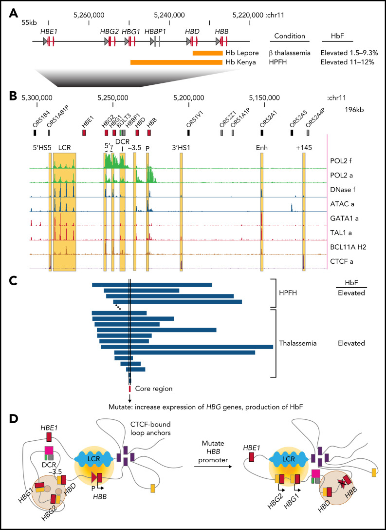
Genetic studies of inherited diseases and variants of hemoglobin have long provided insights and models for problems in biomedical research, ranging from regulation of gene expression to development of novel therapeutics. For example, in studies dating back 50 to 60 years, structural determination of unusual hemoglobins revealed that Hb Lepore contained a hybrid of δ-globin and β-globin and Hb Kenya contained a hybrid of γ-globin and β-globin.2 These results led to a proposed linkage arrangement that allowed formation of these fusion genes by unequal crossing over, and they also revealed a striking contrast in the accompanying phenotypes (see figure panel A). Hb Lepore is associated with anemia, and Hb Kenya is associated with the benign condition hereditary persistence of fetal hemoglobin (HPFH). These differing phenotypes for overlapping gene fusions suggested that an element in the differentially deleted region was required for silencing fetal hemoglobin in adult erythroid cells.2
Activation of fetal HBG genes by disruption of the HBB promoter supports a model of promoter competition for an enhancer for switching in gene expression. (A) The globin genes within the HBB cluster are shown with rectangles for exons (red for genes encoding globins, gray for the pseudogene) and triangles for promoters. The genomic intervals deleted to form fusion genes are indicated as orange rectangles, along with phenotypes in individuals. The interval chr11:5,220,001-5,275,000 in assembly GRCh38 is shown in reverse orientation so that genes are arranged 5′ to 3′ left to right. HBE1 encodes the embryonic ɛ-globin; other gene symbols are defined in the text. (B) An expanded view of the HBB cluster includes surrounding olfactory receptor genes (OR), with genes shown as single rectangles (all are in the same orientation). Selected regulatory (LCR, 5′γ indicating sites immediately upstream from the HBG genes, differential contact region [DCR], −3.5 for an element upstream of HBD,8 P for HBB promoter, and Enh) and structural (CTCF-bound 5′HS5, 3′HS1, +145 sites) elements are marked by yellow underlays for the sequence-based signal tracks of epigenetic features, including occupancy by RNA polymerase II (POL2) in fetal (f) and adult (a) erythroblasts, chromatin accessibility to DNase or transposase (Assay for Transposase-Accessible Chromatin [ATAC]), and occupancy by GATA1, TAL1, and CTCF in adult erythroblasts and BCL11A in HUDEP-2 cells. The genomic interval shown is chr11:5 109,001-5,305,000 in reverse orientation. (C) The intervals deleted in several alleles associated with HPFH or thalassemia (from the HbVar database) are shown as blue rectangles in register with the features shown in panel B; these are a subset of the deletions studied in Topfer et al. A browser session with the tracks shown in panels B and C allows further exploration and gives links to references and further information (https://main.genome-browser.bx.psu.edu/cgi-bin/hgTracks?hgS_doOtherUser=submit&hgS_otherUserName=ross&hgS_otherUserSessionName=BloodCommentary_Crossley and http://usevision.org). (D) A model for competition between the promoters for the adult HBB gene and the fetal HBG genes for activation by proximity to the LCR enhancer (blue lobes), encompassing chromatin interaction results from multiple sources.1,3,8 The diagram indicates the shift in promoter interactions with the LCR enhancer upon disruption of the HBB promoter. The brown disks on the 5′γ elements represent BCL11A, the gold gradient represents a zone of high transcriptional activity centered on the LCR, the light brown disk represents a repressive zone, and the gray line represents DNA.
Over the ensuing decades, myriad studies greatly increased our understanding of the HBB gene cluster and elements regulating tissue-specific and developmental stage–specific expression (see figure panel B). Binding sites for the architectural factor CTCF demarcate domains of chromatin interactions that separate the HBB cluster from adjacent regions.3 The locus control region (LCR), a strong distal enhancer comprising multiple elements marked by chromatin accessibility, transcription, active epigenetic states, and binding by erythroid transcription factors (eg, Xu et al4), mediates high-level expression of any gene in the HBB cluster.5 The transcription factors BCL11A and ZBTB7A play major roles in adult-stage repression of the HBG genes,6 with BCL11A binding directly to regulatory sites upstream of the HBG genes.7 The DNA interval encompassing BGLT3 and the pseudogene HBBP1 contacts different regions of the HBB cluster in fetal and adult erythroblasts (differential contact region), which has been implicated in developmental switching.3 Additional regulatory elements have been mapped upstream of the HBD gene8 (encoding δ-globin) and distal to the locus. However, these and many other studies not summarized here do not fully explain the process of hemoglobin switching and how it is impacted by the diverse, naturally occurring mutations found in this gene cluster.
Topfer et al took a new approach to identifying candidate elements responsible for elevated levels of HbF in individuals who carry deletions within the HBB cluster. Traditionally, these deletions are categorized as associated with either δβ-thalassemia, which is characterized by microcytosis and heterocellular HbF, or with HPFH, a benign condition with increased, pancellular HbF and morphologically normal erythrocytes. HbF levels vary considerably among individuals, but they can be elevated in both conditions. Focusing on levels of HbF as the relevant endophenotype, Topfer et al compared the breakpoints of 23 thalassemia deletions and 13 HPFH deletions (some shown in figure panel C), all of which were associated with elevated (>2%) HbF in adult erythroid cells. A short core region was common to all these deletions. Remarkably, this core region encompassed the promoter and first exon of the HBB gene, which leads to the hypothesis that loss of the HBB promoter causes the observed increases in HBG gene expression. Indeed, such an effect could explain the HbF endophenotype for Hb Lepore and Hb Kenya (see figure panel A). This hypothesis derives from a model positing that reciprocal expression of HBB vs HBG is mediated by competition for the common upstream enhancer (LCR).9
Topfer et al tested this hypothesis through a series of CRISPR/Cas9-directed mutagenesis experiments. By using the HUDEP-2 erythroid cell line, which is capable of undergoing a β- to γ-globin switch, they showed that deletion of the entire HBB promoter or transcription factor binding sites within it caused increased expression of the fetal HBG genes in cis. Similar results were observed in primary human erythroblasts, confirming the hypothesized role of the HBB promoter in silencing HBG genes in adult erythroid cells. Was the mechanism through competition for the LCR enhancer? By using high-resolution mapping of chromatin contacts, Topfer et al observed strong interaction of the HBB gene with this enhancer in HUDEP-2 cells with the HBB promoter intact, but those contacts were diminished upon deletion of the HBB promoter whereas contacts with the HBG genes increased. These results are precisely those predicted by the promoter competition model (see figure panel D).
This new work provides compelling support for a simple enhancer competition model for switches in gene expression. However, the simplicity belies the complex and dynamic structures within which the competition occurs. One can connect multiple elements implicated in hemoglobin switching3-8 in a competition model (see figure panel D), but many questions are still unanswered. The gene configurations diagrammed in figure panel D are static images of interpretations of population averages in which the underlying structures are dynamic and allow for switching between fetal and adult genes.10 This model also accommodates the direct repressive functions of BCL11A and ZBTB7A at the HBG genes: these factors might create a local environment that tips the balance of competition for the LCR in favor of the adult HBB and HBD genes. Promoter competition might be modulated further by architectural elements that facilitate one configuration over another.3 The study by Topfer et al joins many recent articles identifying DNA elements and transcription factors that have roles in reactivating fetal HBG genes in adult erythroid cells. All of these are candidate targets for potential therapeutic interventions, which makes this an exciting time to study hemoglobin switching.
Supplementary Material
Footnotes
Conflict-of-interest disclosure: The author declares no competing financial interests.
REFERENCES
- 1.Topfer SK, Feng R, Huang P, et al. Disrupting the adult globin promoter alleviates promoter competition and reactivates fetal globin gene expression. Blood. 2022;139(14):2107-2118. [DOI] [PMC free article] [PubMed] [Google Scholar]
- 2.Kendall AG, Ojwang PJ, Schroeder WA, Huisman TH. Hemoglobin Kenya, the product of a gamma-beta fusion gene: studies of the family. Am J Hum Genet. 1973; 25(5):548-563. [PMC free article] [PubMed] [Google Scholar]
- 3.Huang P, Keller CA, Giardine B, et al. Comparative analysis of three-dimensional chromosomal architecture identifies a novel fetal hemoglobin regulatory element. Genes Dev. 2017;31(16):1704-1713. [DOI] [PMC free article] [PubMed] [Google Scholar]
- 4.Xu J, Shao Z, Glass K, et al. Combinatorial assembly of developmental stage-specific enhancers controls gene expression programs during human erythropoiesis. Dev Cell. 2012;23(4):796-811. [DOI] [PMC free article] [PubMed] [Google Scholar]
- 5.Li Q, Peterson KR, Fang X, Stamatoyannopoulos G. Locus control regions. Blood. 2002;100(9):3077-3086. [DOI] [PMC free article] [PubMed] [Google Scholar]
- 6.Sankaran VG, Menne TF, Xu J, et al. Human fetal hemoglobin expression is regulated by the developmental stage-specific repressor BCL11A. Science. 2008;322(5909):1839-1842. [DOI] [PubMed] [Google Scholar]
- 7.Martyn GE, Wienert B, Yang L, et al. Natural regulatory mutations elevate the fetal globin gene via disruption of BCL11A or ZBTB7A binding. Nat Genet. 2018;50(4):498-503. [DOI] [PubMed] [Google Scholar]
- 8.Shen Y, Verboon JM, Zhang Y, et al. A unified model of human hemoglobin switching through single-cell genome editing. Nat Commun. 2021;12(1):4991. [DOI] [PMC free article] [PubMed] [Google Scholar]
- 9.Gallarda JL, Foley KP, Yang ZY, Engel JD. The beta-globin stage selector element factor is erythroid-specific promoter/enhancer binding protein NF-E4. Genes Dev. 1989; 3(12A):1845-1859. [DOI] [PubMed] [Google Scholar]
- 10.Bartman CR, Hsu SC, Hsiung CC, Raj A, Blobel GA. Enhancer regulation of transcriptional bursting parameters revealed by forced chromatin looping. Mol Cell. 2016;62(2):237-247. [DOI] [PMC free article] [PubMed] [Google Scholar]
Associated Data
This section collects any data citations, data availability statements, or supplementary materials included in this article.


