Abstract
Functions of the photosynthetic apparatus of C3 (Pisum sativum L.) and C4 (Zea mays L.) plants under physiological conditions and after treatment with different NaCl concentrations (0–200 mM) were investigated using chlorophyll a fluorescence (pulse-amplitude-modulated (PAM) and JIP test) and P700 photooxidation measurement. Data revealed lower density of the photosynthetic structures (RC/CSo), larger relative size of the plastoquinone (PQ) pool (N) and higher electron transport capacity and photosynthetic rate (parameter RFd) in C4 than in C3 plants. Furthermore, the differences were observed between the two studied species in the parameters characterizing the possibility of reduction in the photosystem (PSI) end acceptors (REo/RC, REo/CSo and δRo). Data revealed that NaCl treatment caused a decrease in the density of the photosynthetic structures and relative size of the PQ pool as well as decrease in the electron transport to the PSI end electron acceptors and the probability of their reduction as well as an increase in the thermal dissipation. The effects were stronger in pea than in maize. The enhanced energy losses after high salt treatment in maize were mainly from the increase in the regulated energy losses (ΦNPQ), while in pea from the increase in non-regulated energy losses (ΦNO). The reduction in the electron transport from QA to the PSI end electron acceptors influenced PSI activity. Analysis of the P700 photooxidation and its decay kinetics revealed an influence of two PSI populations in pea after treatment with 150 mM and 200 mM NaCl, while in maize the negligible changes were registered only at 200 mM NaCl. The experimental results clearly show less salt tolerance of pea than maize.
Keywords: NaCl treatment, JIP test, PAM chlorophyll fluorescence, pea, photooxidation of P700, maize
1. Introduction
Plants in their development are exposed to various environmental stresses. Salinity is one of the most widespread environmental factors, which limits plant growth and crop productivity [1,2]. Toxic sodium levels in plants harm biological membranes and subcellular organelles, causing an inhibition of biochemical and physiological processes [3]. The harmful effects of salinity on the plant are caused by osmotic stress and ionic stress, and subsequent oxidative damage often occurs [4,5]. Oxidative damage of lipids and proteins leads to damage of the photosynthetic apparatus and a decrease in the photosynthetic activity [6,7,8]. The enhanced salt-induced reactive oxygen species (ROS) cause lipid peroxidation leading to increased membrane fluidity and permeability [9]. High saline conditions also lead to a reduction in the total pigment content and impact on the chloroplast structure [10,11,12]. It has been shown that salinization leads to swelling of chloroplasts, increasing the number and size of plastoglobuli, destroying of thylakoid membranes in these organelles, and subsequent destruction of the structure of these photosynthetic organelles [8,13,14,15,16]. These changes inhibit the functions of the photosynthetic electron transport chain, which depend on the plant species [12,17]. Salinity affects photosystem II (PSII) more than photosystem I (PSI) [10]. The inhibition of PSII functions is a result of disintegrating the PSII reaction center, the oxygen-evolving complex (OEC) and by limiting the activity of quinone acceptors [10]. The influence of the salt stress on the photosynthetic components, as well as the extent of damage, is determined by the strength and duration of the stress [12,18].
The most common plants can be divided into two major groups, as C3 photosynthesis and C4 photosynthesis, depending on the first product of the CO2 fixation [19,20,21]. The photosynthetic CO2 assimilation rates, the photosynthetic efficiency, the biomass production and the efficient use of light in C4 plants are higher than in C3 plants [22,23]. Previous studies also revealed that the ratios chlorophyll a/b, lipid/chlorophyll and lipid/protein, as well as the mobility of pigment–protein complexes in thylakoid membranes of C3 and C4 plants are different [24,25,26,27]. Furthermore, the plants with these two photosynthetic pathways respond quite differently to changes in the environmental conditions. Differences in antioxidant defense under drought and salinity in C3 and C4 plants have also been shown [28,29].
Chlorophyll fluorescence is widely used to study the influence of the abiotic stress factors on photosynthetic performance [30] and gives information about the energy transfer in the photosynthetic apparatus and related photosynthetic processes [31,32,33,34,35,36]. The widespread use of chlorophyll fluorescence is determined by the fact that it is a fast, informative and non-invasive method. One of the most widely used measurement techniques for characterization of the functions of photosynthetic apparatus is pulse-amplitude modulated (PAM) and chlorophyll a fluorescence induction. The parameters of PAM chlorophyll fluorescence can be used to determine both the structural differences between dark-adapted state and light-adapted state and the photosynthetic performance at light-adapted state [37]. A rapid increase in the chlorophyll fluorescence in dark-adapted plants after exposure to light (JIP parameters) describes the primary photosynthetic reactions and evaluates important PSII characteristics, such as energy trapping, electron transport and dissipation of the excitation energy in the antenna complexes [38,39]. In recent years, the JIP test has been used for understanding the mechanism of action of different stress factors on the plants.
We hypothesized that differences in the structure, functions and antioxidant activity of the C3 and C4 plants would differentially affect the activity of different components of the photosynthetic apparatus under salt stress. Using chlorophyll fluorescence (PAM and chlorophyll a fluorescence induction), we evaluated the effects of different concentrations of NaCl on the primary processes of photosynthesis in pea (Pisum sativum L.) and in maize (Zea mays L.). Data clearly show the influence of NaCl concentration on the various components of the electron transport chain, as well as the higher sensitivity of pea compared with maize at different levels of salinization.
2. Results
2.1. Effects of Salinity on the Chlorophyll a Fluorescence
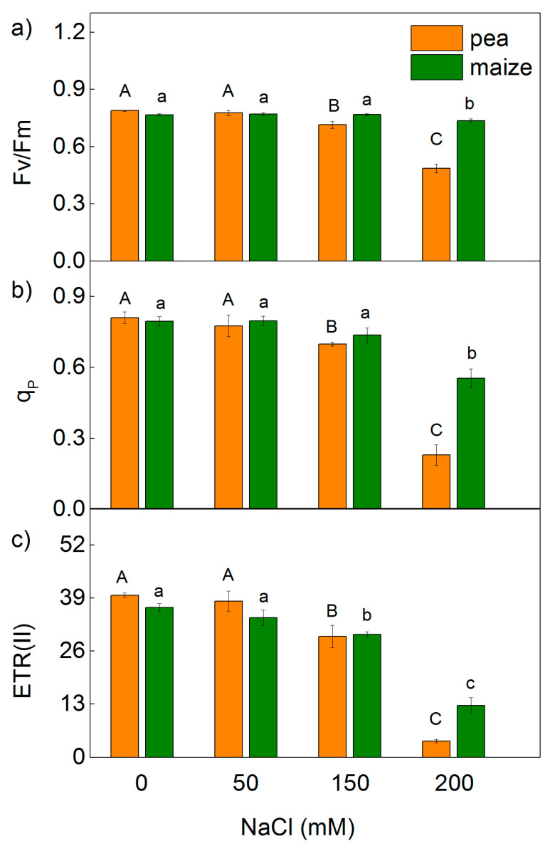
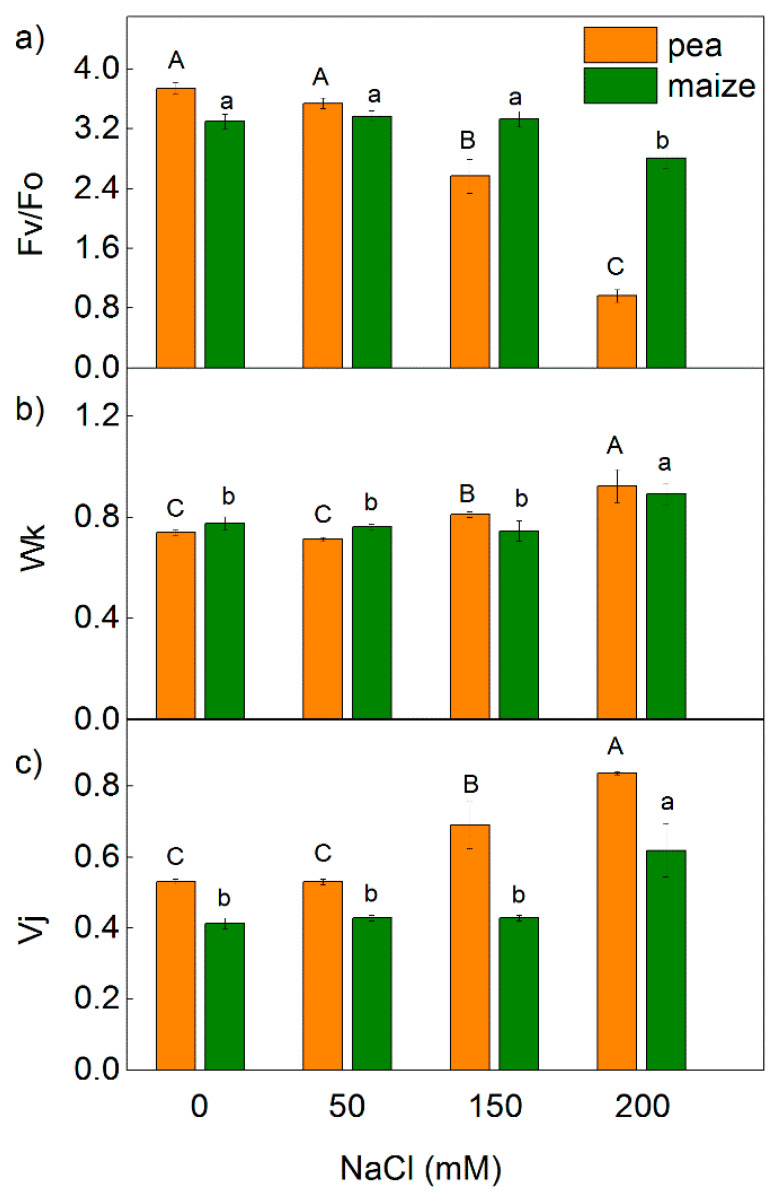
Analysis of the PAM chlorophyll fluorescence signals revealed a strong influence on the maximum quantum yields of primary photochemistry of PSII (Fv/Fm), the ratio of photochemical to non-photochemical processes in PSII (Fv/Fo), the photochemical quenching (qp) and the PSII based electron transport rate (ETR(II)) in both studied species exposed to the highest NaCl concentration (200 mM NaCl) (Figure 1 and Figure 2). The effects on these parameters were stronger in pea than in maize. The decrease in the ETR(II) was 90% in pea and 65% in maize, while qp decreased by 72% and 30% in pea and maize, respectively (Figure 1 and Figure 2). Experimental results also reveal that after treatment with 150 mM, NaCl changes in Fv/Fm, Fv/Fo, qp and ETR(II) were registered only in pea. In addition, data reveal that the changes in the parameters Fv/Fm and Fv/Fo were smaller than ETR(II) and qp. Data also demonstrate that the values of the studied PAM parameters (Fv/Fm, Fv/Fo, qp and ETR(II)) after the treatment with 50 mM NaCl were similar to the untreated plants in both studied species.
Figure 1.
Effects of different NaCl concentrations on the selected parameters of PAM chlorophyll fluorescence in leaves of pea and maize: (a) the maximal quantum yield in dark-adapted state (Fv/Fm); (b) the photochemical quenching (qp); (c) PSII based electron transport rate (ETR(II)). The parameters are in relative units. Mean values (±SE) are from 10 independent measurements. Different letters indicate significant differences for the respective parameters at p < 0.05 (uppercase for pea and lowercase for maize).
Figure 2.
Effects of different NaCl concentrations on the selected parameters of chlorophyll fluorescence (Table 1) in leaves of pea and maize: (a) the ratio of photochemical to non-photochemical processes in PSII (Fv/Fo); (b) JIP parameter Wk; (c) JIP parameter Vj. The parameters are in relative units. Mean values (±SE) are from 10 independent measurements. Different letters indicate significant differences for the respective parameters at p < 0.05 (uppercase for pea and lowercase for maize).
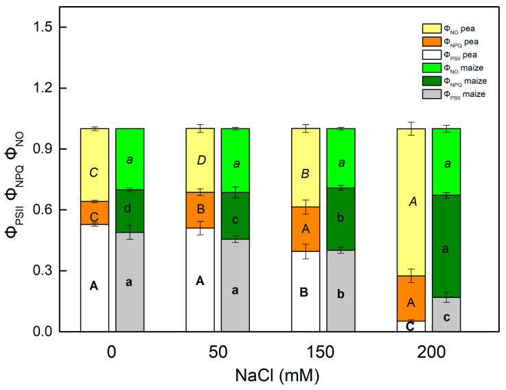
The impact of different levels of salinity on the light energy utilization in PSII in the C3 and C4 plants was determined by the effective quantum yield of the photochemical energy conversion in PSII (ΦPSII) and the quantum yields of regulated (ΦNPQ) and non-regulated (ΦNO) energy losses in PSII. The sum of the parameters ΦPSII, ΦNPQ and ΦNO is equal to 1 [49,50]. High concentrations of NaCl (150–200 mM) more strongly affected the parameter ΦPSII in pea than in maize (Figure 3). After NaCl treatment this parameter in pea decreased by 25% at 150 mM and 90% at 200 mM, while in maize the decrease was from 18% (at 150 mM) to 65% (at 200 mM). Data also reveal that the influence on the quantum yields of regulated (ΦNPQ) and non-regulated (ΦNO) energy losses in PSII after NaCl treatment was different in the studied plants. The enhancement of the energy losses in pea was connected mainly with an increase in the ΦNO, while in maize with an increase in ΦNPQ (Figure 3).
Figure 3.
Effects of different NaCl concentrations in leaves of pea and maize on the effective quantum yield of a photochemical energy conversion of PSII (ΦPSII), the regulated (ΦNPQ) and non-regulated (ΦNO) energy loss in PSII. The parameters are in relative units. Mean values (±SE) are from 10 independent measurements. Different letters indicate significant differences for the respective parameters at p < 0.05 (uppercase for pea and lowercase for maize).
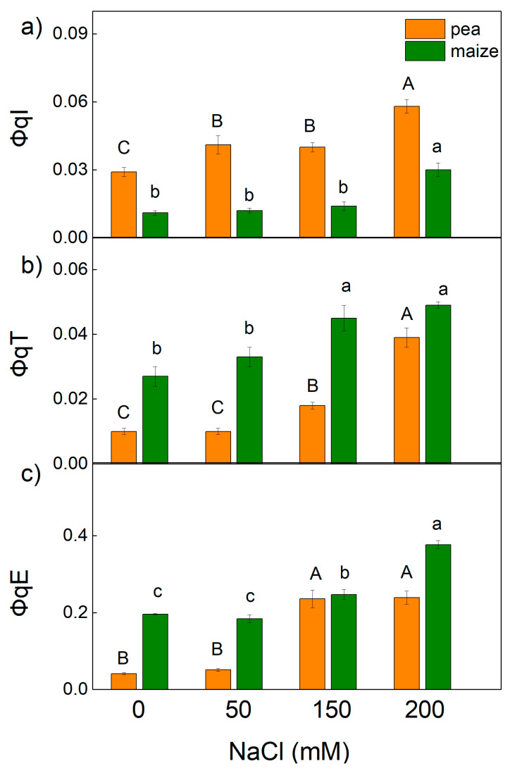
The non-photochemical quenching of chlorophyll fluorescence (NPQ) is an important photoprotective mechanism in plants [51]. It has been shown that NPQ involves three components: the energy-dependent quenching (qE), mediated by the proton gradient across the thylakoid membrane; the state transition quenching (qT), induced by the reversible phosphorylation of the light-harvesting complex of PSII (LHCII); and the photoinhibitory quenching (qI)) [52]. The impact of different levels of salinity on the quantum yields of these components (ΦqE, ΦqT and ΦqI) is shown in Figure 4. The values of components ΦqE and ΦqT were higher in maize compared with pea, but values of ΦqI were higher in pea than in maize in the treated and untreated plants. In addition, data revealed an increase in the ΦqI in pea treated with all NaCl concentrations, while in maize an increase in this component was registered only at 200 mM NaCl. The components ΦqE and ΦqT increased after treatment with higher NaCl concentrations (150 mM and 200 mM) in both studied plant species, but the changes were more pronounced in pea than in maize (Figure 4).
Figure 4.
Effects of different NaCl concentrations on the quantum yields of NPQ components: (a) ΦqI (photoinhibitory component), (b) ΦqT (state transition component) and (c) ΦqE (energy-dependent component) in leaves of pea and maize. The parameters are in relative units. Mean values (±SE) are from 10 independent measurements. Different letters indicate significant differences for the respective parameters at p < 0.05 (uppercase for pea and lowercase for maize).
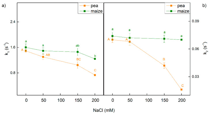
The kinetic of dark relaxation of chlorophyll fluorescence induced by a single saturating light pulse in dark-adapted leaves gives information for the electron transfer between QA and plastoquinone [53,54]. The fluorescence signals could be fitted to two components with fast (k1) and slow (k2) rate constant in treated and untreated plants of pea and maize. These constants were less influenced in maize than in pea after treatment with NaCl (Figure 5). In maize, k1 was influenced only after treatment with 200 mM NaCl, while k2 was similar to control plants. Data also reveal that in pea both constants decreased after treatment with 150 mM and 200 mM NaCl, as the effect was more pronounced for constant k2 (Figure 5).
Figure 5.
Effects of different NaCl concentrations on the dark relaxation of chlorophyll fluorescence induced by a single saturating light pulse in leaves of pea and maize. (a) Constant of the fast component (k1); (b) constant of the slow component (k2). Mean values (±SE) are from 10 independent measurements. Different letters indicate significant differences for the respective parameters at p < 0.05 (uppercase for pea and lowercase for maize).
2.2. Effects of Salinity on the Rate of Photosynthesis
The chlorophyll fluorescence decay ratio (RFd), which correlates with the net assimilation of CO2 [55], was used to assess the influence of salinity on the rate of photosynthesis.
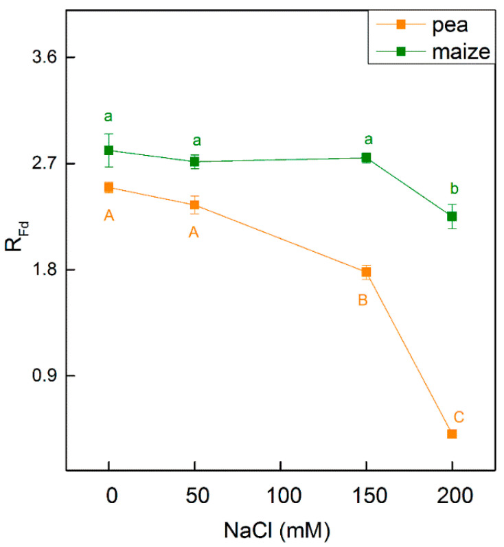
Salt-induced changes in the photosynthetic apparatus led to a decrease in the RFd parameter in both studied species (Figure 6). Data reveal that the parameter RFd decreases in pea treated with 150 mM NaCl (by 29%) and 200 mM NaCl (by 84%), while in maize an influence was observed only at the highest NaCl concentration (at 200 mM NaCl, decreases by 20%) (Figure 6).
Figure 6.
Effects of different NaCl concentrations on the chlorophyll fluorescence decay ratio RFd in leaves of pea and maize. The parameter is in relative units. Mean values (±SE) are from 10 independent measurements. Different letters indicate significant differences for the respective parameters at p < 0.05 (uppercase for pea and lower case are for maize).
2.3. Effects of Salinity on the Chlorophyll Fluorescence Induction
For more information about the influence of the different levels of salinity on the photosynthetic apparatus, selected JIP parameters were calculated (Table 1).
Table 1.
Description of the selected parameters of chlorophyll fluorescence, based on information presented in [40,41,42,43,44,45,46,47,48]. All parameters are in relative units.
| JIP Parameters | |
|---|---|
| ABS/RC | Absorption flux per RC (apparent antenna size of an active RC) |
| ETo/RC | Electron transport flux (further than QA−) per RC |
| REo/RC | Electron flux reducing end electron acceptors at the PSI acceptor side per RC |
| TRo/RC | Trapping flux (leading to QA reduction) per RC |
| DIo/RC | Dissipated energy flux per RC (at t = 0) |
| RC/ABS | The numbers of active RC per PSII antenna chlorophyll |
| ABS/CSo | Light energy (photons) absorption flux per cross section |
| ETo/CSo | Electron transport flux from QA to QB per cross section |
| REo/CSo | Electron transport flux until PSI acceptors per cross section |
| TRo/CSo | Maximum trapped exciton flux per cross section |
| DIo/CSo | Dissipated energy flux per cross section at t = 0 |
| RC/CSo | Density of RCs (QA reducing PSII RC) |
| φPo | Maximum quantum yield of primary photochemistry (at t = 0) |
| φEo | Quantum yield of electron transport (at t = 0) |
| φRo | Quantum yield of reduction in end electron acceptors at the PSI acceptor side |
| ψEo | Moves an electron into the electron transport chain beyond QA− |
| δRo | Efficiency/probability with which an electron from the intersystem electron carriers moves to reduce end electron acceptors at the PSI acceptor side |
| PIABS | Performance index (potential) for energy conservation from exciton to the reduction in intersystem electron acceptors |
| PI total | Performance index (potential) for energy conservation from exciton to the reduction in PSI end acceptors |
| N | Maximum turnovers of QA reduction until Fm was reached |
| Vj | Relative variable fluorescence at the J step |
| Wk | The ratio of the K phase to the J phase |
| PAM parameters | |
| Fv/Fm | The maximum quantum yields of primary photochemistry of PSII |
| Fv/Fo | The ratio of photochemical to non-photochemical processes in PSII |
| qp | The photochemical quenching |
| ETR(II) | PSII based electron transport rate |
| ΦPSII | The photochemical energy conversion in PSII |
| ΦNPQ | The quantum yields of regulated energy losses in PSII |
| ΦNO | The quantum yields of non-regulated energy losses in PSII |
| RFd | The fluorescence decrease from Fm to a steady state chlorophyll fluorescence after continuous saturated illumination |
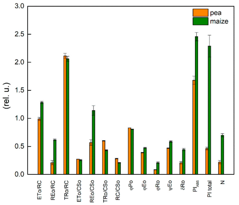
Comparison of two studied plants under physiological conditions revealed differences in parameters ETo/RC, REo/RC, REo/CSo, TRo/CSo, RC/CSo φEo, φRo, ψEo, δRo and N (Figure 7). The data show a low density of the photosynthetic structure (RC/CSo), but a larger relative size of the PQ pool per reaction center (N) and more efficient operation of PSI in the maize compared with the pea. The value for REo/RC was 3 times higher in maize than pea. In addition, the data reveal that parameters which characterized quantum efficiency of the processes in the photosynthetic apparatus (φEo, φRo, ψEo) also had higher values in maize than in pea—i.e., the electron transport efficiency is higher in C4 than in C3 plants. The differences between the studied plants were larger in electron transport flux until the PSI end electron acceptors (REo/RC) and quantum yields for reduction in PSI end electron acceptors (φRo).
Figure 7.
Selected JIP parameters (Table 1) were measured in leaves of pea and maize under physiological conditions. Mean values (±SE) were calculated from 20 independent measurements.
Significant differences between the studied untreated plants were found in the performance indices (PIABS, PI total), as the values of these parameters were much higher in maize compared with pea (Figure 7). The index PIABS includes three parameters: the number of active RC per PSII antenna chlorophyll [γRC2/(1 − γRC2) = RC/ABS], the partial performance of primary photochemistry [φPo/(1 − φPo)] and the performance of thermal reactions of the intersystem electron carriers [ψEo/(1 − ψEo)] [35,41,56]. Higher values of PIABS in maize were the result of the high value of the third component [ψEo/(1 − ψEo)], which characterizes non-light-dependent reactions (Table 2). The observed differences in the PI total between pea and maize were the result of the differences in the efficiency/probability with which an electron is transferred from QB to PSI electron acceptors [(δREo/(1 − δREo)] (Table 2). The values of this component were about 3 times higher in the maize than the pea.
Table 2.
Effects of different NaCl concentrations on the components of the performance indices PIABS and PI total in leaves of pea and maize. The parameters are in relative units. Mean values (±SE) are from 20 independent measurements. Different letters indicate significant differences for the respective parameters at p < 0.05 (uppercase for pea and lowercase for maize).
| RC/ABS | φPo/(1 − φPo) | ψEo/(1 − ψEo) | δREo/(1 − δREo) | |
|---|---|---|---|---|
| pea | ||||
| control | 0.393 ± 0.008 A | 4.837 ± 0.077 A | 0.883 ± 0.025 A | 0.272 ± 0.054 A |
| 50 mM NaCl | 0.425 ± 0.010 A | 4.908 ± 0.084 A | 0.889 ± 0.028 A | 0.201 ± 0.030 A |
| 150 mM NaCl | 0.356 ± 0.012 B | 3.793 ± 0.034 B | 0.668 ± 0.109 B | 0.245 ± 0.056 A |
| 200 mM NaCl | 0.112 ± 0.004 C | 1.360 ± 0.005 C | 0.484 ± 0.025 C | 0.100 ± 0.010 B |
| maize | ||||
| control | 0.392 ± 0.010 a | 4.174 ± 0.101 a | 1.440 ± 0.090 a | 0.801 ± 0.071 a |
| 50 mM NaCl | 0.393 ± 0.013 a | 4.300 ± 0.066 a | 1.341 ± 0.042 a | 0.880 ± 0.086 a |
| 150 mM NaCl | 0.403 ± 0.004 a | 4.079 ± 0.119 a | 1.341 ± 0.040 a | 0.925 ± 0.103 a |
| 200 mM NaCl | 0.313 ± 0.043 b | 2.582 ± 0.009 b | 0.730 ± 0.095 b | 0.619 ± 0.067 b |
Equations for performance indices:
| PIABS = RC/ABS × φPo/(1 − φPo) × ψEo/(1 − ψEo); |
| PI total = PIABS × δREo/(1 − δREo) |
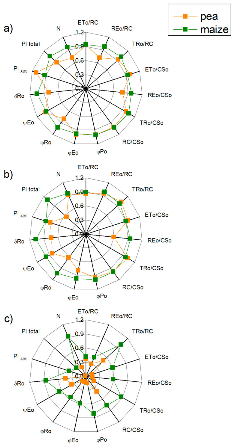
After treatment with 50 mM NaCl, the values of the measured JIP parameters (Table 1) in maize were similar to the values of the control plants, while negligible effects on them in pea were observed. Data revealed a small decrease in the parameters REo/RC, REo/CSo, φRo, δRo and N in the pea than in the control, while in the maize the values of these parameters are similar to the untreated plants (Figure 8).
Figure 8.
Effects of different NaCl concentrations on selected OJIP parameters (Table 1) in leaves of pea and maize plants grown in 50 mM NaCl (a), 150 mM NaCl (b) and 200 mM NaCl (c). The parameters are normalized to the respective control. Mean values (±SE) are from 20 independent measurements.
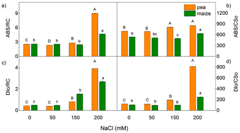
The treatment with 150 mM and 200 mM NaCl influenced all studied parameters, as the effects were more pronounced in the pea compared with the maize (Figure 8 and Figure 9). After treatment of pea with 150 mM NaCl, changes were observed in the electron transport capacity (φEo, φRo, δRo, REo/CSo), absorbance flux per cross-section (ABS/CSo) and the processes related to energy dissipation (DIo/RC and DIo/CSo) (Figure 8 and Figure 9). At the same time, the changes in these parameters were negligible in the maize. In addition, the changes in parameters Vj and Wk were registered in both studied plants after treatment with 200 mM NaCl (Figure 2). The increased value of Wk indicates damage of the OEC after salt treatment. An increase in the values of Vj after treatment with 150 mM NaCl was observed only in pea (Figure 2). After treatment with the highest NaCl concentration (200 mM), the effects on the JIP parameters were stronger in the pea than in the maize (Figure 2, Figure 8 and Figure 9).
Figure 9.
Effects of different NaCl concentrations on the selected JIP parameters (Table 1) in leaves of pea and maize. (a) absorption flux per RC (ABS/RC), (b) light energy absorption flux per CS (ABS/CSo), (c) dissipated energy flux per RC (DIo/RC), (d) dissipated energy flux per CS (DIo/CSo). The parameters are in relative units. Mean values (±SE) are from 20 independent measurements. Different letters indicate significant differences for the respective parameters at p < 0.05 (uppercase for pea and lowercase for maize).
Treatment with high salt concentration led to a stronger influence of salinity on the performance index (PI total) measuring the energy conservation from exciton to the reduction in PSI end acceptor) than the performance index on the absorption base (PIABS, energy conservation from exciton to the reduction in intersystem electron acceptors) (Figure 8). The data reveal that PI total after treatment with 200 mM NaCl decreased by 98% and 73% for pea and maize, respectively. Performance index PI total (45%) also decreased (45%) in pea after treatment with 150 mM NaCl. The performance index PIABS was also less affected by salinity in maize (decreases with 11% at 150 mM and 63% at 200 mM NaCl) than in pea (21% and 99% decrease for 150 mM and 200 mM NaCl, respectively) (Figure 8).
2.4. P700 Photooxidation
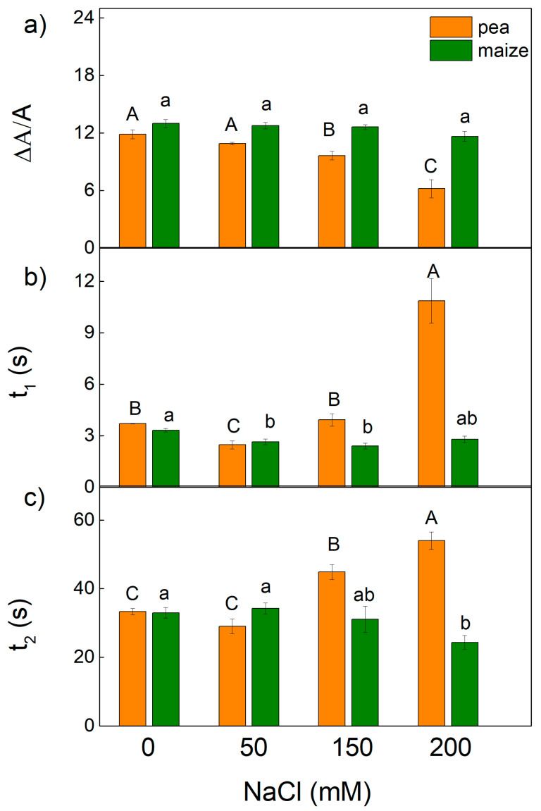
Additional information for the influence of salinity on PSI function was provided by the study of P700 photooxidation by far-red light. The far-red light-induced steady-state oxidation of P700 (ΔA/A) and the times t1 and t2 of the P700+ dark reduction were calculated (Figure 10). The parameter ΔA/A in pea decreased after treatment with high NaCl concentrations (150 mM and 200 mM), while in maize the values at these concentrations were similar to the control. The dark-reduction in P700+ was deconvoluted into two exponential components with times t1 and t2 for the fast and the slow exponents, respectively. The influence of higher NaCl concentrations on the t1 and t2 was less in maize than pea. In addition, data revealed a decrease in t1 in maize after treatment with 50 mM (by 21%) and 150 mM NaCl (by 27%), while in pea the decrease in t1 (by 33%) was registered only after treatment with 50 mM NaCl (Figure 10). At the same time, a decrease in the t2 was registered after treatment with 200 mM in maize plants, while in pea an increase in t2 was observed after treatment with 150 mM and 200 mM NaCl (Figure 10).
Figure 10.
Effects of different NaCl concentrations on the far-red light-induced oxidation of P700 (ΔA/A) (the parameter is in relative units) (a), and the times of fast t1 (b) and slow t2 (c) of two components of dark reduction in the P700+ in leaves of pea and maize. Mean values (±SE) are from 10 independent measurements. Different letters indicate significant differences for the respective parameters at p < 0.05 (uppercase for pea and lowercase for maize).
3. Discussion
The influence of salinity on plants is a complex phenomenon. This study for the first time focused on the effects of different NaCl concentrations on the photosynthetic apparatus in C3 and C4 plants. Pisum sativum L. was used as a model for C3 plants, and Zea mays L. was chosen to represent C4 plants.
Previous studies and the present study revealed some differences in the amount of chlorophyll and Chl a/b ratio in pea and maize under physiological conditions [24,25,26,27,57] and Supplementary Table S1. Based on these studies, different organizations of the thylakoid membranes in C3 and C4 plants could be suggested. It was also shown that the smaller Chl a/b ratio corresponds with the increased amount of chlorophyll in grana and suggests the synthesis of the additional LHCII molecules [58]. Furthermore, the differences in lipid/chlorophyll and in lipid/protein rations in C3 and C4 plants influence the mobility of pigment–protein complexes in the thylakoid membranes [25]. Data in the current study reveal that the value of the widely used parameter Fv/Fm that assesses PSII activity was similar in both studied plant species (Figure 1). The value of this parameter in higher plants is around 0.8 [59,60]. Previous studies have shown a lower or similar value of Fv/Fm ratio in C4 compared with C3 plants [61]. These differences could be due to plant growing conditions as well as to plant species. On the other hand, the reduction in the chlorophyll content could not be the cause for a decrease in the parameter Fv/Fm, which indicates that pigment amount is not associated with the maximum quantum yield of the primary PSII photochemistry. Similar results have been shown in the study of Guha et al. [39]. At the same time, our data reveal a higher photosynthetic rate (parameter RFd) in maize than in pea (Figure 6). This observation is in agreement with the previous studies [22,23,62]. In addition to these observations, the experimental results in this study reveal variation in the functional efficiency of different components of the photosynthetic apparatus in studied plants, which could be a result of differences in the density of the photosynthetic structure (RC/CSo the relative size of the PQ pool (N) and electron transport activity in maize and pea (Figure 7).
Previous investigations revealed the effects of the salinity on the chlorophyll content, the structure of the chloroplast, membrane injury and enhanced amount of ROS [63,64,65]. It has been also shown that high salt concentrations led to a restriction of the electron flow from QA− to the plastoquinone pool, influencing QA− reoxidation by plastoquinone and by the recombination of electrons in QAQB− via the QA−QB ↔ QAQB− charge equilibrium with the oxidized S2 (or S3) state of the OEC [24,45,66]. The results of the present study also show an influence of the salinity on the constants (k1 and k2) characterizing the Fm decay (Figure 5)—i.e., influence on the interaction between QA and PQ. The impact of salinity on these constants was stronger in pea than in maize. All of these salt-induced changes cause a decrease in the efficiency of the photosynthetic machinery. Previous investigations [24,45,67] and the experimental results of the present study revealed a decrease in the potential of PSII efficiency (Fv/Fm) as well as in the ratio of the photochemical to nonphotochemical processes (Fv/Fo) (Figure 1 and Figure 2). Having in mind that the ratio Fv/Fo corresponds to the efficiency of the OEC [68,69,70], it can be concluded that the high salt concentrations induced changes in the donor side of PSII, which were larger in pea than in maize plants. This statement is supported by the changes in the parameter Wk, indicating activity of the OEC (Figure 2). A strong increase in the Wk was found in both studied species after treatment with the highest NaCl concentration (Figure 2), which indicates dissociation of this complex and its damage [71]. Some authors suggest a correlation between changes in this parameter and salt sensitivity of the plants [43]. The suggested modification of the OEC was confirmed by previous study, which revealed that salt-induced changes in the donor side of PSII [72,73]. Data in this study also revealed an increase in the Vj and decrease in the ψEo in both studied plants after treatment with 150 mM and 200 mM NaCl (Figure 2 and Figure 8), which could be a result of the accumulation of reduced QA and limitation of the electron transport beyond QA [74]. In support of this assumption for changes in the PSII acceptor side are the changes in the constants k1 and k2 characterizing the Fm decay (Figure 5), as well as previous studies [24,45,66,72,73].
The changes in the photosynthetic apparatus after salt treatment led to a decrease in the open reaction centers of PSII (qp) and an inhibition of the ETR(II), as these parameters changed differently in pea and maize (Figure 1). The decrease in the efficiency of the open reaction centers [24] and an increase in the closed reaction centers after NaCl treatment (qp decrease, Figure 1) resulted in a decrease in the ΦPSII and an increase in the quantum yields of regulated (ΦNPQ) and non-regulated (ΦNO) energy losses in PSII (Figure 3). The increase in non-regulated energy losses due to PSII inactivation and thermal dissipation indicates an increase in the ROS production, which causes damage of PSII [49,75,76]. The changes in energy losses in maize were mainly due to an increase in regulated energy losses (ΦNPQ), with the largest changes at 200 mM NaCl (Figure 3), but the values of ΦNO were similar to the control. Salt-induced changes in pea after NaCl treatment led to a larger increase in ΦNO in comparison with ΦNPQ. It is well known that non-photochemical quenching (NPQ) is the main photoprotective process that protects photosynthesis under abiotic stress [61]. More detailed information for the dissipation mechanism of the thylakoid membranes gives components of NPQ (qE, qT and qI) [48,77,78,79]. The energy-dependent (qE) and the state transition (qT) quenching are important for photoprotection of the photosynthetic apparatus, while the photoinhibitory quenching can be used to assess the PSII damage [78]. Our data reveal larger values of the qE and qT in maize plants than in pea plants, which could be one of the reasons for better tolerance to high salt concentrations in maize than in pea. Moreover, the strong increase in qI in pea after NaCl treatment suggests damage to the PSII complex (Figure 4).
The impact of salt-induced changes in thylakoid membranes on the PSI activity (the photooxidation of P700+ and its dark reduction kinetics) were different in studied species, and the changes varied in both studied plants depending on NaCl concentrations (Figure 10). It has been suggested that two components of P700+ decay (characterizing with time t1 and t2) originate from two electron-donor systems or due to two different populations of PSI located in different domains of the thylakoid membranes (in stroma lamellae and grana margin) [80,81]. The decrease in t1 corresponds to the stimulation of the cyclic electron transport around PSI, which plays a defensive role in preventing photosynthetic apparatus from oxidative damage under stress conditions [82]. Our data reveal that stimulation of the cyclic electron transport around PSI in pea was detected only at 50 mM NaCl, while in maize at 50 mM NaCl and 150 mM NaCl (Figure 10). The higher NaCl concentration led to a change in t1 and t2 in studied plant species, which suggests an influence on both populations of PSI. Furthermore, the data reveal stronger alteration in PSI in pea than in maize, which is associated with the stronger influence of the electron transport from QA to the PSI electron acceptors and the efficiency of their reduction (Figure 7).
4. Materials and Methods
4.1. Plant Growth Conditions and Treatments
The seedlings from pea (Pisum Sativum L. Ran1) and maize (Zea mays L. Method) were used in this study. Details of seedling growth are given in Stefanov et al. [24]. The plants were grown in a half-strength Hoagland solution. The cultivation of the plants was carried out in a photothermostat under controlled conditions: a 12 h light/dark photoperiod, a light intensity of 150 μmol photons/m2s, 28 °C (daily)/25 °C (night) temperature and 60% relative humidity. After 10 days of growth, NaCl (50 mM, 150 mM and 200 mM) was added to the nutrient solutions for 6 days. The plants that grew without NaCl were used as controls. Two independent experiments (20–25 uniform seedlings for each treatment, about 10 plants in pot) were performed for each treatment. For the analyses, we used the mature leaves (the middle part of the third and the fourth leaves).
4.2. Room-Temperature Chlorophyll Fluorescence
The pulse-amplitude-modulated (PAM) chlorophyll a fluorescence was measured using a PAM fluorometer (model 101/103, Walz GmbH, Effeltrich, Germany). Details for the measurements are described in Stefanov et al. [45]. The dark adaptation of leaves was 20 min. The maximum fluorescence levels in the dark-adapted (Fm) and light-adapted (Fm′) states were obtained with saturated pulses of 3000 μmol photons/m2s, which were provided by a Schott KL 1500 lamp (Schott Glaswerke, Mainz, Germany). The actinic light was 150 μmol photons/m2s. The following parameters were determined: the maximum quantum efficiency of PSII in dark-adapted state, Fv/Fm = (Fm − Fo)Fm; the photochemical quenching, qp = (Fm′ − Fs)/Fv′; the effective quantum yield of photochemical energy conversion of PSII, ΦPSII = (Fm′ − Fs)/Fm′; the PSII based electron transport rate, ETR(II) = ΦPSII × 150 × 0.5; the non-regulated (ΦNO = Fs/Fm) and regulated (ΦNPQ = Fs/Fm′ − Fs/Fm) energy loss in PSII; the ratio of quantum yields of photochemical and concurrent non-photochemical processes in PSII, Fv/Fo = (Fm − Fo)/Fo; [46,47]; the quantum yields of components of the non-photochemical quenching: the energy-dependent quenching, ΦqE, the state transition quenching, ΦqT, and the photoinhibitory quenching, ΦqI [48].
The constants of decay components (k1 for fast and k2 for slow) of the variable fluorescence relaxation after excitation by a saturated light pulse (3000 μmol photons/m2s) in dark-adapted leaves were determined [45,53,54].
The chlorophyll fluorescence decay ratio (RFd = Fd/Fs) was determined, where Fd is the fluorescence decrease from Fm to a steady state chlorophyll fluorescence (Fs) after continuous saturated illumination (3000 μmol photon/m2s). This ratio (RFd) correlates with the net assimilation of CO2 [55].
Induction curves of chlorophyll fluorescence were measured with a Handy PEA+ instrument (Hansatech, Norfolk, UK) as described in Stefanov et al., [24]. The samples were dark-adapted for 20 min at room temperature using leaf clips. Prompt chlorophyll fluorescence was induced by strong light pulse (3000 μmol photons/m2s). All studied variants showed multiphase chlorophyll fluorescence increase during the first second of illumination after dark adaptation. The measured signals were used for calculations of the selected parameters of OJIP transitions (Table 1) [36,41,83,84].
All fluorescence measurements were performed on mature leaves (the middle part of the third and the fourth leaves).
4.3. P700 Photooxidation
The photooxidation of P700 (P700+) was measured on leaf discs, with a dual-wavelength (820 nm) unit (Walz ED 700DW-E) attached to a PAM101E main control unit in the reflectance mode. The details for the measurements are given in Dankov et al. [85]. The dark-adapted (20 min) leaf discs were illuminated with far-red light supplied by a photodiode (102-FR, Walz GmbH, Effeltrich, Germany). Changes in the oxidation of P700 (P700+) were assessed by red light-induced changes in absorbance at 820 nm (∆A). The ∆A/A ratio and the times of dark reduction in P700+ (t1 and t2) were calculated [45].
4.4. Statistical Analysis
Mean values ± SE were calculated from the data for at least two independent treatments with five biological replicates (five plants) of each variant. Statistically significant differences between the studied variants were identified using one-way ANOVA followed by a Tukey’s post hoc test for each parameter. Prior to the test, the assumptions for the normality of raw data (using the Shapiro–Wilk test) and the homogeneity of the variances (using Levene’s test) were checked. The homogeneity of variance test was used to verify the parametric distribution of data. Values were considered statistically different with p < 0.05 after Fisher’s least significant difference post hoc test by using Origin 9.0 software (OriginLab, Northampton, MA, USA).
5. Conclusions
In summary, the data reveal a larger relative size of the PQ pool and higher electron transport activity in maize compared with pea, as well as the lower density of the photosynthetic structure in maize than in pea. A significant difference in the electron transport capacity of the two studied species was observed in the parameters characterizing the possibility of reduction in the PSI end acceptors (REo/RC, REo/CSo and δRo). The treatment with higher NaCl concentrations led to: (i) a decrease in the density of the photosynthetic structure and relative size of the PQ pool; (ii) an increase in the apparent antenna size of an active PSII; (iii) a decrease in the efficiency and quantum yield for reduction in PSI end electron acceptors and increase in the thermal dissipation. All these changes were larger in C3 than C4 plants. Performance indices (PIABS and PI total), which are a sensitive indicator of the activity of the photosynthetic apparatus, were strongly reduced after treatment with high salt concentrations in both studied species, but the effects were more pronounced in C3 than in C4 plants. In addition, our data reveal enhanced energy losses after salt treatment in maize due to an increase in regulated energy losses (ΦNPQ), while in pea due to an increase in non-regulated energy losses (ΦNO). In conclusion, the data clearly show greater salt tolerance in maize than in pea. Furthermore, the experimental results in this study clearly show the high sensitivity of the processes in the photosynthetic apparatus in both C3 and C4 plants under different salinity levels; therefore, salt-induced changes in them could be used for determination of the salt tolerance of plants.
Acknowledgments
This study was supported by the Bulgarian Science Research Fund, KП-06-K36/9, 2019, and the Bulgarian Academy of Sciences, National Research Programme “Young scientists and postdoctoral students” (PMC 353/10.12.2020).
Supplementary Materials
The following supporting information can be downloaded at: https://www.mdpi.com/article/10.3390/ijms23073768/s1.
Author Contributions
Conceptualization, E.L.A.; methodology, M.A.S., G.D.R. and E.L.A.; software, M.A.S.; validation, E.L.A.; formal analysis, E.L.A.; investigation, M.A.S. and G.D.R.; writing—original draft preparation, E.L.A.; writing—review and editing, M.A.S., G.D.R. and E.L.A.; visualization, M.A.S.; supervision and project administration, E.L.A. All authors have read and agreed to the published version of the manuscript.
Funding
This research was funded by the Bulgarian Science Research Fund, KП-06-K36/9, 2019 and the Bulgarian Academy of Sciences, National Research Programme “Young scientists and postdoctoral students” (PMC 353/10.12.2020).
Institutional Review Board Statement
Not applicable.
Informed Consent Statement
Not applicable.
Data Availability Statement
Not applicable.
Conflicts of Interest
The authors declare no conflict of interest.
Footnotes
Publisher’s Note: MDPI stays neutral with regard to jurisdictional claims in published maps and institutional affiliations.
References
- 1.Parihar P., Singh S., Singh R., Singh V.P., Prasad S.M. Effect of salinity stress on plants and its tolerance strategies: A review. Environ. Sci. Pollut. Res. 2015;22:4056–4075. doi: 10.1007/s11356-014-3739-1. [DOI] [PubMed] [Google Scholar]
- 2.Shrivastava P., Kumar R. Soil salinity: A serious environmental issue and plant growth promoting bacteria as one of the tools for its alleviation. Saudi J. Biol. Sci. 2015;22:123–131. doi: 10.1016/j.sjbs.2014.12.001. [DOI] [PMC free article] [PubMed] [Google Scholar]
- 3.Ghezal N., Rinez I., Sbai H., Saad I., Farooq M., Rinez A., Zribi I., Haouala R. Improvement of Pisum sativum salt stress tolerance by bio-priming their seeds using Typha angustifolia leaves aqueous extract. S. Afr. J. Bot. 2016;105:240–250. doi: 10.1016/j.sajb.2016.04.006. [DOI] [Google Scholar]
- 4.AbdElgawad H., Zinta G., Hegab M.M., Pandey R., Asard H., Abuelsoud W. High salinity induces different oxidative stress and antioxidant responses in maize seedlings organs. Front. Plant Sci. 2016;7:276. doi: 10.3389/fpls.2016.00276. [DOI] [PMC free article] [PubMed] [Google Scholar]
- 5.Isayenkov S.V., Maathuis F.J.M. Plant salinity stress: Many unanswered questions remain. Front. Plant Sci. 2019;10:80. doi: 10.3389/fpls.2019.00080. [DOI] [PMC free article] [PubMed] [Google Scholar]
- 6.Arora A., Sairam R.K., Srivastava G.C. Oxidative stress and antioxidative system in plants. Curr. Sci. 2002;82:1227–1238. [Google Scholar]
- 7.Suo J., Zhao Q., David L., Chen S., Dai S. Salinity response in chloroplasts: Insights from gene characterization. Int. J. Mol. Sci. 2017;18:1011. doi: 10.3390/ijms18051011. [DOI] [PMC free article] [PubMed] [Google Scholar]
- 8.Giannakoula A., Therios I., Chatzissavvidis C. Effect of lead and copper on photosynthetic apparatus in citrus (Citrus aurantium L.) plants. The role of antioxidants in oxidative damage as a response to heavy metal stress. Plants. 2021;10:155. doi: 10.3390/plants10010155. [DOI] [PMC free article] [PubMed] [Google Scholar]
- 9.Wong-Ekkabut J., Xu Z., Triampo W., Tang I.M., Tieleman D.P., Monticelli L. Effect of lipid peroxidation on the properties of lipid bilayers: A molecular dynamics study. Biophys. J. 2007;93:4225–4236. doi: 10.1529/biophysj.107.112565. [DOI] [PMC free article] [PubMed] [Google Scholar]
- 10.Arif Y., Singh P., Siddiqui H., Bajguz A., Hayat S. Salinity induced physiological and biochemical changes in plants: An omic approach towards salt stress tolerance. Plant Physiol. Biochem. 2020;156:64–77. doi: 10.1016/j.plaphy.2020.08.042. [DOI] [PubMed] [Google Scholar]
- 11.Zeeshan M., Lu M., Sehar S., Holford P., Wu F. Comparison of biochemical, anatomical, morphological, and physiological responses to salinity stress in wheat and barley genotypes deferring in salinity tolerance. Agronomy. 2020;10:127. doi: 10.3390/agronomy10010127. [DOI] [Google Scholar]
- 12.Hameed A., Ahmed M.Z., Hussain T., Aziz I., Ahmad N., Gul B., Nielsen B.L. Effects of salinity stress on chloroplast structure and function. Cells. 2021;10:2023. doi: 10.3390/cells10082023. [DOI] [PMC free article] [PubMed] [Google Scholar]
- 13.Salama S., Trivedi S., Busheva M., Arafa A.A., Garab G., Erdei L. Effects of NaCl salinity on growth, cation accumulation, chloroplast structure and function in wheat cultivars differing in salt tolerance. J. Plant Physiol. 1994;144:241–247. doi: 10.1016/S0176-1617(11)80550-X. [DOI] [Google Scholar]
- 14.Mitsuya S., Takeoka Y., Miyake H. Effects of sodium chloride on foliar ultrastructure of sweet potato (Ipomoea batatas Lam.) plantlets grown under light and dark conditions in vitro. J. Plant Physiol. 2000;157:661–667. doi: 10.1016/S0176-1617(00)80009-7. [DOI] [Google Scholar]
- 15.Li W. Effect of environmental salt stress on plants and the molecular mechanism of salt stress tolerance. Int. J. Environ. Sci. Nat. Resour. 2017;7:55714. doi: 10.19080/IJESNR.2017.07.555714. [DOI] [Google Scholar]
- 16.Stefanov M., Biswal A.K., Misra M., Misra A.N., Apostolova E.L. Responses of photosynthetic apparatus to salt stress: Structure, function, and protection. In: Pessarakli M., editor. Handbook of Plant and Crop Stress. 4th ed. Taylor & Francis CRC Press; New York, NY, USA: 2019. pp. 233–250. [Google Scholar]
- 17.Demetriou G., Neonaki C., Navakoudis E., Kotzabasis K. Salt stress impact on the molecular structure and function of the photosynthetic apparatus-The protective role of polyamines. Biochim. Biophys. Acta-Bioenerg. 2007;1767:272–280. doi: 10.1016/j.bbabio.2007.02.020. [DOI] [PubMed] [Google Scholar]
- 18.Misra A.N., Srivastava A., Strasser R.J. Utilization of fast chlorophyll a fluorescence technique in assessing the salt/ion sensitivity of mung bean and Brassica seedlings. J. Plant Physiol. 2001;158:1173–1181. doi: 10.1078/S0176-1617(04)70144-3. [DOI] [Google Scholar]
- 19.Ghannoum O., Von Caemmerer S., Taylor N., Millar A.H. Plants in Action. Volume 4. Macmillan Education Australia Pty Ltd.; Melbourne, Australia: 1999. Chapter 2-Carbon Dioxide Assimilation and Respiration; pp. 1–75. [Google Scholar]
- 20.Renné P., Dreßen U., Hebbeker U., Hille D., Flügge U.-I., Westhoff P., Weber A.P.M. The Arabidopsis mutant dct is deficient in the plastidic glutamate/malate translocator DiT2. Plant J. 2003;35:316–331. doi: 10.1046/j.1365-313X.2003.01806.x. [DOI] [PubMed] [Google Scholar]
- 21.Majeran W., Cai Y., Sun Q., Van Wijk K.J. Functional Differentiation of bundle sheath and mesophyll maize chloroplasts determined by comparative proteomics. Plant Cell. 2005;17:3111–3140. doi: 10.1105/tpc.105.035519. [DOI] [PMC free article] [PubMed] [Google Scholar]
- 22.Bräutigam A., Hoffmann-Benning S., Weber A.P.M. Comparative proteomics of chloroplast envelopes from C3 and C4 plants reveals specific adaptations of the plastid envelope to C4 photosynthesis and candidate proteins required for maintaining C4 metabolite fluxes. Plant Physiol. 2008;148:568–579. doi: 10.1104/pp.108.121012. [DOI] [PMC free article] [PubMed] [Google Scholar]
- 23.Wang C., Guo L., Li Y., Wang Z. Systematic comparison of C3 and C4 plants based on metabolic network Analysis. BMC Syst. Biol. 2012;6:S9. doi: 10.1186/1752-0509-6-S2-S9. [DOI] [PMC free article] [PubMed] [Google Scholar]
- 24.Stefanov M.A., Rashkov G.D., Yotsova E.K., Borisova P.B., Dobrikova A.G., Apostolova E.L. Different sensitivity levels of the photosynthetic apparatus in Zea mays L. and Sorghum bicolor L. under salt stress. Plants. 2021;10:1469. doi: 10.3390/plants10071469. [DOI] [PMC free article] [PubMed] [Google Scholar]
- 25.Kirchhoff H., Sharpe R.M., Herbstova M., Yarbrough R., Edwards G.E. Differential mobility of pigment-protein complexes in granal and agranal thylakoid membranes of C3 and C4 plants. Plant Physiol. 2013;161:497–507. doi: 10.1104/pp.112.207548. [DOI] [PMC free article] [PubMed] [Google Scholar]
- 26.Stefanov M.A., Rashkov G.D., Yotsova E.K., Borisova P.B., Dobrikova A.G., Apostolova E.L. Effects of Salt Stress on the Photosynthesis of Maize and Sorghum. Ecol. Balk. 2020;12:147–154. [Google Scholar]
- 27.Elshoky H.A., Yotsova E., Farghali M.A., Farroh K.Y., El-Sayed K., Elzorkany H.E., Rashkov G., Dobrikova A., Borisova P., Stefanov M., et al. Impact of foliar spray of zinc oxide nanoparticles on the photosynthesis of Pisum sativum L. under salt stress. Plant Physiol. Biochem. 2021;167:607–618. doi: 10.1016/j.plaphy.2021.08.039. [DOI] [PubMed] [Google Scholar]
- 28.Stepien P., Klobus G. Antioxidant defense in the leaves of C3 and C4 plants under salinity stress. Physiol. Plant. 2005;125:31–40. doi: 10.1111/j.1399-3054.2005.00534.x. [DOI] [Google Scholar]
- 29.Uzilday B., Turkan I., Sekmen A.H., Ozgur R., Karakaya H.C. Comparison of ROS formation and antioxidant enzymes in Cleome gynandra (C4) and Cleome spinosa (C3) under drought stress. Plant Sci. 2012;182:59–70. doi: 10.1016/j.plantsci.2011.03.015. [DOI] [PubMed] [Google Scholar]
- 30.Baker N.R. Chlorophyll Fluorescence: A probe of photosynthesis in vivo. Annu. Rev. Plant Biol. 2008;59:89–113. doi: 10.1146/annurev.arplant.59.032607.092759. [DOI] [PubMed] [Google Scholar]
- 31.Kalaji M.H., Goltsev V.N., Zuk-Golaszewska K., Zivcak M., Brestic M. Chlorophyll Fluorescence: Understanding Crop Performance—Basic and Applications. CRC Press; Boca Raton, FL, USA: 2017. [Google Scholar]
- 32.Maxwell K., Johnson G.N. Chlorophyll fluorescence-A practical guide. J. Exp. Bot. 2000;51:659–668. doi: 10.1093/jexbot/51.345.659. [DOI] [PubMed] [Google Scholar]
- 33.Bussotti F., Gerosa G., Digrado A., Pollastrini M. Selection of chlorophyll fluorescence parameters as indicators of photosynthetic efficiency in large scale plant ecological studies. Ecol. Indic. 2020;108:105686. doi: 10.1016/j.ecolind.2019.105686. [DOI] [Google Scholar]
- 34.Bayat L., Arab M., Aliniaeifard S., Seif M., Lastochkina O., Li T. Effects of growth under different light spectra on the subsequent high light tolerance in rose plants. AoB Plants. 2018;10:1–17. doi: 10.1093/aobpla/ply052. [DOI] [PMC free article] [PubMed] [Google Scholar]
- 35.Kalaji H.M., Govindjee, Bosa K., Kościelniak J., Zuk-Gołaszewska K. Effects of salt stress on photosystem II efficiency and CO2 assimilation of two Syrian barley landraces. Environ. Exp. Bot. 2011;73:64–72. doi: 10.1016/j.envexpbot.2010.10.009. [DOI] [Google Scholar]
- 36.Kalaji H.M., Jajoo A., Oukarroum A., Brestic M., Zivcak M., Samborska I.A., Cetner M.D., Łukasik I., Goltsev V., Ladle R.J. Chlorophyll a fluorescence as a tool to monitor physiological status of plants under abiotic stress conditions. Acta Physiol. Plant. 2016;38:102. doi: 10.1007/s11738-016-2113-y. [DOI] [Google Scholar]
- 37.Papageorgiou G.C., Govindjee, editors. Chlorophyll a Fluorescence. Volume 19. Springer; Dordrecht, The Netherlands: 2004. Advances in Photosynthesis and Respiration. [Google Scholar]
- 38.Küpper H., Benedikty Z., Morina F., Andresen E., Mishra A., Trtílek M. Analysis of OJIP chlorophyll fluorescence kinetics and Q A reoxidation kinetics by direct fast imaging 1[OPEN] Plant Physiol. 2019;179:369–381. doi: 10.1104/pp.18.00953. [DOI] [PMC free article] [PubMed] [Google Scholar]
- 39.Guha A., Sengupta D., Reddy A.R. Polyphasic chlorophyll a fluorescence kinetics and leaf protein analyses to track dynamics of photosynthetic performance in mulberry during progressive drought. J. Photochem. Photobiol. B Biol. 2013;119:71–83. doi: 10.1016/j.jphotobiol.2012.12.006. [DOI] [PubMed] [Google Scholar]
- 40.Dąbrowski P., Baczewska-Dąbrowska A.H., Bussotti F., Pollastrini M., Piekut K., Kowalik W., Wróbel J., Kalaji H.M. Photosynthetic efficiency of Microcystis ssp. under salt stress. Environ. Exp. Bot. 2021;186:104459. doi: 10.1016/j.envexpbot.2021.104459. [DOI] [Google Scholar]
- 41.Giorio P., Sellami M.H. Polyphasic OKJIP Chlorophyll a fluorescence transient in a landrace and a commercial cultivar of sweet pepper (Capsicum annuum, L.) under long-term salt stress. Plants. 2021;10:887. doi: 10.3390/plants10050887. [DOI] [PMC free article] [PubMed] [Google Scholar]
- 42.Salim Akhter M., Noreen S., Mahmood S., Athar H., Ashraf M., Abdullah Alsahli A., Ahmad P. Influence of salinity stress on PSII in barley (Hordeum vulgare L.) genotypes, probed by chlorophyll-a fluorescence. J. King Saud Univ.-Sci. 2021;33:101239. doi: 10.1016/j.jksus.2020.101239. [DOI] [Google Scholar]
- 43.Rastogi A., Kovar M., He X., Zivcak M., Kataria S., Kalaji H.M., Skalicky M., Ibrahimova U.F., Hussain S., Mbarki S., et al. JIP-test as a tool to identify salinity tolerance in sweet sorghum genotypes. Photosynthetica. 2020;58:518–528. doi: 10.32615/ps.2019.169. [DOI] [Google Scholar]
- 44.Jiang H.X., Chen L.S., Zheng J.G., Han S., Tang N., Smith B.R. Aluminum-induced effects on Photosystem II photochemistry in Citrus leaves assessed by the chlorophyll a fluorescence transient. Tree Physiol. 2008;28:1863–1871. doi: 10.1093/treephys/28.12.1863. [DOI] [PubMed] [Google Scholar]
- 45.Stefanov M., Yotsova E., Rashkov G.D., Ivanova K., Markovska Y., Apostolova E.L. Effects of salinity on the photosynthetic apparatus of two Paulownia lines. Plant Physiol. Biochem. 2016;101:54–59. doi: 10.1016/j.plaphy.2016.01.017. [DOI] [PubMed] [Google Scholar]
- 46.Genty B., Briantais J.M., Baker N.R. The relationship between the quantum yield of photosynthetic electron transport and quenching of chlorophyll fluorescence. Biochim. Biophys. Acta-Gen. Subj. 1989;990:87–92. doi: 10.1016/S0304-4165(89)80016-9. [DOI] [Google Scholar]
- 47.Roháček K. Chlorophyll fluorescence parameters: The definitions, photosynthetic meaning, and mutual relationships. Photosynthetica. 2002;40:13–29. doi: 10.1023/A:1020125719386. [DOI] [Google Scholar]
- 48.Guadagno C.R., Virzo De Santo A., D’Ambrosio N. A revised energy partitioning approach to assess the yields of non-photochemical quenching components. Biochim. Biophys. Acta-Bioenerg. 2010;1797:525–530. doi: 10.1016/j.bbabio.2010.01.016. [DOI] [PubMed] [Google Scholar]
- 49.Sperdouli I., Mellidou I., Moustakas M. Harnessing chlorophyll fluorescence for phenotyping analysis of wild and cultivated tomato for high photochemical efficiency under water deficit for climate change resilience. Climate. 2021;9:154. doi: 10.3390/cli9110154. [DOI] [Google Scholar]
- 50.Kramer D.M., Johnson G., Kiirats O., Edwards G.E. New fluorescence parameters for the determination of QA redox state and excitation energy fluxes. Photosynth. Res. 2004;79:209–218. doi: 10.1023/B:PRES.0000015391.99477.0d. [DOI] [PubMed] [Google Scholar]
- 51.Ruban A.V. Nonphotochemical chlorophyll fluorescence quenching: Mechanism and effectiveness in protecting plants from photodamage. Plant Physiol. 2016;170:1903–1916. doi: 10.1104/pp.15.01935. [DOI] [PMC free article] [PubMed] [Google Scholar]
- 52.Ahn T.K., Avenson T.J., Peers G., Li Z., Dall’Osto L., Bassi R., Niyogi K.K., Fleming G.R. Investigating energy partitioning during photosynthesis using an expanded quantum yield convention. Chem. Phys. 2009;357:151–158. doi: 10.1016/j.chemphys.2008.12.003. [DOI] [Google Scholar]
- 53.Bukhov N.G., Samson G., Carpentier R. Nonphotosynthetic reduction of the intersystem electron transport chain of chloroplasts following heat stress. The pool size of stromal reductants. Photochem. Photobiol. 2001;74:438–443. doi: 10.1562/0031-8655(2001)074<0438:NROTIE>2.0.CO;2. [DOI] [PubMed] [Google Scholar]
- 54.Shirao M., Kuroki S., Kaneko K., Kinjo Y., Tsuyama M., Förster B., Takahashi S., Badger M.R. Gymnosperms have increased capacity for electron leakage to oxygen (Mehler and PTOX reactions) in photosynthesis compared with angiosperms. Plant Cell Physiol. 2013;54:1152–1163. doi: 10.1093/pcp/pct066. [DOI] [PubMed] [Google Scholar]
- 55.Lichtenthaler H.K., Buschmann C., Knapp M. How to correctly determine the different chlorophyll fluorescence parameters and the chlorophyll fluorescence decrease ratio RFd of leaves with the PAM fluorometer. Photosynthetica. 2005;43:379–393. doi: 10.1007/s11099-005-0062-6. [DOI] [Google Scholar]
- 56.Bussotti F., Desotgiu R., Pollastrini M., Cascio C. The JIP test: A tool to screen the capacity of plant adaptation to climate change. Scand. J. For. Res. 2010;25:43–50. doi: 10.1080/02827581.2010.485777. [DOI] [Google Scholar]
- 57.Lichtenthaler H.K., Babani F. Contents of photosynthetic pigments and ratios of chlorophyll a/b and chlorophylls to carotenoids (a+b)/(x+c) in C4 plants as compared to C3 plants. Photosynthetica. 2022;60:1–7. doi: 10.32615/ps.2021.041. [DOI] [Google Scholar]
- 58.Petrova N., Stoichev S., Paunov M., Todinova S., Taneva S.G., Krumova S. Structural organization, thermal stability, and excitation energy utilization of pea thylakoid membranes adapted to low light conditions. Acta Physiol. Plant. 2019;41:188. doi: 10.1007/s11738-019-2979-6. [DOI] [Google Scholar]
- 59.Adams W.W., III, Zarter C.R., Mueh K.E., Amiard V., Demmig-Adams B. Photoprotection, Photoinhibition, Gene Regulation, and Environment. Springer; Dordrecht, The Netherlands: 2008. Energy Dissipation and Photoinhibition: A Continuum of Photoprotection; pp. 49–64. [Google Scholar]
- 60.Zhori A., Meco M., Brandl H., Bachofen R. In situ chlorophyll fluorescence kinetics as a tool to quantify effects on photosynthesis in Euphorbia cyparissias by a parasitic infection of the rust fungus Uromyces pisi. BMC Res. Notes. 2015;8:698. doi: 10.1186/s13104-015-1681-z. [DOI] [PMC free article] [PubMed] [Google Scholar]
- 61.Guidi L., Lo Piccolo E., Landi M. Chlorophyll fluorescence, photoinhibition and abiotic stress: Does it make any difference the fact to be a C3 or C4 species? Front. Plant Sci. 2019;10:174. doi: 10.3389/fpls.2019.00174. [DOI] [PMC free article] [PubMed] [Google Scholar]
- 62.Karki S., Rizal G., Quick W.P. Improvement of photosynthesis in rice (Oryza sativa L.) by inserting the C4 pathway. Rice. 2013;6:28. doi: 10.1186/1939-8433-6-28. [DOI] [PMC free article] [PubMed] [Google Scholar]
- 63.Bomle D.V., Kiran A., Kumar J.K., Nagaraj L.S., Pradeep C.K., Ansari M.A., Alghamdi S., Kabrah A., Assaggaf H., Dablool A.S., et al. Plants saline environment in perception with rhizosphere bacteria containing 1-aminocyclopropane-1-carboxylate deaminase. Int. J. Mol. Sci. 2021;22:11461. doi: 10.3390/ijms222111461. [DOI] [PMC free article] [PubMed] [Google Scholar]
- 64.Taïbi K., Taïbi F., Ait Abderrahim L., Ennajah A., Belkhodja M., Mulet J.M. Effect of salt stress on growth, chlorophyll content, lipid peroxidation and antioxidant defence systems in Phaseolus vulgaris L. S. Afr. J. Bot. 2016;105:306–312. doi: 10.1016/j.sajb.2016.03.011. [DOI] [Google Scholar]
- 65.Huang L., Li Z., Liu Q., Pu G., Zhang Y., Li J. Research on the adaptive mechanism of photosynthetic apparatus under salt stress: New directions to increase crop yield in saline soils. Ann. Appl. Biol. 2019;175:1–17. doi: 10.1111/aab.12510. [DOI] [Google Scholar]
- 66.Deák Z., Sass L., Kiss É., Vass I. Characterization of wave phenomena in the relaxation of flash-induced chlorophyll fluorescence yield in cyanobacteria. Biochim. Biophys. Acta-Bioenerg. 2014;1837:1522–1532. doi: 10.1016/j.bbabio.2014.01.003. [DOI] [PubMed] [Google Scholar]
- 67.Stefanov M., Yotsova E., Markovska Y., Apostolova E.L. Effect of high light intensity on the photosynthetic apparatus of two hybrid lines of Paulownia grown on soils with different salinity. Photosynthetica. 2018;56:832–840. doi: 10.1007/s11099-017-0735-y. [DOI] [Google Scholar]
- 68.Moustakas M., Bayçu G., Sperdouli I., Eroğlu H., Eleftheriou E.P. Arbuscular mycorrhizal symbiosis Enhances photosynthesis in the medicinal herb Salvia fruticosa by improving photosystem II photochemistry. Plants. 2020;9:962. doi: 10.3390/plants9080962. [DOI] [PMC free article] [PubMed] [Google Scholar]
- 69.Govindachary S., Bukhov N.G., Joly D., Carpentier R. Photosystem II inhibition by moderate light under low temperature in intact leaves of chilling-sensitive and -tolerant plants. Physiol. Plant. 2004;121:322–333. doi: 10.1111/j.0031-9317.2004.00305.x. [DOI] [PubMed] [Google Scholar]
- 70.Mosadegh H., Trivellini A., Lucchesini M., Ferrante A., Maggini R., Vernieri P., Sodi A.M. UV-B physiological changes under conditions of distress and eustress in sweet basil. Plants. 2019;8:306. doi: 10.3390/plants8100396. [DOI] [PMC free article] [PubMed] [Google Scholar]
- 71.de Oliveira A.B., Alencar N.L.M., Prisco J.T., Gomes-Filho E. Accumulation of organic and inorganic solutes in NaCl-stressed sorghum seedlings from aged and primed seeds. Sci. Agric. 2011;68:632–637. doi: 10.1590/S0103-90162011000600004. [DOI] [Google Scholar]
- 72.Athar H.U.R., Zafar Z.U., Ashraf M. Glycinebetaine improved photosynthesis in Canola under salt stress: Evaluation of chlorophyll fluorescence parameters as potential indicators. J. Agron. Crop Sci. 2015;201:428–442. doi: 10.1111/jac.12120. [DOI] [Google Scholar]
- 73.Mehta P., Jajoo A., Mathur S., Bharti S. Chlorophyll a fluorescence study revealing effects of high salt stress on Photosystem II in wheat leaves. Plant Physiol. Biochem. 2010;48:16–20. doi: 10.1016/j.plaphy.2009.10.006. [DOI] [PubMed] [Google Scholar]
- 74.Haldimann P., Strasser R.J. Effects of anaerobiosis as probed by the polyphasic chlorophyll a fluorescence rise kinetic in pea (Pisum sativum L.) Photosynth. Res. 1999;62:67–83. doi: 10.1023/A:1006321126009. [DOI] [Google Scholar]
- 75.Yang Y.-J., Liu T., Zhang S.-B., Huang W. Photoinhibition of oxygen-evolving complex and photosystem II at chiling stress in the tropical tree species Dalbergia odorifera. Photosynthetica. 2020;58:245–252. doi: 10.32615/ps.2019.138. [DOI] [Google Scholar]
- 76.Busch F., Hunter N.P.A., Ensminger I. Biochemical constrains limit the potential of the photochemical reflectance index as a predictor of effective quantum efficiency of photosynthesis during the winter spring transition in Jack pine seedlings. Funct. Plant Biol. 2009;36:1016–1026. doi: 10.1071/FP08043. [DOI] [PubMed] [Google Scholar]
- 77.Derks A., Schaven K., Bruce D. Diverse mechanisms for photoprotection in photosynthesis. Dynamic regulation of photosystem II excitation in response to rapid environmental change. Biochim. Biophys. Acta. 2015;1847:468–485. doi: 10.1016/j.bbabio.2015.02.008. [DOI] [PubMed] [Google Scholar]
- 78.Ruban A.V., Johnson M.P., Duffy C.D.P. The photoprotective mo-lecular switch in the photosystem II antenna. Biochim. Biophys. Acta. 2012;1817:167–181. doi: 10.1016/j.bbabio.2011.04.007. [DOI] [PubMed] [Google Scholar]
- 79.Stirbet A., Riznichenko G., Rubin A.B., Govindjee Modeling chlorophyll a fluorescence transient: Relation to photosynthesis. Biochemistry. 2014;79:291–323. doi: 10.1134/S0006297914040014. [DOI] [PubMed] [Google Scholar]
- 80.Albertsson P.Å. The structure and function of the chloroplast photosynthetic membrane-a model for the domain organization. Photosynth. Res. 1995;46:141–149. doi: 10.1007/BF00020424. [DOI] [PubMed] [Google Scholar]
- 81.Bukhov N., Egorova E., Carpentier R. Electron flow to photosystem I from stromal reductants in vivo: The size of the pool of stromal reductants controls the rate of electron donation to both rapidly and slowly reducing photosystem I units. Planta. 2002;215:812–820. doi: 10.1007/s00425-002-0808-3. [DOI] [PubMed] [Google Scholar]
- 82.Huang W., Yang Y.-J., Zhang S.-B. Specific roles of cyclic electron flow around photosystem I in photosynthetic regulation in immature and mature leaves. J. Plant Physiol. 2017;209:76–83. doi: 10.1016/j.jplph.2016.11.013. [DOI] [PubMed] [Google Scholar]
- 83.Kalaji H.M., Rastogi A., Živčák M., Brestic M., Daszkowska-Golec A., Sitko K., Alsharafa K.Y., Lotfi R., Stypiński P., Samborska I.A., et al. Prompt chlorophyll fluorescence as a tool for crop phenotyping: An example of barley landraces exposed to various abiotic stress factors. Photosynthetica. 2018;56:953–961. doi: 10.1007/s11099-018-0766-z. [DOI] [Google Scholar]
- 84.Tsimilli-Michael M., Strasser R.J. The energy flux theory 35 years later: Formulations and applications. Photosynth. Res. 2013;117:289–320. doi: 10.1007/s11120-013-9895-1. [DOI] [PubMed] [Google Scholar]
- 85.Dankov K., Busheva M., Stefanov D., Apostolova E.L. Relationship between the degree of carotenoid depletion and function of the photosynthetic apparatus. J. Photochem. Photobiol. B Biol. 2009;96:49–56. doi: 10.1016/j.jphotobiol.2009.04.004. [DOI] [PubMed] [Google Scholar]
Associated Data
This section collects any data citations, data availability statements, or supplementary materials included in this article.
Supplementary Materials
Data Availability Statement
Not applicable.


