

Abstract
The relative curvature energetics of two lipids are tested using thermodynamic integration (TI) on four topologically distinct lipid phases. Simulations use TI to switch between choline head group lipids (POPC; that prefers to be flat) and ethanolamine head group lipids (POPE; that prefer, for example, the inner monolayer of vesicles). Thermodynamically moving the lipids between planar, inverse hexagonal (HII), cubic (QII; Pn3m space group), and vesicle topologies reveals differences in material parameters that were previously inaccessible. The methodology allows for predictions of two important lipid material properties: the difference in POPC/POPE monolayer intrinsic curvature (ΔJ0) and the difference in POPC/POPE monolayer Gaussian curvature modulus (), both of which are connected to the energetics of topological variation. Analysis of the TI data indicates that, consistent with previous experiment and simulation, the J0 of POPE is more negative than POPC (ΔJ0 = −0.018 ± 0.001 Å−1). The theoretical framework extracts significant differences in , of which, POPE is less negative than POPC by 2.0 to 4.0 kcal/mol. The range of these values is determined by considering subsets of the simulations, and disagreement between these subsets suggests separate mechanical parameters at very high curvature. Finally, the fit of the TI data to the model indicates that the position of the pivotal plane of curvature is not constant across topologies at high curvature. Overall, the results offer insights into lipid material properties, the limits of a single HC model, and how to test them using simulation.
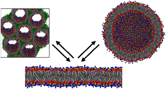
Graphical Abstract
Introduction
Work must be performed by the cell to reshape its many lipid membranes into functional structures, such as the organelles and the vesicles that are trafficked into, out of, and throughout the cell. The standard model for quantifying membrane shape energies is the Helfrich/Canham (HC) Hamiltonian,1,2 a second-order (harmonic) model that takes as input the fundamental mathematical variables describing a local deformation of a planar surface: the principal curvatures c1 and c2. Here we apply this model, developed for bilayers, to single monolayers with:
| (1) |
where EHC is the HC bending free energy of the monolayer, A is the surface area, κm is the monolayer bending modulus, J = c1 + c2 is the monolayer curvature, J0 is the monolayer local intrinsic curvature, is the monolayer Gaussian bending modulus, and K = c1c2 is the Gaussian curvature.
Each membrane composition has a specific set of these mechanical properties based on individual lipid chemistry and lipid-lipid interactions. The HC model (Equation 1) then acts as a framework to connect molecular interactions to the lengthscales of cellular biophysics. This approach has proved useful in identifying the mechanisms and biophysical factors behind the modulation of embedded protein function3-16 as well as the formation of topologically diverse membrane structures (e.g., the structures formed during membrane fusion and fission).17-24
Previous research to quantify bending energetics of diverse surfaces has typically employed an HC model with a single set of parameters that is applicable to arbitrary topology up to some limiting curvature. Published values of simulation-derived κm 25,26 and J0 26-29 from planar bilayers have typically compared well26,30 to HII experiments31 at high curvature. However, there is likely a limit to extending the single HC model to high curvature, and the limits may depend on the magnitude of the curvature, which is different in typical examples of topologically different morphologies. Therefore, restricting an HC model to a single set of parameters means missing phenomena that the model simply cannot assess.
Beyond defining the limits of κm and J0 extensibility, another problem in attempting to use a single HC model is that is notoriously difficult to extract by either experimental or computational means.26,32-38 However, appropriate modeling of fusion and fission events requires quantifying – the value that determines the energy required for a surface to change its topology (e.g., poration, vesicle fusion / fission, etc.) via the Gauss-Bonnet theorem.39 Currently, the best estimates of a single phospholipid’s have been provided by Siegel and Kozlov, who demonstrated a ≈ −0.83 to −0.9 depending on methodology. 40,41
This manuscript’s simulations focus on glycerophospholipids with 1-palmitoyl-2-oleoyl acyl chains (PO). The net-neutral, zwitterionic head groups are either phosphocholine (PC) or phosphoethanolamine (PE), yielding lipids named POPC and POPE. PC and PE head groups have contrasting curvature preferences, as confirmed by all-atom molecular dynamics, 30 coarse-grained molecular dynamics, 42 and experiment.20,31,43-45 In the coarse-grained Martini force field, 42,46 the POPC and POPE chemical structures are reduced into 12 interaction-sites per lipid – differing only by a single site at the head group. These lipids are simulated in four distinct lipid topologies: i) the planar lipid bilayer, ii) the inverse lipid hexagonal (HII) phase,46 iii) the lipid cubic (QII) phase,47-49 and iv) the vesicle50 (Figure 1).
Figure 1:

Cut-through images of the four topologically distinct systems simulated. Clockwise from top left: the planar topology with zero curvature; the HII topology with negative curvature (perpendicular to the cylindrical axis) and zero Gaussian curvature; a vesicle with non-zero monolayer curvature and positive Gaussian curvature; and the QII topology with zero midplane total curvature and negative Gaussian curvature. The HII and QII figures include surrounding periodic images. The head group interaction-sites are shown in blue, phosphate in mustard, glycerol in red, and the tail sites in grey. Interstitial alkane in the HII topology is colored green. Water is not shown for clarity.
Thermodynamic integration (TI)51 is used to test whether a single HC model can describe the relative curvature energetics of PC and PE head groups across a range of curvature and topology. TI has previously been applied to lipid tails to quantify the free energy involved in swapping an unsaturated and saturated lipid between the liquid disordered and liquid ordered states. 52 The TI results generated in this manuscript are used to train an HC model parameterized by the differences in J0 and , as well as by δ (the lipidic neutral surface, where bending and area compression energetics are decoupled). Using differences in J0 and and assuming an uniform HC model ( and are the same for POPC and POPE) reduces the parameter space and simplifies the mathematical expressions.
In this work we neglect molecular factors such as lipid “tilt”, in which the local orientation of a lipid differs, on average, from the collective surface normal. Energetic models for the tilt degree of freedom are available,53,54 however, tilting requires a gradient of curvature, which is either zero (planar, HII, vesicle) or small (QII).
The fit is most robust when we assume that κm and (the area per lipid) are equal for the two lipids, upon which the difference for J0 and between the two lipids is obtained. In this framework, TI produces predictions of these values as parameters of a single HC model. A complication is that the expected HC model parameters depend on the position of δ. Simulations indicate that the Martini pivotal planes (and thus, δ) are not consistent among the four topologies – an indication that the high curvature crosses the limits of universal applicability. The modest disagreement found between the molecular and HC models allows us to address unknowns in the simulations, and the limits of a unified model.
Methods and background
The neutral surface (δ) and pivotal plane
The concept of the pivotal plane is critical for analysis of experimental HII x-ray scattering.55 In these experiments, HII phases are formed and placed under varying osmotic stress. The volume of the water cylinder increases as the phase is hydrated, and the phase’s radius changes accordingly. Because of the phase’s cylindrical geometry, the area per lipid () depends on the atom at which the radius is measured. For example, atoms close to the head group will have a smaller radius than at the tails. However, at a particular atomic position, the will be constant as the radius is varied – the position of the pivotal plane. The pivotal plane location serves as a common metric of the phase’s radius when analyzing HII experiments.
Another lipidic position, the neutral surface (δ), is closely related. It is the lipidic position at which curvature and area dilation energetics are uncoupled. The difference between the pivotal plane and neutral surface is subtle. At the pivotal plane, the surface will bend at constant area, however, the monolayer may be compressed laterally to attain that higher curvature, or to increase the number of lipids at that curvature.
The difference between the two values is small for reasonable values of J0. Even for DOPE with its high negative curvature preference (−1/29.4 Å−1),31 the neutral surface is about 1.0–1.5 Å less than the pivotal plane.44 Therefore, for the purposes of this work we considered them to be equivalent.
Simulation parameters
All simulations were performed with the GROMACS 2019.3 simulation package.56,57 Constant temperature was set at 310.15 K by velocity rescaling. Semi-isotropic pressure of 1 bar was set with a Parrinello-Rahman coupling algorithm (with a compressibility of 3×10⁻⁴ bar−1 and pressure coupling constant of 12.0 ps)58,59 for bilayer and HII simulations. The Parrinello-Rahman algorithm produces good ensembles for systems that do not undergo large strains.60 Isotropic pressure was used for the QII and vesicle simulations. The timestep was 20 fs, and the dielectric constant was set to 15. Each system was simulated for 3 μs unless otherwise noted. Independent 100 ns TI runs were seeded from the 1, 2, and 3 μs configurations. See Supporting Material Table 1 for a complete list of systems and their purpose.
Building the planar topology
Planar bilayers were built using the CHARMM-GUI Martini Maker – Bilayer Builder module scripts.61,62 Systems were built with and without a ~5 Å layer of hexadecane in the bilayer midplane. Hexadecane is represented by four Martini saturated acyl-chain interaction sites and was initially packed using Packmol.63
Building the HII (cylindrical) topology
Five HII sizes were considered. Those with approximate initial radii of 30, 35, 40, or 65 Å contained 400 total lipids (named HII,30, HII,35, HII,40, and HII,65, respectively), while the 90 Å systems contained 500 total lipids (HII,90). In this context, the radius is defined as the xy distance from the lipid C1A bead to the origin. Each unit cell contained one water cylinder, and the equilibrated unit cell z ranged from ~50–90 Å depending on the radius.
All HII systems were built using modified scripts from the CHARMM-GUI Hex Phase Builder module first described in Jo et al.64 The procedure is similar to CHARMM-GUI’s build of planar systems, except that the lipids are placed on a cylinder. The specific lipid area in the cylindrical geometry was measured at the C1A bead, near the pivotal plane, and the interior of the cylinder is filled with water. The HII topology has a geometry such that cylinders are packed on a hexagonal lattice, naturally leaving interstitial spaces that were filled with hexadecane. The geometry of the lipids and water pore (and therefore, presumably the bending energetics) are affected by the amount of interstitial alkane. Therefore, two amounts of interstitial hexadecane in the HII,65 and HII,90 systems were tested to determine if the TI results were sensitive to this packing effect. Detailed build information for the HII topology, including analysis of its pivotal plane, is included in the Supporting Material.
Building the QII (cubic) topology
Periodic minimal surfaces can be defined by a Weierstrass-Enneper parameterization, in which, given two-dimensional surface coordinates u and v, the Cartesian r can be found by performing an integral over complex coordinate ω = u + ιv. For the Pn3m space group minimal surface used here, the expression is:
| (2) |
The structure is one-eighth of the real Pn3m unit cell. The full Pn3m structure consists of eight units that differ by the orientation of the bilayer, i.e., what is the “upper” monolayer in one cell is the “lower” monolayer in its direct neighbors. Simulating one-eighth of the total allows the efficient simulation of a single monolayer that, if the unit cell is traversed in one direction, flips to join the opposite monolayer. Figure S2 in the Supporting Material illustrates how the simulated cell is related to the crystallographic system.
First, a three dimensional mesh corresponding to the Pn3m space group was created, providing the surface midplane. Lipids were placed at approximately evenly distributed sites and oriented such that a lipid’s major moment of inertia was normal to the midplane surface. Water was then added to non-lipid regions to achieve the expected density of the system. The pivotal plane was set to 12 Å from the midplane. Three different sizes (containing 1544, 2791, and 4040 lipids, respectively) of single-component QII phases were built. After constructing the single-component systems, 2 and 10 mol% of the total lipid content were mutated yielding 2:98 POPC:POPE, 10:90 POPC:POPE, 98:2 POPC:POPE, and 90:10 POPC:POPE. These systems are referred to as 2 and 10 mol% QII phases.
Detailed build information for the QII topology is available in the Supporting Material, including energetic considerations for the tension of QII monolayers.
Building the vesicle topology
Vesicular systems were built using the CHARMM-GUI Martini Maker – Vesicle Builder methodology.62 Generally, lipids do not readily flip between monolayers, which may lead to an area imbalance between the lipids and consequential area elasticity strain simply from improperly building the system.65 To mitigate this effect, the Vesicle Builder includes an additional step to artificially equilibrate lipids between monolayers. It works by building a spherical vesicle with six holes punched (±x, ±y, ±z) where the lipids are repelled using an artificial potential first described by Risselada et al.66,67 When simulated, lipids at the hole edges rapidly shield themselves by forming toroidal pores because of the hydrophobic effect. The pores are important for two reasons: i) water density equilibrates between inside and outside of the vesicle, and ii) lipids readily exchange between the inside and outside of the vesicle at the pore edges, allowing lipid densities to equilibrate. The pores are slowly relaxed, resulting in a continuous, equilibrated vesicles. Because of the build procedure, it is presumed that these vesicles are under no tension or differential stress.68
First, pure POPC and pure POPE vesicles were built and equilibrated at midplane radii of 65 and 90 Å (V65 and V90, respectively). Then, after the CHARMM-GUI minimization and equilibration, one of four possible mutations was made to create a minor component: i) 2 mol% of the outer monolayer, ii) 2 mol% of the inner monolayer; iii) 10 mol% of the outer monolayer, or iv) 10 mol% of the inner monolayer. The mutated lipids were chosen randomly. Therefore, initial systems were 2:98 and 10:90 POPC:POPE (inner or outer), as well as 98:2 and 90:10 POPC:POPE (inner or outer). The mutation must occur after the pores are closed, otherwise there would be flip-flop at the pore edges.
It could be straightforward to verify that a vesicle is under zero differential stress by taking the neutral surface determined from the inverse hexagonal phase and calculating for the inner and outer monolayers. If they match the , the vesicle is under minimal stress. However, this only applies if the mechanical model is valid to high curvature. Instead, the position of the neutral surface may change at high curvature. The nature of this paper is to test the limits of lipid mechanical parameters at high curvature, including and δ. In the pivotal plane diagnostic plots shown in Figure 2, the inner and outer monolayers do not have consistent pivotal planes. Instead, the inner monolayer (sloping down) pivots around 17 Å, while the outer monolayer (sloping up) pivots around 7 Å. The areas of the inner and outer monolayer are computed in the Results section to estimate the effects of area strains on perturbation thermodynamics.
Figure 2:

Diagnostic plots for the vesicle pivotal plane for the R = 65 Å and R = 90 Å vesicles containing 10 mol% POPE on the inner monolayer. The is plotted against the height above the bilayer midplane of a Martini site in the planar configuration. The outer monolayer data are shown with brighter blue. The R = 90 vesicle is shown with thicker lines. The black horizontal line is .
Thermodynamic integration and Helfrich/Canham free energies
TI is a computational method that gives a change in free energy (ΔF) by alchemically mutating a “species A” into “species B” along a space of intermediate forcefield parameters. TI is typically used to quantify ΔF between protein residues (e.g., it could be more favorable to have a tryptophan than an alanine at “site X”), but herein, the method is applied to mutating lipid head groups. Technical details regarding the application of TI are included in the Supporting Material.
The Helfrich/Canham model applied to individual lipids
The HC model is applied to a patch of monolayer area around a single lipid:
| (3) |
where X refers to the lipid species (either PE or PC) and Y refers to the topology. The free energy difference, per unit area, between the planar and curved phase is labeled , while extending the difference to that between POPC and POPE is labeled :
| (4) |
| (5) |
In Equations 4 and 5, the final state is POPE, mutated from the initial state, POPC. The planar state is used as a reference for each system.
Each topology has a unique range of ⟨J⟩ and ⟨K⟩, and these values are summarized in Table 1. The QII surface is complicated and so deserves further comment. For those QII surfaces considered here, J is uniformly zero at the midplane (but nonzero away from the midplane). The total K is the same for each Pn3m QII midplane surface, but varies locally with a distribution of negative values (for example, in some regions the surface is planar). According to the rules of differential geometry,69 and discussed in more detail in the Supporting Material, J is negative for QII when measured a distance δ from the midplane. The total and Gaussian curvature at δ for the QII topology are:
| (6) |
| (7) |
Table 1:
Values of total and Gaussian curvature for each system, when measured at the estimated neutral surface of the monolayer with curvature J, ΔJ.
| System | ⟨J⟩ | ⟨K⟩ |
|---|---|---|
| Planar | zero | zero |
| HII | zero | |
| QII | −4πA−1 | |
| Vesicle (inner) | ||
| Vesicle (outer) |
Via the Gauss-Bonnet theorem, K depends only on the topology of the phase. Therefore, it does not depend on thermal fluctuations or simple external perturbations. This makes studying this property challenging. For example, κ can be determined by the magnitude of thermal fluctuations of the bilayer. Yet, the EHC contribution from is unchanged by thermal fluctuations. Differences between phases can be detected because ⟨K⟩ varies between phases (Table 1). Thus, a perturbation that moves a POPE lipid from the planar phase (with ⟨K⟨ = 0) to the QII phase (with ⟨K⟩ = −4πA−1) will be affected by differences in between POPE and the background (POPC). Furthermore, the contribution is modulated trivially by the size of the phase, with the average ⟨K⟩ scaling with the inverse of area.
Modeled bending modulus, area elasticity, and area per lipid
Values for κm were obtained from the normal-director fluctuation spectrum developed by Watson et al.,25 and values were calculated from the average box dimensions of pure POPC or POPE simulations. The bilayer area elasticity KA, used in the tension correction described in the Supporting Material, was computed according to:70
| (8) |
Results & Discussion
TI was performed on the four topologies depicted in Figure 1: i) bilayer patches, with and without alkane; ii) the HII topology with five radii, two with added alkane; iii) three sizes of the QII topology; iv) and a smaller and larger vesicle, with mutations on the inner and outer monolayers considered separately. A range of POPC:POPE ratios (see Table S1 of the Supporting Material) were mutated simultaneously.
HC model parameters extracted from TI data
There is high statistical precision in the TI procedure (typically < 0.005 kcal/mol/lipid), and therefore, the stochastic error in the fit is less than 2% for all calculated values of J0 and . Therefore, disagreements in the fit between the HC model and TI data (to be discussed in the Results section) reveal the limitations of a single HC model. The range of fit values obtained therefore reflects systematic variation in δ between different topologies, or due to latent tension in the monolayer. Controls for these factors are explored in detail in the Supporting Material.
Under the assumption that κm and are roughly equal for POPC and POPE, Equation 5 simplifies to an expression linear in J for the HII and QII systems, but quadratic in J for the vesicle monolayers:
| (9) |
| (10) |
| (11) |
Note that these expressions are all relative to the planar bilayer. Changing the sizes of the three topologies yields changes in J. The TI data can then be compared as a direct test of Equations 9, 10, and 11. Data for are plotted in Figure 3.
Figure 3:

ΔΔF values for the HII topology (left) and the QII and vesicle systems (right) with 10% of the minority lipid (either POPE or POPC). Mutations (ΔΔFTI) of POPC→POPE are shown as circles, while POPE→POPC are shown as squares. Dotted lines indicate fits according to functional forms predicted by Equations 9, 10, and 11 (ΔΔFHC). Simultaneously fitting all the functional forms, in addition to area-elastic corrections, yields the fit in Table 2 that is depicted in Figure 4.
TI demonstrates, as expected, that the PC→PE transition is more favorable with increasing negative curvature (smaller radius). According to Equation 9, the slope of ΔΔFHII versus J is with a y-intercept (the J axis) of zero. By fitting the slope of ΔΔFHII versus J, and with the preceding assumptions regarding κm and , ΔJ0 = −0.016 Å−1 for POPC→POPE, and ΔJ0 = −0.018 Å−1 for POPE→POPC, with both values given as POPE minus POPC (fit together, the best fit ΔJ0 = −0.018 Å−1). However, the intercept of the fit in Figure 3 (bright green data, at left) is clearly non-zero. A modified reference is thus formed by adding alkane to the planar system, suitable for comparison with the HII topology. With this modified reference, is extrapolated to be nearly zero (dark green data, at left), indicating an energetically consistent reference state.
The functional forms for the QII and vesicle topologies (Equations 10 and 11) are consistent with the data for these systems. Simple fits to the data extrapolate close to the origin.
Preliminary mechanical properties and assumptions
Testing Equations 9, 10, and 11 with the TI data requires information on κm and . Values of κm were determined to be 9.05 ± 0.01 kcal/mol (14.68 ± 0.02 kBT) and 7.05 ± 0.10 kcal/mol (11.44 ± 0.16 kBT) for POPC and POPE, respectively. Values of were 65.43 ± 0.01 Å2 and 61.48 ± 0.01 Å2 for POPC and POPE, respectively, determined from the planar bilayer simulations. The values of KA extracted from the fluctuation of the planar bilayer simulation of POPC and POPC were 0.42 ± 0.03 kcal/mol/Å2 (292 ± 21 mN/m) and 0.46 ± 0.03 kcal/mol/Å2 (320 ± 21 mN/m), respectively.
It is an open question to what extent a lipid influences the local mechanical parameters surrounding it. We introduce a simplifying assumption: that each lipid of the bilayer contributes to the overall J0, , and KA of a uniform patch with area in its vicinity. It is not clear that, for example, when the pure POPE bilayer has a smaller bending modulus than POPC, that the local environment of POPE is softer in a POPE/POPC mixture. The alternative, which we adopt, is that the bending modulus is uniform across the entire surface; we set the value κm to the average of the two lipids, 8.05 kcal/mol. We further assume that the area and KA (0.44 kcal/mol/Å) are the same for both PC and PE lipids in any mixture. The differences in KA (~13%), (~3%) and κm (~15%) are modest when making this simplification. At its most general, five unknowns can be determined from the fit: J0,PC, J0,PE, , , and δ. The above assumptions permit robust determination of ΔJ0 and . If instead the individual and κm are used the fit is substantially worse (the assumptions are compared in the Supporting Material in Figure S3).
Whereas for the vesicle and HII systems curvature is only weakly dependent on δ for the simulations here, it affects the curvature of the QII structures proportionally. An error in δ of 6 Å for an R = 30 Å HII system results in a 17% change in curvature (1/30 Å−1 versus 1/36 Å−1), whereas for QII the curvature with δ = 12 is twice that of δ = 6 Å. Moreover, with the HII and vesicle simulations, δ is available from the diagnostic plots in Figure 2 and Figure S1 of the Supporting Material. For the purposes of computing vesicle curvature, the value of δ can be refined by either sampling the neutral surface atom’s radius directly (obviating the use of the midplane) or by computing the expected change in the neutral surface position with curvature, as in Ref.71 We adopt the latter approach, with
| (12) |
where δ′ is the position of the neutral surface in the curved monolayer. Therefore, we focus here on the HC model obtained by varying δ for the QII topology. A range of values were used (7, 12, and 17 Å) for the QII systems that span the vesicle’s δ location. Wang and Deserno determined that the pivotal plane, and thus δ, is near the middle of Martini monolayers.71
Fitting the TI data yields ΔJ0 and
Simultaneously fitting all three curved topologies (relative to the appropriate planar reference) yields ΔJ0 = −0.018 Å−1 and kcal/mol with δ = 12 Å for QII (Figure 4). The HII, QII, and vesicle data are all fit by the same value of ΔJ0, however, the topologies produce different estimates of (Table 2). In part this is because the best fit to the QII data depends strongly on the position of δ used (see the Supporting Material). With large values of δ (near that indicated for the HII topology in the Supporting Material), values approaching zero are optimal. For smaller values of δ (near that indicated for the outer monolayer of the vesicle), increases. However, for the vesicle to match that of the QII topology, δ would need to be nearly zero. Such a small value would be unreasonable; it would require all of the stiffness of the bilayer to be concentrated at the midplane, inconsistent with our knowledge of how bilayers bend (see Ref.72 for a discussion of a reasonable monolayer material model). While a high δ seems justified for the HII systems and the inner monolayer of the vesicle (see Figure 2 and Figure S1 of the Supporting Material), it is not consistent for the outer monolayer of the vesicle or with the analysis in Wang and Deserno.71 Given the range of and inconsistency of δ, we conclude that POPE’s is between 2.0 and 4.0 kcal/mol less negative than POPC, and that POPE’s J0 is −0.018 ± 0.001 A−1 more intrinsically curved than POPC.
Figure 4:

The resulting fit of to the directly calculated values using δQII = 12 Å. Circles represent POPC→POPE mutations, while squares are in the POPE→POPC direction. Results for the HII systems are green, QII results are red, and vesicle results are blue.
Table 2:
Calculated values of ΔJ0 and . These values are obtained by a least-squares fit between and values. The values are reported in the POPC→POPE sense (i.e., Δ = POPE – POPC). That is, POPE has a stronger negative curvature than POPC, and a less negative . The error for each value was calculated by fits to Monte Carlo generated data sets.
| System | ΔJ0 [Å−1] | [kcal/mol] |
|---|---|---|
| HII | −0.017 | N/A |
| QII + HII (δQII = 12) | −0.017 | 0.7 |
| Vesicle | −0.018 | 4.0 |
| Universal (δQII = 7) | −0.018 | 3.1 |
| Universal (δQII = 12) | −0.018 | 2.0 |
As expected, the value of ΔJ0 suggests a POPE lipid preferring a more negative curvature than POPC. The ΔJ0 inferred, −0.018 ± 0.001 Å−1, is consistent with the experimental difference (JDOPE = −1/29.4 Å−1 and JDOPC = −1/87.3 Å−1; ΔJDOPE–DOPC = −0.023 Å−1).31 In addition to the values inferred by Siegel and Kozlov, an important bound is set on the ratio of to κm on the basis of stability of vesicle and HII phases:33 that the ratio . This inequality is based on membrane stability considerations. This is consistent with the interpretation that and κm should be related to a fundamental stiffness parameter like the Young’s modulus of solid isotropic materials. 73 For anisotropic, laterally fluid materials like a bilayer, the origins are currently poorly defined. For example, a molecular explanation in terms of the second moment of the lateral pressure profile28 appears to be invalid or incomplete. 74,75
Our determination that is less negative for Martini POPE than POPC is consistent with POPE being softer than POPC as judged by its bending modulus. For example, if a particular Martini phospholipid obeys the same ratio as experimentally determined by Siegel and Kozlov (−0.83),40 the for POPE would be 1.7 kcal/mol higher. This is roughly consistent with our conclusion that is between 2.0 and 4.0 kcal/mol. The high observation, which relies on very small vesicles that may be under tension, would still be consistent with . The same analysis is performed while relaxing the preliminary mechanical assumptions (that κm and are the same in a mixture) in the Supporting Material, where qualitatively similar differences in mechanical constants are obtained with a substantially worse fit.
Conclusions
The molecular simulations in this work established that the difference in spontaneous curvature (ΔJ0) between models of POPE and POPC lipids is consistent across a broad range of curvature and topology, but indicate significant differences in the Gaussian curvature modulus () most relevant to topological changes. To determine the mechanical model, the Martini coarse-grained force field was utilized to simulate four distinct lipidic topologies: i) the planar lipid bilayer, ii) the inverse hexagonal phase (HII), iii) the inverse cubic phase (QII), and iv) the vesicle. TI was used to obtain the free energy change of mutating POPC↔POPE on these diversely curved lipidic topologies, yielding for each topology and subsystem. By comparing results from the curved topologies to the planar bilayer, a set of was obtained that were directly compared to the universal HC model, . To make the comparison, a least-squares fit between and was performed, ultimately allowing two adjustable parameters: ΔJ0 and . Using different subsets of the data, the least-squares fits yielded a ΔJ0 of −0.018 ± 0.001 Å−1 and a ranging from 2.0 to 4.0 kcal/mol (with the Δ in the POPE minus POPC sense). Both the ΔJ0 and values broadly agree with previous estimates, and in particular, the value adds to the dearth of information for how changes as a function of lipid type.
The results of this manuscript test the limits of a universal HC model, which we defined as a model that supposes material properties extracted from one lipid topology can be extended to other topologies up to some limiting curvature. The data presented here demonstrate the incredible sensitivity of the TI method – discriminating between changes in lipid conformation based on the topological context. The fit of TI data to the universal HC model demonstrated that the HII phase with and without alkane were inconsistent. We explained this discrepancy by first observing that alkane addition in the HII phase increases relative to the planar topology. It is plausible that the higher of POPC makes this lipid more favorable in alkane-rich environments. Through an alternative methodology, Johner et al. observed a small decrease in the apparent κm of Martini HII phases with curvature, and, furthermore, observed a decrease in κm with added tetradecane.76 While our data do not indicate a significant change in κm with curvature, they also do not report the pure κm.
The fit results as plotted in Figure 4 suggest room for improvement of our model parameters. For example, the QII results in red appear to have a different slope than the HII results in green; that is, the simulation error is not simply noise. This discrepancy is a result of the interplay between the vesicle and QII data, which appear to have disagreements in . Alternatively, the discrepancies between vesicle and cubic phases suggest the possibility of latent tension in vesicles built using a method that should presumably relieve tensions because of area and water content strain. It is ambiguous whether the monolayers of these small vesicles were under differential stress (which would perturb ΔΔF values in the range of disagreement) or if these highly positively-curved monolayers had simply reached the limit of the linear response of stress to strain. The latter seems likely, given that the surface defined by δ is so far from the tail region of the bilayer and these tails must reach a hard limit as to their extension.
The TI results echo experimental data from small unilamellar vesicles (SUVs) made of diverse PC lipids that demonstrate asymmetrical lipid packing between the inner and outer monolayers.77-82 The asymmetrical packing is manifested by a thinned inner monolayer that has a reduced area between head groups, while the outer monolayer has increased area between head groups. The difference in packing changes how cholesterol partitions in SUVs – above 30 mol%, cholesterol selectively partitions into the inner monolayer of cis unsaturated PC lipids and into the outer monolayer of saturated PC lipids.78,80
The stability of these small vesicles is limited by the very high curvature stresses in these bilayers. Yet, due to packing effects at high curvature, the energetics of the inner and outer monolayer may not correspond to a single intrinsic curvature. Perturbing the lipid chemistry to see how shifts in δ correlate with ΔΔF, for example, may reveal how to reduce these stresses. While it is computationally convenient to apply these approaches using the Martini force field, the approach is feasible for all-atom force fields, which would allow fine-grained insight to be paired with the powerful TI approach.
Supplementary Material
Acknowledgments
This work was supported by the Intramural Research Program of the Eunice Kennedy Shriver National Institute of Child Health and Human Development (NICHD) at the National Institutes of Health. A.H.B. was supported by a Postdoctoral Research Associate (PRAT) fellowship from the National Institute of General Medical Sciences (NIGMS), award number 1Fi2GM137844-01. Molecular rendering was performed with Tachyon software written by John E. Stone. Simulations were performed on computational resources provided by the both the Intramural Research Program of the NICHD and the Biowulf high performance computing system of the National Institutes of Health. This work was partially performed under the auspices of the U.S. DOE by the Lawrence Livermore National Laboratory under contract DE-AC52-07NA27344, Release LLNL-JRNL-819145.
Footnotes
Supporting Information
A document of supplemental material is provided that includes a description of the neutral surface and pivotal plane, extended build information for the phases, additional information regarding the QII phase geometry, additional information regarding thermodynamic integration, an expanded discussion of the fitting procedure, and a description of the alkane distribution in the planar bilayers.
References
- (1).Helfrich W Elastic Properties of Lipid Bilayers: Theory and Possible Experiments. Zeitschrift fur Naturforsch. - Sect. C J. Biosci 1973, 28, 693–703. [DOI] [PubMed] [Google Scholar]
- (2).Canham PB The minimum energy of bending as a possible explanation of the biconcave shape of the human red blood cell. J. Theor. Biol 1970, 26. [DOI] [PubMed] [Google Scholar]
- (3).Lundbæk JA; Maer AM; Andersen OS Lipid bilayer electrostatic energy, curvature stress, and assembly of gramicidin channels. Biochemistry 1997, 36, 5695–5701. [DOI] [PubMed] [Google Scholar]
- (4).Lundbæk JA; Andersen OS Spring constants for channel-induced lipid bilayer deformations estimates using gramicidin channels. Biophys. J 1999, 76, 889–895. [DOI] [PMC free article] [PubMed] [Google Scholar]
- (5).Nielsen C; Goulian M; Andersen OS Energetics of Inclusion-Induced Bilayer Deformations. Biophys. J 1998, 74, 1966–1983. [DOI] [PMC free article] [PubMed] [Google Scholar]
- (6).Beaven AH; Maer AM; Sodt AJ; Rui H; Pastor RW; Andersen OS; Im W Gramicidin A Channel Formation Induces Local Lipid Redistribution I: Experiment and Simulation. Biophys. J 2017, 112, 1185–1197. [DOI] [PMC free article] [PubMed] [Google Scholar]
- (7).Sodt AJ; Beaven AH; Andersen OS; Im W; Pastor RW Gramicidin A Channel Formation Induces Local Lipid Redistribution II: A 3D Continuum Elastic Model. Biophys. J 2017, 112, 1198–1213. [DOI] [PMC free article] [PubMed] [Google Scholar]
- (8).Beaven A; Sodt A; Pastor R; Koeppe R; Andersen O; Im W Characterizing Residue-Bilayer Interactions Using Gramicidin A as a Scaffold and Tryptophan Substitutions as Probes. J. Chem. Theory Comput 2017, 13. [DOI] [PMC free article] [PubMed] [Google Scholar]
- (9).Brown MF Modulation of rhodopsin function by properties of the membrane bilayer. Chem. Phys. Lipids 1994, 73, 159–180. [DOI] [PubMed] [Google Scholar]
- (10).Brown MF In Curr. Top. Membr. Transp; Epand R. M. B. T. C. T. i. M., Ed.; Academic Press, 1997; Vol. 44; pp 285–356. [Google Scholar]
- (11).Botelho AV; Huber T; Sakmar TP; Brown MF Curvature and Hydrophobic forces drive oligomerization and modulate activity of rhodopsin in membranes. Biophys. J 2006, 91, 4464–4477. [DOI] [PMC free article] [PubMed] [Google Scholar]
- (12).Soubias O; Teague WE; Hines KG; Mitchell DC; Gawrisch K Contribution of membrane elastic energy to rhodopsin function. Biophys. J 2010, 99, 817–824. [DOI] [PMC free article] [PubMed] [Google Scholar]
- (13).Brown MF Curvature forces in membrane lipid-protein interactions. Biochemistry 2012, 51, 9782–9795. [DOI] [PMC free article] [PubMed] [Google Scholar]
- (14).Teague WE; Soubias O; Petrache H; Fuller N; Hines KG; Rand RP; Gawrisch K Elastic properties of polyunsaturated phosphatidylethanolamines influence rhodopsin function. Faraday Discuss. 2012, 161, 383–395. [DOI] [PMC free article] [PubMed] [Google Scholar]
- (15).Soubias O; Teague WE; Hines KG; Gawrisch K The role of membrane curvature elastic stress for function of rhodopsin-like G protein-coupled receptors. Biochimie 2014, 107 Pt A, 28–32. [DOI] [PMC free article] [PubMed] [Google Scholar]
- (16).Soubias O; Teague WE; Hines KG; Gawrisch K Rhodopsin/lipid hydrophobic matching - Rhodopsin oligomerization and function. Biophys. J 2015, 108, 1125–1132. [DOI] [PMC free article] [PubMed] [Google Scholar]
- (17).Chernomordik LV; Vogel SS; Sokoloff A; Onaran HO; Leikina EA; Zimmerberg J Lysolipids reversibly inhibit Ca2+−, GTP− and pH− dependent fusion of biological membranes. FEBS Lett. 1993, 318, 71–76. [DOI] [PubMed] [Google Scholar]
- (18).Chernomordik L; Chanturiya A; Green J; Zimmerberg J The hemifusion intermediate and its conversion to complete fusion: regulation by membrane composition. Biophys. J 1995, 69, 922–929. [DOI] [PMC free article] [PubMed] [Google Scholar]
- (19).Chizmadzhev YA; Cohen FS; Shcherbakov A; Zimmerberg J Membrane mechanics can account for fusion pore dilation in stages. Biophys. J 1995, 69, 2489–2500. [DOI] [PMC free article] [PubMed] [Google Scholar]
- (20).Fuller N; Rand RP The influence of lysolipids on the spontaneous curvature and bending elasticity of phospholipid membranes. Biophys. J 2001, 81, 243–254. [DOI] [PMC free article] [PubMed] [Google Scholar]
- (21).Markin VS; Albanesi JP Membrane fusion: Stalk model revisited. Biophys. J 2002, 82, 693–712. [DOI] [PMC free article] [PubMed] [Google Scholar]
- (22).Kozlovsky Y; Kozlov MM Stalk model of membrane fusion: Solution of energy crisis. Biophys. J 2002, 82, 882–895. [DOI] [PMC free article] [PubMed] [Google Scholar]
- (23).Ryham RJ; Klotz TS; Yao L; Cohen FS Calculating Transition Energy Barriers and Characterizing Activation States for Steps of Fusion. Biophys. J 2016, 110, 1110–1124. [DOI] [PMC free article] [PubMed] [Google Scholar]
- (24).Leikina E; Gamage DG; Prasad V; Goykhberg J; Crowe M; Diao J; Kozlov MM; Chernomordik LV; Millay DP Myomaker and Myomerger Work Independently to Control Distinct Steps of Membrane Remodeling during Myoblast Fusion. Dev. Cell 2018, 46, 767–780.e7. [DOI] [PMC free article] [PubMed] [Google Scholar]
- (25).Watson MC; Brandt EG; Welch PM; Brown FL Determining biomembrane bending rigidities from simulations of modest size. Phys. Rev. Lett 2012, 109. [DOI] [PubMed] [Google Scholar]
- (26).Venable RM; Sodt AJ; Rogaski B; Rui H; Hatcher E; MacKerell AD; Pastor RW; Klauda JB CHARMM all-atom additive force field for sphingomyelin: Elucidation of hydrogen bonding and of positive curvature. Biophys. J 2014, 107, 134–145. [DOI] [PMC free article] [PubMed] [Google Scholar]
- (27).Schofield P; Henderson JR Statistical mechanics of inhomogeneous fluids. Proc. R. Soc. London. A. Math. Phys. Sci 1982, 379, 231–246. [Google Scholar]
- (28).Szleifer L; Kramer D; Ben-Shaul A; Gelbart WM; Safran SA Molecular theory of curvature elasticity in surfactant films. J. Chem. Phys 1990, 92, 6800–6817. [Google Scholar]
- (29).Goetz R; Lipowsky R Computer simulations of bilayer membranes: Self-assembly and interfacial tension. J. Chem. Phys 1998, 108, 7397–7409. [Google Scholar]
- (30).Sodt AJ; Pastor RW Bending free energy from simulation: Correspondence of planar and inverse hexagonal lipid phases. Biophys. J 2013, 104, 2202–2211. [DOI] [PMC free article] [PubMed] [Google Scholar]
- (31).Chen Z; Rand RP The influence of cholesterol on phospholipid membrane curvature and bending elasticity. Biophys. J 1997, 73, 267–276. [DOI] [PMC free article] [PubMed] [Google Scholar]
- (32).Lorenzen S; Servuss RM; Helfrich W Elastic Torques about Membrane Edges: A Study of Pierced Egg Lecithin Vesicles. Biophys. J 1986, 50, 565–572. [DOI] [PMC free article] [PubMed] [Google Scholar]
- (33).Templer RH; Khoo BJ; Seddon JM Gaussian curvature modulus of an amphiphilic monolayer. Langmuir 1998, 14, 7427–7434. [Google Scholar]
- (34).Marsh D Elastic curvature constants of lipid monolayers and bilayers. Chem. Phys. Lipids 2006, 144, 146–159. [DOI] [PubMed] [Google Scholar]
- (35).Baumgart T; Das S; Webb WW; Jenkins JT Membrane elasticity in giant vesicles with fluid phase coexistence. Biophys. J 2005, [DOI] [PMC free article] [PubMed] [Google Scholar]
- (36).Semrau S; Idema T; Holtzer L; Schmidt T; Storm C Accurate determination of elastic parameters for multicomponent membranes. Phys. Rev. Lett 2008, [DOI] [PubMed] [Google Scholar]
- (37).Hu M; Briguglio JJ; Deserno M Determining the Gaussian curvature modulus of lipid membranes in simulations. Biophys. J 2012, 102, 1403–1410. [DOI] [PMC free article] [PubMed] [Google Scholar]
- (38).Hu M; De Jong DH; Marrink SJ; Deserno M Gaussian curvature elasticity determined from global shape transformations and local stress distributions: A comparative study using the MARTINI model. Faraday Discuss. 2012, 161, 365–382. [DOI] [PubMed] [Google Scholar]
- (39).Kreyszig E Differential Geometry; Dover Publications Inc., New York, 1991. [Google Scholar]
- (40).Siegel DP; Kozlov MM The Gaussian curvature elastic modulus of N-monomethylated dioleoylphosphatidylethanolamine: Relevance to membrane fusion and lipid phase behavior. Biophys. J 2004, 87, 366–374. [DOI] [PMC free article] [PubMed] [Google Scholar]
- (41).Siegel DP Determining the ratio of the Gaussian curvature and bending elastic moduli of phospholipids from QII phase unit cell dimensions. Biophys. J 2006, [DOI] [PMC free article] [PubMed] [Google Scholar]
- (42).Marrink SJ; Risselada HJ; Yefimov S; Tieleman DP; De Vries AH The MARTINI force field: Coarse grained model for biomolecular simulations. J. Phys. Chem. B 2007, 111, 7812–7824. [DOI] [PubMed] [Google Scholar]
- (43).Rand RP; Fuller NL; Gruner SM; Parsegian VA Membrane Curvature, Lipid Segregation, and Structural Transitions for Phospholipids under Dual-Solvent Stress. Biochemistry 1990, 29, 76–87. [DOI] [PubMed] [Google Scholar]
- (44).Leikin S; Kozlov MM; Fuller NL; Rand RP Measured effects of diacylglycerol on structural and elastic properties of phospholipid membranes. Biophys. J 1996, 71, 2623–2632. [DOI] [PMC free article] [PubMed] [Google Scholar]
- (45).Chen Z; Rand RP Comparative Study of the Effects of Several n-Alkanes on Phospholipid Hexagonal Phases. Biophys. J 1998, 74, 944–952. [DOI] [PMC free article] [PubMed] [Google Scholar]
- (46).Marrink SJ; De Vries AH; Mark AE Coarse Grained Model for Semiquantitative Lipid Simulations. J. Phys. Chem. B 2004, 108, 750–760. [Google Scholar]
- (47).Marrink S-J; Tieleman DP Molecular Dynamics Simulation of a Lipid Diamond Cubic Phase. J. Am. Chem. Soc 2001, 123, 12383–12391. [DOI] [PubMed] [Google Scholar]
- (48).Marrink SJ; Peter Tieleman D Molecular dynamics simulation of spontaneous membrane fusion during a cubichexagonal phase transition. Biophys. J 2002, 83, 2386–2392. [DOI] [PMC free article] [PubMed] [Google Scholar]
- (49).Fuhrmans M; Knecht V; Marrink SJ A single bicontinuous cubic phase induced by fusion peptides. J. Am. Chem. Soc 2009, 131, 9166–9167. [DOI] [PubMed] [Google Scholar]
- (50).Marrink SJ; Mark AE Molecular Dynamics Simulation of the Formation, Structure, and Dynamics of Small Phospholipid Vesicles. J. Am. Chem. Soc 2003, 125, 15233–15242. [DOI] [PubMed] [Google Scholar]
- (51).Kirkwood JG Statistical mechanics of fluid mixtures. J. Chem. Phys 1935, 3, 300–313. [Google Scholar]
- (52).Bennett WF; Shea JE; Tieleman DP Phospholipid Chain Interactions with Cholesterol Drive Domain Formation in Lipid Membranes. Biophys. J 2018, 114, 2595–2605. [DOI] [PMC free article] [PubMed] [Google Scholar]
- (53).Hamm M; Kozlov MM Elastic energy of tilt and bending of fluid membranes. Eur. Phys. J. E 2000, 3, 323–335. [Google Scholar]
- (54).Terzi MM; Deserno M Novel tiltcurvature coupling in lipid membranes. 2017. [DOI] [PubMed] [Google Scholar]
- (55).Gruner SM; Parsegian VA; Rand RP Directly measured deformation energy of phospholipid HII hexagonal phases. Faraday Discuss. Chem. Soc 1986, 81, 29–37. [DOI] [PubMed] [Google Scholar]
- (56).Van Der Spoel D; Lindahl E; Hess B; Groenhof G; Mark AE; Berendsen HJ GROMACS: Fast, flexible, and free. J. Comput. Chem 2005, 26, 1701–1718. [DOI] [PubMed] [Google Scholar]
- (57).Lindahl E; Hess B; van der Spoel D GROMACS 2019.March. 10.5281/zenodo.3243833. [DOI] [PubMed] [Google Scholar]
- (58).Parrinello M; Rahman A Crystal structure and pair potentials: A moleculardynamics study. Phys. Rev. Lett 1980, 45, 1196–1199. [Google Scholar]
- (59).Parrinello M; Rahman A Polymorphic transitions in single crystals: A new molecular dynamics method. J. Appl. Phys 1981, 52, 7182–7190. [Google Scholar]
- (60).Miller RE; Tadmor EB; Gibson JS; Bernstein N; Pavia F Molecular dynamics at constant Cauchy stress. J. Chem. Phys 2016, [DOI] [PubMed] [Google Scholar]
- (61).Jo S; Kim T; Iyer VG; Im W CHARMM-GUI: A web-based graphical user interface for CHARMM. J. Comput. Chem 2008, 29, 1859–1865. [DOI] [PubMed] [Google Scholar]
- (62).Qi Y; Ingólfsson HI; Cheng X; Lee J; Marrink SJ; Im W CHARMM- GUI Martini Maker for Coarse-Grained Simulations with the Martini Force Field. J. Chem. Theory Comput 2015, 11, 4486–4494. [DOI] [PubMed] [Google Scholar]
- (63).Martínez L; Andrade R; Birgin EG; Martínez JM PACKMOL: A package for building initial configurations for molecular dynamics simulations. J. Comput. Chem 2009, 30, 2157–2164. [DOI] [PubMed] [Google Scholar]
- (64).Jo S et al. CHARMM-GUI 10 years for biomolecular modeling and simulation. J. Comput. Chem 2017, 38, 1114–1124. [DOI] [PMC free article] [PubMed] [Google Scholar]
- (65).Park S; Beaven AH; Klauda JB; Im W How Tolerant are Membrane Simulations with Mismatch in Area per Lipid between Leaflets? J. Chem. Theory Comput 2015, 11, 3466–3477. [DOI] [PMC free article] [PubMed] [Google Scholar]
- (66).Risselada HJ; Mark AE; Marrink SJ Application of mean field boundary potentials in simulations of lipid vesicles. J. Phys. Chem. B 2008, 112, 7438–7447. [DOI] [PubMed] [Google Scholar]
- (67).Risselada HJ; Marrink SJ Curvature effects on lipid packing and dynamics in liposomes revealed by coarse grained molecular dynamics simulations. Phys. Chem. Chem. Phys 2009, 11, 2056–2067. [DOI] [PubMed] [Google Scholar]
- (68).Hossein A; Deserno M Spontaneous Curvature, Differential Stress, and Bending Modulus of Asymmetric Lipid Membranes. Biophys. J 2020, 118, 624–642. [DOI] [PMC free article] [PubMed] [Google Scholar]
- (69).do Carmo M Differential Geometry of Curves and Surfaces; Prentice-Hall, 1976. [Google Scholar]
- (70).Feller SE; Pastor RW Constant surface tension simulations of lipid bilayers: The sensitivity of surface areas and compressibilities. J. Chem. Phys 1999, 111, 1281–1287. [Google Scholar]
- (71).Wang X; Deserno M Determining the pivotal plane of fluid lipid membranes in simulations. J. Chem. Phys 2015, 143, 164109. [DOI] [PubMed] [Google Scholar]
- (72).Campelo F; McMahon HT; Kozlov MM The hydrophobic insertion mechanism of membrane curvature generation by proteins. Biophys. J 2008, 95, 2325–2339. [DOI] [PMC free article] [PubMed] [Google Scholar]
- (73).Deserno M Fluid lipid membranes: From differential geometry to curvature stresses. Chem. Phys. Lipids 2015, [DOI] [PubMed] [Google Scholar]
- (74).Venable RM; Brown FL; Pastor RW Mechanical properties of lipid bilayers from molecular dynamics simulation. Chem. Phys. Lipids 2015, 192, 60–74. [DOI] [PMC free article] [PubMed] [Google Scholar]
- (75).Terzi MM; Ergüder MF; Deserno M A consistent quadratic curvature-tilt theory for fluid lipid membranes. J. Chem. Phys 2019, 151. [DOI] [PubMed] [Google Scholar]
- (76).Johner N; Harries D; Khelashvili G Curvature and lipid packing modulate the elastic properties of lipid assemblies: Comparing HII and lamellar phases. J. Phys. Chem. Lett 2014, 5, 4201–4206. [DOI] [PubMed] [Google Scholar]
- (77).Sheetz MP; Chan SI Effect of sonication on the structure of lecithin bilayers. Biochemistry 1972, 11, 4573–4581. [DOI] [PubMed] [Google Scholar]
- (78).Huang CH; Sipe JP; Chow ST; Martin RB Differential interaction of cholesterol with phosphatidylcholine on the inner and outer surfaces of lipid bilayer vesicles. Proc. Natl. Acad. Sci. U. S. A 1974, 71, 359–362. [DOI] [PMC free article] [PubMed] [Google Scholar]
- (79).Longmuir KJ; Dahlquist FW Direct spectroscopic observation of inner and outer hydrocarbon chains of lipid bilayer vesicles. Proc. Natl. Acad. Sci. U. S. A 1976, 73, 2716–2719. [DOI] [PMC free article] [PubMed] [Google Scholar]
- (80).De Kruijff B; Cullis PR; Radda GK Outside-inside distributions and sizes of mixed phosphatidylcholine-cholesterol vesicles. BBA - Biomembr. 1976, 436, 729–740. [DOI] [PubMed] [Google Scholar]
- (81).Chrzeszczyk A; Wishnia A; Springer CS The intrinsic structural asymmetry of highly curved phospholipid bilayer membranes. BBA - Biomembr. 1977, 470, 161–169. [DOI] [PubMed] [Google Scholar]
- (82).Huang C; Mason JT Geometric packing constraints in egg phosphatidylcholine vesicles. Proc. Natl. Acad. Sci. U. S. A 1978, 75, 308–310. [DOI] [PMC free article] [PubMed] [Google Scholar]
Associated Data
This section collects any data citations, data availability statements, or supplementary materials included in this article.