Abstract
HIF-1α is a master regulator of oxygen homeostasis involved in different stages of cancer development. Thus, HIF-1α inhibition represents an interesting target for anti-cancer therapy. It was recently shown that the HIF-1α interaction with NQO1 inhibits proteasomal degradation of the former, thus suggesting that targeting the stability and/or function of NQO1 could lead to the destabilization of HIF-1α as a therapeutic approach. Since the molecular interactions of NQO1 with HIF-1α are beginning to be unraveled, in this review we discuss: (1) Structure–function relationships of HIF-1α; (2) our current knowledge on the intracellular functions and stability of NQO1; (3) the pharmacological modulation of NQO1 by small ligands regarding function and stability; (4) the potential effects of genetic variability of NQO1 in HIF-1α levels and function; (5) the molecular determinants of NQO1 as a chaperone of many different proteins including cancer-associated factors such as HIF-1α, p53 and p73α. This knowledge is then further discussed in the context of potentially targeting the intracellular stability of HIF-1α by acting on its chaperone, NQO1. This could result in novel anti-cancer therapies, always considering that the substantial genetic variability in NQO1 would likely result in different phenotypic responses among individuals.
Keywords: HIF-1α, NQO1, hypoxia, angiogenesis, cancer, protein: protein interactions, ligand binding, proteasomal degradation, genetic variability
1. HIF-1α: Structure, Function, Regulation and Disease
Oxygen homeostasis is one of the main principles of life for most organisms. The availability of oxygen regulates many physiological processes that are strictly controlled by a complex network of molecules in the organism, with hypoxia-inducible factors (HIFs) playing a central role. These transcription factors are heterodimers composed of an alpha (HIF-α) and a beta subunit (HIF-β). There are three isoforms of HIF-α: HIF-1α, HIF-2α and HIF-3α. Both HIF-1α and HIF-2α regulate the expression of a multitude of genes in response to oxygen availability and play a fundamental role in many physiological and pathological processes [1,2,3]. These two isoforms share structural features and some functional roles but they also have unique, sometimes opposing activities [3]. Since the focus of this review is the chaperone effect of NQO1, which has been studied on HIF-1α, we will mostly refer to this isoform.
1.1. HIF-1α Structure and Function
HIF-1 is a DNA-binding complex composed of two subunits (α and β), both basic helix-loop-helix (bHLH) proteins of the PER-ARNT-SIM (PAS) family. HIF-1β (initially named aryl hydrocarbon nuclear receptor translocator, ARNT) is constitutively expressed and, thus, its expression and activity are oxygen-independent. In contrast, HIF-1α is strongly repressed in normoxia and induced in hypoxia [4]. In response to low oxygen, HIF-1α dimerizes with HIF-1β, and HIF-1 binds to the hypoxia response element (HRE) of the promoter or enhancer region of the target genes to induce transcription. HIF-1 induces the transcription of numerous genes [5,6] involved in many biological processes, such as angiogenesis, erythropoiesis, anaerobic metabolism, cell survival, and cell proliferation.
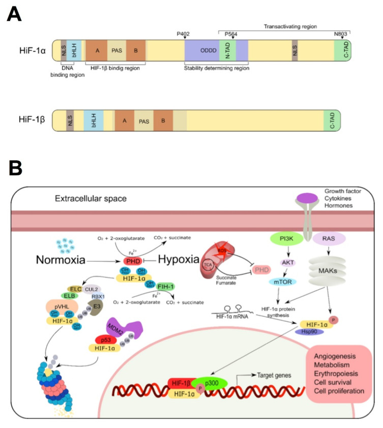
The basic domain and the carboxy-terminus of PAS are specifically required for the DNA binding of HIF-1 to the HRE DNA sequence 5-‘RCGTG-3’, whereas the HLH domain and the amino-terminus of the two subunits of PAS proteins mediate their dimerization [7]. In addition to ubiquitous HIF-1α and HIF-1β, both subunits exist as a series of isoforms encoded by distinct genetic loci with different splice variants [8,9,10,11]. HIF-1α constitutes the most prominent member of the three HIF-α subunits in the human genome. The most important structural elements of HIF-1α are summarized in Figure 1A. HIF-1 has two nuclear location sequences (NLS) at the amino (residues 17–74) and carboxyl (residues 718–721) ends of the protein [12]. The stability of the protein is mainly regulated at the oxygen dependent degradation domain (ODDD). This domain contains two peptide sequences rich in proline (P), glutamic acid (E), serine (S), and threonine (T) at residues 499–518 and 581–600 [4]. PEST-like motifs are present in short-lived proteins rapidly targeted for intracellular degradation. They mediate HIF-1 degradation by the ubiquitin-proteasome pathway [13,14]. HIF-1α also contains two transactivation domains, N-TAD (residues 531–575) and C-TAD (residues 786–826) [15]. Under hypoxia, several co-activators such as CBP/p300/SRC-1 interact with the C-TAD inducing HIF-1α transcriptional activity, while N-TAD is responsible for stabilizing HIF-1α against degradation [16].
Figure 1.
Structure and regulation of HIF. (A) Schematic representation of human HIF-1α and HIF-1β structure. Both proteins form part of the HLH–PAS transcription factor family and contain a N-terminal bHLH domain (implicated in DNA binding) and two PAS domains (responsible of its dimerization). HIF-1α contains an oxygen-dependent degradation domain (ODDD) that mediates oxygen-regulated stability, and two transactivation domains (TAD), that mediate its transcriptional activity and its stability. (B) In normoxia, HIF-1α is subjected to oxygen-dependent hydroxylation by Prolyl hydroxylase domain (PHD) hydroxylases, conducting HIF-1α to ubiquitination by the von Hippel-Lindau protein (pVHL) and proteasomal degradation. MDM2/p53 are also involved in ubiquitination and degradation of HIF1α protein in a pVHL-independent manner. Under low oxygen levels (as well as some metabolites of TCA cycle), PHD is inhibited, HIF-1α translocates to the nucleus and promotes the activation of target genes. Transactivation of HIF-1α could be also induced by PI3K/MAPKs signaling pathways upon activation of some growth factor, hormones and cytokines.
1.2. Regulation of HIF-1α Expression
Although HIF-1α is tightly regulated at the transcriptional, translational and posttranslational levels [5], protein degradation plays an important role in regulating its activity and is the main topic of this review, focused on the interaction between NQO1 and HIF-1α.
1.2.1. Oxygen-Dependent Regulation
Most of HIF-1 regulation is mediated by the HIF-1α subunit. The best known mode of HIF-1α regulation, and more related to the topic reviewed here, takes place at the postranslational level, where oxygen regulates HIF-1α stability. Under normoxic conditions, HIF-1α protein is rapidly targeted for ubiquitination and proteasomal degradation, thus keeping minimal steady-state levels. In this scenario, HIF-1α becomes hydroxylated using O2 as substrate by members of the prolyl hydroxylase domain (PHD) family on two specific prolines (P402 and P564) of the ODDD and NTAD domains, respectively [17,18,19]. Hydroxylation of these proline residues generates a binding site for an E3-ubiquitin ligase complex that contains the von Hippel Lindau protein (VHL), elongin B (ELB), elongin C (ELC), cullin 2 (CUL2), and ring box protein 1 (RBX1). As a result, HIF-1α is polyubiquitinated and degraded by the proteasome [20,21,22,23] (Figure 1B). Oxygen also affects DNA binding and transcriptional activity. This effect is mediated by the factor inhibiting HIF-1α (FIH-1), an asparagyl hydroxylase that catalyzes the hydroxylation of Q803 within the C-TAD domain of HIF-1α. Hydroxylation of Q803 leads to a steric clash that prevents the binding of the p300 and CBP coactivators to HIF-1α, abolishing their transactivation [24].
In addition, HIF1A transcription is also regulated [25] and more recent evidence indicates that the regulation of HIF1A mRNA translation is a crucial element of HIF1α induction under hypoxia and stress conditions, making the translational machinery an important target for cancer therapy (reviewed in [26]).
In hypoxia, impaired hydroxylation of HIF-1α allows dimerization of the transcriptional complex and the subsequent expression of its target genes. The mitochondrial electron transport chain (MEC) consumes the vast majority of cellular oxygen and may play an important role in HIF-1α regulation by determining the amount of oxygen available to PHD enzymes [27,28,29]. In this sense, pharmacological or genetic inhibition of mitochondrial respiration may prevent the accumulation of HIF-1α in hypoxia [30,31]. However, there are other ways in which mitochondria affect the stability of HIF-1α (Figure 1B). Thus, in addition to requiring 2-oxoglutarate as a co-substrate, several metabolites of the tricarboxylic acid (TCA) cycle, especially succinate and fumarate, additionally inhibit the HIF hydroxylases [29,32,33,34]. Reactive oxygen species produced by MEC in hypoxia also increase the stability of HIF-1α [30,35,36,37,38].
1.2.2. Growth Factor Signaling Pathways
Growth factors, cytokines and hormones [39,40,41,42,43,44] also regulate HIF-1α. These signals lead to the activation of mTOR (by the PI3K/AKT pathway) and the mitogen activated protein kinases (MAPK) (by the RAS/RAF/MEK/ERK kinases cascade) which can phosphorylate 4E-BP1, favoring translation of HIF1A mRNA [45,46,47]. The translational regulation of HIF1A mRNA has been recently reviewed [25]. MAPKs also play an important role in the regulation of the transactivation activity of HIF-1. Once activated, members of this family phosphorylate HIF1α, enhancing the binding of the coactivator p300/CBP [46,48].
1.2.3. The Mdm2 Pathway
A complex interplay exists between p53 and HIF-1α. On one hand, HIF-1α binds to Mdm2 (the mouse double minute 2 homologue), inhibiting Mdm2-dependent degradation of p53 [49]. On the other hand, p53 also mediates HIF-1α activity. In this sense, at moderate expression levels, p53 competes with HIF-1α for p300, consequently decreasing the transcriptional activity of HIF-1α. Moreover, high levels of p53 expression lead to HIF-1α degradation [50]. This is due to the binding of p53 to HIF-1α that allows Mdm2-mediated ubiquitination of HIF-1α and its proteasomal degradation [51] (Figure 1B).
1.2.4. Heat Shock Protein 90 (Hsp90)
HIFα stability is also regulated by other signaling pathways, since Hsp90 inhibitors promote HIFα degradation in a pVHL-independent manner [52,53]. In addition, Hsp90 is known to bind directly with HIF-1α, inducing some conformational changes in its structure that improve the fitting and coupling with HIF-1β, thus initiating its transactivation [54] (Figure 1B).
1.3. HIF-1α in Cancer
Cancer is defined as the autonomous growth and spread of a clone of somatic cells, under selective pressure, which is able to use multiple cellular pathways to modify its environment in favor of its own proliferation and against the needs of the organism, escaping physiological constraints on cell growth, including immune surveillance [55].
As the cell clone grows, its core becomes further removed from the blood supply and changes in oxygen availability are key for tumor cells to reorganize their metabolism in order to match oxygen supply with cellular demands. Thus, hypoxia becomes one of the key features of the tumor microenvironment [56], a fact first reported in early studies where hypoxic cells were shown to surround the necrotic center in histological sections of lung carcinomas [57] and later was demonstrated in essentially all solid tumors (reviewed in [56]). Intratumoral hypoxia is widespread and almost independent of tumor size, grade, stage, or histology [58]. Numerous studies have elucidated the molecular mechanisms involved in the cellular response to hypoxia, and three leaders in the field have been awarded the 2019 Nobel Prize in Medicine for their achievements, including their seminal work on HIF-1α, a critical transcription factor for adaptation to hypoxia [59,60] (https://www.nobelprize.org/prizes/medicine/2019/summary/, accessed on 1 April 2022).
HIF-1α and HIF-2α regulate a number of physiological pathways involved in cancer, such as cell proliferation, survival, apoptosis, angiogenesis, glucose metabolism, immune cell activation and stem cells [59,60,61]. In particular, they play an important role in at least two of the main biological capabilities acquired during the multistep process of tumor development (the “hallmarks” of cancer [55]): reprogramming cell metabolism and inducing angiogenesis.
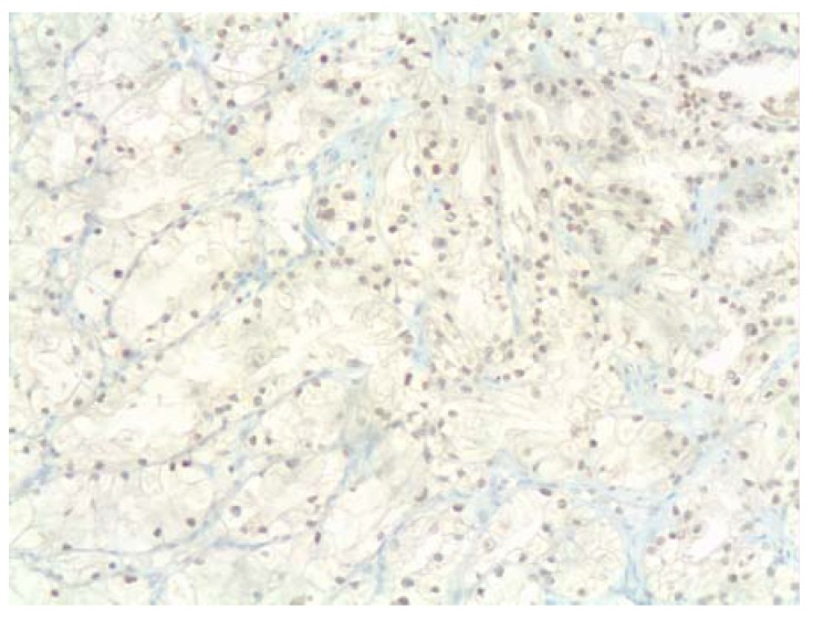
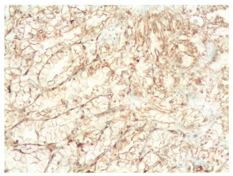
The complexities of cancer prognosis (the high variability of tumor mutations, histological types, grades and stages) make it too simplistic to think that the levels of expression of a single gene such as HIF1A might have a significant impact on prognosis. However, increased HIF-1α and HIF-2α protein levels in diagnostic tumor biopsies have been correlated with poor prognosis in a variety of cancer types (reviewed in [1]). Recently, high HIF-1α expression has been studied in triple-negative breast cancer (TNBC) [62]. TNBC are a heterogeneous group of poorly differentiated, highly aggressive and metastatic breast cancer type. They lack specific molecular-targeted therapy and there is an urgent need to find effective biomarkers rather than relying of their lack of hormone receptors and Her2 expression to define this subgroup. These authors [62] found that HIF-1α and c-Myc expression detected by immunohistochemistry could add to the development of a prognostic nomogram together with the histological grade and stage of cancer (based on tumor size (T), lymph node involvement (N) and distant metastases (M), known as TNM status). In an effort to validate the proposed nomogram in our institution, we are currently evaluating the feasibility of including HIF-1α immunohistochemistry in the biomarker profile (Figure 2). As transcription factors induced in tumor cells by hypoxic environmental stress, HIF-1α and HIF-2α could regulate a variety of downstream genes, thereby increasing the invasive ability of tumors and their resistance against standard radiotherapy and chemotherapy.
Figure 2.
Nuclear HIF-1α (top) and NQO1 (bottom) expression (brown staining) in serial sections of clear cell renal carcinoma (ccRCC; this particular case carried p.L89L mutation in one allele and a deletion of the other one). Common deletions of the VHL gene result in lack of HIF1 degradation, with the subsequent activation of HIF1 as a nuclear transcription factor that drives tumor cell proliferation. NQO1 is widely expressed in the cytoplasm of tumor cells, including clear cell renal carcinoma, and it might play a role in the postranslational regulation of HIF-1α. Clear cell ovarian carcinoma is another example with strong hypoxic signature where NQO1 might play a role, however we have had no access to cases of clear cell ovarian carcinoma driven by ARID1A mutations. Much more work is needed to address this interesting connection. Primary antibodies used: rabbit polyclonal anti-HIF1α (ab2185, dilution 1:100, Abcam, Cambridge, UK) and mouse monoclonal anti-NQO1 (clone A180, dilution 1:600, Thermofisher scientific, Madrid, Spain); peroxidase signal development with Optiview, Ventana.
2. NQO1 as a Protein Chaperone: Current Knowledge and Potential Application to Target HIF-1α Stability
The description of NQO1 as a protein chaperone protecting HIF-1α against degradation [63] supports that targeting this protein–protein interaction (PPI) could allow inactivation of HIF-1α. In this section we will describe a wealth of information available for the activity and stability of NQO1, focusing on what we know about other PPI, by which NQO1 acts as a chaperone and how small ligands and missense mutations are capable of modulating these PPI. We hope that this information will boost interest in targeting NQO1 as a modulator of HIF-1α.
2.1. Overview of NQO1 Expression, Regulation and Functions: On the Potential Roles of O2 Levels and HIF-1α
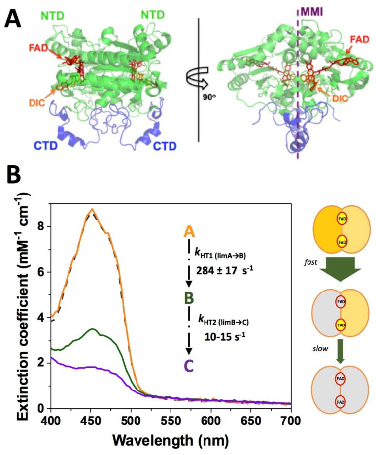
NAD(P)H quinone oxidoreductase 1 (NQO1; DT-diaphorase; EC 1.6.5.2) functions as a homodimer of 62 kDa [64,65]. Each subunit contains two domains: an N-terminal domain (NTD, comprising approximately residues 1–225), which tightly binds one molecule of FAD and is capable of folding and assembly into dimers autonomously, and a C-terminal domain (CTD, comprising the last 40–50 residues) that completes the monomer:monomer interface (MMI) and the binding sites of NAD(P)H and the substrates (Figure 3A) [64,66,67,68,69,70]. Its main enzymatic function is to catalyze the two-electron reduction of quinones, thus avoiding the production of reactive, and potentially cytotoxic, semiquinones (Table A1) [71]. Since one of its substrates is menadione (vitamin K3) it may function in blood clotting, although its role appears to be minor compared to vitamin K oxidoreductase [72]. Other substrates include α-tocopherol-quinone and ubiquinone, which are maintained in their reduced (antioxidant) forms by the enzyme [73,74]. NQO1 can also catalyze the reduction of superoxide radicals and thus plays a role in directly protecting the cell from reactive oxygen species (ROS) [75].
Figure 3.
Structure and catalytic function of NQO1. (A) Structural representation of the NQO1 dimer (PDB 2F1O) [83]. NTD and CTD refer to N-terminal and C-terminal domains, respectively. The location of the FAD and the inhibitor dicoumarol (DIC) binding sites is also indicated. The monomer:monomer interface is indicated as MMI. (B) Reduction of FAD by NADH shows two different pathways. In the left panel, the spectral properties of the different spectroscopic species (A, B, C) stabilized upon reduction are indicated as well as their conversion limiting rate constants. The right panel shows the model proposed by us for the sequential reduction of the two FAD cofactors in the protein homo-dimer, in which this large difference in kinetics represents a type of functional negative cooperativity. Adapted from [76].
The enzymatic cycle of NQO1 follows a ping-pong mechanism with two main steps or semi-reactions: in the first one, the rate-limiting step, NAD(P)H binds to the holo-form (NQO1holo) of the enzyme (FAD is not released during the cycle) [65]. Consequently, NAD(P)H reduces the flavin to FADH2 and the NAD(P)+ is released. In the second half-reaction, a two-electron reduction occurs between FADH2 and the substrate (Q). However, the efficiency of catalytic sites in the dimer, either in the reductive or oxidative half-reactions, has been shown to be non-equivalent [76,77]. Steady-state and pre-steady-state studies have supported the presence of a substantial functional negative cooperativity in the NQO1 catalytic cycle (even in the FAD binding affinity) [76,78,79]. Essentially, one of the active sites operates one-to-two orders of magnitude faster than the other one, indicating the existence of strong allosteric communication between the active sites during the catalytic cycle [76,77] (Figure 3B). We must note that the rate of this reductive half-reaction is strictly NAD(P)H concentration dependent, and thus, should be accelerated due to the HIF-mediated enhancement of aerobic glycolysis (raising cytosolic NADH levels). In the second and faster oxidative half-reaction [76], the substrate binds and is reduced by the FADH2, thus releasing the reduced substrate and regenerating the holo-enzyme [65,68]. Dicoumarol, a potent anticoagulant and mitochondrial uncoupling agent, acts as a competitive inhibitor of NAD(P)H and the substrate during the full NQO1 cycle (Table A1) [76,80]. The high level of NQO1 expression in some cancer cells, coupled with its role in defense against ROS, has led to the proposal that NQO1 inhibition may be a novel and plausible therapy for this disease. Actually, dicoumarol is an effective inhibitor of pancreatic cancer cells in culture, but no NQO1 inhibitors are yet in clinical use [81]. However, the situation is far from simple, and it has also been shown that NQO1 promotes AMPK activation and induces cancer cell death under oxygen–glucose deprivation [82].
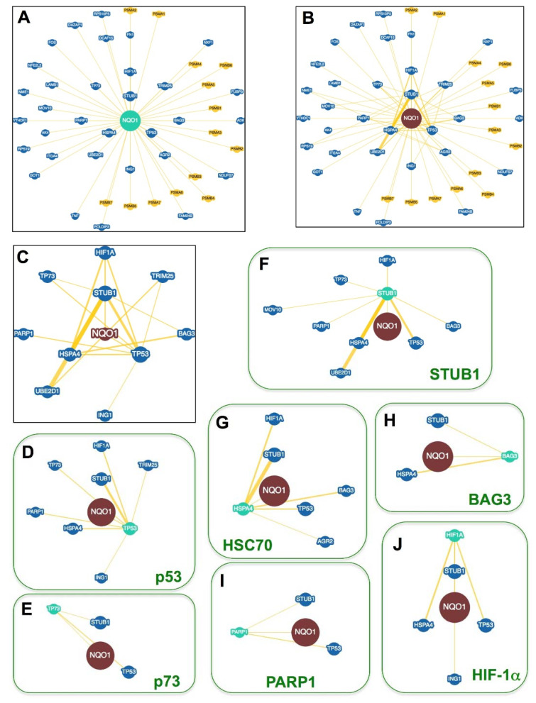
The NQO1 protein also has non-enzymatic functions in which it mostly participates in PPI, modulating the stability of its binding partners (Figure 4 and Table A2). Indeed, and as we will further discuss in Section 2.2 and Section 2.3, several of the ligation states populated by NQO1 during the catalytic cycle (NQO1holo, either oxidized and reduced, or the dead-end complex of the holo-enzyme with dicoumarol, NQO1dic) are very relevant to the discussion of PPIs developed by NQO1. Among them, the reduced form of NQO1 seems to be most critical for strengthening PPI as well as the change in the subcellular location of the enzyme [84,85,86]. From a (bio)chemical point of view, its intrinsically low kinetic stability (i.e., a very small half-life, in the order of a few ms for the reduced flavin state) in the presence of model substrates is intriguing [76]. However, the results from Ross and coworkers have additionally supported that from a cellular/physiological viewpoint; this NQO1 state with the flavin reduced could be of high relevance, locally controlling the NADH/NAD+ ratio, the cellular location of NQO1 and α-tubulin organization [85,86].
Figure 4.
The NQO1 interactome. (A) NQO1 protein partners described in at least one report. (B) Connectivity between NQO1 partners. The shorter the radial distance between a partner and NQO1 indicates a higher interconnectivity. Partners in yellow are from mouse and in blue from human. (C) A zoom from panel (B) shows the most highly interconnected partners of NQO1. (D–J) Interactions between NQO1 partners in panel (C) and other proteins to highlight their different interconnectivity. The thickness of connecting yellow lines is related to the number of reports describing the interaction. Data were retrieved from the BioGRID database (https://thebiogrid.org/108072, accessed on 1 April 2022) [111].
NQO1 is known to undergo different post-translational modifications (PTMs), some of which have been characterized in a site-specific manner. These studies have revealed that phosphorylation may modulate NQO1 function and stability in different manners. Phosphorylation of NQO1 at T128 by the Akt kinase results in polyubiquitination and proteasomal degradation of NQO1 [87]. A phosphomimetic mutation on the residue S82 showed reduced FAD binding affinity and consequent loss of intracellular protein stability, although the kinase involved in this phosphorylation event has not been identified yet [88]. Therefore, it is reasonable to propose that phosphorylation of NQO1 described at multiple sites (https://www.phosphosite.org/proteinAction.action?id=14721&showAllSites=true, accessed on 1 April 2022) may have an impact on the intracellular stability of its protein partners through the destabilization of NQO1.
NQO1 is expressed in most human cell types (https://www.genecards.org/cgi-bin/carddisp.pl?gene=NQO1, accessed on 1 April 2022) and its expression can be upregulated in response to stress. Two control elements are located upstream of the human NQO1 gene: the antioxidant response element (ARE) and the xenobiotic response element (XRE) [89,90,91,92]. A key regulator of the ARE is nuclear factor erythroid 2-related factor 2 (Nrf2). This transcription factor from the basic leucine zipper family is located in the cytosol under non-stressed conditions. Here it interacts with Kelch like-ECH-associated protein 1 (KEAP1) that targets it for ubiquitin-mediated proteasomal degradation [93,94]. Oxidative stress is sensed by the oxidation of disulfide bonds in KEAP1 [95]. This releases Nrf2, which translocates to the nucleus and, together with members of the small musculoaponeurotic fibrosarcoma protein family (sMaF), binds to AREs [96]. This results in the recruitment of the mediator complex, the acetylation of Nrf2 and the activation of a range of genes involved in antioxidant defense, including NQO1 [97,98,99]. Nrf2 also upregulates genes for NADPH generation, thus ensuring that enzymes such as NQO1 have sufficient reduced cofactor [100,101]. The ARE also responds to some antioxidants (e.g., resveratrol and quercetin), by upregulating the expression of NQO1 and other antioxidant enzymes [102,103]. Interestingly, the ARE that controls the expression of NQO1 is also activated by hypoxic conditions [104]. Thus, NQO1 protein levels increase under hypoxic conditions [105]. Consequently, NQO1 is overexpressed in tumors due to their hypoxic environment.
The XRE is activated by the aryl hydrocarbon receptor (AhR), a basic helix-loop-helix transcription factor. This protein binds to a wide range of hydrophobic xenobiotics and translocates to the nucleus in response. Here, it heterodimerizes with aryl hydrocarbon nuclear translocator (ARNT or HIF-1β) and binds the XRE [106,107]. In the case of the NQO1 gene, AhR at the XRE and Nrf2 functionally cooperate to regulate its expression [108]. Like the ARE, the XRE can be induced by hypoxia [109]. Although HIF-1α also partners with HIF-1β to regulate transcription, siRNA silencing of the HIF1A gene does not affect NQO1 expression [110].
2.2. NQO1 Macromolecular Interactions
To date, there are 47 protein partners of NQO1 compiled in the BioGRID database (https://thebiogrid.org/108072, accessed on 1 April 2022) [112]. This set includes thirty-three human proteins (actually one is a pseudo-gene product). The remaining fourteen are from rodents and belong to different subunits of the 20S proteasome (Figure 4A). In addition, NQO1 can interact specifically with certain RNAs modulating their expression [113]. Focusing on protein partners, there is certain evidence within the NQO1 interactome supporting that different partners (i.e., nodes) of this network can interact not only with NQO1 but also among each other. Consequently, this could generate regulatory circuits upon the interaction of different partners in two- or multiple-way manners (i.e., binary, ternary, quaternary, etc.…complexes; Figure 4B). This also suggests that we may affect the properties (e.g., intracellular activity or stability) of one node by targeting different adjacent nodes in the network, an effect that could be amplified particularly in nodes with high connectivity. The most representative and consistently reported nodes with such a high connectivity for NQO1 are shown in Figure 4C and Table A2. Importantly, HIF-1α belongs to this group.
A critical point to achieve a better understanding of the functional consequences of NQO1 interactomics emerges from a well-known effect. This is the interaction of NQO1 with protein partners that enhances the stability of these partners against both Ub-dependent or –independent proteasomal degradation [63,114,115]. Thus, effects on the intracellular levels of NQO1 or in the strength of its interaction with partners should translate into altered stability of the nodes in the NQO1 interactomic network. For instance, reducing the availability of the FAD precursor riboflavin leads to an increased population of the flavin-free NQO1apo state, which is highly sensitive to proteasomal degradation through ubiquitin-dependent and -independent mechanisms [116,117]. This is in part caused by the destabilization of the CTD of NQO1 in the absence of bound FAD that promotes its ubiquitination and degradation [117]. Remarkably, addition of the inhibitor dicoumarol does not stabilize WT NQO1 protein intracellularly and seems to reduce its ability to interact with and stabilize these protein partners (see Table A2). The binding of NAD(P)H, which reduces the bound flavin cofactor, seems to enhance the interaction of NQO1 with these protein partners ([84], Table A2). A recent study also suggested that NQO1 with a reduced flavin cofactor could be a physiologically important state in the cell [85], and thus very relevant to understand the interactome dynamics of NQO1 (note that the population of this state can be promoted due to HIF-mediated increased levels of cytosolic NADH). Additional factors modulating NQO1 intracellular stability, such as protein phosphorylation events, small ion binding or single amino acid changes [111,118,119,120,121], may thus propagate and amplify these effects to the stability of the NQO1 protein partners.
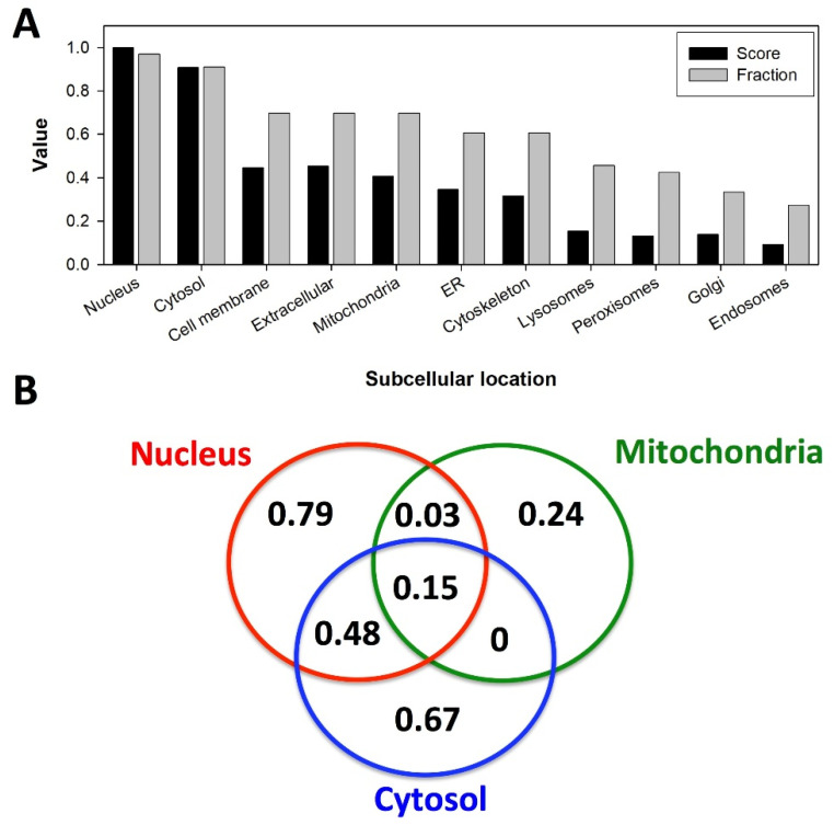
Another critical issue for understanding the interaction of NQO1 with its partners and subsequent effects of these interactions on the stability of its interactomic consequences is the subcellular location of NQO1 and its partners. It is generally accepted that NQO1 subcellular location is primarily cytosolic, where it can fulfill its roles as antioxidant/detoxifiying enzyme and is able to interact with protein and RNA partners. However, there is also certain evidence supporting its location in other subcellular compartments, mostly mitochondria and the nucleus (with low confidence in other organelles) (Figure 5A; GeneCards®; https://www.genecards.org/cgi-bin/carddisp.pl?gene=NQO1&keywords=NQO1, accessed on 1 April 2022). Importantly, most of the protein partners of NQO1 also seem to primarily localize in the nucleus and the cytosol, and to a moderate extent in the mitochondria, cell membrane and extracellularly, apparently coexisting between more than one subcellular location (Figure 5A,B). In the case of the cytosol and the nucleus, about 50% of NQO1 partners may operate in any of these locations and translocate between these two to fulfill their roles in cytosolic and nuclear processes such as the control of DNA expression (p53, HIF-1, c-FOS, NRF-2, NME1, FUBP3), the regulation of RNA and translation (POLDIP3, YTHDF1, NXF1, MOV10, DAZAP1, RPS19), the regulation of nuclear protein function (PARP1 and TRIM25), protein folding and degradation (BAG-3, eIF4G1, DCAF15, HSPA4, STUB1 and UBE2D1). In addition, a number of metabolic enzymes in the cytosol and mitochondria (ODC, NDUSF7, NME1, ADK, GOT1 and AK4) could also interact with NQO1 in these compartments. Indeed, NQO1 overexpression in mice is known to enhance glycolytic and mitochondrial respiration activities and enhance metabolic flexibility, mimicking the beneficial effects of caloric restriction [122]. It is worth noting that the proteasomal protein degradation machinery may operate through rather similar mechanisms in the cytosol and the nucleus [123,124], and thus, the well-known chaperone role of NQO1 for different protein partners (see Table A2) may indeed operate in the cytosol, thus increasing the levels of cytosolic proteins amenable to importing to other organelles (such as nucleus or mitochondria [63,125]) and plausibly by stabilizing these proteins upon import of both the partner and NQO1 in these organelles. It is noteworthy, although more rarely described, that the presence of NQO1 in other subcellular locations such as the cytoskeleton may explain other roles of the multifunctional NQO1 protein. For instance, recent studies have supported that NQO1 may provide an adequate supply of NAD+ for the deacetylase activity of different sirtuins associated with microtubule dynamics [85,122,126]. These studies also highlight the potential plasticity of NQO1 subcellular location during different cellular conditions or stages (e.g., the recruitment of cytosolic NQO1 to cytoskeletal structures during cell division) [85,86].
Figure 5.
Subcellular location of NQO1 interacting human partners based on data from GeneCards® (https://www.genecards.org/, accessed on 1 April 2022). For all the interactors, the subcellular compartment (location) and its confidence (1–5, from the lowest to the highest) were retrieved from GeneCards®. (A) Accumulated score for each organelle as the sum of the numerical degree of confidence for all partners found in a given compartment. The highest accumulated score (i.e., for the nucleus) was used to normalize yielding the Score. As Fraction, we refer to the fraction of all the partners found in a given organelle. Note that the ratio Score:Fraction gives a measure of the degree of confidence for finding a given partner in a given subcellular location. (B) Subcellular location of NQO1 partners in the three subcellular locations of NQO1 reported with confidence (i.e., equal or higher than 3) as the fraction of the total of NQO1 partners. Overlapping regions in the Venn diagram reveal the presence of an NQO1 partner in at least two subcellular locations. Numbers in the different regions of the Venn diagram represent the fraction of NQO1 partners present in different subcellular locations.
2.3. Changes in NQO1 Stability, Structure and Dynamics upon Mutation and Ligand Binding: Implications for the Stability of Its Protein Partners
The functional chemistry of NQO1 is remarkably complex [65,127]. At least four different ligation/redox states (with small molecules bound or oxidoreductive states of the flavin) are relevant to understanding the intracellular stability and enzymatic activity of NQO1. Similarly, these states have different functional consequences as well as effects on its macromolecular partners: NQO1apo, which has no ligand bound; NQO1holo, which contains a molecule of oxidized FAD per NQO1 monomer; NQO1holo-red, containing the FAD cofactor reduced upon interaction with NAD(P)H and ready for hydride transfer to the substrate; NQO1dic, a ternary complex of NQO1holo with the inhibitor dicoumarol bound that competitively inhibits NAD(P)H and substrate binding. These different ligation states interact differently with NQO1 protein partners [67,84,125]. High-resolution structural models for these complexes are, to the best of our knowledge, not reported. Thus, we will focus in this section on the structural and dynamic consequences of ligand binding to NQO1 in an attempt to obtain some insight into their regulatory effects on NQO1 interactions with protein partners (always from an NQO1 point of view). NQO1apo exists as a stable and expanded dimer characterized by significantly large conformational flexibility [65,67,68,69,111,128,129,130,131]. Although no high resolution structural model is available for this state, recent kinetic studies using hydrogen–deuterium exchange mass spectrometry (HDXMS) have identified a minimal stable core that holds the protein dimer, while most of the protein exists forming a highly dynamic structural ensemble, including the FAD and substrate binding sites in non-competent states for binding [132]. The local stability of this state is very sensitive to mutations [131]. This remarkable conformational flexibility makes NQO1apo likely the most relevant state to understand the intracellular stability of NQO1, since the flexible CTD acts as an initiation site for rapid degradation through ubiquitin-dependent and -independent proteasomal pathways [88,117,131]. It is plausible that this unstable and highly dynamic NQO1apo state is not capable of interacting with NQO1 protein partners, or at least, does so with lower affinity [67].
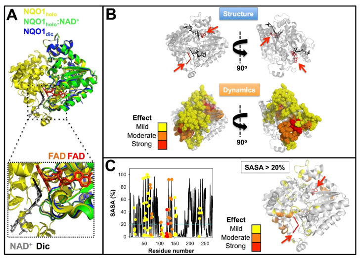
Binding of FAD triggers a large conformational change leading to compaction of the protein dimer, an increase in ordered secondary structure, overall conformational stabilization and a large decrease of protein dynamics that is sensed in almost the entire protein structure [67,68,69,78,111,128,129,131,132]. This NQO1holo state is amenable for high-resolution structural studies [70] (Figure 6A). This state is also known to be intracellularly stable and likely to interact with protein partners quite efficiently [85,111,117,118,122].
Figure 6.
Structure and dynamics of NQO1 upon binding different ligands. (A) Structural overlay of the X-ray structures of NQO1holo (1D4A) [81], NQO1holo:NAD+ (kindly supplied by Profs. Mario Bianchet and Mario Amzel, John Hopkins University Medical School, Baltimore, Maryland, USA) and NQO1dic (2F1O). The lower panel shows a zoom highlighting the position of the FAD (orange, NQO1dic and red, NQO1holo:NAD+), NAD+ (in grey) and dicoumarol (Dic, in black). (B) Dicoumarol binding causes long-range effects on the structural dynamics of NQO1 WT. Residues shown in dot representation are those for which the structural dynamics is reduced according to HDXMS [132]. (C) Most of the residues whose dynamics are reduced upon dicoumarol binding are solvent-exposed. The plot in the left shows the solvent accessible surface area (SASA) for the each residue as calculated in [132] and color circles indicate the magnitude of the change in structural dynamics. The figure on the right shows the structural location of solvent-exposed residues (SASA > 20%). The color scales in panels B and C reflect the magnitude of the changes in protein dynamics according to [132] and red arrows indicate the position of dicoumarol.
The binding of NAD(P)H results in the FAD reduction, leading to a state we may name NQO1holo-red. This reaction is very fast (Figure 3) [68,76] and this state is likely unstable unless strongly reducing and/or anaerobic conditions are used (at least in vitro [68,76,86]). Consequently, detailed structural analyses of this state are not yet available. However, it is worth commenting on some studies carried out under less stringent (i.e., aerobic) conditions, particularly because a wealth of data support that, generally, this state may strengthen the interaction of NQO1 protein with protein partners [84,125]. Using biochemical in vitro assays with NAD(P)H, it has been reported that NQO1holo-red influences the conformation of the CTD affecting the interaction with antibodies raised against it [85], apparently increasing the thermodynamic stability of this domain [69,85] and targeting the protein to the microtubules [86]. Intriguingly, the changes in conformation and stability of the CTD reported for NQO1dic and NQO1holo-red are strikingly similar, suggesting again that some subtle differences in structure and dynamics between these two states are likely responsible for their opposing effects on the interaction of NQO1 with protein partners.
The binding of dicoumarol (or NAD+) causes small structural rearrangements in the protein structure [64,67,70,83,129] (Figure 6A). Therefore, from an NQO1 structural point of view, it is not straightforward to understand how NQO1dic prevents binding to or largely decreases binding affinity for macromolecular partners with subsequent effects on the stability of these partners [67,125]. An alternative explanation has emerged from studies on NQO1 protein dynamics and local stability by HDXMS [132]. The binding of dicoumarol to wild-type (WT) NQO1 causes strong effects on NQO1 protein dynamics, affecting the local stability of the protein core, and these effects propagate through long distances in the protein structure (Figure 6B). Some of these effects are sensed by the CTD (Figure 6B), which may explain how dicoumarol binding mildly increases the thermodynamic stability of this domain by about 1.5 kJ·mol−1 [69,131]. Interestingly, a significant fraction of the residues whose stabilities are affected by dicoumarol binding appear on the protein surface and far from the ligand-binding site (Figure 6C). This may suggest that NQO1dic interacts differently with macromolecular partners through changes in the local stability and dynamics of this (these) binding site(s), which are likely located on NQO1 protein surface.
2.4. Mutations and Polymorphisms in NQO1: Disease and Protein Interactions
The association of NQO1 activity with several human diseases having a huge social impact, particularly cancer, HIV infection and neurological and cardiovascular diseases, has attracted attention on the effects of naturally-occurring single amino acid changes on NQO1 multifunctionality and the potential predisposition provided by these changes towards disease development [65,68,78,111,117,120,121,128,133,134,135,136,137,138]. There were 106 missense variants described in the human population (ExAC and gnomAD databases; https://gnomad.broadinstitute.org/gene/ENSG00000181019?dataset=gnomad_r2_1, 1 January 2021) and 47 missense variants in the COSMIC database (https://cancer.sanger.ac.uk/cosmic/gene/analysis?ln=NQO1#variants, 1 January 2021). By far, the two most common single amino acid variants are p.P187S and p.R139W, with allelic frequencies in the human population of about 0.25 and 0.03, respectively. Consequently, these two polymorphisms have been characterized in a large detail [67,68,78,111,118,121,128,129,139]. Additionally, over 25 mutations in NQO1, including rare natural variants (i.e., from gnomAD or COSMIC databases), evolutionarily divergent mutations and artificial mutations have been characterized in vitro and in cellulo. These mutations were originally (but not exclusively) aimed at characterizing the structural propagation of stability effects across the NQO1 structure. Therefore, the mutations may potentially affect different functional sites to different extents [65,120,129,131,133,134,140]. Overall, these studies strongly supported that mutational effects (either from natural, designed or evolutionary-derived mutations) can be transmitted to long distances (over 40 Å within the NQO1 dimeric structure, a distance that represents basically the entire size of the NQO1 monomer), and that these effects are strongly dependent on the ligation state. Actually, we have recently demonstrated these are long-range at high resolutions with HDXMS on p.S82D and p.P187S mutants [131]. It is known that the protein:protein interactions (PPIs) developed by NQO1 may be weakened when the inhibitor dicoumarol is bound to NQO1 [67,84] and, fortunately, high resolution structural and energetic information for this NQO1 state is available [83,132]. However, this type of information is not available for the NQO1 state with the flavin reduced, which is likely essential to understanding some physio-pathological roles of NQO1 [85,86,127].
The intracellular effects of p.P187S, the most common polymorphism in NQO1, can be ascribed to changes in both stability and activity. This polymorphism decreases, by 10- to 40-fold, the affinity for FAD thus favoring the population of NQO1apo [68,78,79,118,120,133]. Unlike WT NQO1, p.P187S, in both NQO1holo and NQO1apo, is degraded similarly due to a large thermodynamic destabilization of the CTD that triggers ubiquitination and subsequent proteasomal degradation [67,68,111,117,118,120,131]. This effect is spectacular in NQO1, in which the CTD of p.P187S is about 17 kJ·mol−1 less stable than that of the WT protein [131]. Remarkably, the crystal structure of p.P187S in the NQO1dic state is virtually identical to that of the WT enzyme [69]. This may seem to be paradoxical, because the residue P187 is fully buried in the protein structure, close to the NQO1 dimer interface, and belongs to the minimally stable core of NQO1apo and, thus, a mutation to serine should have catastrophic effects on protein structure and function [132,133]. Detailed biochemical, biophysical, computational and mutational studies have shown that the effects of p.P187S are pleiotropic (i.e., affect many functional features) in the structure, function and stability of NQO1apo and NQO1holo, highlighting the critical role of the propagation of the local stability effects due to p.P187S to long distances (over 20 Å) in the protein structure, thus affecting the dynamics and stability of critical regions of NQO1 function [67,68,87,88,111,118,120,129,131,133,134]. These studies on p.P187S have also supported that different functional sites located far (over 20 Å) in the structure are functionally and energetically coupled [131] and, thus, the interaction of NQO1 with its partners could be modulated by an allosteric site (i.e., a ligand binding or mutated site) far from the protein:protein binding site (e.g., with p53, p73, HIF-1α…) [63,84,115]). p.P187S is known to lead to the destabilization of NQO1 protein partners. However, it is not clear whether the origin of effects resides in altered PPIs (i.e., binding affinity) or they are simply due to the decreased intracellular stability of this variant that is reflected in those of the partners [67,111,117,118,121,125,131,141].
The polymorphism p.R139W represents a beautiful example in which a single nucleotide change may affect protein functionality at post-transcriptional and translational levels. On the one hand, it affects the normal processing of NQO1 mRNA leading to exon 4 skipping, which produces a shorter version of the NQO1 protein that is extremely unstable inside cells due to the lack of part of the catalytic site (residues 102–139) critical for protein folding and stability [139]. On the other hand, full-length NQO1 is also translated, containing the single amino acid exchange p.R139W, which has only mild effects on protein stability and function [78,111,128]. Consequently, the former effect is likely the prevalent one to explain the loss of intracellular NQO1 activity due to this polymorphism [78,111,128,139]. In addition, it must be expected that the decrease in full-length NQO1 protein caused by this polymorphism may negatively impact the stability of protein partners, although to our knowledge, no experimental evidence for this is available.
The rare mutation p.K240Q, found in the COSMIC database, was later investigated. Additionally, the aim of this study was to determine whether different (i.e., mutational) structural perturbations from glutamine (one of the mildest exchange from lysine) to more perturbing (such as glycine and glutamate) in the CTD could propagate differently through the NQO1 structure to affect multiple functional sites [120,133]. K240 is a solvent-exposed residue involved in a highly stabilizing electrostatic network in the CTD [120]. This site held different types of mutations without compromising the soluble protein levels, activity or thermal stability upon expression in E. coli [120,133]. However, detailed biophysical and biochemical experiments actually revealed that the p.K240Q mutation affected the stability of the CTD locally, and this effect was much more pronounced in the more disruptive mutations p.K240G and p.K240E [120,133]. A similar gradual mutational effect was observed on the affinity for FAD, a remarkable result considering that the FAD binding site is located at 20 Å from the mutated site [120,133]. Studies with these mutations thus highlighted the plasticity of the NQO1 protein to respond to mutations in a single site in different ligation states in different functional features (e.g., FAD binding affinity).
Due to this plasticity in terms of genotype–phenotype and structure–energetic relationships, we hypothesized that rare mutations found in cancer cell lines (COSMIC) or whole-genome sequence studies (gnomAD) such as p.K240Q would have certain effects on NQO1 protein function and, consequently, on its interaction with protein partners. This would imply that NQO1 function and its ability to develop PPI might be affected in the global human population and, thus, not only related to confirmed cancer cell lines. This concept was recently confirmed with a remarkable outcome [140]. We carried out extensive experimental and theoretical analyses on eight additional mutations, naturally found either in COSMIC or gnomAD databases [140]. The obtained results indicated that mutations found in both databases may display mild to catastrophic consequences in NQO1 function and stability. A plausible consequence of these results is that the PPI developed by NQO1 (for instance with HIF-1α) could be strongly affected in the overall population.
3. Targeting the Chaperone Role of NQO1 to Inactivate HIF-1α: Future Perspectives
The chaperone role of NQO1 on HIF-1α has been recently addressed in some detail in relation to hypoxia and normoxia [63,142]. While hypoxic and normoxic conditions provided somewhat different effects, the overall consequence of NQO1:HIF-1α interaction was the stabilization of HIF-1α. The expression levels of NQO1 consistently modulated the levels of HIF-1α, likely due to the specific interaction of NQO1 with the HIF-1α ODD domain in the cytosol, rather than to changes in HIF-1α mRNA at the transcriptional level [64]. This interaction enhances the intracellular stability of HIF-1α by suppressing its ubiquitination in the cytosol, and thus promoting its nuclear import and transcriptional activity [64]. This chaperone effect is enhanced under hypoxic conditions due to increased expression of NQO1 [64,105]. Intriguingly, the presence of the p.P187S polymorphism did not prevent the chaperone action of NQO1, although this polymorphism is known to reduce NQO1 protein levels due to intracellular destabilization (see Section 2.4).
We could envision different strategies to disrupt NQO1 interaction with HIF-1α leading to increased degradation of the latter:
-
(i)
To prevent PPI by targeting the protein:protein binding site or an allosteric site. We must note that allosteric communication of conformational information is well documented and likely a critical feature for the multifunctionality of both NQO1 (Section 2.3 and Section 2.4) and HIF-1α [143,144]. Allosterism in HIF-1α is dramatically exemplified by the negative effector CITED2, which competes with HIF-1α for the same binding site on CBP/p300, attenuating the transcriptional activity of HIF-1α [143,144]. Remarkably, CITED2 and HIF-1α show the same binding affinity for CBP/p300, although the former is much more efficient in displacing the latter from binary complexes with CBP/p300 due to enhanced HIF-1α release linked to the intrinsic disorder of the C-TAD domain. It is worth noting that an important caveat to rational screening for ligands is the lack of high-resolution structural models neither for a complex of NQO1 with a partner nor for the ODD of HIF-1α. There is also no detailed biochemical mapping of the interaction site or plausible molecular models. In addition to identifying non-covalent binders, potential, specific covalent modifiers should also be considered (e.g., the cysteine modification of Giardia lamblia triose phosphate isomerase by omeprazole which destabilizes the dimer [145]);
-
(ii)
A high-throughput screening for ligands that targets the formation of the NQO1:HIF-1α complex. Both (i) and (ii) would require a rapid, reproducible, robust assay for the interaction in vitro, for example labeling of the proteins with a fluorophore and a quencher. A cell-based assay would also be required to test hits from the screen under in vivo conditions, for example proteins labeled with FRET donors and acceptors, co-immunoprecipitation or indirectly by measuring amounts of HIF1α. Although challenging due to several highly disordered regions in the HIF-1α protein, such screening would still be feasible as recently reported for inhibiting PPI in the cancer-associated and intrinsically disordered protein NUPR1 [146,147];
-
(iii)
To use dicoumarol-like molecules that may target that interaction with lower second-site effects. It must be noted regarding this approach that the effects of NADH or dicoumarol have not been tested for the interaction of NQO1 with HIF-1α. Many dicoumarol analogues that function as NQO1 inhibitors have been reported [148,149,150]. It is reasonable to assume that they would also antagonize the NQO1 interactions with binding partners in a similar way to dicoumarol. As such, they represent immediately available compounds that could be tested for their ability to antagonize the NQO1:HIF1α interaction. However, they are likely to inhibit or antagonize many of NQO1′s functions and may, therefore, cause significant off-target effects. Some of these may be undesirable in the context of cancer therapy, e.g., the antagonism of the NQO1/p53 interaction and consequent down-regulation of p53-mediated apoptosis [77,151].
Appendix A
Table A1 and Table A2 compile information on known NQO1 substrates, inhibitors and protein partners.
Table A1.
Selected substrates and inhibitors of NQO1.
| Compound | Comments | References |
|---|---|---|
| Substrates | ||
| Dichlorophenolindolphenol (DCPIP) | Non-physiological, but often used in in vitro assays | [152,153] |
| Menadione (Vitamin K3) | Reaction occurs in vivo but enzyme likely to play only a minor role in blood clotting | [72,153] |
| Coenzyme Q10 (ubiquinone) | NQO1 maintains this, and related compounds, in the reduced form | [154,155] |
| Superoxide ion | Likely physiological role in directly combatting oxidative stress in vivo. | [75] |
| Fe(III) ion | Probably non-physiological | [156] |
| Idebenone | Important in the metabolism of this drug (a coenzyme Q10 mimic) | [157] |
| (3-hydroxymethyl-5-aziridinyl-1-methyl-2-(H-indole-4, 7-indione)-propenol) EO9 | Reduction activates this anticancer compound | [158] |
| Quinone epoxides | Potentially important if members of this group of compounds used as drugs. | [159] |
| β-lapachone | Futile cycling involving NQO1 results in reduced cellular concentrations of NAD(P)H contributing to cell death | [160,161] |
| Mitomycin C | Reduction activates this akylating cytotoxic drug | [162,163] |
| Tirapazamine and other heteroaromatic N-oxides | Slow reaction. Superoxide also produced | [164] |
| Benzofuroxans | Reduction by NQO1 may play a minor role in cytotoxicity | [165] |
| Nitroaromatics | Reactivity correlates with electrode potential | [166,167] |
| Aminochrome | Reaction is important in protection against Parkinson’s Disease and other neurological diseases | [168,169] |
| Inhibitors | ||
| Dicoumarol and derivatives thereof | High affinity; competes with NAD(P)H; negatively cooperative; often used in experimental studies; derivatives may be anticancer lead compounds; dissociates NQO1-p53 complexes resulting in increased p53 degradation and inhibition of apoptosis | [77,141,149,153,170,171] |
| Curcumin | May dissociate NQO1-p53 complexes resulting in increased p53 degradation and inhibition of apoptosis. Other studies suggest it may enhance the NQO1-p53 interaction in vivo | [172,173] |
| Resveratrol | Potent inhibitor of the related protein NQO2; only weakly inhibits NQO1 | [174] |
| Warfarin | NQO1 is a secondary target for this anticoagulant | [171,175] |
Table A2.
Macromolecular binding partners of NQO1 and effects of ligand binding.
| Macromolecule | Effect on Partner Stability | Effect of NAD(P)H | Effect of Dicoumarol | General Comments | References |
|---|---|---|---|---|---|
| p53 | Binds to, and stabilizes, the full-length protein, protecting it from proteasomal degradation | Increases affinity of interaction. | Antagonizes interaction. | Interaction promotes p53-mediated apoptosis. Dicoumarol down-regulates this by promoting release of p53 from NQO1 and consequent degradation of p53. | [84,141,151,176,177] |
| p73α | Binds to, and stabilizes, the full-length protein, protecting it from proteasomal degradation. | Increases affinity of interaction; effect not observed with NAD+. | Antagonizes interaction. | Interaction promotes p73α-mediated apoptosis. Dicoumarol down-regulates this by promoting release of p73α from NQO1 and consequent degradation of p73α. No interaction with p73β which lacks a SAM domain at the C-terminus. The SAM in p73α is responsible for the interaction. | [67,84] |
| Ornithine decarboxylase (ODC) | NQO1 binds to, and stabilizes ODC, preventing proteasomal degradation | Not known. | Antagonizes interaction | ODC monomer (inactive) degradation is enhanced by binding antizyme 1 (AZ1) which targets the ODC/AZ1 complex to the proteasome. NQO1 protects monomeric ODC by binding the ODC/AZ1 heterodimer. | [114,178] |
| mRNA encoding SERPINA1 (α-1-antitrypsin) | No effect on stability | Not known | Not known | Does not affect the amount of mRNA, but does enhance the translation by binding to 3′-UTR. This results in more protein | [113] |
| 20S proteasomal subunit | Not known | No effect | Not known | NQO1 interacts with the proteasome and negatively regulates proteolytic activity. NQO1-apo is degraded by the proteasome. | [116] |
| HIF-1α | NQO1 binds HIF-1α, stabilizes it and prevent proteasomal degradation | Not known | Not known | Interaction occurs in cytoplasm. NQO1 enhances transcription of HIF-1α regulated genes, presumably by increasing the amount of HIF-1α. See also main text. |
[63] |
| Hsp70/HSPA4 | Interaction most likely occurs with newly synthesized NQO1, presumably stabilizing it and assisting folding. | Not known | Not known | NQO1-p.P187S only interacts very weakly. | [179] |
| STUB1/CHIP | Not known | Not known | Not known | NQO1 ubiquitination is mediated upon binding to STUB1 which triggers NQO1 degradation. NQO1-p.P187S and apo-NQO1 are degraded efficiently by this mechanism. STUB1′s TPR domain is required for the interaction. |
[117,180] |
| c-FOS | NQO1 binding to c-FOS protects the former from 20S proteasomal degradation | Not known | Not known | NQO1 localizes c-Fos (at least partly) to the cytoplasm. Free c-Fos or c-Fos in complex with other transcription factors, is localized to the nucleus. | [181] |
| Bcl2-associated Athanogene 3 (BAG3) | Not known | Not known | Not known | BAG3 regulates the proteasome. siRNA knockdown of BAG3 reduces proteasomal activity. | [182] |
| ING1B (p33) | NQO1 binds to p33ING1b tumor suppressor protecting the former from 20S proteasomal degradation. | Increases affinity of interaction | Not known | NQO1 binds preferentially to Ser-126 phosphorylated p33ING1b. This phosphorylation is induced by genotoxoic stress and increases the in vivo half-life of the protein from 5.7 h to 16.8 h. | [183,184] |
| HIV-1 Tat | HIV-1 Tat binds to NQO1 stabilizing the former | Not known, but assumed to promote interaction. | Antagonizes interaction. | HIV Rev downregulates NQO1 destabilizing HIV/Tat complex and thus resulting in increased Tat degradation. Dicoumarol inhibits HIV replication. |
[135] |
| eIF4GI | eIF4GI binds to NQO1 leading to protection of the former against proteasomal degradation | Not known. | Antagonizes interaction. | This interaction modulates mRNA translation. Dicoumarol downregulates translation. Interaction does not require RNA binding by eIF4GI. Oxidative stress reduces the amount of cellular eIF4GI. |
[185] |
| Homocysteine-induced endoplasmic reticulum protein (Herp) | Stabilizes Herp by protecting it from proteasomal degradation | Not known | Antagonizes interaction | Herp is up-regulated in the unfolded protein response (UPR). Herp’s cellular half-life is increased by NQO1. | [186,187] |
| PGC-1α | Stabilizes this intrinsically disordered protein and protects it from proteasomal degradation. | Enhances interaction | Antagonizes interaction | Cellular levels of NQO1 and PGC-1α are correlated. Stabilization of PGC-1α by NQO1 leads to induction of genes encoding enzymes of gluconeogénesis. | [188] |
| RIL (reversion-induced LIM domain protein; PDLIM4) | NQO1 binds and stabilizes unstructured C-terminal region of one alternately spliced isoform (RILaltCterm) protecting it from proteasomal degradation. | Not known | Increases RIL proteasomal degradation, presumably by antagonizing RIL/NQO1 interaction | RILaltCterm accumulates in response to oxidative stress and stimulates actin cytoskeleton rearrangement. | [189] |
| TAp63γ | Stabilizes TAp63γ and protects it from proteasomal degradation. | Not known | Not known | NQO1-p.P187S does not interact. Interaction occurs in response to genotoxic stress. |
[190] |
Author Contributions
All authors contributed to writing—original draft and editing. All authors have read and agreed to the published version of the manuscript.
Institutional Review Board Statement
Not applicable.
Informed Consent Statement
Not applicable.
Data Availability Statement
All primary data will be provided upon appropriate request.
Conflicts of Interest
The authors declare no conflict of interest. The funders had no role in the design of the study; in the collection, analyses, or interpretation of data; in the writing of the manuscript, or in the decision to publish the results.
Funding Statement
This research was funded by the ERDF/Spanish Ministry of Science, Innovation and Universities—State Research Agency (Grant RTI2018-096246-B-I00, to A.L.P., PID2019-110900GB-I00 to M.M. and SAF2015-69796 to E.S.), Consejeriía de Economiía, Conocimiento, Empresas y Universidad, Junta de Andalucía (Grant P18-RT-2413, to A.L.P.), and the Government of Aragón-FEDER (Grant E35-20R to M.M.).
Footnotes
Publisher’s Note: MDPI stays neutral with regard to jurisdictional claims in published maps and institutional affiliations.
References
- 1.Semenza G.L. Hypoxia-inducible factors in physiology and medicine. Cell. 2012;148:399–408. doi: 10.1016/j.cell.2012.01.021. [DOI] [PMC free article] [PubMed] [Google Scholar]
- 2.Nakazawa M.S., Keith B., Simon M.C. Oxygen availability and metabolic adaptations. Nat. Rev. Cancer. 2016;16:663–673. doi: 10.1038/nrc.2016.84. [DOI] [PMC free article] [PubMed] [Google Scholar]
- 3.Keith B., Johnson R.S., Simon M.C. HIF-1α and HIF-2α: Sibling rivalry in hypoxic tumour growth and progression. Nat. Rev. Cancer. 2012;12:9–22. doi: 10.1038/nrc3183. [DOI] [PMC free article] [PubMed] [Google Scholar]
- 4.Wang G.L., Jiang B.H., Rue E.A., Semenza G.L. Hypoxia-inducible factor 1 is a basic-helix-loop-helix-PAS heterodimer regulated by cellular O2 tension. Proc. Natl. Acad. Sci. USA. 1995;92:5510–5514. doi: 10.1073/pnas.92.12.5510. [DOI] [PMC free article] [PubMed] [Google Scholar]
- 5.Yee Koh M., Spivak-Kroizman T.R., Powis G. HIF-1 regulation: Not so easy come, easy go. Trends Biochem. Sci. 2008;33:526–534. doi: 10.1016/j.tibs.2008.08.002. [DOI] [PubMed] [Google Scholar]
- 6.Taylor C.T., Doherty G., Fallon P.G., Cummins E.P. Hypoxia-dependent regulation of inflammatory pathways in immune cells. J. Clin. Investig. 2016;126:3716–3724. doi: 10.1172/JCI84433. [DOI] [PMC free article] [PubMed] [Google Scholar]
- 7.Jiang B.H., Rue E., Wang G.L., Roe R., Semenza G.L. Dimerization, DNA binding, and transactivation properties of hypoxia-inducible factor 1. J. Biol. Chem. 1996;271:17771–17778. doi: 10.1074/jbc.271.30.17771. [DOI] [PubMed] [Google Scholar]
- 8.Makino Y., Cao R., Svensson K., Bertilsson G., Asman M., Tanaka H., Cao Y., Berkenstam A., Poellinger L. Inhibitory PAS domain protein is a negative regulator of hypoxia-inducible gene expression. Nature. 2001;414:550–554. doi: 10.1038/35107085. [DOI] [PubMed] [Google Scholar]
- 9.Marti H.H., Katschinski D.M., Wagner K.F., Schaffer L., Stier B., Wenger R.H. Isoform-specific expression of hypoxia-inducible factor-1alpha during the late stages of mouse spermiogenesis. Mol. Endocrinol. 2002;16:234–243. doi: 10.1210/mend.16.2.0786. [DOI] [PubMed] [Google Scholar]
- 10.Maynard M.A., Qi H., Chung J., Lee E.H., Kondo Y., Hara S., Conaway R.C., Conaway J.W., Ohh M. Multiple splice variants of the human HIF-3 alpha locus are targets of the von Hippel-Lindau E3 ubiquitin ligase complex. J. Biol. Chem. 2003;278:11032–11040. doi: 10.1074/jbc.M208681200. [DOI] [PubMed] [Google Scholar]
- 11.Depping R., Hagele S., Wagner K.F., Wiesner R.J., Camenisch G., Wenger R.H., Katschinski D.M. A dominant-negative isoform of hypoxia-inducible factor-1 alpha specifically expressed in human testis. Biol. Reprod. 2004;71:331–339. doi: 10.1095/biolreprod.104.027797. [DOI] [PubMed] [Google Scholar]
- 12.Kallio P.J., Okamoto K., O’Brien S., Carrero P., Makino Y., Tanaka H., Poellinger L. Signal transduction in hypoxic cells: Inducible nuclear translocation and recruitment of the CBP/p300 coactivator by the hypoxia-inducible factor-1alpha. EMBO J. 1998;17:6573–6586. doi: 10.1093/emboj/17.22.6573. [DOI] [PMC free article] [PubMed] [Google Scholar]
- 13.Salceda S., Caro J. Hypoxia-inducible factor 1alpha (HIF-1alpha) protein is rapidly degraded by the ubiquitin-proteasome system under normoxic conditions. Its stabilization by hypoxia depends on redox-induced changes. J. Biol. Chem. 1997;272:22642–22647. doi: 10.1074/jbc.272.36.22642. [DOI] [PubMed] [Google Scholar]
- 14.Huang L.E., Gu J., Schau M., Bunn H.F. Regulation of hypoxia-inducible factor 1alpha is mediated by an O2-dependent degradation domain via the ubiquitin-proteasome pathway. Proc. Natl. Acad. Sci. USA. 1998;95:7987–7992. doi: 10.1073/pnas.95.14.7987. [DOI] [PMC free article] [PubMed] [Google Scholar]
- 15.Jiang B.H., Zheng J.Z., Leung S.W., Roe R., Semenza G.L. Transactivation and inhibitory domains of hypoxia-inducible factor 1alpha. Modulation of transcriptional activity by oxygen tension. J. Biol. Chem. 1997;272:19253–19260. doi: 10.1074/jbc.272.31.19253. [DOI] [PubMed] [Google Scholar]
- 16.Yamashita K., Discher D.J., Hu J., Bishopric N.H., Webster K.A. Molecular regulation of the endothelin-1 gene by hypoxia. Contributions of hypoxia-inducible factor-1, activator protein-1, GATA-2, AND p300/CBP. J. Biol. Chem. 2001;276:12645–12653. doi: 10.1074/jbc.M011344200. [DOI] [PubMed] [Google Scholar]
- 17.Ivan M., Kondo K., Yang H., Kim W., Valiando J., Ohh M., Salic A., Asara J.M., Lane W.S., Kaelin W.G., Jr. HIFalpha targeted for VHL-mediated destruction by proline hydroxylation: Implications for O2 sensing. Science. 2001;292:464–468. doi: 10.1126/science.1059817. [DOI] [PubMed] [Google Scholar]
- 18.Jaakkola P., Mole D.R., Tian Y.M., Wilson M.I., Gielbert J., Gaskell S.J., von Kriegsheim A., Hebestreit H.F., Mukherji M., Schofield C.J., et al. Targeting of HIF-alpha to the von Hippel-Lindau ubiquitylation complex by O2-regulated prolyl hydroxylation. Science. 2001;292:468–472. doi: 10.1126/science.1059796. [DOI] [PubMed] [Google Scholar]
- 19.Masson N., Willam C., Maxwell P.H., Pugh C.W., Ratcliffe P.J. Independent function of two destruction domains in hypoxia-inducible factor-alpha chains activated by prolyl hydroxylation. EMBO J. 2001;20:5197–5206. doi: 10.1093/emboj/20.18.5197. [DOI] [PMC free article] [PubMed] [Google Scholar]
- 20.Maxwell P.H., Wiesener M.S., Chang G.W., Clifford S.C., Vaux E.C., Cockman M.E., Wykoff C.C., Pugh C.W., Maher E.R., Ratcliffe P.J. The tumour suppressor protein VHL targets hypoxia-inducible factors for oxygen-dependent proteolysis. Nature. 1999;399:271–275. doi: 10.1038/20459. [DOI] [PubMed] [Google Scholar]
- 21.Cockman M.E., Masson N., Mole D.R., Jaakkola P., Chang G.W., Clifford S.C., Maher E.R., Pugh C.W., Ratcliffe P.J., Maxwell P.H. Hypoxia inducible factor-alpha binding and ubiquitylation by the von Hippel-Lindau tumor suppressor protein. J. Biol. Chem. 2000;275:25733–25741. doi: 10.1074/jbc.M002740200. [DOI] [PubMed] [Google Scholar]
- 22.Kamura T., Sato S., Iwai K., Czyzyk-Krzeska M., Conaway R.C., Conaway J.W. Activation of HIF1alpha ubiquitination by a reconstituted von Hippel-Lindau (VHL) tumor suppressor complex. Proc. Natl. Acad. Sci. USA. 2000;97:10430–10435. doi: 10.1073/pnas.190332597. [DOI] [PMC free article] [PubMed] [Google Scholar]
- 23.Ohh M., Park C.W., Ivan M., Hoffman M.A., Kim T.Y., Huang L.E., Pavletich N., Chau V., Kaelin W.G. Ubiquitination of hypoxia-inducible factor requires direct binding to the beta-domain of the von Hippel-Lindau protein. Nat. Cell Biol. 2000;2:423–427. doi: 10.1038/35017054. [DOI] [PubMed] [Google Scholar]
- 24.Mahon P.C., Hirota K., Semenza G.L. FIH-1: A novel protein that interacts with HIF-1alpha and VHL to mediate repression of HIF-1 transcriptional activity. Genes Dev. 2001;15:2675–2686. doi: 10.1101/gad.924501. [DOI] [PMC free article] [PubMed] [Google Scholar]
- 25.BelAiba R.S., Bonello S., Zähringer C., Schmidt S., Hess J., Kietzmann T., Görlach A. Hypoxia up-regulates hypoxia-inducible factor-1alpha transcription by involving phosphatidylinositol 3-kinase and nuclear factor kappaB in pulmonary artery smooth muscle cells. Mol. Biol. Cell. 2007;18:4691–4697. doi: 10.1091/mbc.e07-04-0391. [DOI] [PMC free article] [PubMed] [Google Scholar]
- 26.El-Naggar A.M., Sorensen P.H. Translational control of aberrant stress responses as a hallmark of cancer. J. Pathol. 2018;244:650–666. doi: 10.1002/path.5030. [DOI] [PubMed] [Google Scholar]
- 27.Hagen T., Taylor C.T., Lam F., Moncada S. Redistribution of intracellular oxygen in hypoxia by nitric oxide: Effect on HIF1alpha. Science. 2003;302:1975–1978. doi: 10.1126/science.1088805. [DOI] [PubMed] [Google Scholar]
- 28.Doege K., Heine S., Jensen I., Jelkmann W., Metzen E. Inhibition of mitochondrial respiration elevates oxygen concentration but leaves regulation of hypoxia-inducible factor (HIF) intact. Blood. 2005;106:2311–2317. doi: 10.1182/blood-2005-03-1138. [DOI] [PubMed] [Google Scholar]
- 29.Selak M.A., Armour S.M., MacKenzie E.D., Boulahbel H., Watson D.G., Mansfield K.D., Pan Y., Simon M.C., Thompson C.B., Gottlieb E. Succinate links TCA cycle dysfunction to oncogenesis by inhibiting HIF-alpha prolyl hydroxylase. Cancer Cell. 2005;7:77–85. doi: 10.1016/j.ccr.2004.11.022. [DOI] [PubMed] [Google Scholar]
- 30.Chandel N.S., McClintock D.S., Feliciano C.E., Wood T.M., Melendez J.A., Rodriguez A.M., Schumacker P.T. Reactive oxygen species generated at mitochondrial complex III stabilize hypoxia-inducible factor-1alpha during hypoxia: A mechanism of O2 sensing. J. Biol. Chem. 2000;275:25130–25138. doi: 10.1074/jbc.M001914200. [DOI] [PubMed] [Google Scholar]
- 31.Agani F.H., Pichiule P., Chavez J.C., LaManna J.C. The role of mitochondria in the regulation of hypoxia-inducible factor 1 expression during hypoxia. J. Biol. Chem. 2000;275:35863–35867. doi: 10.1074/jbc.M005643200. [DOI] [PubMed] [Google Scholar]
- 32.Dalgard C.L., Lu H., Mohyeldin A., Verma A. Endogenous 2-oxoacids differentially regulate expression of oxygen sensors. Biochem. J. 2004;380:419–424. doi: 10.1042/bj20031647. [DOI] [PMC free article] [PubMed] [Google Scholar]
- 33.Isaacs J.S., Jung Y.J., Mole D.R., Lee S., Torres-Cabala C., Chung Y.L., Merino M., Trepel J., Zbar B., Toro J., et al. HIF overexpression correlates with biallelic loss of fumarate hydratase in renal cancer: Novel role of fumarate in regulation of HIF stability. Cancer Cell. 2005;8:143–153. doi: 10.1016/j.ccr.2005.06.017. [DOI] [PubMed] [Google Scholar]
- 34.Koivunen P., Hirsila M., Remes A.M., Hassinen I.E., Kivirikko K.I., Myllyharju J. Inhibition of hypoxia-inducible factor (HIF) hydroxylases by citric acid cycle intermediates: Possible links between cell metabolism and stabilization of HIF. J. Biol. Chem. 2007;282:4524–4532. doi: 10.1074/jbc.M610415200. [DOI] [PubMed] [Google Scholar]
- 35.Chandel N.S., Maltepe E., Goldwasser E., Mathieu C.E., Simon M.C., Schumacker P.T. Mitochondrial reactive oxygen species trigger hypoxia-induced transcription. Proc. Natl. Acad. Sci. USA. 1998;95:11715–11720. doi: 10.1073/pnas.95.20.11715. [DOI] [PMC free article] [PubMed] [Google Scholar]
- 36.Brunelle J.K., Bell E.L., Quesada N.M., Vercauteren K., Tiranti V., Zeviani M., Scarpulla R.C., Chandel N.S. Oxygen sensing requires mitochondrial ROS but not oxidative phosphorylation. Cell Metab. 2005;1:409–414. doi: 10.1016/j.cmet.2005.05.002. [DOI] [PubMed] [Google Scholar]
- 37.Guzy R.D., Hoyos B., Robin E., Chen H., Liu L., Mansfield K.D., Simon M.C., Hammerling U., Schumacker P.T. Mitochondrial complex III is required for hypoxia-induced ROS production and cellular oxygen sensing. Cell Metab. 2005;1:401–408. doi: 10.1016/j.cmet.2005.05.001. [DOI] [PubMed] [Google Scholar]
- 38.Mansfield K.D., Guzy R.D., Pan Y., Young R.M., Cash T.P., Schumacker P.T., Simon M.C. Mitochondrial dysfunction resulting from loss of cytochrome c impairs cellular oxygen sensing and hypoxic HIF-alpha activation. Cell Metab. 2005;1:393–399. doi: 10.1016/j.cmet.2005.05.003. [DOI] [PMC free article] [PubMed] [Google Scholar]
- 39.Feldser D., Agani F., Iyer N.V., Pak B., Ferreira G., Semenza G.L. Reciprocal positive regulation of hypoxia-inducible factor 1alpha and insulin-like growth factor 2. Cancer Res. 1999;59:3915–3918. [PubMed] [Google Scholar]
- 40.Forsythe J.A., Jiang B.H., Iyer N.V., Agani F., Leung S.W., Koos R.D., Semenza G.L. Activation of vascular endothelial growth factor gene transcription by hypoxia-inducible factor 1. Mol. Cell. Biol. 1996;16:4604–4613. doi: 10.1128/MCB.16.9.4604. [DOI] [PMC free article] [PubMed] [Google Scholar]
- 41.Zelzer E., Levy Y., Kahana C., Shilo B.Z., Rubinstein M., Cohen B. Insulin induces transcription of target genes through the hypoxia-inducible factor HIF-1alpha/ARNT. EMBO J. 1998;17:5085–5094. doi: 10.1093/emboj/17.17.5085. [DOI] [PMC free article] [PubMed] [Google Scholar]
- 42.Jung Y., Isaacs J.S., Lee S., Trepel J., Liu Z.G., Neckers L. Hypoxia-inducible factor induction by tumour necrosis factor in normoxic cells requires receptor-interacting protein-dependent nuclear factor kappa B activation. Biochem. J. 2003;370:1011–1017. doi: 10.1042/bj20021279. [DOI] [PMC free article] [PubMed] [Google Scholar]
- 43.Jung Y.J., Isaacs J.S., Lee S., Trepel J., Neckers L. IL-1beta-mediated up-regulation of HIF-1alpha via an NFkappaB/COX-2 pathway identifies HIF-1 as a critical link between inflammation and oncogenesis. FASEB J. 2003;17:2115–2117. doi: 10.1096/fj.03-0329fje. [DOI] [PubMed] [Google Scholar]
- 44.Albina J.E., Mastrofrancesco B., Vessella J.A., Louis C.A., Henry W.L., Jr., Reichner J.S. HIF-1 expression in healing wounds: HIF-1alpha induction in primary inflammatory cells by TNF-alpha. Am. J. Physiol. Cell Physiol. 2001;281:C1971–C1977. doi: 10.1152/ajpcell.2001.281.6.C1971. [DOI] [PubMed] [Google Scholar]
- 45.Gingras A.C., Raught B., Sonenberg N. Regulation of translation initiation by FRAP/mTOR. Genes Dev. 2001;15:807–826. doi: 10.1101/gad.887201. [DOI] [PubMed] [Google Scholar]
- 46.Sang N., Stiehl D.P., Bohensky J., Leshchinsky I., Srinivas V., Caro J. MAPK signaling up-regulates the activity of hypoxia-inducible factors by its effects on p300. J. Biol. Chem. 2003;278:14013–14019. doi: 10.1074/jbc.M209702200. [DOI] [PMC free article] [PubMed] [Google Scholar]
- 47.Sonenberg N., Hinnebusch A.G. New modes of translational control in development, behavior, and disease. Mol. Cell. 2007;28:721–729. doi: 10.1016/j.molcel.2007.11.018. [DOI] [PubMed] [Google Scholar]
- 48.Richard D.E., Berra E., Gothie E., Roux D., Pouyssegur J. p42/p44 mitogen-activated protein kinases phosphorylate hypoxia-inducible factor 1alpha (HIF-1alpha) and enhance the transcriptional activity of HIF-1. J. Biol. Chem. 1999;274:32631–32637. doi: 10.1074/jbc.274.46.32631. [DOI] [PubMed] [Google Scholar]
- 49.Chen D., Li M., Luo J., Gu W. Direct interactions between HIF-1 alpha and Mdm2 modulate p53 function. J. Biol. Chem. 2003;278:13595–13598. doi: 10.1074/jbc.C200694200. [DOI] [PubMed] [Google Scholar]
- 50.Schmid T., Zhou J., Kohl R., Brune B. p300 relieves p53-evoked transcriptional repression of hypoxia-inducible factor-1 (HIF-1) Biochem. J. 2004;380:289–295. doi: 10.1042/bj20031299. [DOI] [PMC free article] [PubMed] [Google Scholar]
- 51.Ravi R., Mookerjee B., Bhujwalla Z.M., Sutter C.H., Artemov D., Zeng Q., Dillehay L.E., Madan A., Semenza G.L., Bedi A. Regulation of tumor angiogenesis by p53-induced degradation of hypoxia-inducible factor 1alpha. Genes Dev. 2000;14:34–44. doi: 10.1101/gad.14.1.34. [DOI] [PMC free article] [PubMed] [Google Scholar]
- 52.Isaacs J.S., Jung Y.J., Mimnaugh E.G., Martinez A., Cuttitta F., Neckers L.M. Hsp90 regulates a von Hippel Lindau-independent hypoxia-inducible factor-1 alpha-degradative pathway. J. Biol. Chem. 2002;277:29936–29944. doi: 10.1074/jbc.M204733200. [DOI] [PubMed] [Google Scholar]
- 53.Kong X., Lin Z., Liang D., Fath D., Sang N., Caro J. Histone deacetylase inhibitors induce VHL and ubiquitin-independent proteasomal degradation of hypoxia-inducible factor 1alpha. Mol. Cell. Biol. 2006;26:2019–2028. doi: 10.1128/MCB.26.6.2019-2028.2006. [DOI] [PMC free article] [PubMed] [Google Scholar]
- 54.Gradin K., McGuire J., Wenger R.H., Kvietikova I., Fhitelaw M.L., Toftgard R., Tora L., Gassmann M., Poellinger L. Functional interference between hypoxia and dioxin signal transduction pathways: Competition for recruitment of the Arnt transcription factor. Mol. Cell. Biol. 1996;16:5221–5231. doi: 10.1128/MCB.16.10.5221. [DOI] [PMC free article] [PubMed] [Google Scholar]
- 55.Hanahan D., Weinberg R.A. Hallmarks of cancer: The next generation. Cell. 2011;144:646–674. doi: 10.1016/j.cell.2011.02.013. [DOI] [PubMed] [Google Scholar]
- 56.Harris A.L. Hypoxia—A key regulatory factor in tumour growth. Nat. Rev. Cancer. 2002;2:38–47. doi: 10.1038/nrc704. [DOI] [PubMed] [Google Scholar]
- 57.Thomlinson R.H., Gray L.H. The histological structure of some human lung cancers and the possible implications for radiotherapy. Br. J. Cancer. 1955;9:539–549. doi: 10.1038/bjc.1955.55. [DOI] [PMC free article] [PubMed] [Google Scholar]
- 58.Moulder J.E., Rockwell S. Hypoxic fractions of solid tumors: Experimental techniques, methods of analysis, and a survey of existing data. Int. J. Radiat. Oncol. Biol. Phys. 1984;10:695–712. doi: 10.1016/0360-3016(84)90301-8. [DOI] [PubMed] [Google Scholar]
- 59.Semenza G.L. Targeting HIF-1 for cancer therapy. Nat. Rev. Cancer. 2003;3:721–732. doi: 10.1038/nrc1187. [DOI] [PubMed] [Google Scholar]
- 60.Schito L., Semenza G.L. Hypoxia-Inducible Factors: Master Regulators of Cancer Progression. Trends Cancer. 2016;2:758–770. doi: 10.1016/j.trecan.2016.10.016. [DOI] [PubMed] [Google Scholar]
- 61.Semenza G.L. HIF-1 mediates metabolic responses to intratumoral hypoxia and oncogenic mutations. J. Clin. Investig. 2013;123:3664–3671. doi: 10.1172/JCI67230. [DOI] [PMC free article] [PubMed] [Google Scholar]
- 62.Cui J., Jiang H. Prediction of postoperative survival of triple-negative breast cancer based on nomogram model combined with expression of HIF-1alpha and c-myc. Medicine. 2019;98:e17370. doi: 10.1097/MD.0000000000017370. [DOI] [PMC free article] [PubMed] [Google Scholar]
- 63.Oh E.T., Kim J.W., Kim J.M., Kim S.J., Lee J.S., Hong S.S., Goodwin J., Ruthenborg R.J., Jung M.G., Lee H.J., et al. NQO1 inhibits proteasome-mediated degradation of HIF-1alpha. Nat. Commun. 2016;7:13593. doi: 10.1038/ncomms13593. [DOI] [PMC free article] [PubMed] [Google Scholar]
- 64.Li R., Bianchet M.A., Talalay P., Amzel L.M. The three-dimensional structure of NAD(P)H:quinone reductase, a flavoprotein involved in cancer chemoprotection and chemotherapy: Mechanism of the two-electron reduction. Proc. Natl. Acad. Sci. USA. 1995;92:8846–8850. doi: 10.1073/pnas.92.19.8846. [DOI] [PMC free article] [PubMed] [Google Scholar]
- 65.Beaver S.K., Mesa-Torres N., Pey A.L., Timson D.J. NQO1: A target for the treatment of cancer and neurological diseases, and a model to understand loss of function disease mechanisms. Biochim. Biophys. Acta Proteins Proteom. 2019;1867:663–676. doi: 10.1016/j.bbapap.2019.05.002. [DOI] [PubMed] [Google Scholar]
- 66.Pey A.L., Megarity C.F., Medina-Carmona E., Timson D.J. Natural Small Molecules as Stabilizers and Activators of Cancer-Associated NQO1 Polymorphisms. Curr. Drug Targets. 2016;17:1506–1514. doi: 10.2174/1389450117666160101121610. [DOI] [PubMed] [Google Scholar]
- 67.Medina-Carmona E., Neira J.L., Salido E., Fuchs J.E., Palomino-Morales R., Timson D.J., Pey A.L. Site-to-site interdomain communication may mediate different loss-of-function mechanisms in a cancer-associated NQO1 polymorphism. Sci. Rep. 2017;7:44352. doi: 10.1038/srep44532. [DOI] [PMC free article] [PubMed] [Google Scholar]
- 68.Lienhart W.D., Gudipati V., Uhl M.K., Binter A., Pulido S.A., Saf R., Zangger K., Gruber K., Macheroux P. Collapse of the native structure caused by a single amino acid exchange in human NAD(P)H:quinone oxidoreductase(1.) FEBS J. 2014;281:4691–4704. doi: 10.1111/febs.12975. [DOI] [PMC free article] [PubMed] [Google Scholar]
- 69.Chen S., Deng P.S., Bailey J.M., Swiderek K.M. A two-domain structure for the two subunits of NAD(P)H:quinone acceptor oxidoreductase. Protein Sci. 1994;3:51–57. doi: 10.1002/pro.5560030107. [DOI] [PMC free article] [PubMed] [Google Scholar]
- 70.Faig M., Bianchet M.A., Talalay P., Chen S., Winski S., Ross D., Amzel L.M. Structures of recombinant human and mouse NAD(P)H:quinone oxidoreductases: Species comparison and structural changes with substrate binding and release. Proc. Natl. Acad. Sci. USA. 2000;97:3177–3182. doi: 10.1073/pnas.97.7.3177. [DOI] [PMC free article] [PubMed] [Google Scholar]
- 71.Anusevicius Z., Sarlauskas J., Cenas N. Two-electron reduction of quinones by rat liver NAD(P)H:quinone oxidoreductase: Quantitative structure-activity relationships. Arch. Biochem. Biophys. 2002;404:254–262. doi: 10.1016/S0003-9861(02)00273-4. [DOI] [PubMed] [Google Scholar]
- 72.Ingram B.O., Turbyfill J.L., Bledsoe P.J., Jaiswal A.K., Stafford D.W. Assessment of the contribution of NAD(P)H-dependent quinone oxidoreductase 1 (NQO1) to the reduction of vitamin K in wild-type and NQO1-deficient mice. Biochem. J. 2013;456:47–54. doi: 10.1042/BJ20130639. [DOI] [PubMed] [Google Scholar]
- 73.Landi L., Fiorentini D., Galli M.C., Segura-Aguilar J., Beyer R.E. DT-Diaphorase maintains the reduced state of ubiquinones in lipid vesicles thereby promoting their antioxidant function. Free Radic. Biol. Med. 1997;22:329–335. doi: 10.1016/S0891-5849(96)00294-8. [DOI] [PubMed] [Google Scholar]
- 74.Siegel D., Bolton E.M., Burr J.A., Liebler D.C., Ross D. The reduction of alpha-tocopherolquinone by human NAD(P)H: Quinone oxidoreductase: The role of alpha-tocopherolhydroquinone as a cellular antioxidant. Mol. Pharmacol. 1997;52:300–305. doi: 10.1124/mol.52.2.300. [DOI] [PubMed] [Google Scholar]
- 75.Siegel D., Gustafson D.L., Dehn D.L., Han J.Y., Boonchoong P., Berliner L.J., Ross D. NAD(P)H:quinone oxidoreductase 1: Role as a superoxide scavenger. Mol. Pharmacol. 2004;65:1238–1247. doi: 10.1124/mol.65.5.1238. [DOI] [PubMed] [Google Scholar]
- 76.Anoz-Carbonell E., Timson D.J., Pey A.L., Medina M. The Catalytic Cycle of the Antioxidant and Cancer-Associated Human NQO1 Enzyme: Hydride Transfer, Conformational Dynamics and Functional Cooperativity. Antioxidants. 2020;9:772. doi: 10.3390/antiox9090772. [DOI] [PMC free article] [PubMed] [Google Scholar]
- 77.Megarity C.F., Abdel-Bettley H., Caraher M.C., Scott K.A., Jowitt T.A., Gutierrez A., Bryce R.A., Nolan K.A., Stratford I.J., Timson D., et al. Negative cooperativity in NADP(H) quinone oxidoreductase 1 (NQO1) ChemBioChem. 2019;20:2841–2849. doi: 10.1002/cbic.201900313. [DOI] [PubMed] [Google Scholar]
- 78.Pey A.L., Megarity C.F., Timson D.J. FAD binding overcomes defects in activity and stability displayed by cancer-associated variants of human NQO1. Biochim. Biophys. Acta. 2014;1842:2163–2173. doi: 10.1016/j.bbadis.2014.08.011. [DOI] [PubMed] [Google Scholar]
- 79.Claveria-Gimeno R., Velazquez-Campoy A., Pey A.L. Thermodynamics of cooperative binding of FAD to human NQO1: Implications to understanding cofactor-dependent function and stability of the flavoproteome. Arch. Biochem. Biophys. 2017;636:17–27. doi: 10.1016/j.abb.2017.10.020. [DOI] [PubMed] [Google Scholar]
- 80.Timson D.J. Dicoumarol: A Drug which Hits at Least Two Very Different Targets in Vitamin K Metabolism. Curr. Drug Targets. 2017;18:500–510. doi: 10.2174/1389450116666150722141906. [DOI] [PubMed] [Google Scholar]
- 81.Cullen J.J., Hinkhouse M.M., Grady M., Gaut A.W., Liu J., Zhang Y.P., Weydert C.J., Domann F.E., Oberley L.W. Dicumarol inhibition of NADPH:quinone oxidoreductase induces growth inhibition of pancreatic cancer via a superoxide-mediated mechanism. Cancer Res. 2003;63:5513–5520. [PubMed] [Google Scholar]
- 82.Lee H., Oh E.T., Choi B.H., Park M.T., Lee J.K., Lee J.S., Park H.J. NQO1-induced activation of AMPK contributes to cancer cell death by oxygen-glucose deprivation. Sci. Rep. 2015;5:7769. doi: 10.1038/srep07769. [DOI] [PMC free article] [PubMed] [Google Scholar]
- 83.Asher G., Dym O., Tsvetkov P., Adler J., Shaul Y. The crystal structure of NAD(P)H quinone oxidoreductase 1 in complex with its potent inhibitor dicoumarol. Biochemistry. 2006;45:6372–6378. doi: 10.1021/bi0600087. [DOI] [PubMed] [Google Scholar]
- 84.Asher G., Tsvetkov P., Kahana C., Shaul Y. A mechanism of ubiquitin-independent proteasomal degradation of the tumor suppressors p53 and p73. Genes Dev. 2005;19:316–321. doi: 10.1101/gad.319905. [DOI] [PMC free article] [PubMed] [Google Scholar]
- 85.Siegel D., Dehn D.D., Bokatzian S.S., Quinn K., Backos D.S., Di Francesco A., Bernier M., Reisdorph N., de Cabo R., Ross D. Redox modulation of NQO1. PLoS ONE. 2018;13:e0190717. doi: 10.1371/journal.pone.0190717. [DOI] [PMC free article] [PubMed] [Google Scholar]
- 86.Siegel D., Bersie S., Harris P., Di Francesco A., Armstrong M., Reisdorph N., Bernier M., de Cabo R., Fritz K., Ross D. A redox-mediated conformational change in NQO1 controls binding to microtubules and α-tubulin acetylation. Redox Biol. 2021;39:101840. doi: 10.1016/j.redox.2020.101840. [DOI] [PMC free article] [PubMed] [Google Scholar]
- 87.Luo S., Su Kang S., Wang Z.H., Liu X., Day J.X., Wu Z., Peng J., Xiang D., Springer W., Ye K. Akt Phosphorylates NQO1 and Triggers its Degradation, Abolishing its Antioxidative Activities in Parkinson’s Disease. J. Neurosci. 2019;39:7291–7305. doi: 10.1523/JNEUROSCI.0625-19.2019. [DOI] [PMC free article] [PubMed] [Google Scholar]
- 88.Medina-Carmona E., Rizzuti B., Martin-Escolano R., Pacheco-Garcia J.L., Mesa-Torres N., Neira J.L., Guzzi R., Pey A.L. Phosphorylation compromises FAD binding and intracellular stability of wild-type and cancer-associated NQO1: Insights into flavo-proteome stability. Int. J. Biol. Macromol. 2019;125:1275–1288. doi: 10.1016/j.ijbiomac.2018.09.108. [DOI] [PubMed] [Google Scholar]
- 89.Ross D., Kepa J.K., Winski S.L., Beall H.D., Anwar A., Siegel D. NAD(P)H:quinone oxidoreductase 1 (NQO1): Chemoprotection, bioactivation, gene regulation and genetic polymorphisms. Chem. Biol. Interact. 2000;129:77–97. doi: 10.1016/S0009-2797(00)00199-X. [DOI] [PubMed] [Google Scholar]
- 90.Rushmore T.H., Morton M.R., Pickett C.B. The antioxidant responsive element. Activation by oxidative stress and identification of the DNA consensus sequence required for functional activity. J. Biol. Chem. 1991;266:11632–11639. doi: 10.1016/S0021-9258(18)99004-6. [DOI] [PubMed] [Google Scholar]
- 91.Nioi P., Hayes J.D. Contribution of NAD(P)H:quinone oxidoreductase 1 to protection against carcinogenesis, and regulation of its gene by the Nrf2 basic-region leucine zipper and the arylhydrocarbon receptor basic helix-loop-helix transcription factors. Mutat. Res. 2004;555:149–171. doi: 10.1016/j.mrfmmm.2004.05.023. [DOI] [PubMed] [Google Scholar]
- 92.Brauze D., Widerak M., Cwykiel J., Szyfter K., Baer-Dubowska W. The effect of aryl hydrocarbon receptor ligands on the expression of AhR, AhRR, ARNT, Hif1alpha, CYP1A1 and NQO1 genes in rat liver. Toxicol. Lett. 2006;167:212–220. doi: 10.1016/j.toxlet.2006.09.010. [DOI] [PubMed] [Google Scholar]
- 93.Itoh K., Wakabayashi N., Katoh Y., Ishii T., Igarashi K., Engel J.D., Yamamoto M. Keap1 represses nuclear activation of antioxidant responsive elements by Nrf2 through binding to the amino-terminal Neh2 domain. Genes Dev. 1999;13:76–86. doi: 10.1101/gad.13.1.76. [DOI] [PMC free article] [PubMed] [Google Scholar]
- 94.Furukawa M., Xiong Y. BTB protein Keap1 targets antioxidant transcription factor Nrf2 for ubiquitination by the Cullin 3-Roc1 ligase. Mol. Cell. Biol. 2005;25:162–171. doi: 10.1128/MCB.25.1.162-171.2005. [DOI] [PMC free article] [PubMed] [Google Scholar]
- 95.Dinkova-Kostova A.T., Holtzclaw W.D., Cole R.N., Itoh K., Wakabayashi N., Katoh Y., Yamamoto M., Talalay P. Direct evidence that sulfhydryl groups of Keap1 are the sensors regulating induction of phase 2 enzymes that protect against carcinogens and oxidants. Proc. Natl. Acad. Sci. USA. 2002;99:11908–11913. doi: 10.1073/pnas.172398899. [DOI] [PMC free article] [PubMed] [Google Scholar]
- 96.Motohashi H., O’Connor T., Katsuoka F., Engel J.D., Yamamoto M. Integration and diversity of the regulatory network composed of Maf and CNC families of transcription factors. Gene. 2002;294:1–12. doi: 10.1016/S0378-1119(02)00788-6. [DOI] [PubMed] [Google Scholar]
- 97.Tonelli C., Chio I.I.C., Tuveson D.A. Transcriptional Regulation by Nrf2. Antioxid. Redox Signal. 2018;29:1727–1745. doi: 10.1089/ars.2017.7342. [DOI] [PMC free article] [PubMed] [Google Scholar]
- 98.Sun Z., Chin Y.E., Zhang D.D. Acetylation of Nrf2 by p300/CBP augments promoter-specific DNA binding of Nrf2 during the antioxidant response. Mol. Cell. Biol. 2009;29:2658–2672. doi: 10.1128/MCB.01639-08. [DOI] [PMC free article] [PubMed] [Google Scholar]
- 99.Sekine H., Okazaki K., Ota N., Shima H., Katoh Y., Suzuki N., Igarashi K., Ito M., Motohashi H., Yamamoto M. The Mediator Subunit MED16 Transduces NRF2-Activating Signals into Antioxidant Gene Expression. Mol. Cell. Biol. 2016;36:407–420. doi: 10.1128/MCB.00785-15. [DOI] [PMC free article] [PubMed] [Google Scholar]
- 100.Hayes J.D., Dinkova-Kostova A.T. The Nrf2 regulatory network provides an interface between redox and intermediary metabolism. Trends Biochem. Sci. 2014;39:199–218. doi: 10.1016/j.tibs.2014.02.002. [DOI] [PubMed] [Google Scholar]
- 101.Wu K.C., Cui J.Y., Klaassen C.D. Beneficial role of Nrf2 in regulating NADPH generation and consumption. Toxicol. Sci. 2011;123:590–600. doi: 10.1093/toxsci/kfr183. [DOI] [PMC free article] [PubMed] [Google Scholar]
- 102.Valerio L.G., Jr., Kepa J.K., Pickwell G.V., Quattrochi L.C. Induction of human NAD(P)H:quinone oxidoreductase (NQO1) gene expression by the flavonol quercetin. Toxicol. Lett. 2001;119:49–57. doi: 10.1016/S0378-4274(00)00302-7. [DOI] [PubMed] [Google Scholar]
- 103.Hsieh T.C., Lu X., Wang Z., Wu J.M. Induction of quinone reductase NQO1 by resveratrol in human K562 cells involves the antioxidant response element ARE and is accompanied by nuclear translocation of transcription factor Nrf2. Med. Chem. 2006;2:275–285. doi: 10.2174/157340606776930709. [DOI] [PubMed] [Google Scholar]
- 104.Waleh N.S., Calaoagan J., Murphy B.J., Knapp A.M., Sutherland R.M., Laderoute K.R. The redox-sensitive human antioxidant responsive element induces gene expression under low oxygen conditions. Carcinogenesis. 1998;19:1333–1337. doi: 10.1093/carcin/19.8.1333. [DOI] [PubMed] [Google Scholar]
- 105.O’Dwyer P.J., Yao K.S., Ford P., Godwin A.K., Clayton M. Effects of hypoxia on detoxicating enzyme activity and expression in HT29 colon adenocarcinoma cells. Cancer Res. 1994;54:3082–3087. [PubMed] [Google Scholar]
- 106.Beischlag T.V., Luis Morales J., Hollingshead B.D., Perdew G.H. The aryl hydrocarbon receptor complex and the control of gene expression. Crit. Rev. Eukaryot. Gene Expr. 2008;18:207–250. doi: 10.1615/CritRevEukarGeneExpr.v18.i3.20. [DOI] [PMC free article] [PubMed] [Google Scholar]
- 107.Rowlands J.C., Gustafsson J.A. Aryl hydrocarbon receptor-mediated signal transduction. Crit. Rev. Toxicol. 1997;27:109–134. doi: 10.3109/10408449709021615. [DOI] [PubMed] [Google Scholar]
- 108.Ma Q., Kinneer K., Bi Y., Chan J.Y., Kan Y.W. Induction of murine NAD(P)H:quinone oxidoreductase by 2,3,7,8-tetrachlorodibenzo-p-dioxin requires the CNC (cap ‘n’ collar) basic leucine zipper transcription factor Nrf2 (nuclear factor erythroid 2-related factor 2): Cross-interaction between AhR (aryl hydrocarbon receptor) and Nrf2 signal transduction. Biochem. J. 2004;377:205–213. doi: 10.1042/BJ20031123. [DOI] [PMC free article] [PubMed] [Google Scholar]
- 109.Gassmann M., Kvietikova I., Rolfs A., Wenger R.H. Oxygen- and dioxin-regulated gene expression in mouse hepatoma cells. Kidney Int. 1997;51:567–574. doi: 10.1038/ki.1997.81. [DOI] [PubMed] [Google Scholar]
- 110.Yeligar S.M., Machida K., Kalra V.K. Ethanol-induced HO-1 and NQO1 are differentially regulated by HIF-1alpha and Nrf2 to attenuate inflammatory cytokine expression. J. Biol. Chem. 2010;285:35359–35373. doi: 10.1074/jbc.M110.138636. [DOI] [PMC free article] [PubMed] [Google Scholar]
- 111.Medina-Carmona E., Palomino-Morales R.J., Fuchs J.E., Padín-Gonzalez E., Mesa-Torres N., Salido E., Timson D.J., Pey A.L. Conformational dynamics is key to understanding loss-of-function of NQO1 cancer-associated polymorphisms and its correction by pharmacological ligands. Sci. Rep. 2016;6:20331. doi: 10.1038/srep20331. [DOI] [PMC free article] [PubMed] [Google Scholar]
- 112.Stark C., Breitkreutz B.J., Reguly T., Boucher L., Breitkreutz A., Tyers M. BioGRID: A general repository for interaction datasets. Nucleic Acids Res. 2006;34:D535–D539. doi: 10.1093/nar/gkj109. [DOI] [PMC free article] [PubMed] [Google Scholar]
- 113.Di Francesco A., Di Germanio C., Panda A.C., Huynh P., Peaden R., Navas-Enamorado I., Bastian P., Lehrmann E., Diaz-Ruiz A., Ross D., et al. Novel RNA-binding activity of NQO1 promotes SERPINA1 mRNA translation. Free Radic. Biol. Med. 2016;99:225–233. doi: 10.1016/j.freeradbiomed.2016.08.005. [DOI] [PMC free article] [PubMed] [Google Scholar]
- 114.Asher G., Bercovich Z., Tsvetkov P., Shaul Y., Kahana C. 20S proteasomal degradation of ornithine decarboxylase is regulated by NQO1. Mol. Cell. 2005;17:645–655. doi: 10.1016/j.molcel.2005.01.020. [DOI] [PubMed] [Google Scholar]
- 115.Ben-Nissan G., Sharon M. Regulating the 20S proteasome ubiquitin-independent degradation pathway. Biomolecules. 2014;4:862–884. doi: 10.3390/biom4030862. [DOI] [PMC free article] [PubMed] [Google Scholar]
- 116.Moscovitz O., Tsvetkov P., Hazan N., Michaelevski I., Keisar H., Ben-Nissan G., Shaul Y., Sharon M. A mutually inhibitory feedback loop between the 20S proteasome and its regulator, NQO1. Mol. Cell. 2012;47:76–86. doi: 10.1016/j.molcel.2012.05.049. [DOI] [PubMed] [Google Scholar]
- 117.Martinez-Limon A., Alriquet M., Lang W.H., Calloni G., Wittig I., Vabulas R.M. Recognition of enzymes lacking bound cofactor by protein quality control. Proc. Natl. Acad. Sci. USA. 2016;113:12156–12161. doi: 10.1073/pnas.1611994113. [DOI] [PMC free article] [PubMed] [Google Scholar]
- 118.Medina-Carmona E., Fuchs J.E., Gavira J.A., Mesa-Torres N., Neira J.L., Salido E., Palomino-Morales R., Burgos M., Timson D.J., Pey A.L. Enhanced vulnerability of human proteins towards disease-associated inactivation through divergent evolution. Hum. Mol. Genet. 2017;26:3531–3544. doi: 10.1093/hmg/ddx238. [DOI] [PubMed] [Google Scholar]
- 119.Pey A.L. Anion-specific interaction with human NQO1 inhibits flavin binding. Int. J. Biol. Macromol. 2019;126:1223–1233. doi: 10.1016/j.ijbiomac.2019.01.016. [DOI] [PubMed] [Google Scholar]
- 120.Pey A.L. Biophysical and functional perturbation analyses at cancer-associated P187 and K240 sites of the multifunctional NADP(H):quinone oxidoreductase 1. Int. J. Biol. Macromol. 2018;118:1912–1923. doi: 10.1016/j.ijbiomac.2018.07.051. [DOI] [PubMed] [Google Scholar]
- 121.Siegel D., Anwar A., Winski S.L., Kepa J.K., Zolman K.L., Ross D. Rapid polyubiquitination and proteasomal degradation of a mutant form of NAD(P)H:quinone oxidoreductase 1. Mol. Pharmacol. 2001;59:263–268. doi: 10.1124/mol.59.2.263. [DOI] [PubMed] [Google Scholar]
- 122.Diaz-Ruiz A., Lanasa M., Garcia J., Mora H., Fan F., Martin-Montalvo A., Di Francesco A., Calvo-Rubio M., Salvador-Pascual A., Aon M.A., et al. Overexpression of CYB5R3 and NQO1, two NAD(+)-producing enzymes, mimics aspects of caloric restriction. Aging Cell. 2018;17:e12767. doi: 10.1111/acel.12767. [DOI] [PMC free article] [PubMed] [Google Scholar]
- 123.Sontag E.M., Samant R.S., Frydman J. Mechanisms and Functions of Spatial Protein Quality Control. Annu. Rev. Biochem. 2017;86:97–122. doi: 10.1146/annurev-biochem-060815-014616. [DOI] [PubMed] [Google Scholar]
- 124.Enam C., Geffen Y., Ravid T., Gardner R.G. Protein Quality Control Degradation in the Nucleus. Annu. Rev. Biochem. 2018;87:725–749. doi: 10.1146/annurev-biochem-062917-012730. [DOI] [PubMed] [Google Scholar]
- 125.Fang Q., Andrews J., Sharma N., Wilk A., Clark J., Slyskova J., Koczor C.A., Lans H., Prakash A., Sobol R.W. Stability and sub-cellular localization of DNA polymerase beta is regulated by interactions with NQO1 and XRCC1 in response to oxidative stress. Nucleic Acids Res. 2019;47:6269–6286. doi: 10.1093/nar/gkz293. [DOI] [PMC free article] [PubMed] [Google Scholar]
- 126.Janke C., Montagnac G. Causes and Consequences of Microtubule Acetylation. Curr. Biol. 2017;27:R1287–R1292. doi: 10.1016/j.cub.2017.10.044. [DOI] [PubMed] [Google Scholar]
- 127.Ross D., Siegel D. The diverse functionality of NQO1 and its roles in redox control. Redox Biol. 2021;41:101950. doi: 10.1016/j.redox.2021.101950. [DOI] [PMC free article] [PubMed] [Google Scholar]
- 128.Lienhart W.D., Strandback E., Gudipati V., Koch K., Binter A., Uhl M.K., Rantasa D.M., Bourgeois B., Madl T., Zangger K., et al. Catalytic competence, structure and stability of the cancer-associated R139W variant of the human NAD(P)H:quinone oxidoreductase 1 (NQO1) FEBS J. 2017;284:1233–1245. doi: 10.1111/febs.14051. [DOI] [PMC free article] [PubMed] [Google Scholar]
- 129.Munoz I.G., Morel B., Medina-Carmona E., Pey A.L. A mechanism for cancer-associated inactivation of NQO1 due to P187S and its reactivation by the consensus mutation H80R. FEBS Lett. 2017;591:2826–2835. doi: 10.1002/1873-3468.12772. [DOI] [PubMed] [Google Scholar]
- 130.Pey A.L., Megarity C.F., Timson D.J. NAD(P)H quinone oxidoreductase (NQO1): An enzyme which needs just enough mobility, in just the right places. Biosci. Rep. 2019;39:BSR20180459. doi: 10.1042/BSR20180459. [DOI] [PMC free article] [PubMed] [Google Scholar]
- 131.Pacheco-Garcia J.L., Anoz-Carbonell E., Vankova P., Kannan A., Palomino-Morales R., Mesa-Torres N., Salido E., Man P., Medina M., Naganathan A.N., et al. Structural basis of the pleiotropic and specific phenotypic consequences of missense mutations in the multifunctional NAD(P)H:quinone oxidoreductase 1 and their pharmacological rescue. Redox Biol. 2021;46:102112. doi: 10.1016/j.redox.2021.102112. [DOI] [PMC free article] [PubMed] [Google Scholar]
- 132.Vankova P., Salido E., Timson D.J., Man P., Pey A.L. A dynamic core in human NQO1 controls the functional and stability effects of ligand binding and their communication across the enzyme dimer. Biomolecules. 2019;9:728. doi: 10.3390/biom9110728. [DOI] [PMC free article] [PubMed] [Google Scholar]
- 133.Medina-Carmona E., Betancor-Fernández I., Santos J., Mesa-Torres N., Grottelli S., Batlle C., Naganathan A.N., Oppici O., Cellini B., Ventura S., et al. Insight into the specificity and severity of pathogenic mechanisms associated with missense mutations through experimental and structural perturbation analyses. Hum. Mol. Genet. 2019;28:1–15. doi: 10.1093/hmg/ddy323. [DOI] [PubMed] [Google Scholar]
- 134.Mesa-Torres N., Betancor-Fernández I., Oppici E., Cellini B., Salido E., Pey A.L. Evolutionary Divergent Suppressor Mutations in Conformational Diseases. Genes. 2018;9:352. doi: 10.3390/genes9070352. [DOI] [PMC free article] [PubMed] [Google Scholar]
- 135.Lata S., Ali A., Sood V., Raja R., Banerjea A.C. HIV-1 Rev downregulates Tat expression and viral replication via modulation of NAD(P)H:quinine oxidoreductase 1 (NQO1) Nat. Commun. 2015;6:7244. doi: 10.1038/ncomms8244. [DOI] [PubMed] [Google Scholar]
- 136.Moran J.L., Siegel D., Ross D. A potential mechanism underlying the increased susceptibility of individuals with a polymorphism in NAD(P)H:quinone oxidoreductase 1 (NQO1) to benzene toxicity. Proc. Natl. Acad. Sci. USA. 1999;96:8150–8155. doi: 10.1073/pnas.96.14.8150. [DOI] [PMC free article] [PubMed] [Google Scholar]
- 137.Siegel D., McGuinness S.M., Winski S.L., Ross D. Genotype-phenotype relationships in studies of a polymorphism in NAD(P)H:quinone oxidoreductase 1. Pharmacogenetics. 1999;9:113–121. doi: 10.1097/00008571-199902000-00015. [DOI] [PubMed] [Google Scholar]
- 138.Traver R.D., Siegel D., Beall H.D., Phillips R.M., Gibson N.W., Franklin W.A., Ross D. Characterization of a polymorphism in NAD(P)H: Quinone oxidoreductase (DT-diaphorase) Br. J. Cancer. 1997;75:69–75. doi: 10.1038/bjc.1997.11. [DOI] [PMC free article] [PubMed] [Google Scholar]
- 139.Pan S.S., Forrest G.L., Akman S.A., Hu L.T. NAD(P)H:quinone oxidoreductase expression and mitomycin C resistance developed by human colon cancer HCT 116 cells. Cancer Res. 1995;55:330–335. [PubMed] [Google Scholar]
- 140.Pacheco-Garciía J.L., Cano-Munñoz M., Saánchez-Ramos I., Salido E., Pey A.L. Naturally-Occurring Rare Mutations Cause Mild to Catastrophic Effects in the Multifunctional and Cancer-Associated NQO1 Protein. J. Pers. Med. 2020;10:207. doi: 10.3390/jpm10040207. [DOI] [PMC free article] [PubMed] [Google Scholar]
- 141.Asher G., Lotem J., Kama R., Sachs L., Shaul Y. NQO1 stabilizes p53 through a distinct pathway. Proc. Natl. Acad. Sci. USA. 2002;99:3099–3104. doi: 10.1073/pnas.052706799. [DOI] [PMC free article] [PubMed] [Google Scholar]
- 142.Perez-Perri J.I., Dengler V.L., Audetat K.A., Pandey A., Bonner E.A., Urh M., Mendez J., Daniels D.L., Wappner P., Galbraith M.D., et al. The TIP60 Complex Is a Conserved Coactivator of HIF1A. Cell Rep. 2016;16:37–47. doi: 10.1016/j.celrep.2016.05.082. [DOI] [PMC free article] [PubMed] [Google Scholar]
- 143.Berlow R.B., Dyson H.J., Wright P.E. Hypersensitive termination of the hypoxic response by a disordered protein switch. Nature. 2017;543:447–451. doi: 10.1038/nature21705. [DOI] [PMC free article] [PubMed] [Google Scholar]
- 144.Wang Y., Brooks Iii C.L. Electrostatic Forces Control the Negative Allosteric Regulation in a Disordered Protein Switch. J. Phys. Chem. Lett. 2020;11:864–868. doi: 10.1021/acs.jpclett.9b03618. [DOI] [PubMed] [Google Scholar]
- 145.Garcia-Torres I., de la Mora-de la Mora I., Marcial-Quino J., Gomez-Manzo S., Vanoye-Carlo A., Navarrete-Vazquez G., Colin-Lozano B., Gutierrez-Castrellon P., Sierra-Palacios E., Lopez-Velazquez G., et al. Proton pump inhibitors drastically modify triosephosphate isomerase from Giardia lamblia at functional and structural levels, providing molecular leads in the design of new antigiardiasic drugs. Biochim. Biophys. Acta. 2016;1860:97–107. doi: 10.1016/j.bbagen.2015.10.021. [DOI] [PubMed] [Google Scholar]
- 146.Santofimia-Castano P., Rizzuti B., Pey A.L., Soubeyran P., Vidal M., Urrutia R., Iovanna J.L., Neira J.L. Intrinsically disordered chromatin protein NUPR1 binds to the C-terminal region of Polycomb RING1B. Proc. Natl. Acad. Sci. USA. 2017;114:E6332–E6341. doi: 10.1073/pnas.1619932114. [DOI] [PMC free article] [PubMed] [Google Scholar]
- 147.Santofimia-Castano P., Xia Y., Lan W., Zhou Z., Huang C., Peng L., Soubeyran P., Velazquez-Campoy A., Abian O., Rizzuti B., et al. Ligand-based design identifies a potent NUPR1 inhibitor exerting anticancer activity via necroptosis. J. Clin. Investig. 2019;129:2500–2513. doi: 10.1172/JCI127223. [DOI] [PMC free article] [PubMed] [Google Scholar]
- 148.Nolan K.A., Scott K.A., Barnes J., Doncaster J., Whitehead R.C., Stratford I.J. Pharmacological inhibitors of NAD(P)H quinone oxidoreductase, NQO1: Structure/activity relationships and functional activity in tumour cells. Biochem. Pharmacol. 2010;80:977–981. doi: 10.1016/j.bcp.2010.06.024. [DOI] [PubMed] [Google Scholar]
- 149.Nolan K.A., Zhao H., Faulder P.F., Frenkel A.D., Timson D.J., Siegel D., Ross D., Burke T.R., Jr., Stratford I.J., Bryce R.A. Coumarin-based inhibitors of human NAD(P)H:quinone oxidoreductase-1. Identification, structure-activity, off-target effects and in vitro human pancreatic cancer toxicity. J. Med. Chem. 2007;50:6316–6325. doi: 10.1021/jm070472p. [DOI] [PubMed] [Google Scholar]
- 150.Scott K.A., Barnes J., Whitehead R.C., Stratford I.J., Nolan K.A. Inhibitors of NQO1: Identification of compounds more potent than dicoumarol without associated off-target effects. Biochem. Pharmacol. 2011;81:355–363. doi: 10.1016/j.bcp.2010.10.011. [DOI] [PubMed] [Google Scholar]
- 151.Asher G., Lotem J., Cohen B., Sachs L., Shaul Y. Regulation of p53 stability and p53-dependent apoptosis by NADH quinone oxidoreductase 1. Proc. Natl. Acad. Sci. USA. 2001;98:1188–1193. doi: 10.1073/pnas.98.3.1188. [DOI] [PMC free article] [PubMed] [Google Scholar]
- 152.Lind C., Cadenas E., Hochstein P., Ernster L. DT-diaphorase: Purification, properties, and function. Methods Enzymol. 1990;186:287–301. doi: 10.1016/0076-6879(90)86122-c. [DOI] [PubMed] [Google Scholar]
- 153.Ernster L., Danielson L., Ljunggren M. DT diaphorase. I. Purification from the soluble fraction of rat-liver cytoplasm, and properties. Biochim. Biophys. Acta. 1962;58:171–188. doi: 10.1016/0006-3002(62)90997-6. [DOI] [PubMed] [Google Scholar]
- 154.Ross D., Siegel D. Functions of NQO1 in Cellular Protection and CoQ10 Metabolism and its Potential Role as a Redox Sensitive Molecular Switch. Front. Physiol. 2017;8:595. doi: 10.3389/fphys.2017.00595. [DOI] [PMC free article] [PubMed] [Google Scholar]
- 155.Beyer R.E., Segura-Aguilar J., Di Bernardo S., Cavazzoni M., Fato R., Fiorentini D., Galli M.C., Setti M., Landi L., Lenaz G. The role of DT-diaphorase in the maintenance of the reduced antioxidant form of coenzyme Q in membrane systems. Proc. Natl. Acad. Sci. USA. 1996;93:2528–2532. doi: 10.1073/pnas.93.6.2528. [DOI] [PMC free article] [PubMed] [Google Scholar]
- 156.Onyenwoke R.U., Wiegel J. Iron (III) reduction: A novel activity of the human NAD(P)H:oxidoreductase. Biochem. Biophys. Res. Commun. 2007;353:389–393. doi: 10.1016/j.bbrc.2006.12.022. [DOI] [PubMed] [Google Scholar]
- 157.Haefeli R.H., Erb M., Gemperli A.C., Robay D., Courdier Fruh I., Anklin C., Dallmann R., Gueven N. NQO1-dependent redox cycling of idebenone: Effects on cellular redox potential and energy levels. PLoS ONE. 2011;6:e17963. doi: 10.1371/journal.pone.0017963. [DOI] [PMC free article] [PubMed] [Google Scholar]
- 158.Walton M.I., Smith P.J., Workman P. The role of NAD(P)H: Quinone reductase (EC 1.6.99.2, DT-diaphorase) in the reductive bioactivation of the novel indoloquinone antitumor agent EO9. Cancer Commun. 1991;3:199–206. doi: 10.3727/095535491820873164. [DOI] [PubMed] [Google Scholar]
- 159.Brunmark A., Cadenas E., Lind C., Segura-Aguilar J., Ernster L. DT-diaphorase-catalyzed two-electron reduction of quinone epoxides. Free Radic. Biol. Med. 1987;3:181–188. doi: 10.1016/0891-5849(87)90003-7. [DOI] [PubMed] [Google Scholar]
- 160.Pink J.J., Planchon S.M., Tagliarino C., Varnes M.E., Siegel D., Boothman D.A. NAD(P)H:Quinone oxidoreductase activity is the principal determinant of beta-lapachone cytotoxicity. J. Biol. Chem. 2000;275:5416–5424. doi: 10.1074/jbc.275.8.5416. [DOI] [PubMed] [Google Scholar]
- 161.Silvers M.A., Deja S., Singh N., Egnatchik R.A., Sudderth J., Luo X., Beg M.S., Burgess S.C., DeBerardinis R.J., Boothman D.A., et al. The NQO1 bioactivatable drug, beta-lapachone, alters the redox state of NQO1+ pancreatic cancer cells, causing perturbation in central carbon metabolism. J. Biol. Chem. 2017;292:18203–18216. doi: 10.1074/jbc.M117.813923. [DOI] [PMC free article] [PubMed] [Google Scholar]
- 162.Siegel D., Gibson N.W., Preusch P.C., Ross D. Metabolism of mitomycin C by DT-diaphorase: Role in mitomycin C-induced DNA damage and cytotoxicity in human colon carcinoma cells. Cancer Res. 1990;50:7483–7489. [PubMed] [Google Scholar]
- 163.Siegel D., Beall H., Senekowitsch C., Kasai M., Arai H., Gibson N.W., Ross D. Bioreductive activation of mitomycin C by DT-diaphorase. Biochemistry. 1992;31:7879–7885. doi: 10.1021/bi00149a019. [DOI] [PubMed] [Google Scholar]
- 164.Nemeikaite-Ceniene A., Sarlauskas J., Jonusiene V., Maroziene A., Miseviciene L., Yantsevich A.V., Cenas N. Kinetics of Flavoenzyme-Catalyzed Reduction of Tirapazamine Derivatives: Implications for Their Prooxidant Cytotoxicity. Int. J. Mol. Sci. 2019;20:4602. doi: 10.3390/ijms20184602. [DOI] [PMC free article] [PubMed] [Google Scholar]
- 165.Sarlauskas J., Miseviciene L., Maroziene A., Karvelis L., Stankeviciute J., Krikstopaitis K., Cenas N., Yantsevich A., Laurynenas A., Anusevicius Z. The study of NADPH-dependent flavoenzyme-catalyzed reduction of benzo [1,2-c]1,2,5-oxadiazole N-oxides (benzofuroxans) Int. J. Mol. Sci. 2014;15:23307–23331. doi: 10.3390/ijms151223307. [DOI] [PMC free article] [PubMed] [Google Scholar]
- 166.Miseviciene L., Anusevicius Z., Sarlauskas J., Cenas N. Reduction of nitroaromatic compounds by NAD(P)H:quinone oxidoreductase (NQO1): The role of electron-accepting potency and structural parameters in the substrate specificity. Acta Biochim. Pol. 2006;53:569–576. doi: 10.18388/abp.2006_3329. [DOI] [PubMed] [Google Scholar]
- 167.Sarlauskas J., Dickancaite E., Nemeikaite A., Anusevicius Z., Nivinskas H., Segura-Aguilar J., Cenas N. Nitrobenzimidazoles as substrates for DT-diaphorase and redox cycling compounds: Their enzymatic reactions and cytotoxicity. Arch. Biochem. Biophys. 1997;346:219–229. doi: 10.1006/abbi.1997.0285. [DOI] [PubMed] [Google Scholar]
- 168.Munoz P., Cardenas S., Huenchuguala S., Briceno A., Couve E., Paris I., Segura-Aguilar J. DT-Diaphorase Prevents Aminochrome-Induced Alpha-Synuclein Oligomer Formation and Neurotoxicity. Toxicol. Sci. 2015;145:37–47. doi: 10.1093/toxsci/kfv016. [DOI] [PMC free article] [PubMed] [Google Scholar]
- 169.Graumann R., Paris I., Martinez-Alvarado P., Rumanque P., Perez-Pastene C., Cardenas S.P., Marin P., Diaz-Grez F., Caviedes R., Caviedes P., et al. Oxidation of dopamine to aminochrome as a mechanism for neurodegeneration of dopaminergic systems in Parkinson’s disease. Possible neuroprotective role of DT-diaphorase. Pol. J. Pharmacol. 2002;54:573–579. [PubMed] [Google Scholar]
- 170.Hosoda S., Nakamura W., Hayashi K. Properties and reaction mechanism of DT diaphorase from rat liver. J. Biol. Chem. 1974;249:6416–6423. doi: 10.1016/S0021-9258(19)42173-X. [DOI] [PubMed] [Google Scholar]
- 171.Rase B., Bartfai T., Ernster L. Purification of DT-diaphorase by affinity chromatography. Occurrence of two subunits and nonlinear Dixon and Scatchard plots of the inhibition by anticoagulants. Arch. Biochem. Biophys. 1976;172:380–386. doi: 10.1016/0003-9861(76)90089-8. [DOI] [PubMed] [Google Scholar]
- 172.Tsvetkov P., Asher G., Reiss V., Shaul Y., Sachs L., Lotem J. Inhibition of NAD(P)H:quinone oxidoreductase 1 activity and induction of p53 degradation by the natural phenolic compound curcumin. Proc. Natl. Acad. Sci. USA. 2005;102:5535–5540. doi: 10.1073/pnas.0501828102. [DOI] [PMC free article] [PubMed] [Google Scholar]
- 173.Patino-Morales C.C., Soto-Reyes E., Arechaga-Ocampo E., Ortiz-Sanchez E., Antonio-Vejar V., Pedraza-Chaverri J., Garcia-Carranca A. Curcumin stabilizes p53 by interaction with NAD(P)H:quinone oxidoreductase 1 in tumor-derived cell lines. Redox Biol. 2020;28:101320. doi: 10.1016/j.redox.2019.101320. [DOI] [PMC free article] [PubMed] [Google Scholar]
- 174.Megarity C.F., Timson D.J. Cancer-associated variants of human NQO1: Impacts on inhibitor binding and cooperativity. Biosci. Rep. 2019;39:BSR20191874. doi: 10.1042/BSR20191874. [DOI] [PMC free article] [PubMed] [Google Scholar]
- 175.Tie J.K., Jin D.Y., Straight D.L., Stafford D.W. Functional study of the vitamin K cycle in mammalian cells. Blood. 2011;117:2967–2974. doi: 10.1182/blood-2010-08-304303. [DOI] [PMC free article] [PubMed] [Google Scholar]
- 176.Asher G., Lotem J., Sachs L., Kahana C., Shaul Y. Mdm-2 and ubiquitin-independent p53 proteasomal degradation regulated by NQO1. Proc. Natl. Acad. Sci. USA. 2002;99:13125–13130. doi: 10.1073/pnas.202480499. [DOI] [PMC free article] [PubMed] [Google Scholar]
- 177.Anwar A., Dehn D., Siegel D., Kepa J.K., Tang L.J., Pietenpol J.A., Ross D. Interaction of human NAD(P)H:quinone oxidoreductase 1 (NQO1) with the tumor suppressor protein p53 in cells and cell-free systems. J. Biol. Chem. 2003;278:10368–10373. doi: 10.1074/jbc.M211981200. [DOI] [PubMed] [Google Scholar]
- 178.Zhang M., Pickart C.M., Coffino P. Determinants of proteasome recognition of ornithine decarboxylase, a ubiquitin-independent substrate. EMBO J. 2003;22:1488–1496. doi: 10.1093/emboj/cdg158. [DOI] [PMC free article] [PubMed] [Google Scholar]
- 179.Anwar A., Siegel D., Kepa J.K., Ross D. Interaction of the molecular chaperone Hsp70 with human NAD(P)H:quinone oxidoreductase 1. J. Biol. Chem. 2002;277:14060–14067. doi: 10.1074/jbc.M111576200. [DOI] [PubMed] [Google Scholar]
- 180.Tsvetkov P., Adamovich Y., Elliott E., Shaul Y. E3 ligase STUB1/CHIP regulates NAD(P)H:quinone oxidoreductase 1 (NQO1) accumulation in aged brain, a process impaired in certain Alzheimer disease patients. J. Biol. Chem. 2011;286:8839–8845. doi: 10.1074/jbc.M110.193276. [DOI] [PMC free article] [PubMed] [Google Scholar]
- 181.Adler J., Reuven N., Kahana C., Shaul Y. c-Fos proteasomal degradation is activated by a default mechanism, and its regulation by NAD(P)H:quinone oxidoreductase 1 determines c-Fos serum response kinetics. Mol. Cell. Biol. 2010;30:3767–3778. doi: 10.1128/MCB.00899-09. [DOI] [PMC free article] [PubMed] [Google Scholar]
- 182.Chen Y., Yang L.N., Cheng L., Tu S., Guo S.J., Le H.Y., Xiong Q., Mo R., Li C.Y., Jeong J.S., et al. Bcl2-associated athanogene 3 interactome analysis reveals a new role in modulating proteasome activity. Mol. Cell. Proteom. 2013;12:2804–2819. doi: 10.1074/mcp.M112.025882. [DOI] [PMC free article] [PubMed] [Google Scholar]
- 183.Garate M., Wong R.P., Campos E.I., Wang Y., Li G. NAD(P)H quinone oxidoreductase 1 inhibits the proteasomal degradation of the tumour suppressor p33(ING1b) EMBO Rep. 2008;9:576–581. doi: 10.1038/embor.2008.48. [DOI] [PMC free article] [PubMed] [Google Scholar]
- 184.Garate M., Campos E.I., Bush J.A., Xiao H., Li G. Phosphorylation of the tumor suppressor p33(ING1b) at Ser-126 influences its protein stability and proliferation of melanoma cells. FASEB J. 2007;21:3705–3716. doi: 10.1096/fj.07-8069com. [DOI] [PubMed] [Google Scholar]
- 185.Alard A., Fabre B., Anesia R., Marboeuf C., Pierre P., Susini C., Bousquet C., Pyronnet S. NAD(P)H quinone-oxydoreductase 1 protects eukaryotic translation initiation factor 4GI from degradation by the proteasome. Mol. Cell. Biol. 2010;30:1097–1105. doi: 10.1128/MCB.00868-09. [DOI] [PMC free article] [PubMed] [Google Scholar]
- 186.Maeda T., Tanabe-Fujimura C., Fujita Y., Abe C., Nanakida Y., Zou K., Liu J., Liu S., Nakajima T., Komano H. NAD(P)H quinone oxidoreductase 1 inhibits the proteasomal degradation of homocysteine-induced endoplasmic reticulum protein. Biochem. Biophys. Res. Commun. 2016;473:1276–1280. doi: 10.1016/j.bbrc.2016.04.057. [DOI] [PubMed] [Google Scholar]
- 187.Hori O., Ichinoda F., Yamaguchi A., Tamatani T., Taniguchi M., Koyama Y., Katayama T., Tohyama M., Stern D.M., Ozawa K., et al. Role of Herp in the endoplasmic reticulum stress response. Genes Cells. 2004;9:457–469. doi: 10.1111/j.1356-9597.2004.00735.x. [DOI] [PubMed] [Google Scholar]
- 188.Adamovich Y., Shlomai A., Tsvetkov P., Umansky K.B., Reuven N., Estall J.L., Spiegelman B.M., Shaul Y. The protein level of PGC-1alpha, a key metabolic regulator, is controlled by NADH-NQO1. Mol. Cell. Biol. 2013;33:2603–2613. doi: 10.1128/MCB.01672-12. [DOI] [PMC free article] [PubMed] [Google Scholar]
- 189.Guryanova O.A., Drazba J.A., Frolova E.I., Chumakov P.M. Actin cytoskeleton remodeling by the alternatively spliced isoform of PDLIM4/RIL protein. J. Biol. Chem. 2011;286:26849–26859. doi: 10.1074/jbc.M111.241554. [DOI] [PMC free article] [PubMed] [Google Scholar]
- 190.Hershkovitz Rokah O., Shpilberg O., Granot G. NAD(P)H quinone oxidoreductase protects TAp63gamma from proteasomal degradation and regulates TAp63gamma-dependent growth arrest. PLoS ONE. 2010;5:e11401. doi: 10.1371/journal.pone.0011401. [DOI] [PMC free article] [PubMed] [Google Scholar]
Associated Data
This section collects any data citations, data availability statements, or supplementary materials included in this article.
Data Availability Statement
All primary data will be provided upon appropriate request.


