Abstract
Anabasis articulata is medicinally used to treat various diseases. In this study, A. articulata was initially subjected to extraction, and the resultant extracts were then evaluated for their antimicrobial, antioxidant, and antidiabetic potentials. After obtaining the methanolic extract, it was subjected to a silica gel column for separation, and fractions were collected at equal intervals. Out of the obtained fractions (most rich in bioactive compounds confirmed through HPLC), designated as A, B, C, and D as well hexane fraction, were subjected to GC-MS analysis, and a number of valuable bioactive compounds were identified from the chromatograms. The preliminary phytochemical tests were positive for the extracts where fraction A exhibited the highest total phenolic and flavonoid contents. The hexane fraction as antimicrobial agent was the most potent, followed by the crude extract, fraction A, and fraction D. DPPH and ABTS assays were used to estimate the free radical scavenging potential of the extracts. Fraction C was found to contain potent inhibitors of both the tested radicals, followed by fraction D. The potential antidiabetic extracts were determined using α-glucosidase and amylase as probe enzymes. The former was inhibited by crude extract, hexane, and A, B, C and D fractions to the extent of 85.32 ± 0.20, 61.14 ± 0.49, 62.15 ± 0.84, 78.51 ± 0.45, 72.57 ± 0.92 and 70.61 ± 0.91%, respectively, at the highest tested concentration of 1000 µg/mL with their IC50 values 32, 180, 200, 60, 120 and 140 µg/mL correspondingly, whereas α-amylase was inhibited to the extent of 83.98 ± 0.21, 58.14 ± 0.75, 59.34 ± 0.89, 81.32 ± 0.09, 74.52 ± 0.13 and 72.51 ± 0.02% (IC50 values; 34, 220, 240, 58, 180, and 200 µg/mL, respectively). The observed biological potentials might be due to high phenolic and flavonoid content as detected in the extracts. The A. articulata might thus be considered an efficient therapeutic candidate and could further be investigated for other biological potentials along with the isolation of pure responsible ingredients.
Keywords: diabetes, antibacterial activity, total phenolic contents, total flavonoid contents, DPPH, ABTS, antidiabetic enzymes, HPLC-UV, GC-MS
1. Introduction
Humans have employed medicinal plants not only for therapeutic purposes but also for other applications. Since the beginning of human life on earth, they have constantly been utilized by humans for medicinal purposes, which is thus considered the beginning of the exploration of plants for medicinal purposes. Medicinal plants are valuable sources of biodiversity for humanity in providing multiple bioactive secondary metabolites such as sterols, saponins, tri-terpenes, alkaloids, polyphenols, flavonoids, tannins, and essential oils [1]. The mentioned phytochemicals cause different physiological and therapeutic effects when utilized by a human. These effects are broadly summed up as antioxidant, antimicrobial, anti-constitutive, anti-plasmodial, antidiabetic, spasmolytic, and neuroprotective potentials [2,3,4].
Free radicals are chemical species with unpaired electrons that are capable of attacking other chemical substances, especially those containing double bonds. The oxygen and nitrogen-based free radicals are constantly produced in the human body, which can attack biologically important substances, such as DNA and protein, causing a number of health complications from aging to life-threatening cancer and diabetes mellites. Oxidative stress is a general term used to describe such health complications, whereas antioxidants are chemical substances capable of scavenging the responsible free radicals. Most antioxidants contain a benzene ring, which can delocalize the free electrons associated with free radicals. Most of the plant’s secondary metabolites fall into these categories, especially flavonoids and phenolics [5].
Diabetes mellitus is one of the top 10 causative factors of human deaths globally. In individuals with diabetes, there is either less/no production of insulin (type-1) or resistance to the reception of insulin by its receptors (type-2) [6,7]. Type 2 is more prevalent and generally appears as a result of a combination of resistance to insulin action along with an inadequate insulin secretory response [6,7,8]. Although a number of therapies are used to control this dreadful disease, a 100% efficient therapy is still not available. Scientists around the globe are constantly exploring plants for their antidiabetic potentials, and few of them have produced far-reaching results. Extensive research in this regard is still needed as, according to the world health organization, 1.6 million deaths occurred due to diabetes mellitus in 2016. For the treatment and management of this disorder, either insulin is taken or other strategies collectively known as non-insulin treatment are followed. In the non-insulin treatment category, the most popular approach used is to inhibit the carbohydrate metabolic enzymes (α-amylase and α-glucosidase), thus resulting in the minimum release of glucose molecules into the bloodstream [9,10]. Several synthetic inhibitors are commercially available that are taken orally by patients, and although effective, they are associated with more side effects as compared to natural products [11]. Importantly, the trend of pharmacological screening for hypoglycemic and antidiabetic potential has increased manyfold in the last few decades [11]. For ages, medicinal plants have been used as anti-hyperglycemic agents in folk medicine [12,13,14].
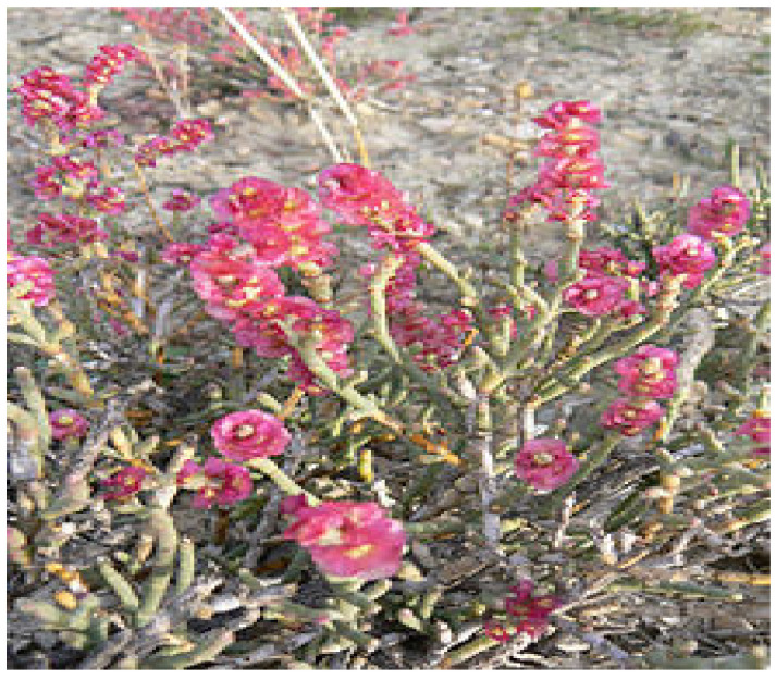
Anabasis articulata (A. articulata; Figure 1) belongs to the genus Anabasis, and the family Amaranthaceae is a medicinally valuable plant that is subjected to scientific exploration very little and needs to be explored in line with modern approaches. It is a xerophyte primarily found in deserts. In many parts of the world, it is used in folk medicine to treat skin conditions such as eczema and other ailments, including diabetes, headache, and fever [8].
Figure 1.
The A. articulata plant.
As mentioned above, A. articulata has been reported to have medicinal properties by very few researchers, and thus, the present study is an attempt to explore A. articulata for its antimicrobial, antioxidant, and antidiabetic potential in connection to its phytochemical composition, which was investigated using preliminary phytochemical tests, HPLC and GC-MS analysis.
2. Materials and Methods
2.1. Plant Material Collection
The leaves and stem (1 kg; equal proportions) of A. articulata were collected from Mohmand Agency (34.5356° N, 71.2874° E, a deserted tribal area), Pakistan, in the year 2020–2021. The collected plant samples were identified by the Taxonomist, working as an expert in Herbarium, University of Malakand, Pakistan. A voucher specimen (BGH-UOM-190) was kept for record in the herbarium. After cleaning, the samples were shade dried, grounded, and then subjected to the extraction process.
2.2. Chemicals and Reagents
All chemicals used in the study were of analytical grade except those used as solvents in HPLC analysis. The reagents; 2,2-Diphenyle-1 picrylhydrazyl (DPPH), sodium carbonate, Folin-Ciocalteu (F-C) reagent were obtained from Sigma-Aldrich CHEMIE GmbH, St. Louis, MO, USA, whereas quercetin and 2,2′-Azino-Bis-3 ethyl benzothiazoline-6-sulfonic Acid (ABTS) aluminum chloride, methanol, sodium hydroxide, ethanol, sodium nitrite, and ascorbic acid were purchased from Sigma-Aldrich, Taufkirchen, Germany.
2.3. Extraction and Fractionation
Extraction and fractionation were carried out according to already reported protocols [6,13]. About 200 g of the powdered sample was soaked in methanol, filtered through the Whatman filter paper, and concentrated using a rotary evaporator (Rotavapor R-200, Buchi, Flawil, Switzerland). About 20 g of crude extract was obtained and used in subsequent experiments. The extract was eluted through a silica gel column for fractionation as per the following details: The extract was mixed with silica gel slurry and then allowed to dry in the air. The sample loaded silica was then carefully loaded to a large silica gel column with an internal diameter of 10 cm and packed height of 50 cm using a gradient of increasing polarity from n-hexane to ethyl acetate as the mobile phase. The oil fraction was extracted through n-hexane solvent. The effluents of columns were separated into four purified fractions designated as A, B, C, and D. Fraction A was separated by silica gel column chromatography in a solvent system of ethyl acetate and n-hexane (5:95), fraction B was separated by silica gel column chromatography in a solvent system of ethyl acetate and n-hexane (10:90), fraction C was separated in a solvent system of ethyl acetate and n-hexane (20:80), while fraction D was separated by silica gel column chromatography in a solvent system of ethyl acetate and n-hexane (30:70). The resultant extracts were stored at a temperature of 4 °C in a refrigerator till their screening for in vitro biological activities.
2.4. Preliminary Phytochemical Analysis
Reported protocols were followed for the identification of major phytochemicals in the extracts [15].
2.5. Estimation of Total Phenolic Content (TPC) and Total Flavonoid Content (TFC)
Shirazi et al.’s method was used for the estimation of TPC [16] in extract, whereas Kim et al.’s method [17] was followed for TFC estimation. From the stock sample (5 mg/5 mL), 1 mL was added to 9 mL of distilled water, to which 1 mL F-C reagent was added and incubated for 6 min, after which 10 mL of 7% sodium carbonate solution and 25 mL of distilled water were added. The absorbance was measured at 760 nm after 90 min of incubation time. The standard gallic acid solution’s curve was used for the estimation of the TPC expressed as mg GAE (Gallic acid equivalent)/g of the dry sample.
In distilled water (500 µL), 100 µL of each sample, 150 µL of aluminum chloride, 100 µL of 5% sodium nitrate, and 200 µL of 1M sodium hydroxide were added for assessment of TFC. The absorbance of the mixture was recorded at 510 nm after being incubated for 5 min. The TFC was calculated as mg QE (quercetin equivalents)/g of the dry sample.
2.6. HPLC-UV Characterization
Methanolic extract, hexane, and purified fraction were each added to a distilled water:methanol (1:1) mixture, heated at 50 °C for 1 h, dually filtered, and poured into HPLC vials [18]. The extract’s phytochemicals were separated using HPLC Agilent 1260 with an eclipsed C18 column (Santa Clara, CA, USA). The spectrums were recorded at 320 nm. Retention times of the available standards were employed to identify the unknown compounds present in the analyzed samples. Quantification of antioxidants was measured by formula (Equation (1)):
| (1) |
where Cx = concentration of unknown sample, As = peak area of standard, Ax = peak area of unknown sample, Cs = concentration of standard (0.09 µg/mL).
2.7. GC-MS Analysis
The extracts were further analyzed for volatile components using GC-MS (Agilent Technological USA) analysis [19]. The mass spectra and retention time of the compounds present in samples were compared with those of Willy and NIST libraries [20].
2.8. Antibacterial Screening
The antibacterial potential of methanolic crude extract, purified fractions (A, B, C, and D), and oil fraction was assessed against Shigella dysentery (S. dynasties), Escherichia coli (E. coli), and Salmonella Typhi (S. Typhi) using the agar disc diffusion method. The strains were grown on nutrient broth, whereas the antibacterial spectrum of the extracts and fractions was assessed by the agar disc diffusion method, as mentioned before. A control (ampicillin) disc was also placed. The Clinical Laboratory Standards Institute guidelines (CLSI 2012) were followed [3] while determining the zone of inhibition (ZI) encountered by each extract and fractions against selected bacterial strains. The activity for each extract, purified fractions, and oil fraction was performed in triplicate and presented as the mean values.
2.9. Antioxidant Activities
2.9.1. DPPH Assay
A slightly modified DPPH assay as used before by Brand William was followed [21]. The absorbance of 3 mL from stock DPPH (20 mg in 100 mL of methanol) was adjusted to 0.75 at 517 nm. The DPPH stock solution was covered and kept in the dark overnight to generate free radicals. About 2 mL of each dilution (1000, 500, 250, 125, 62.5 µg/mL) prepared from methanolic extract stock (5 mg/5 mL of methanol) were mixed with 2 mL of pre-incubated DPPH stock solution and incubated for 15 min. Ascorbic acid was used as a standard. The absorbance of the reaction mixtures was recorded at 517 nm and the % inhibition was calculated as (Equation (2)):
| (2) |
where A = absorbance of pure DPPH in oxidized form, B = absorbance of the sample, which was measured after 15 min of reaction with DPPH.
2.9.2. ABTS Assay
A standard protocol for ABTS free radical scavenging potential of extracts was followed [22]. ABTS (7 mM) and K2S2O8 (2.45 mM) were mixed (in methanol) and put in the dark for 24 h for free radicals’ formation, which was then used as stock solution. The absorbance of 3 mL from the stock ABTS was adjusted to 0.75 at 745 nm, which was considered a control. About 300 μl of each of the serial dilutions of methanolic extract (1000, 500, 250, 125, 62.5 µg/mL) and 3 mL of stock ABTS were mixed and incubated for 15 min at 25 °C, and their absorbance was measured at 745 nm. Ascorbic acid was used as a control. The scavenging activity was calculated by Equation (2).
2.10. In Vitro Antidiabetic Activities
2.10.1. Inhibition of α-Amylase
The extracts were assessed as inhibitors of α-amylase following a standard protocol reported in the literature with some modifications [23]. In distilled water, an alpha-amylase stock solution (10 mg/100 mL) was prepared. About 10 µL of alpha-amylase stock solution was mixed with 30 µL of each sample dilution and 40 mL of starch solution, and these were kept at 37 °C for 30 min. After incubation, 20 µL of HCl (1M) was added to the reaction mixture, and its absorbance was measured at 580 nm. Acarbose was used as the reference standard. Alpha-amylase % inhibition was calculated as (Equation (3)):
| (3) |
2.10.2. Inhibition of α-Glucosidase
The reported protocol was followed to assess the α-glucosidase inhibitory potential of the extracts [24]. To 100 µL α-glucosidase (0.5 units/mL), 50 µL of each sample dilutions and 600 µL of 0.1 M phosphate buffer (pH 6.9) were mixed. From the substrate (p-nitrophenyl-α-D-glucopyranoside) prepared as a 5 Mm solution in 0.1 M phosphate buffer, 100 µL of the substrate was added to the reaction mixture and kept for 15 min at 37 °C. The absorbance was recorded at 405 nm. The reaction mixture without α-glucosidase was labeled as blank, whereas the reaction mixture without the sample was taken as the control. The degree of the enzyme’s activity inhibition was measured as:
| (4) |
2.11. Statistical Analysis
All in vitro experiments were performed in three replicates. All results have been presented as mean ± SEM. The Student’s t-test and one-way ANOVA followed by Dunnett’s post hoc multiple comparison test was used to evaluate the significance of the data obtained. p ≤ 0.05 was considered significant.
3. Results
3.1. Yield from Fractionation
Crude extract: 10 g, fraction A: 200 mg, B: 150 mg, C: 120 mg, and D: 100 mg were obtained. About 80 mg of purified hexane fraction was obtained in semi-solid form. In the subsequent studies, these components have been tested.
3.2. Qualitative Phytochemical Screening
Plants contain millions of compounds that are classified into roader groups of phytochemicals. Such constituents are determined as preliminary evaluations to decide the medicinal value of a plant. Table 1 represents the presence of different phytochemical groups showing that this plant is worthy of being investigated for its medicinal profile, being a rich source of phytochemicals.
Table 1.
Phytochemical screening (qualitative) of A. articulata crude extract.
| Phytochemical | Reagent | Observation | Result |
|---|---|---|---|
| Alkaloids | Dragendroff’s | Reddish-orange precipitate | + |
| Tannins | Gelatin | Dirty (brownish) green precipitates | + |
| Flavonoids | Ferric chloride | The yellowish appearance that becomes clear after acid (HCL) addition | + |
| Triterpenoids | Liebermann Burchard | Brown ring | + |
| Glycosides | Keller Killiani | Reddish-brown layer | + |
3.3. TPC and TFC in Resultant Extracts
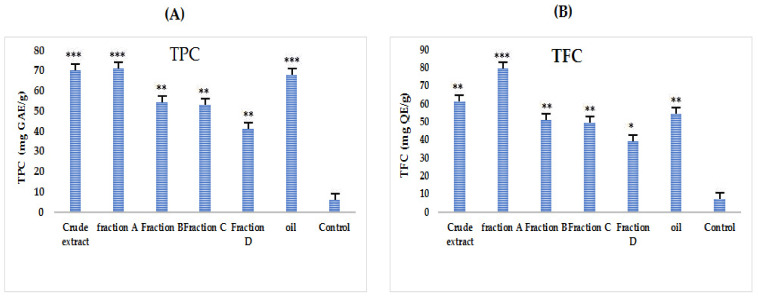
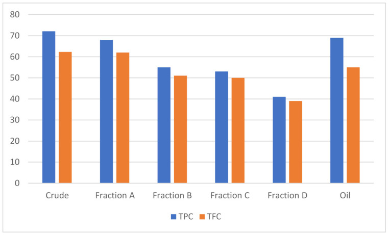
A standard Gallic acid curve was constructed by preparing the dilutions 20, 40, 60, 80 and 100 mg/mL to estimate the TPC in different tested samples of A. articulata using a graphical regression method (Figure 2A). Comparatively higher TPC contents were estimated in almost all extracts as compared to TFC. Results show that the highest TPC (Figure 3) values were observed for crude extract and then oil and fraction A (72.1 ± 0.2, 69.0 ± 1.1 and 68.0 ± 0.4 mg GAE/g of dry sample, respectively).
Figure 2.
Total phenolic (A) and total flavonoids (B) contents in different tested samples (crude extract, purified fractions, and oil) of A. articulata. (A) TPC expressed as gallic acid equivalents (mg GAE)/g dry plant sample; (B) TFC expressed as quercetin equivalents (mg QE)/g dry plant sample. The data is represented as Mean ± SEM, n = 3. Values are significantly different as compared to positive control * p < 0.05, ** p < 0.01, *** p < 0.001.
Figure 3.
Total phenolic and total flavonoid content in different tested samples (crude extract, purified fractions, and oil) of A. articulata. TPC expressed as gallic acid equivalents (mg GAE)/g of dry plant sample, and TFC expressed as quercetin equivalents (mg QE)/g of dry plant sample. The data are represented as mean ± SEM, n = 3.
To estimate the TFC in different tested samples of A. articulata, a regression curve of standard quercetin was constructed by preparing the dilutions 20, 40, 60, 80 and 100 mg/mL. The estimated contents are graphically presented in Figure 2B. Fraction A/crude extract followed by the oil fraction has shown the highest total flavonoid contents (62.0 ± 0.1, 62.3 ± 1.2, and 55.1 ± 0.3 mg QE/g of dry sample, respectively).
3.4. HPLC-UV Analysis
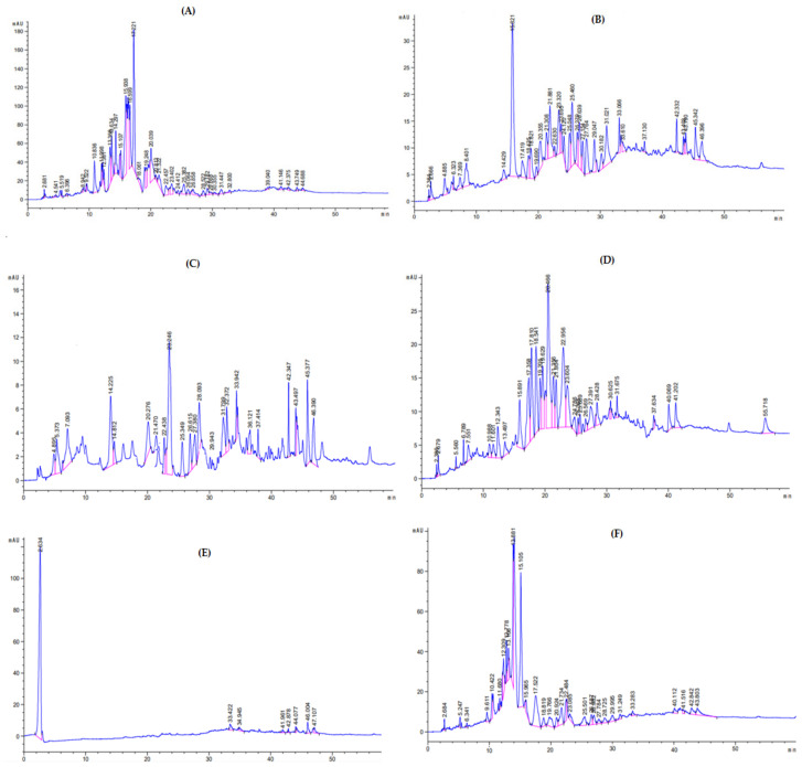
The HPLC chromatograms of the crude extract are presented in Figure 4A, purified fractions in Figure 4B–E, and the n-hexane fraction in Figure 4F. The compounds identified were: malic acid, gallic acid, chlorogenic acid, epigallocatechin gallate, Bis-HHDP-hex (pedunculagin), morin, 3-0-caffcoylquinic acid, Ellagic acid, kaempferol-3-(p-coumaroyl-diglucoside)-7-glucoside, catechin hydrate, rutin, syringic acid, quercetin-7-O-sophoroside, kaempferol-3-(caffeoyl-diglucoside)-7-rhamnosyl, mannose, pyrogallol, caffeic acid, mandelic acid, quercetin-3-(caffeoyldiglucoside)-7-glucoside, p-Coumaric acid, galactose, vitamin C, 3-0-caffeoylquinic acid, apigenin-7-O-rutinoside, quercetin 3,7-di-O-glucoside, rhamnose, 3,5-dicaffeoylquinic acid, mandelic acid, xylulose, quercetin 3,7-O-glucoside, glucose, quercitin-3-0-glysides, quercitin-3-O-rutinoside, quercitin-3-O-glycosides, quercetin, quercetin-3-(caffeoyldiglucoside)7-glucoside, quercitin-3-0-glycosides, in crude extract, various fraction (A, B, C, D, and n-hexane fraction. Each peak in the given chromatograms represents a phytoconstituent. For the identification of such constituent retention times of each component were compared with that of external standards. The quantification of each phenolic compound with their particular peak position and retention time (Rt) in chromatogram is presented in Table 2, Table 3, Table 4, Table 5, Table 6 and Table 7.
Figure 4.
HPLC chromatograms of crude extract (A), various purified fractions (B–E), and n-hexane fraction (F) of A. articulata.
Table 2.
Identified phytochemicals in crude extract of A. articulata through the HPLC-UV technique.
| Retention Time (min) | Phytochemical Compounds | HPLC-UV λmax (nm) | Peak Area of Sample | Peak Area of Standard | Concentration (µg/mL) | Identification Reference |
|---|---|---|---|---|---|---|
| 2 | Malic acid | 320 | 53.130 | 40.323 | 1.186 | Ref. Stand |
| 4 | Gallic acid | 320 | 41.239 | 195.40 | 0.189 | Ref. Stand |
| 6 | Chlorogenic acid | 320 | 32.966 | 12.929 | 2.295 | Ref. Stand |
| 8 | Epigallocatechin gallate | 320 | 44.782 | 7261.474 | 0.005 | Ref. Stand |
| 11 | Bis-HHDP-hex(pedunculagin) | 320 | 171.562 | - | - | [25] |
| 12 | Morin | 320 | 103.604 | 2.00 | 46.622 | Ref. Stand |
| 14 | 3-0-caffcoylquinic acid | 320 | 514.593 | - | - | [25] |
| 16 | Ellagic acid | 320 | 912.321 | 319.242 | 2.572 | Ref. Stand |
| 18 | Kaempferol-3-(p-coumaroyl-diglucoside)-7-glucoside | 320 | 149.535 | - | - | [25] |
| 20 | Catechin hydrate | 320 | 810.747 | 78.00 | 9.355 | Ref. Stand |
| 22 | Rutin | 320 | 264.573 | 22.40 | 10.630 | Ref. Stand |
| 23 | Syringic acid | 320 | 254.546 | - | - | [26] |
| 24 | Quercetin-7-O-sophoroside | 320 | 50.8907 | - | - | [26] |
| 25 | Kaempferol-3-(caffeoyl-diglucoside)-7-rhamnosyl | 320 | 261.997 | - | - | [26] |
| 26 | Mannose | 320 | 85.1536 | - | - | [25] |
| 28 | Pyrogallol | 320 | 101.640 | 1.014 | 90.213 | Ref. Stand |
| 29 | Caffeic Acid | 320 | 129.708 | - | - | [25] |
| 30 | Mandelic acid | 320 | 45.405 | 72.00 | 0.567 | Ref. Stand |
| 31 | Quercetin-3-(caffeoyldiglucoside)-7-glucoside | 320 | 107.633 | - | - | [27] |
| 32 | p-Coumaric acid | 320 | 52.304 | - | - | [27] |
| 42 | Galactose | 320 | 57.017 | - | [25] |
Table 3.
Identified phytochemical compounds in fraction A of A. articulata through the HPLC-UV technique.
| Retention Time (min) | Phytochemical Compounds | HPLC-UV λmax (nm) | Peak Area of Sample | Peak Area of Standard | Concentration (µg/mL) | Identification Reference |
|---|---|---|---|---|---|---|
| 4 | Vitamin C | 320 | 64.43 | 22.4 | 2.588 | Ref. Stand |
| 6 | Chlorogenic acid | 320 | 18.64 | 2.929 | 5.727 | Ref. Stand |
| 8 | Epigallocatechin gallate | 320 | 132.75 | 7261.474 | 0.0164 | Ref. Stand |
| 14 | 3-0-caffeoylquinic acid | 320 | 45.345 | - | - | [26] |
| 18 | Kaempferol-3-(p-coumaroyl-diglucoside)-7-glucoside | 320 | 86.189 | - | - | [26] |
| 18 | Apigenin-7-O-rutinoside | 320 | 128.126 | - | - | [26] |
| 20 | Catechin hydrate | 320 | 134.971 | 78.00 | 1.557 | Ref. Stand |
| 22 | Rutin | 320 | 23.593 | 22.40 | 0.947 | Ref. Stand |
| 23 | Syringic acid | 320 | 176.298 | - | - | [25] |
| 23 | Quercetin 3,7-di-O-glucoside | 320 | 77.612 | - | - | [25] |
| 24 | Quercetin-7-O-sophoroside | 320 | 58.34 | - | - | [25] |
| 25 | Kaempferol-3-(caffeoyl-diglucoside)-7-rhamnosyl | 320 | 248.248 | - | - | [26] |
| 26 | Rhamnose | 320 | 103.713 | - | - | [26] |
| 27 | 3,5-dicaffeoylquinic acid | 320 | 97.727 | - | - | [26] |
| 29 | Caffeic acid | 320 | 113.173 | - | - | [27] |
| 30 | Mandellic acid | 320 | 63.944 | 72.00 | 0.799 | Ref. Stand |
| 31 | Quercetin-3-(caffeoyldiglucoside)-7-glucoside | 320 | 223.444 | - | - | [26] |
| 42 | Galactose | 320 | 60.609 | - | - | [27] |
| 43 | Xylulose | 320 | 31.328 | - | - | [27] |
Table 4.
Identified phytochemicals in purified fraction B of A. articulata through the HPLC-UV technique.
| Retention Time (min) | Phytochemical Compounds | HPLC-UV λmax (nm) | Peak Area of Sample | Peak Area of Standard | Concentration (µg/mL) | Identification Reference |
|---|---|---|---|---|---|---|
| 4 | Gallic acid | 320 | 36.4984 | 195.40 | 0.168 | Ref. Stand |
| 14 | 3-0-caffeoylquinic acid | 320 | 160.818 | - | - | [26] |
| 20 | Catechin hydrate | 320 | 78.1380 | 78.00 | 0.902 | Ref. Stand |
| 22 | Rutin | 320 | 42.5292 | 22.40 | 1.709 | Ref. Stand |
| 23 | Syringic acid | 320 | 324.245 | - | - | [26] |
| 25 | Kaempferol-3-(caffeoyl-diglucoside)-7-rhamnosyl | 320 | 58.9445 | - | - | [26] |
| 27 | 3,5-dicaffeoylquinic acid | 320 | 88.6597 | - | - | [26] |
| 28 | Pyrogallol | 320 | 85.8767 | 1.014 | 76.221 | Ref. Stand |
| 31 | Quercetin-3-(caffeoyldiglucoside)-7-glucoside | 320 | 88.6597 | - | - | [26] |
| 32 | p-Coumaric acid | 320 | 56.5793 | - | - | [26] |
| 43 | Xylulose | 320 | 36.3262 | - | - | [27] |
Table 5.
Identified phytochemicals in fraction C of A. articulata through the HPLC-UV technique.
| Retention Time (min) | Phytochemical Compounds | HPLC-UV λmax (nm) | Peak Area of Sample | Peak Area of Standard | Concentration (µg/mL) | Identification Reference |
|---|---|---|---|---|---|---|
| 2 | Malic acid | 320 | 24.7858 | 40.323 | 0.554 | Ref. Stand |
| 11 | Bis-HHDP-hex (pedunculagin | 320 | 47.4527 | - | - | [25] |
| 12 | Morin | 320 | 100.496 | 2.00 | 45.223 | Ref. Stand |
| 18 | Apigenin-7-O-rutinoside | 320 | 246.3633 | - | - | [26] |
| 22 | Rutin | 320 | 424.706 | 22.40 | 17.064 | Ref. Stand |
| 23 | Quercetin 3,7-O-glucoside | 320 | 201.7868 | - | - | [26] |
| 25 | Kaempferol-3-(caffeoyl-diglucoside)-7-rhamnosyl | 320 | 30.1412 | - | - | [26] |
| 26 | Rhamnose | 320 | 32.8371 | - | - | [27] |
| 27 | 3,5-dicaffeoylquinic acid | 320 | 120.040 | - | - | [26] |
| 28 | Pyrogallol | 320 | 66.255 | 1.014 | 58.806 | Ref. Stand |
| 30 | Mandelic acid | 320 | 29.289 | 72.00 | 0.366 | Ref. Stand |
| 31 | Quercetin-3-(caffeoyldiglucoside)-7-glucoside | 320 | 32.0566 | - | - | [26] |
| 37 | Glucose | 320 | 21.6425 | - | - | [27] |
| 41 | Quercitin-3-0-glysides | 320 | 78.9891 | - | - | [26] |
Table 6.
Identified phytochemicals in purified fraction D of A. articulata through the HPLC-UV technique.
| Retention Time (min) | Phytochemical Compounds | HPLC-UV λmax (nm) | Peak Area of Sample | Peak Area of Standard | Concentration (µg/mL) | Identification Reference |
|---|---|---|---|---|---|---|
| 2 | Malic acid | 320 | 2514.20 | 40.323 | 56.116 | Ref. Stand |
| 34 | Quercitin-3-O-rutinoside | 320 | 41.0693 | - | - | [26] |
| 41 | Quercitin-3-O-glycosides | 320 | 30.249 | - | - | [26] |
| 42 | Galactose | 320 | 16.116 | - | - | [27] |
Table 7.
Identified phytochemicals in n-hexane fraction of A. articulata through the HPLC-UV technique.
| Retention Time (min) | Phytochemical Compounds | HPLC-UV λmax (nm) | Peak Area of Sample | Peak Area of Standard | Concentration (µg/mL) | Identification Reference |
|---|---|---|---|---|---|---|
| 2 | Malic acid | 320 | 28.2673 | 40.323 | 0.631 | Ref. Stand |
| 6 | Chlorogenic acid | 320 | 14.9574 | 2.929 | 4.595 | Ref. Stand |
| 10 | Quercetin | 320 | 137.065 | 90.90 | 1.357 | Ref. Stand |
| 11 | Bis-HHDP-hex (pedunculagin) | 320 | 44.509 | - | - | [26] |
| 12 | Morin | 320 | 231.396 | 2.00 | 104.128 | Ref. Stand |
| 18 | Apigenin-7-O-rutinoside | 320 | 89.384 | - | - | [26] |
| 22 | Rutin | 320 | 159.626 | 22.40 | 6.414 | Ref. Stand |
| 23 | Syringic acid | 320 | 76.785 | - | - | [26] |
| 25 | Kaempferol-3-(caffeoyl-diglucoside)-7-rhamnosyl | 320 | 152.318 | - | - | [26] |
| 26 | Rhamnose | 320 | 100.004 | - | - | [26] |
| 31 | Quercetin-3-(caffeoyldiglucoside)7-glucoside | 320 | 51.863 | - | - | [26] |
| 41 | Quercitin-3-0-glycosides | 320 | 31.616 | - | - | [26] |
| 42 | Galactose | 320 | 100.903 | - | - | [27] |
3.5. GC-MS Characterization of the Different Fractions
3.5.1. Purified Fraction A
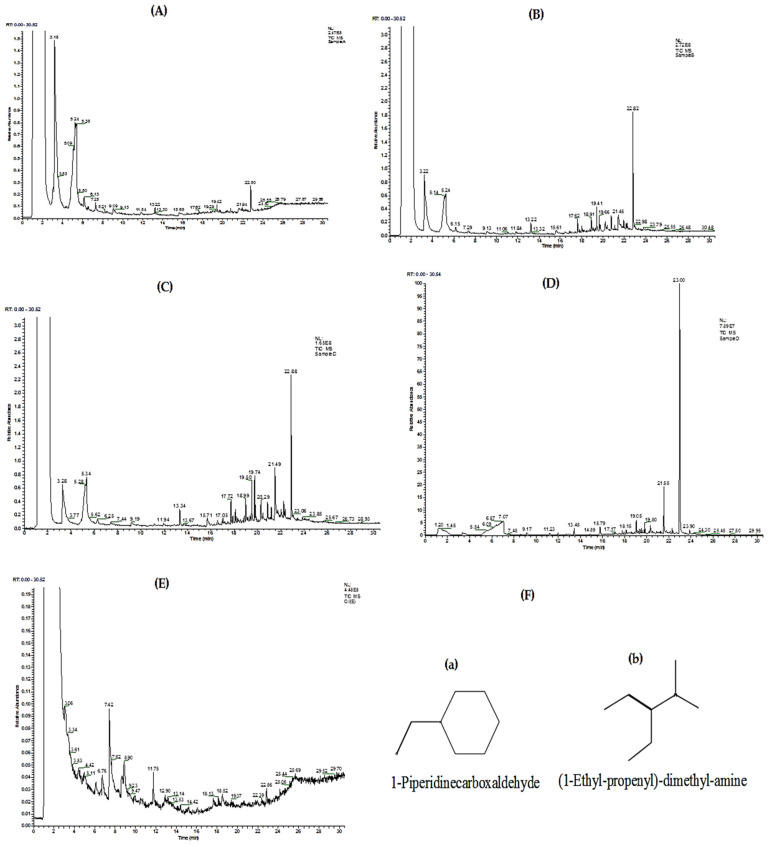
The GC-MS chromatogram of fraction A is indicated in Figure 5A. Figure S1 represents the structural formulas of five phytochemical compounds identified from the given chromatogram, whereas Figure S2 represents their mass fragmentation pattern and Table S1 represents different parameters of the major phytochemical compounds identified.
Figure 5.
GC-MS chromatogram of A. articulata purified fractions (A–D), oil fraction (E), and major phytochemical compounds (a,b) identified in the oil fraction (F).
3.5.2. Purified Fraction B
The GC-MS chromatogram of fraction B is indicated in Figure 5B. The technique confirmed the presence of 12 phytochemical compounds in fraction B, and their other parameters are presented in Table S2. Figures S3 and S4 represent the structural formulas of 12 phytochemical compounds and the pattern of their mass fragmentation, respectively.
3.5.3. Purified Fraction C
Figure 5C shows the GC-MS chromatogram of fraction C. Table S3 represents 13 phytochemical compounds identified along with some basic parameters related to the analysis performed. Figure S5 indicates the structural formulas of 13 phytochemical compounds, and Figure S6 indicates their mass fragmentation pattern.
3.5.4. Purified Fraction D
The GC-MS chromatogram of fraction D is presented in Figure 5D, where the presence of 13 phytochemical compounds was confirmed as presented in Table S4 along with analysis-related parameters. Figure S7 represents the structural formulas of phytochemical compounds, whereas Figure S8 represents the pattern of their mass fragmentation.
3.5.5. Oil Fraction
Figure 5E represents the GC-MS chromatogram of the purified oil fraction. Figure 5F shows the structural formulas of major phytochemical compounds (a and b), and Table S5 shows their different parameters.
3.6. Antibacterial Activity
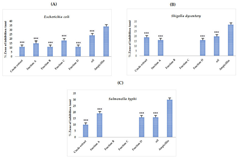
The results of the antibacterial potential of the samples have been tabulated in Table S6 and graphically represented in Figure 6. The results depicted that all the samples except for B and C showed activity against the tested bacterial strains. The broad-spectrum antibiotic ampicillin was used as a positive control. The n-hexane (oil) fraction showed the highest ZI against all tested strains: S. dysentery, E. coli, and S. Typhi as 20, 24, and 16 mm respectively. An appreciable degree of antibacterial bacterial potential suggests the plant’s possible usage as a source for isolating antibacterial compounds.
Figure 6.
Antibacterial potential of crude extract, purified fractions (A, B, C, and D), and oil fraction of A. articulata against (A) E. coli, (B) Shigella dysentery and (C) S. typhi. The data are represented as mean ± SEM, n = 3. Values are significantly different as compared to positive control *** p < 0.001.
3.7. Antioxidant Activity of Crude and Purified Fractions of A. articulata
3.7.1. DPPH Assay
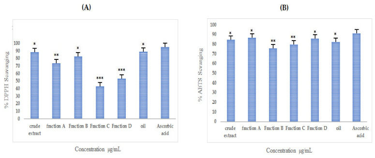
Almost all extracts inhibited DPPH free radicals; however, among them, n-hexane fraction, crude extract, and fraction B showed significant free radical inhibition with IC50 values of 45, 90, and 62 µg/mL, respectively, as presented in Table S7 and Figure 7A.
Figure 7.
Antioxidant activity ((A) = DPPH; (B) = ABTS) of crude extract, purified fractions (A, B, C, and D), and oil fraction of A. articulata. The data plotted is mean ± SEM, n = 3. Values are significantly different as compared to positive control * p < 0.05, ** p < 0.01, *** p < 0.001.
3.7.2. ABTS Assay
The ABTS scavenging potential of extracts is presented in Table S7 and Figure 7B. The results depict that fraction A and n-hexane extract possesses significant free radical inhibition with the lowest IC50 values of 75 and 71 µg/mL, respectively, as compared to the ascorbic acid used as the standard, which showed an IC50 value of 32 µg/mL.
3.8. In Vitro Antidiabetic Activities of Purified Fraction and Extract
3.8.1. In Vitro α-Glucosidase Inhibition
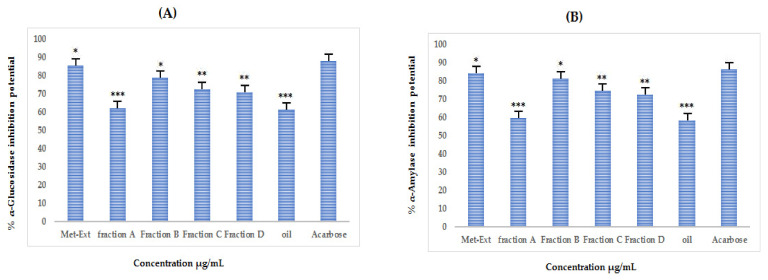
Figure 8A and Table S8 represent the %α-glucosidase inhibition of crude extract, oil, and the purified fractions. The crude extract showed the highest inhibition of the enzyme with an IC50 of 32 µg/mL followed by fraction B, which produced an IC50 of 60 µg/mL.
Figure 8.
(A) %α-glucosidase inhibition potential and (B) %α-amylase inhibition potential of crude extract, purified fractions (A, B, C, and D), and oil fraction of A. articulata at different concentrations. The data are represented as mean ± SEM, n = 3. Values are significantly different as compared to positive control * p < 0.05, ** p < 0.01, *** p < 0.001.
3.8.2. In Vitro α Amylase Inhibition
As shown in Figure 8B and Table S8, the %α-amylase inhibition is appreciable, and the highest inhibition was caused by crude extract with an IC50 of 34 µg/mL followed by fraction B, which produced an IC50 of 58 µg/mL.
4. Discussion
Presently, insulin therapies are the treatment of choice to control hyperglycemia in diabetes mellitus. Other strategies are the inhibition of alpha-amylase and glucosidase through different inhibitors, as both enzymes are responsible for releasing glucose from starch taken in food [28,29]. In this context, an attempt has been made in this study to identify the possible antidiabetic phytochemical that could inhibit the activity of carbohydrate digesting enzymes (α-amylase and α-glucosidase). The study revealed that the plant could be a potential candidate for isolating antidiabetic compounds.
With the increasing reports about the side effects of synthetic drugs, researchers have focused on plants to isolate effective therapeutic precursors with low or no side effects. Drug resistance is the other overwhelming problem in the modern era, and the search for new antibiotics of plant origin is in progress. The plant crude extract and purified fractions showed appreciable antibacterial activity, which is evident from the zones of inhibitions against selected bacterial strains, as shown in Figure 5.
Oxidative stress caused by free radicals that are constantly produced during normal metabolic processes is a serious health threat. Although these are constantly deactivated by the human defense system, in the modern era, humans have started relying on processed foods, which have given rise to the overproduction of free radicals. Research shows that plants could neutralize the free radicals because of their constituent phenolics [30], as benzene rings in such compounds can stabilize the singlet electron of the free radicals. Collectively, such phytoconstituents are named antioxidant compounds, which play an important role in human health by combating reactive oxygen species and, in turn, is the main contributor to a number of human diseases, including insulin resistance, cardiovascular diseases, atherosclerosis, and coronary heart disease. Butylated hydroxytoluene and butylated hydroxy anisole are strong synthetic antioxidant agents, but they are carcinogenic and toxic to humans. Therefore, plant-based phenolic compounds can be used as antioxidants to scavenge or stabilize free radicals involved in oxidative stress generated in human bodies as a result of oxidation of certain substances. It is found that the use of synthetic antioxidants is injurious to human health, and individuals taking them are at risk of cancer and other liver disorders. The antioxidants in plants have become a hotspot for researchers in recent decades due to the mentioned fact of low or no side effects. Studies have indicated that the use of natural antioxidants can reduce oxidative stress and reduce the risk of major human diseases, including oxidative stress [3,6]. The n-hexane fraction, crude extract, and fraction B were more potent against DPPH radicals, whereas against ABTS, the n-hexane fraction and fraction A were more potent, indicating that these extracts contained certain phytoconstituents capable of scavenging free radicals, which could thus be further investigated for the isolation of responsible compounds. The DPPH radicals in acholic medium undergo a reduction in the presence of hydrogen donating antioxidants. Phytochemicals such as flavonoids and phenolics are good antioxidants and play a vital role in scavenging the free radicals due to the presence of benzene rings in their structures [6,7,8,9,10,11,12,13]. The observed antioxidant potential can be correlated with the estimated TFC and TPC values, as these are the responsible scavengers in the extracts. The total polyphenol and flavonoid content in the fractions increased in the following order: crude extract, fraction A, and oil fraction. The crude extract has the highest TPC and TFC, i.e., (TPC = 72.1 mg GAE/g and TFC = 62 mg QE/g) followed by purified fraction A, which has the highest TPC and TFC, i.e., 68 GAE/g and 62 mg QE/g, inferring the plant is a good source of flavonoids and phenolics. As mentioned before, due to the presence of benzene rings in the structure, flavonoids and phenolics have been found to be excellent scavengers of the free radicals, which is why the tested radicals, ABTS and DPPH, were potently scavenged by the extracts, i.e., the total phenolic and flavonoid contents in the extracts and purified fractions were positively proportional to the antioxidant activities. The current results were in line with the previously reported studies [6,14]. The study of Kim et al. [31] showed plants that contained high TFC and TPC, and by virtue of these components, they exhibit various biological potentials. Their conclusion was based on findings of extracts from 40 plant species in Korea. As mentioned, phenolic and flavonoid compounds are strong antioxidants that can deactivate free radicals by offering their hydrogen atoms and electrons [32], which is the reason that plants with high TFC and TPC inhibit DPPH and ABTS radicals more potently in laboratory-scale experiments. The positive correlation between the total phenolic content and flavonoid content in the plant extracts and the antioxidant activities have been observed by other researchers as well [32]. The plants in the form of extracts could, therefore, offer strong activity against a wide range of oxidants and thus would have great medicinal applications. It can be seen from Table S7 that the crude extract and fractions exhibited significant activities against the DPPH and ABTS tested radicals, which needs to be further investigated. Furthermore, for the crude extract, the preliminary phytochemical tests (Table 1) were positive, indicating the presence of broad phytochemical groups and, consequently, the wider range of their therapeutic action.
The HPLC analysis of crude and purified fractions of A. articulata showed the presence of several possible compounds that might be responsible for antioxidant and antidiabetic activities. The antidiabetic properties of A. articulata crude extracts and fractions (Figure 7 and Table S8) were determined based on the inhibitory effect against two carbohydrate hydrolyzing enzymes, namely α-amylase and α-glucosidase. As mentioned before, starch is converted into disaccharides and oligosaccharides by pancreatic α-amylase, while disaccharides are broken down into glucose by intestinal α-glucosidase [3,6] and, thus, if inhibited, will lessen the glucose burden in diabetic patients as their inhibition could retard the breakdown of starch in the gastrointestinal tract and, therefore, would ameliorate hyperglycemia in human. The detected compounds are known to be antioxidant and antidiabetic agents, as indicated in the previously reported studies [3,6,9,14]. The current results of the screening are in close accordance with the already reported study of Nazir et al., where they confirmed the presence of quercetin, morin, and rutin in the methanolic extract of Silybum marianum (L.) seeds [33] and in the methanolic extracts of the fruit of Elaeagnus umbellata Thunb. [6]. The results of this study are in agreement with the findings of other studies where strong antioxidant activities were observed along with strong α-glucosidase and α-amylase inhibitions [3,6,9].
The medicinal plant has become a vital source of antioxidants in the last few decades. Literature surveys have shown that the ingestion of natural antioxidants can reduce oxidative stress-related diseases. Various studies have shown that the presence of malic acid, gallic acid, quercetin, morin, ellagic acid, rutin, chlorogenic acid, and epigallocatechin gallate can be liable for the antioxidant capacity observed [14,34,35]. It is evident from the literature that gallic acid, chlorogenic acid, epigallocatechin gallate, and morin have strong antioxidants and antidiabetic potentials [36,37].
The GC-MS analysis of the purified fraction also confirmed the presence of certain valuable phytochemical compounds: Acetdimethylamide, N-Nitrosomorpholine, 1,2-Benzenedicarboxylic acid, Mono(2-ethylhexyl) phthalate, Bis(2-ethylhexyl) phthalate, N-Acetyl-l-methioninamide, 2-Propanamine, Phenol, 2,4-di-tert-butyl, Benzene, (1-dodecyltridecyl)-, Benzene, (1-hexyltetradecyl)-, Benzene, (1-hexylheptyl)-, Isopropyl Palmitate, 10-Octadecenoic acid, methyl ester, 1-Docosene, Methyl ricinoleate, Oleic acid, tetradecyl ester, Diisooctyl phthalate, Asparagine, entacosane, 13-phenyl Eicosane, 7-phenyl, Dodecane, 6-phenyl, Palmitic acid, methyl ester, tert-Hexadecanethiol, Decyl oleate, octadecyl ester, Elaidic acid, isopropyl ester, Phenethyl alcohol, á-methyl, Benzyl-3-hydroxypyrrolidine, Diethyl Phthalate, 2,6-Dimethyl-pyridine-3,5-dicarboxylic acid, dihydrazide, Methoxycarbonyl-2-methoxyphenyl isothiocyanate, Phosphoric acid, dibutyl 3-trifluoromethyl-3-pentyl ester, 4-Acetylaminophthalic acid, dimethyl ester, Benzene-1,3-dicarboxylic acid, 5-acetylamino-, (2-Phenyl-1,3-dioxolan-4-yl) methyl (9E)-9-octadecenoate, 1-Heneicosyl formate, 18,19-Secoyohimban-19-oic acid, Cleavamine, 18á-carboxy-3,4à-dihydro-, 1-Piperidinecarboxaldehyde, and (1-Ethyl-propenyl)-dimethyl-amine, which could possibly have their share in the observed biological potentials. The findings of the present study could be correlated with the reported studies [19,38]. From the rich phytochemical composition of the selected plant, we hypothesized that the different levels of antidiabetic activity of the extract and different fractions of A. articulata are due to the varying levels of various phytochemicals in each extract/fraction. The purified fraction A followed by crude extract A. articulata exhibited higher levels of TPC and TFC, together with antioxidant and antidiabetic activity as compared with the other extracts/fractions. This indicates that phenolic compounds, including flavonoids, are key active compounds found in these extracts, and the plant could thus be a good candidate for further studies to isolate inhibitors of the tested radicals and enzymes.
5. Conclusions
From the current study results, it can be concluded that A. articulata in extract form should be considered an effective source to relieve oxidative stress and health complications associated with diabetes. This plant also has the potential to be used as an antimicrobial agent as it effectively inhibited the growth of selected bacterial strains. The α-amylase and α-glucosidase enzymes were inhibited by extracts to an appreciable extent suggesting that this plant could be used as a potential candidate to isolate effective antidiabetic drugs. The observed biological activities were at the expense of its rich phytochemical composition, confirmed through HPLC-UV and GC-MS techniques in this study. Further studies are needed to isolate pure compounds responsible for the observed biological potentials.
Acknowledgments
The authors are thankful to Aljouf and Malakand Universities for providing research facilities.
Abbreviations
| WHO | World Health Organization |
| UV | Ultraviolet |
| HPLC | High performance liquid chromatography |
| GC-MS | Gas chromatography-mass spectroscopy |
| DPPH | 2,2-Diphenyle-1 picrylhydrazyl |
| ABTS | 2,2’-Azino-Bis-3 Ethylbenzothiazoline-6-Sulfonic Acid |
| TLC | Thin layer chromatography |
| TPC | Total phenolic content |
| TFC | Total Flavonoid Content |
| GAE | Gallic acid equivalent |
| QE | Quercetin equivalent |
| FID | Flame ionization detector |
| NIST | National Institute of Standards and Technology |
Supplementary Materials
The following supporting information can be downloaded at: https://www.mdpi.com/article/10.3390/molecules27113526/s1, Figure S1. Major phytochemical compounds identified in fraction A of A. articulata. Figure S2. Individual mass fragmentation patterns of each compound: (A) Acetdimethylamide (B) N-Nitrosomorpholine (C) 1,2-Benzenedicarboxylic acid (D) Mono(2-ethylhexyl) phthalate (E) Bis(2-ethylhexyl) phthalate. Figure S3. Major phytochemical compounds identified in fraction B of A. articulata. Figure S4. Individual mass fragmentation patterns of each compound: (A) N-Acetyl-l-methioninamide (B) 2-Propanamine (C) Phenol, 2,4-di-tert-butyl (D) Benzene, (1-dodecyltridecyl) (E) Benzene, (1-hexyltetradecyl) (F) Benzene, (1-hexylheptyl) (G) Isopropyl Palmitate (H) 10-Octadecenoic acid, methyl ester (I) 1-Docosene (J) Methyl ricinoleate (K) Oleic acid, tetradecyl ester (L) Diisooctyl phthalate. Figure S5. Major phytochemical compounds identified in fraction C of A. articulata. Figure S6. Individual mass fragmentation patterns of each compound: (A) Asparagine (B) entacosane, 13-phenyl (C) Eicosane, 7-phenyl (D) Dodecane, 6-phenyl (E) Palmitic acid, methyl ester (F) Isopropyl Palmitate (G) tert-Hexadecanethiol (H) Methyl ricinoleate (I) Decyl oleate (J) Oleic acid, octadecyl ester (K) Elaidic acid, isopropyl ester (L) Diisooctyl phthalate (M) Mono(2-ethylhexyl) phthalate. Figure S7. Major phytochemical compounds identified in fraction D of A. articulata. Figure S8. Individual mass fragmentation patterns of each compound: (A) Phenethyl alcohol, á-methyl (B) Benzyl-3-hydroxypyrrolidine (C) Diethyl Phthalate (D) 2,6-Dimethyl-pyridine-3,5-dicarboxylic acid, dihydrazide (E) Methoxycarbonyl-2-methoxyphenyl isothiocyanate (F) Phosphoric acid, dibutyl 3-trifluoromethyl-3-pentyl ester (G) Oleic Acid (H) 4-Acetylaminophthalic acid, dimethyl ester (I) Benzene-1,3-dicarboxylic acid (J) (2-Phenyl-1,3-dioxolan-4-yl) methyl (9E)-9-octadecenoate (K) 1-Heneicosyl formate (L) 18,19-Secoyohimban-19-oic acid (M) Cleavamine, 18á-carboxy-3,4à-dihydro-, methyl ester. Table S1. Major phytochemical compounds identified in fraction A of A. articulata and their various parameters. Table S2. Major phytochemical compounds and their parameters identified in fraction B of A. articulata. Table S3. List of major components and their parameters identified in fraction C of A. articulata. Table S4. Major phytochemical compounds and their parameters identified in fraction D of A. articulata. Table S5. Major phytochemical compounds and their parameters identified in the oil fraction of A. articulata. Table S6. Antibacterial potential of crude extract, oil, and purified fraction of A. articulata. Table S7. Antioxidant potential (DPPH and ABTS) of A. articulata crude extract, hexane, and subsequent fractions. Table S8. α-glucosidase and α-amylase inhibition of A. articulata crude extract and subsequent purified fractions at various concentrations.
Author Contributions
F.A.A.-J. and M.Z.: conceptualization; F.A.A.-J. and M.J.: methodology and formal analysis; N.N. and S.N.: writing—original draft; M.Z.: writing—review and editing; F.A.K. and M.T.: visualization; M.Z.: supervision and project administration, and validation; S.N.: formal analysis and data curation; A.N. and A.S. helped in the GC-MS analysis and biological activities. All authors have read and agreed to the published version of the manuscript.
Institutional Review Board Statement
Not applicable.
Informed Consent Statement
Not applicable.
Data Availability Statement
The data presented in this manuscript belong to the research work supervized under Muhammad Zahoor and have not been deposited in any repository yet. However, the data are available to the researchers upon request.
Conflicts of Interest
The authors declare that they have no conflict of interest.
Funding Statement
This research received no external funding.
Footnotes
Publisher’s Note: MDPI stays neutral with regard to jurisdictional claims in published maps and institutional affiliations.
References
- 1.Patel S.B., Ghane S.G. Phyto-constituents profiling of Luffa echinata and in vitro assessment of antioxidant, antidiabetic, anticancer and anti-acetylcholine esterase activities. Saudi J. Biol. Sci. 2021;28:3835–3846. doi: 10.1016/j.sjbs.2021.03.050. [DOI] [PMC free article] [PubMed] [Google Scholar]
- 2.Beshah F., Hunde Y., Getachew M., Bachheti R.K., Husen A., Bachheti A. Ethnopharmacological, phytochemistry and other potential applications of Dodonaea genus: A comprehensive review. Curr. Res. Biotechnol. 2020;2:103–119. doi: 10.1016/j.crbiot.2020.09.002. [DOI] [Google Scholar]
- 3.Nazir N., Zahoor M., Nisar M., Khan I., Ullah R., Alotaibi A. Antioxidants Isolated from Elaeagnus umbellata (Thunb.) Protect against Bacterial Infections and Diabetes in Streptozotocin-Induced Diabetic Rat Model. Molecules. 2021;26:4464. doi: 10.3390/molecules26154464. [DOI] [PMC free article] [PubMed] [Google Scholar]
- 4.Nazir N., Zahoor M., Nisar M., Karim N., Latif A., Ahmad S., Uddin Z. Evaluation of neuroprotective and anti-amnesic effects of Elaeagnus umbellata Thunb. On scopolamine-induced memory impairment in mice. BMC Complement. Med. Ther. 2020;20:143. doi: 10.1186/s12906-020-02942-3. [DOI] [PMC free article] [PubMed] [Google Scholar]
- 5.Juan C.A., Pérez de la Lastra J.M., Plou F.J., Pérez-Lebeña E. The Chemistry of Reactive Oxygen Species (ROS) Revisited: Outlining Their Role in Biological Macromolecules (DNA, Lipids and Proteins) and Induced Pathologies. Int. J. Mol. Sci. 2021;22:4642. doi: 10.3390/ijms22094642. [DOI] [PMC free article] [PubMed] [Google Scholar]
- 6.Nazir N., Zahoor M., Nisar M., Khan I., Karim N., Abdel-Halim H., Ali A. Phytochemical analysis and antidiabetic potential of Elaeagnus umbellata (Thunb.) in streptozotocin-induced diabetic rats: Pharmacological and computational approach. BMC Complement. Altern. Med. 2018;18:332. doi: 10.1186/s12906-018-2381-8. [DOI] [PMC free article] [PubMed] [Google Scholar]
- 7.Li Z., Cheng Y., Wang D., Chen H., Chen H., Ming W.K., Wang Z. Incidence rate of type 2 diabetes mellitus after gestational diabetes mellitus: A systematic review and meta-analysis of 170,139 women. J. Diabetes Res. 2020;2020:3076463. doi: 10.1155/2020/3076463. [DOI] [PMC free article] [PubMed] [Google Scholar]
- 8.Kambouche N., Merah B., Derdour A., Bellahouel S., Bouayed J., Dicko A., Younos C., Soulimani R. Hypoglycemic and anti-hyperglycemic effects of Anabasis articulata (Forssk) Moq (Chenopodiaceae), an Algerian medicinal plant. Afr. J. Biotechnol. 2009;8:5589–5594. [Google Scholar]
- 9.Nazir N., Zahoor M., Ullah R., Ezzeldin E., Mostafa G.A. Curative effect of catechin isolated from Elaeagnus umbellata Thunb. berries for diabetes and related complications in streptozotocin-induced diabetic rats model. Molecules. 2021;26:137. doi: 10.3390/molecules26010137. [DOI] [PMC free article] [PubMed] [Google Scholar]
- 10.Zafar R., Ullah H., Zahoor M., Sadiq A. Isolation of bioactive compounds from Bergenia ciliata (haw.) Sternb rhizome and their antioxidant and anticholinesterase activities. BMC Complement. Altern. Med. 2019;19:296. doi: 10.1186/s12906-019-2679-1. [DOI] [PMC free article] [PubMed] [Google Scholar]
- 11.Bari W.U., Zahoor M., Zeb A., Sahibzada M.U.K., Ullah R., Shahat A.A., Mahmood H.M., Khan I. Isolation, pharmacological evaluation and molecular docking studies of bioactive compounds from Grewia optiva. Drug Des. Dev. 2019;13:3029. doi: 10.2147/DDDT.S220510. [DOI] [PMC free article] [PubMed] [Google Scholar]
- 12.Nazir N., Zahoor M., Nisar M. A review on traditional uses and pharmacological importance of genus Elaeagnus species. Bot. Rev. 2020;86:247–280. doi: 10.1007/s12229-020-09226-y. [DOI] [Google Scholar]
- 13.Nazir N., Nisar M., Ahmad S., Wadood S.F., Jan T., Zahoor M., Ahmad M., Ullah A. Characterization of phenolic compounds in two novel lines of Pisum sativum L. along with their in vitro antioxidant potential. Environ. Sci. Pollut. Res. 2020;27:7639–7646. doi: 10.1007/s11356-019-07065-y. [DOI] [PubMed] [Google Scholar]
- 14.Nazir N., Khalil A.A.K., Nisar M., Zahoor M., Ahmad S. HPLC-UV characterisation, anticholinesterase, and free radical-scavenging activities of Rosa moschata Herrm. leaves and fruits methanolic extracts. Braz. J. Bot. 2020;43:523–530. doi: 10.1007/s40415-020-00635-2. [DOI] [Google Scholar]
- 15.Barros L., Oliveira S., Carvalho A.M., Ferreira I.C. In vitro antioxidant properties and characterisation in nutrients and phytochemicals of six medicinal plants from the Portuguese folk medicine. Ind. Crops Prod. 2010;32:572–579. doi: 10.1016/j.indcrop.2010.07.012. [DOI] [Google Scholar]
- 16.Khan I., Abbas T., Anjum K., Abbas S.Q., Shagufta B.I., Shah S.A.A., Akhter N., ul Hassan S.S. Antimicrobial potential of aqueous extract of Camellia sinensis against representative microbes. Pak. J. Pharm. Sci. 2019;32:631–636. [PubMed] [Google Scholar]
- 17.Xiao Y., Zhu S., Wu G., ul Hassan S.S., Xie Y., Ishaq M., Sun Y., Yan S.K., Qian X.P., Jin H.Z. Chemical Constituents of Vernonia parishii. Chem. Nat. Compd. 2020;56:134–136. doi: 10.1007/s10600-020-02963-x. [DOI] [Google Scholar]
- 18.Zeb A. A reversed phase HPLC-DAD method for the determination of phenolic compounds in plant leaves. Anal. Methods. 2015;7:7753–7757. doi: 10.1039/C5AY01402F. [DOI] [Google Scholar]
- 19.Nazir N., Zahoor M., Uddin F., Nisar M. Chemical composition, in vitro antioxidant, anticholinesterase, and antidiabetic potential of essential oil of Elaeagnus umbellata Thunb. BMC Complement. Med. Ther. 2021;21:73. doi: 10.1186/s12906-021-03228-y. [DOI] [PMC free article] [PubMed] [Google Scholar]
- 20.Xie Y.G., Zhao X.C., ul Hassan S.S., Zhen X.Y., Muhammad I., Yan S.K., Yuan X., Li H.L., Jin H.Z. One new sesquiterpene and one new iridoid derivative from Valeriana amurensis. Phytochem. Lett. 2019;32:6–9. doi: 10.1016/j.phytol.2019.04.020. [DOI] [Google Scholar]
- 21.Shams ul Hassan S., Ishaq M., Zhang W., Jin H.-Z. An overview of the mechanisms of marine fungi-derived antiinflammatory and anti-tumor agents and their novel role in drug targeting. Curr. Pharm. Des. 2021;27:2605–2614. doi: 10.2174/1381612826666200728142244. [DOI] [PubMed] [Google Scholar]
- 22.Shams S., Zhang W., Jin H., Basha S.H., Priya S.V.S.S. In-silico anti-inflammatory potential of guaiane dimers from Xylopia vielana targeting COX-2. J. Biomol. Struct. Dyn. 2020;40:484–498. doi: 10.1080/07391102.2020.1815579. [DOI] [PubMed] [Google Scholar]
- 23.Miller G.L. Use of dinitrosalicylic acid reagent for determination of reducing sugar. Anal. Chem. 1959;31:426–428. doi: 10.1021/ac60147a030. [DOI] [Google Scholar]
- 24.Ranilla L.G., Kwon Y.I., Apostolidis E., Shetty K. Phenolic compounds, antioxidant activity and in vitro inhibitory potential against key enzymes relevant for hyperglycemia and hypertension of commonly used medicinal plants, herbs and spices in Latin America. Bioresour. Technol. 2010;101:4676–4689. doi: 10.1016/j.biortech.2010.01.093. [DOI] [PubMed] [Google Scholar]
- 25.Suryanti V., Marliyana S.D., Putri H.E. Effect of germination on antioxidant activity, total phenolics, β-carotene, ascorbic acid and α-tocopherol contents of lead tree sprouts (Leucaena leucocephala (lmk.) de Wit) Int. Food Res. J. 2016;23:167–172. [Google Scholar]
- 26.Hamid A.A., Aiyelaagbe O.O., Usman L.A., Ameen O.M., Lawal A. Antioxidants: Its medicinal and pharmacological applications. Afr. J. Pure Appl. Chem. 2010;4:142–151. [Google Scholar]
- 27.Ngueyem T.A., Brusotti G., Caccialanza G., Finzi P.V. The genus Bridelia: A phytochemical and ethnopharmacological review. J. Ethnopharmacol. 2009;124:339–349. doi: 10.1016/j.jep.2009.05.019. [DOI] [PubMed] [Google Scholar]
- 28.Buttermore E., Campanella V., Priefer R. The increasing trend of Type 2 diabetes in youth: An overview. Diabetes Metab. Syndr. Clin. Res. Rev. 2021;15:102253. doi: 10.1016/j.dsx.2021.102253. [DOI] [PubMed] [Google Scholar]
- 29.Tiji S., Bouhrim M., Addi M., Drouet S., Lorenzo J.M., Hano C., Bnouham M., Mimouni M. Linking the phytochemicals and the α-glucosidase and α-amylase enzyme inhibitory effects of Nigella sativa seed extracts. Foods. 2021;10:1818. doi: 10.3390/foods10081818. [DOI] [PMC free article] [PubMed] [Google Scholar]
- 30.Gani M.A., Shama M. Bioactive Compounds-Biosynthesis, Characterisation and Applications. Intechopen; London, UK: 2021. Phenolic Compounds. [Google Scholar]
- 31.Kim E.J., Choi J.Y., Yu M.R., Kim M.Y., Lee S.H., Lee B.H. Total polyphenols, total flavonoid contents, and antioxidant activity of Korean natural and medicinal plants. Korean J. Food Sci. Technol. 2012;44:337–342. doi: 10.9721/KJFST.2012.44.3.337. [DOI] [Google Scholar]
- 32.Aryal S., Baniya M.K., Danekhu K., Kunwar P., Gurung R., Koirala N. Total phenolic content, flavonoid content and antioxidant potential of wild vegetables fromWestern Nepal. Plants. 2019;8:96. doi: 10.3390/plants8040096. [DOI] [PMC free article] [PubMed] [Google Scholar]
- 33.Nazir N., Karim N., Abdel-Halim H., Khan I., Wadood S.F., Nisar M. Phytochemical analysis, molecular docking and antiamnesic effects of methanolic extract of Silybum marianum (L.) Gaertn seeds in scopolamine induced memory impairment in mice. J. Ethnopharmacol. 2018;210:198–208. doi: 10.1016/j.jep.2017.08.026. [DOI] [PubMed] [Google Scholar]
- 34.Ihsan M., Nisar M., Nazir N., Zahoor M., Khalil A.A.K., Ghafoor A., Khan A., Mothana R.A., Ullah R., Ahmad N. Genetic diversity in nutritional composition of oat (Avena sativa L.) germplasm reported from Pakistan. Saudi J. Biol. Sci. 2022;29:1487–1500. doi: 10.1016/j.sjbs.2021.11.023. [DOI] [PMC free article] [PubMed] [Google Scholar]
- 35.Khan J., Majid A., Nazir N., Nisar M., Khan Khalil A.A., Zahoor M., Ihsan M., Ullah R., Bari A., Shah A.B. HPLC Characterization of Phytochemicals and Antioxidant Potential of Alnus nitida (Spach) Endl. Horticulturae. 2021;7:232. doi: 10.3390/horticulturae7080232. [DOI] [Google Scholar]
- 36.Qin L., Zang M., Xu Y., Zhao R., Wang Y., Mi Y., Mei Y. Chlorogenic Acid Alleviates Hyperglycemia-Induced Cardiac Fibrosis through Activation of the NO/cGMP/PKG Pathway in Cardiac Fibroblasts. Mol. Nutr. Food Res. 2021;65:2000810. doi: 10.1002/mnfr.202000810. [DOI] [PubMed] [Google Scholar]
- 37.Liu B., Kang Z., Yan W. Synthesis, Stability, and Antidiabetic Activity Evaluation of (−)-Epigallocatechin Gallate (EGCG) Palmitate Derived from Natural Tea Polyphenols. Molecules. 2021;26:393. doi: 10.3390/molecules26020393. [DOI] [PMC free article] [PubMed] [Google Scholar]
- 38.Nwosu L.C., Edo G.I., Onyibe P.N., Ozgor E. The Phytochemical, Proximate, Pharmacological, FTIR, GC-MS Analysis of Cyperus Esculentus (Tiger Nut): A Fully Validated Approach in Health, Food and Nutrition. Food Nutr. 2022;46:101551. doi: 10.2139/ssrn.3946265. [DOI] [Google Scholar]
Associated Data
This section collects any data citations, data availability statements, or supplementary materials included in this article.
Supplementary Materials
Data Availability Statement
The data presented in this manuscript belong to the research work supervized under Muhammad Zahoor and have not been deposited in any repository yet. However, the data are available to the researchers upon request.


