Abstract
Background
There is growing evidence of a strong relationship between COVID-19 and myocarditis. However, there are few bioinformatics-based analyses of critical genes and the mechanisms related to COVID-19 Myocarditis. This study aimed to identify critical genes related to COVID-19 Myocarditis by bioinformatic methods, explore the biological mechanisms and gene regulatory networks, and probe related drugs.
Methods
The gene expression data of GSE150392 and GSE167028 were obtained from the Gene Expression Omnibus (GEO), including cardiomyocytes derived from human induced pluripotent stem cells infected with SARS-CoV-2 in vitro and GSE150392 from patients with myocarditis infected with SARS-CoV-2 and the GSE167028 gene expression dataset. Differentially expressed genes (DEGs) (adjusted P-Value <0.01 and |Log2 Fold Change| ≥2) in GSE150392 were assessed by NetworkAnalyst 3.0. Meanwhile, significant modular genes in GSE167028 were identified by weighted gene correlation network analysis (WGCNA) and overlapped with DEGs to obtain common genes. Functional enrichment analyses were performed by using the "clusterProfiler" package in the R software, and protein-protein interaction (PPI) networks were constructed on the STRING website (https://cn.string-db.org/). Critical genes were identified by the CytoHubba plugin of Cytoscape by 5 algorithms. Transcription factor-gene (TF-gene) and Transcription factor-microRibonucleic acid (TF-miRNA) coregulatory networks construction were performed by NetworkAnalyst 3.0 and displayed in Cytoscape. Finally, Drug Signatures Database (DSigDB) was used to probe drugs associated with COVID-19 Myocarditis.
Results
Totally 850 DEGs (including 449 up-regulated and 401 down-regulated genes) and 159 significant genes in turquoise modules were identified from GSE150392 and GSE167028, respectively. Functional enrichment analysis indicated that common genes were mainly enriched in biological processes such as cell cycle and ubiquitin-protein hydrolysis. 6 genes (CDK1, KIF20A, PBK, KIF2C, CDC20, UBE2C) were identified as critical genes. TF-gene interactions and TF-miRNA coregulatory network were constructed successfully. A total of 10 drugs, (such as Etoposide, Methotrexate, Troglitazone, etc) were considered as target drugs for COVID-19 Myocarditis.
Conclusions
Through bioinformatics method analysis, this study provides a new perspective to explore the pathogenesis, gene regulatory networks and provide drug compounds as a reference for COVID-19 Myocarditis. It is worth highlighting that critical genes (CDK1, KIF20A, PBK, KIF2C, CDC20, UBE2C) may be potential biomarkers and treatment targets of COVID-19 Myocarditis for future study.
Introduction
Coronavirus disease 2019 (COVID-19) has been defined as a global pandemic by the WHO since March 2020 and is still ravaging the world with high morbidity and mortality. Globally, as of 5:20 pm CET, 18 February 2022, there have been 418,650,474 confirmed cases of COVID-19, including 5,856,224 deaths, reported to WHO (https://covid19.who.int). The common clinical manifestations of SARS-CoV-2 infection are pneumonia, fever, cough, myalgia, and fatigue, and in severe cases, respiratory distress and lymphopenia, with complications including respiratory distress syndrome, secondary infections, and acute heart injury [1].
Numerous studies have shown that [2–4] ACE2 is one of the potential pathogenic targets of SARS-CoV-2 which uses serine protease to activate the S protein to bind to the ACE2 receptor of the cell and enter the cell for virus transmission [5]. ACE2 is broadly expressed in various tissues including the lung, heart, and kidney [6–8], which make these tissues at a higher risk of infection with the new coronavirus. Further studies showed that SARS-CoV-2 binding to receptor proteins in target cells resulted in reduced ACE2 expression levels [9] (low levels of ACE2 are a risk factor for heart disease [6]) and TLR4 activation is a potential mechanism leading to cardiac diseases, especially myocarditis [10]. This is consistent with the prevalence of myocardial injury in COVID-19 patients [11, 12].
With the increase in clinical cases [13, 14] and reported deaths [15] of SARS-CoV-2 associated respiratory and cardiac complications, people increased interest in COVID-19 Myocarditis. However, in clinical practice, the diagnosis of COVID-19 Myocarditis is not well established, and the biological pathways associated with the two are not fully understood, which has caused widespread concern in the medical community [16, 17].
Generally, myocarditis is caused by various viral infections, poisoning, and immune reactions [18]. Although endomyocardial biopsy (EMB) is the gold standard for the diagnosis of myocarditis, it is limited by the level of medical facilities [19] and the requirements of epidemic prevention and control, and there is evidence that EMB has the potential to further aggravate the patient’s condition [17]. On the other hand, a large number of mildly ill patients with clinical symptoms suspicious of COVID-19 Myocarditis are often advised to use non-invasive diagnostic tools such as cardiac magnetic resonance imaging; cardiac magnetic resonance (CMR) imaging is the current non-invasive diagnostic tool for patients with suspected myocarditis [20]. However, for patients with chronic myocarditis, the diagnostic performance of CMR is poor. These add to the difficulty of clarifying the diagnosis of COVID-19 myocarditis [17]. Physicians are often limited to vague diagnoses in clinical decision-making, which may account for the low diagnosis rate of COVID-19 Myocarditis [16]. Consequently, there is a strong need to explore valid, objective, and reliable biomarkers, such as mRNA and protein markers that can be used to diagnose COVID-19 Myocarditis and to explore the pathogenesis of COVID-19 Myocarditis to lay the foundation for further treatment.
Although a large number of antiviral drugs are currently used in the treatment of COVID-19. However, clinical efficacy data for antivirals in patients with COVID-19 Myocarditis are lacking [17]. At present, it is urgent to explore drugs related to the treatment of COVID-19 Myocarditis.
High-throughput screening offers the possibility to screen for mRNA and protein diagnostic markers, clarify biological pathways and relationships in COVID-19 and myocarditis, and screen for gene-targeted drugs.
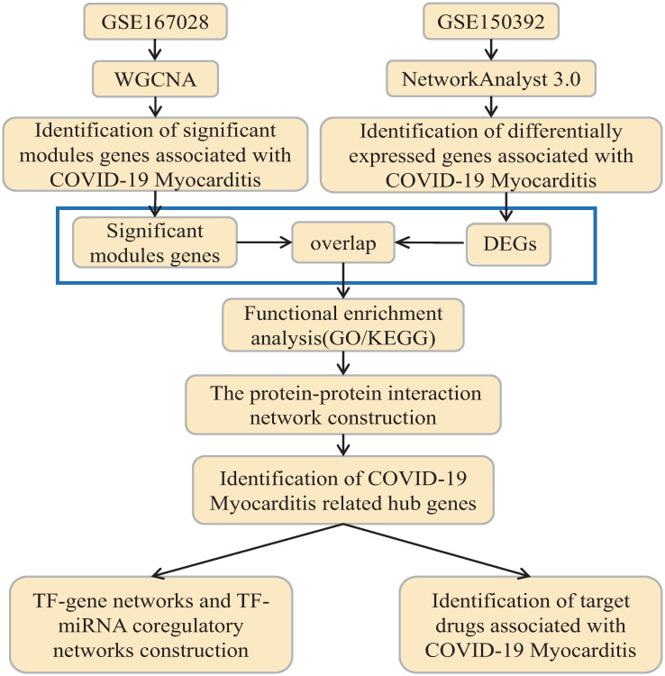
Our study was conducted by downloading differential analysis of SARS-CoV-2 infected cardiac stem cell data from the GEO database, performing weighted gene correlation network analysis (WGCNA) on human myocarditis dataset to identify disease-related modules and associated genes, and matching with the DEGs to identify the common genes. Kyoto Encyclopedia of Genes and Genomes (KEGG) pathway analysis and Gene Ontology (GO) analysis were performed for the common genes to identify potential biological pathways and pathogenesis. The PPI networks were constructed to screen out critical genes and proteins to clarify disease diagnostic markers to screen out small molecule drugs based on critical genes. Subsequently, based on critical genes, the TF-gene networks and TF-miRNA coregulatory networks were studied for related pathway analysis to lay the foundation for further research and clinical diagnosis and treatment of COVID-19 Myocarditis.
The flow chart for this study is presented in Fig 1.
Fig 1. Workflow chart of this study.
WGCNA, weighted gene correlation network analysis; DEGs, differentially expressed genes; GO, Gene Ontology; KEGG, Kyoto Encyclopedia of Genes and Genomes; TF, Transcription factor; miRNA, microRibonucleic acid.
Materials and methods
RNA-sequencing data collection
Dataset related to SARS-CoV-2 infected cardiomyocytes was obtained from the Gene Expression Omnibus (GEO) datasets (https://www.ncbi.nlm.nih.gov/gds/) with accession number GSE150392(https://www.ncbi.nlm.nih.gov/geo/query/acc.cgi?acc=GSE150392) [21, 22] which from GPL18573 Illumina NextSeq 500 (Homo sapiens). There were 6 groups of the GSE150392 dataset, including SARS-CoV-2 infected human induced pluripotent stem cell-derived cardiomyocytes (hiPSC-CMs) groups (n = 3) and Mock hiPSC-CMs (n = 3) groups.
Identification of the DEGs from GSE150392 dataset
NetworkAnalyst 3.0 (https://www.networkanalyst.ca) [23] is a user-friendly online bioinformatics tool for performing comprehensive gene expression analyses, meta-analyses, and network analyses, which agrees with five data inputs, including one or multiple gene lists, single or multiple gene expression data, raw RNA-seq reads, and serial matrix files. This potent online visualization tool integrates transcription factor-gene interaction networks, RNA-gene interaction networks, and other biological regulatory networks which includes data processing, analysis and data update, integrated knowledge base, and synergistic visualization analysis.
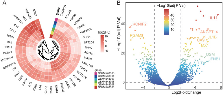
GSE150392 dataset was uploaded to the NetworkAnalyst 3.0 for screening and normalizing. The adjusted P-Value (adj P Val) was analyzed to correct for false-positive results in GEO datasets. “adj P Val <0.01 and |Log2 Fold Change|≥2” were set as the threshold values to screen the differential expression of mRNAs. Circular heatmap and gradient volcano plot were generated by using the “gheatmap” function of the R package “ggtree” (version 3.2.1) and “ggplot2” (version 3.3.5), respectively.
Weighted correlation network analysis (WGCNA) and matching common genes
WGCNA is a systems biology approach to characterize correlation patterns between genes in microarray samples [24]. This analysis method is designed to find co-expressed gene modules, explore the associations between gene networks and phenotypes of interest, and core genes in the network, and this approach is used to identify candidate biomarkers or therapeutic targets.
The normalized gene expression data were downloaded from GSE167028 (https://www.ncbi.nlm.nih.gov/geo/query/acc.cgi?acc=GSE167028) dataset, which included 32 samples and used to construct a co-expression network by using WGCNA (version 1.7.3) package [24] in R-4.1.1. To be clear, WGCNA package doesn’t recommend attempting WGCNA on a data set consisting of fewer than 15 samples. If at all possible, one should have at least 20 samples. We eliminated the KD groups, divided the COVID-19 positive samples into the disease groups, and combined the remaining samples as control groups to maintain study consistency.
According to the correlation of the trait genes, the neighborhood degree of the trait genes was calculated to investigate the co-expression similarity of each module. To identify the association between the general expression module and the clinical group, the p-value, and the correlation coefficient were calculated to visualize the characteristic heatmap of the modules. Modules with a p-value< 0.05 were considered significant.
Finally, a Venn diagram summarizing the overlapping DEGs and significant module genes was generated by using the OmicShare online tool (https://www.omicshare.com).
Functional enrichment analysis
Gene Ontology (GO) is a widely-used tool to annotate the functions of genes, especially biological pathways (BP), cellular components (CC), and molecular function (MF) [25]. KEGG Enrichment Analysis is a practical resource for analyzing gene function and related high-level genomic function information [26, 27].
GO pathway analysis of common genes was performed using the R package org.Hs.eg.db (version 3.1.0) as background. Meanwhile, the KEGG pathway gene annotations (c2.cp.kegg.v7.4.symbols.gmt subset) were obtained from the Molecular Signatures Database [28] to perform the KEGG pathway analysis. In addition, the gene set enrichment results were obtained by using the R package clusterProfiler (version 3.14.3). GO terms and KEGG pathways with P-value<0.05 were considered as a significant enrichment.
PPI network construction to identify critical genes and module analysis
The protein-protein interaction (PPI) network was constructed through the STRING (v11.5; https://cn.string-db.org), a web-based tool for detecting protein interactions by uploading the gene dataset [29, 30]. In this study, the interaction score was set at 0.4. Subsequently, the PPI network data was exported into Cytoscape version 3.7.2 for analysis and visualization.
CytoHybba(https://apps.cytoscape.org/apps/cytoHubba/), a plug-in of Cytoscape software, was used to get the top ten genes through 5 different algorithms: Degree, EPC, Closeness, Betweenness, and Stress, separately [31, 32]. The intersecting genes obtained by the above five methods were considered as critical genes and displayed in the form of a Venn diagram generated by the R package “ggVennDiagram” (version 1.2.0) based on the “ggplot2” (version 3.3.5) package of R [33].
TF-gene networks and TF-miRNA coregulatory networks construction
TF regulates gene expression by binding to specific regions of genes to form feedforward and feedback loops involved in a variety of biological processes and disease processes [34]. On the other hand, TF binds to miRNAs to co-regulate gene expression [35]. In our study, based on critical genes, TF-gene networks and TF-miRNA coregulatory networks were identified by NetworkAnalyst 3.0 (https://www.networkanalyst.ca) [23]. In subsequent work, gene regulatory networks were imported into the Cytoscape 3.7.2 for visualization and analysis.
Identification of target drugs associated with COVID-19 myocarditis
The Drug Signatures Database (DSigDB) is an online gene set linking drugs and their target genes which contains 22 527 gene sets, consists of 17 389 unique compounds, and covers 19 531 genes [36]. The Enrichr (https://maayanlab.cloud/Enrichr/) website provides links to access to the DSigDB. In this study, critical genes were uploaded to the Enricher website to identify the drugs associated with COVID-19 Myocarditis.
Results
Identification of COVID-19 myocarditis related DEGs
The GSE150392 dataset contains 3 SARS-CoV-2 infected pluripotent stem cell-derived cardiomyocytes (hiPSC-CMs) and 3 Mock pluripotent stem cell-derived cardiomyocytes (hiPSC-CMs) for identification of DEGs in COVID-19 Myocarditis. Under the screening criteria, 850 DEGs were obtained, including 449 up-regulated and 401 down-regulated genes (data in S1 Text).
A gradient volcano plot was exhibited in Fig 2A, which showed the upregulated and downregulated genes that varied as the expression fold changes for the GSE150392 dataset [37]. A circular heatmap exhibited the top 40 DEGs in Fig 2B, while the top 10 DEGs’ details were shown in Table 1.
Fig 2. Identification of differentially expressed genes in the GSE150392 dataset by differential analysis.
(A) The circular heatmap exhibited the top 40 DEGs sorted by the Log2 FC for the GSE150392 dataset. (B) DEGs in the gradient volcano plot. The top 10 genes were labeled on the plot which was sorted by the Log2 FC and most of them were upregulated (except KCNIP2 and PGAM2 which were downregulated). Two vertical lines indicated |Log2 FoldChange| ≥2, severally, and the horizontal line indicated the adj P Val of 0.01. The color of the plots represents the Log2 FoldChange levels [37]. DEGs, differentially expressed genes; FC, fold change; adj P Val, adjusted P-Value.
Table 1. The top ten differentially expressed genes (DEGs) in the GSE150392 dataset.
| Gene | Log2FoldChange | P-value | Adj P Value | Regulate |
|---|---|---|---|---|
| IL11 | 7.6191 | 1.03E-52 | 5.49E-49 | Up |
| ANGPTL4 | 6.1073 | 2.08E-42 | 3.33E-39 | Up |
| MX1 | 6.101 | 2.39E-33 | 1.37E-30 | Up |
| IFNB1 | 6.0943 | 3.20E-19 | 4.80E-17 | Up |
| LTA | 5.9337 | 4.18E-19 | 6.10E-17 | Up |
| IL1B | 5.9318 | 1.43E-34 | 8.85E-32 | Up |
| KCNIP2 | -5.7287 | 8.10E-44 | 2.17E-40 | Down |
| OSM | 5.5858 | 4.27E-22 | 8.78E-20 | Up |
| PGAM2 | -5.5819 | 1.84E-34 | 1.10E-31 | Down |
| CA9 | 5.5093 | 7.12E-37 | 7.62E-34 | Up |
Identification of COVID-19 myocarditis associated modules by WGCNA
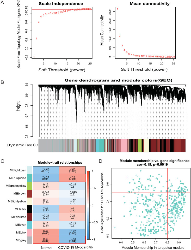
In this study, we constructed a co-expression network for normalized gene expression data of the GSE167028 dataset by the WGCNA package (version 1.7.3) in the R application. The soft threshold was set to 20 to fit a scale-free network and the maximum mean connectivity, while the scale-free R^2 was 0.88 (Fig 3A). Meanwhile, a total of 10 co-expression modules, each with more than 80 genes, were identified using the DynamicTreeCut method (Fig 3B). Among the 10 significant modules (Fig 3C), the lightcyan, turquoise (r = 0.15, p = 0.0019, Fig 3D), black, darkred modules were positively related to COVID-19 Myocarditis, whereas the green, brown, lightyellow, cyan, pink, and grey modules were negatively associated with COVID-19 Myocarditis. Nevertheless, only the turquoise module met the criteria which had a p-value of 0.04 (except for the gray module, which contained high amounts of un-classified genes). Therefore, this module was identified as a significant module for further analysis.
Fig 3. Identification of significant modules and genes of GSE167028 by WGCNA.
(A) Network topology analysis with different soft thresholds. The scale-free R^2 was 0.88 and the soft threshold was 20. (B) A cluster dendrogram of module-specific colors showed 10 co-expressed gene modules, each containing more than 80 genes. (C) Correlation between disease groupings and gene modules. (D) The scatter plot of Gene significance vs Module membership in the turquoise co-expression module.
We calculated the expression correlation of module feature vectors with genes to obtain module membership (MM) and gene significance (GS). Based on the cut-off criteria |MM| > 0.8 and |GS|>0.2, 159 of 427 genes with high connectivity in the turquoise module were identified.
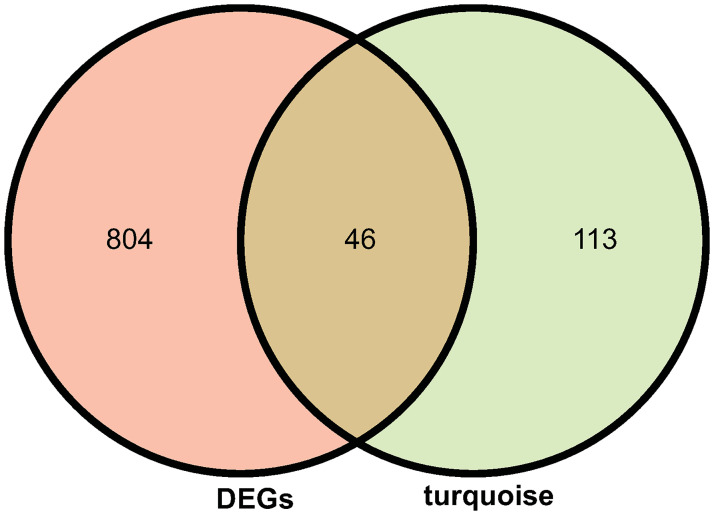
Ultimately, 46 common genes were obtained by intersecting with the DEGs of GSE150392 and the genes in the turquoise module of GSE167028 (Fig 4, data in S2 Text).
Fig 4. The intersection of DEGs in the GSE150392 dataset and turquoise module genes in the GSE167028.
There were 850 DEGs in the GSE150392 dataset and 159 genes in the turquoise module of the GSE167028 dataset, and 150 genes were obtained as common genes by overlapping the two datasets.
Gene function annotations of COVID-19 myocarditis related the common genes
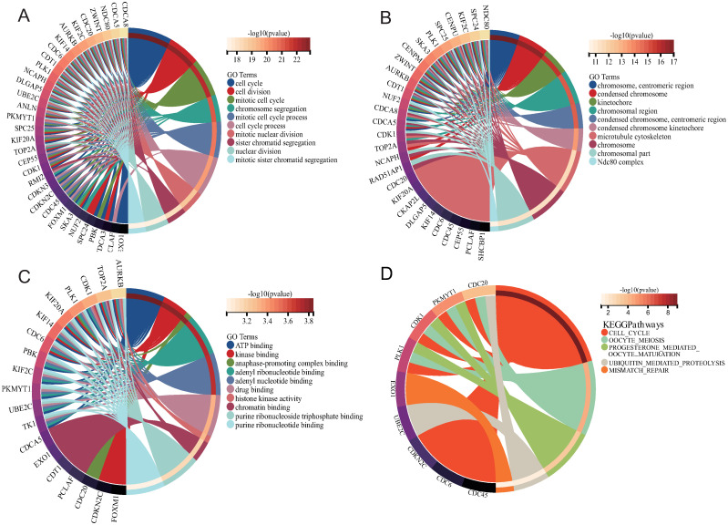
After obtaining common genes with COVID-19 Myocarditis, GO enrichment and KEGG pathway analysis were performed to understand the biological pathways. The top ten Go terms for biological process, cellular component and molecular function were shown in Table 2 and Fig 5A–5C. The data of GO terms indicated that the common genes were significantly enhanced in cell cycle/division and mitotic cell cycle of biological process. Cellular components revealed significant involvement of microtubule cytoskeleton and chromosome in common genes. For the molecular function subsection, it was apparent that ATP binding was involved in the common genes.
Table 2. GO category, GO description, GO ID, and their corresponding P-value.
| Category | Description | GO ID | P-value |
|---|---|---|---|
| BP | cell cycle | GO:0007049 | 1.31E-23 |
| BP | cell division | GO:0051301 | 3.12E-23 |
| BP | mitotic cell cycle | GO:0000278 | 1.33E-22 |
| BP | chromosome segregation | GO:0007059 | 9.27E-22 |
| BP | mitotic cell cycle process | GO:1903047 | 3.41E-21 |
| BP | cell cycle process | GO:0022402 | 1.86E-20 |
| BP | mitotic nuclear division | GO:0140014 | 1.59E-19 |
| BP | sister chromatid segregation | GO:0000819 | 8.04E-19 |
| BP | nuclear division | GO:0000280 | 3.57E-18 |
| BP | mitotic sister chromatid segregation | GO:0000070 | 4.42E-18 |
| CC | chromosome, centromeric region | GO:0000775 | 9.69E-18 |
| CC | condensed chromosome | GO:0000793 | 2.73E-17 |
| CC | kinetochore | GO:0000776 | 2.7E-16 |
| CC | chromosomal region | GO:0098687 | 1.59E-15 |
| CC | condensed chromosome, centromeric region | GO:0000779 | 1.99E-15 |
| CC | condensed chromosome kinetochore | GO:0000777 | 4.25E-14 |
| CC | microtubule cytoskeleton | GO:0015630 | 3.45E-12 |
| CC | chromosome | GO:0005694 | 5.64E-12 |
| CC | chromosomal part | GO:0044427 | 7.3E-12 |
| CC | Ndc80 complex | GO:0031262 | 3.22E-11 |
| MF | ATP binding | GO:0005524 | 0.000143 |
| MF | kinase binding | GO:0019900 | 0.00016 |
| MF | anaphase-promoting complex binding | GO:0010997 | 0.000166 |
| MF | adenyl ribonucleotide binding | GO:0032559 | 0.000198 |
| MF | adenyl nucleotide binding | GO:0030554 | 0.000207 |
| MF | drug binding | GO:0008144 | 0.000496 |
| MF | histone kinase activity | GO:0035173 | 0.000534 |
| MF | chromatin binding | GO:0003682 | 0.000674 |
| MF | purine ribonucleoside triphosphate binding | GO:0035639 | 0.000737 |
| MF | purine ribonucleotide binding | GO:0032555 | 0.00099 |
Fig 5. GO and KEGG analysis of COVID-19 myocarditis related to the common genes.
(A) Biological Process. (B) Cellular Component. (C) Molecular Function. (D) KEGG pathways analysis.
The next section of the functional enrichment analysis was concerned with the KEGG analysis which was demonstrated in Table 3 and Fig 5D. KEGG analysis found that common genes were mainly enriched in the cell cycle, oocyte meiosis, progesterone mediated oocyte maturation, ubiquitin mediated proteolysis, and mismatch repair.
Table 3. KEGG pathways and their corresponding P-values and Q-values, and common genes enriched in their pathways.
| KEGG pathways | P-value | Q-value | Gene ID |
|---|---|---|---|
| cell cycle | 1.13E-09 | 5.93E-09 | PLK1/CDK1/CDC45/CDC6/PKMYT1/ |
| CDKN2C/CDC20 | |||
| oocyte meiosis | 6.00E-05 | 1.58E-04 | PLK1/CDK1/PKMYT1/CDC20 |
| progesterone mediated oocyte maturation | 6.17E-04 | 1.08E-03 | PLK1/CDK1/PKMYT1 |
| ubiquitin mediated proteolysis | 3.11E-02 | 4.09E-02 | UBE2C/CDC20 |
| mismatch repair | 4.72E-02 | 4.97E-02 | EXO1 |
Construction of a PPI network and identification of critical genes
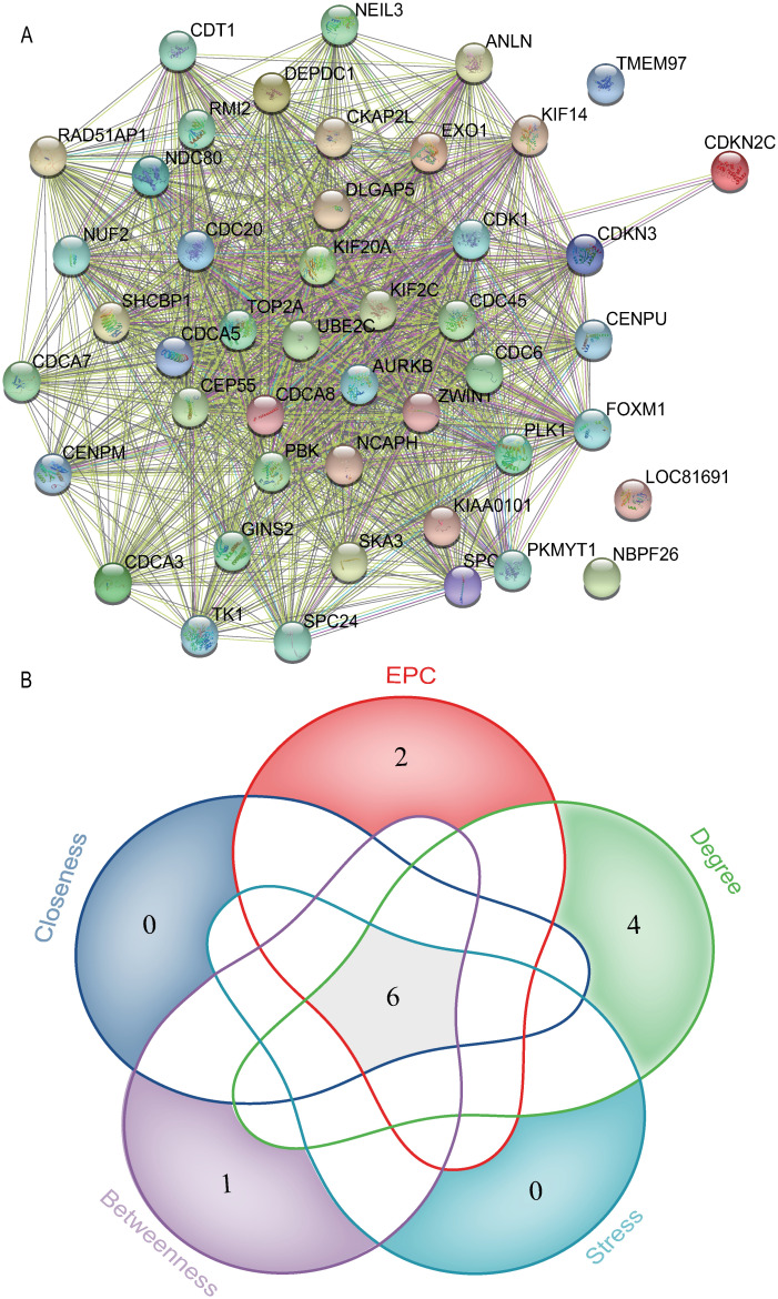
Among these 46 common genes, a PPI network (46 nodes and 778 edges) was generated by STRRING (v11.5; https://cn.string-db.org). Thereafter, the network was imported into the Cytoscape version 3.7.2 for analysis and visualization. Based on 5 algorithms of the CytoHubba plug-in, six genes (CDK1, KIF20A, PBK, KIF2C, CDC20, UBE2C) were confirmed as critical genes related to COVID-19 Myocarditis (Fig 6). All topological features of critical genes were shown in Table 4. Since these critical genes may be potential biomarkers, they may provide a reference for the diagnosis and treatment of COVID-19 Myocarditis.
Fig 6. The number of critical genes were shown by using the cytoHubba plugin of cytoscape and the Venn diagram.
EPC, Edge Percolated Component.
Table 4. The top 10 genes in the PPI network were calculated using five algorithms.
| Gene | Degree | Gene | EPC | Gene | Closeness | Gene | Betweenness | Gene | Stress |
|---|---|---|---|---|---|---|---|---|---|
| CDK1 | 42 | KIF2C | 16.979 | CDK1 | 42 | CDK1 | 48.66956 | CDK1 | 250 |
| KIF20A | 41 | PBK | 16.82 | KIF2C | 41.5 | CDKN3 | 42.99438 | CDKN3 | 204 |
| TOP2A | 41 | RAD51AP1 | 16.708 | PBK | 41.5 | KIF2C | 6.66956 | KIF2C | 170 |
| PBK | 41 | CDK1 | 16.659 | RAD51AP1 | 41.5 | PBK | 6.66956 | PBK | 170 |
| AURKB | 41 | CDCA5 | 16.594 | CDCA5 | 41.5 | RAD51AP1 | 6.66956 | RAD51AP1 | 170 |
| KIF2C | 41 | UBE2C | 16.576 | UBE2C | 41.5 | CDCA5 | 6.66956 | CDCA5 | 170 |
| CDCA8 | 41 | EXO1 | 16.562 | KIF20A | 41.5 | UBE2C | 6.66956 | UBE2C | 170 |
| CDC20 | 41 | KIF20A | 16.384 | CDC20 | 41.5 | KIF20A | 6.66956 | KIF20A | 170 |
| UBE2C | 41 | CDC20 | 16.344 | DLGAP5 | 41.5 | CDC20 | 6.66956 | CDC20 | 170 |
| NUF2 | 41 | NCAPH | 16.339 | CDC6 | 41.5 | DLGAP5 | 6.66956 | DLGAP5 | 170 |
Identification of gene regulatory networks related to critical genes
NetworkAnalyst 3.0 was used to identify the TF-gene networks and TF-miRNA coregulatory networks based on critical genes which were visualized in Fig 8A, 8B.
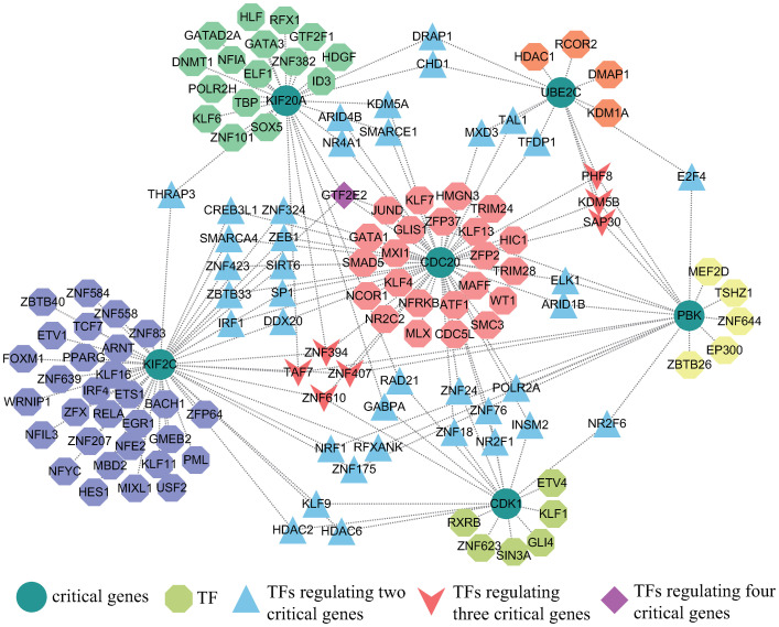
The TF-gene networks comprised 136 nodes and 185 edges. The entire network consists of 130 TF genes and 6 critical genes. CDC20 was regulated by 55 TF genes and KIF2C was regulated by 52 TF genes. In addition, 130 TF genes regulated more than one common gene, which indicated that TF genes were highly regulatory of critical genes. Interestingly, we found that GTF2E2 had high connectivity in the TF-gene regulatory network, regulating four critical genes simultaneously. Fig 7 showed the TF-gene networks.
Fig 7. Transcription factor-gene regulatory network in COVID-19 myocarditis.
The circular dots represent critical genes, and the octagonal dots attached next to the critical genes represent transcription factors that regulated the critical genes. In addition, the triangular-shaped transcription factors indicated regulation of two critical genes, and the V-shaped transcription factors regulated three key genes. Obviously, the diamond-shaped transcription factor GF2E2 regulated four critical genes. The network was composed of 142 nodes and 180 edges.
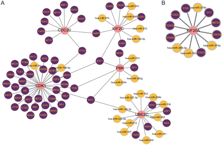
On the contrary, the TF-miRNA coregulatory networks consist of two parts, one including 83 nodes and 85 edges, and the other including 13 nodes and 12 edges which were shown in Fig 8. A total of 25 miRNAs and 64 TF genes co-regulated critical genes.
Fig 8. Transcription factors-miRNA-gene regulatory networks in COVID-19 myocarditis.
There were two TF-miRNA networks. Pink plots represent critical genes, purple plots represent TF genes, and the others represent miRNAs. Network (A) had 83 plots and 85 edges, which was consisted of 5 critical genes, 56 TF genes, and 21 miRNAs. Network (B) had 13 plots and 12 edges including 1 critical gene, 8 TF genes, and 4 miRNAs.
Identification of target drugs associated with COVID-19 myocarditis
Based on critical genes, the drugs related to the COVID-19 Myocarditis were identified by the DSigDB database that was built on the Enrichr website. In the integration of the DSigDB dataset, 308 drug compounds were identified (data in S5 Text). Finally, the top ten drug compounds were screened according to the p-value. Etoposide and methotrexate are two notable genetically linked drug compounds. Meanwhile, CDC20 and KIF2C were associated with the most drug compounds, suggesting that they act prominent roles in drug efficacy. Table 5 showed information on potentially effective drug compounds for COVID-19 Myocarditis.
Table 5. COVID-19 myocarditis gene-targeted drugs.
| Term | P-value | Combined Score | Genes |
|---|---|---|---|
| etoposide MCF7 DOWN | 4.36E-10 | 19544.37 | CDC20; UBE2C; KIF2C; KIF20A |
| methotrexate MCF7 DOWN | 6.07E-10 | 17637.87 | CDC20; UBE2C; KIF2C; KIF20A |
| LUCANTHONE CTD 00006227 | 7.78E-10 | 9976.195 | CDC20; CDK1; PBK; KIF2C; KIF20A |
| troglitazone CTD 00002415 | 1.16E-09 | 2388335 | CDC20; UBE2C; CDK1; PBK; KIF2C; KIF20A |
| ciclopirox MCF7 DOWN | 1.72E-09 | 12770.53 | CDC20; UBE2C; KIF2C; KIF20A |
| 5109870 MCF7 DOWN | 1.82E-09 | 12532.66 | CDC20; UBE2C; KIF2C; KIF20A |
| thalidomide CTD 00006858 | 3.51E-08 | 4975.73 | CDC20; UBE2C; CDK1; KIF2C |
| genistein CTD 00007324 | 5.37E-08 | 1885048 | CDC20; UBE2C; CDK1; PBK; KIF2C; KIF20A |
| dmnq CTD 00002569 | 5.41E-08 | 4339.482 | CDC20; UBE2C; PBK; KIF2C |
| testosterone CTD 00006844 | 5.81E-08 | 1874705 | CDC20; UBE2C; CDK1; PBK; KIF2C; KIF20A |
Discussion
As previously described, a large number of patients with myocarditis were identified during clinical treatment of COVID-19. However, the understanding of COVID-19 and myocarditis is insufficient, and many patients with COVID-19 Myocarditis are not properly diagnosed and well treated. To our knowledge, research on the key genes and pathways by bioinformatics methods between COVID-19 and myocarditis has hardly been reported. The present study was designed to elaborate on the bioinformatics lessons about the key genes and pathways between COVID-19 and myocarditis.
In our study, 850 DEGs and 159 significant module genes from GSE150392 and GSE167028 were identified by bioinformatics-related methods, respectively. For constructing the relationship of the COVID-19 and myocarditis, 46 common genes were overlapped. The remaining studies were functional enrichment analysis, PPI network construction, TF-gene networks, TF-miRNA coregulatory networks construction, and gene targeting drug screening [38]. Eventually, 6 genes (CDK1, KIF20A, PBK, KIF2C, CDC20, UBE2C) were identified by CytoHubba plug-in of Cytoscape as critical genes of COVID-19 Myocarditis for future study.
Based on the common genes, GO terms were identified as a threshold of P-value of < 0.05. According to biological process, the top ten GO terms were cell cycle, cell division, mitotic cell cycle, chromosome segregation, mitotic cell cycle process, cell cycle process, mitotic nuclear division, sister chromatid segregation, nuclear division, and mitotic sister chromatid segregation [39]. The cell cycle is the complete process of cell division and replication and consists of a specific series of events such as cell division, DNA replication, nuclear membrane rupture, spindle formation, and preparation for chromosome segregation [40]. Numerous studies have shown that viruses provide powerful conditions for viral replication and survival by regulating different processes of the cell cycle of host cells [41–44]. For molecular function, ATP binding, kinase binding, anaphase-promoting complex binding, adenyl ribonucleotide binding, adenyl nucleotide binding, drug binding, histone kinase activity, chromatin binding, purine ribonucleoside triphosphate binding, and purine ribonucleotide binding were the top ten GO terms. Adenosine triphosphate (ATP), is an energy metabolite that plays a role in energy transfer and information transmission in various cellular metabolic processes. Recent findings uncovered that SARS-CoV-2 N protein regulates the cell cycle of host cells by specifically binding ATP, which provides us with a new idea to fight against the SARS-CoV-2 pandemic [45]. In addition, the previous study has shown that ATP is also a specific autoantibody for myocarditis [46]. As for cellular components, the top GO terms are chromosome, centromeric region, condensed chromosome, and kinetochore.
The KEGG pathways analysis was achieved from the common genes for identifying similar pathways between COVID-19 and myocarditis. KEGG pathway analysis mainly focused on cell cycle, oocyte meiosis, progesterone mediated oocyte maturation, ubiquitin mediated proteolysis, and mismatch repair, which indicate that they are crucial to the biological progression of COVID-19 Myocarditis. The ubiquitin protein hydrolysis plays an essential role in a range of underlying cellular processes, such as immune responses and inflammatory responses [47]. In addition, it has been shown that ubiquitin protein hydrolysis is associated with myocardial remodelings, such as Atrophy of the heart [48].
PPI network analysis was the most important step in this study, laying the foundation for the subsequent screening of critical genes. Based on topological algorithms (i.e., degree), in this study, CDK1, KIF20A, PBK, KIF2C, CDC20, and UBE2C were identified as critical genes that may be potential biomarkers for COVID-19 Myocarditis.
CDK1 (Cyclin-dependent kinase 1) plays a critical role in eukaryotic cell cycle control by regulating centrosome cycling and mitotic initiation. There is growing evidence found that CDK1 can be used as a potential biomarker for a variety of diseases, such as Rhabdomyosarcoma [49], endometrioid endometrial cancer [50]. Furthermore, in recent studies, CDK1, promotes the phosphorylation of RAPTOR during mitosis, leading to mTORC1 phosphorylation and affecting the autophagic process [51], which plays an important role in cardiac diseases as a degradation process of cellular self, especially in myocarditis or cardiomyopathy [52]. In addition, CDK1 regulates the cell cycle leading to cell cycle arrest in cardiomyocytes, the latter being an important factor involved in oxidative stress leading to heart failure [53]. Similarly, in previous studies, CDK1 was identified as a potential target of COVID-19 [54], so the role of CDK1 in myocarditis needs to be further investigated.
KIF20A (Kinesin Family Member 20A), is a member of the Kinesin-like proteins that play an important role in intracellular transport and cell division [55]. According to the literature, KIF20A plays an important role in several cardiovascular diseases, such as restrictive cardiomyopathy and acute type A aortic coarctation. In one case report, exome sequencing analysis of children with congenital cardiomyopathy identified the KIF20A complex and in subsequent in vitro experiments in zebrafish, KIF20A was identified as the phenotypic gene for cardiomyopathy [56]. Chen et al. found that KIF20A was identified as a hub gene involved in the infection of the intestine by the SARS-CoV-2 [57]. All of the above studies provide implications for the study of KIF20A in COVID-19 and myocarditis.
PBK (PDZ Binding Kinase) plays a regulatory role in cell cycle regulation and cell mitosis. It was reported that using the PathExt tool was able to identify PBK as the most common central gene target in activated TopNets to suppress SARS-CoV-2 [58]. Ekaterina et al. showed that genes such as PBK regulated myofibril formation and thus caused cardiac hypertrophy [59].
As for the remaining genes, CDC20 and KIF2C were identified as target genes of COVID-19 by bioinformatics means and machine learning [54]. Meanwhile, UBE2C, Ubiquitin-Conjugating Enzyme E2 C, is closely related to the cardiovascular system which induced endothelial cell inflammation and endothelial mesenchymal transition, exacerbating aortic sclerosis and calcification [60].
According to critical genes, the TF-gene networks and TF-miRNA co-regulatory networks were established. In our knowledge, transcription factors play important roles in many biological processes by binding specific sequences of genes, such as regulation of gene transcription, control of metabolism, and immune response [61]. And further studies have shown that transcription factors are closely related to a variety of diseases. From the network, it can be seen that CDC20 has a high rate of interactions with other TF genes. In the TF-gene coregulatory networks, the degree value of CDC20 was 55. This was closely followed by KIF2C with a degree of 52 and KIF2C had higher connectivity with CDC20 with a value of 14. Notably, GTF2E2 was identified as the transcription factor that regulates the most genes which has been reported to exert inhibitory effects on lung adenocarcinoma in the mTOR pathway. It is well known that the mTOR pathway plays an important role in the autophagic process and the pathogenesis of myocarditis [62], and autophagy has been shown to be closely related to COVID-19 and myocarditis [63], which provides us with a new idea to study the regulatory role of GTF2E2 in COVID-19 Myocarditis. Meanwhile, in the TF-miRNA coregulatory networks, MYC, E2F1, PTTG1, GABPA, TP53 regulated more than one critical gene.
Through the drug database, drug molecules associated with critical genes were identified and sorted by p-value. Etoposide MCF7 DOWN, Methotrexate MCF7 DOWN, Lucanthone CTD 00006227, Troglitazone CTD 00002415, Ciclopirox MCF7 DOWN, STL264925 MCF7 DOWN, Thalidomide CTD 00006858, Genistein CTD 00007324, Dmnq CTD 00002569, Testosterone CTD 00006844 are potentially investigational and therapeutic agents associated with COVID-19 Myocarditis. Because of superior anti-cancer activity, Etoposide plays an important role in cancer treatment. Meanwhile, as a TOP II inhibitor, Etoposide effectively inhibits intracellular replication of SARS-CoV-2’s structural proteins [64] and has a rescue effect on the cytokine storm of the COVID-19 [65]. It has been shown that immunosuppressive therapy has a therapeutic effect on myocarditis and can improve the prognosis of myocarditis [45], which offers the possibility of drug targeting for the treatment of COVID-19 Myocarditis. Methotrexate, an immunosuppressant, also has an inhibitory effect on the COVID-19 cytokine storm [66]. Troglitazone as a type 2 diabetes oral medication, has the effect of improving the sensitivity of muscle and adipose tissue to insulin and inhibiting hepatic gluconeogenesis. A recent study has shown that Troglitazone has the potential to inhibit SARS-CoV-2 NSP9 which plays a vital role in viral replication [67].
Several limitations need to be noted regarding the present study. Firstly, since there are few studies related to COVID-19 Myocarditis, we selected only two datasets from GEO database for bioinformatics study. In addition, due to the limitations of the data set sample, we unified the experimental group in the GSE167028 dataset as the group of patients with myocarditis and unified the healthy adult and pediatric groups as the healthy control group in order to maintain disease grouping consistency and statistical accuracy, which may result in a degree of study heterogeneity. Thirdly, although rigorous bioinformatics analysis was performed in this study, the findings need to be validated with more samples and cellular and animal experiments. A further study with more focus on COVID-19 myocarditis is therefore suggested.
Conclusion
The bioinformatics study of the GSE167028 and GSE150392 datasets identified 6 critical genes (CDK1, KIF20A, PBK, KIF2C, CDC20, UBE2C) involved in COVID-19 Myocarditis and explored the biological processes between COVID-19 and myocarditis, confirming previous studies and providing some insights into the pathogenesis of COVID-19 Myocarditis, demonstrating that SARS-CoV-2 contributes to myocarditis through pathophysiological processes such as cell cycle and the ubiquitin-protein hydrolysis. At the same time, this study also provides relevant drugs for the clinical treatment of COVID-19 Myocarditis. There are few studies on COVID-19 Myocarditis, and if more samples are available in the future, the role of this study will be more effective in the context of the SARS-CoV-2 pandemic.
Supporting information
(TXT)
(TXT)
(TXT)
(TXT)
(TXT)
(CYS)
Acknowledgments
We acknowledge the GEO database for providing its platforms and contributors for uploading their meaningful datasets. Meanwhile, we thank Dr. Guangli Sun, Chief Physician, Department of Traditional Chinese Medicine, Laixi City Hospital, for her financial assistance with this study.
Abbreviations
- adj P Val
adjusted P-Value
- CMR
cardiac magnetic resonance
- COVID-19
Coronavirus disease 2019
- DEGs
Differentially expressed genes
- DSigDB
Drug Signatures Database
- EMB
endomyocardial biopsy
- FC
Fold Change
- GEO
Gene Expression Omnibus
- GO
Gene Ontology
- hiPSC-CMs
human induced pluripotent stem cell-derived cardiomyocytes
- KEGG
Kyoto Encyclopedia of Genes and Genomes
- PPI
protein-protein interaction
- TF-gene
Transcription factor-gene
- TF-miRNA
Transcription factor-microRibonucleic acid
- WGCNA
weighted gene correlation network analysis
Data Availability
Data are available within the GEO database (GSE150392 and GSE167028). The NetworkAnalyst 3.0 repository (https://www.networkanalyst.ca/), STRING repository (https://string-db.org/), DSigDB repository (https://maayanlab.cloud/Enrichr/) are the three databases used to process the data and are all online sites that the reader can access and use directly to process the data, as described in the "Methods and Materials" section of the article.
Funding Statement
This study was supported by the National Natural Science Foundation of China (82074388); project of shaoxing medical key discipline construction plan(2019SZD06); the Project of Health and Family Planning Commission of Zhejiang province (2021KY1139); and Shaoxing Medical and Health Science and Technology Plan Project(2020A13026). The funders had no role in study design, data collection and analysis, decision to publish, or preparation of the manuscript.
References
- 1.Huang C, Wang Y, Li X, Ren L, Zhao J, Hu Y, et al. Clinical features of patients infected with 2019 novel coronavirus in Wuhan, China. The Lancet. 2020;395(10223):497–506. doi: 10.1016/s0140-6736(20)30183-5 [DOI] [PMC free article] [PubMed] [Google Scholar]
- 2.Hoffmann M, Kleine-Weber H, Schroeder S, Kruger N, Herrler T, Erichsen S, et al. SARS-CoV-2 Cell Entry Depends on ACE2 and TMPRSS2 and Is Blocked by a Clinically Proven Protease Inhibitor. Cell. 2020;181(2):271–80 e8. Epub 2020/03/07. doi: 10.1016/j.cell.2020.02.052 . [DOI] [PMC free article] [PubMed] [Google Scholar]
- 3.Datta PK, Liu F, Fischer T, Rappaport J, Qin X. SARS-CoV-2 pandemic and research gaps: Understanding SARS-CoV-2 interaction with the ACE2 receptor and implications for therapy. Theranostics. 2020;10(16):7448–64. Epub 2020/07/10. doi: 10.7150/thno.48076 . [DOI] [PMC free article] [PubMed] [Google Scholar]
- 4.Li W, Moore MJ, Vasilieva N, Sui J, Wong SK, Berne MA, et al. Angiotensin-converting enzyme 2 is a functional receptor for the SARS coronavirus. Nature. 2003;426(6965):450–4. doi: 10.1038/nature02145 . [DOI] [PMC free article] [PubMed] [Google Scholar]
- 5.Coronaviridae Study Group of the International Committee on Taxonomy of V. The species Severe acute respiratory syndrome-related coronavirus: classifying 2019-nCoV and naming it SARS-CoV-2. Nat Microbiol. 2020;5(4):536–44. Epub 2020/03/04. doi: 10.1038/s41564-020-0695-z . [DOI] [PMC free article] [PubMed] [Google Scholar]
- 6.Patel VB, Zhong JC, Grant MB, Oudit GY. Role of the ACE2/Angiotensin 1–7 Axis of the Renin-Angiotensin System in Heart Failure. Circ Res. 2016;118(8):1313–26. Epub 2016/04/16. doi: 10.1161/CIRCRESAHA.116.307708 . [DOI] [PMC free article] [PubMed] [Google Scholar]
- 7.Hamming I, Timens W, Bulthuis ML, Lely AT, Navis G, van Goor H. Tissue distribution of ACE2 protein, the functional receptor for SARS coronavirus. A first step in understanding SARS pathogenesis. J Pathol. 2004;203(2):631–7. Epub 2004/05/14. doi: 10.1002/path.1570 . [DOI] [PMC free article] [PubMed] [Google Scholar]
- 8.Nicin L, Abplanalp WT, Mellentin H, Kattih B, Tombor L, John D, et al. Cell type- specific expression of the putative SARS-CoV-2 receptor ACE2 in human hearts. Eur Heart J. 2020;41(19):1804–6. Epub 2020/04/16. doi: 10.1093/eurheartj/ehaa311 . [DOI] [PMC free article] [PubMed] [Google Scholar]
- 9.Glowacka I, Bertram S, Herzog P, Pfefferle S, Steffen I, Muench MO, et al. Differential downregulation of ACE2 by the spike proteins of severe acute respiratory syndrome coronavirus and human coronavirus NL63. J Virol. 2010;84(2):1198–205. Epub 2009/10/30. doi: 10.1128/JVI.01248-09 . [DOI] [PMC free article] [PubMed] [Google Scholar]
- 10.Aboudounya MM, Heads RJ. COVID-19 and Toll-Like Receptor 4 (TLR4): SARS-CoV- 2 May Bind and Activate TLR4 to Increase ACE2 Expression, Facilitating Entry and Causing Hyperinflammation. Mediators Inflamm. 2021;2021:8874339. Epub 2021/01/29. doi: 10.1155/2021/8874339 . [DOI] [PMC free article] [PubMed] [Google Scholar]
- 11.Guo T, Fan Y, Chen M, Wu X, Zhang L, He T, et al. Cardiovascular Implications of Fatal Outcomes of Patients With Coronavirus Disease 2019 (COVID-19). JAMA cardiology. 2020;5(7):811–8. doi: 10.1001/jamacardio.2020.1017 . [DOI] [PMC free article] [PubMed] [Google Scholar]
- 12.Shi S, Qin M, Shen B, Cai Y, Liu T, Yang F, et al. Association of Cardiac Injury With Mortality in Hospitalized Patients With COVID-19 in Wuhan, China. JAMA cardiology. 2020;5(7):802–10. doi: 10.1001/jamacardio.2020.0950 . [DOI] [PMC free article] [PubMed] [Google Scholar]
- 13.Sala S, Peretto G, Gramegna M, Palmisano A, Villatore A, Vignale D, et al. Acute myocarditis presenting as a reverse Tako-Tsubo syndrome in a patient with SARS-CoV-2 respiratory infection. Eur Heart J. 2020;41(19):1861–2. Epub 2020/04/09. doi: 10.1093/eurheartj/ehaa286 . [DOI] [PMC free article] [PubMed] [Google Scholar]
- 14.Inciardi RM, Lupi L, Zaccone G, Italia L, Raffo M, Tomasoni D, et al. Cardiac Involvement in a Patient With Coronavirus Disease 2019 (COVID-19). JAMA cardiology. 2020;5(7):819–24. doi: 10.1001/jamacardio.2020.1096 . [DOI] [PMC free article] [PubMed] [Google Scholar]
- 15.Fox SE, Akmatbekov A, Harbert JL, Li G, Quincy Brown J, Vander Heide RS. Pulmonary and cardiac pathology in African American patients with COVID-19: an autopsy series from New Orleans. The Lancet Respiratory Medicine. 2020;8(7):681–6. doi: 10.1016/S2213-2600(20)30243-5 [DOI] [PMC free article] [PubMed] [Google Scholar]
- 16.Abou Hassan OK, Sheng CC, Wang TKM, Cremer PC. SARS-CoV-2 Myocarditis: Insights Into Incidence, Prognosis, and Therapeutic Implications. Curr Cardiol Rep. 2021;23(9):129. Epub 2021/08/04. doi: 10.1007/s11886-021-01551-x . [DOI] [PMC free article] [PubMed] [Google Scholar]
- 17.Mele D, Flamigni F, Rapezzi C, Ferrari R. Myocarditis in COVID-19 patients: current problems. Intern Emerg Med. 2021;16(5):1123–9. Epub 2021/01/24. doi: 10.1007/s11739-021-02635-w . [DOI] [PMC free article] [PubMed] [Google Scholar]
- 18.Sagar S, Liu PP, Cooper LT. Myocarditis. The Lancet. 2012;379(9817):738–47. doi: 10.1016/S0140-6736(11)60648-X [DOI] [PMC free article] [PubMed] [Google Scholar]
- 19.Cooper LT, Baughman KL, Feldman AM, Frustaci A, Jessup M, Kuhl U, et al. The role of endomyocardial biopsy in the management of cardiovascular disease: a scientific statement from the American Heart Association, the American College of Cardiology, and the European Society of Cardiology Endorsed by the Heart Failure Society of America and the Heart Failure Association of the European Society of Cardiology. Eur Heart J. 2007;28(24):3076–93. Epub 2007/10/26. doi: 10.1093/eurheartj/ehm456 . [DOI] [PubMed] [Google Scholar]
- 20.Friedrich MG, Sechtem U, Schulz-Menger J, Holmvang G, Alakija P, Cooper LT, et al. Cardiovascular magnetic resonance in myocarditis: A JACC White Paper. J Am Coll Cardiol. 2009;53(17):1475–87. Epub 2009/04/25. doi: 10.1016/j.jacc.2009.02.007 . [DOI] [PMC free article] [PubMed] [Google Scholar]
- 21.Sharma A, Garcia G Jr., Wang Y, Plummer JT, Morizono K, Arumugaswami V, et al. Human iPSC-Derived Cardiomyocytes Are Susceptible to SARS-CoV-2 Infection. Cell Rep Med. 2020;1(4):100052. Epub 2020/08/25. doi: 10.1016/j.xcrm.2020.100052 . [DOI] [PMC free article] [PubMed] [Google Scholar]
- 22.Kumar S, Curran JE, Kumar K, DeLeon E, Leandro AC, Peralta J, et al. Disease Modeling and Disease Gene Discovery in Cardiomyopathies: A Molecular Study of Induced Pluripotent Stem Cell Generated Cardiomyocytes. Int J Mol Sci. 2021;22(7). Epub 2021/04/04. doi: 10.3390/ijms22073311 . [DOI] [PMC free article] [PubMed] [Google Scholar]
- 23.Zhou G, Soufan O, Ewald J, Hancock REW, Basu N, Xia J. NetworkAnalyst 3.0: a visual analytics platform for comprehensive gene expression profiling and meta-analysis. Nucleic Acids Res. 2019;47(W1):W234–W41. Epub 2019/04/02. doi: 10.1093/nar/gkz240 . [DOI] [PMC free article] [PubMed] [Google Scholar]
- 24.Langfelder P, Horvath S. WGCNA: an R package for weighted correlation network analysis. BMC Bioinformatics. 2008;9:559. Epub 2008/12/31. doi: 10.1186/1471-2105-9-559 . [DOI] [PMC free article] [PubMed] [Google Scholar]
- 25.Doms A, Schroeder M. GoPubMed: exploring PubMed with the Gene Ontology. Nucleic Acids Res. 2005;33(Web Server issue):W783–6. Epub 2005/06/28. doi: 10.1093/nar/gki470 . [DOI] [PMC free article] [PubMed] [Google Scholar]
- 26.Kanehisa M, Goto S. KEGG: kyoto encyclopedia of genes and genomes. Nucleic Acids Res. 2000;28(1):27–30. doi: 10.1093/nar/28.1.27 . [DOI] [PMC free article] [PubMed] [Google Scholar]
- 27.Kanehisa M, Furumichi M, Tanabe M, Sato Y, Morishima K. KEGG: new perspectives on genomes, pathways, diseases and drugs. Nucleic Acids Res. 2017;45(D1):D353–D61. doi: 10.1093/nar/gkw1092 . [DOI] [PMC free article] [PubMed] [Google Scholar]
- 28.Liberzon A, Subramanian A, Pinchback R, Thorvaldsdottir H, Tamayo P, Mesirov JP. Molecular signatures database (MSigDB) 3.0. Bioinformatics. 2011;27(12):1739–40. Epub 2011/05/07. doi: 10.1093/bioinformatics/btr260 . [DOI] [PMC free article] [PubMed] [Google Scholar]
- 29.Szklarczyk D, Gable AL, Nastou KC, Lyon D, Kirsch R, Pyysalo S, et al. The STRING database in 2021: customizable protein-protein networks, and functional characterization of user-uploaded gene/measurement sets. Nucleic Acids Res. 2021;49(D1):D605–D12. doi: 10.1093/nar/gkaa1074 . [DOI] [PMC free article] [PubMed] [Google Scholar]
- 30.Szklarczyk D, Gable AL, Lyon D, Junge A, Wyder S, Huerta-Cepas J, et al. STRING v11: protein-protein association networks with increased coverage, supporting functional discovery in genome-wide experimental datasets. Nucleic Acids Res. 2019;47(D1):D607–D13. doi: 10.1093/nar/gky1131 . [DOI] [PMC free article] [PubMed] [Google Scholar]
- 31.Chin CH, Chen SH, Wu HH, Ho CW, Ko MT, Lin CY. cytoHubba: identifying hub objects and sub-networks from complex interactome. BMC Syst Biol. 2014;8 Suppl 4:S11. Epub 2014/12/19. doi: 10.1186/1752-0509-8-S4-S11 . [DOI] [PMC free article] [PubMed] [Google Scholar]
- 32.Ceylan H. A bioinformatics approach for identifying potential molecular mechanisms and key genes involved in COVID-19 associated cardiac remodeling. Gene Rep. 2021;24:101246. Epub 2021/06/17. doi: 10.1016/j.genrep.2021.101246 . [DOI] [PMC free article] [PubMed] [Google Scholar]
- 33.Gao CH, Yu G, Cai P. ggVennDiagram: An Intuitive, Easy-to-Use, and Highly Customizable R Package to Generate Venn Diagram. Front Genet. 2021;12:706907. Epub 2021/09/25. doi: 10.3389/fgene.2021.706907 . [DOI] [PMC free article] [PubMed] [Google Scholar]
- 34.Sigova AA, Abraham BJ, Ji X, Molinie B, Hannett NM, Guo YE, et al. Transcription factor trapping by RNA in gene regulatory elements. Science. 2015;350(6263):978–81. Epub 2015/10/31. doi: 10.1126/science.aad3346 . [DOI] [PMC free article] [PubMed] [Google Scholar]
- 35.Zhang HM, Kuang S, Xiong X, Gao T, Liu C, Guo AY. Transcription factor and microRNA co-regulatory loops: important regulatory motifs in biological processes and diseases. Brief Bioinform. 2015;16(1):45–58. Epub 2013/12/07. doi: 10.1093/bib/bbt085 . [DOI] [PubMed] [Google Scholar]
- 36.Yoo M, Shin J, Kim J, Ryall KA, Lee K, Lee S, et al. DSigDB: drug signatures database for gene set analysis. Bioinformatics (Oxford, England). 2015;31(18):3069–71. doi: 10.1093/bioinformatics/btv313 . [DOI] [PMC free article] [PubMed] [Google Scholar]
- 37.Han G, Yang G, Hao D, Lu Y, Thein K, Simpson BS, et al. 9p21 loss confers a cold tumor immune microenvironment and primary resistance to immune checkpoint therapy. Nat Commun. 2021;12(1):5606. Epub 2021/09/25. doi: 10.1038/s41467-021-25894-9 . [DOI] [PMC free article] [PubMed] [Google Scholar]
- 38.Taz TA, Ahmed K, Paul BK, Kawsar M, Aktar N, Mahmud SMH, et al. Network-based identification genetic effect of SARS-CoV-2 infections to Idiopathic pulmonary fibrosis (IPF) patients. Brief Bioinform. 2021;22(2):1254–66. Epub 2020/10/08. doi: 10.1093/bib/bbaa235 . [DOI] [PMC free article] [PubMed] [Google Scholar]
- 39.Shen M, Cao J, Shi H. Effects of Estrogen and Estrogen Receptors on Transcriptomes of HepG2 Cells: A Preliminary Study Using RNA Sequencing. Int J Endocrinol. 2018;2018:5789127. doi: 10.1155/2018/5789127 . [DOI] [PMC free article] [PubMed] [Google Scholar]
- 40.Fan Y, Sanyal S, Bruzzone R. Breaking Bad: How Viruses Subvert the Cell Cycle. Front Cell Infect Microbiol. 2018;8:396. Epub 2018/12/05. doi: 10.3389/fcimb.2018.00396 . [DOI] [PMC free article] [PubMed] [Google Scholar]
- 41.Berger G, Lawrence M, Hue S, Neil SJ. G2/M cell cycle arrest correlates with primate lentiviral Vpr interaction with the SLX4 complex. J Virol. 2015;89(1):230–40. Epub 2014/10/17. doi: 10.1128/JVI.02307-14 . [DOI] [PMC free article] [PubMed] [Google Scholar]
- 42.Li FQ, Tam JP, Liu DX. Cell cycle arrest and apoptosis induced by the coronavirus infectious bronchitis virus in the absence of p53. Virology. 2007;365(2):435–45. Epub 2007/05/12. doi: 10.1016/j.virol.2007.04.015 . [DOI] [PMC free article] [PubMed] [Google Scholar]
- 43.Yi R, Saito K, Isegawa N, Shirasawa H. Alteration of cell cycle progression by Sindbis virus infection. Biochem Biophys Res Commun. 2015;462(4):426–32. Epub 2015/05/16. doi: 10.1016/j.bbrc.2015.04.148 . [DOI] [PubMed] [Google Scholar]
- 44.Wu W, Munday DC, Howell G, Platt G, Barr JN, Hiscox JA. Characterization of the interaction between human respiratory syncytial virus and the cell cycle in continuous cell culture and primary human airway epithelial cells. J Virol. 2011;85(19):10300–9. Epub 2011/07/29. doi: 10.1128/JVI.05164-11 . [DOI] [PMC free article] [PubMed] [Google Scholar]
- 45.Cooper LT Jr., Hare JM, Tazelaar HD, Edwards WD, Starling RC, Deng MC, et al. Usefulness of immunosuppression for giant cell myocarditis. Am J Cardiol. 2008;102(11):1535–9. Epub 2008/11/26. doi: 10.1016/j.amjcard.2008.07.041 . [DOI] [PMC free article] [PubMed] [Google Scholar]
- 46.Schulze K, Becker BF, Schultheiss HP. Antibodies to the ADP/ATP carrier, an autoantigen in myocarditis and dilated cardiomyopathy, penetrate into myocardial cells and disturb energy metabolism in vivo. Circ Res. 1989;64(2):179–92. 2536302. doi: 10.1161/01.res.64.2.179 [DOI] [PubMed] [Google Scholar]
- 47.Ciechanover A, Orian A, Schwartz AL. Ubiquitin-mediated proteolysis: biological regulation via destruction. BioEssays: news and reviews in molecular, cellular and developmental biology. 2000;22(5):442–51. doi: . [DOI] [PubMed] [Google Scholar]
- 48.Razeghi P, Sharma S, Ying J, Li YP, Stepkowski S, Reid MB, et al. Atrophic remodeling of the heart in vivo simultaneously activates pathways of protein synthesis and degradation. Circulation. 2003;108(20):2536–41. Epub 2003/11/12. doi: 10.1161/01.CIR.0000096481.45105.13 . [DOI] [PubMed] [Google Scholar]
- 49.Lu S, Sun C, Chen H, Zhang C, Li W, Wu L, et al. Bioinformatics Analysis and Validation Identify CDK1 and MAD2L1 as Prognostic Markers of Rhabdomyosarcoma. Cancer Manag Res. 2020;12:12123–36. Epub 2020/12/05. doi: 10.2147/CMAR.S265779 . [DOI] [PMC free article] [PubMed] [Google Scholar]
- 50.Ying X, Che X, Wang J, Zou G, Yu Q, Zhang X. CDK1 serves as a novel therapeutic target for endometrioid endometrial cancer. J Cancer. 2021;12(8):2206–15. Epub 2021/03/25. doi: 10.7150/jca.51139 . [DOI] [PMC free article] [PubMed] [Google Scholar]
- 51.Odle RI, Florey O, Ktistakis NT, Cook SJ. CDK1, the Other ’Master Regulator’ of Autophagy. Trends Cell Biol. 2021;31(2):95–107. Epub 2020/12/05. doi: 10.1016/j.tcb.2020.11.001 . [DOI] [PubMed] [Google Scholar]
- 52.Oka T, Hikoso S, Yamaguchi O, Taneike M, Takeda T, Tamai T, et al. Mitochondrial DNA that escapes from autophagy causes inflammation and heart failure. Nature. 2012;485(7397):251–5. doi: 10.1038/nature10992 . [DOI] [PMC free article] [PubMed] [Google Scholar]
- 53.Sun W, Zhu J, Li S, Tang C, Zhao Q, Zhang J. Selenium supplementation protects against oxidative stress-induced cardiomyocyte cell cycle arrest through activation of PI3K/AKT. Metallomics. 2020;12(12):1965–78. Epub 2020/11/26. doi: 10.1039/d0mt00225a . [DOI] [PubMed] [Google Scholar]
- 54.Auwul MR, Rahman MR, Gov E, Shahjaman M, Moni MA. Bioinformatics and machine learning approach identifies potential drug targets and pathways in COVID-19. Brief Bioinform. 2021;22(5). doi: 10.1093/bib/bbab120 . [DOI] [PMC free article] [PubMed] [Google Scholar]
- 55.Lai F, Fernald AA, Zhao N, Le Beau MM. cDNA cloning, expression pattern, genomic structure and chromosomal location of RAB6KIFL, a human kinesin-like gene. Gene. 2000;248(1–2):117–25. doi: 10.1016/s0378-1119(00)00135-9 . [DOI] [PubMed] [Google Scholar]
- 56.Louw JJ, Nunes Bastos R, Chen X, Verdood C, Corveleyn A, Jia Y, et al. Compound heterozygous loss-of-function mutations in KIF20A are associated with a novel lethal congenital cardiomyopathy in two siblings. PLoS Genet. 2018;14(1):e1007138. doi: 10.1371/journal.pgen.1007138 . [DOI] [PMC free article] [PubMed] [Google Scholar]
- 57.Chen J-C, Xie T-A, Lin Z-Z, Li Y-Q, Xie Y-F, Li Z-W, et al. Identification of Key Pathways and Genes in SARS-CoV-2 Infecting Human Intestines by Bioinformatics Analysis. Biochem Genet. 2021. doi: 10.1007/s10528-021-10144-w . [DOI] [PMC free article] [PubMed] [Google Scholar]
- 58.Agrawal P, Sambaturu N, Olgun G, Hannenhalli S. A path-based analysis of infected cell line and COVID-19 patient transcriptome reveals novel potential targets and drugs against SARS-CoV-2. Research Square. 2022. doi: 10.21203/rs.3.rs-1474136/v1 . [DOI] [PMC free article] [PubMed] [Google Scholar]
- 59.Ovchinnikova E, Hoes M, Ustyantsev K, Bomer N, de Jong TV, van der Mei H, et al. Modeling Human Cardiac Hypertrophy in Stem Cell-Derived Cardiomyocytes. Stem Cell Reports. 2018;10(3):794–807. doi: 10.1016/j.stemcr.2018.01.016 . [DOI] [PMC free article] [PubMed] [Google Scholar]
- 60.Fernandez Esmerats J, Villa-Roel N, Kumar S, Gu L, Salim MT, Ohh M, et al. Disturbed Flow Increases UBE2C (Ubiquitin E2 Ligase C) via Loss of miR-483-3p, Inducing Aortic Valve Calcification by the pVHL (von Hippel-Lindau Protein) and HIF-1α (Hypoxia-Inducible Factor-1α) Pathway in Endothelial Cells. Arteriosclerosis, Thrombosis, and Vascular Biology. 2019;39(3):467–81. doi: 10.1161/ATVBAHA.118.312233 . [DOI] [PMC free article] [PubMed] [Google Scholar]
- 61.Singh H, Khan AA, Dinner AR. Gene regulatory networks in the immune system. Trends Immunol. 2014;35(5):211–8. Epub 2014/04/29. doi: 10.1016/j.it.2014.03.006 . [DOI] [PubMed] [Google Scholar]
- 62.Buckley BJR, Harrison SL, Fazio-Eynullayeva E, Underhill P, Lane DA, Lip GYH. Prevalence and clinical outcomes of myocarditis and pericarditis in 718,365 COVID-19 patients. Eur J Clin Invest. 2021;51(11):e13679. doi: 10.1111/eci.13679 . [DOI] [PMC free article] [PubMed] [Google Scholar]
- 63.Bello-Perez M, Sola I, Novoa B, Klionsky DJ, Falco A. Canonical and Noncanonical Autophagy as Potential Targets for COVID-19. Cells. 2020;9(7). doi: 10.3390/cells9071619 . [DOI] [PMC free article] [PubMed] [Google Scholar]
- 64.Lovetrue B. The AI-discovered aetiology of COVID-19 and rationale of the irinotecan+ etoposide combination therapy for critically ill COVID-19 patients. Med Hypotheses. 2020;144:110180. Epub 2020/12/02. doi: 10.1016/j.mehy.2020.110180 . [DOI] [PMC free article] [PubMed] [Google Scholar]
- 65.Patel M, Dominguez E, Sacher D, Desai P, Chandar A, Bromberg M, et al. Etoposide as Salvage Therapy for Cytokine Storm Due to Coronavirus Disease 2019. Chest. 2021;159(1):e7–e11. Epub 2020/09/16. doi: 10.1016/j.chest.2020.09.077 . [DOI] [PMC free article] [PubMed] [Google Scholar]
- 66.Ganjei Z, Faraji Dana H, Ebrahimi-Dehkordi S, Alidoust F, Bahmani K. Methotrexate as a safe immunosuppressive agent during the COVID-19 pandemic. Int Immunopharmacol. 2021;101:108324. doi: 10.1016/j.intimp.2021.108324 [DOI] [PMC free article] [PubMed] [Google Scholar]
- 67.Chandra A, Gurjar V, Ahmed MZ, Alqahtani AS, Qamar I, Singh N. Exploring potential inhibitor of SARS-CoV2 replicase from FDA approved drugs using insilico drug discovery methods. J Biomol Struct Dyn. 2021:1–8. Epub 2021/01/26. doi: 10.1080/07391102.2020.1871416 . [DOI] [PubMed] [Google Scholar]
Associated Data
This section collects any data citations, data availability statements, or supplementary materials included in this article.
Supplementary Materials
(TXT)
(TXT)
(TXT)
(TXT)
(TXT)
(CYS)
Data Availability Statement
Data are available within the GEO database (GSE150392 and GSE167028). The NetworkAnalyst 3.0 repository (https://www.networkanalyst.ca/), STRING repository (https://string-db.org/), DSigDB repository (https://maayanlab.cloud/Enrichr/) are the three databases used to process the data and are all online sites that the reader can access and use directly to process the data, as described in the "Methods and Materials" section of the article.


