Abstract
We previously found that a 10-min bout of moderate-intensity exercise (50% maximal oxygen uptake) under normobaric and hypoxic conditions (fraction of inspired oxygen [] = 0.135) reduced executive performance and neural activity in the left dorsolateral prefrontal cortex (DLPFC). To examine whether this cognitive fatigue is due to a decrease in SpO2 during exercise, we compared executive performance and related prefrontal activation between two experimental conditions, in which the participants inhaled normobaric hypoxic gas (= 0.135) (hypoxic exercise [HE]) or hypoxic gas adjusted so that SpO2 during exercise remained at the resting level (milder hypoxic exercise [ME]). ME condition showed that reaction time in executive performance decreased (t[13] = 2.228, P < 0.05, d = 0.34, paired t-test) and left DLPFC activity increased (t[13] = -2.376, P < 0.05, d = 0.63, paired t-test) after exercise compared with HE condition. These results showed that the HE-induced reductions in the left DLPFC activity and executive performance were both suppressed in the ME condition, supporting the hypothesis that exercise-induced cognitive fatigue under hypoxic environment is due to hypoxemia during exercise. This may lead to the development of a method of coping with cognitive fatigue due to exercise that causes hypoxemia.
Subject terms: Cognitive neuroscience, Human behaviour, Circulation, Neurological disorders
Introduction
Acute exercise improves executive function in the lateral prefrontal cortex1–3. However, exercise may negatively affect not only physical4–6, but also executive performance7 in hypoxic environments. In fact, by using functional near-infrared spectroscopy (fNIRS), we showed that moderate-intensity exercise in a hypoxic environment (fraction of inspired oxygen [] = 0.135; corresponding to an altitude of 3,500 m) impairs executive function and left dorsolateral prefrontal cortex (l-DLPFC) activity8. Executive function is related to motor performance, such as motor coordination9 as well as behavioral inhibition and decision-making10,11, which are important for physical activity under hypoxic conditions, e.g., mountaineering and high-altitude training. However, the underlying physiological mechanism of executive underperformance (cognitive fatigue) remains unclear.
Cerebral hypoxia, which involves a substantial decrease in percutaneous arterial oxygen saturation (SpO2), may be a potential mechanism for cognitive fatigue. SpO2 is strongly correlated with oxygen saturation in the prefrontal cortex12,13, and exercise-induced low SpO2 may indirectly reflect cerebral hypoxia14. Indeed, we confirmed that just sitting quietly in severely hypoxic environments ( = 0.115; equivalent to an altitude of 5000 m), where SpO2 decreases below 80%, reduces executive function15. In addition, previous studies, wherein SpO2 reduced to 80% during moderate-intensity exercise on the semi-recumbent cycle ergometer, even in moderately hypoxic environments ( = 0.125–0.135; equivalent to an altitude of 3500–4100 m), reported impaired executive function7,8. These results suggest that severely low SpO2 caused by exercise in a hypoxic environment might affect cortical activation, which leads to cognitive fatigue. Interestingly, inhibition of motor cortex activation and cerebral hypoxia is thought to be a factor in central fatigue, which limits exercise execution16,17. Moreover, 2 weeks of acclimatization to hypoxic environments suppressed cerebral hypoxia by suppressing low SpO2 during exercise under hypoxic conditions and ameliorated the reduction in the central command from the motor cortex (central fatigue)18. If cerebral hypoxia associated with low SpO2 is also a factor in cognitive fatigue, then cognitive fatigue can be improved by suppressing low SpO2 during exercise in hypoxic environments. However, while previous studies on the effects of cerebral hypoxia associated with low SpO2 during exercise have focused on the motor cortex, no studies have focused on cognitive fatigue associated with decreased prefrontal neural activity.
Using fNIRS, we previously found that a 10-min bout of moderate-intensity exercise (50% maximal oxygen uptake: ) under normobaric and hypoxic conditions ( = 0.135) reduced executive performance measured by the color-word Stroop task (CWST) and neural activity in the l-DLPFC, which is responsible for executive functions8. One of the potential physiological reasons for this would be the decrease in SpO2 that occurs during exercise in hypoxic environments8. To test this hypothesis, we aimed to examine whether low SpO2 is due to executive underperformance after exercise in hypoxic environments. We compared moderate-intensity exercise in a moderately hypoxic environment ( = 0.135), which impairs executive function, with exercise in conditions in which the participant breathed a mildly hypoxic gas (= 0.161 ± 0.018) during exercise so that their SpO2 did not decrease from the resting state.
Results
Physiological parameters
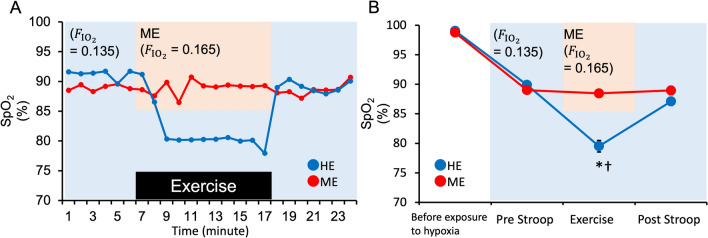
Table 1 summarizes the results of the physiological parameters. Heart rate (HR), ratings of perceived exertion (RPE), ventilation (), end-tidal carbon dioxide concentration (ETCO2), and SpO2 were subjected to repeated measures two-way analysis of variance (ANOVA) with condition (hypoxic exercise [HE]/milder hypoxic exercise [ME]) and time (before exposure to hypoxia/pre-Stroop/during exercise/post-Stroop) as within-subject factors. Other respiratory gas parameters are shown in the Supplementary materials. There was a significant interaction between time and condition for HR (F[3, 39] = 10.52, P < 0.001, η2p = 0.44) and ETCO2 (F[3, 39] = 22.626, P < 0.001, η2p = 0.64). We found significant increases in HR and ETCO2 during exercise compared to during pre-Stroop sessions in both conditions. HR during exercise was higher during HE than during ME, and ETCO2 during exercise was lower during HE than during ME. There were the significant main effects in RPE and , and there was a predominant increase during exercise under ME and HE conditions. There was a significant interaction between time and condition for SpO2 (F[3, 39] = 83.995, P < 0.001, η2p = 0.87). We also found a significant decrease in SpO2 during exercise compared to during the pre-Stroop session in the HE condition. Post hoc analyses showed that SpO2 values during exercise periods under the HE condition were lower than those under the ME condition (Fig. 1). In the HE condition, SpO2 during exercise was comparable to that in our previous study (this study, 79.5 ± 3.6%; previous study, 81.7 ± 1.4%)8; we confirmed that the experimental model in which cognitive fatigue occurs was replicated in this study.
Table 1.
Physiological parameters.
| Variable | Condition | Before exposure to hypoxia | Pre-Stroop | During exercise | Post-Stroop |
|---|---|---|---|---|---|
| HR (bpm) | ME | 69.6 (1.8) | 76.2 (2.2) | 128.7 (3.1)† | 85.7 (2.5)† |
| RPE (point) | 7.9 (0.7) | 12.9 (0.5)† | |||
| 10.8 (0.5) | 10.3 (0.4) | 42.8 (2.2)† | 10.4 (0.5) | ||
| ETCO2 (%/min) | 5.1 (0.1) | 5.3 (0.0) | 6.1 (0.1)† | 5.2 (0.0) | |
| HR (bpm) | HE | 70.3 (2.6) | 76.7 (2.8) | 138.7 (2.8)*† | 89.2 (3.1)*† |
| RPE (point) | 7.8 (0.5) | 13.9 (0.5)† | |||
| 10.2 (0.9) | 9.3 (0.7) | 44.6 (3.2)† | 9.3 (0.7) | ||
| ETCO2 (%/min) | 5.1 (0.1) | 5.3 (0.1) | 5.7 (0.1)*† | 5.2 (0.1) |
The value of each indicator is averaged over the 3 min before exposure to hypoxia, during the Stroop task (Pre and Post, 6.5 min each), and during exercise (10 min). HE, hypoxic exercise; ME, milder hypoxic exercise; HR, heart rate; RPE, ratings of perceived exertion; , ventilation; ETCO2, end-tidal carbon dioxide concentration; bpm, beats per minute.
Values are presented as mean (standard error). *P < 0.05 versus (vs.) ME condition, †P < 0.05 vs. pre-Stroop.
Figure 1.
(A) A typical example of the percutaneous arterial oxygen saturation (SpO2) during exercise. Immediately before exercise, the inhaled hypoxic gas was switched from hypoxia (= 0.135; blue background) to a milder hypoxic gas (= 0.161 ± 0.018; orange background), which was adjusted so that SpO2 during exercise remained at the resting level in the ME condition (red). Immediately after the end of the exercise, the inhaled hypoxic gas was again returned to hypoxia ( = 0.135). (B) The mean and standard deviation of SpO2 under hypoxic exercise (HE) (blue) and milder hypoxic exercise (ME) (red) conditions. Values are presented as mean ± standard error. *P < 0.05 versus (vs.) ME condition, †P < 0.05 vs. pre-Stroop.
Executive performance: Stroop interference
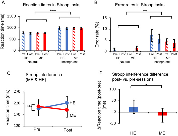
We first examined whether a general tendency in the CWST could be reproduced under all conditions. The reaction time (RT) and error rate were subjected to a repeated-measures three-way ANOVA with trial (incongruent/neutral), condition (HE/ME), and session (pre/post) as within-subject factors. ANOVA revealed significant main effects of trial on the RT (F[1, 13] = 60.274, P < 0.001, η2p = 0.82, Fig. 2A) and error rate (F[1, 13] = 12.138, P < 0.005, η2p = 0.48, Fig. 2B). These results verified that Stroop interference was generally observed in all the sessions used in this experiment as in our previous studies1–3,8,15,38–40,42,45. Therefore, to clarify the effect of an acute bout of exercise on a specifically defined cognitive process, we focused on analyses of Stroop interference (incongruent – neutral). ANOVA for the RT of Stroop interference revealed a significant interaction between condition and session factors (F[1, 13] = 4.964, P < 0.05, η2p = 0.28, Fig. 2C). There was no significant interaction or main effect on the error rate.
Figure 2.
(A) Comparison of the reaction time (RT) between the incongruent and neutral conditions. The incongruent condition exhibits a significantly slower RT than the neutral condition (***P < 0.001). (B) Comparison of the error rate between the incongruent and neutral conditions. Significant Stroop interference effects are observed (**P < 0.005). (C) The mean difference of the RT in incongruent and neutral trials indicates Stroop interference for each condition. (D) Stroop interference (RT) differences between post- and pre-sessions for each condition. Stroop interference differences are significantly more positive in the hypoxic exercise (HE) condition than in the milder hypoxic exercise (ME) condition (*P < 0.05). Error bars indicate the standard error.
Next, to examine the interaction for the RT, we calculated the difference in the degree of Stroop interference between post- and pre-sessions: ([incongruent–neutral] of pre-session–[incongruent–neutral] of post-session); contrasts for both the HE and ME conditions were calculated, and the differences between them were compared. The RT difference was significantly more negative in the HE condition than in the ME condition (t[13] = 2.228, P < 0.05, d = 0.34, paired t-test; Fig. 2D). These results demonstrate that the negative effect of exercise on executive performance under the HE condition was diminished under the ME condition.
Results of fNIRS
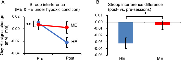
Since a previous study8 showed that the increased RT of Stroop interference by moderate exercise under hypoxic conditions was associated with decreased l-DLPFC activity, we assessed the effects of moderate exercise under hypoxic conditions on l-DLPFC activation, focusing on Stroop interference (Fig. 3). The oxygenated hemoglobin (oxy-Hb) signal differences (incongruent–neutral) in response to Stroop interference in the l-DLPFC were analyzed with repeated measures two-way ANOVA, including condition (HE/ME) and session (pre/post) as within-subject factors. In this design, the effect of an acute bout of moderate exercise on Stroop interference was expected to appear as an interaction between the two factors, because pre-HE and pre-ME were virtually identical. ANOVA performed on the regions of interest (ROIs) of l-DLPFC revealed a significant interaction (F[1, 13] = 8.63, P < 0.05, η2p = 0.40, Fig. 4A). There were no significant differences between pre-sessions. To clarify the exercise-session interaction in the ROIs of l-DLPFC, differences in hemodynamic responses due to Stroop interference between the post- and pre-sessions were analyzed. Oxy-Hb signal differences in response to Stroop interference in the l-DLPFC were significantly lower in the HE condition than in the ME condition (t[13] = −2.376, P < 0.05, d = 0.63, paired t-test; Fig. 4B). However, no significant interactions or main effects of deoxygenated hemoglobin (deoxy-Hb) were observed.
Figure 3.
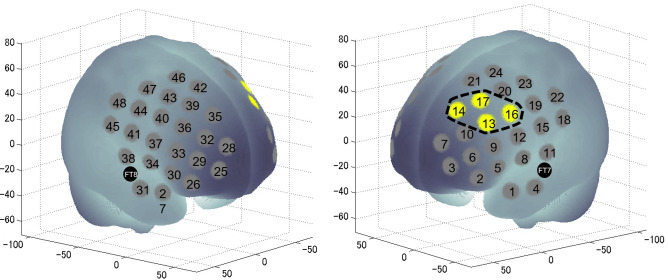
The spatial profiles of functional near-infrared spectroscopy channels and region of interest segmentation used in the current study; they were introduced in previous studies46,50. Channel numbers and FT7 and FT8 in the international 10–20 electroencephalography standard positions are denoted above the corresponding locations. The channels enclosed by the black broken lines were defined as the l-DLPFC, and their data were integrated for further analyses.
Figure 4.
(A) Stroop interference differences between post- and pre-sessions for oxygenated hemoglobin (oxy-Hb) signal contrasts in both conditions. (B) Oxy-Hb signal differences for the hypoxic exercise (HE) condition are significantly lower than those for the milder hypoxic exercise (ME) condition (P < 0.05). Error bars indicate the standard error.
Association of executive performance and l-DLPFC activity with physiological parameters
We examined the correlation between the delayed Stroop interference-related RT and altered , ETCO2, and SpO2 during exercise in the HE condition. There was no notable correlation between the RT and any parameter ( : r = 0.203, P > 0.05; ETCO2: r = − 0.109, P > 0.05; SpO2: r = 0.08, P > 0.05). Moreover, we examined the correlation between reduced activation in the l-DLPFC and altered , ETCO2, and SpO2 during exercise under the HE condition. There was no significant correlation between l-DLPFC activation and any of the parameters (: r = -0.406, P > 0.05; ETCO2: r = − 0.025, P > 0.05; SpO2: r = 0.227, P > 0.05).
Discussion
This study aimed to examine the involvement of low SpO2 during exercise in executive underperformance and prefrontal deactivation caused by exercise in hypoxic environments. We found that suppression of low SpO2 during exercise in hypoxic environments diminished the negative effects on CWST performance and task-related l-DLPFC activity. These result suggest that exercise-induced executive underperformance under hypoxic environment is due to decrease in SpO2 during exercise.
Behavioral measurements revealed a shorter RT and lower error rate in neutral trials than in incongruent trials. Therefore, we confirmed that Stroop interference could be stably induced before and after an acute bout of moderate exercise under both conditions. Based on these data, we first compared the effects of the HE and ME conditions on Stroop interference. Stroop interference increased after 10 min of exercise in the HE condition compared with the ME condition. This finding is consistent with those of previous studies that have demonstrated that an acute bout of moderate exercise under normobaric hypoxic conditions decreases inhibitory control function8. However, there was no delay in Stroop interference in the ME condition. The aspirated oxygen concentration and SpO2 during the CWST did not differ between the two conditions, suggesting that the decline in executive function was associated with hypoxemia during exercise.
In the subsequent analysis, we examined the brain regions that were deactivated after exercise in a hypoxic environment. Previous studies have confirmed that the activity of the l-DLPFC decreases after exercise under normobaric hypoxic conditions8. Therefore, in the present study, we also focused on the decreased activity of the l-DLPFC in response to increased Stroop interference. As a result, a decrease in neural activity of the l-DLPFC was observed after exercise in the HE condition, similar to the results of previous studies, whereas in the ME condition, the decrease in neural activation after exercise improved. These results suggest that the suppression of low behavioral data after exercise in the ME condition may involve the suppression of task-specific decreased activity in the l-DLPFC.
In this study, SpO2, , and ETCO2, which are involved in cerebral vasodilation during the CWST, were not significantly different before and after exercise or between the conditions. This finding suggests that the negative effect on CWST performance and l-DLPFC activity is related to altered physiological parameters during exercise. Our previous study showed that CWST performance was impaired in severely hypoxic environments where SpO2 decreased below 80%15, suggesting that the impaired executive function in this study was due to low SpO2 during exercise. Similarly, a previous study reported that hypoxemia during exercise in hypoxic environments is associated with decreased neural activity in the motor cortex and decreased motor performance18. In addition, moderate exercise under normobaric hypoxic conditions ( = 0.10) led to a decreased cerebral metabolic rate for oxygen (CMRO2)19, and a reduction in CMRO2 with exposure to normobaric hypoxic conditions ( = 0.08) did not return to baseline after re-oxygenation20. Therefore, we considered that exercise-induced cognitive decline in the HE condition might be due to the low SpO2 during exercise.
During exercise in a hypoxic environment, SpO2 decreases due to the ventilation-to-perfusion ratio inequality and diffusion limitation which is due to the reduction in the erythrocyte transit time in the pulmonary capillaries with the increase in cardiac output21. Although a decrease in SpO2 limits oxygen supply to the brain, only low SpO2 but with normal cerebral blood flow might not affect the neural activity of an intact organism22. This is thought to be due to the cerebral blood flow increase as the SpO2 decreases23–25. However, this vascular response is not uniform across the brain. It has been reported that changes are greater in paleocortical regions than in neocortical regions, suggesting that the blood flow change during decreasing SpO2 may protect regions of the brain with essential homeostatic roles26. In addition, since exercise in a hypoxic environment has been suggested to cause competition for blood flow distribution between muscles and the brain27, it is possible that this cerebral blood flow response in the prefrontal cortex was more inadequate, resulting in impaired executive function. Although the mechanism by which neural activity decreases under cerebral hypoxia still remains unclear, one possible cause is neural deactivation associated with inflammatory response. In an animal study, it has been shown that hypoxia is accompanied by an inflammatory response via hypoxia-inducible factor and nuclear factor-kB28, and the combination of hypoxia and inflammation rapidly decreases the neural excitability in the rodent hippocampal CA1 neurons29. An acute exercise, on the other hand, also induces acute inflammatory markers, which results in increased inflammatory cytokine levels in the animal cortex30. If so, in this study, it is possible that the combined effects of hypoxia and inflammation occur in the human prefrontal cortex with hypoxic exercise, which in turn may cause cognitive fatigue. This hypothesis, however, needs to be verified in the future. A limitation of this study is that we were unable to evaluate the relationship between oxygen metabolism in the brain during exercise and post-exercise decline in executive function. Since SpO2 is strongly correlated with prefrontal oxygen saturation12,13,31, it is assumed that cerebral hypoxia during exercise in the HE condition decreases executive function and l-DLPFC activity. However, in the present study, no significant correlation was found between decreased SpO2 and decreased executive function during exercise, and it remains unclear how low SpO2 is related to negative effects on executive function. Although fMRI and fNIRS still have issues in evaluating neural activity during exercise, such as the effects of body movement and skin blood flow, transcranial magnetic stimulation (TMS) has been used to evaluate neural activity in the motor cortex during exercise under hypoxia. Future evaluation of prefrontal neural activity and oxygen metabolism during exercise under hypoxia using TMS will help elucidate the mechanism of cognitive fatigue.
Another limitation of the present study is that it was conducted in healthy adults who do not normally engage in strenuous exercise, so there was insufficient inter-subject evaluation. In fact, elite mountaineers, compared to non-mountaineers, showed a smaller decrease in SpO2 in the quadriceps muscle under hypoxic conditions ( = 0.125) and might maintain oxygen supply to the tissues32. In addition, no sex-related differences were detected for reaction time and DLPFC activity or physiological indices. This lack of difference is supposedly due to the small sample size of female participants. The ventilatory response in hypoxic environments in women is higher than that in men33; some authors hypothesize that women have a strong protective mechanism against acute exposure to hypoxia34. It is possible that cognitive fatigue due to exercise under hypoxic conditions is also attenuated in women, but this needs to be verified in the future. Furthermore, some previous studies have reported that acclimatization to hypoxic environments increased the oxygen supply to the brain during the resting state35 and exercise18 in hypoxic environments compared to before acclimatization and improved hypoxia-induced motor cortex hypoactivity18. Based on the present study, further studies are needed to examine whether acclimatization to hypoxic environments improves cognitive fatigue in mountaineers and trail runners who have been acclimatized to hypoxic environments through hypoxic training. This may lead to the elucidation of the actual situation of exercise-induced cognitive fatigue and the development of methods to cope with it.
In conclusion, the current study revealed that the decline in executive function and l-DLPFC activity after moderate-intensity exercise under hypoxic environments could be prevented by suppressing the decrease in SpO2 during exercise, suggesting that the exercise-induced cognitive fatigue under hypoxic environment is due to hypoxemia during exercise. The results of this research will contribute to the elucidation of the mechanism of cognitive fatigue caused by a decrease in blood oxygen concentration due to exercise, as well as activities in hypoxic environments, e.g., high altitude, and to the development of methods to cope with such central fatigue.
Methods
Ethics statements
This study was approved by the Institutional Ethics Committee of the University of Tsukuba, Faculty of Health and Sport Sciences (approval number: Tai 025–120) and was conducted in accordance with the latest version of the Declaration of Helsinki. The study participants provided written informed consent for participation and publication of their details.
Participants
Fourteen right-handed young adults (12 men and 2 women) participated in this study (Table 2). The sample size was determined by assuming that the effects of exercise in hypoxic environments would be similar to those in our previous study8. All participants were Japanese native speakers, healthy, and naive to the experimental procedures for which they volunteered. None of the participants had a history of neurological, psychiatric, or respiratory disorders or a disease requiring medical care. All the participants had normal or corrected-to-normal vision and normal color vision. All participants were asked to refrain from exercise and the consumption of alcohol and caffeine for at least 24 h prior to each experiment to control for external factors that could affect cardiovascular and executive functions. Post-hoc sensitivity analysis performed based on this sample with 80% power and 0.05 alpha demonstrated sufficient sensitivity to detect repeated-measures effects exceeding f = 0.40 and t-test differences exceeding d = 0.81 (with a two-tailed alpha), as computed using G*Power (3.1.9.2).
Table 2.
Participants’ characteristics.
| Age (years) | Height (cm) | Weight (kg) | (mL·kg·min-1) | Workload (W) | |
|---|---|---|---|---|---|
| Average (SD) | 21.4 (1.7) | 171.7 (7.1) | 63.3 (7.2) | 44.7 (9.5) | 116.6 (23.7) |
Age, height, weight, peak oxygen intake (), and relative workload for moderate-intensity exercise are presented as the mean and standard deviation for the 14 participants.
Experimental procedures
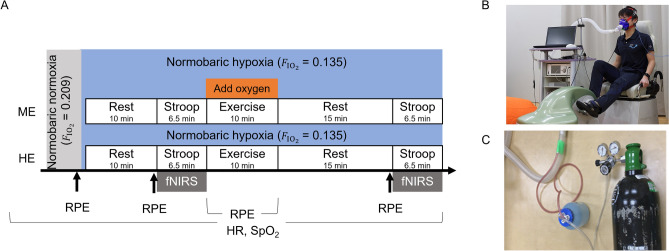
On the first day, participants underwent a graded exercise test to measure their and determine the appropriate individual intensity for moderate exercise, which was defined as 50% of a participant’s based on the American College of Sports Medicine's classification of physical activity intensity36. The detailed procedures for the graded exercise test were the same as those in our previous study8. The participants practiced the CWST twice before being subjected to the main experimental conditions. A few days after the first visit, two main experimental conditions were conducted in a single-blind (participant being blinded) crossover study design: exercise under moderate normobaric hypoxia (HE) or ME, which improved the inhaled oxygen concentration to suppress SpO2 during exercise. All participants participated in both the HE and ME conditions, each on separate days, with the order counterbalanced across participants (Fig. 5). In both conditions, participants underwent the CWST before and 15 min after 10 min of moderate-intensity exercise on a recumbent cycle ergometer (Strength Ergo 240 W, Mitsubishi Electric Corp., Tokyo, Japan) at 60 revolutions per minute, based on our previous study methods8. Cortical hemodynamic changes in the l-DLPFC were monitored using fNIRS while the participants performed the CWST.
Figure 5.
(A) The two conditions, breathing a moderately hypoxic gas (hypoxic exercise [HE]) and breathing a milder hypoxic gas, during which oxygen was added to the moderately hypoxic gas to maintain the oxygen saturation (SpO2) level during milder hypoxic exercise (ME). Cortical hemodynamic changes were monitored with functional near-infrared spectroscopy (fNIRS) while participants performed the Stroop task. HR, heart rate. (B) In both conditions, the exercise and color-word Stroop task (CWST) were performed on a recumbent cycle ergometer. Hypoxic gas ( = 0.135) stored in a Douglas bag was inhaled through a mask. (C) In the ME condition, SpO2 was adjusted during exercise by adding oxygen gas with humidity to prevent the participant’s throat from drying out to the hose connecting the Douglas bag to the mask.
In the HE condition, participants breathed hypoxic gas, as in our previous study8, which was a mixture of 13.5% oxygen and 0.03% carbon dioxide in nitrogen ( = 0.135; equivalent to an altitude of approximately 3,500 m) through a mask connected to a Douglas bag. In the ME condition, as in the HE condition, participants breathed a moderately hypoxic gas during the CWST, but only during exercise; they breathed a milder hypoxic gas ( = 0.161 ± 0.018) to regulate the hypoxic gas so their SpO2 did not decrease from the resting state. To adjust the oxygen concentration, oxygen was injected directly into the hose while monitoring the SpO2 during exercise. Expired air was exhausted directly outside the mask so participants did not re-breathe it. The participants were exposed to a moderately hypoxic gas 10 min before the pre-Stroop session while sitting on a cycle ergometer. HR was monitored by a heart rate monitor (V800, Polar Electro, Kempele, Finland), SpO2 was monitored by a pulse oximeter (OLV-3100, Nihon Kohden, Tokyo, Japan) placed on the left earlobe, and exhaled gas was monitored every minute by a gas analyzer (Aeromonitor AE-310S; Minato Medical Science, Osaka, Japan). The participants’ RPE37 were recorded before exposure to hypoxia, every minute during exercise, and before the CWST.
Behavioral measurements
The CWST1–3,8,15,38–42 was adopted in an event-related design. The CWST, which included two rows containing letters or words, was presented on a screen, and the participants were instructed to decide whether the color of the letters or words in the top row corresponded to the color name presented in the bottom row. Participants pressed a “yes” or “no” button with their right forefinger or middle finger to respond. The RT and error rates were measured.
The CWST consisted of three trials, including 10 neutral, 10 congruent, and 10 incongruent trials. For neutral trials, the top row contained sets of X’s (XXXX) written in red, blue, green, or yellow, and the bottom row contained the words “RED,” “BLUE,” “GREEN,” or “YELLOW” written in black. For congruent trials, the top row contained the words “RED,” “BLUE,” “GREEN,” or “YELLOW” written in a color congruent with that of the bottom row. For incongruent trials, the color word in the top row was written in an incongruent color to produce interference between the color of the word and the color name. All words were written in Japanese. The correct answer rate assigned to “yes” and “no” was 50%. Each stimulus was separated by an inter-stimulus interval showing a fixation cross for 9–13 s to avoid predicting the timing of the subsequent trial1,2,8,15,38–40,42. The stimulus remained on the screen until a response was given or for 2 s. In the present study, Stroop interference, a specifically defined cognitive process, was used to elucidate the effect of an acute bout of moderate exercise under hypoxic conditions on executive function. Therefore, the (incongruent-neutral) contrast, which is assumed to represent Stroop interference, was calculated.
fNIRS measurements
We used a multichannel fNIRS optical topography system (ETG-7000, Hitachi Medical Corporation, Chiba, Japan) set with two wavelengths of near-infrared light (785 and 830 nm). We analyzed the optical data from fNIRS based on the modified Beer–Lambert law43, as previously described44. This method allowed us to calculate signals reflecting the oxy-Hb, deoxy-Hb, and total hemoglobin concentration changes, calculated in millimolar-millimeters (mM⋅mm)44. The composition of the fNIRS probe holder and its placement followed the same procedure used in our previous studies1–3,8,38–40,42,45. To register fNIRS data in the Montreal Neurological Institute (MNI) space, we used virtual registration46,47. Briefly, this method enables the placement of a virtual probe holder on the scalp by stimulating the holder’s deformation and registering probes and channels onto a reference brain in a preconstructed magnetic resonance imaging database48,49. We probabilistically estimated the MNI coordinate values for the fNIRS channels to obtain the most likely estimate of the location of the given channels for the group of participants and the spatial variability associated with the estimation50,51.
Analysis of the fNIRS data
In this study, neural activity related to the CWST was evaluated by examining changes in oxy-Hb, as shown in our previous studies1–3,8,38–40,42,45. Individual timeline data for the oxy-Hb signal of each channel were preprocessed with a band-pass filter using a cut-off frequency of 0.04 Hz to remove baseline drift and 0.3 Hz to filter out heartbeat pulsations. Channel-wise and subject-wise contrasts were obtained by calculating the inter-trial mean of differences between the oxy-Hb signals of the peak (6–10 s after trial onset) and baseline (0–2 s before trial onset) periods based on our study8. The contrasts were calculated as prefrontal activation elicited by a cognitive task, and the contrasts obtained were subjected to second-level random-effects group analysis.
Based on a method widely used in anatomical labeling systems, such as the LBPA4049, channels 13, 14, 16, and 17 were combined to analyze l-DLPFC activity, which was found to decrease after moderate-intensity exercise under hypoxic conditions in a previous study8.
Statistical analysis
The HR, RPE, and SpO2 were subjected to repeated measures two-way ANOVA with condition (HE/ME) and time (before exposure to hypoxia/pre-Stroop/during exercise/post-Stroop) as within-subject factors. The RT and error rate were subjected to repeated measures three-way ANOVA with trial (incongruent/neutral), condition (HE/ME), and session (pre/post) as within-subject factors to examine whether the general tendencies for the Stroop task could be reproduced in all conditions. The Stroop effect associated with acute moderate exercise on all outcome measures was analyzed using repeated measures two-way ANOVA with condition (HE/ME) and session (pre/post) as within-subject factors. When a significant F-value was obtained, a post hoc test using the Bonferroni method for multiple corrections was applied to identify significant differences among the mean values.
Moreover, to clarify the relationships of physiological parameters (SpO2, ETCO2, and ) during exercise with executive performance and task-related brain activation, we conducted parametric Pearson correlation analyses.
All data are presented as mean ± standard error. Statistical significance was set a priori at P < 0.05 for all comparisons. Statistical analyses were performed using the Statistical Package for the Social Sciences (SPSS) version 26 (SPSS Inc., Chicago, IL, USA).
Supplementary Information
Acknowledgements
This work was supported in part by the Japan Society for the Promotion of Science Grants JP15J00782 (G.O.), JP16H06405 (H.S.), JP18H04081 (H.S.), and JP19K20036 (G.O.), and the Japan Science and Technology Agency Grants JPMJMI19D5 (H.S.). We would like to thank Editage (www.editage.com) for English language editing.
Author contributions
G.O. and H.S. contributed to the study design. G.O. played a role in data collection. G.O., R.K., K. H., T.F., K.S. performed data analysis. G.O. wrote the manuscript and R.K., K. S., T.F., K. H., and H.S. edited the manuscript and contributed to the study concept and funding acquisition. All authors have read and approved the final version of the manuscript.
Data availability
The datasets generated and/or analyzed during the current study are available from the corresponding author on reasonable request.
Competing interests
The authors declare no competing interests.
Footnotes
Publisher's note
Springer Nature remains neutral with regard to jurisdictional claims in published maps and institutional affiliations.
Supplementary Information
The online version contains supplementary material available at 10.1038/s41598-022-14146-5.
References
- 1.Byun K, et al. Positive effect of acute mild exercise on executive function via arousal-related prefrontal activations: an fNIRS study. Neuroimage. 2014;98:336–345. doi: 10.1016/j.neuroimage.2014.04.067. [DOI] [PubMed] [Google Scholar]
- 2.Hyodo K, et al. Acute moderate exercise enhances compensatory brain activation in older adults. Neurobiol. Aging. 2012;33:2621–2632. doi: 10.1016/j.neurobiolaging.2011.12.022. [DOI] [PubMed] [Google Scholar]
- 3.Yanagisawa H, et al. Acute moderate exercise elicits increased dorsolateral prefrontal activation and improves cognitive performance with Stroop test. Neuroimage. 2010;50:1702–1710. doi: 10.1016/j.neuroimage.2009.12.023. [DOI] [PubMed] [Google Scholar]
- 4.Chapman RF. The individual response to training and competition at altitude. Br. J. Sports Med. 2013;47(Suppl 1):i40–44. doi: 10.1136/bjsports-2013-092837. [DOI] [PMC free article] [PubMed] [Google Scholar]
- 5.Fulco CS, Rock PB, Cymerman A. Maximal and submaximal exercise performance at altitude. Aviat. Space Environ. Md. 1998;69:793–801. [PubMed] [Google Scholar]
- 6.Wehrlin JP, Hallen J. Linear decrease in .VO2max and performance with increasing altitude in endurance athletes. Eur. J. Appl. Physiol. 2006;96:404–412. doi: 10.1007/s00421-005-0081-9. [DOI] [PubMed] [Google Scholar]
- 7.Lefferts WK, et al. Effect of hypoxia on cerebrovascular and cognitive function during moderate intensity exercise. Physiol. Behav. 2016;165:108–118. doi: 10.1016/j.physbeh.2016.07.003. [DOI] [PubMed] [Google Scholar]
- 8.Ochi G, et al. Neural basis for reduced executive performance with hypoxic exercise. Neuroimage. 2018;171:75–83. doi: 10.1016/j.neuroimage.2017.12.091. [DOI] [PubMed] [Google Scholar]
- 9.Piek JP, et al. The relationship between motor coordination, executive functioning and attention in school aged children. Arch. Clin. Neuropsychol. 2004;19:1063–1076. doi: 10.1016/j.acn.2003.12.007. [DOI] [PubMed] [Google Scholar]
- 10.Vestberg T, Reinebo G, Maurex L, Ingvar M, Petrovic P. Core executive functions are associated with success in young elite soccer players. PLoS ONE. 2017;12:e0170845. doi: 10.1371/journal.pone.0170845. [DOI] [PMC free article] [PubMed] [Google Scholar]
- 11.Vestberg T, Gustafson R, Maurex L, Ingvar M, Petrovic P. Executive functions predict the success of top-soccer players. PLoS ONE. 2012;7:e34731. doi: 10.1371/journal.pone.0034731. [DOI] [PMC free article] [PubMed] [Google Scholar]
- 12.Eichhorn L, et al. Evaluation of near-infrared spectroscopy under apnea-dependent hypoxia in humans. J. Clin. Monit. Comput. 2015;29:749–757. doi: 10.1007/s10877-015-9662-2. [DOI] [PubMed] [Google Scholar]
- 13.Ricci M, et al. Near-infrared spectroscopy to monitor cerebral oxygen saturation in single-ventricle physiology. J. Thorac. Cardiovasc. Surg. 2006;131:395–402. doi: 10.1016/j.jtcvs.2005.07.039. [DOI] [PubMed] [Google Scholar]
- 14.Subudhi AW, Dimmen AC, Roach RC. Effects of acute hypoxia on cerebral and muscle oxygenation during incremental exercise. J. Appl. Physiol. 2007;1985(103):177–183. doi: 10.1152/japplphysiol.01460.2006. [DOI] [PubMed] [Google Scholar]
- 15.Ochi G, et al. Hypoxia-induced lowered executive function depends on arterial oxygen desaturation. J. Physiol. Sci. 2018;68:847–853. doi: 10.1007/s12576-018-0603-y. [DOI] [PMC free article] [PubMed] [Google Scholar]
- 16.Goodall S, González-Alonso J, Ali L, Ross EZ, Romer LM. Supraspinal fatigue after normoxic and hypoxic exercise in humans. J. Physiol. 2012;590:2767–2782. doi: 10.1113/jphysiol.2012.228890. [DOI] [PMC free article] [PubMed] [Google Scholar]
- 17.Rasmussen P, et al. Reduced muscle activation during exercise related to brain oxygenation and metabolism in humans. J. Physiol. 2010;588:1985–1995. doi: 10.1113/jphysiol.2009.186767. [DOI] [PMC free article] [PubMed] [Google Scholar]
- 18.Goodall S, et al. AltitudeOmics: exercise-induced supraspinal fatigue is attenuated in healthy humans after acclimatization to high altitude. Acta Physiol. (Oxf.) 2014;210:875–888. doi: 10.1111/apha.12241. [DOI] [PMC free article] [PubMed] [Google Scholar]
- 19.Rasmussen P, et al. Cerebral oxygenation is reduced during hyperthermic exercise in humans. Acta Physiol. (Oxf.) 2010;199:63–70. doi: 10.1111/j.1748-1716.2010.02084.x. [DOI] [PubMed] [Google Scholar]
- 20.Tichauer KM, Brown DW, Hadway J, Lee TY, St Lawrence K. Near-infrared spectroscopy measurements of cerebral blood flow and oxygen consumption following hypoxia-ischemia in newborn piglets. J. Appl. Physiol. (1985) 2006;100:850–857. doi: 10.1152/japplphysiol.00830.2005. [DOI] [PubMed] [Google Scholar]
- 21.Calbet JAL, Lundby C. Air to muscle O2 delivery during exercise at altitude. High Alt. Med. Biol. 2009;10:123–134. doi: 10.1007/s00421-021-04667-8. [DOI] [PubMed] [Google Scholar]
- 22.Miyamoto O, Auer RN. Hypoxia, hyperoxia, ischemia, and brain necrosis. Neurology. 2000;54:362–371. doi: 10.1212/wnl.54.2.362,10668697. [DOI] [PubMed] [Google Scholar]
- 23.Ellingsen I, Hauge A, Nicolaysen G, Thoresen M, Walloe L. Changes in human cerebral blood flow due to step changes in PAO2 and PACO2. Acta Physiol. Scand. 1987;129:157–163. doi: 10.1111/j.1748-1716.1987.tb08054.x. [DOI] [PubMed] [Google Scholar]
- 24.Cohen PJ, Alexander SC, Smith TC, Reivich M, Wollman H. Effects of hypoxia and normocarbia on cerebral blood flow and metabolism in conscious man. J. Appl. Physiol. 1967;23:183–189. doi: 10.1152/jappl.1967.23.2.183. [DOI] [PubMed] [Google Scholar]
- 25.Kety SS, Schmidt CF. The effects of altered arterial tensions of carbon dioxide and oxygen on cerebral blood flow and cerebral oxygen consumption of normal young men. J. Clin. Invest. 1948;27:484–492. doi: 10.1172/JCI101995. [DOI] [PMC free article] [PubMed] [Google Scholar]
- 26.Binks AP, Cunningham VJ, Adams L, Banzett RB. Gray matter blood flow change is unevenly distributed during moderate isocapnic hypoxia in humans. J. Appl. Physiol. 2008;1985(104):212–217. doi: 10.1152/japplphysiol.00069.2007. [DOI] [PubMed] [Google Scholar]
- 27.Verges S, et al. Cerebral perturbations during exercise in hypoxia. Am. J. Physiol. Regul. Integr. Comp. Physiol. 2012;302(8):R903–R916. doi: 10.1152/ajpregu.00555.2011. [DOI] [PubMed] [Google Scholar]
- 28.Taylor CT. Interdependent roles for hypoxia inducible factor and nuclear factor-κB in hypoxic inflammation. J. Physiol. 2008;586:4055–4059. doi: 10.1113/jphysiol.2008.157669. [DOI] [PMC free article] [PubMed] [Google Scholar]
- 29.Yang YS, Son SJ, Choi JH, Rah JC. Synaptic transmission and excitability during hypoxia with inflammation and reoxygenation in hippocampal CA1 neurons. Neuropharmacology. 2018;138:20–31. doi: 10.1016/j.neuropharm.2018.05.011. [DOI] [PubMed] [Google Scholar]
- 30.Carmichael MD, et al. Role of brain IL-1beta on fatigue after exercise-induced muscle damage. Am. J. Physiol. Regul. Integr. Comp. Physiol. 2006;291(5):R1344–R1348. doi: 10.1152/ajpregu.00141.2006. [DOI] [PubMed] [Google Scholar]
- 31.Kusaka T, et al. Quantification of cerebral oxygenation by full-spectrum near-infrared spectroscopy using a two-point method. Comp. Biochem. Physiol. A Mol. Integr. Physiol. 2002;132:121–132. doi: 10.1016/S1095-6433(01)00538-4. [DOI] [PubMed] [Google Scholar]
- 32.Puthon L, et al. Physiological characteristics of elite high-altitude climbers. Scand. J. Med. Sci Sports. 2016;26:1052–1059. doi: 10.1111/sms.12547. [DOI] [PubMed] [Google Scholar]
- 33.Paterson DJ, Pinnington H, Pearce AR, Morton AR. Maximal exercise cardiorespiratory responses of men and women during acute exposure to hypoxia. Aviat. Space Environ. Med. 1987;58:243–247. [PubMed] [Google Scholar]
- 34.Woorons X, Mollard P, Lamberto C, Letournel M, Richalet J-P. Effect of acute hypoxia on maximal exercise in trained and sedentary women. Med. Sci. Sports Exerc. 2005;37:147–154. doi: 10.1249/01.Mss.0000150020.25153.34. [DOI] [PubMed] [Google Scholar]
- 35.Subudhi AW, et al. AltitudeOmics: Effect of ascent and acclimatization to 5260 m on regional cerebral oxygen delivery. Exp Physiol. 2014;99:772–781. doi: 10.1113/expphysiol.2013.075184. [DOI] [PubMed] [Google Scholar]
- 36.Medicine, A. C. o. S. ACSM’s Guidelines for Exercise Testing and Prescription (Wolters Kluwer Health, 2014)
- 37.Borg G. Perceived exertion as an indicator of somatic stress. Scand. J. Rehabil. Med. 1970;2:92–98. [PubMed] [Google Scholar]
- 38.Damrongthai C, et al. Benefit of human moderate running boosting mood and executive function coinciding with bilateral prefrontal activation. Sci. Rep. 2021;11:22657. doi: 10.1038/s41598-021-01654-z. [DOI] [PMC free article] [PubMed] [Google Scholar]
- 39.Kuwamizu R, et al. Spontaneous eye blink rate connects missing link between aerobic fitness and cognition. Med. Sci. Sports Exerc. 2021;53:1425–1433. doi: 10.1249/MSS.0000000000002590. [DOI] [PubMed] [Google Scholar]
- 40.Suwabe K, et al. Positive mood while exercising influences beneficial effects of exercise with music on prefrontal executive function: A functional NIRS study. Neuroscience. 2021;454:61–71. doi: 10.1016/j.neuroscience.2020.06.007. [DOI] [PubMed] [Google Scholar]
- 41.Zysset S, Müller K, Lohmann G, von Cramon DY. Color-word matching stroop task: Separating interference and response conflict. Neuroimage. 2001;13:29–36. doi: 10.1006/nimg.2000.0665. [DOI] [PubMed] [Google Scholar]
- 42.Kujach S, et al. A transferable high-intensity intermittent exercise improves executive performance in association with dorsolateral prefrontal activation in young adults. Neuroimage. 2018;169:117–125. doi: 10.1016/j.neuroimage.2017.12.003. [DOI] [PubMed] [Google Scholar]
- 43.Cope M, et al. Methods of quantitating cerebral near infrared spectroscopy data. Adv. Exp. Med. Biol. 1988;222:183–189. doi: 10.1007/978-1-4615-9510-6_2. [DOI] [PubMed] [Google Scholar]
- 44.Maki A, et al. Spatial and temporal analysis of human motor activity using noninvasive NIR topography. Med. Phys. 1995;22:1997–2005. doi: 10.1118/1.597496. [DOI] [PubMed] [Google Scholar]
- 45.Hyodo K, et al. The association between aerobic fitness and cognitive function in older men mediated by frontal lateralization. Neuroimage. 2016;125:291–300. doi: 10.1016/j.neuroimage.2015.09.062. [DOI] [PubMed] [Google Scholar]
- 46.Brett M, Johnsrude IS, Owen AM. The problem of functional localization in the human brain. Nat. Rev. Neurosci. 2002;3:243–249. doi: 10.1038/nrn756. [DOI] [PubMed] [Google Scholar]
- 47.Tsuzuki D, Dan I. Spatial registration for functional near-infrared spectroscopy: From channel position on the scalp to cortical location in individual and group analyses. Neuroimage. 2014;85:92–103. doi: 10.1016/j.neuroimage.2013.07.025. [DOI] [PubMed] [Google Scholar]
- 48.Okamoto M, et al. Three-dimensional probabilistic anatomical cranio-cerebral correlation via the international 10–20 system oriented for transcranial functional brain mapping. Neuroimage. 2004;21:99–111. doi: 10.1016/j.neuroimage.2003.08.026. [DOI] [PubMed] [Google Scholar]
- 49.Okamoto M, Dan I. Automated cortical projection of head-surface locations for transcranial functional brain mapping. Neuroimage. 2005;26:18–28. doi: 10.1016/j.neuroimage.2005.01.018. [DOI] [PubMed] [Google Scholar]
- 50.Shattuck DW, et al. Construction of a 3D probabilistic atlas of human cortical structures. Neuroimage. 2008;39:1064–1080. doi: 10.1016/j.neuroimage.2007.09.031. [DOI] [PMC free article] [PubMed] [Google Scholar]
- 51.Singh AK, Okamoto M, Dan H, Jurcak V, Dan I. Spatial registration of multichannel multi-subject fNIRS data to MNI space without MRI. Neuroimage. 2005;27:842–851. doi: 10.1016/j.neuroimage.2005.05.019. [DOI] [PubMed] [Google Scholar]
Associated Data
This section collects any data citations, data availability statements, or supplementary materials included in this article.
Supplementary Materials
Data Availability Statement
The datasets generated and/or analyzed during the current study are available from the corresponding author on reasonable request.


