Abstract
The high cost of biofabricated titanium mesh plates can make them out of reach for hospitals in low-income countries. To increase the availability of cranioplasty, the authors of this work investigated the production of polymer-based endoprostheses. Recently, cheap, popular desktop 3D printers have generated sufficient opportunities to provide patients with on-demand and on-site help. This study also examines the technologies of 3D printing, including SLM, SLS, FFF, DLP, and SLA. The authors focused their interest on the materials in fabrication, which include PLA, ABS, PET-G, PEEK, and PMMA. Three-dimensional printed prostheses are modeled using widely available CAD software with the help of patient-specific DICOM files. Even though the topic is insufficiently researched, it can be perceived as a relatively safe procedure with a minimal complication rate. There have also been some initial studies on the costs and legal regulations. Early case studies provide information on dozens of patients living with self-made prostheses and who are experiencing significant improvements in their quality of life. Budget 3D-printed endoprostheses are reliable and are reported to be significantly cheaper than the popular counterparts manufactured from polypropylene polyester.
Keywords: cranioplasty, 3D cranioplasty, additive manufacturing, PMMA, PEEK, PLA, ABS, PET-G
1. Introduction
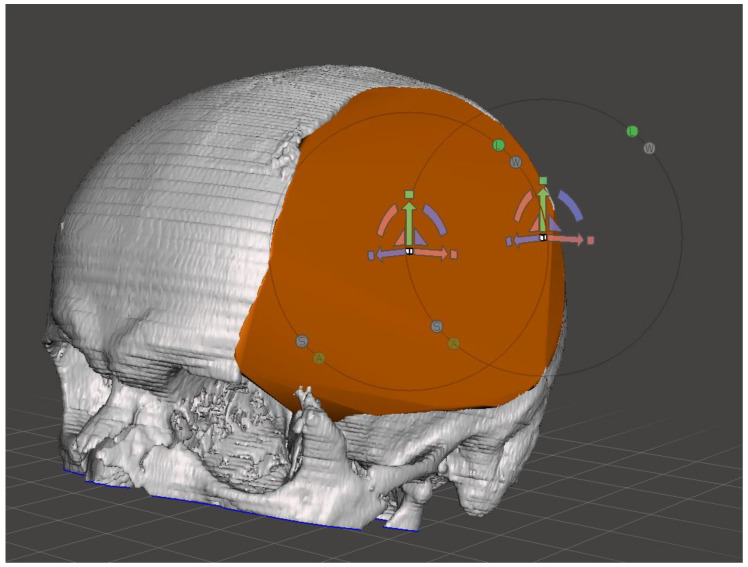
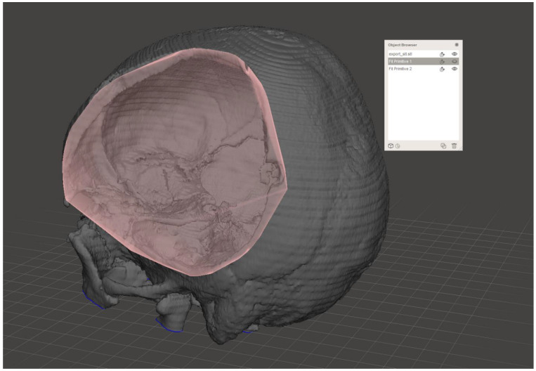
The human skull is one of the most complex regions of the human body in terms of maintaining visually pleasing results. In the case of a post-traumatic or postoperative defect, cranioplasty requires constant medical development to provide a decent quality of life to affected patients by producing personalized endoprostheses [1]. Although widely adapted titanium endoprostheses often provide satisfactory results, they tend to be too expensive for low-income patients. Despite high biocompatibility, titanium leads to a significant artifact in CT and MRI imaging. Prostheses made of polypropylene–polyester are often used in cranioplasty, for which prices in the case of standard products (regular, oval shape) do not exceed USD 500 [2]. However, if there is a necessity to use a personalized prosthesis, the cost escalates to nearly USD 2000. In recent years, FDM- (fused deposition modeling) and SLA- (stereolithography) 3D-printing have become increasingly popular [3]. These methods provide a terrific opportunity for medicine to bring widely available solutions with inexpensive planning, prototyping, guiding, and even creating on-demand 3D-printed tools, models, and prostheses [4,5,6,7]. The personalized anatomical models can then be implemented in various engineering applications, such as the Materialise Mimics or Materialise 3-matic and CAD/CAM/CAE software, allowing their further analysis using, e.g., the finite element method (FEM) [8,9]. The materials used during these processes can be chosen accordingly, e.g., PET-G for surgical guides, PLA/resin for patient skull models, or PEEK for the production of prostheses. Implants are modeled in the CAD software (such as MeshLab®) using the STL files harvested from the DICOM data (Chart 1) (for example, by using RadiAnt® or OsiriX®). This systematic review focuses on various utilizations of 3D-printing technology during surgical procedures in the craniofacial area and consists of a comparison of the multidisciplinary approaches that involve the manufacturing of surgical guides, skull models, partial cranial prostheses, CAD modeling, material analyses and case studies.
Chart 1.
Cranial defect recreated using Autodesk® Meshmixer.
2. Methods of Article Selection
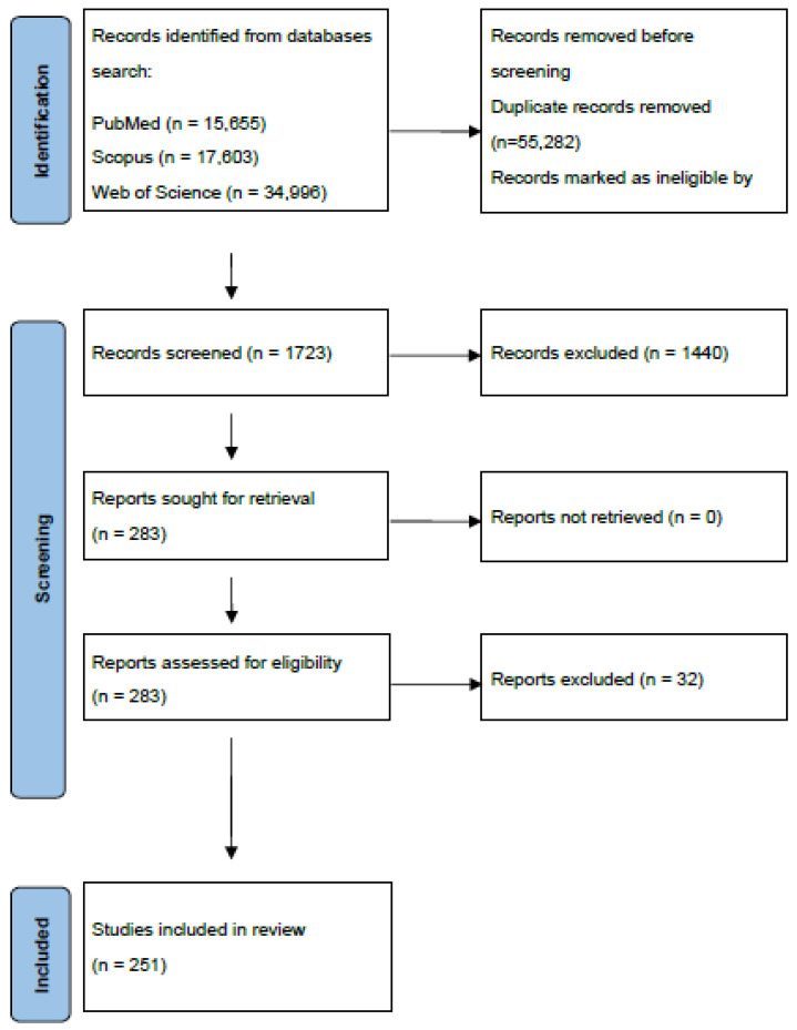
The search for articles, conducted on 15 April 2022, supporting the topic of our review, focused on the 3D-printing techniques employed in cranioplasty.
During the search, the following portals were used: PubMed, Scopus, Web of Science and Google Scholar. The searched articles were in English and not older than 7 years. The keywords used were “3D-printing cranioplasty”; “3D-printing endoprosthesis”; “3D-printing methods”; “3D-printing materials”; “3D-printing costs”; “3D-printing regulations”. These searched keywords resulted in 37,872 articles found. The screening inclusion and exclusion criteria provided at least 250 full-text articles suitable for this review. Inclusion criteria were at least one of: 3D printing in cranioplasty; 3D-printing costs in medicine; 3D printing of endoprostheses; comparison of 3D-printing methods; 3D-printing legal regulations.
Exclusion criteria: 3D printing of endoprostheses, which is much more expensive than the traditional methods; modeling of endoprostheses without the use of 3D printing; articles in a language other than English; duplicates; incomplete articles; bone endoprostheses other than those of the skull; 3D bioprinting with live cells; articles older than 7 years (Figure 1).
Figure 1.
Flowchart of the identification and selection of studies according to the 2020 PRISMA statement.
3. Methods of 3D Printing
Additive manufacturing employs the data computer-aided design (CAD) software to create 3D objects through the use of layer-by-layer various material depositions [10]. Furthermore, 3D printing encompasses several production technologies that involve solid (FDM—fused deposition modeling) [11], powder (SLS—selective laser sintering, SLM—selective laser melting) [12,13,14] or liquid-based materials (SLA—stereolithography, DLP—digital light processing) [15,16,17]. They differ in the manner that they arrange elements, as well as in their surface finish, operating time, material selection, durability, and value (Table 1).
Table 1.
Summary of properties of the 3D-printing technologies.
| Technology | Materials Used | Printing Technique | Application in Medicine |
|---|---|---|---|
| Selective Laser Melting (SLM) | metal alloys (e.g., titanium) | processing with a laser beam |
|
| Fused Filament Fabrication (FFF, FDM) | PLA, ABS, PET-G, PP, PMMA, PEEK, PVDF [19] | forming layers by depositing material through a heated, moving nozzle | |
| Stereolithography (SLA) | photosensitive polymers | focusing a UV laser onto a vat of polymer resin | |
| Digital Light Processing (DLP) | photosensitive polymers | like SLA, but instead of the laser beam, the entire layer is cured at the same time | |
| Selective Laser Sintering (SLS) | PA11 (nylon), PA12, PS, EOS TPE, PEEK | sintering with a laser beam |
|
3.1. Fused Deposition Modeling (FDM)
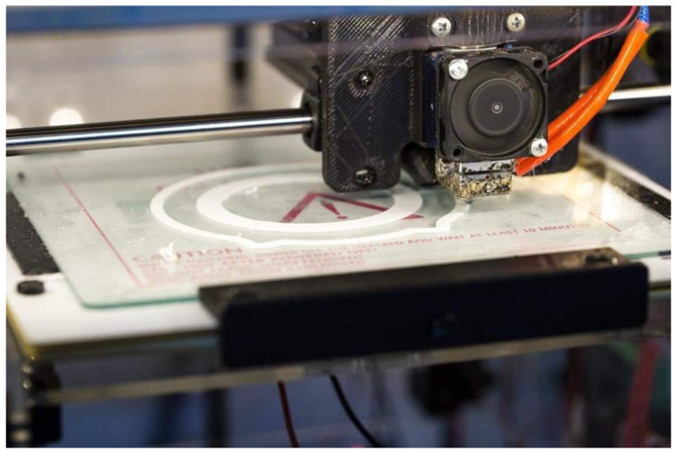
FDM was invented in 1988 by Scott Crump—a co-founder of Stratasys® [39]. This method is extrusion-based, where thermoplastic polymers in the form of filaments are wound on spools and are fed to the printer’s nozzle, where, after heating and melting, are subsequently selectively distributed layer-by-layer in accordance with the outline of the geometric model previously processed by the 3D-printing software (Chart 2) [40,41]. The characteristic of printing depends on the material’s dropping pressure, size of the nozzle, as well as the rate of feeding [42,43].
Chart 2.
FDM 3D Printer.
FDM has become increasingly prevalent of late, due to its immense accuracy, simplicity, relatively low price and eligibility for house printing [44,45]. It also utilizes strong engineering-grade materials that include PLA, ABS, PET-G and PP, among others [46]. Further materials worth mentioning in the implants market comprise PMMA, PEEK, or PVDF [19]. It is worth mentioning here, that the fused filament fabrication of metal is capable of fabricating patient-specific implants made out of titanium alloy [47].
FDM-based additional manufacturing is widespread in medicine and frequently utilized in the production of models that aid in clinical diagnoses and preoperative planning [20,21,22,48]. FDM enables the design and production of bone scaffolds and implants, and takes part in drug development research [24,25,26,27,28,49]. Novel literature also presents numerous examples where it was used to prepare cranioplasty implants [29,30,31,32]. For example, Thakur et al. used an FDM printer to prepare an ABS-made template for subsequent PMMA-implant fabrication, reporting high accuracy, precision, and excellent esthetics, which were obtained postoperatively [50]. However, in this indication, PCL and PLA belong to most manufactured polyesters used in FDM printers [51].
3.2. Selective Laser Melting (SLM)
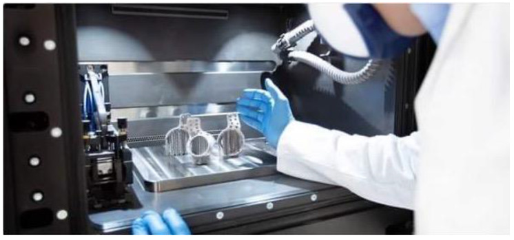
SLM is a powder-bed fusion technology that uses a powdered metallic alloy that is processed with a high-density-focused laser beam, layer-by-layer [52], building a compact three-dimensional structure characterized by considerable hardness and low porosity [53]. The novel literature presents numerous examples of research dedicated to the type of materials constructed by SLM (Chart 3). Among them, the most employed are titanium-, nickel-, and ferrous-based alloys [54]. The SLM-manufactured titanium 3D-prostheses perfectly emulate the biomechanical properties of natural human bone due to its precise internal and external architecture [55]. It is characterized by a high resistance to rusting [56] and its mechanical strength at room temperature [57]. This method is often used in modern medicine due to the high durability of Ti, radiolucency, and the minimal risk of post-treatment infections compared to other artificial materials [58]. Additionally, 3D-printed Ti prostheses are already utilized in the production of hip prostheses [18], but are also applicable in 3D-cranial reconstruction [46,59].
Chart 3.
SLM 3D Printer (Source: Sandvik).
The SLM-titanium-made prostheses represent an interesting direction for the cranial reconstruction market; however, their high manufacturing costs discourage its widespread use by patients and hospitals of a lower socioeconomic status [60].
3.3. Stereolithography (SLA)
SLA is a solid free-form (SFF) technique that uses a photosensitive polymer resin that reacts under the influence of a computer-controlled laser beam. The illumination-induced chemical reaction causes the formation of polymers by a cross-link fusion of chemical particles (monomers and oligomers) [49]. A previously designed three-dimensional project is converted into a stack of two-dimensional images. Then, a computer-derived pattern is illuminated on the surface of a liquid resin. Each 2D layer is then photochemically solidified, layer-by-layer with the use of a building stage and moving laser beam to form a desired three-dimensional solid representation of the pre-programmed CAD-CAM model. Subsequent solvent cleansing of wet resin from the model surface is then performed [61,62].
Stereolithography presents an advantage over other printing methods through its greater speed, controlled integrity, high-print resolution, and heat-free printing [63,64].
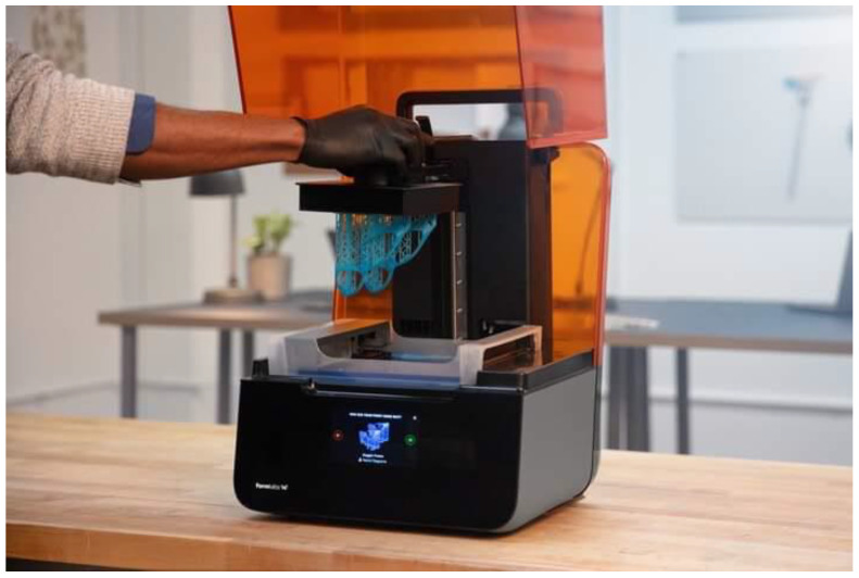
The method was patented by Charles W. Hull in 1986 [28], and in addition to industrial applications, stereolithography is now widely used in various fields of medicine (Chart 4). Polyethylene glycol (PEG), poly (D, L-lactide) (PDLLA), poly-ε-caprolactone (PCL), and poly(propylene fumarate) (PPF) comprise the most common photopolymerizable resins for tissue-engineered implants [65]. SLA finds its application in the medical field to create personalized endocardial implants [33], accurate models of reconstructing intestinal epithelial architecture [34], and innovative treatment of focal cartilage lesions [35,66]. SLA-3D printing has also been used to improve a cranioplasty procedure’s performance. Pöppe et al. described the technique of creating a patient-specific SLA-printed template for subsequent PMMA-implant modeling. In 14 surgeries, all cranioplasties perfectly matched the patient’s anatomy. Out of those, the three patients who bore clinically relevant coagulation-related risk factors required revision surgery due to a postoperative hematoma. Apart from that, no other complications have been recorded [67]. The analogous method was used by da Silva Junior, where a polycarbonate-made SLA-printed two-piece mold accompanied an intraoperative PMMA final implant formation, achieving satisfying results without any complications on a 3–26-month follow-up [68]. A similar method has also been reported by other authors [51].
Chart 4.
SLA 3D Printer (Source: Formlabs).
3.4. Digital Light Processing (DLP)
DLP is a technique analogous to SLA, with the difference being that instead of using a moving laser beam, the entire layer is cured simultaneously by a onetime projection of the whole image in an x/y space into photosensitive liquid resin, which guarantees excellent dimensional accuracy by avoiding the quality losses caused by a traveling laser [69,70]. The mechanical strength of the objects manufactured with the use of resin in this technology is sufficient to produce exoprostheses used in the rehabilitation of hands and wrists [36]. It can also be applied in the production of dentures [37].
3.5. Selective Laser Sintering (SLS)
SLS is a 3D-printing technology that involves a powder bed for an object’s construction analogously to SLM. However, in contrast to SLM, it utilizes a combination of high and low melting point powders, which results in a lower energy beam required for particle fusion [71]. The SLS printer involves an enclosed chamber containing three main components, i.e., the laser source, powder spreading platform and powder solidifying bed [72]. A moving roller transfers powder from a delivery platform to a solidifying platform, where it is selectively heated by a laser system that draws shapes layer-by-layer, causing fusion between the particles. The roller (or a blade) redistributes the powder after each layer. The process of the powder distribution and laser processing is repeated until the completion of the three-dimensional object [73,74,75]. Following the sintering process, an object is cleansed from unexploited remains by compressed air and may necessitate post-processing [76]. The SLS-processable materials in biomedical engineering include polymers, ceramics, biomaterials, and composites, e.g., ABS, PEEK, PMMA, hydroxyapatite, cellulose, glass-ceramics, calcium phosphate, and poly-L-lactic acid [77]. It can also use pure titanium or alloy powder, such as Ti-6Al-4V [78]. The quality of the SLS products contributes to their widespread commercial distribution [79], provided by its structural freedom of residual stress and internal defects. SLS is used in the manufacturing of medical devices and pharmaceutics, e.g., disc matrices for drug delivery devices [80,81]. SLS-fabricated surgical instrumentation is attracting growing attention in the orthopedics industry [82]. SLS can be used to produce various endoprostheses [83,84]. In the context of cranioplasty, Bertani et al. described the technique of printing cranial prosthesis’ molds made from polyamide (PA12) with a subsequent PMMA-made final implant preparation. According to this study, the total time required for implant preparation has not exceeded 42 h and has resulted in a 15-fold decrease in the total costs in comparison to the other methods [85]. Titanium implants, manufactured by selective laser sintering, were used in patients with Chiari malformation Type 1 after a suboccipital craniectomy [86].
4. Cranioplasty
Neurosurgical procedures occasionally lead to the formation of a bone defect within the cranial convexity. Craniectomies are predominantly performed to reduce intracranial pressure after trauma or as a part of radical tumor resection [87,88], and are customarily followed by cranioplasty [89,90]. A reconstruction of the cranial vault aims to re-establish the initial cerebrospinal fluid dynamics, restore esthesis as well as provide appropriate brain tissue protection, which is essential for further rehabilitation processes [91,92,93,94].
The restoration of the head’s natural symmetry improves a patient’s mental state and positively affects further psychosocial development [95]. Cranioplasty is also the method of choice for the syndrome of the trephined, characterized by various neurological and behavioral disorders [96], concerning up to 24% of those patients suffering from cranial defects [97].
The available novel literature does not deliver a universal consensus regarding optimal cranioplasty timing [98]. Normally, it varies on clinical circumstances and substantially depends on the initial surgery’s indication. Routinely, the procedure is scheduled at a 3-month interval, especially in patients with a post-decompressive neuroinfection or in the case of treatment-resistant intracranial hypertension. According to some sources, early cranioplasty improves the dynamics of cerebrospinal fluid circulation [99]. Other authors highlight no difference in infection rates but underline better outcomes in cognitive function as well as wound healing [100,101,102]. Nevertheless, recent studies indicate that there are no statistically significant differences in the frequency of hydrocephalus morbidity between an early and delayed cranioplasty following a decompression craniectomy [99].
The cranioplasty procedure has since evolved from when Dutch surgeon van Meekeren described the first-ever successful bone transplant that was performed by using canine bone as an implant in 1668 [103]. Nowadays, a vast range of methods and materials significantly enlarge surgeons’ artillery. To cover the cranial defects, there is a choice of allografts, autografts, xenografts, or other bone substitutes [104,105,106,107]. Cranioplasty with the use of bone, derived from decompressive surgery from the same patient via allograft, is often associated with bone resorption, auto-immunologic reactions and a high infection risk [108,109], and is often not feasible due to skull fractures.
There is a wide variety of substitute materials available on the market. One of them is methyl methacrylate, which is a polymerized ester of acrylic acid [110] (PMMA). It provides strength, comparable to natural bone tissue, and allows the prosthesis to be accurately adjusted to the shape of the defect. Its biggest and most significant disadvantage, from a clinical point of view, is its greater susceptibility to infections [103]. Another example of useful material is calcium phosphate bone cement [111,112]. Calcium phosphate bone cement is readily used for minor craniofacial defects and, in the case of cranioplasty, is often implemented in the treatment of skull defects in pediatric patients because, unlike methyl methacrylate, it does not prevent further expansion of the growing skull. In addition, some materials from this group, including those containing hydroxyapatite, possess osteoconductive properties [113,114]. Another material frequently used in cranioplasty is titanium, which can be used alone or together with methyl methacrylate. Titanium is noninflammatory, noncorrosive and nonferromagnetic and provides satisfactory cosmetic results [94,115].
Various techniques exist for bone flap fixation procedures. Ideally, it should ensure proper stabilization, a good cosmetic effect, minimize artifacts in neuroradiological imaging and be inexpensive [116]. The most described methods in the literature utilize sutures, wires, or mini plates, each of which has advantages and weaknesses. Among the above-mentioned, titanium mini plates seem to be the most beneficial due to their high biocompatibility and favorable osteointegrative properties [103,117,118] (Table 2).
Table 2.
Comparison of bone-substitute materials used for cranioplasty.
| Properties | PMMA | Hydroxyapatite | Titanium |
|---|---|---|---|
| Physical | |||
| Biocompatibility | Good [120] | High [103] | High [103] |
| Risk of fracture, fragmentation | High in larger defects [103] | High [103] | Low [113] |
| Osteointegration | Poor [106,113] | Excellent [103,113] | Poor [113] |
| Intraoperative modifying | Yes | Yes | No |
| Accommodation with skull growth (pediatric use) | No | Yes | No |
| Infection rate | 5.8–12.7% [103,113,122] | 0.6–2.1% [111,112,113] | 2.6–5.4% [103,112,113] |
| Mean implant cost | $80 (for intra-op. molded version) $650–$800 (for CAD/CAM version) [120,122] |
$7900 [120] | $25,100 |
A retrospective review based on 329 medical cases showed that the most common complications after a cranioplasty procedure are infections (39%), followed by incorrectly fitting implants (30%). Furthermore, there is a risk of cerebrospinal fluid leakage and cerebral edema (5%) [119]. However, adverse effects may occur regardless of the type of prosthesis material.
The risk of infection depends on the material used during the procedure, the location of a previous craniectomy and appropriate wound care following surgery. Overall, according to a systematic review designed by van de Vijfeijken, autografts carry higher complication and failure risks in comparison to allografts [106].
5. 3D Cranioplasty
3D printing, which is gaining increasing popularity in various fields of science, has also found an application in medicine. It is used by various specialists, including neurosurgeons. Thanks to this technology, it has become possible to print precisely fitted, low-cost bone prostheses for patients requiring postoperative cranioplasty [30,31,123]. It is also used in surgeries for pre-planning incision lines, which allows the operators to perform a precise opening of the skull, reducing the risk of complications and improving the cosmetic effect [22]. In justified cases, print castings can be used to reconstruct a cranial base, for instance, in Arnold–Chiari syndrome [29]. Modern techniques for the repair of sphenoid dysplasia are also based on the pre-operative 3D-printing models of the skull base [32,124]. The use of 3D printing also contributes to the preparation of autologous transplants for craniofacial reconstruction surgeries in patients after severe burns [125]. Prostheses made with the use of 3D technology have high aesthetic value, which has become more important for patients nowadays [126]. In combination with the CAD software, it opens the way for the reconstruction of the continuity of the bone involving even complicated anatomical structures, e.g., the orbit [127,128]. The method employed to design a 3D image and replicate it to a 3D virtual model is called reverse engineering [129]. In addition to its universal availability, 3D printing allows for the preparation of personalized, perfectly-sized bone prostheses, which significantly reduces the time of surgery compared to the procedures that utilize standard implants. As an example, an intraoperative placement of the tegmen plate in temporal bone defects can be shortened from 60 to less than 1 min [130].
The improvement in 3D-printing techniques, the introduction of newer advanced materials and the easy availability of affordable 3D printers have made the creation of 3D-bone prostheses easy and generally available for hospitals. The relatively uncomplicated production makes it possible to create personalized bone prostheses, even on the premises of a medical facility. Creating an appropriate design and printing a prosthesis saves time, both at the preoperative stage and during the procedure of applying it to the patient, thanks to the precise adjustment to the bone defect. The employment of 3D printing for cranial defect implant fabrication has some limitations. The most important factor is the choice of appropriate material that gives the best end result in terms of its expense, regulations, durability, function, and safety [131]. Selecting appropriate material reduces the time of patient care and enables patients’ faster recovery [132,133,134]. An ideal implant is inert, biocompatible, eligible for sterilization, strong and possesses long-term stability, durability, and intraoperative workability [135].
In this article, we aimed to address these concerns and analyzed the available materials that are commonly selected for the prosthetic 3D-printing market: PLA, PET-G, ABS, PEEK, and PMMA.
5.1. PLA
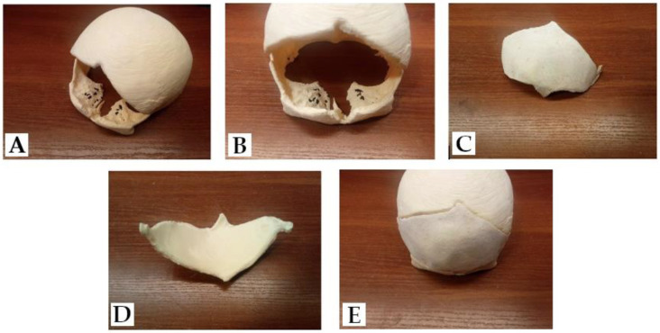
Polylactic acid—PLA—is a versatile biopolymer classified as an aliphatic polyester. PLA is an FDA-approved, non-toxic, biodegradable material that is widely used in a plethora of applications, including in the biomedical and automotive engineering industries [136,137,138,139]. In healthcare, it finds employment, for example, in regenerative medicine, cancer therapy, tissue engineering, or cardiovascular implants, naming a few [140]. Currently, PLA is gaining rising attention because of its excellent performance as a three-dimensional printing material, especially with the employment of FMD technology [141], which is attributable to its mechanical properties. Polylactic acid is characterized by a low melting point (about 220 °C) as well as a low level of distortions during the printing process [142]. In tests, PLA shows two times greater resistance to bending and pressure than ABS [143]. Nonetheless, its poor toughness with a 10% elongation at break prevents its use when high-stress resilience is to be expected [144]. In the medical field, PLA shows potential in radiotherapy, as it appears to be undamaged by radiation exposure [145]. PLA blended with hydroxyapatite may potentially be used to repair bone defects [23,146,147]; however, there is little research and information on the ability of HA/PLA to stimulate bone formation in vivo [148]. PLA might also be used for anatomical model preparation and preoperative planning [149,150]. In addition, polylactic acid, along with PLGA—poly(lactic-co-glycolic acid)—represents the polymers most abundantly fabricated to create biodegradable scaffolds for the regeneration of cranial bone [51,151]. The novel literature delivers several examples of PLA employment in cranioplasty surgeries. De la Pena et al. used a 3D-printed-PLA calvarial defect model to create a plaster mold over it. The mold was then used as a form for the ultimate PMMA implant [121]. Other authors decided to print a 3D-PLA mold directly, and again, PMMA was used as a cranial prosthesis [152,153]. Similarly, Lannon et al. used desktop 3D printers to produce a PLA-made patient-specific mold for a PMMA-made final cranial implant and reported a significant reduction in the total procedure’s expenditure [154]. None of the mentioned authors experienced any significant complications that would stand out in comparison to the standard cranioplasty methods. The overall complication rate is difficult to assess due to the small number of patients involved.
In Macedonia, Apriawan et al. implemented PLA as a material for the cranioplasty of two patients following decompressive craniectomies. In both cases, no postoperative complications have been recorded [155]. A simulation of cranial reconstruction with a use of PLA is presented in Chart 5 below.
Chart 5.
(A,B) CT-derived FDM-printed PLA model of a complicated skull defect post-craniectomy. (C,D) Implant ready for cranioplasty; (E) model simulating cranioplasty’s procedure outcome.
5.2. PET-G
PET-G, an amorphous thermoplastic polyester resin belonging to the PET family and one that is subjected to secondary glycol modifications, is another option. It is characterized by its good impact strength, transparency, and chemical resistance [23]. It is also approved by the FDA as a water and food container [156]. The glass transition temperature of PET-G is around 80 °C, making it suitable for thermal forming [147], but unfortunately, it is not autoclave-resistant. It is considered an improvement over ABS due to more flexibility and biocompatibility [157]. In medicine, PET-G is used in ligament [158], joint [159] or vascular [160] prostheses. Additionally, PET-G prints can be potentially used to produce artificial breasts for mammography imaging techniques [161]. Katsching et al. successfully printed well-fitting, mechanically stable, and cleanly printed implants for maxillofacial defects ex vivo [162]. Although it has been used as a prosthetics material in various branches of medicine or for bone tissue engineering [163], we found no studies where PET-G was used as a direct material for cranioplasty in vivo. Nevertheless, there are multiple studies where PET-G was utilized to create a mold for the PMMA cranial implants. Kim et al. [164], Parthasarathyet [165] as well as Surme [166] used various techniques of mold preparation, whereas they all reported the avoidance of local and systemic PMMA-associated toxic adverse reactions as well as brain or dural tissue destruction. In each study, the operation time was significantly decreased with the preservation of high aesthetic value.
5.3. ABS
Products made from ABS, i.e., acrylonitrile butadiene styrene terpolymer, also show satisfactory durability. They are stiff but still more malleable than PLA [167]. In comparison to PLA, ABS is more difficult to print due to its greater expansion at higher temperatures [168]. In addition, it produces toxic fumes during the printing process [169]. The printing temperature for ABS and PET-G is similar, around 240 °C [170]. ABS implants are inert, biocompatible and permit tissue integration [171]. Ziąbka et al. explored ABS’s properties for the purpose of middle ear implant surgeries, where ABS proved to have great biocompatibility as well as low cytotoxicity [172]. Sarah E. Mowry used ABS to replicate human temporal bone for surgery simulation purposes. According to the evaluating specialists, the ABS-3D model proved to be accurate and may serve as a valuable alternative for cadaveric training [173]. In 2020, the FDA approved ABS as a low-risk polymer when in contact with intact skin. Kinsman et al. utilized ABS to prepare a patient-specific 3D model, which was used to customize the titanium mesh preoperatively, excluding the need for intraoperative molding [174]. The method proved to be resource and time-saving in comparison to the use of prefabricated personalized PEEK implants. ABS plastic, due to its mechanical properties as well as the printing method, is not an exquisite candidate to be used as an implant [175], although it might be successfully used as a material for PMMA molding. Yerragunta et al. prepared a 3D-printed ABS template for molding PMMA in 10 patients with post-craniotomy defects, reaching excellent calvarial contour restorations as well as patient satisfaction [176]. No long-term complications or infections have been noted on a follow-up. The same method was chosen by Unterhofer et al. and was conducted on 46 patients with 41 months of mean follow-up. Among these, 7 required reoperations, but overall, 42 achieved excellent cosmetic results [120]. These lead to a conclusion that the fabricated in-house PMMA implants, by direct molding with the use of the ABS model, are easy to produce, provide satisfying aesthetic results and are inexpensive in comparison to the industrially manufactured personalized cranial implants.
5.4. PEEK
PEEK (polyether ether ketone) is a poly-aromatic, partly crystalline thermoplastic polymer increasingly used in orthopedics and dentistry [177,178,179,180]. The melting temperature of PEEK is around 343 °C [181]. The tensile strength is comparable to enamel, and its wear resistance is similar to metal alloys [182,183]. PEEK is inert and noncytotoxic; however, it is hydrophobic, making it susceptible to the adhesion of bacterial cells [184]. Platelet adhesion to PEEK is much lower than to polished medical steel and is similar to titanium alloys [185]. Because of the lack of osteogenic properties, it is more prone to infection and displacement [103]. Unfortunately, it is also considered the most expensive among other types, which limits its commercial dissemination [60,106]. It has great strength and a low Young’s modulus, comparable to human bone [104,186], which allows for an equal dispersion of induced stress and limits osteolysis [187]. It is also radiolucent and CT and MRI compatible, and due to its properties, it can be sterilized and re-sterilized in an autoclave without deformations [188]. The use of PEEK in combination with CAD technology allows us to obtain very satisfactory aesthetic effects in the production of bone implants, e.g., those used in maxillofacial surgery [189]. Nevertheless, the novel literature presents scarce examples of PEEK applications as a material for cranioplasty, regardless of its undeniable potential. According to Thien et al., cranioplasties with PEEK exhibit a lower incidence of revision surgeries (12.5%) than those with the use of titanium implants (25%) [190]. Meta-analyses performed by Punchak et al. and van de Vijfeijken proved that surgeries performed with the use of PEEK carry lower risks of infection than PMMA and autologous bone cranioplasties [106,191]. In a study performed by Mian et al., 3D-printed PEEK implants delivered great precision, minimal deviation (0.5919 mm), with maximal von Mises stress (8.15 MPa), von Mises strain (0.002) and minimal deformation (0.18 mm), which underlines the implant’s extraordinary load-bearing capability [192]. Similar observations have been made in a preclinical study designed by Sharma et al. [193]. De Barros described a one-step customized PEEK cranioplasty, reaching a favorable outcome without any major complications during a 3-month follow-up [194]. Mohsen et al. evaluated 20 patients who underwent a cranioplasty with prefabricated-3D-printed PEEK implants in the years 2017–2018. In this study, excellent cosmetic results were achieved by all participants. Apart from one superficial wound infection and one clinically irrelevant extradural hematoma, no major complications have been encountered [195].
The data from the current literature are encouraging, but the routine use of 3D-printed PEEK cranial implants requires further studies and is limited due to a lack of testing and validation. This might be accelerated by pending studies, which aim to establish the safety and efficacy of the house-manufactured PEEK implants used in cranioplasty in comparison to conventional methods [196].
5.5. PMMA
PMMA—(polymethylmethacrylate)—is a popular resin that is strong, light, stable and nonferromagnetic [197,198]. Among the alloplastic materials, PMMA is the most widely used by prosthesis manufacturers because it is less expensive than PEEK and titanium, which brings additional importance to its value in countries where funds reserved for the implant must be covered by the patient’s own resources [121]. Adding PMMA powder to other additive manufacturing processes can increase the porosity of parts and thus increase bone ingrowth [199]. PMMA, due to its high biocompatibility, is a useful material in dentistry and orthopedics [200,201,202,203]. Prostheses made with this technique adequately protect the tissues and present a mechanical strength analogous to natural human bone [122]. In addition, PMMA-based bone cement can be used to stiffen endoprostheses with the patient’s bone [204,205].
The PMMA castings built on printed forms can be further refined, e.g., by drilling holes, thus enabling a better flow, adhesion to soft tissues and providing greater stability [31].
Apart from their favorable mechanical properties, the advantages of PMMA implants also include their permeability for imaging studies [206]. Unfortunately, they can sometimes cause an immunologic reaction in the surrounding tissues, leading to subcutaneous seroma [207]. Moreover, the placement of polymers and their slow release of toxic monomers seldom initiates hypersensitivity [123,208].
The review of 41 articles regarding cranioplasty materials presents that there is no significant difference in the average infection rate for autologous bone (10.5; relative risk = 1.00) and hand-formed PMMA implant (10.98; relative risk = 1.05). In comparison, a prefabricated PMMA’s relative infection risk reaches about 0.66 [209,210,211]. Apart from the possible complications after implanting the 3D-printed bone prosthesis, it is difficult to predict its mechanical properties. The resulting strength is highly dependent on the method of printing and on a variety of parameters, which can be selected in accordance with a contractor’s experience. Material shrinkage greatly affects the accuracy of a material, as well as the errors induced by the CAD/CAM software and post-processing. The size of a printer restricts the size of the implant; however, in the case of cranial bones is not a remarkable limitation [4].
The available literature delivers numerous examples of PMMA applications in 3D cranioplasty. The safety and efficacy of long-term use of PMMA prostheses covering large cranial defects were assessed and approved after a long-term follow-up of randomized, blinded, multi-centered trials conducted by Jaberi et al. [212,213,214]. Morales-Gomez examined 22 patients who underwent cranioplasty that utilized PMMA with the use of a desktop 3D printer. Apart from a great cosmetic result achieved, no infection, implant rejection or wound dehiscence were reported in the 6-month follow-up [215] Yerragunta et al. used the same method in India and achieved comparable results, determining this technique as the most economically effective among all [176].
Dabadi et al., in a study conducted in Nepal, implemented 3D-printed PMMA cranial implants made on a silicon mold in three cases in patients after craniectomies in search of improved efficacy, expense- and time-wise. This has been achieved with no major complications reported with the acquisition of a great aesthetic and economic outcome [216].
6. Sterilization
FDM-printed parts can be sterilized in an autoclave, although it links to a 0.1–3% dimension shrinkage, which must be considered during manufacturing [217,218]. A similar phenomenon occurs in SLA-printed parts. The autoclaved prints present unwanted bends, causing displacement of the SLA-printed surgical guides, and contain numerous cracks [219].
Hydrostatic high-pressure post-processing may be a promising alternative to traditional sterilization techniques. Overall, it preserves better tensile properties than autoclave. Unfortunately, the DLP-processed samples are still damaged in the sterilization process, and thus, the mechanical properties are drastically decreased. Microbiological screenings in HHP adhere to the Good Laboratory Practices; however, further evaluation should be executed [220]. Titanium alloy implants fabricated with the SLM technique can be fully sterilized, both by dry heat and steam. However, there is no available ISO protocol for porous implant sterilization [221].
PLA, in consequence of a relatively low melting point (180–220 °C) as well as glass transition temperature (57 °C), is not applicable for autoclave sterilization. Nevertheless, PLA implants can undergo a sterilization process with the use of a low-temperature hydrogen peroxide gas plasma system (HPGP) and new scCO2 (supercritical carbon dioxide) technique [222]. Aguado-Maestro suggests EtOH sterilization for in-hospital 3D-printed PLA materials, whereas, for gas plasma, an infill application of 100% should be applied [221]. While PET-G is not eligible for autoclave sterilization, ultraviolet radiation sterilization, and ethylene oxide sterilization provide adequate alternatives [162]. Dautzenberg reported the instability of ABS following steam sterilization [217]. According to Toro et al., vaporized hydrogen peroxide sterilization provides dimensional stability for sterilized ABS-3D printed elements [223]. The same was reported by other authors [224,225]. PEEK implants are known to retain their mechanical properties after multiple steam sterilization cycles [226,227]. Münker et al. inspected the micromechanical properties of PMMA after various sterilization systems and reported significant changes in the material’s mechanical behavior after autoclave sterilization. A favorable outcome was achieved with the use of ethylene oxide, hydrogen peroxide gas plasma and γ-irradiation sterilization [228].
7. Costs and Legal Regulations
The 3D-printing market share grew to USD 5.165 billion in 2015 and is predicted to reach USD 30 billion in 2022 [229].
Pricing of a 3D printer ranges from a few hundred for low-end devices to hundreds of thousands (USD) for industrial printers. An affordable, ready-to-use Ultimaker, which costs around USD 2750–3000, is fully capable of printing a human skull model. The 3D-printing market offers an abundance of materials, with the simplest Poly(lactic acid) filament for as low as USD 20 per 1 kg, with an average cost for the 3D-printed craniofacial models ranging from USD 20 to 100. In a study conducted by Apriawan et al., the PLA cost for a 3D-cranioplasty model reached around USD 50–150 [155]. A PLA-made-cranial model for surgical consulting purposes costs around USD 5.20 [230]. The cost of a 3D printer and ABS (acrylonitrile butadiene styrene) oscillates around USD 1350 and USD 25/kg, with one kg of ABS being able to generate around 7–10 3D models [174]. A higher price is usually associated with the more specialized printing processes. In a study by Unterhofer et al., conducted on 46 patients, the expenditure of PMMA-implant production oscillated around EUR 81 [120]. For comparison, the manufactured personalized PEEK implants cost around USD 10,450, titanium implants cost USD 10,265 and HA is USD 7849–8960 [60,155] (Table 3).
Table 3.
Characteristics of 3D-printed materials.
| Injection-Molded PMMA | Injection-Molded PEEK | 3D-Printed PET-G | 3D-Printed ABS | 3D-Printed PLA | |
|---|---|---|---|---|---|
| Used for 3D cranioplasty as | Implant | Implant | Mold | Mold | Mold |
| Tensile strength [MPa] | 48–76 [231] | 80 [231] | 50 [232] | 37 [233] | 47–52 [234] |
| Elastic modulus [GPa] | 3–5 [231] | 3–4 [231] | 1.1–1.3 [232] | 1.8 [143] | 3.4–5.7 [234] |
| Biocompatibility | Good [177,235] | High [231] | Yes, in specialized filaments [163,236] | Yes, in specialized filaments [172,236] | High (in pure materials) [236] |
| Osteointegration | Poor [115,123] | Poor [231] | Under research [163] | Under research [172] | With coating [237] |
| Complication rate [%] | 7.4 [231] | 9.1 [231] | N/A | N/A | N/A |
| Cost of material [USD per kg] | 30 | 275–400 [238] | 40 | 25 [175] | 20 |
| Total cost of 3D-printed implant manufacturing [USD] | 600 [152,239] | 10,450 [156] | N/A | N/A | N/A |
Abbreviations: PMMA = polymethylmethacrylate; PEEK = polyether ether ketone; PET-G = gylocol-modified polyethylene terephthalate; ABS = acrylonitrile butadiene styrene; PLA = polylactic acid; N/A = non-applicable.
Ballard et al. calculated the potential economic profits related to the additive manufacturing of cranial prostheses. Assuming the average operating theater cost exceeds 62 USD/min, the possible net savings were estimated at USD 19,284–USD 129,586 for a low (5%) and USD 77,536–USD 518,358 for a high (20%) utilization rate [240].
In some countries, i.e., Mexico, cranioplasty procedures are not refunded by the national health service, and the cost of the operation, as well as the implant, needs to be covered by the individual. With the cost of widely used personalized titanium implants that reach around USD 5000 and those made of PEEK of around USD 7000, it is undemanding to assume that these prices reach financial levels that are not achievable for medium- to low-income patients. For comparison, a personalized prosthesis, created with 3D-printing technology and comprised of PMMA, wavers around USD 600, including the 3D design, prototype, and the prosthesis itself, which diminishes the overall expense by over tenfold [121,241]. Around 85 3D-printed devices were approved by the FDA for medical purposes through 510(K) processes [229]. The 510(K) is a premarket notification—another pathway to apply for an NDA (new drug application). Other pathways include premarket approval (PMA) and a biologics license application (BLA) [239].
8. Conclusions
Cranioplasty is a common procedure performed by neurosurgeons all over the world. In the cases of a complicated, uneven, and large deficit, where a random-sized prosthesis is not sufficient, personalization of the material is inevitable. Unfortunately, this comes with a vast increase in price, even 5- to 10-fold. Therefore, it is crucial to obtain a method that allows us to manufacture complicated, personalized cranial prostheses that are designed for departments located in developing countries and patients with a low socioeconomic status. This is achievable due to the development and spread of 3D-printing technology.
There are numerous factors that influence the cost of the prosthesis, i.e., size, type of material and manufacturing technology. A low-cost prosthesis can be successfully made using home and even DIY printers as well as typically industrial and professional devices, such as Stratasys®. The market offers a wide range of CAD software which enables the personalization of an implant (Chart 6). It is even possible to design and prepare the model by using free software solutions. Apart from its employment in cranioplasty, 3D-assisted preoperative planning has become particularly popular, allowing surgeons to better understand the site of the planned reconstruction. Three-dimensional printing often allows us to obtain satisfactory cosmetic effects, restoring the correct skull contour and thus increasing the patient’s quality of life. In fact, there is no shortage of companies dealing with the more expensive technologies, such as the production of titanium prostheses; however, there is a niche for start-ups with low-share capital who are aiming to provide services for patients living in poorer regions. A plethora of various technologies of additive manufacturing exist, so it is currently difficult to decide which one of them will prove to be the best in the future, as in many cases, the research is still pending. As 3D-printed PMMA could be considered too expensive, more attention should be pointed to using prints from the PLA in the PMMA prosthesis creation process, especially due to the material’s satisfactory strength and low manufacturing price. This method has been attempted in several cited publications, achieving satisfactory results in terms of cost, esthetic value, as well as outcome.
Chart 6.
Automatically generated part is user-checked on the fly, adjusted using tools provided with Autodesk® Meshmixer and exported to printable file.
In conclusion, 3D-printed cranioplasty might be an interesting, efficient, and inexpensive alternative to the methods that are currently routinely used.
Abbreviations
| ABS | Acrylonitrile butadiene styrene | CAD/CAM/CAE | Computer-aided design/Computer-aided Manufacturing/Computer-aided Engineering |
| DICOM | Digital Imaging and Communications in Medicine | DLP | Digital Light Processing |
| FDM® | Fused deposition modelling | FFF | Fused filament fabrication |
| HA | Hydroxyapatite | PEEK | Poly(ether ether ketone) |
| PET-G | Poly(ethylene terephthalate glycol) | PLA | Poly(lactic acid) |
| PMMA | Poly(methylmethacrylate) | PP | Polypropylene |
| PVDF | Poly(vinylidene fluoride) | SLA | Stereolithography |
| SLM | Selective laser melting | SLS | Selective laser sintering |
| STL | stereolithography CAD software file format | ||
Institutional Review Board Statement
Not applicable.
Informed Consent Statement
Not applicable.
Conflicts of Interest
The authors declare no conflict of interest.
Funding Statement
The article was supported by a research grant by Medical Univeristy of Lublin to KT-DS495.
Footnotes
Publisher’s Note: MDPI stays neutral with regard to jurisdictional claims in published maps and institutional affiliations.
References
- 1.Worm P.V., Finger G., Ludwig do Nascimento T., Rynkowski C.B., Collares M.V.M. The impact of cranioplasty on the patients’ quality of life. J. Cranio-Maxillofac. Surg. 2019;47:715–719. doi: 10.1016/j.jcms.2019.01.040. [DOI] [PubMed] [Google Scholar]
- 2.Codubix Proteza Kości Czaszki Niejałowa. [(accessed on 19 June 2022)]. Available online: https://www.matopat24.pl/codubix-proteza-kosci-czaszki-niejalowa_7341-14736.
- 3.Warsi M.H., Yusuf M., Al Robaian M., Khan M., Muheem A., Khan S. 3D Printing Methods for Pharmaceutical Manufacturing: Opportunity and Challenges. Curr. Pharm. Des. 2019;24:4949–4956. doi: 10.2174/1381612825666181206121701. [DOI] [PubMed] [Google Scholar]
- 4.Kate J.T., Smit G., Breedveld P. 3D-printed upper limb prostheses: A review. Disabil. Rehabil. Assist. Technol. 2017;12:300–314. doi: 10.1080/17483107.2016.1253117. [DOI] [PubMed] [Google Scholar]
- 5.Karpiński R., Jaworski Ł., Zubrzycki J. Structural analysis of articular cartilage of the hip joint using finite element method. Adv. Sci. Technol. Res. J. 2016;10:240–246. doi: 10.12913/22998624/64064. [DOI] [Google Scholar]
- 6.Karpiński R., Jaworski Ł., Zubrzycki J. The design and structural analysis of the endoprosthesis of the shoulder joint. ITM Web Conf. 2017;15:7015. doi: 10.1051/itmconf/20171507015. [DOI] [Google Scholar]
- 7.Karpiński R., Jaworski Ł., Szala M., Mańko M. Influence of patient position and implant material on the stress distribution in an artificial intervertebral disc of the lumbar vertebrae. ITM Web Conf. 2017;15:07006. doi: 10.1051/itmconf/20171507006. [DOI] [Google Scholar]
- 8.Karpiński R., Jaworski Ł., Jonak J., Krakowski P. The influence of the nucleus pulposus on the stress distribution in the natural and prosthetic intervertebral disc. MATEC Web Conf. 2019;252:07006. doi: 10.1051/matecconf/201925207006. [DOI] [Google Scholar]
- 9.Zubrzycki J., Каприньски R., Jaworski L., Ausiyevich A.M., Smidova N. Structural analysis of the pelvic girdle before and after hip replacement procedure. Sci. Tech. 2018;17:165–172. doi: 10.21122/2227-1031-2018-17-2-165-172. [DOI] [Google Scholar]
- 10.Kafle A., Luis E., Silwal R., Pan H.M., Shrestha P.L., Bastola A.K. 3D/4D Printing of Polymers: Fused Deposition Modelling (FDM), Selective Laser Sintering (SLS), and Stereolithography (SLA) Polymers. 2021;13:3101. doi: 10.3390/polym13183101. [DOI] [PMC free article] [PubMed] [Google Scholar]
- 11.Khalid G.M., Billa N. Solid Dispersion Formulations by FDM 3D Printing—A Review. Pharmaceutics. 2022;14:690. doi: 10.3390/pharmaceutics14040690. [DOI] [PMC free article] [PubMed] [Google Scholar]
- 12.Hou Y., Su H., Zhang H., Wang X., Wang C. Fabricating Homogeneous FeCoCrNi High-Entropy Alloys via SLM In Situ Alloying. Metals. 2021;11:942. doi: 10.3390/met11060942. [DOI] [Google Scholar]
- 13.Ponnusamy P., Rahman Rashid R.A., Masood S.H., Ruan D., Palanisamy S. Mechanical Properties of SLM-Printed Aluminium Alloys: A Review. Materials. 2020;13:4301. doi: 10.3390/ma13194301. [DOI] [PMC free article] [PubMed] [Google Scholar]
- 14.Idriss A.I.B., Li J., Wang Y., Guo Y., Elfaki E.A., Adam S.A. Selective Laser Sintering (SLS) and Post-Processing of Prosopis Chilensis/Polyethersulfone Composite (PCPC) Materials. 2020;13:3034. doi: 10.3390/ma13133034. [DOI] [PMC free article] [PubMed] [Google Scholar]
- 15.Miedzińska D., Gieleta R., Popławski A. Experimental Study on Influence of Curing Time on Strength Behavior of SLA-Printed Samples Loaded with Different Strain Rates. Materials. 2020;13:5825. doi: 10.3390/ma13245825. [DOI] [PMC free article] [PubMed] [Google Scholar]
- 16.Martín-Montal J., Pernas-Sánchez J., Varas D. Experimental Characterization Framework for SLA Additive Manufacturing Materials. Polymers. 2021;13:1147. doi: 10.3390/polym13071147. [DOI] [PMC free article] [PubMed] [Google Scholar]
- 17.Komissarenko D.A., Sokolov P.S., Evstigneeva A.D., Shmeleva I.A., Dosovitsky A.E. Rheological and Curing Behavior of Acrylate-Based Suspensions for the DLP 3D Printing of Complex Zirconia Parts. Materials. 2018;11:2350. doi: 10.3390/ma11122350. [DOI] [PMC free article] [PubMed] [Google Scholar]
- 18.Liang H., Ji T., Zhang Y., Wang Y., Guo W. Reconstruction with 3D-printed pelvic endoprostheses after resection of a pelvic tumour. Bone Jt. J. 2017;99-B:267–275. doi: 10.1302/0301-620X.99B2.BJJ-2016-0654.R1. [DOI] [PubMed] [Google Scholar]
- 19.Petersmann S., Spoerk M., Van De Steene W., Üçal M., Wiener J., Pinter G., Arbeiter F. Mechanical properties of polymeric implant materials produced by extrusion-based additive manufacturing. J. Mech. Behav. Biomed. Mater. 2019;104:103611. doi: 10.1016/j.jmbbm.2019.103611. [DOI] [PubMed] [Google Scholar]
- 20.Brouwers L., Teutelink A., van Tilborg F.A.J.B., de Jongh M.A.C., Lansink K.W.W., Bemelman M. Validation study of 3D-printed anatomical models using 2 PLA printers for preoperative planning in trauma surgery, a human cadaver study. Eur. J. Trauma Emerg. Surg. 2018;45:1013–1020. doi: 10.1007/s00068-018-0970-3. [DOI] [PMC free article] [PubMed] [Google Scholar]
- 21.Guo X.-Y., He Z.-Q., Duan H., Lin F.-H., Zhang G.-H., Zhang X.-H., Chen Z.-H., Sai K., Jiang X.-B., Wang Z.-N., et al. The utility of 3-dimensional-printed models for skull base meningioma surgery. Ann. Transl. Med. 2020;8:370. doi: 10.21037/atm.2020.02.28. [DOI] [PMC free article] [PubMed] [Google Scholar]
- 22.Matsui C., Tokuyama E., Senoo T., Yamada K., Kameda M., Takeuchi T., Kimata Y. Utilization of a Simple Surgical Guide for Multidirectional Cranial Distraction Osteogenesis in Craniosynostosis. Plast. Reconstr. Surg.-Glob. Open. 2020;8:e2797. doi: 10.1097/GOX.0000000000002797. [DOI] [PMC free article] [PubMed] [Google Scholar]
- 23.Kim H., Ryu K.-H., Baek D., Khan T.A., Kim H.-J., Shin S., Hyun J., Ahn J.S., Ahn S.-J., Koo J. 3D Printing of Polyethylene Terephthalate Glycol–Sepiolite Composites with Nanoscale Orientation. ACS Appl. Mater. Interfaces. 2020;12:23453–23463. doi: 10.1021/acsami.0c03830. [DOI] [PubMed] [Google Scholar]
- 24.Rindone A.N., Nyberg E., Grayson W.L. 3D-Printing Composite Polycaprolactone-Decellularized Bone Matrix Scaffolds for Bone Tissue Engineering Applications. Methods Mol. Biol. 2017;1577:209–226. doi: 10.1007/7651_2017_37. [DOI] [PubMed] [Google Scholar]
- 25.Shie M.-Y., Chang W.-C., Wei L.-J., Huang Y.-H., Chen C.-H., Shih C.-T., Chen Y.-W., Shen Y.-F. 3D Printing of Cytocompatible Water-Based Light-Cured Polyurethane with Hyaluronic Acid for Cartilage Tissue Engineering Applications. Materials. 2017;10:136. doi: 10.3390/ma10020136. [DOI] [PMC free article] [PubMed] [Google Scholar]
- 26.Hung K.-C., Tseng C.-S., Hsu S.-H. Synthesis and 3D Printing of Biodegradable Polyurethane Elastomer by a Water-Based Process for Cartilage Tissue Engineering Applications. Adv. Health Mater. 2014;3:1578–1587. doi: 10.1002/adhm.201400018. [DOI] [PubMed] [Google Scholar]
- 27.Alhijjaj M., Belton P., Qi S. An investigation into the use of polymer blends to improve the printability of and regulate drug release from pharmaceutical solid dispersions prepared via fused deposition modeling (FDM) 3D printing. Eur. J. Pharm. Biopharm. 2016;108:111–125. doi: 10.1016/j.ejpb.2016.08.016. [DOI] [PubMed] [Google Scholar]
- 28.Costa P.F., Puga A.M., Díaz-Gomez L., Concheiro A., Busch D.H., Alvarez-Lorenzo C. Additive manufacturing of scaffolds with dexamethasone controlled release for enhanced bone regeneration. Int. J. Pharm. 2015;496:541–550. doi: 10.1016/j.ijpharm.2015.10.055. [DOI] [PubMed] [Google Scholar]
- 29.Pijpker P.A., Wagemakers M., Kraeima J., Vergeer R.A., Kuijlen J.M., Groen R.J. Three-Dimensional Printed Polymethylmethacrylate Casting Molds for Posterior Fossa Reconstruction in the Surgical Treatment of Chiari I Malformation: Technical Note and Illustrative Cases. World Neurosurg. 2019;129:148–156. doi: 10.1016/j.wneu.2019.05.191. [DOI] [PubMed] [Google Scholar]
- 30.Low P.H., Abdullah J.Y., Abdullah A.M., Yahya S., Idris Z., Mohamad D. Patient-Specific Reconstruction Utilizing Computer Assisted Three-Dimensional Modelling for Partial Bone Flap Defect in Hybrid Cranioplasty. J. Craniofac. Surg. 2019;30:e720–e723. doi: 10.1097/SCS.0000000000005713. [DOI] [PubMed] [Google Scholar]
- 31.Lal B., Ghosh M., Agarwal B., Gupta D., Roychoudhury A. A novel economically viable solution for 3D printing-assisted cranioplast fabrication. Br. J. Neurosurg. 2020;34:280–283. doi: 10.1080/02688697.2020.1726289. [DOI] [PubMed] [Google Scholar]
- 32.Abbate V., Iaconetta G., Califano L., Pansini A., Bonavolonta P., Romano A., Salzano G., Somma T., D’Andrea L., Orabona G.D. Self-Made Rapid Prototyping Technique for Orbital Floor Reconstruction. J. Craniofac. Surg. 2019;30:2106–2110. doi: 10.1097/SCS.0000000000006004. [DOI] [PubMed] [Google Scholar]
- 33.Robinson S.S., Aubin C.A., Wallin T.J., Gharaie S., Xu P.A., Wang K., Dunham S., Mosadegh B., Shepherd R.F. Stereolithography for Personalized Left Atrial Appendage Occluders. Adv. Mater. Technol. 2018;3:1800233. doi: 10.1002/admt.201800233. [DOI] [PMC free article] [PubMed] [Google Scholar]
- 34.Creff J., Courson R., Mangeat T., Foncy J., Souleille S., Thibault C., Besson A., Malaquin L. Fabrication of 3D scaffolds reproducing intestinal epithelium topography by high-resolution 3D stereolithography. Biomaterials. 2019;221:119404. doi: 10.1016/j.biomaterials.2019.119404. [DOI] [PubMed] [Google Scholar]
- 35.Aisenbrey E.A., Tomaschke A., Kleinjan E., Muralidharan A., Pascual-Garrido C., Mcleod R., Ferguson V.L., Bryant S.J. A Stereolithography-Based 3D Printed Hybrid Scaffold for In Situ Cartilage Defect Repair. Macromol. Biosci. 2017;18:1700267. doi: 10.1002/mabi.201700267. [DOI] [PMC free article] [PubMed] [Google Scholar]
- 36.Keller M., Guebeli A., Thieringer F., Honigmann P. In-hospital professional production of patient-specific 3D-printed devices for hand and wrist rehabilitation. Hand Surg. Rehabil. 2020;40:126–133. doi: 10.1016/j.hansur.2020.10.016. [DOI] [PubMed] [Google Scholar]
- 37.You S.-M., Lee B.-I., Kim J.-H. Evaluation of trueness in a denture base fabricated by using CAD-CAM systems and adaptation to the socketed surface of denture base: An in vitro study. J. Prosthet. Dent. 2020;127:108–114. doi: 10.1016/j.prosdent.2020.09.027. [DOI] [PubMed] [Google Scholar]
- 38.Yu J.M., Kang S.Y., Lee J.S., Jeong H.S., Lee S.Y. Mechanical Properties of Dental Alloys According to Manufacturing Process. Materials. 2021;14:3367. doi: 10.3390/ma14123367. [DOI] [PMC free article] [PubMed] [Google Scholar]
- 39.Awad A., Trenfield S.J., Gaisford S., Basit A.W. 3D printed medicines: A new branch of digital healthcare. Int. J. Pharm. 2018;548:586–596. doi: 10.1016/j.ijpharm.2018.07.024. [DOI] [PubMed] [Google Scholar]
- 40.Cailleaux S., Sanchez-Ballester N.M., Gueche Y.A., Bataille B., Soulairol I. Fused Deposition Modeling (FDM), the new asset for the production of tailored medicines. J. Control. Release. 2020;330:821–841. doi: 10.1016/j.jconrel.2020.10.056. [DOI] [PubMed] [Google Scholar]
- 41.Elhattab K., Bhaduri S.B., Sikder P. Influence of Fused Deposition Modelling Nozzle Temperature on the Rheology and Mechanical Properties of 3D Printed β-Tricalcium Phosphate (TCP)/Polylactic Acid (PLA) Composite. Polymers. 2022;14:1222. doi: 10.3390/polym14061222. [DOI] [PMC free article] [PubMed] [Google Scholar]
- 42.Mahmood M.A. 3D Printing in Drug Delivery and Biomedical Applications: A State-of-the-Art Review. Compounds. 2021;1:94–115. doi: 10.3390/compounds1030009. [DOI] [Google Scholar]
- 43.Arrigo R., Frache A. FDM Printability of PLA Based-Materials: The Key Role of the Rheological Behavior. Polymers. 2022;14:1754. doi: 10.3390/polym14091754. [DOI] [PMC free article] [PubMed] [Google Scholar]
- 44.Tan D.K., Münzenrieder N., Maniruzzaman M., Nokhodchi A. A Low-Cost Method to Prepare Biocompatible Filaments with Enhanced Physico-Mechanical Properties for FDM 3D Printing. Curr. Drug Deliv. 2021;18:700–711. doi: 10.2174/1567201817999201103195456. [DOI] [PubMed] [Google Scholar]
- 45.Takagishi K., Umezu S. Development of the Improving Process for the 3D Printed Structure. Sci. Rep. 2017;7:39852. doi: 10.1038/srep39852. [DOI] [PMC free article] [PubMed] [Google Scholar]
- 46.Park E.-K., Lim J.-Y., Yun I.-S., Kim J.-S., Woo S.-H., Kim D.-S., Shim K.-W. Cranioplasty Enhanced by Three-Dimensional Printing. J. Craniofac. Surg. 2016;27:943–949. doi: 10.1097/SCS.0000000000002656. [DOI] [PubMed] [Google Scholar]
- 47.Shaikh M.Q., Nath S.D., Akilan A.A., Khanjar S., Balla V.K., Grant G.T., Atre S.V. Investigation of Patient-Specific Maxillofacial Implant Prototype Development by Metal Fused Filament Fabrication (MF3) of Ti-6Al-4V. Dent. J. 2021;9:109. doi: 10.3390/dj9100109. [DOI] [PMC free article] [PubMed] [Google Scholar]
- 48.Kim S.R., Jang S., Ahn K.-M., Lee J.-H. Evaluation of Effective Condyle Positioning Assisted by 3D Surgical Guide in Mandibular Reconstruction Using Osteocutaneous Free Flap. Materials. 2020;13:2333. doi: 10.3390/ma13102333. [DOI] [PMC free article] [PubMed] [Google Scholar]
- 49.Singh M., Jonnalagadda S. Advances in bioprinting using additive manufacturing. Eur. J. Pharm. Sci. 2019;143:105167. doi: 10.1016/j.ejps.2019.105167. [DOI] [PubMed] [Google Scholar]
- 50.Thakur A., Chauhan D., Viswambaran M., Yadav R.K., Sharma D. Rapid prototyping technology for cranioplasty: A case series. J. Indian Prosthodont. Soc. 2019;19:184–189. doi: 10.4103/jips.jips_295_18. [DOI] [PMC free article] [PubMed] [Google Scholar]
- 51.De Santis R., Russo T., Rau J.V., Papallo I., Martorelli M., Gloria A. Design of 3D Additively Manufactured Hybrid Structures for Cranioplasty. Materials. 2021;14:181. doi: 10.3390/ma14010181. [DOI] [PMC free article] [PubMed] [Google Scholar]
- 52.Tan X., Tan Y., Chow C., Tor S., Yeong W. Metallic powder-bed based 3D printing of cellular scaffolds for orthopaedic implants: A state-of-the-art review on manufacturing, topological design, mechanical properties and biocompatibility. Mater. Sci. Eng. C. 2017;76:1328–1343. doi: 10.1016/j.msec.2017.02.094. [DOI] [PubMed] [Google Scholar]
- 53.Cheng B., Shrestha S., Chou K. Stress and deformation evaluations of scanning strategy effect in selective laser melting. Addit. Manuf. 2016;12:240–251. doi: 10.1016/j.addma.2016.05.007. [DOI] [Google Scholar]
- 54.Yasa E. Additive Manufacturing. Elsevier; Amsterdam, The Netherlands: 2021. Selective laser melting: Principles and surface quality; pp. 77–120. [DOI] [Google Scholar]
- 55.Xu X., Luo D., Guo C., Rong Q. A custom-made temporomandibular joint prosthesis for fabrication by selective laser melting: Finite element analysis. Med. Eng. Phys. 2017;46:1–11. doi: 10.1016/j.medengphy.2017.04.012. [DOI] [PubMed] [Google Scholar]
- 56.Kong D.C., Ni X.Q., Dong C.F., Lei X.W., Zhang L., Man C., Yao J.Z., Cheng X.Q., Li X.G. Bio-functional and anti-corrosive 3D printing 316L stainless steel fabricated by selective laser melting. Mater. Des. 2018;152:88–101. doi: 10.1016/j.matdes.2018.04.058. [DOI] [Google Scholar]
- 57.Trevisan F., Calignano F., Lorusso M., Pakkanen J., Aversa A., Ambrosio E.P., Lombardi M., Fino P., Manfredi D. On the Selective Laser Melting (SLM) of the AlSi10Mg Alloy: Process, Microstructure, and Mechanical Properties. Materials. 2017;10:76. doi: 10.3390/ma10010076. [DOI] [PMC free article] [PubMed] [Google Scholar]
- 58.Minto J., Zhou X., Osborn J., Zhang L.G., Sarkar K., Rao R.D. Three-Dimensional Printing: A Catalyst for a Changing Orthopaedic Landscape. JBJS Rev. 2020;8:e0076. doi: 10.2106/JBJS.RVW.19.00076. [DOI] [PubMed] [Google Scholar]
- 59.Binhammer A., Jakubowski J., Antonyshyn O., Binhammer P. Comparative Cost-Effectiveness of Cranioplasty Implants. Plast. Surg. 2019;28:29–39. doi: 10.1177/2292550319880922. [DOI] [PMC free article] [PubMed] [Google Scholar]
- 60.Tuan D.N., Kashani A., Imbalzano G., Nguyen K.T.Q., Hui D. Additive manufacturing (3D printing): A review of materials, methods, applications and challenges. Compos. Part B Eng. 2018;143:172–196. doi: 10.1016/j.compositesb.2018.02.012. [DOI] [Google Scholar]
- 61.Grothe T., Brockhagen B., Storck J. Three-dimensional printing resin on different textile substrates using stereolithography: A proof of concept. J. Eng. Fibers Fabr. 2020;15:1–17. doi: 10.1177/1558925020933440. [DOI] [Google Scholar]
- 62.Chang P.S.-H., Parker T.H., Patrick C.W., Miller M.J. The Accuracy of Stereolithography in Planning Craniofacial Bone Replacement. J. Craniofac. Surg. 2003;14:164–170. doi: 10.1097/00001665-200303000-00006. [DOI] [PubMed] [Google Scholar]
- 63.Deshmane S., Kendre P., Mahajan H., Jain S. Stereolithography 3D printing technology in pharmaceuticals: A review. Drug Dev. Ind. Pharm. 2021;47:1362–1372. doi: 10.1080/03639045.2021.1994990. [DOI] [PubMed] [Google Scholar]
- 64.Triacca A., Pitzanti G., Mathew E., Conti B., Dorati R., Lamprou D.A. Stereolithography 3D printed implants: A preliminary investigation as potential local drug delivery systems to the ear. Int. J. Pharm. 2022;616:121529. doi: 10.1016/j.ijpharm.2022.121529. [DOI] [PubMed] [Google Scholar]
- 65.Guerra A.J., Lara-Padilla H., Becker M.L., Rodriguez C.A., Dean D., Catalani L.H. Photopolymerizable Resins for 3D-Printing Solid-Cured Tissue Engineered Implants. Curr. Drug Targets. 2019;20:823–838. doi: 10.2174/1389450120666190114122815. [DOI] [PubMed] [Google Scholar]
- 66.Bajaj P., Chan V., Jeong J.H., Zorlutuna P., Kong H., Bashir R. 3-D biofabrication using stereolithography for biology and medicine; In Proceeding of the 2012 Annual International Conference of the IEEE Engineering in Medicine and Biology Society; San Diego, CA, USA. 28 August–1 September 2012; [DOI] [PubMed] [Google Scholar]
- 67.Pöppe J.P., Spendel M., Schwartz C., Winkler P.A., Wittig J. The “springform” technique in cranioplasty: Custom made 3D-printed templates for intraoperative modelling of polymethylmethacrylate cranial implants. Acta Neurochir. 2021;164:679–688. doi: 10.1007/s00701-021-05077-7. [DOI] [PMC free article] [PubMed] [Google Scholar]
- 68.Da Silva Júnior E.B., de Aragão A.H., De Paula Loureiro M., Lobo C.S., Oliveti A.F., de Oliveira R.M., Ramina R. Cranioplasty with three-dimensional customised mould for polymethylmethacrylate implant: A series of 16 consecutive patients with cost-effectiveness consideration. 3D Print. Med. 2021;7:4. doi: 10.1186/s41205-021-00096-7. [DOI] [PMC free article] [PubMed] [Google Scholar]
- 69.He R., Landowne J., Currie J., Amoah J., Shi W., Yunus D., Liu Y. Three-dimensional printing of large objects with high resolution by scanning lithography. Int. J. Adv. Manuf. Technol. 2019;105:4147–4157. doi: 10.1007/s00170-019-03862-4. [DOI] [Google Scholar]
- 70.Unkovskiy A., Schmidt F., Beuer F., Li P., Spintzyk S., Fernandez P.K. Stereolithography vs. Direct Light Processing for Rapid Manufacturing of Complete Denture Bases: An In Vitro Accuracy Analysis. J. Clin. Med. 2021;10:1070. doi: 10.3390/jcm10051070. [DOI] [PMC free article] [PubMed] [Google Scholar]
- 71.Padmakumar M. Additive Manufacturing of Tungsten Carbide Hardmetal Parts by Selective Laser Melting (SLM), Selective Laser Sintering (SLS) and Binder Jet 3D Printing (BJ3DP) Techniques. Lasers Manuf. Mater. Process. 2020;7:338–371. doi: 10.1007/s40516-020-00124-0. [DOI] [Google Scholar]
- 72.Awad A., Fina F., Goyanes A., Gaisford S., Basit A.W. Advances in powder bed fusion 3D printing in drug delivery and healthcare. Adv. Drug Deliv. Rev. 2021;174:406–424. doi: 10.1016/j.addr.2021.04.025. [DOI] [PubMed] [Google Scholar]
- 73.Charoo N.A., Ali S.F.B., Mohamed E.M., Kuttolamadom M.A., Ozkan T., Khan M.A., Rahman Z. Selective laser sintering 3D printing—An overview of the technology and pharmaceutical applications. Drug Dev. Ind. Pharm. 2020;46:869–877. doi: 10.1080/03639045.2020.1764027. [DOI] [PubMed] [Google Scholar]
- 74.Sunny S., Mathews R., Yu H., Malik A. Effects of microstructure and inherent stress on residual stress induced during powder bed fusion with roller burnishing. Int. J. Mech. Sci. 2022;219:107092. doi: 10.1016/j.ijmecsci.2022.107092. [DOI] [Google Scholar]
- 75.Karolewska K., Ligaj B. AIP Conference Proceedings. AIP Publishing LLC; Melville, NY, USA: 2019. Comparison analysis of titanium alloy Ti6Al4V produced by metallurgical and 3D printing method. [DOI] [Google Scholar]
- 76.Awad A., Fina F., Goyanes A., Gaisford S., Basit A.W. 3D printing: Principles and pharmaceutical applications of selective laser sintering. Int. J. Pharm. 2020;586:119594. doi: 10.1016/j.ijpharm.2020.119594. [DOI] [PubMed] [Google Scholar]
- 77.Riza S.H., Masood S.H., Rashid R.A.R., Chandra S. Metallic Biomaterials Processing and Medical Device Manufacturing. Woodhead Publishing; Sawston, UK: 2020. Selective laser sintering in biomedical manufacturing; pp. 193–233. [DOI] [Google Scholar]
- 78.Harada Y., Ishida Y., Miura D., Watanabe S., Aoki H., Miyasaka T., Shinya A. Mechanical Properties of Selective Laser Sintering Pure Titanium and Ti-6Al-4V, and Its Anisotropy. Materials. 2020;13:5081. doi: 10.3390/ma13225081. [DOI] [PMC free article] [PubMed] [Google Scholar]
- 79.Ma F., Zhang H., Hon K., Gong Q. An optimization approach of selective laser sintering considering energy consumption and material cost. J. Clean. Prod. 2018;199:529–537. doi: 10.1016/j.jclepro.2018.07.185. [DOI] [Google Scholar]
- 80.Gueche Y.A., Sanchez-Ballester N.M., Cailleaux S., Bataille B., Soulairol I. Selective Laser Sintering (SLS), a New Chapter in the Production of Solid Oral Forms (SOFs) by 3D Printing. Pharmaceutics. 2021;13:1212. doi: 10.3390/pharmaceutics13081212. [DOI] [PMC free article] [PubMed] [Google Scholar]
- 81.Trenfield S.J., Januskaite P., Goyanes A., Wilsdon D., Rowland M., Gaisford S., Basit A.W. Prediction of Solid-State Form of SLS 3D Printed Medicines Using NIR and Raman Spectroscopy. Pharmaceutics. 2022;14:589. doi: 10.3390/pharmaceutics14030589. [DOI] [PMC free article] [PubMed] [Google Scholar]
- 82.Kozlovsky K., Schiltz J., Kreider T., Kumar M., Schmid S. Mechanical Properties of Reused Nylon Feedstock for Powder-bed Additive Manufacturing in Orthopedics. Procedia Manuf. 2018;26:826–833. doi: 10.1016/j.promfg.2018.07.103. [DOI] [Google Scholar]
- 83.Mumith A., Coathup M., Chimutengwende-Gordon M., Aston W., Briggs T., Blunn G. Augmenting the osseointegration of endoprostheses using laser-sintered porous collars. Bone Jt. J. 2017;99-B:276–282. doi: 10.1302/0301-620X.99B2.BJJ-2016-0584.R1. [DOI] [PubMed] [Google Scholar]
- 84.Huo Y., Lyu Y., Bosiakov S., Han F. A Critical Review of the Design, Manufacture, and Evaluation of Bone Joint Replacements for Bone Repair. Materials. 2021;15:153. doi: 10.3390/ma15010153. [DOI] [PMC free article] [PubMed] [Google Scholar]
- 85.Bertani R., Moreno Perret Novo C., Henrique Freitas P., Nunes A.N., Palhares T.N., Noritomi P.Y., Oliveira M.F., Vicentini J.C., de Morais Lima T.M., Pilon B.C., et al. A Comprehensive 3D-Molded Bone Flap Protocol for Patient-Specific Cranioplasty. Res. Sq. 2021 doi: 10.21203/RS.3.PEX-1384/V1. [DOI] [Google Scholar]
- 86.Mishinov S., Samokhin A., Panchenko A., Stupak V. A titanium implant for Chiari malformation Type 1 surgery. Surg. Neurol. Int. 2021;12:72. doi: 10.25259/SNI_960_2020. [DOI] [PMC free article] [PubMed] [Google Scholar]
- 87.Yue J.K., Rick J.W., Deng H., Feldman M.J., Winkler E.A. Efficacy of decompressive craniectomy in the management of intracranial pressure in severe traumatic brain injury. J. Neurosurg. Sci. 2019;63:425–440. doi: 10.23736/S0390-5616.17.04133-9. [DOI] [PubMed] [Google Scholar]
- 88.Sahuquillo J., Dennis J.A. Decompressive craniectomy for the treatment of high intracranial pressure in closed traumatic brain injury. Cochrane Database Syst. Rev. 2019;12:CD003983. doi: 10.1002/14651858.CD003983.pub3. [DOI] [PMC free article] [PubMed] [Google Scholar]
- 89.Cho Y.J., Kang S.H. Review of Cranioplasty after Decompressive Craniectomy. Korean J. Neurotrauma. 2017;13:9–14. doi: 10.13004/kjnt.2017.13.1.9. [DOI] [PMC free article] [PubMed] [Google Scholar]
- 90.Iaccarino C., Kolias A., Roumy L.-G., Fountas K., Adeleye A.O. Cranioplasty Following Decompressive Craniectomy. Front. Neurol. 2020;10:1357. doi: 10.3389/fneur.2019.01357. [DOI] [PMC free article] [PubMed] [Google Scholar]
- 91.Henker C., Hoppmann M.-C., Sherman M.U.S., Glass A., Piek J. Validation of a Novel Clinical Score: The Rostock Functional and Cosmetic Cranioplasty Score. J. Neurotrauma. 2018;35:1030–1036. doi: 10.1089/neu.2017.5512. [DOI] [PubMed] [Google Scholar]
- 92.Satapathy D., Nadeem M., Shukla D.P., Prabhuraj A., Devi B.I. Cosmetic Outcome of Cranioplasty After Decompressive Craniectomy—An Overlooked Aspect. World Neurosurg. 2019;129:e81–e86. doi: 10.1016/j.wneu.2019.05.027. [DOI] [PubMed] [Google Scholar]
- 93.Garg R., Aggarwal A., Salunke P. Importance of Calvaria in Cerebrospinal Fluid Dynamics: A Case of Ventriculomegaly and Sinking Flap Syndrome after Decompressive Craniectomy. Asian J. Neurosurg. 2018;13:128–129. doi: 10.4103/1793-5482.175631. [DOI] [PMC free article] [PubMed] [Google Scholar]
- 94.Piazza M., Grady M.S. Cranioplasty. Neurosurg. Clin. N. Am. 2017;28:257–265. doi: 10.1016/j.nec.2016.11.008. [DOI] [PubMed] [Google Scholar]
- 95.Shih F.-Y., Lin C.-C., Wang H.-C., Ho J.-T., Lin C.-H., Lu Y.-T., Chen W.-F., Tsai M.-H. Risk factors for seizures after cranioplasty. Seizure. 2019;66:15–21. doi: 10.1016/j.seizure.2018.12.016. [DOI] [PubMed] [Google Scholar]
- 96.Tarr J.T., Hagan M., Zhang B., Tanna N., Andrews B.T., Lee J.C., Bradley J.P. Syndrome of the Trephined: Quantitative Functional Improvement after Large Cranial Vault Reconstruction. Plast. Reconstr. Surg. 2020;145:1486–1494. doi: 10.1097/PRS.0000000000006836. [DOI] [PubMed] [Google Scholar]
- 97.Annan M., De Toffol B., Hommet C., Mondon K. Sinking skin flap syndrome (or Syndrome of the trephined): A review. Br. J. Neurosurg. 2015;29:314–318. doi: 10.3109/02688697.2015.1012047. [DOI] [PubMed] [Google Scholar]
- 98.Malcolm J.G., Rindler R.S., Chu J.K., Grossberg J.A., Pradilla G., Ahmad F.U. Complications following cranioplasty and relationship to timing: A systematic review and meta-analysis. J. Clin. Neurosci. 2016;33:39–51. doi: 10.1016/j.jocn.2016.04.017. [DOI] [PubMed] [Google Scholar]
- 99.Nasi D., Dobran M. Can early cranioplasty reduce the incidence of hydrocephalus after decompressive craniectomy? A meta-analysis. Surg. Neurol. Int. 2020;11:94. doi: 10.25259/SNI_120_2020. [DOI] [PMC free article] [PubMed] [Google Scholar]
- 100.Oh J.-S., Lee K.-S., Shim J.-J., Yoon S.-M., Doh J.-W., Bae H.-G. Which One Is Better to Reduce the Infection Rate, Early or Late Cranioplasty? J. Korean Neurosurg. Soc. 2016;59:492–497. doi: 10.3340/jkns.2016.59.5.492. [DOI] [PMC free article] [PubMed] [Google Scholar]
- 101.Bjornson A., Tajsic T., Kolias A.G., Wells A., Naushahi M.J., Anwar F., Helmy A., Timofeev I., Hutchinson P.J. A case series of early and late cranioplasty—Comparison of surgical outcomes. Acta Neurochir. 2019;161:467–472. doi: 10.1007/s00701-019-03820-9. [DOI] [PMC free article] [PubMed] [Google Scholar]
- 102.Talwar A.A., Bhat D.K., Heiman A.J., Ricci J.A. Outcomes of Immediate Titanium Cranioplasty Following Post-Craniotomy Infection. J. Craniofac. Surg. 2020;31:1404–1407. doi: 10.1097/SCS.0000000000006488. [DOI] [PubMed] [Google Scholar]
- 103.Alkhaibary A., Alharbi A., Alnefaie N., Almubarak A.O., Aloraidi A., Khairy S. Cranioplasty: A Comprehensive Review of the History, Materials, Surgical Aspects, and Complications. World Neurosurg. 2020;139:445–452. doi: 10.1016/j.wneu.2020.04.211. [DOI] [PubMed] [Google Scholar]
- 104.Qin L., Yao S., Zhao J., Zhou C., Oates T., Weir M., Wu J., Xu H. Review on Development and Dental Applications of Polyetheretherketone-Based Biomaterials and Restorations. Materials. 2021;14:408. doi: 10.3390/ma14020408. [DOI] [PMC free article] [PubMed] [Google Scholar]
- 105.Arnaoutakis D., Bahrami A., Cohn J.E., Smith J.E. Cranioplasty Using a Mixture of Biologic and Nonbiologic Agents. JAMA Facial Plast. Surg. 2018;20:9–13. doi: 10.1001/jamafacial.2017.0437. [DOI] [PMC free article] [PubMed] [Google Scholar]
- 106.van de Vijfeijken S.E., Münker T.J., Spijker R., Karssemakers L.H., Vandertop W.P., Becking A.G., Ubbink D.T., Dubois L., Milstein D., Depauw P., et al. Autologous Bone Is Inferior to Alloplastic Cranioplasties: Safety of Autograft and Allograft Materials for Cranioplasties, a Systematic Review. World Neurosurg. 2018;117:443–452.e8. doi: 10.1016/j.wneu.2018.05.193. [DOI] [PubMed] [Google Scholar]
- 107.Ganau M., Cebula H., Fricia M., Zaed I., Todeschi J., Scibilia A., Gallinaro P., Coca A., Chaussemy D., Ollivier I., et al. Surgical preference regarding different materials for custom-made allograft cranioplasty in patients with calvarial defects: Results from an internal audit covering the last 20 years. J. Clin. Neurosci. 2020;74:98–103. doi: 10.1016/j.jocn.2020.01.087. [DOI] [PubMed] [Google Scholar]
- 108.Kasprzak P., Ormezowska E., Jaskólski D. Cranioplasty as the return-to-work factor—112 patients with cranial defects treated in the Department of Neurosurgery at the Medical University of Lodz. Int. J. Occup. Med. Environ. Health. 2017;30:803–809. doi: 10.13075/ijomeh.1896.00908. [DOI] [PubMed] [Google Scholar]
- 109.Malcolm J.G., Mahmooth Z., Rindler R.S., Allen J.W., Grossberg J.A., Pradilla G., Ahmad F.U. Autologous Cranioplasty is Associated with Increased Reoperation Rate: A Systematic Review and Meta-Analysis. World Neurosurg. 2018;116:60–68. doi: 10.1016/j.wneu.2018.05.009. [DOI] [PubMed] [Google Scholar]
- 110.El-Ghani W.M.A.A. Cranioplasty with polymethyl methacrylate implant: Solutions of pitfalls. Egypt. J. Neurosurg. 2018;33:7. doi: 10.1186/s41984-018-0002-y. [DOI] [Google Scholar]
- 111.Linder L.K.B., Birgersson U., Lundgren K., Illies C., Engstrand T. Patient-Specific Titanium-Reinforced Calcium Phosphate Implant for the Repair and Healing of Complex Cranial Defects. World Neurosurg. 2019;122:e399–e407. doi: 10.1016/j.wneu.2018.10.061. [DOI] [PubMed] [Google Scholar]
- 112.Foster K.A., Shin S.S., Prabhu B., Fredrickson A., Sekula R.F. Calcium Phosphate Cement Cranioplasty Decreases the Rate of Cerebrospinal Fluid Leak and Wound Infection Compared with Titanium Mesh Cranioplasty: Retrospective Study of 672 Patients. World Neurosurg. 2016;95:414–418. doi: 10.1016/j.wneu.2016.02.071. [DOI] [PubMed] [Google Scholar]
- 113.Zanotti B., Zingaretti N., Verlicchi A., Robiony M., Alfieri A., Parodi P.C. Cranioplasty. J. Craniofac. Surg. 2016;27:2061–2072. doi: 10.1097/SCS.0000000000003025. [DOI] [PubMed] [Google Scholar]
- 114.Litak J., Czyzewski W., Szymoniuk M., Pastuszak B., Litak J., Litak G., Grochowski C., Rahnama-Hezavah M., Kamieniak P. Hydroxyapatite Use in Spine Surgery—Molecular and Clinical Aspect. Materials. 2022;15:2906. doi: 10.3390/ma15082906. [DOI] [PMC free article] [PubMed] [Google Scholar]
- 115.Yang J., Sun T., Yuan Y., Li X., Yu H., Guan J. Evaluation of titanium mesh cranioplasty and polyetheretherketone cranioplasty: Protocol for a multicentre, assessor-blinded, randomised controlled trial. BMJ Open. 2019;9:e033997. doi: 10.1136/bmjopen-2019-033997. [DOI] [PMC free article] [PubMed] [Google Scholar]
- 116.Winston K.R., Wang M.C. Cranial bone fixation: Review of literature and description of a new procedure. J. Neurosurg. 2003;99:484–488. doi: 10.3171/jns.2003.99.3.0484. [DOI] [PubMed] [Google Scholar]
- 117.Wang Y.R., Su Z.P., Yang S.X., Guo B.Y., Zeng Y.J. Biomechanical Evaluation of Cranial Flap Fixation Techniques. Ann. Plast. Surg. 2007;58:388–391. doi: 10.1097/01.sap.0000239352.89088.26. [DOI] [PubMed] [Google Scholar]
- 118.Bukhari S., Junaid M. Mini titanium plates and screws for cranial bone flap fixation; an experience from Pakistan. Surg. Neurol. Int. 2015;6:75. doi: 10.4103/2152-7806.156774. [DOI] [PMC free article] [PubMed] [Google Scholar]
- 119.Hacherl C.-C., Patel N.A., Jones K., Ruh N.B., Gendreau J.L., Abraham M.E., Mammis A. Characterizing Adverse Events of Cranioplasty Implants After Craniectomy: A Retrospective Review of the Federal Manufacturer and User Facility Device Experience Database. Cureus. 2021;13:e16795. doi: 10.7759/cureus.16795. [DOI] [PMC free article] [PubMed] [Google Scholar]
- 120.Unterhofer C., Wipplinger C., Verius M., Recheis W., Thomé C., Ortler M. Reconstruction of large cranial defects with poly-methyl-methacrylate (PMMA) using a rapid prototyping model and a new technique for intraoperative implant modeling. Neurol. Neurochir. Pol. 2017;51:214–220. doi: 10.1016/j.pjnns.2017.02.007. [DOI] [PubMed] [Google Scholar]
- 121.De La Peña A., De La Peña-Brambila J., La Torre J.P.-D., Ochoa M., Gallardo G.J. Low-cost customized cranioplasty using a 3D digital printing model: A case report. 3D Print. Med. 2018;4:4. doi: 10.1186/s41205-018-0026-7. [DOI] [PMC free article] [PubMed] [Google Scholar]
- 122.Lee S.-C., Wu C.-T., Lee S.-T., Chen P.-J. Cranioplasty using polymethyl methacrylate prostheses. J. Clin. Neurosci. 2009;16:56–63. doi: 10.1016/j.jocn.2008.04.001. [DOI] [PubMed] [Google Scholar]
- 123.Peciul A., Strîșca S., Dogaru C., Sîrbu D., Șontea V., Saviţchi E. Syndrome of the Trephined and Custom Made Cranioplasty Using Virtual Surgical Planning. A Series of 10 Cases. Technical University of Moldova; Chișinău, Moldova: 2020. [DOI] [Google Scholar]
- 124.Spazzapan P., Verdenik M., Eberlinc A. Preoperative planning with a 3D model and repair of sphenoid dysplasia: A technical note. Child’s Nerv. Syst. 2020;36:2079–2082. doi: 10.1007/s00381-020-04759-4. [DOI] [PubMed] [Google Scholar]
- 125.Wang Y., Qi H. Perfect combination of the expanded flap and 3D printing technology in reconstructing a child’s craniofacial region. Head Face Med. 2020;16:3. doi: 10.1186/s13005-020-00219-1. [DOI] [PMC free article] [PubMed] [Google Scholar]
- 126.Ghosh M., Kaur H., Dua M., Nanda A., Verma M. Cranioplast fabrication in a comatose patient: A clinical report. J. Prosthet. Dent. 2020;125:834–838. doi: 10.1016/j.prosdent.2020.02.014. [DOI] [PubMed] [Google Scholar]
- 127.Weadock W.J., Heisel C.J., Kahana A., Kim J. Use of 3D Printed Models to Create Molds for Shaping Implants for Surgical Repair of Orbital Fractures. Acad. Radiol. 2019;27:536–542. doi: 10.1016/j.acra.2019.06.023. [DOI] [PubMed] [Google Scholar]
- 128.Mian S.H., Moiduddin K., Abdo B.M.A., Sayeed A., Alkhalefah H. Modelling and evaluation of meshed implant for cranial reconstruction. Int. J. Adv. Manuf. Technol. 2021;118:1967–1985. doi: 10.1007/s00170-021-08161-5. [DOI] [Google Scholar]
- 129.Pappas G., Vidakis N., Petousis M., Maniadi A. Individualized Ophthalmic Exoplants by Means of Reverse Engineering and 3D Printing Technologies for Treating High Myopia Complications with Macular Buckles. Biomimetics. 2020;5:54. doi: 10.3390/biomimetics5040054. [DOI] [PMC free article] [PubMed] [Google Scholar]
- 130.VanKoevering K.K., Gao R.W., Ahmed S., Green G.E., Arts H.A. A 3D-printed Lateral Skull Base Implant for Repair of Tegmen Defects: A Case Series. Otol. Neurotol. 2020;41:1108–1115. doi: 10.1097/MAO.0000000000002771. [DOI] [PubMed] [Google Scholar]
- 131.Rukskul P., Suvannapruk W., Suwanprateeb J. Cranial reconstruction using prefabricated direct 3DP porous polyethylene. Rapid Prototyp. J. 2019;26:278–287. doi: 10.1108/RPJ-08-2018-0209. [DOI] [Google Scholar]
- 132.Thomas D.J., Singh D. 3D printing for developing patient specific cosmetic prosthetics at the point of care. Int. J. Surg. 2020;80:241–242. doi: 10.1016/j.ijsu.2020.04.023. [DOI] [PubMed] [Google Scholar]
- 133.Aimar A., Palermo A., Innocenti B. The Role of 3D Printing in Medical Applications: A State of the Art. J. Health Eng. 2019;2019:5340616. doi: 10.1155/2019/5340616. [DOI] [PMC free article] [PubMed] [Google Scholar]
- 134.Daly A., Freeman F., Gonzalez-Fernandez T., Critchley S.E., Nulty J., Kelly D.J. 3D Bioprinting for Cartilage and Osteochondral Tissue Engineering. Adv. Health Mater. 2017;6:8277. doi: 10.1002/adhm.201700298. [DOI] [PubMed] [Google Scholar]
- 135.Petersmann S., Spoerk M., Huber P., Lang M., Pinter G., Arbeiter F. Impact Optimization of 3D-Printed Poly(methyl methacrylate) for Cranial Implants. Macromol. Mater. Eng. 2019;304:1900263. doi: 10.1002/mame.201900263. [DOI] [Google Scholar]
- 136.Plaza E.G., López P.J.N., Torija M.C., Muñoz J.M.C. Analysis of PLA Geometric Properties Processed by FFF Additive Manufacturing: Effects of Process Parameters and Plate-Extruder Precision Motion. Polymers. 2019;11:1581. doi: 10.3390/polym11101581. [DOI] [PMC free article] [PubMed] [Google Scholar]
- 137.Blasi P. Poly(lactic acid)/poly(lactic-co-glycolic acid)-based microparticles: An overview. J. Pharm. Investig. 2019;49:337–346. doi: 10.1007/s40005-019-00453-z. [DOI] [Google Scholar]
- 138.Wojtyła S., Klama P., Baran T. Is 3D printing safe? Analysis of the thermal treatment of thermoplastics: ABS, PLA, PET, and nylon. J. Occup. Environ. Hyg. 2017;14:D80–D85. doi: 10.1080/15459624.2017.1285489. [DOI] [PubMed] [Google Scholar]
- 139.Tyler B., Gullotti D., Mangraviti A., Utsuki T., Brem H. Polylactic acid (PLA) controlled delivery carriers for biomedical applications. Adv. Drug Deliv. Rev. 2016;107:163–175. doi: 10.1016/j.addr.2016.06.018. [DOI] [PubMed] [Google Scholar]
- 140.DeStefano V., Khan S., Tabada A. Applications of PLA in modern medicine. Eng. Regen. 2020;1:76–87. doi: 10.1016/j.engreg.2020.08.002. [DOI] [PMC free article] [PubMed] [Google Scholar]
- 141.Pugliese R., Beltrami B., Regondi S., Lunetta C. Polymeric biomaterials for 3D printing in medicine: An overview. Ann. 3D Print. Med. 2021;2:100011. doi: 10.1016/j.stlm.2021.100011. [DOI] [Google Scholar]
- 142.Gonabadi H., Yadav A., Bull S.J. The effect of processing parameters on the mechanical characteristics of PLA produced by a 3D FFF printer. Int. J. Adv. Manuf. Technol. 2020;111:695–709. doi: 10.1007/s00170-020-06138-4. [DOI] [Google Scholar]
- 143.Tymrak B.M., Kreiger M., Pearce J.M. Mechanical properties of components fabricated with open-source 3-D printers under realistic environmental conditions. Mater. Des. 2014;58:242–246. doi: 10.1016/j.matdes.2014.02.038. [DOI] [Google Scholar]
- 144.Farah S., Anderson D.G., Langer R. Physical and mechanical properties of PLA, and their functions in widespread applications—A comprehensive review. Adv. Drug Deliv. Rev. 2016;107:367–392. doi: 10.1016/j.addr.2016.06.012. [DOI] [PubMed] [Google Scholar]
- 145.Van der Walt M., Crabtree T., Albantow C. PLA as a suitable 3D printing thermoplastic for use in external beam radiotherapy. Australas. Phys. Eng. Sci. Med. 2019;42:1165–1176. doi: 10.1007/s13246-019-00818-6. [DOI] [PubMed] [Google Scholar]
- 146.Kaya I., Şahin M.C., Cingöz I.D., Aydin N., Atar M., Kizmazoğlu C., Kavuncu S., Aydin H.E. Three Dimensional Printing and Biomaterials in the Repairment of Bone Defects; Hydroxyapatite PLA Filaments. Turk. J. Med Sci. 2019;49:922–927. doi: 10.3906/sag-1901-184. [DOI] [PMC free article] [PubMed] [Google Scholar]
- 147.Paszkiewicz S., Irska I., Piesowicz E. Environmentally Friendly Polymer Blends Based on Post-Consumer Glycol-Modified Poly(Ethylene Terephthalate) (PET-G) Foils and Poly(Ethylene 2,5-Furanoate) (PEF): Preparation and Characterization. Materials. 2020;13:2673. doi: 10.3390/ma13122673. [DOI] [PMC free article] [PubMed] [Google Scholar]
- 148.Chen X., Gao C., Jiang J., Wu Y., Zhu P., Chen G. 3D printed porous PLA/nHA composite scaffolds with enhanced osteogenesis and osteoconductivity in vivo for bone regeneration. Biomed. Mater. 2019;14:065003. doi: 10.1088/1748-605X/ab388d. [DOI] [PubMed] [Google Scholar]
- 149.Arnold J., Sarkar K., Smith D. 3D printed bismuth oxide-polylactic acid composites for radio-mimetic computed tomography spine phantoms. J. Biomed. Mater. Res. Part B Appl. Biomater. 2020;109:789–796. doi: 10.1002/jbm.b.34744. [DOI] [PubMed] [Google Scholar]
- 150.Apriawan T., Bajamal A.H., Hermawan Y., Fitra F., Darlan D., Kamal I.H., Kuswanto D., Dhafin F.R. Three-dimensional (3D)-printed model reconstruction in pre-operative planning for wooden penetrating brain injury. Bioprinting. 2021;24:e00168. doi: 10.1016/j.bprint.2021.e00168. [DOI] [Google Scholar]
- 151.Chao M.T., Jiang S., Smith D., DeCesare G.E., Cooper G.M., Pollack I.F., Girotto J., Losee J.E. Demineralized Bone Matrix and Resorbable Mesh Bilaminate Cranioplasty: A Novel Method for Reconstruction of Large-Scale Defects in the Pediatric Calvaria. Plast. Reconstr. Surg. 2009;123:976–982. doi: 10.1097/PRS.0b013e31819ba46f. [DOI] [PubMed] [Google Scholar]
- 152.Hay J.A., Smayra T., Moussa R. Customized Polymethylmethacrylate Cranioplasty Implants Using 3-Dimensional Printed Polylactic Acid Molds: Technical Note with 2 Illustrative Cases. World Neurosurg. 2017;105:971–979.e1. doi: 10.1016/j.wneu.2017.05.007. [DOI] [PubMed] [Google Scholar]
- 153.Gürkan G., Husemoglu R.B., Yuceer N. Polymethylmethacrylate cranioplasty implant customized using a polylactic acid mold and prepared with a 3D printer: An example case. J. Med. Innov. Technol. 2021;3:14–17. doi: 10.51934/jomit.958365. [DOI] [Google Scholar]
- 154.Lannon M., Algird A., Alsunbul W., Wang B.H. Cost-Effective Cranioplasty Utilizing 3D Printed Molds: A Canadian Single-Center Experience. Can. J. Neurol. Sci. J. Can. des Sci. Neurol. 2021;49:196–202. doi: 10.1017/cjn.2021.57. [DOI] [PubMed] [Google Scholar]
- 155.Apriawan T., Permana K.R., Darlan D., Arifianto M.R., Fitra F., Alfauzi A., Bajamal A.H. Polylactic Acid Implant for Cranioplasty with 3-dimensional Printing Customization: A Case Report. Open Access Maced. J. Med Sci. 2020;8:151–155. doi: 10.3889/oamjms.2020.5156. [DOI] [Google Scholar]
- 156.Freeland B., McCarthy E., Balakrishnan R., Fahy S., Boland A., Rochfort K.D., Dabros M., Marti R., Kelleher S.M., Gaughran J. A Review of Polylactic Acid as a Replacement Material for Single-Use Laboratory Components. Materials. 2022;15:2989. doi: 10.3390/ma15092989. [DOI] [PMC free article] [PubMed] [Google Scholar]
- 157.Basurto-Vázquez O., Sánchez-Rodríguez E., McShane G., Medina D. Load Distribution on PET-G 3D Prints of Honeycomb Cellular Structures under Compression Load. Polymers. 2021;13:1983. doi: 10.3390/polym13121983. [DOI] [PMC free article] [PubMed] [Google Scholar]
- 158.Nguyen T.N., Rangel A., Grainger D.W., Migonney V. Influence of spin finish on degradation, functionalization and long-term storage of polyethylene terephthalate fabrics dedicated to ligament prostheses. Sci. Rep. 2021;11:4258. doi: 10.1038/s41598-021-83572-8. [DOI] [PMC free article] [PubMed] [Google Scholar]
- 159.Silva A., Guilhon D. XXVI Brazilian Congress on Biomedical Engineering. Springer; Berlin, Germany: 2019. Comparative Analysis of Ankle Prosthesis Connector Adapters in 3D Printed Using PLA and PETG; pp. 155–161. [DOI] [Google Scholar]
- 160.Dieval F., Khoffi F., Mir R., Chaouch W., Le Nouen D., Chakfe N., Durand B. Long-Term Biostability of Pet Vascular Prostheses. Int. J. Polym. Sci. 2012;2012:646578. doi: 10.1155/2012/646578. [DOI] [Google Scholar]
- 161.Ivanov D., Bliznakova K., Buliev I., Popov P., Mettivier G., Russo P., Di Lillo F., Sarno A., Vignero J., Bosmans H., et al. Suitability of low density materials for 3D printing of physical breast phantoms. Phys. Med. Biol. 2018;63:175020. doi: 10.1088/1361-6560/aad315. [DOI] [PubMed] [Google Scholar]
- 162.Katschnig M., Wallner J., Janics T., Burgstaller C., Zemann W., Holzer C. Biofunctional Glycol-Modified Polyethylene Terephthalate and Thermoplastic Polyurethane Implants by Extrusion-Based Additive Manufacturing for Medical 3D Maxillofacial Defect Reconstruction. Polymers. 2020;12:1751. doi: 10.3390/polym12081751. [DOI] [PMC free article] [PubMed] [Google Scholar]
- 163.Hassan M.H., Omar A.M., Daskalakis E., Hou Y., Huang B., Strashnov I., Grieve B.D., Bártolo P. The Potential of Polyethylene Terephthalate Glycol as Biomaterial for Bone Tissue Engineering. Polymers. 2020;12:3045. doi: 10.3390/polym12123045. [DOI] [PMC free article] [PubMed] [Google Scholar]
- 164.Kim J.C., Hong I.P. Split-Rib Cranioplasty Using a Patient-Specific Three-Dimensional Printing Model. Arch. Plast. Surg. 2016;43:379–381. doi: 10.5999/aps.2016.43.4.379. [DOI] [PMC free article] [PubMed] [Google Scholar]
- 165.Parthasarathy J. 3D modeling, custom implants and its future perspectives in craniofacial surgery. Ann. Maxillofac. Surg. 2014;4:9–18. doi: 10.4103/2231-0746.133065. [DOI] [PMC free article] [PubMed] [Google Scholar]
- 166.Sürme M.B., Hergunsel O.B., Akgun B., Kaplan M. Cranioplasty with preoperatively customized Polymethyl-methacrylate by using 3-Dimensional Printed Polyethylene Terephthalate Glycol Mold. J. Neurosci. Neurol. Disord. 2018;2:52–64. doi: 10.29328/journal.jnnd.1001016. [DOI] [Google Scholar]
- 167.Penumakala P.K., Santo J., Thomas A. A critical review on the fused deposition modeling of thermoplastic polymer composites. Compos. Part B Eng. 2020;201:108336. doi: 10.1016/j.compositesb.2020.108336. [DOI] [Google Scholar]
- 168.Priya M.S., Naresh K., Jayaganthan R., Velmurugan R. A comparative study between in-house 3D printed and injection molded ABS and PLA polymers for low-frequency applications. Mater. Res. Express. 2019;6:085345. doi: 10.1088/2053-1591/ab2776. [DOI] [Google Scholar]
- 169.Dilberoglu U.M., Simsek S., Yaman U. Shrinkage compensation approach proposed for ABS material in FDM process. Mater. Manuf. Process. 2019;34:993–998. doi: 10.1080/10426914.2019.1594252. [DOI] [Google Scholar]
- 170.Beloshenko V.A., Beygelzimer Y.E., Voznyak Y.V., Savchenko B.M., Dmitrenko V.Y. Reinforcing effect caused by equal channel multiple angular extrusion of polymers manufactured by the FDM process: Experimental investigation and mathematical modeling. J. Appl. Polym. Sci. 2017;135:45727. doi: 10.1002/app.45727. [DOI] [Google Scholar]
- 171.Júnior D.G., Nassar E., Neto F.D., Lima A., Filho E.M., Oriá A. Experimental acrylonitrile butadiene styrene and polyamide evisceration implant: A rabbit clinical and histopathology study. Arq. Bras. Med. Veterinária Zootec. 2016;68:1168–1176. doi: 10.1590/1678-4162-8680. [DOI] [Google Scholar]
- 172.Ziąbka M., Dziadek M., Menaszek E. Biocompatibility of Poly(acrylonitrile-butadiene-styrene) Nanocomposites Modified with Silver Nanoparticles. Polymers. 2018;10:1257. doi: 10.3390/polym10111257. [DOI] [PMC free article] [PubMed] [Google Scholar]
- 173.Mowry S.E., Jammal H., Myer C., Solares C.A., Weinberger P. A Novel Temporal Bone Simulation Model Using 3D Printing Techniques. Otol. Neurotol. 2015;36:1562–1565. doi: 10.1097/MAO.0000000000000848. [DOI] [PubMed] [Google Scholar]
- 174.Kinsman M., Aljuboori Z., Ball T., Nauta H., Boakye M. Rapid high-fidelity contour shaping of titanium mesh implants for cranioplasty defects using patient-specific molds created with low-cost 3D printing: A case series. Surg. Neurol. Int. 2020;11:288. doi: 10.25259/SNI_482_2020. [DOI] [PMC free article] [PubMed] [Google Scholar]
- 175.Amza C., Zapciu A., Constantin G., Baciu F., Vasile M. Enhancing Mechanical Properties of Polymer 3D Printed Parts. Polymers. 2021;13:562. doi: 10.3390/polym13040562. [DOI] [PMC free article] [PubMed] [Google Scholar]
- 176.Yerragunta T., Kanala R.R., Yerramneni V.K., Kolpakawar S., Rangan V. Designer Cranioplasty at Budget Prices: A Novel Use of 3D Printing Technology. Indian J. Neurosurg. 2021;10:194–198. doi: 10.1055/s-0040-1716995. [DOI] [Google Scholar]
- 177.Ma H., Suonan A., Zhou J., Yuan Q., Liu L., Zhao X., Lou X., Yang C., Li D., Zhang Y.-G. PEEK (Polyether-ether-ketone) and its composite materials in orthopedic implantation. Arab. J. Chem. 2021;14:102977. doi: 10.1016/j.arabjc.2020.102977. [DOI] [Google Scholar]
- 178.Garrido B., Albaladejo-Fuentes V., Cano I.G., Dosta S. Development of Bioglass/PEEK Composite Coating by Cold Gas Spray for Orthopedic Implants. J. Therm. Spray Technol. 2022;31:186–196. doi: 10.1007/s11666-021-01312-w. [DOI] [PMC free article] [PubMed] [Google Scholar]
- 179.Haleem A., Javaid M. Polyether ether ketone (PEEK) and its manufacturing of customised 3D printed dentistry parts using additive manufacturing. Clin. Epidemiol. Glob. Health. 2019;7:654–660. doi: 10.1016/j.cegh.2019.03.001. [DOI] [Google Scholar]
- 180.Dua R., Rashad Z., Spears J., Dunn G., Maxwell M. Applications of 3D-Printed PEEK via Fused Filament Fabrication: A Systematic Review. Polymers. 2021;13:4046. doi: 10.3390/polym13224046. [DOI] [PMC free article] [PubMed] [Google Scholar]
- 181.Flejszar M., Chmielarz P. Surface Modifications of Poly(Ether Ether Ketone) via Polymerization Methods—Current Status and Future Prospects. Materials. 2020;13:999. doi: 10.3390/ma13040999. [DOI] [PMC free article] [PubMed] [Google Scholar]
- 182.Alexakou E. PEEK High Performance Polymers: A Review of Properties and Clinical Applications in Prosthodontics and Restorative Dentistry. Eur. J. Prosthodont. Restor. Dent. 2019;27:113–121. doi: 10.1922/EJPRD_01892ZOIDIS09. [DOI] [PubMed] [Google Scholar]
- 183.Litak J., Szymoniuk M., Czyżewski W., Hoffman Z., Litak J., Sakwa L., Kamieniak P. Metallic Implants Used in Lumbar Interbody Fusion. Materials. 2022;15:3650. doi: 10.3390/ma15103650. [DOI] [PMC free article] [PubMed] [Google Scholar]
- 184.Mrówka M., Machoczek T., Jureczko P., Joszko K., Gzik M., Wolański W., Wilk K. Mechanical, Chemical, and Processing Properties of Specimens Manufactured from Poly-Ether-Ether-Ketone (PEEK) Using 3D Printing. Materials. 2021;14:2717. doi: 10.3390/ma14112717. [DOI] [PMC free article] [PubMed] [Google Scholar]
- 185.Komorowski P., Siatkowska M., Kamińska M., Jakubowski W., Walczyńska M., Walkowiak-Przybyło M., Szymański W., Piersa K., Wielowski P., Sokołowska P., et al. Comprehensive Biological Evaluation of Biomaterials Used in Spinal and Orthopedic Surgery. Materials. 2020;13:4769. doi: 10.3390/ma13214769. [DOI] [PMC free article] [PubMed] [Google Scholar]
- 186.Papathanasiou I., Kamposiora P., Papavasiliou G., Ferrari M. The use of PEEK in digital prosthodontics: A narrative review. BMC Oral Health. 2020;20:271. doi: 10.1186/s12903-020-01202-7. [DOI] [PMC free article] [PubMed] [Google Scholar]
- 187.Liao C., Li Y., Tjong S.C. Polyetheretherketone and Its Composites for Bone Replacement and Regeneration. Polymers. 2020;12:2858. doi: 10.3390/polym12122858. [DOI] [PMC free article] [PubMed] [Google Scholar]
- 188.Em G., Sr I., Verma K., Murali M.S. Evaluation of PEEK composite dental implants: A comparison of two different loading protocols. Dent. Res. Pract. 2018;1 doi: 10.15761/JDRR.1000101. [DOI] [Google Scholar]
- 189.Panayotov I.V., Orti V., Cuisinier F., Yachouh J. Polyetheretherketone (PEEK) for medical applications. J. Mater. Sci. Mater. Med. 2016;27:118. doi: 10.1007/s10856-016-5731-4. [DOI] [PubMed] [Google Scholar]
- 190.Thien A., King N.K., Ang B.T., Wang E., Ng I. Comparison of Polyetheretherketone and Titanium Cranioplasty after Decompressive Craniectomy. World Neurosurg. 2015;83:176–180. doi: 10.1016/j.wneu.2014.06.003. [DOI] [PubMed] [Google Scholar]
- 191.Punchak M., Chung L.K., Lagman C., Bui T.T., Lazareff J., Rezzadeh K., Jarrahy R., Yang I. Outcomes following polyetheretherketone (PEEK) cranioplasty: Systematic review and meta-analysis. J. Clin. Neurosci. 2017;41:30–35. doi: 10.1016/j.jocn.2017.03.028. [DOI] [PubMed] [Google Scholar]
- 192.Mian S.H., Moiduddin K., Elseufy S.M., Alkhalefah H. Adaptive Mechanism for Designing a Personalized Cranial Implant and Its 3D Printing Using PEEK. Polymers. 2022;14:1266. doi: 10.3390/polym14061266. [DOI] [PMC free article] [PubMed] [Google Scholar]
- 193.Sharma N., Aghlmandi S., Dalcanale F., Seiler D., Zeilhofer H.-F., Honigmann P., Thieringer F. Quantitative Assessment of Point-of-Care 3D-Printed Patient-Specific Polyetheretherketone (PEEK) Cranial Implants. Int. J. Mol. Sci. 2021;22:8521. doi: 10.3390/ijms22168521. [DOI] [PMC free article] [PubMed] [Google Scholar]
- 194.De Barros A., Brauge D., Quehan R., Cavallier Z., Roux F.E., Moyse E. One-step customized peek cranioplasty after 3D printed resection template assisted surgery for a frontal intraosseous meningioma: A case report. Turk. Neurosurg. 2020;31:142–147. doi: 10.5137/1019-5149.JTN.30192-20.2. [DOI] [PubMed] [Google Scholar]
- 195.Mohsen A., Gabr M. The Use of Prefabricated 3D Printed PEEK Implants for Repair of Skull Defects. [(accessed on 11 June 2022)];Med. J. Cairo Univ. 2022 90:345–350. Available online: https://www.medicaljournalofcairouniversity.net/ [Google Scholar]
- 196.3D Printed PEEK Implants for Cranioplasty|Clinical Research Trial Listing (Skull Defect) ( NCT05291754) [(accessed on 11 June 2022)]. Available online: https://www.centerwatch.com/clinical-trials/listings/296190/3d-printed-peek-implants-for-cranioplasty/?study_type=Interventional&page=3&query=traumatic-brain-injury&rnk=9.
- 197.Choudhary A., Banerjee M., Mukherjee G.S., Joshi A. Magnetic and structural properties of poly methyl methacrylate (PMMA)/Fe film. AIP Conf. Proc. 2019;2100:020181. doi: 10.1063/1.5098735. [DOI] [Google Scholar]
- 198.Polzin C., Spath S., Seitz H. Characterization and evaluation of a PMMA-based 3D printing process. Rapid Prototyp. J. 2013;19:37–43. doi: 10.1108/13552541311292718. [DOI] [Google Scholar]
- 199.Meenashisundaram G., Xu Z., Nai M., Lu S., Ten J., Wei J. Binder Jetting Additive Manufacturing of High Porosity 316L Stainless Steel Metal Foams. Materials. 2020;13:3744. doi: 10.3390/ma13173744. [DOI] [PMC free article] [PubMed] [Google Scholar]
- 200.Szabelski J., Karpiński R., Krakowski P., Jonak J. The Impact of Contaminating Poly (Methyl Methacrylate) (PMMA) Bone Cements on Their Compressive Strength. Materials. 2021;14:2555. doi: 10.3390/ma14102555. [DOI] [PMC free article] [PubMed] [Google Scholar]
- 201.Babo S., Ferreira J.L., Ramos A.M., Micheluz A., Pamplona M., Casimiro M.H., Ferreira L.M., Melo M.J. Characterization and Long-Term Stability of Historical PMMA: Impact of Additives and Acrylic Sheet Industrial Production Processes. Polymers. 2020;12:2198. doi: 10.3390/polym12102198. [DOI] [PMC free article] [PubMed] [Google Scholar]
- 202.Aziz S.B., Abdullah O.G., Hussein A.M., Ahmed H.M. From Insulating PMMA Polymer to Conjugated Double Bond Behavior: Green Chemistry as a Novel Approach to Fabricate Small Band Gap Polymers. Polymers. 2017;9:626. doi: 10.3390/polym9110626. [DOI] [PMC free article] [PubMed] [Google Scholar]
- 203.Karpiński R., Szabelski J., Maksymiuk J. Seasoning Polymethyl Methacrylate (PMMA) Bone Cements with Incorrect Mix Ratio. Materials. 2019;12:3073. doi: 10.3390/ma12193073. [DOI] [PMC free article] [PubMed] [Google Scholar]
- 204.Saruta J., Ozawa R., Hamajima K., Saita M., Sato N., Ishijima M., Kitajima H., Ogawa T. Prolonged Post-Polymerization Biocompatibility of Polymethylmethacrylate-Tri-n-Butylborane (PMMA-TBB) Bone Cement. Materials. 2021;14:1289. doi: 10.3390/ma14051289. [DOI] [PMC free article] [PubMed] [Google Scholar]
- 205.Karpiński R., Szabelski J., Krakowski P., Jojczuk M., Jonak J., Nogalski A. Evaluation of the Effect of Selected Physiological Fluid Contaminants on the Mechanical Properties of Selected Medium-Viscosity PMMA Bone Cements. Materials. 2022;15:2197. doi: 10.3390/ma15062197. [DOI] [PMC free article] [PubMed] [Google Scholar]
- 206.Zafar M.S. Prosthodontic Applications of Polymethyl Methacrylate (PMMA): An Update. Polymers. 2020;12:2299. doi: 10.3390/polym12102299. [DOI] [PMC free article] [PubMed] [Google Scholar]
- 207.Maricevich J.B.R., Cezar A., de Oliveira E., e Silva J.M.V., Maricevich R., Almeida N., Azevedo-Filho H.C. Adhesion sutures for seroma reduction in cranial reconstructions with polymethyl methacrylate prosthesis in patients undergoing decompressive craniectomy: A clinical trial. Surg. Neurol. Int. 2018;9:168. doi: 10.4103/sni.sni_102_18. [DOI] [PMC free article] [PubMed] [Google Scholar]
- 208.Pemberton M.A., Lohmann B.S. Risk Assessment of residual monomer migrating from acrylic polymers and causing Allergic Contact Dermatitis during normal handling and use. Regul. Toxicol. Pharmacol. 2014;69:467–475. doi: 10.1016/j.yrtph.2014.05.013. [DOI] [PubMed] [Google Scholar]
- 209.Kwarcinski J., Boughton P., Ruys A., Doolan A., Van Gelder J. Cranioplasty and Craniofacial Reconstruction: A Review of Implant Material, Manufacturing Method and Infection Risk. Appl. Sci. 2017;7:276. doi: 10.3390/app7030276. [DOI] [Google Scholar]
- 210.Klinger D.R., Madden C., Beshay J., White J., Gambrell K., Rickert K. Autologous and Acrylic Cranioplasty: A Review of 10 Years and 258 Cases. World Neurosurg. 2014;82:e525–e530. doi: 10.1016/j.wneu.2013.08.005. [DOI] [PubMed] [Google Scholar]
- 211.Chaudhary A., Sinha V.D., Chopra S., Shekhawat J., Jain G. Low-Cost Customized Cranioplasty with Polymethyl Methacrylate Using 3D Printer Generated Mold: An Institutional Experience and Review of Literature. Indian J. Neurotrauma. 2020;17:104–109. doi: 10.1055/s-0040-1713459. [DOI] [Google Scholar]
- 212.Jaberi J., Gambrell K., Tiwana P., Madden C., Finn R. Long-Term Clinical Outcome Analysis of Poly-Methyl-Methacrylate Cranioplasty for Large Skull Defects. J. Oral Maxillofac. Surg. 2013;71:e81–e88. doi: 10.1016/j.joms.2012.09.023. [DOI] [PubMed] [Google Scholar]
- 213.Yu Q., Chen L., Qiu Z., Zhang Y., Song T., Cui F. Skull Repair Materials Applied in Cranioplasty: History and Progress. Transl. Neurosci. Clin. 2017;3:48–57. doi: 10.18679/CN11-6030_R.2017.007. [DOI] [Google Scholar]
- 214.Feroze A.H., Walmsley G.G., Choudhri O., Lorenz H.P., Grant G.A., Edwards M.S.B. Evolution of cranioplasty techniques in neurosurgery: Historical review, pediatric considerations, and current trends. J. Neurosurg. 2015;123:1098–1107. doi: 10.3171/2014.11.JNS14622. [DOI] [PubMed] [Google Scholar]
- 215.Morales-Gómez J.A., Garcia-Estrada E., Leos-Bortoni J.E., Delgado-Brito M., Flores-Huerta L.E., De La Cruz-Arriaga A.A., Torres-Díaz L.J., de León R.M.-P. Cranioplasty with a low-cost customized polymethylmethacrylate implant using a desktop 3D printer. J. Neurosurg. 2019;130:1721–1727. doi: 10.3171/2017.12.JNS172574. [DOI] [PubMed] [Google Scholar]
- 216.Sharma U., Dabadi S., Dhungel R.R., Shrestha D., Gurung P., Shrestha R., Pant B. Customized cost-effective polymethyl-methacrylate cranioplasty implant using three-dimensional printer. Asian J. Neurosurg. 2021;16:150–154. doi: 10.4103/ajns.AJNS_441_20. [DOI] [PMC free article] [PubMed] [Google Scholar]
- 217.Dautzenberg P., Volk H.A., Huels N., Cieciora L., Dohmen K., Lüpke M., Seifert H., Harms O. The effect of steam sterilization on different 3D printable materials for surgical use in veterinary medicine. BMC Vet. Res. 2021;17:389. doi: 10.1186/s12917-021-03065-8. [DOI] [PMC free article] [PubMed] [Google Scholar]
- 218.Landaeta F.J., Shiozawa J.N., Erdman A., Piazza C. Low cost 3D printed clamps for external fixator for developing countries: A biomechanical study. 3D Print. Med. 2020;6:31. doi: 10.1186/s41205-020-00084-3. [DOI] [PMC free article] [PubMed] [Google Scholar]
- 219.Keßler A., Dosch M., Reymus M., Folwaczny M. Influence of 3D-printing method, resin material, and sterilization on the accuracy of virtually designed surgical implant guides. J. Prosthet. Dent. 2021 doi: 10.1016/j.prosdent.2020.08.038. [DOI] [PubMed] [Google Scholar]
- 220.Linares-Alvelais J.A.R., Figueroa-Cavazos J.O., Chuck-Hernandez C., Siller H.R., Rodríguez C.A., Martínez-López J.I. Hydrostatic High-Pressure Post-Processing of Specimens Fabricated by DLP, SLA, and FDM: An Alternative for the Sterilization of Polymer-Based Biomedical Devices. Materials. 2018;11:2540. doi: 10.3390/ma11122540. [DOI] [PMC free article] [PubMed] [Google Scholar]
- 221.Manea A., Bran S., Baciut M., Armencea G., Pop D., Berce P., Vodnar D.-C., Hedesiu M., Dinu C., Petrutiu A., et al. Sterilization protocol for porous dental implants made by Selective Laser Melting. Med. Pharm. Rep. 2018;91:452–457. doi: 10.15386/cjmed-987. [DOI] [PMC free article] [PubMed] [Google Scholar]
- 222.Davila S.P., Rodríguez L.G., Chiussi S., Serra J., González P. How to Sterilize Polylactic Acid Based Medical Devices? Polymers. 2021;13:2115. doi: 10.3390/polym13132115. [DOI] [PMC free article] [PubMed] [Google Scholar]
- 223.Toro M., Cardona A., Restrepo D., Buitrago L. Does vaporized hydrogen peroxide sterilization affect the geometrical properties of anatomic models and guides 3D printed from computed tomography images? 3D Print. Med. 2021;7:29. doi: 10.1186/s41205-021-00120-w. [DOI] [PMC free article] [PubMed] [Google Scholar]
- 224.Hsu C.-P., Lin C.-S., Fan C.-H., Chiang N.-Y., Tsai C.-W., Chang C.-M., Liu I.-L. Geometric accuracy of an acrylonitrile butadiene styrene canine tibia model fabricated using fused deposition modelling and the effects of hydrogen peroxide gas plasma sterilisation. BMC Vet. Res. 2020;16:478. doi: 10.1186/s12917-020-02691-y. [DOI] [PMC free article] [PubMed] [Google Scholar]
- 225.Shea G.K.-H., Wu K.L.-K., Li I.W.-S., Leung M.-F., Ko A.L.-P., Tse L., Pang S.S.-Y., Kwan K.Y.-H., Wong T.-M., Leung F.K.-L., et al. A review of the manufacturing process and infection rate of 3D-printed models and guides sterilized by hydrogen peroxide plasma and utilized intra-operatively. 3D Print. Med. 2020;6:7. doi: 10.1186/s41205-020-00061-w. [DOI] [PMC free article] [PubMed] [Google Scholar]
- 226.Kumar A., Yap W.T., Foo S.L., Lee T.K. Effects of Sterilization Cycles on PEEK for Medical Device Application. Bioengineering. 2018;5:18. doi: 10.3390/bioengineering5010018. [DOI] [PMC free article] [PubMed] [Google Scholar]
- 227.Godara A., Raabe D., Green S. The influence of sterilization processes on the micromechanical properties of carbon fiber-reinforced PEEK composites for bone implant applications. Acta Biomater. 2007;3:209–220. doi: 10.1016/j.actbio.2006.11.005. [DOI] [PubMed] [Google Scholar]
- 228.Münker T., van de Vijfeijken S., Mulder C., Vespasiano V., Becking A., Kleverlaan C., Dubois L., Karssemakers L., Milstein D., Depauw P., et al. Effects of sterilization on the mechanical properties of poly(methyl methacrylate) based personalized medical devices. J. Mech. Behav. Biomed. Mater. 2018;81:168–172. doi: 10.1016/j.jmbbm.2018.01.033. [DOI] [PubMed] [Google Scholar]
- 229.Rahman Z., Ali S.F.B., Ozkan T., Charoo N.A., Reddy I.K., Khan M.A. Additive Manufacturing with 3D Printing: Progress from Bench to Bedside. AAPS J. 2018;20:101. doi: 10.1208/s12248-018-0225-6. [DOI] [PubMed] [Google Scholar]
- 230.Alshomer F., Alfaqeeh F., Alariefy M., Altweijri I., Alhumsi T. Low-Cost Desktop-Based Three-Dimensional-Printed Patient-Specific Craniofacial Models in Surgical Counseling, Consent Taking, and Education of Parent of Craniosynostosis Patients. J. Craniofac. Surg. 2019;30:1652–1656. doi: 10.1097/SCS.0000000000005401. [DOI] [PubMed] [Google Scholar]
- 231.Zhang J., Tian W., Chen J., Yu J., Zhang J., Chen J. The application of polyetheretherketone (PEEK) implants in cranioplasty. Brain Res. Bull. 2019;153:143–149. doi: 10.1016/j.brainresbull.2019.08.010. [DOI] [PubMed] [Google Scholar]
- 232.Mercado-Colmenero J.M., La Rubia M.D., Mata-Garcia E., Rodriguez-Santiago M., Martin-Doñate C. Experimental and Numerical Analysis for the Mechanical Characterization of PETG Polymers Manufactured with FDM Technology under Pure Uniaxial Compression Stress States for Architectural Applications. Polymers. 2020;12:2202. doi: 10.3390/polym12102202. [DOI] [PMC free article] [PubMed] [Google Scholar]
- 233.Wu W., Geng P., Li G., Zhao D., Zhang H., Zhao J. Influence of Layer Thickness and Raster Angle on the Mechanical Properties of 3D-Printed PEEK and a Comparative Mechanical Study between PEEK and ABS. Materials. 2015;8:5834–5846. doi: 10.3390/ma8095271. [DOI] [PMC free article] [PubMed] [Google Scholar]
- 234.Tang C., Liu J., Yang Y., Liu Y., Jiang S., Hao W. Effect of process parameters on mechanical properties of 3D printed PLA lattice structures. Compos. Part C Open Access. 2020;3:100076. doi: 10.1016/j.jcomc.2020.100076. [DOI] [Google Scholar]
- 235.Pituru S.M., Greabu M., Totan A., Imre M., Pantea M., Spinu T., Tancu A.M.C., Popoviciu N.O., Stanescu I.-I., Ionescu E. A Review on the Biocompatibility of PMMA-Based Dental Materials for Interim Prosthetic Restorations with a Glimpse into their Modern Manufacturing Techniques. Materials. 2020;13:2894. doi: 10.3390/ma13132894. [DOI] [PMC free article] [PubMed] [Google Scholar]
- 236.Health Guidelines for 3D Printing Medical Devices and Personal Protective Equipment During COVID-19 Response N95 Working Group Report. [(accessed on 19 June 2022)]. Available online: https://c19hcc.org.
- 237.Zhang C., Hayashi K., Ishikawa K. Enhancement of bone to polylactic acid plate bonding by carbonate apatite coating. Ceram. Int. 2021;47:28348–28356. doi: 10.1016/j.ceramint.2021.06.252. [DOI] [Google Scholar]
- 238.PEEK in India—A Growing Market with Many Challenges|Precision Parts from Poly Fluoro Ltd. [(accessed on 19 June 2022)]. Available online: https://polyfluoroltd.com/blog/peek-in-india-a-growing-market-with-many-challenges/
- 239.Ricles L.M., Coburn J.C., Di Prima M., Oh S.S. Regulating 3D-printed medical products. Sci. Transl. Med. 2018;10:eaan6521. doi: 10.1126/scitranslmed.aan6521. [DOI] [PubMed] [Google Scholar]
- 240.Ballard D.H., Mills P., Duszak R., Weisman J.A., Rybicki F.J., Woodard P.K. Medical 3D Printing Cost-Savings in Orthopedic and Maxillofacial Surgery: Cost Analysis of Operating Room Time Saved with 3D Printed Anatomic Models and Surgical Guides. Acad. Radiol. 2020;27:1103–1113. doi: 10.1016/j.acra.2019.08.011. [DOI] [PMC free article] [PubMed] [Google Scholar]
- 241.Marchac D., Greensmith A. Long-term experience with methylmethacrylate cranioplasty in craniofacial surgery. J. Plast. Reconstr. Aesthetic Surg. 2008;61:744–752. doi: 10.1016/j.bjps.2007.10.055. [DOI] [PubMed] [Google Scholar]


