Abstract
Background
Although there are numerous postoperative surveillance guidelines for non-small cell lung cancer (NSCLC), most guidelines recommend the same protocol for patients with different recurrence dynamics. In this study, we investigated the recurrence dynamics of NSCLC patients according to their clinical factors.
Methods
We retrospectively reviewed the data from NSCLC patients who underwent complete resection between 2007 and 2017. Recurrence dynamics were estimated using the hazard rate and displayed with kernel smoothing method according to tumor stage, sex, and histology.
Results
During the period, a total of 6,012 patients were enrolled: 3,687 (61.3%) in stage I, 1,194 (19.9%) in stage II, and 1,131 (18.8%) in stage III. The highest recurrence hazard rate was shown at about 12 months, regardless of tumor stage, but the maximum of hazard rate for stage III was 7 times higher than that in stage I. Depending on tumor histology, the highest peak of hazard curve was observed at different periods, 9 months in squamous cell carcinoma and 15 months in adenocarcinoma. These trends were similar when analyzed based on sex, 9 months in male patients and 15 months in female patients. In stage I adenocarcinoma, recurrence hazard rates were significantly different depending on histologic subtypes and tumor differentiation grade.
Conclusions
Adopting the same follow-up strategy may be undesirable in NSCLC patients who have different clinical and pathological characteristics. Adequate consideration of these factors will help clinicians develop detailed follow-up strategy in lung cancer patients with different recurrence dynamics.
Keywords: Lung cancer, follow-up, surveillance, recurrence
Introduction
Despite recent developments in multimodality approaches and targeted therapies, non-small cell lung cancer (NSCLC) remains the leading cause of cancer-related deaths worldwide (1). This is partly due to the high recurrence rates and the incremental risk of developing new primary lung cancers after complete resection (2). Thus, periodic surveillance for lung cancer survivors is a vital component of comprehensive survivorship care. Various organizations have suggested the differing postoperative surveillance regimens (3-8), but the optimal one for NSCLC survivors remains unclear.
To establish a rational surveillance regimen for NSCLC, a detailed insight of the timing and patterns of recurrences should be given priority. Previously, cumulative incidence curves, which represents the cumulative failure rates over time due to a particular cause, have been used frequently to obtain information on tumor recurrence. However, this method is not suitable for identifying the propensity change of an event failure depending on time it has reached (i.e., event dynamics), which can be computed by event-specific hazard rates over the follow-up time interval (9). In addition, most surveillance guidelines adopted the same protocol for all patients who received surgical resection (3-8), without distinguishing between tumor stage and histology that are known to have different recurrence rates (10,11).
In this study, we sought to investigate the recurrence pattern and timing of NSCLC patients who received complete resection using the hazard rate estimates. To develop an individualized surveillance protocol, we compared the recurrence dynamics of those patients according to their pathological stage, tumor histology, histologic grade, and histologic subtype. We present the following article in accordance with the STROBE reporting checklist (available at https://tlcr.amegroups.com/article/view/10.21037/tlcr-21-1028/rc).
Methods
Patients
From January 2007 and December 2017, we retrospectively reviewed the data from patients with primary NSCLC who received surgical resection at Asan Medical Center, Seoul, South Korea. Patients with concurrent malignancies, neoadjuvant therapy, incomplete resection, stage IV, lung cancer history, and deaths within 30 days after surgery or during the initial hospitalization were excluded from the study (Figure S1). The study was conducted in accordance with the Declaration of Helsinki (as revised in 2013) and approved by the institutional review board of Asan Medical Center in Seoul, South Korea (IRB No. 2021–0166). The requirement for individual patient consent was waived due to the retrospective nature of this study.
Patient work-up for diagnosis, staging, and surgical resection were conducted according to well-established, widely accepted protocols, the details of which are previously described elsewhere (12). Sublobar resection was generally performed when patients had a tumor size of 2 cm or less without suspicious lymph node metastasis. Patients with a borderline pulmonary reserve (forced expiratory volume in 1 second <60% and diffusing capacity of the lungs for carbon monoxide <60%) and comorbidities were also considered candidates for sublobar resection. Whether to perform wedge resection or segmentectomy was decided according to the depth of the nodule to the lung surface (i.e., feasibility of sufficient resection margin). The pathological staging was performed retrospectively, based on the 8th edition of American Joint Committee on Cancer (AJCC) criteria (13). For the simplicity of the study, adenocarcinoma in situ (AIS) or minimally invasive adenocarcinoma (MIA) was considered stage IA1.
Follow-up information on the patients was obtained through clinic follow-up notes every 3 months for the first two years after surgery, every 6 months for the next three years, and annually thereafter (8). Chest CT was performed concomitantly with clinic visits. When cancer recurrence was suspected on chest CT images, patient’s symptoms, or physical exam, positron emission tomography-computed tomography (PET-CT) was additionally performed. Whole-brain CT or brain magnetic resonance imaging (MRI) and other imaging techniques were not routinely performed in patients with early-stage NSCLC. For pathological stage III NSCLC, brain assessment with imaging at 6 and 12 months postoperatively was routinely performed. Extrathoracic recurrence including bone, liver, adrenal gland, and kidney was detected by chest CT, and additional imaging modalities were performed accordingly. Recurrence was diagnosed based on patient’s symptoms, physical examinations, imaging findings, and, if necessary, biopsy specimens. Recurrence in the ipsilateral hemithorax and mediastinum was defined as local recurrence, whereas that in the contralateral lung or outside the hemithorax and mediastinum was distant recurrence. Second primary lung cancers [defined as: (I) different histologic type; (II) different lung site, in the absence of mediastinal node involvement; or (III) time to occurrence >4 years] were excluded in this study (14,15). Recurrence-free survival (RFS) was defined as the interval between the date of operation and recurrence, and patients who did not occur recurrence were censored at the latest time known to be recurrence-free. Treatment modalities and chemotherapeutic regimens in relapsed cases were determined at the discretion of the attending physician.
Statistical analysis
Continuous variables are presented as medians and interquartile range, and categorical variables are shown as percentages. Using the Kaplan-Meier method, RFS was analyze and the differences were calculated through the log-rank test. Bonferroni correction was adopted to assess the P values of log-rank test for multiple comparisons of the survival curves (≥3 curves). For the simplicity of the study, histologic subtypes of adenocarcinoma were grouped into four categories (AIS/MIA vs. lepidic vs. acinar/papillary vs. solid/micropapillary) based on internal exploratory data analysis. In terms of recurrence dynamics, the life-table method was used to measure the hazard rate for recurrence, that is, the conditional probability of manifesting recurrence within a certain time interval. To display easier underlying pattern, some instability owing to random variation in the hazard rate estimates was dealt with the kernel smoothing method at 2-month intervals (16). With the kernel smoothing approach and discrete hazards, smoothed risk estimates were obtained using a flexible piecewise exponential regression model (17). We used natural cubic splines, i.e., with linearity constraints on the tails, to place internal knots equidistantly within the month range (0–72 months). The number of knots, which represented to the number of basic cubic spline functions, was selected depending on the Akaike Information Criteria.
All statistical analyses were performed using R version 3.4.2 (The R Foundation for Statistical Computing, Vienna, Austria). P values less than 0.05 were considered statistically significant.
Results
Overall patients
A total of 6,012 patients fitting the inclusion criteria were identified (Figure S1). The mean follow-up after surgery was 58.5±30.4 months. During the study period, 27.6% (1,658/6,012) of patients had developed recurrence. In detail, 409 patients had only local recurrence, 1,074 patients had only distant recurrence, and 188 patients showed mixed pattern (local plus distant recurrence simultaneously). The clinicopathologic characteristics of the patients are summarized in Table 1. There were 3,687 (61.3%), 1,194 (19.9%), 1,131 (18.8%) patients with pathological stage I, II, and III. Adjuvant chemoradiotherapy, chemotherapy, and radiotherapy was performed in 309 (5.1%), 933 (15.5%), and 320 (5.3%) patients in overall cohort and the detailed rates according to pathological stage are described in Table S1. The most frequent recurrence site was chest wall (32.3%) in loco-regional metastasis and the contralateral lung in distant metastasis (48.2%) (Table S2). Among patients with cancer recurrence (n=1,658), there were 441 (26.6%) cases of pathologically confirmed recurrence. According to the recurrence pattern, cases of pathologically confirmed recurrence were 30.2% (130/431), 25.1% (272/1,084), and 21.0% (39/186) in patients with local, distant, and mixed recurrence. The details of the mode for recurrence detection are described in the Table S3.
Table 1. Characteristics of enrolled patients (N=6,012).
| Variable | Number (%) or median [IQR] |
|---|---|
| Age (years) | 63 [56–70] |
| Sex | |
| Male | 3,578 (59.5) |
| Female | 2,434 (40.5) |
| Smoking status | |
| Smoker | 3,226 (53.7) |
| Never-smoker | 2,786 (46.3) |
| Histologic structure | |
| ADC | 4,395 (73.1) |
| SqCC | 1,290 (21.5) |
| Others | 327 (5.4) |
| Tumor location | |
| Right upper | 1,700 (28.3) |
| Right middle | 401 (6.7) |
| Right lower | 1,440 (23.9) |
| Left upper | 1,421 (23.6) |
| Left lower | 1,050 (17.5) |
| Surgical approach | |
| VATS | 4,303 (71.6) |
| Thoracotomy conversion | 254 (4.2) |
| Thoracotomy | 1,455 (24.2) |
| Pathologic tumor size (mm) | 26 [18–38] |
| Operative method | |
| Wedge resection | 635 (10.6) |
| Segmentectomy | 405 (6.7) |
| Lobectomy | 4,242 (77.2) |
| Bilobectomy | 209 (3.5) |
| Pneumonectomy | 121 (2.0) |
| 8th pathologic stage | |
| IA1 | 338 (5.6) |
| IA2 | 1,215 (20.2) |
| IA3 | 1,026 (17.1) |
| IB | 1,108 (18.4) |
| IIA | 287 (4.8) |
| IIB | 907 (15.1) |
| IIIA | 910 (15.1) |
| IIIB | 221 (3.7) |
| EGFR mutation | |
| Present | 668 (11.1) |
| Absent | 791 (13.2) |
| Unknown | 4,553 (75.7) |
| Adjuvant therapy | |
| Chemoradiotherapy | 309 (5.1) |
| Chemotherapy | 933 (15.5) |
| Radiotherapy | 320 (5.3) |
IQR, interquartile range; ADC, adenocarcinoma; SqCC, squamous cell carcinoma; VATS, video-assisted thoracoscopic surgery; EGFR, epidermal growth factor receptor.
Recurrence dynamics
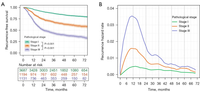
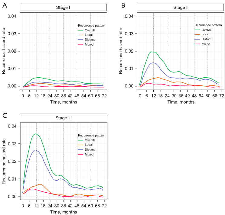
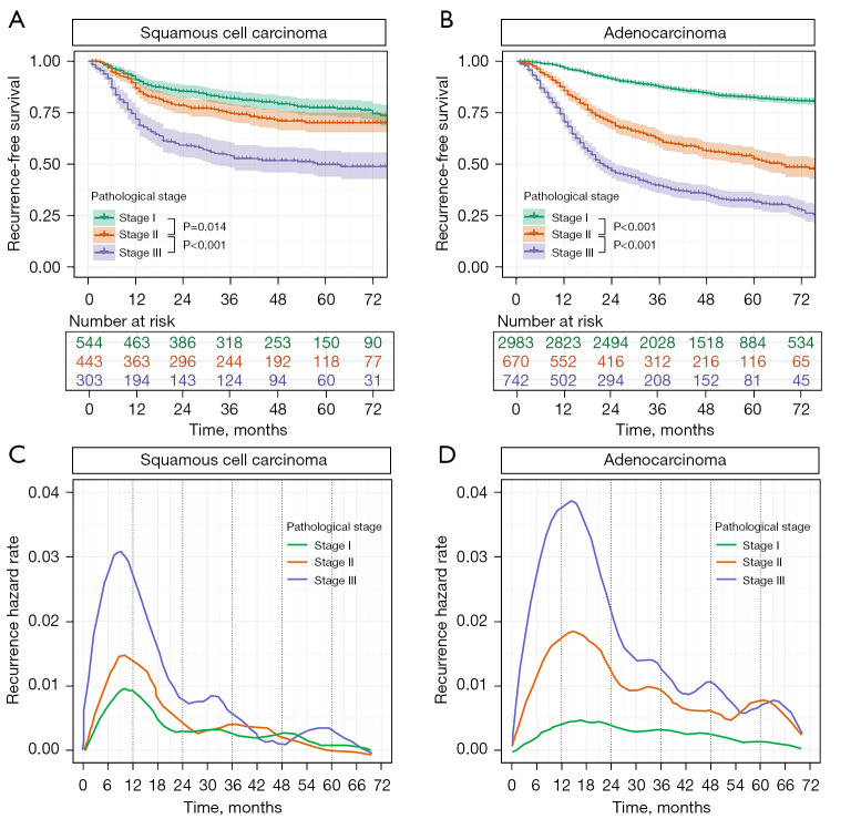
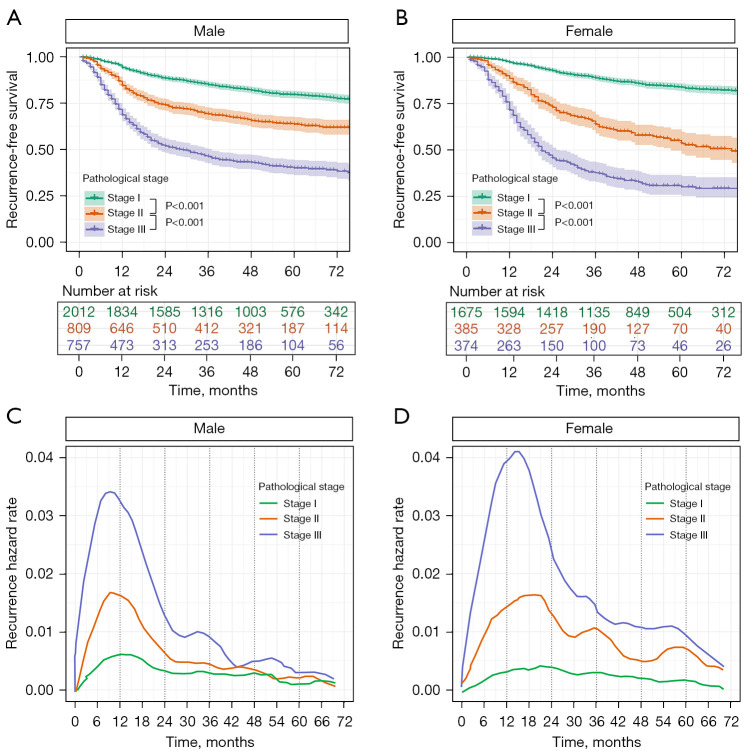
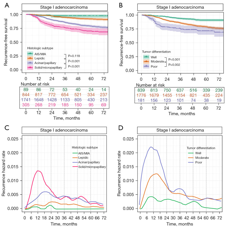
Figure 1 describes the RFS and the hazard rate for recurrence based on the pathological stage. Survival curves for recurrence between patients with stage I, stage II, and stage III were significantly different (all P<0.001, Figure 1A). In spite of different hazard rates depending on pathological stage, all hazard rate curves displayed similar patterns with the highest peak at around 12 months and the second peak at around 33 months after surgery (Figure 1B). When patients with stage II and II were divided according to the performance of adjuvant therapy, all hazard rate curves still showed a similar pattern with the highest peak at about 12 months, although there were slight differences (Figure S2). In accordance with the recurrence pattern, the rate of distant recurrence was higher than that of local or mixed recurrence, regardless of pathological stage (Figure 2). In addition, the highest peak for distant recurrence was shown around 9–12 months, whereas the peak for local recurrence was observed at around 15 months in patients with stage II and III (Figures 2B,2C). As for histological type, the 5-year RFS rate for adenocarcinoma was higher than squamous cell carcinoma in stage I (82.4% vs. 77.5%), but it was reversed in stage III (31.7% vs. 49.9%) (Figure 3A,3B). The highest peak of hazard rate was also observed at different period, which was 9 months for squamous cell carcinoma and 15 months for adenocarcinoma (Figures 3C,3D). Similar to histological type, the 5-year RFS rate for female patients was higher than male patients in stage I (83.8% vs. 79.7%), but it was reversed in stage III (30.1% vs. 40.1%) (Figure 4A,4B). In addition, the highest peak of hazard rate was shown at 9 months in female patients and 15 months in male patients (Figure 4C,4D). When it comes to histologic subtypes among patients with stage I adenocarcinoma, a phased degradation was found within the RFS curves from AIS/MIA to solid/micropapillary (Figure 5A). Each curve of RFS according to the grade of histologic differentiation was also different in stage I adenocarcinoma (Figure 5B). Stage I adenocarcinoma with solid/micropapillary pattern had a distinct peak of recurrence hazard rate at 12 months, but other subtypes did not (Figure 5C). The highest peak of hazard rate was displayed at 9 months in well differentiated stage I adenocarcinoma, whereas it was at 15 months in moderately differentiated tumor (Figure 5D).
Figure 1.
Recurrence-free survival (A) and the hazard rate for recurrence (B) based on the pathological stage in patients with non-small cell lung cancer.
Figure 2.
The hazard rate for recurrence following the recurrence pattern in patients with pathological stage I (A), stage II (B), and stage III (C) non-small cell lung cancer.
Figure 3.
Recurrence-free survival following the pathological stage in patients with squamous cell carcinoma (A) and adenocarcinoma (B). The hazard rate for recurrence following the pathological stage in patients with squamous cell carcinoma (C) and adenocarcinoma (D).
Figure 4.
Recurrence-free survival following the pathological stage in male (A) and female (B) patients. The hazard rate for recurrence following the pathological stage in male (C) and female (D).
Figure 5.
Recurrence-free survival following the histologic subtype (A) and tumor differentiation (B) in patients with stage I adenocarcinoma. The hazard rate for recurrence following the histologic subtype (C) and tumor differentiation (D).
Discussion
In this study, we investigated the recurrence dynamics of NSCLC patients who received complete resection according to their pathological stage, tumor histology, sex, histologic grade, and histologic subtype. Based on the results in our study, the main findings could be summarized as follows. First, irrespective of pathological stage, the highest recurrence hazard rate was shown at about 12 months and distant recurrence accounted for the largest portion of the overall recurrence pattern. However, the maximum of hazard rate for stage I was only one-seventh of that in stage III. Second, depending on tumor histology, the highest peak of recurrence hazard curve was observed at different periods, 9 months in squamous cell carcinoma and 15 months in adenocarcinoma. These trends based on tumor histology were similar when analyzed based on sex, 9 months in male patients and 15 months in female patients. Lastly, RFS and recurrence hazard rate were significantly different in accordance with histologic subtype and tumor differentiation grade in stage I adenocarcinoma. These findings indicate that adopting the same follow-up strategy may be undesirable in NSCLC patients who have different clinical and pathological characteristics.
As with our findings, several studies have reported a structured recurrence pattern with multiple peaks in NSCLC (18-20). This pattern contradicts the conventional notion that tumor cells continue to proliferate in disorder, leading to disease progression. Demicheli et al. adopted the hypothesis of a metastasis growth model based on breast cancer to explain this specific recurrence pattern in NSCLC (18). It was that the first peak of recurrence at around 1 year is closely related to disruption of homeostasis and proliferation of dormant tumor cells triggered by surgical invasion (21). Accordingly, the subsequent peaks of recurrence could be explained by the proliferation of residual tumor cells and the development of micro-metastasis after entering a temporary state of dormancy (20). However, the detailed mechanisms for the hypothesis of tumor dormancy have not been fully elucidated to date.
It should be noted that the recurrence dynamics of patients who undergo curative surgical treatment for NSCLC might be changed by various confounding factors, such as the frequency of the follow-up visits and radiologic examinations, the diagnostic modalities, and the interruption. In our institution, patients were strictly followed up every 3 months for the first 2 years after surgery and every 6 months thereafter. Chest CT was performed concomitantly with clinic visits, which seems more frequent than those recommended in any guidelines, such as National Comprehensive Cancer Network, European Society for Medical Oncology, and American Association of Thoracic Surgeons (3-8). Many previous studies examined optimal surveillance strategies that potentially contribute to overall survival (22). Westeel et al. reported that symptomatic patients with recurrence had worse survival than asymptomatic patients in whom recurrence was diagnosed on intensive imaging studies after surgery (23). Williams et al. also insisted that symptomatic patients at the time of recurrence have more than doubled the risk of death compared to asymptomatic patients, which supports intensive follow-up after complete resection (24). Accumulated evidence from prospective randomized studies and meta-analysis suggests intensive local therapy for oligo–recurrence may improve outcomes in a meaningful way (25-27). Furthermore, the adoption of molecular targeted therapy with an epidermal growth factor receptor (EGFR) gene mutation and an anaplastic lymphoma kinase (ALK) gene mutation for recurrent NSCLC has improved post-recurrence survival (28-30). However, the benefit of postoperative surveillance was also questioned from the perspectives of efficacy and cost-effectiveness (8,31). To date, there have been no large, prospective, randomized trials comparing different surveillance strategies in patients with NSCLC, and it remains unclear whether the early detection of recurrence contributes to improved outcomes. Consequently, it should be cautious to recommend individualized postoperative surveillance protocol according to clinical factors. However, we believe several tips based on our findings will help clinicians develop appropriate follow-up strategy for NSCLC patients with various clinical information.
First, given that no apparent peak of recurrence hazard curve has emerged in stage I patients, it does not seem mandatory to follow-up aggressively (e.g., hospital visit for every 3 months over the first 2 years or standard dose CT for 5 years) for these patients, as do patients in higher stages. Second, we have shown different hazard rates for recurrence depending on histologic subtype and tumor differentiation grade in stage I adenocarcinoma. Thus, aggressive surveillance should be maintained even in stage I adenocarcinoma, if patients have poor prognostic indicators, such as solid/micropapillary histologic subtype or poor tumor differentiation. Last, considering that the timing at which the peak of hazard curve was seen differed according to tumor histology and sex, it is suggested to take the intensive follow-up strategy longer in adenocarcinoma and female, compared to squamous cell carcinoma and male.
This study has some limitations. Selection bias may be present due to the retrospective, single-center design of the study. Although intensive surveillance system was adopted in our study, especially for the first two years, the timing of the first event obviously depends on the timing of imaging studies or hospital visits. Thus, we acknowledge there might be lead-time and length-time bias on our results. In addition, given that this study is focusing on descriptive analysis, no conclusions about further effects on survival outcomes, cost-effectiveness, or patient’s quality of life can be made. The impact on these outcomes would necessarily be performed with randomized prospective design.
In summary, patients who received complete surgical resection for NSCLC have various recurrence dynamics depending on clinical factors, such as pathological stage, sex, tumor histology and its subtype, and tumor differentiation grade. Adequate consideration of these factors will help clinicians develop detailed follow-up strategy in lung cancer patients with different recurrence dynamics.
Supplementary
The article’s supplementary files as
Acknowledgments
Funding: None.
Ethical Statement: The authors are accountable for all aspects of the work in ensuring that questions related to the accuracy or integrity of any part of the work are appropriately investigated and resolved. The study was conducted in accordance with the Declaration of Helsinki (as revised in 2013) and approved by the institutional review board of Asan Medical Center in Seoul, South Korea (IRB No. 2021–0166). The requirement for individual patient consent was waived due to the retrospective nature of this study.
Footnotes
Reporting Checklist: The authors have completed the STROBE reporting checklist. Available at https://tlcr.amegroups.com/article/view/10.21037/tlcr-21-1028/rc
Data Sharing Statement: Available at https://tlcr.amegroups.com/article/view/10.21037/tlcr-21-1028/dss
Peer Review File: Available at https://tlcr.amegroups.com/article/view/10.21037/tlcr-21-1028/prf
Conflicts of Interest: All authors have completed the ICMJE uniform disclosure form (available at https://tlcr.amegroups.com/article/view/10.21037/tlcr-21-1028/coif). The authors have no conflicts of interest to declare.
References
- 1.Bray F, Ferlay J, Soerjomataram I, et al. Global cancer statistics 2018: GLOBOCAN estimates of incidence and mortality worldwide for 36 cancers in 185 countries. CA Cancer J Clin 2018;68:394-424. 10.3322/caac.21492 [DOI] [PubMed] [Google Scholar]
- 2.Sugimura H, Nichols FC, Yang P, et al. Survival after recurrent nonsmall-cell lung cancer after complete pulmonary resection. Ann Thorac Surg 2007;83:409-17; discussioin 417-8. [DOI] [PubMed]
- 3.Ettinger DS, Wood DE, Aisner DL, et al. Non-Small Cell Lung Cancer, Version 5.2017, NCCN Clinical Practice Guidelines in Oncology. J Natl Compr Canc Netw 2017;15:504-35. 10.6004/jnccn.2017.0050 [DOI] [PubMed] [Google Scholar]
- 4.Vansteenkiste J, Crinò L, Dooms C, et al. 2nd ESMO Consensus Conference on Lung Cancer: early-stage non-small-cell lung cancer consensus on diagnosis, treatment and follow-up. Ann Oncol 2014;25:1462-74. 10.1093/annonc/mdu089 [DOI] [PubMed] [Google Scholar]
- 5.Jaklitsch MT, Jacobson FL, Austin JH, et al. The American Association for Thoracic Surgery guidelines for lung cancer screening using low-dose computed tomography scans for lung cancer survivors and other high-risk groups. J Thorac Cardiovasc Surg 2012;144:33-8. 10.1016/j.jtcvs.2012.05.060 [DOI] [PubMed] [Google Scholar]
- 6.Colt HG, Murgu SD, Korst RJ, et al. Follow-up and surveillance of the patient with lung cancer after curative-intent therapy: Diagnosis and management of lung cancer, 3rd ed: American College of Chest Physicians evidence-based clinical practice guidelines. Chest 2013;143:e437S-54S. [DOI] [PubMed] [Google Scholar]
- 7.Sause WT, Byhardt RW, Curran WJ, Jr, et al. Follow-up of non-small cell lung cancer. American College of Radiology. ACR Appropriateness Criteria. Radiology 2000;215 Suppl:1363-72. [PubMed] [Google Scholar]
- 8.Calman L, Beaver K, Hind D, et al. Survival benefits from follow-up of patients with lung cancer: a systematic review and meta-analysis. J Thorac Oncol 2011;6:1993-2004. 10.1097/JTO.0b013e31822b01a1 [DOI] [PubMed] [Google Scholar]
- 9.Simes RJ, Zelen M. Exploratory data analysis and the use of the hazard function for interpreting survival data: an investigator's primer. J Clin Oncol 1985;3:1418-31. 10.1200/JCO.1985.3.10.1418 [DOI] [PubMed] [Google Scholar]
- 10.Taylor MD, Nagji AS, Bhamidipati CM, et al. Tumor recurrence after complete resection for non-small cell lung cancer. Ann Thorac Surg 2012;93:1813-20; discussion 1820-1. 10.1016/j.athoracsur.2012.03.031 [DOI] [PubMed] [Google Scholar]
- 11.Hung JJ, Yeh YC, Jeng WJ, et al. Prognostic Factors of Survival after Recurrence in Patients with Resected Lung Adenocarcinoma. J Thorac Oncol 2015;10:1328-36. 10.1097/JTO.0000000000000618 [DOI] [PubMed] [Google Scholar]
- 12.Yun JK, Bok JS, Lee GD, et al. Long-term outcomes of upfront surgery in patients with resectable pathological N2 non-small-cell lung cancer. Eur J Cardiothorac Surg 2020;58:59-69. 10.1093/ejcts/ezaa042 [DOI] [PubMed] [Google Scholar]
- 13.Detterbeck FC, Boffa DJ, Kim AW, et al. The Eighth Edition Lung Cancer Stage Classification. Chest 2017;151:193-203. [DOI] [PubMed] [Google Scholar]
- 14.Martini N, Melamed MR. Multiple primary lung cancers. J Thorac Cardiovasc Surg 1975;70:606-12. 10.1016/S0022-5223(19)40289-4 [DOI] [PubMed] [Google Scholar]
- 15.Detterbeck FC, Jones DR, Kernstine KH, et al. Lung cancer. Special treatment issues. Chest 2003;123:244S-58S. 10.1378/chest.123.1_suppl.244S [DOI] [PubMed] [Google Scholar]
- 16.Gefeller O, Dette H. Nearest neighbour kernel estimation of the hazard function from censored data. J Stat Comput Simul 1992;43:93-101. 10.1080/00949659208811430 [DOI] [Google Scholar]
- 17.Boracchi P, Biganzoli E, Marubini E. Joint modelling of cause-specific hazard functions with cubic splines: an application to a large series of breast cancer patients. Comput Stat Data Anal 2003;42:243-62. 10.1016/S0167-9473(02)00122-6 [DOI] [Google Scholar]
- 18.Demicheli R, Fornili M, Ambrogi F, et al. Recurrence dynamics for non-small-cell lung cancer: effect of surgery on the development of metastases. J Thorac Oncol 2012;7:723-30. 10.1097/JTO.0b013e31824a9022 [DOI] [PubMed] [Google Scholar]
- 19.Kelsey CR, Fornili M, Ambrogi F, et al. Metastasis dynamics for non-small-cell lung cancer: effect of patient and tumor-related factors. Clin Lung Cancer 2013;14:425-32. 10.1016/j.cllc.2013.01.002 [DOI] [PubMed] [Google Scholar]
- 20.Watanabe K, Tsuboi M, Sakamaki K, et al. Postoperative follow-up strategy based on recurrence dynamics for non-small-cell lung cancer. Eur J Cardiothorac Surg 2016;49:1624-31. 10.1093/ejcts/ezv462 [DOI] [PubMed] [Google Scholar]
- 21.Hedley BD, Chambers AF. Tumor dormancy and metastasis. Adv Cancer Res 2009;102:67-101. 10.1016/S0065-230X(09)02003-X [DOI] [PubMed] [Google Scholar]
- 22.Mollberg NM, Ferguson MK. Postoperative surveillance for non-small cell lung cancer resected with curative intent: developing a patient-centered approach. Ann Thorac Surg 2013;95:1112-21. 10.1016/j.athoracsur.2012.09.075 [DOI] [PubMed] [Google Scholar]
- 23.Westeel V, Choma D, Clément F, et al. Relevance of an intensive postoperative follow-up after surgery for non-small cell lung cancer. Ann Thorac Surg 2000;70:1185-90. 10.1016/S0003-4975(00)01731-8 [DOI] [PubMed] [Google Scholar]
- 24.Williams BA, Sugimura H, Endo C, et al. Predicting postrecurrence survival among completely resected nonsmall-cell lung cancer patients. Ann Thorac Surg 2006;81:1021-7. 10.1016/j.athoracsur.2005.09.020 [DOI] [PubMed] [Google Scholar]
- 25.Ashworth AB, Senan S, Palma DA, et al. An individual patient data metaanalysis of outcomes and prognostic factors after treatment of oligometastatic non-small-cell lung cancer. Clin Lung Cancer 2014;15:346-55. 10.1016/j.cllc.2014.04.003 [DOI] [PubMed] [Google Scholar]
- 26.Stephens SJ, Moravan MJ, Salama JK. Managing Patients With Oligometastatic Non-Small-Cell Lung Cancer. J Oncol Pract 2018;14:23-31. 10.1200/JOP.2017.026500 [DOI] [PubMed] [Google Scholar]
- 27.Gomez DR, Blumenschein GR, Jr, Lee JJ, et al. Local consolidative therapy versus maintenance therapy or observation for patients with oligometastatic non-small-cell lung cancer without progression after first-line systemic therapy: a multicentre, randomised, controlled, phase 2 study. Lancet Oncol 2016;17:1672-82. 10.1016/S1470-2045(16)30532-0 [DOI] [PMC free article] [PubMed] [Google Scholar]
- 28.Maemondo M, Inoue A, Kobayashi K, et al. Gefitinib or chemotherapy for non-small-cell lung cancer with mutated EGFR. N Engl J Med 2010;362:2380-8. 10.1056/NEJMoa0909530 [DOI] [PubMed] [Google Scholar]
- 29.Thatcher N, Chang A, Parikh P, et al. Gefitinib plus best supportive care in previously treated patients with refractory advanced non-small-cell lung cancer: results from a randomised, placebo-controlled, multicentre study (Iressa Survival Evaluation in Lung Cancer). Lancet 2005;366:1527-37. 10.1016/S0140-6736(05)67625-8 [DOI] [PubMed] [Google Scholar]
- 30.Novello S, Mazières J, Oh IJ, et al. Alectinib versus chemotherapy in crizotinib-pretreated anaplastic lymphoma kinase (ALK)-positive non-small-cell lung cancer: results from the phase III ALUR study. Ann Oncol 2018;29:1409-16. 10.1093/annonc/mdy121 [DOI] [PMC free article] [PubMed] [Google Scholar]
- 31.Virgo KS, McKirgan LW, Caputo MC, et al. Post-treatment management options for patients with lung cancer. Ann Surg 1995;222:700-10. 10.1097/00000658-199512000-00003 [DOI] [PMC free article] [PubMed] [Google Scholar]
Associated Data
This section collects any data citations, data availability statements, or supplementary materials included in this article.
Supplementary Materials
The article’s supplementary files as


