Abstract
Mono-, di-, and trimethylation of histone H3 lysine 4 (H3K4me1/2/3) are associated with transcription, yet it remains controversial whether H3K4me1/2/3 promote or result from transcription. Our previous characterizations of Arabidopsis H3K4 demethylases suggest roles for H3K4me1 in transcription. However, the control of H3K4me1 remains unexplored in Arabidopsis, in which no methyltransferase for H3K4me1 has been identified. Here, we identify three Arabidopsis methyltransferases that direct H3K4me1. Analyses of their genome-wide localization using ChIP-seq and machine learning reveal that one of the enzymes cooperates with the transcription machinery, while the other two are associated with specific histone modifications and DNA sequences. Importantly, these two types of localization patterns are also found for the other H3K4 methyltransferases in Arabidopsis and mice. These results suggest that H3K4me1/2/3 are established and maintained via interplay with transcription as well as inputs from other chromatin features, presumably enabling elaborate gene control.
Subject terms: Epigenomics, Histone post-translational modifications, Plant molecular biology
Methylation of histone H3 lysine 4 is associated with transcription. Here the authors describe three Arabidopsis methyltransferases that direct H3K4me1 and propose that one methyltransferase works co-transcriptionally while the others are recruited to genomic and epigenomic features.
Introduction
Posttranslational modifications of histone residues shape the epigenome and affect gene expression1. Histone H3 lysine 4 methylation (H3K4me) is a conserved histone mark found in actively transcribed genes; however, the molecular basis and functional significance of the interrelationship between H3K4me and transcription remain elusive2,3. It has been proposed that H3K4me can be viewed as a memory of transcription because transcription directs H3K4 methylation4–7. On the other hand, in some experimental systems, H3K4me is deposited independent of transcription8,9. The underlying mechanisms that coordinate transcription-coupled and transcription-independent H3K4me are largely unexplored.
Another layer of H3K4me complexity is its occurrence in three states (mono-, di-, and trimethylation; me1, me2 and me3, respectively). Generally, H3K4me3 and H3K4me2 are found on the first several nucleosomes of genes, while H3K4me1 is found in downstream regions within the gene bodies10–12. Our previous genetic and genomic studies in Arabidopsis thaliana (hereafter Arabidopsis) have demonstrated key roles of the gene body H3K4me1 in various epigenetic phenomena. A putative H3K4me1 demethylase, LYSINE-SPECIFIC DEMETHYLASE1-LIKE2 (LDL2), mediates silencing of genes with the repressive histone mark H3K9me2 by removal of H3K4me113. Another related demethylase, FLOWERING LOCUS D (FLD), downregulates transcriptional elongation and initiation by removing H3K4me1 in genes that have high levels of convergent overlapping transcription (i.e., antisense transcription)14. Although these findings regarding the H3K4 demethylases of Arabidopsis suggest a link of H3K4me1 with other chromatin features and transcription, the mechanisms of H3K4me1 control remain elusive, mainly because methyltransferase(s) for H3K4me1 has not been identified in plants.
In both animals and plants, two types of H3K4 methyltransferases are known: Set1-type and Trithorax/Trithorax-related (Trx/Trr)-type H3K4 methyltransferases15. However, yeasts have lost Trx/Trr-type H3K4 methyltransferases during evolution16, and yeast Set1 is the sole H3K4 methyltransferase responsible for all H3K4me1/2/3. Arabidopsis has five Trx/Trr-type enzymes (Arabidopsis Trithorax (ATX) 1 to 5) and at least one Set1-type enzyme (Arabidopsis Trithorax Related (ATXR) 7) (Fig. 1a). Additionally, ATXR3, which has a domain structure characteristic of Set1-type enzymes but contains an atypical catalytic domain as H3K4 methyltransferase17,18, can catalyze all three states of H3K4me1-3 in vitro19. Loss-of-function mutations in the ATXR3 gene cause substantial H3K4me3 loss genome-wide, but H3K4me1 and H3K4me2 are largely unaffected19. In addition, triple loss-of-function of ATX3, ATX4, and ATX5 leads to global decreases in H3K4me3 and H3K4me2 but not H3K4me120. However, methyltransferase mutants with a genome-wide decrease of H3K4me1 levels have not been reported. The functions of ATX1, ATX2, and ATXR7 genes have been characterized in the context of development, flowering regulation, and plant immunity19–27. For example, they were shown to redundantly inhibit flowering by activating the transcription of the flowering repressor FLOWERING LOCUS C (FLC) by increasing H3K4me1/2/3 levels within the FLC locus21,24. In the control of FLC, ATXR7 was shown to counteract FLD24, which demethylates H3K4me114. However, the genome-wide impacts of these putative H3K4 methyltransferases have not been elucidated.
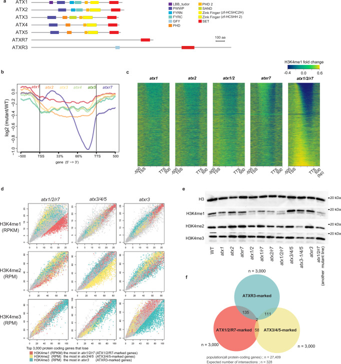
Fig. 1. ATX1, ATX2, and ATXR7 redundantly contribute to H3K4me1.
a Domain architectures of ATX(R) proteins from the UniProt database. b Metaplot illustrates the averaged pattern of H3K4me1 changes of the 6 mutants compared to WT. c Mutations of atx1, atx2, and atxr7 synergistically cause reduced H3K4me1 in the gene body. H3K4me1 changes from WT are visualized as heat maps. All genes are ordered so that genes that lost H3K4me1 the most in atx1/2/r7 come to the top. d ChIP-seq for H3Kme1, H3K4me2, and H3K4me3 in mutants (y-axis) compared to WT (x-axis). Each dot represents each gene. Axes are trimmed to 0.98 quantiles. Top genes (n = 3000) that lose H3K4me1 most in atx1/2/r7 (hereafter ATX1/2/R7-marked genes), lose H3K4me2 in atx3/4/5 (ATX3/4/5-marked genes), and lose H3K4me3 in atxr3 (ATXR3-marked genes) are colored red, yellow, and blue, respectively. Values for H3K4me2/3 are RPM normalized instead of RPKM as in H3K4me1 because unlike H3K4me1, which covers the gene body, the amounts of H3K4me2 and H3K4me3 are less dependent on gene length10. e Western blotting of H3K4 methylations on bulk histone extracted from the mutants. The atx1/2/r7 mutant on the right consists of three T-DNA alleles that are different from those in atx1/2/r7 mutant mainly used in this study. Western blotting for H3K4me1 in atx1/2/r7 were repeated and quantified (Supplementary Fig. 2a). f Activities of ATX1/2/R7, ATX3/4/5, and ATXR3 are observed in mutually exclusive genes. All pairwise ATX1/2/R7-marked genes, ATX3/4/5-marked genes, and ATXR3-marked genes have significantly fewer overlaps than expected (n = 328). Significances of the under-representation are tested by hypergeometric tests (p < 1e-4 for all pairs).
Here, we explored the involvement of seven H3K4 methyltransferase genes in H3K4me1 as well as H3K4me2/3 by analyzing single and multiple mutants using chromatin immunoprecipitation sequencing (ChIP-seq). H3K4me1 levels are substantially decreased by the simultaneous loss of the ATX1, ATX2, and ATXR7 genes. Our results clarify the division of labor among H3K4 methyltransferase genes and provide powerful genetic materials for elucidating the functions of each H3K4 methylation state. In addition, we analyzed the genomic localization of these enzymes. The subsequent application of machine learning algorithms revealed that the ATXR7 protein colocalizes with the transcription machinery, while ATX1 and ATX2 are associated with other chromatin modifications (epigenome) and specific DNA sequences (genome). These two types of localization patterns were also found by the reanalysis of other Set1- and Trx/Trr-type H3K4 methyltransferases, including those of animals. These findings lead us to a new perspective: some H3K4me marks may function as records of transcription, while other H3K4me marks may function as mediators of information encoded in the epigenome and/or genome.
Results
H3K4me1 levels are decreased by the triple loss of ATX1/2/R7 genes
To identify enzyme(s) involved in H3K4me1, we examined mutants of six predicted H3K4 methyltransferases: ATX1 to 5 and ATXR7 (Fig. 1a). Among the six single mutants, atx2 and atxr7 showed relatively large decreases in H3K4me1 levels (Supplementary Fig. 1a). Interestingly, the atx2 and atxr7 mutants differed in the affected regions within genes; atxr7 showed a marked decrease in H3K4me1 in the 3’ half of the gene bodies, while atx2 showed a decrease over a broader region (Fig. 1b). Cluster analyses based on the intragenic H3K4me1 pattern in the atx1 to 5 and atxr7 mutants revealed that atx1 and atx2 formed one cluster, while atx3, atx4, and atx5 formed another cluster (Supplementary Fig. 1b). These similarities in the effects on the H3K4me1 profile coincide with the similarity of the domain architecture of the corresponding proteins (Fig. 1a).
As the lack of a strong effect in each of the single mutants may reflect redundancy, we examined mutants that lose function of multiple ATX(R) genes. Based on the similarities of the protein domain structures and the effects on H3K4me1 patterns, we examined atx1/atx2 double mutants and found stronger effects on H3K4me1 than were observed in either of the single mutants (Fig. 1c, Supplementary Fig. 1c). Additionally, atx1/atxr7 and atx2/atxr7 showed stronger H3K4me1 decreases than each single mutant (Supplementary Fig. 1c, Supplementary Data 1). The effect was still stronger in triple atx1/atx2/atxr7 mutants (hereafter referred to as atx1/2/r7) (Fig. 1c, d, Supplementary Fig. 2a, Supplementary Data 1). ChIP-seq replicates with spike-in control confirmed that atx1, atx2, and atxr7 additively impact H3K4me1 (Supplementary Fig. 1d, Supplementary Data 2). Western blot analyses confirmed that triple atx1/2/r7 mutation (with two different sets of T-DNA insertion alleles) resulted in the loss of H3K4me1 (Fig. 1e, Supplementary Fig. 2a), while H3K4me2 and H3K4me3 were largely unaffected (Fig. 1e). Consistent with the idea that ATX1/2/R7 mediates monomethylation of H3K4, ChIP-seq analysis targeting unmethylated H3K4 (H3K4me0) revealed that ATXR1/2/R7-marked genes showed increased signals of H3K4me0 in atx1/2/r7 compared to wild type (Supplementary Fig. 2b). Taken together, these results demonstrate that ATX1, ATX2, and ATXR7 contribute to H3K4me1 in a partially redundant manner.
Consistent with previous reports19,20,26, our Western blot and ChIP-seq analyses showed that H3K4me3 and H3K4me2 modifications are mainly mediated by ATXR3 and ATX3/4/5, respectively (Fig. 1d, e, Supplementary Fig. 2c–f). Collectively, the results showed that H3K4me1, H3K4me2, and H3K4me3 levels were specifically reduced in atx1/2/r7, atx3/4/5, and atxr3, respectively (Fig. 1d, e). The genes that were strongly affected in each of these mutants (colored red, yellow, or blue in Fig. 1d) showed high corresponding H3K4me levels in the wild type (e.g., ATX1/2/R7-marked genes showed high H3K4me1 levels relative to those of other genes) (Supplementary Fig. 1f–h), and had specific characteristics (Supplementary Fig. 1e). For example, ATX3/4/5-marked genes showed lower levels of expression compared to others (Supplementary Fig. 1e), in line with the body of research that suggests that H3K4me2 is a repressive mark in plants; H3K4me2 colocalizes with other repressive marks such as H3K27me328 and anti-correlates with transcription29. Hypo-H3K4me2 activates transcription in rice29 and during regeneration of Arabidopsis30. Interestingly, the target genes of each ATX(R) group were mutually exclusive (Fig. 1f), suggesting that distinct mechanisms direct each of the H3K4 methyltransferases to distinct target genes.
ATXR7 localization is associated with RNAP2, while ATX1 and ATX2 localization is associated with other chromatin modifications
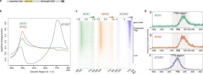
To investigate the mechanisms that specify the targets of each ATX(R) protein that regulates H3K4me1, we determined the genome-wide localization of the ATX1, ATX2 and ATXR7 proteins using transgenic plants expressing FLAG-tagged proteins (Fig. 2a). ChIP-seq analyses revealed that ATX1 and ATX2 localized around transcription start sites (TSSs), typically in the range of −150 ~ +300 bp from the TSS. ATXR7 localized around transcription termination sites (TTSs) in the range of −200 ~ +200 bp from the TTS (Fig. 2b–d). Genes showing H3K4me1 loss in atx1/2/r7 mutants tended to be bound by ATX1, ATX2 and ATXR7 (Supplementary Fig. 3), supporting the conclusion that these enzymes mediate H3K4me1 on chromatin.
Fig. 2. ATX1 and ATX2 occupy TSS while ATXR7 occupies TTS.
a The transgene structure of tagged ATX(R) proteins used for ChIP-seq. b, c Metaplot (b) and heat maps (c) illustrating ATX1, ATX2, and ATXR7 distributions in the gene body region corrected with a non-transgenic control. The heat map was sorted so that highly transcribed genes (measured as mRNA-seq in WT) come to the top. d Position of ATX1 and ATX2 ChIP-seq peak summits relative to TSS and ATXR7 peaks relative to TTS (x-axis), visualized as a frequency of peak summits (y-axis), which are detected against non-transgenic control using MACS2 peak caller. The numbers of peaks are summarized in Supplementary Data 3. Most of the ATX1 and ATX2 peaks belong to the region spanning from 150 bp upstream to 300 bp downstream of TSS (hereafter ‘TSS region’). Most of the ATXR7 peaks are between 200 bp upstream and 200 bp downstream of TTS (hereafter ‘TTS region’).
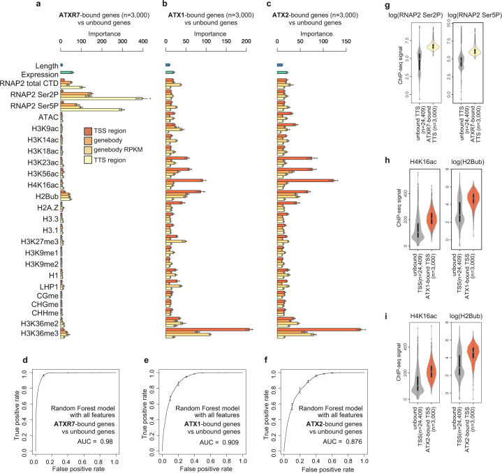
To elucidate the chromatin-targeting mechanism(s) of these ATX1/2/R7 proteins, we screened for the determinants of ATX1/2/R7 localization by using a machine learning algorithm (random forest). Using chromatin and genomic features listed in Fig. 3a, the algorithm was trained to distinguish ATX1/2/R7-bound genes from unbound genes. After training, the random forest algorithm reported the ‘importance (mean decrease in Gini)’ of each feature as a relative score reflecting how informative the feature was for classification (Fig. 3a–c). In principle, the values of the features with high ‘importance’ scores can be positively (colocalize) or negatively (exclusive) correlated with the localization level of the corresponding ATX1/2/R7 protein. By taking all the features into account, the localization of each ATX1/2/R7 protein was well explained (Fig. 3d–f; note the high area under the curve (AUC) values).
Fig. 3. ATXR7 localization is associated with RNAP2, while ATX1 and ATX2 localization is associated with other chromatin modifications.
a–c Chromatin features predictive of ATXR7 (a), ATX1 (b), and ATX2 (c) localization. ATXR7 (or ATX1,2)-bound genes were defined as the top 3000 genes with the highest ChIP-seq signal in ‘TTS region (or TSS region)’ compared to nontransgenic control. Random forest models were trained to predict ATXR7 (or ATX1,2)-bound and unbound genes. Bars indicate ‘mean decrease in Gini’ or in other words ‘importance’ derived from the random forest model, which is a relative score reflecting how informative the feature was for classification. ‘Importance’ are averaged from 5 repeats of training, each of which independently chose negative samples (unbound genes). Error bars are the standard deviation of the 5 repetitions. d–f ROC plots showing the prediction accuracy of the random forest models. AUC indicates the area under the ROC curve. ROC and AUC are calculated with data on Chr. 5, which were held out from training as test data. Average and standard deviation of the 5 repeats of training are plotted. g–i Violin plots showing the abundance of two most predictively ‘important’ features for each protein. Analyses with ChIP-seq datasets of biological replicates showed similar results (Supplementary Fig. 5). The center line of violin plot, median; box limits, upper and lower quartiles; whiskers, 1.5× interquartile range.
The most ‘important’ feature for distinguishing ATXR7-bound genes was the level of RNA Polymerase II (RNAP2) around the TTS (Fig. 3a). ATXR7-bound genes showed higher levels of total RNAP2 and RNAP2 phosphorylation at Ser2 and Ser5 sites in the carboxy-terminal domain (CTD) (Fig. 3g, Supplementary Fig. 4), indicating the strong colocalization of ATXR7 and RNAP2, especially when phosphorylated at Ser2 and Ser5. These results suggest that ATXR7, similarly to yeast Set1, may be recruited to chromatin in a transcription-coupled manner.
In contrast, the most ‘important’ feature for the prediction of ATX1- and ATX2-bound genes was H3K36me3 (Fig. 3b, c, Supplementary Fig. 5), which gives rise to three nonexclusive hypotheses: the distributions of ATX1/2 and H3K36me3 are driven by a shared factor; the presence of H3K36me3 drives ATX1/2 localization; or ATX1/2 drives H3K36me3 modifications. Consistent with the last hypothesis, atx1/2/r7 induced the concomitant loss of H3K4me1 and H3K36me3 (Supplementary Fig. 6a), while H3K4me1 was unaffected by the ash1 homolog 2 (ashh2) mutation, which causes a drastic reduction in H3K36me3 levels (Supplementary Fig. 6b)31,32. However, ashh2 mutant keeps relatively high levels of H3K36me3 around TSS33 where ATX1/2 localize (Fig. 2b–d), thus we cannot exclude the possibility that H3K36me3 acts upstream in addition to downstream of ATX1/2-H3K4me1. Among mutants for five H3K36 methyltransferase family genes, the ashh3 mutant affects H3K36me3 at ATX1/2/R7-marked genes, while others including ASHH2, which has a H3K4me1-binding domain34, markes genes that are not largely overlapped with ATX1/2/R7-marked genes (Supplementary Fig. 6c, d), consistent with the view that ASHH3 functions downstream of ATX1/2/R7-marked H3K4me1 to mediate H3K36me3.
In addition to H3K36me3, H2Bub and H4K16ac in TSS regions were indicated to be ‘important’ (Fig. 3b, c). ATX1- and ATX2-bound TSSs were rich in both modifications (Fig. 3h, i, Supplementary Fig. 4). Genetic depletion of H2Bub using histone mono-ubiquitination 2 (hub2) mutant resulted in decreased localization of ATX2 protein in genes that are predicted to lose ATX2 by in silico simulation of H2Bub loss (Supplementary Fig. 7a–c; for details of in silico simulation of H2Bub loss, see ‘Random forest’ section in Methods). Furthermore, those genes that become unbound by ATX2 in hub2 mutant or by ATX1/2 in the in silico hub tend to lose H3K4me1 in hub mutants (hub1 and hub2) (Supplementary Fig. 7d), consistent with the view that H2Bub promotes localization of ATX1/2, which then promotes H3K4me1. Conversely, H2Bub was also decreased in H3K4me1-decreased genes in atx1/2/r7 (Supplementary Fig. 7e).
These results indicate that H2Bub and H3K4me1 are mutually promoting each other. H4K16ac was not specifically affected at ATX1/2/R7-marked genes or ATX1/2-bound genes in atx1/2/r7 (Supplementary Fig. 7e), suggesting that H4K16ac is not regulated downstream of ATX1/2.
On the other hand, the relative importance values of total or phosphorylated RNAP2 were markedly lower for ATX1/2 than for ATXR7 (Fig. 3a–c). These results suggest that ATX1/2 localization is governed by other chromatin modification(s), rather than by transcription. A previous study reported that physical interaction between ATX1 and RNAP2 phospho-Ser5 is involved in the recruitment of ATX1 to several ATX1-regulated genes35. Our results do not exclude the possibility that phospho-RNAP2 is also involved in the chromatin recruitment of ATX1/2 in addition to chromatin modification(s) such as H2Bub (Fig. 3, Supplementary Fig. 7).
The above results show that there are two modes of chromatin targeting. ATXR7 seems to cooperate with the transcriptional machinery, while ATX1 and ATX2 seem to target genes in a manner that is informed by chromatin modifications, regardless of the abundance of the transcriptional machinery. Interestingly, many genes occupied by ATXR7 are also bound by the H3K4me1 demethylase FLD14, Supplementary Fig. 8a, b). In addition, genes showing the loss of H3K4me1 in the atxr7 mutant tended to show a gain of H3K4me1 in the fld mutant (Supplementary Fig. 8c). Thus, ATXR7 and FLD localize in the TTS region and have opposite effects on H3K4me1.
The DNA sequences of ATX1- and ATX2-bound TSSs have distinct architectures
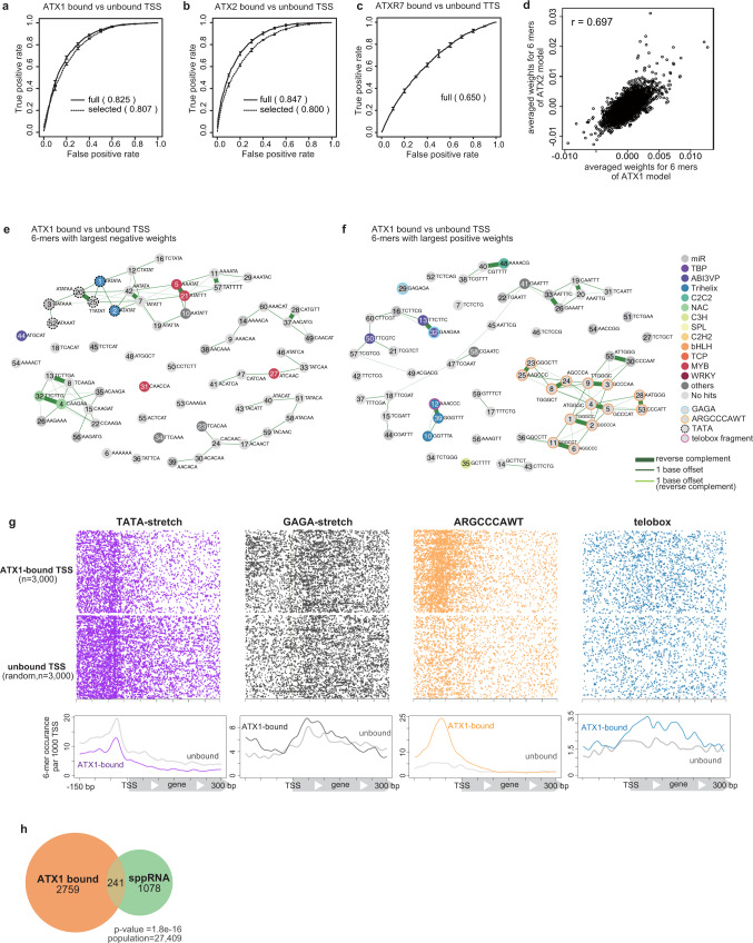
Next, we asked whether the DNA sequences to which ATX1,2 or ATXR7 bind also have specific features. We converted the DNA sequences of TSS or TTS regions into a vector that represents the occurrence of all possible 6 base sequences (6-mer frequency vector) and trained the linear support vector machine (lSVM) algorithm to distinguish ATX1 (or ATX2)-bound TSS regions from unbound TSS regions, and ATXR7-bound TTS regions from unbound TTS regions. This process achieved good accuracy for ATX1 and ATX2 (Fig. 4a, b) but not for ATXR7 (Fig. 4c). The lSVM algorithm learns to assign greater weights to predictively important features. Consistent with their overlapping localization (Fig. 2c), the ATX1 and ATX2 models weighted similar sets of 6-mers (Fig. 4d), suggesting that ATX1 and ATX2 have similar mechanisms of sequence-based targeting.
Fig. 4. The DNA sequences of ATX1- and ATX2-bound TSSs have distinct architectures.
a–c ROC plots that show predicting accuracy of linear SVM models, which are trained with the full set of 6- mers (solid line) and trained with the 120 6-mers which were highly weighted in the full model (dashed line), to discriminate (a) ATX1- or (b) ATX2-bound vs -unbound TSS DNA sequence, and (c) ATXR7-bound and-unbound TTS sequence. ROC and AUC are calculated with data on Chr 5, which are held out from the training as test data. Averaged scores of 5 cross-fold validation models are plotted. Error bars represent the standard deviation of the 5 repeats of training. Analyses with ChIP-seq datasets of biological replicates (see Methods) showed similar results (Supplementary Fig. 9). d Averaged SVM weights from ATX1 models (x-axis) correlates with those from ATX2 models (y-axis), indicating that similar sets of sequences predict ATX1 and ATX2 localizations. r = Pearson’s correlation coefficient. e, f Clustering and annotation of predictive 6-mers. Each circle represents the top sixty 6-mers with negative (e) or positive (f) averaged SVM weights in the ATX1 models. Numbers within the circle are ranks of weights’ absolute values (the more ‘predictive’ a 6-mer is, the smaller the number labels it). Pairs of related 6-mers are connected with lines; the thickest lines connect reverse complement pairs, the thinner lines connect neighboring 6-mers with 1 base offset, the thinnest lines connect reverse complement pairs with 1 base offset. Each 6-mers was searched for matching motifs (see Methods), then 6-mer circles were colored corresponding to the category of its top matched motif that meets q-value <0.1 criteria. 6-mers corresponding to ARGCCCAWT, telobox, GAGA, and TATA-stretch are manually highlighted with border circles. Some of the highly weighted motifs are evolutionarily conserved among TSS regions of the land plants, suggesting their functionality (Supplementary Fig. 13). g Positional distributions of the highly weighted motifs in the TSS region. h ATX1-bound TSS significantly overlaps with sppRNA-harboring TSS detected in the hen2-2 background. The significance of the overlap was tested using a hypergeometric test.
To better understand the underlying mechanism, we sought to annotate the sets of DNA motifs that specify ATX1/2 binding. We selected the 120 predictive 6-mers, including the sixty 6-mers with the highest positive weights (when the TSS region contains these 6-mers, ATX1/2 are more likely to bind) and the sixty 6-mers with the greatest negative weights (opposing binding). The prediction accuracy for these 120 features was not much lower than that of the full model (Fig. 4a, b). Many of the 120 predictive 6-mers in ATX1/2 models were clustered into groups of overlapping 6-mer sequences (Fig. 4e, f, Supplementary Fig. 10a, b), in contrast to the situation for random or nonpredictive 6-mers (Supplementary Fig. 11a, b, 10c), suggesting the presence of >6 bp motifs that are predictive of ATX1/2 binding.
We annotated these discriminative DNA motifs by searching for matching sequences in the literature and in databases (Fig. 4e, f, Supplementary Fig. 10a, b, Supplementary Data 5, 6). In both the ATX1 and ATX2 models, TATA stretch was negatively weighted (Fig. 4e, Supplementary Fig. 10a, dotted circles). Consistent with this result, the occurrence of TATA stretch was less frequent in ATX-bound TSSs than in ATX-unbound TSSs (Fig. 4g, Supplementary Fig. 10d). On the other hand, GAGA stretch was positively weighted (Fig. 4f, Supplementary Fig. 10b, cyan circles), and ATX-bound TSSs showed broad GAGA peaks (Fig. 4g, Supplementary Fig. 10d). The positively weighted motifs also included ‘ARGCCCAWT’ (in both the ATX1 and ATX2 models) and a telobox fragment (in the ATX1 model), the latter of which coincided with a trihelix transcription factor (TF) binding site (Fig. 4f, g and Supplementary Fig. 10b, d. Supplementary Fig. 12). RGCCCAW is likely bound by TCP family TFs36–38. While the ATX protein itself is not known to show DNA binding specificity, these additional DNA binders, such as the GAGA binding factor BPC639,40 and TCP and trihelix TFs, may physically link ATX to DNA motifs. The combination of GAGA and RGCCCAW is characteristic of TSSs that harbor short noncoding RNAs (short promoter proximal RNAs, sppRNAs) with unknown functions41. Indeed, ATX1/2-bound genes were found to significantly overlap with sppRNA genes (Fig. 4h, Supplementary Fig. 10e), suggesting the existence of some links between sppRNA and ATX1/2 localization.
These sequence-based learning results further support the idea that ATX1 and ATX2 target genes independent of transcription events. The predictive motif sets are further interpreted in the discussion.
A positive correlation between transcription and H3K4me1 is mediated by ATXR7 and is disturbed by ATX1/2
From the above results we hypothesized that the ATX1 and ATX2 proteins monomethylate H3K4 irrespective of transcription, while ATXR7 does cotranscriptionally. We tested this hypothesis by analyzing the correlation between transcription and H3K4me1. In the wild type, the amount of H3K4me1 covering a gene is moderately correlated with the transcription level (see below). The hypothesis predicts that ATXR7 is responsible for this correlation through H3K4me1 deposition in transcribed genes, while ATX1 and ATX2 disrupt this correlation by depositing H3K4me1 regardless of transcription. In agreement with this prediction, the correlation became significantly stronger in atx1/2 (Fig. 5a) compared to wild type (Fig. 5b), and weaker in atxr7 (Fig. 5c), as quantified by Spearman’s correlation coefficient or visualized by density heatmaps. This alteration in the correlation landscape was due to a change in H3K4me1 rather than transcription because the correlation showed the same trend when the transcription data for the wild type were used rather than those for the mutants (Supplementary Fig. 14a, b).
Fig. 5. Positive correlation between transcription and H3K4me1 is mediated by ATXR7 and is disturbed by ATX1/2.
a–c Protein-coding genes are ranked in order of H3K4me1 (x-axis, RPKM) and expression levels (y-axis, mRNA-seq FPKM, mean of the three RNA-seq replicates). The densities of genes are visualized as heat maps. ρ is the Spearman’s correlation coefficient. Three replicates of mRNA-seq resulted in three ρ for each genotype, and the average and standard deviation of ρ are presented. P values indicate that correlation estimates ρ significantly differ between genotypes (Welch’s two sample t-test). Another ChIP-seq datasets replicated consistent trends (Supplementary Fig. 14c–h).
Localization and functional mode of other Arabidopsis H3K4 methyltransferases
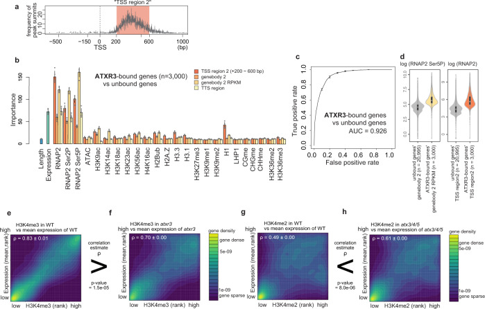
In addition to ATX1, ATX2, and ATXR7, Arabidopsis has at least four other putative H3K4 methyltransferases. Hence, we wondered how the localization patterns of the other methyltransferases were determined. A random forest analysis using the reported genome-wide distribution of the ATXR3 protein42 revealed that the levels of total and phosphorylated RNAP2 and transcription presented the highest ‘importance’ and that they were significantly higher in ATXR3-bound genes than in ATXR3-unbound genes (Fig. 6a–d), suggesting a cotranscriptional mode of H3K4me3 deposition by ATXR3. Consistent with this interpretation, the strong correlation between transcript levels and H3K4me3 decreased in the atxr3 mutant (Fig. 6e, f, Supplementary Fig. 15a, b).
Fig. 6. Localization and functional mode of other Arabidopsis H3K4 methyltransferases.
a Position of ATXR3 peaks relative to TSS, visualized as a frequency of ChIP-seq peak summits around TSS. x-axis, distance from TSS; y-axis, number of peak summits. Most of the ATXR3 peaks belong to the region spanning from 200 bp to 600 bp downstream of TSS, which we hereafter refer to as ‘TSS region 2’. b Chromatin features predictive of ATXR3 localization. Error bars represent the standard deviation of the 5 repeats of training. c ROC plot showing the prediction accuracy of the random forest models. AUC indicates the area under the ROC curve. ROC and AUC are calculated with data on Chr 5, which are held out from the training as test data. Average and standard deviation of the 5 repeats of training are plotted. d Violin plots showing the abundance of the two most predictively ‘important’ features. The center line of violin plot, median; box limits, upper and lower quartiles; whiskers, 1.5× interquartile range. e–h Protein-coding genes are ranked in order of H3K4me3 (e, f) or H3K4me2 (g, h) (x-axis, RPM) and expression levels (y-axis, mRNA-seq FPKM, mean of the three RNA-seq replicates). The densities of genes are visualized as heat maps. ρ is the Spearman’s correlation coefficient. Three replicates of mRNA-seq resulted in three ρ for each genotype, and the average and standard deviation of ρ are represented. ChIP-seq data used is the data sets shown in Fig. 1d. P values indicate that correlation estimates ρ significantly differ between genotypes (Welch’s two sample t-test).
The triple mutation of three other methyltransferases, ATX3/4/5, resulted in H3K4me2 loss; and the loss was also observed for H3K4me3 in the atx3-1/4/5 mutant (for the details of two atx3 alleles, see ‘Plant materials’ section in Methods) (Fig. 1d, e, Supplementary Fig. 2c, e, f, and ref. 20). The genome-wide distributions of the ATX3/4/5 proteins have not been reported. However, the correlation between H3K4me2 and transcription became more prominent in the absence of ATX3/4/5 (as observed for H3K4me3 in the atx3-1/4/5 mutant) (Fig. 6g, h, Supplementary Fig. 15a, c–i), suggesting that these enzymes introduce H3K4me modifications in a transcription-independent manner, which could be instructed by other chromatin modifications or DNA sequences, as discussed for ATX1/2.
Localization and functional mode of mammalian H3K4 methyltransferases
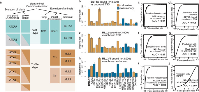
We wondered whether these two functional modes of H3K4 methyltransferases also occur in organisms other than plants. Figure 7a summarizes the lineage of H3K4 methyltransferases in eukaryotes. H3K4 methyltransferases are classified into Set1-type and Trx/Trr-type methyltransferases. ATXR7 and, probably, ATXR3 are Set1-type methyltransferases, while ATX1 to ATX5 belong to the Trx and/or Trr-type methyltransferases (Fig. 7a18,43,). Fungi have lost Trx/Trr-type genes during evolution and exhibit only one Set1-type gene16. We analyzed mammalian SET1A, MLL2, and MLL3/4, for which genome-wide localization data in mouse ESCs are available. The reanalysis of the ChIP-seq datasets revealed that SET1A44 and MLL245 localize around TSSs (typically in the range of −150~300 bp) and, to a lesser extent, around enhancers (Supplementary Fig. 16a, b). On the other hand, MLL3/446,47 localize to enhancers (typically in the range of −900~900 bp from the center of the enhancer) (Supplementary Fig. 16a, b).
Fig. 7. Localization and functional mode of mammalian H3K4 methyltransferases.
a Evolution of H3K4 methyltransferases in eukaryotes based on18,43. b, e, h Chromatin features predictive of the localizations of SET1A (b) and MLL2 (e) at the TSS regions, and MLL3/4 (h) at the enhancer regions. Bars are colored brown if the mean abundance of the feature is bound region > unbound region (i.e. colocalize), and colored blue otherwise (i.e. exclusive). Error bars are the standard deviation of the 5 repeats of training. c, f, i ROC plot showing the prediction accuracy of the random forest models corresponding to (b, e, h). d, g, j ROC plot showing the prediction accuracy using levels of RNAP2 as the sole predictor. All ROC and AUC are calculated with test data (25% of the original data). Error bars represent the standard deviation of the 5 repeats of training. All the genomic data used here are curated from previous works (Supplementary Data 7).
According to the random forest model, the best predictor of both SET1A-bound TSSs and enhancers was the level of RNAP2 (Fig. 7b, c, Supplementary Fig. 16c, d). SET1A strongly colocalizes with RNAP2 (Fig. 7b, d). MLL2-bound TSSs were also best predicted by RNAP2 (Fig. 7e, f, Supplementary Fig. 16e, f). However, the prediction accuracy for the localization of MLL2 according to the RNAP2 level was lower than that for SET1A (Fig. 7d, g), and RNAP2 was not an outstanding predictor (Fig. 7e), suggesting that the colocalization of RNAP2-MLL2 was weaker than that of RNAP2-SET1A. The best predictor of MLL3/4-bound regions was not RNAP2 but H3K27ac, at both enhancers and TSSs (Fig. 7h, i, Supplementary Fig. 16g, h). At enhancers, which are the major localization sites of MLL3/4 (ref. 46,47, and Supplementary Fig. 16a, b), the observed localization was exclusive to sites of RNAP2 occurrence (Fig. 7h, j). These results suggest that SET1A is a cotranscriptional H3K4 methyltransferase, as is the case for its Arabidopsis homolog ATXR7 (and perhaps ATXR3), while MLL3/4 target chromatin irrespective of transcription, similarly to ATX1/2. MLL2 appears to be an intermediate. Therefore, it is plausible that Set1-type H3K4 methyltransferases tend to function in a cotranscriptional manner across eukaryotes, while Trx/Trr-type H3K4 methyltransferases are informed by other chromatin and DNA sequence features (Fig. 8).
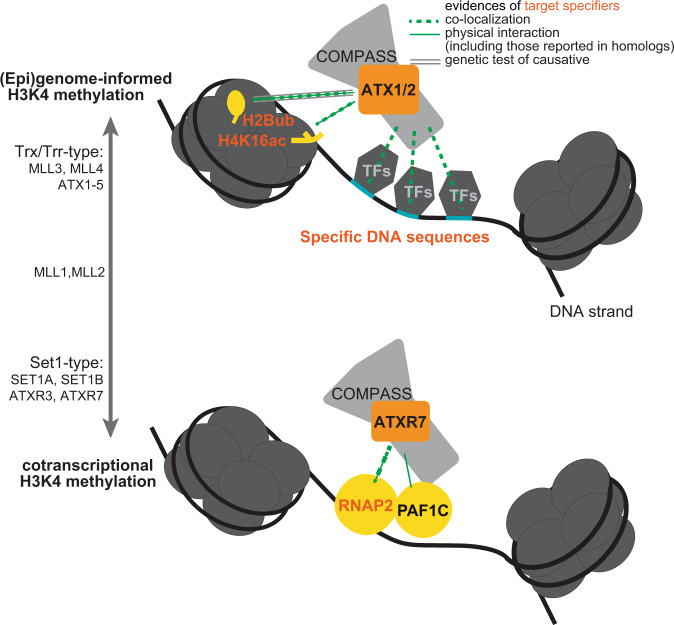
Fig. 8. Suggested models of (epi)genome-informed and cotranscriptional modes of H3K4 methylation.
Random forest and SVM modelings suggest that ATX1 and ATX2 localize based on chromatin features such as H2Bub, H4K16ac (Fig. 3) and several DNA sequences (Fig. 4). Mechanistic studies on ATX homologs indicate that H2Bub53 and H4K16ac52 tether H3K4 methyltransferases to nucleosomes. Some TFs may also bridge ATX to specific DNA sequences. On the other hand, ATXR7 is suggested to be cotranscriptional, based on its colocalization with RNAP2 (Fig. 3) and its contribution to the transcription-H3K4me1 correlation (Fig. 5). Studies in yeast homolog SET1 clarified Polymerase II Associated Factor 1 Complex (PAF1C) interacts with RNAP2 and COMPASS, thus providing a scaffold for H3K4 methyltransferases to work with RNAP26,7. These (epi)genome-informed and cotranscriptional modes of H3K4 methylation may also be generalized to other Arabidopsis H3K4 methyltransferases (Fig. 6) and other organisms (Fig. 7).
Discussion
Here, we revealed in Arabidopsis that the simultaneous loss of the SET-domain histone methyltransferase genes ATX1, ATX2, and ATXR7 causes substantial H3K4me1 loss. In addition, consistent with previous studies19,20,26, our results showed that ATX3, ATX4, and ATX5 redundantly regulate H3K4me2/3 and that ATXR3 regulates H3K4me3. By using a weak allele of ATX3, we were also able to obtain a mutant with specific H3K4me2 loss. Interestingly, these three groups of SET-domain genes are responsible for H3K4me1, H3K4me2/3, and H3K4me3 modifications among a distinct spectrum of genes. The genes that are affected by the loss of these three groups are mutually exclusive, and mutations within them affect H3K4me independently. For example, triple atx1/2/r7 mutation causes the loss of only H3K4me1 and not H3K4me2/3 on a set of genes (ATX1/2/R7-marked genes). This observation implies that other H3K4 methyltransferases can catalyze H3K4me2/3 modifications on unmethylated H3K4 (H3K4me0) in their target regions. Accordingly, ATXR3, for example, was shown to catalyze the H3K4me3 from H3K4me0 in vitro19. In addition, the observation that H3K4me2/3 of ATX1/2/R7-marked genes being not largely affected by atxr3 or atx3/4/5 mutation implies that multiple methyltransferases from the ATX1/2/R7, ATX3/4/5 and ATXR3 groups (and/or other unidentified H3K4 methyltransferase(s)) are redundantly involved in their deposition. Our study clarified the functional division of labor among ATX(R)s genome wide. In contrast to Arabidopsis, yeasts have a single H3K4 methyltransferase, Set1, which is responsible for all H3K4me modifications. The Arabidopsis mutants showing the specific reduction of each H3K4me state identified here will serve as powerful materials for understanding the specific functions of H3K4me1/2/3.
Previous works have reported that atx1 shows H3K4me3 loss21–23,48,49 and that atx2 shows H3K4me2 loss22, on the basis of ChIP-qPCR analyses of selected loci, while our data show that the redundant roles of these enzymes have the largest effect on the global H3K4me1 level. The catalytic domains of ATX1/2/R7 contain a bulky tyrosine at the tyrosine/phenylalanine switch (Supplementary Fig. 17), which is proposed to act as an obstacle to higher-order methylation50, consistent with our conclusion that ATX1/2/R7 primarily regulates H3K4me1. The results of previous studies might reflect indirect effects triggered by altered transcription and/or minor locus-specific effects. Our conclusion is also supported by the results showing that ATX1/2/R7 redundantly repress flowering via FLC activation and that ATXR7 and FLD counteract each other in FLC regulation, probably by modulating H3K4me114,24,51.
Random forest analyses of the genome-wide localization of ATX1 and ATX2 suggested that H3K36me3, H4K16ac and H2Bub are candidate chromatin features for the recruitment of ATX1/2 (Fig. 3, Supplementary Fig. 4). Further examination clarified H2Bub indeed promotes localization of ATX1/2 (Supplementary Fig. 7), while H3K36me3 is promoted by H3K4me1 (Supplementary Fig. 6). The contributions of H4K16ac and H2Bub to ATX1/2 recruitment agree well with previous reports in other species. MLL3 and MLL4 (mammalian homologs of ATX) directly bind to H4K16ac via plant homeodomains (PHDs)52. ATX1 and ATX2 (but not ATXR7) also have PHD domains (Fig. 1a). H2Bub has long been known to promote H3K4 methyltransferase in yeasts and mammals and various explanatory mechanisms have been proposed, while in plants ATXR3 was found not to follow the rule42. Recently, cryo-EM studies revealed that MLL-type H3K4 methyltransferase-containing complexes (complex proteins associated with Set1, COMPASS) attach more firmly to ubiquitinated nucleosomes53.
Motif mining with the lSVM algorithm revealed three notable characteristics of the ATX1/2-bound TSS sequences that coincided with known features of animal Trx/Trr-bound sequences. First, the ATX1/2-bound regions are GAGA-type promoters rather than TATA-box promoters. In Arabidopsis, promoters without TATA boxes (a major core promoter motif) tend to contain GAGA stretch, which are suggested to be equivalent to animal CpG islands54. MLL2 also binds to non-TATA(= CpG)-type promoters45. Second, ATX1/2-bound TSS regions potentially recruit polycomb-group (PcG) proteins, considering that the combination of telobox and GAGA sequences is necessary and sufficient for the recruitment of Arabidopsis PcGs55, which are antagonists of trithorax group (TrxG) proteins. In both plants and animals, TrxG and PcG often target the same genomic regions. Third, both ATX1/2-bound and MLL3/4-bound regions produce noncoding RNAs: sppRNAs and enhancer RNAs46. These results suggest that Trx/Trr methyltransferases bind to a common set of features across eukaryotes.
Our analyses suggested that Set1-type methyltransferases typically function in a cotranscriptional manner and that Trx/Trr-type methyltransferases are less informed by transcription. Although this bifurcation has not been explicitly noted previously, it may underlie the observations of a number of previous works. For instance, the concept that H3K4 methylation occurs cotranscriptionally was founded on studies of Set1 in budding yeast4–7. An interactome analysis of RNAP2 recovered Set1-type methyltransferases but not Trx/Trr-type methyltransferases in human cells56, and the colocalization of RNAP2 with Set1-type, but not Trr/Trx-type methyltransferases, is prominent at the microscopic level in the Drosophila polytene chromosome57. In contrast, various recruitment factors in addition to RNAP2 have been proposed for Trx/Trr-type methyltransferases, including chromatin modifications as described above, TREs, and long noncoding RNAs58. These genome- and/or epigenome-informed methyltransferases may have provided the foundation for multicellularity via the elaborate control of H3K4me, considering that Trr/Trx-type methyltransferases are present in animals and plants but are absent in yeasts. Regulation by both transcription-coupled and (epi)genome-encoded mechanisms may be further generalized to other chromatin modifications, such as H3K36me59.
In addition to transcription-coupled and (epi)genome-encoded pathways which regulate H3K4 methyltransferases, demethylases also shape the pattern of H3K4me. In Arabidopsis, the histone demethylase LDL2 removes H3K4me1 marks from the gene bodies that accumulate H3K9me2 and silences gene expression13. Another histone demethylase, FLD, removes H3K4me1 from sites where convergent overlapping transcription takes place14. Although the biological function of H3K4me remains unclarified, these diverse mechanisms converging on H3K4me1 suggest that the roles of H3K4me require the coordination and integration of a wide range of information. H3K4me is proposed to coordinate splicing60, transcriptional stability61, cryptic transcription62, sense-antisense transcriptions14,63 and to confer transcriptional memory64,65. These apparent multifaceted functions may reflect the multiple regulation mechanisms of this modification. In future studies, the dissection of H3K4me based on its controlling mechanisms may help elucidate the functions of H3K4me.
Methods
Plant materials
T-DNA insertion mutants66,67 used in this study were previously described. Names of mutant alleles such as “atx1-1” or “atx1-2” collide between papers, and the names provided below are not consensus, but examples; atx1-2 (sdg27, SALK_149002C), atx1-3 (SALK_119016C), atx2-1 (sdg30, SALK_074806C), atx2-2 (SALK_117262), atx3-2 (sdg14, SAIL_582_H12, UTR insertion), atx3-1 (sdg14, GK-128H01), atx4 (sdg16, SALK_060156), atx5 (sdg29, SAIL_705_H05), atxr7-1 (sdg25, SALK_149692C), atxr7-2 (SAIL_446_F12), atxr3 (sdg2, SALK_021008), ashh1 (sdg26, SALK_013895), ashh2 (sdg8, SALK_065480), ashh3 (sdg7, SALK_131218C), ashh4 (sdg24, SK22803), ashr3 (sdg4, SALK_128444), hub1-4 (SALK 122512) and hub2-2 (SALK 071289 C). For atx1, atx2, and atxr7 mutants, atx1-2, atx2-1, and atxr7-1 alleles were mainly used. atx1-3, atx2-2, atxr7-2 were used as a replicate to confirm bulk H3K4 methylation levels of atx1/2/r7 mutant in Fig. 1e. For atx3 mutants, we used SAIL_582_H12 (UTR insertion) if not stated otherwise. atx3-1 allele (exon insertion20) was used in Fig. 1e and Supplementary Fig. 2f to compare with preceding research20. For tagged ATX1, ATX2, and ATXR7 lines, the constructs were made with the uniform design; native promoter, which is 3 kb region upstream from initiation codon, followed by 3 x FLAG sequence, coding regions (genomic DNA sequence including introns until just before stop codon) and HA sequence, cloned into pPLV01 vector68. The plasmids were transferred into respective single mutants (ATX1-tag into atx1-2, ATX2-tag into atx2-1, and ATXR7-tag into atxr7-1) via Agrobacterium tumefaciens GV3101::pMP90. Plant lines having homozygous transgene were selected in later generations. All the mutants and ‘wild type (WT)’ are in the Columbia-0 background. For all the experiments, seeds were sown on Murashige and Skoog (MS) plates and kept in dark at 4 °C for a few days, then grown for 15 days under long-day conditions (8 h dark and 16 h light) at 22 °C. Whole seedlings were used for experiments.
Western blotting
Bulk histones for western blotting were prepared using Histone extraction kit (Active Motif) or essentially as described in ref. 69 briefly, 200 mg of frozen seedlings were disrupted into fine powder and suspended in 5 ml of nuclei isolation buffer (NIB: 10 mM MES-KOH pH 5.3, 10 mM NaCl, 10 mM KCl, 250 mM sucrose, 2.5 mM EDTA, 2.5 mM ß-mercaptoethanol, 0.1 mM spermine, 0.1 mM spermidine, 0.3% Triton X-100) supplemented with cOmplete proteinase inhibitor (Roche). The suspension was filtered through 40 µm nylon cell strainer (Falcon) and pelleted by centrifugation at 3000 rpm at 4 °C for 5 min. The pellet was washed twice with NIB, re-suspended in Laemmli loading buffer, and boiled for 5 min. Proteins were resolved on SDS-PAGE, transferred to PVDF membrane (BioRad), and blocked with Blocking one solution (Nacalai). The loaded sample quantities were normalized with H3 or H4. When normalizing with H3, identically loaded blots were respectively incubated with H3 modification specific antibodies (H3K4me1 (ab8895; Abcam), H3K4me2 (ab32356; Abcam), H3K4me3 (ab8580; Abcam)) and H3 antibody (ab1791; Abcam) in 0.5 µg/ml dilution. When normalizing with H4, the membrane was cut horizontally between H3 and H4 using protein ladder markers as a guide. Membranes containing H3 and H4 bands were respectively incubated with H3 modification specific antibodies and H4 antibody (raised and gifted by Dr. Akihisa Osakabe). Second antibody was Anti-Rabbit IgG HRP (NA934; cytiva, 1/10^5 dilution). The blots were developed with ECL prime solutions, and the signal was quantified by iBright Imager (Invitrogen). The signal of H3 modification was divided by H4 or H3 signal to normalize for the amount of protein loaded. The WT to mutant ratios of the normalized modification signals are shown in Supplementary Fig. 2a.
ChIP-seq
ChIP targeting histone modifications, RNAP2, and epitope-tagged proteins were essentially carried out as described previously14. Briefly, approximately 1.5 g frozen seedlings were ground into fine powder and suspended in 25 ml of nuclei isolation buffer (for histone modifications, 10 mM HEPES pH7.6, 1 M sucrose, 5 mM KCl, 5 mM MgCl2, 5 mM EDTA, 1% formaldehyde, 0.1% β-mercaptoethanol, 0.6% Triton X-100, supplemented with 1 tablet/50 ml cOmplete proteinase inhibitor and 1 mM Pefabloc SC (Roche)). The buffer for epitope-tagged proteins additionally contains 0.1 mM spermine, 0.5 mM spermidine and 1.5 mM ethylene glycol bis(succinimidyl succcinate) (EGS; ThermoFisher). Also, 0.6% Triton X-100 was replaced with 0.3% IGEPAL CA-630. The suspension was incubated 10 min in RT to crosslink, quenched with 130 mM glycine, filtered through 40 µm nylon cell strainer and pelleted by centrifugation at 3000 rpm at 4 °C for 10 min. The pellet was resuspended in 300 µl of nuclei isolation buffer and layered on top of 500 µl of nuclei separation buffer (10 mM HEPES pH 7.6, 1 M sucrose, 5 mM KCl, 5 mM MgCl2, 5 mM EDTA pH 8.0, 15% Percoll) and pelleted by centrifugation at 3000 × g for 5 min at 4 °C. For histone modifications, the nuclear pellet was lysed with 150 μl of lysis buffer (50 mM Tris–HCl, pH 7.8, 10 mM EDTA, 1% SDS) then diluted with 800 μl of dilution buffer (50 mM Tris–HCl, pH 7.8, 0.167 M NaCl, 1.1% Triton X-100, 0.11% sodium deoxycholate). For epitope-tagged proteins, the pellet was directly resuspended in 950 µl RIPA buffer (50 mM Tris-HCl pH7.8, 150 mM NaCl, 1 mM EDTA, 0.1% SDS, 1% Triton X-100, 0.1% Sodium deoxycholate and cOmplete proteinase inhibitor). The suspension was sonicated using a Covaris S220 Focused-ultrasonicator (Covaris) and milliTUBE 1 ml AFA Fiber (Covaris) with the following settings: time, 20 min; duty factor, 5%; Cycles per Burst, 200; temperature (water bath), 4–6 °C; and Peak Incident Power, 140 for histones and repeat alternately 105 and 140 for epitope tagged proteins. The sonicated chromatin was then centrifuged at 13,000 g for 3 min, and the supernatant was diluted with RIPA buffer and aliquoted. The chromatin solution was incubated with 1 μg of antibodies overnight at 4 °C. Antibodies are H3, H3K4me1/2/3 (see the section of Western blot), H3K4me0 (MABI0301, MBL, sold as anti-H3 but specific to unmodified H3K4 as shown in ref. 70), H3K36me2 (MABI0332; MBL), H3K36me3 (MABI0333; MBL), H3K27me3 (MABI0323; MBL), H2Bub (MM-0029; Medimabs), H4K16ac (07-329; Millipore), RNAP2 total CTD (MABI0601; MBL), RNAP2 phospho S2 (MABI0602; MBL), RNAP2 phospho S5 (MABI0603; MBL), FLAG (F1804; SIGMA). Then the antibody-chromatin mix was incubated for 2 h at 4 °C with magnetic beads; Dynabeads M280 Sheep anti-mouse IgG in the cases of MBL antibodies, and with Dynabeads Protein G in the cases for the others. For Histone modifications, the incubated beads were washed once with 1 ml of RIPA buffer, twice with 1 ml of High-salt RIPA buffer (RIPA buffer with 500 mM NaCl), once with 1 ml of LiCl buffer (10 mM Tris–HCl, pH 7.8, 1 mM EDTA, 0.25 M LiCl, 1% IGEPAL CA-630, 1% sodium deoxycholate) and once with 1 ml of TE buffer (10 mM Tris–HCl, pH 7.8, 1 mM EDTA), each time rotating for 10 min in 4 °C. For epitope-tagged proteins, beads were washed once with RIPA buffer, medium salt RIPA buffer (RIPA buffer with 300 mM NaCl), high salt RIPA buffer and TE buffer. DNA was eluted from the beads and reverse-crosslinked by adding 100 μl of elution buffer (10 mM Tris–HCl, pH 7.8, 0.3 M NaCl, 5 mM EDTA, 0.5% SDS) and incubating overnight at 65 °C. The DNA samples were then treated with RNase A (Nippon Gene) for 30 min and with Proteinase K (ThermoFisher) for 2 h at 37 °C, and purified using the Monarch PCR & DNA Cleanup Kit (NEB). For spike-in ChIP-seq, chromatin samples of Arabidopsis, adjusted to have the same concentration of DNA, were mixed with chromatin of Schizosaccharomyces pombe (spike-in). Chromatin of S. pombe was prepared as follows; cells were fixed with 1% formaldehyde for 10 min in 37 °C, quenched with glycine, resuspended in Buffer 1 (50 mM HEPES-KOH pH 7.5, 140 mM NaCl, 1 mM EDTA, 1% Triton X-100 and 0.1% sodium deoxycholate supplemented with proteinase inhibitors) and disrupted with bead shocker. The suspension was sonicated by Covaris S220 with a setting similar to that of Arabidopsis histone, except the sonication time being 15 min. The sonicated suspension was centrifuged for 5 min at 13,000 rpm at 4 °C. ChIP-seq libraries were made with KAPA Hyper Prep Kit (Kapa Biosystems), and dual size-selected using AMPureXP (Beckman Coulter) to enrich 200–500 bp fragments. The libraries were 50-bp single-end sequenced by HiSeq4000 sequencer (Illumina) in Vincent J. Coates Genomics Sequencing Laboratory at UC Berkeley, or 150 bp paired-end sequenced by the HiSeqX Ten sequencer (illumina). Biological replicates were conducted on independently grown plants.
ATAC-seq
For ATAC-seq, nuclei preparation, tagmentation and library prep procedure followed sucrose sedimentation protocol of ref. 71. Briefly, 100 mg of frozen seedlings were ground into fine powder, thoroughly resuspended in 10 ml of NPB (20 mM MOPS pH7, 40 mM NaCl, 90 mM KCl, 2 mM EDTA, 0.5 mM EGTA, 0.5 mM spermidine, 0.2 mM spermine, cOmplete protease inhibitor), filtered through 40 µl nylon mesh, then centrifuged for 10 min at 1200 g at 4 °C. The pellet was resuspended in 1 ml of NEB2 (0.25 M Sucrose, 10 mM Tris-HCl pH 8, 10 mM MgCl 2, 1% Triton X-100, cOmplete Protease Inhibitor) and centrifuged for 10 min at 12,000 g at 4 °C. The pellet was resuspended in 300 µl NEB3 (1.7 M Sucrose, 10 mM Tris-HCl pH 8, 2 mM MgCl2, 0.15% Triton X-100, cOmplete proteinase inhibitor), layered on top of 300 µl of NEB3, then centrifuged for 10 min at 16,000 g at 4 °C. This step was repeated twice before the pellet was suspended in NPB. Tagmentation was performed on approximately 25,000 nuclei for 30 min at 37 °C using Tagment DNA TDE1 Enzyme and Buffer Kits (Illumina), with the reaction volume of 25 µl. DNA was immediately purified, amplified with ATAC Primers71 and NEB Next High Fidelity PCR Mix for 7 cycles, and purified with Agencourt AMPure XP. The libraries were 50-bp single-end sequenced.
BS-seq
Whole-genome bisulfite sequencing (BS-seq) was conducted as described before72. DNA was extracted with Nucleon PhytoPure (cytiva) and subjected to bisulphite conversion using MethylCode Bisulfite Conversion Kit (Life Technologies). Bisulphite-treated DNA molecules were PCR amplified with 10 cycles using KAPA HiFi HotStart Uracil+ ReadyMix (Kapa Biosystems) and purified with Agencourt AMPure XP (Beckman Coulter). The libraries were 150 bp paired-end sequenced by the HiSeqX Ten sequencer (llumina).
RNA-seq
Total RNA was extracted from seedling with RNeasy Plant Mini Kit (Qiagen), then polyA selected, fragmented, and made into a library with KAPA Stranded mRNA-seq Kit (Kapa Biosystems) following the manufacturer’s protocol. The libraries were 50-bp single-end sequenced as described above. One sample corresponds to one individual seedling, and three samples were sequenced for one genotype.
Genomes and annotations
For Arabidopsis, TAIR10 reference genome and Araport 11 gene annotations were used. In this paper, ‘gene’ otherwise stated refers to all the transcribed features in Araport11 annotation, including TE genes or non-coding RNAs. ‘protein_coding’ genes refer to the nuclear genes that are annotated as such in Araport11 annotation, excluding the ones that lack primary transcript annotation. For Mus musculus, we used reference genome mm9, which is not the latest, for compatibility reasons. Gene annotations are from NCBI RefSeq. Enhancer annotations are from Enhancer Atlas V273. Corresponding to the cell types of the data origin, MLL3/4 data was analyzed with R1 enhancer annotation, while MLL2 and SET1A data were analyzed with V6.5. For S. pombe, the Ensembl_EF2 genome was used.
Sources of reanalyzed data
For Arabidopsis, the genomic data were reanalyzed from the following works; H3K9me2, DRR02325113; H3K9ac, GSM701925 and H3K18ac, GSM70192774; H3.1, GSM856055 and H3.3, GSM85605475; H1, GSM2544793 and H2A.Z, GSM254479176; H3K14ac, GSM205128577; Like-Heterochromatin Protein 1, GSM202810878; H3K56ac, GSM202781879; H3K23ac, GSM1701017 and H4K16ac, GSM170101880; H3K9me1, GSM124241181; and H2B ubiquitination, GSM309201682. Growth conditions and the tissue-of-origin of these data are summarized as Supplementary Data 4.
For M. musculus, the genomic data were reanalyzed from the following works; SET1A44, MLL2 and its control45, MLL3/4, its control, RNAP2, ATAC and H3K27ac46, H4K16ac83, H3K36me284, H3K9ac, H3K56ac, H3K36me3 and H3K79me385, H4K12ac, H3K9me3 and H4K20me286, H3K18ac, H3K122ac, H4K5ac, H4K8ac and H3K27me387, and DNAme88. The exact names of the analyzed files are provided in Supplementary Data 7. All data is from mESC.
Mapping and counting coverages
For Arabidopsis, ChIP-seq and ATAC-seq data were processed essentially as described in14. Briefly, our sequenced data were mapped to the TAIR10 reference genome using bowtie89 -v 2 -m 1 option. In cases of spike-in ChIP-seq, the sequenced data were also mapped to the S. pombe genome. Mapped reads for single-end sequence reads were extended to 250 bp to represent sequenced fragments, then counted for coverage over the region of interest specified as bed files, using SAMTools90 and BEDTools91. The coverage was RPM/RPKM normalized with custom scripts. Metaplot profiles and heat maps were generated with ngs.plot92. mRNA-seq data were also processed as described in14 using STAR93. BS-seq data were processed as described in13 using Bismark94. For the reanalyses of the published data, SRA data were adapter trimmed following the original papers’ procedure, mapped, and counted essentially the same as above.
For M. musculus, SRR data of ChIP-seq targeting H3K4 methyltransferases were adapter-trimmed and mapped to mm9 reference genome with -v 2 -m 1 option, then counted and normalized as described above. The other genomics data of M. musculus, namely the data of chromatin modifications used for the random forest, were curated and parsed from bigWig format provided as supplementary files at Gene Expression Omnibus (GEO) (Supplementary Data 7). bigWig files were first converted to bedGraph with bigWigtoBedGraph95 of UCSC tools, then to bed file with the awk command of Unix, and when applicable, genome version was converted into mm9 with liftOver of UCSC tools, then coverage were counted over the regions of interest.
Clustering of the 6 single mutants
We selected the top 3000 genes that had the highest reduction of H3K4me1 (RPKM) compared to WT for each mutant. The regions from TSS to TTS of the 3000 genes were split into 6 bins. For the WT and the six mutants, the number of reads mapped to each of the 6 sub-gene regions were counted, RPM normalized, and the difference between WT and the mutant was calculated. To capture the trend of which ones of the 6 bins lose H3K4me1, 6 bins in each one gene were standardized. Thus, for each mutant, a matrix of 6 regions x 3000 genes was obtained. These matrices were vectorized, and Euclid distances were calculated, which were used for hierarchical clustering (Fig. 1b). To explain the idea behind this clustering, considering if a hypothetical methyltransferase catalyzing H3K4me1 into H3K4me2 is lost, H3K4me1 level would be elevated in the region that H3K4me2 originally occupied. We reasoned that similarity in affected sub-gene regions reflects the similarity in function.
Peak calling
For Arabidopsis, differentially modified regions were identified by comparing mutant to WT using MACS296 with option -g 1.3e8. Peaks detected in centromeres are removed. Peaks were assigned to overlapping genes by bedtools ‘intersect’ with default options. If a peak is assigned to multiple genes, all genes are kept. If multiple peaks are assigned to one gene, one gene is added to the list.
Enrichment peaks of H3K4 methyltransferases were identified by comparing against negative control (ChIP-seq with FLAG antibody in non-transgenic WT) using MACS296 with two settings; the default (-g 1.3e8) and more relaxed option (-g 1.3e8 -q 0.3 –nomodel). Both counts are provided as Supplementary Data. 3. The latter relaxed peaks are used to measure the distances of the peak summits of from the nearest TSS (in the cases of ATX1, ATX2, and ATXR3) or TTS (in the case of ATXR7), which were visualized as a histogram (e.g. Fig. 2d). Based on the histogram, we identified the region that ATX1, ATX2 typically occupy to be 150 bp upstream and 300 bp downstream of TSS, which we named ‘TSS region’. Similarly, ‘TTS region’ is defined as 200 bp upstream to 200 bp downstream of TTS, where ATXR7 localizes, and ‘TSS region 2’ starts from 200 bp downstream of TSS to further 400 bp, where ATXR3 localizes. For M. musculus, enrichment peaks of H3K4 methyltransferases were called with default options of MACS2. The distances of the summits of the called peaks from the nearest TSS or the midpoint of enhancer region (in the cases of MLL2 and SET1A, V6.5 enhancer, while in the case of MLL3/4, R1 enhancer) were measured and visualized as a histogram. Based on the histogram, we identified the region that SET1A and MLL2 typically occupy to be 150 bp upstream and 300 bp downstream of TSS, coinciding with the ‘TSS region’ of Arabidopsis. Similarly, we defined ‘enhancer region’, where MLL3/4 localizes, to be 900 bp on both sides from the center of Enhancer Atlas’s annotation.
Spike-in normalization
The summary statistic R, plotted in Supplementary Fig. 1d was calculated as follows; R = ratio of the number of reads that mapped to Arabidopsis and to S/S.p.). RWT_H3 : Rmutant_H3 = RWT_H3K4me1 : Expected Rmutant_H3K4me. (Actual Rmutant_H3K4me1)/(Expected Rmutant_H3K4me1) for each mutant was plotted in the bar graph. The raw counts of reads are summarized in Supplementary Data 2.
Random forest
For ATX1, ATX2, and ATXR7 models, genes shorter than 500 bp were filtered out. Gene was divided into three regions; TSS region, TTS region (definitions described above), and “gene body” region. The “gene body” region is the transcribed region minus TSS and TTS regions. Similarly, for ATXR3 models, the transcribed regions of genes longer than 800 bp were divided into TSS, TSS2, TTS, and “gene body2” regions. “Gene body2” region is the coding region minus TSS, TSS2, and TTS regions.
Explanatory features are levels of chromatin modifications/chromatin states calculated over these three regions per gene. We generated data from samples under the same conditions as ATX(R) ChIP for some modifications that seemed to be particularly variable or well-studied (Expression, RNAP2, H3K27me3, H3K36me3, DNAm) to minimize tissue/stage derived variation. For the other modifications, raw read files reported by preceding works (see ‘sources of the reanalyzed data’ section and Supplementary Data 4) were used. For “gene body” and “gene body2” regions, both length-normalized and unnormalized values are included. These data combined with expression level (FPKM of mRNA-seq in WT, from this work) and gene length were used as the predictor variables.
Objective variables were set as two classes; ATX(R)-bound and -unbound genes. ATX(R)-bound genes are defined as the top 3000 genes that have the highest ChIP-seq signal (RPM values of ATX(R)-Tag expressing lines - negative control) in the typical occupying region defined from the above-mentioned peak calling; TSS region for ATX1 and ATX2, TTS region for ATXR7, TSS2 region for ATXR3. ATX(R)-unbound genes are the rest of protein coding genes (n = all protein coding genes longer than 500 or 800 bp − 3000). We uniformly set the number of bound genes to 3000, at the potential expense of predictive accuracy, in order to reduce parameter variance between models.
To balance the two classes, 3000 genes out of the ATX(R)-unbound genes were randomly selected, and the data on chromosome 1 to 4 were used for training random forest (R package ‘RandomForest’), while data on chromosome 5 were held out as test data.
For SET1A, MLL2, and MLL3/4 models, explanatory features are levels of chromatin states calculated over TSS regions or enhancer regions, using previously reported data in mESC (see ‘sources of the reanalyzed data’ section). Objective variables were set in the same manner as ATX(R)s models; that is, Set1/MLL2/MLL3/4-bound regions were defined as the top 3000 regions with the highest TSS region signals regions or enhancer regions, and unbound regions were down-sampled into 3000. 25% of the data were held out from the training and used as test data.
Both for Arabidopsis and M. musculus, the random sampling and training steps were repeated 5 times, and the average and the standard deviation of the importance derived from the 5 models were plotted. ROC was plotted based on the prediction of the 5 models, using test data.
In order to simulate ATX1/2 localization in the absence of H2Bub, a feature matrix, in which all H2Bub data was replaced with 0, was given to the trained random forest models to make predictions. ‘Genes that lose ATX1 in the in silico hub’ was defined as genes that are predicted to be ATX1-bound at least once among the 5 models that constitute the original random forest model, and predicted to not to be bound at least once among the 5 models that are given the H2Bub-0 feature matrix.
Support vector machine
Application of SVM to DNA sequence essentially followed previous work on human enhancer classification97. Briefly, DNA sequences on the coding strand of TSS regions were converted to k-mer frequency vectors of 4^k features. The frequency vectors were standardized and given bound or unbound labels in the same way as random forest.
The data on chromosome 5 were held out as test data. Data on the chromosome 1 to 4 were used for 5 cross-fold validation for training to select the optimal parameters. For each trial of cross-fold validation, data of the unbound class were randomly sampled independently to balance the two classes. The averaged weights of features (k-mer sequences), which were calculated based on the 5 independently trained models using the best parameters, were used for further motif annotations. ROC was plotted based on the prediction of the 5 models with test data. We trained 5,6,7-mer lSVM or 6-mer kernel-SVM to find that the accuracy of 6-mer lSVM was about the same or better than the rest (Supplementary Fig. 18).
Motif annotation
The decision function of linear SVM is the sum of weighted features (in this case, standardized occurrence of each k-mers), which allows us to interpret the highly weighted motifs to be predictively important ones. Using the weights averaged from the 5 best models, sixty 6-mers that had the largest positive weights, largest negative weights, and smallest absolute weights, and sixty random 6-mers were selected.
To make clusters of related motifs within each group of sixty 6-mers, similarity scores were calculated between all pairs of the 6-mers. Similarity scores are defined as; reverse complement (=1) > 1 nt offset > 1 nt offset on the opposite strand > the others (=0). The resulting similarity score matrices were visualized with the qgraph98 package in R.
Next, to annotate the 6-mers while considering the context the 6-mer is located, we calculated the ATGC frequency in the flanking three bases on both sides of each 6-mer. The flanking ATGC frequency of the positively weighted sixty 6-mers was calculated with the sequence of ATX-bound TSS region, while negatively weighted and random/near-zero weighted 6-mers were calculated with the sequence of ATX-unbound and random TSS region, respectively. The resulting probability matrices representing 3 nt + 6 nt + 3 nt were converted into meme format and searched for matching motifs using TOMTOM against all the Arabidopsis databases included in the MEME Suite’s motif_databases.12.1999. For each motif, TOMTOM hits were summarized in Supplementary Data 6, and top-hit satisfying q.value< 0.1 is indicated by colors representing TF families (e.g. Fig. 4e, f). The distribution of the motifs of interest, i.e., GAGA-stretch, TATA-box, telobox, and RGCCCAW, was visualized by the presence of the 6-mers comprising the motifs. Namely, {AAGAGA, GAGAGA, AAGAGG, GAGAGG, AGAGAA, GGAGAA, AGAGAG, GGAGAG} represented GAGA-stretch, {TATAAA, TATATA, ATATAA, ATAAAT, TAAATA, ATATAT, TTATAA, TTATAT} represented TATA, {AAACCC, AACCCT, ACCCAA, ACCCTA} represented telobox and {AAGCCC, AGGCCC, GGCCCA, AGCCCA, GCCCAA, GCCCAT, GGGCTT, GGGCCT, TGGGCC, TGGGCT, TTGGGC, ATGGGC, AATGGG, CCCATT} represented ARGCCCAWT.
The PhastCon100 score calculated by ref. 101, based on 63 angiosperm species was averaged per nucleotide on each 6-mer appearing in the directional sequence of the TSS region of all Arabidopsis genes as an index of sequence conservation.
Correlation between H3K4me and transcription
Spearman correlation between mRNA-seq and ChIP-seq data of H3K4me was calculated using all protein-coding genes. mRNA-seq data was FPKM normalized, ChIP-seq data targeting H3K4me1 was RPKM, H3K4me2/H3K4me3 was RPM normalized. For color-filled contour plots, genes for which no transcription was detected were excluded.
Reporting summary
Further information on research design is available in the Nature Research Reporting Summary linked to this article.
Supplementary information
Description of Additional Supplementary Files
Acknowledgements
We thank all Kakutani laboratory members for helpful discussion and technical assistance. This work used the Vincent J. Coates Genomics Sequencing Laboratory at UC Berkeley, supported by NIH S10 OD018174 Instrumentation Grant. The computations were partially performed on the NIG supercomputer at NIG, Japan. We thank NASC/ABRC for distributing the seeds. The ashr3 mutant was kindly gifted by Taku Sasaki. This work was supported by grants from Japan Science and Technology Agency (JST) PRESTO (no. JPMJPR17Q1) to S.I., JST CREST (no. JPMJCR15O1) to T.K., and Japan Society for the Promotion of Science (JSPS) (nos. JP26221105, JP15H05963 and JP19H00995 to T.K., JP20H05913 and JP22H02299 to S.I. and JP19J21882 to S.O.). S.O. is supported by the JSPS Research Fellowship for Young Scientists.
Source data
Author contributions
S.O., T.K. and S.I. conceived the study. S.O., M.T., K.T., and S.I. performed the experiments. S.O. conducted the analysis. S.O. drafted and S.O., T.K. and S.I. edited the manuscript.
Peer review
Peer review information
Nature Communications thanks Alexandre Berr, Zhaobo Lang and the other, anonymous, reviewer(s) for their contribution to the peer review of this work. Peer reviewer reports are available.
Data availability
The high-throughput sequencing data generated in this study is available in the NCBI database under the accession number PRJNA732996. Source data are provided with this paper.
Code availability
Codes are available on GitHub (https://github.com/Satoyo08/Arabidopsis_H3K4me1).
Competing interests
The authors declare no competing interests.
Footnotes
Publisher’s note Springer Nature remains neutral with regard to jurisdictional claims in published maps and institutional affiliations.
Contributor Information
Satoyo Oya, Email: o.satoyo@bs.s.u-tokyo.ac.jp.
Tetsuji Kakutani, Email: tkak@bs.s.u-tokyo.ac.jp.
Soichi Inagaki, Email: soinagak@bs.s.u-tokyo.ac.jp.
Supplementary information
The online version contains supplementary material available at 10.1038/s41467-022-32165-8.
References
- 1.Allis CD, Jenuwein T. The molecular hallmarks of epigenetic control. Nat. Rev. Genet. 2016;17:487–500. doi: 10.1038/nrg.2016.59. [DOI] [PubMed] [Google Scholar]
- 2.Howe FS, Fischl H, Murray SC, Mellor J. Is H3K4me3 instructive for transcription activation? Bioessays. 2017;39:1–12. doi: 10.1002/bies.201600095. [DOI] [PubMed] [Google Scholar]
- 3.Henikoff S, Shilatifard A. Histone modification: cause or cog? Trends Genet. 2011;27:389–396. doi: 10.1016/j.tig.2011.06.006. [DOI] [PubMed] [Google Scholar]
- 4.Soares LM, et al. Determinants of Histone H3K4 Methylation Patterns. Mol. Cell. 2017;68:773–785. doi: 10.1016/j.molcel.2017.10.013. [DOI] [PMC free article] [PubMed] [Google Scholar]
- 5.Bae, H. J. et al. The Set1 N-terminal domain and Swd2 interact with RNA polymerase II CTD to recruit COMPASS. Nat. Commun. 11, 2181 (2020). [DOI] [PMC free article] [PubMed]
- 6.Krogan NJ, et al. The Paf1 complex is required for Histone H3 Methylation by COMPASS and Dot1p: linking transcriptional elongation to Histone Methylation. Mol. Cell. 2003;11:721–729. doi: 10.1016/S1097-2765(03)00091-1. [DOI] [PubMed] [Google Scholar]
- 7.Ng HH, Robert F, Young RA, Struhl K. Targeted recruitment of Set1 histone methylase by elongating Pol II provides a localized mark and memory of recent transcriptional activity. Mol. Cell. 2003;11:709–719. doi: 10.1016/S1097-2765(03)00092-3. [DOI] [PubMed] [Google Scholar]
- 8.Wachter E, et al. Synthetic CpG islands reveal DNA sequence determinants of chromatin structure. Elife. 2014;3:e03397. doi: 10.7554/eLife.03397. [DOI] [PMC free article] [PubMed] [Google Scholar]
- 9.Hontelez S, et al. Embryonic transcription is controlled by maternally defined chromatin state. Nat. Commun. 2015;6:10148. doi: 10.1038/ncomms10148. [DOI] [PMC free article] [PubMed] [Google Scholar]
- 10.Zhang X, Bernatavichute YV, Cokus S, Pellegrini M, Jacobsen SE. Genome-wide analysis of mono-, di- and trimethylation of histone H3 lysine 4 in Arabidopsis thaliana. Genome Biol. 2009;10:R62. doi: 10.1186/gb-2009-10-6-r62. [DOI] [PMC free article] [PubMed] [Google Scholar]
- 11.Leng X, Thomas Q, Rasmussen SH, Marquardt S. A G(enomic)P(ositioning)S(ystem) for plant RNAPII transcription. Trends Plant Sci. 2020;25:744–764. doi: 10.1016/j.tplants.2020.03.005. [DOI] [PubMed] [Google Scholar]
- 12.Barski A, et al. High-resolution profiling of histone methylations in the human genome. Cell. 2007;129:823–837. doi: 10.1016/j.cell.2007.05.009. [DOI] [PubMed] [Google Scholar]
- 13.Inagaki S, et al. Gene-body chromatin modification dynamics mediate epigenome differentiation in Arabidopsis. EMBO J. 2017;36:970–980. doi: 10.15252/embj.201694983. [DOI] [PMC free article] [PubMed] [Google Scholar]
- 14.Inagaki S, Takahashi M, Takashima K, Oya S, Kakutani T. Chromatin-based mechanisms to coordinate convergent overlapping transcription. Nat. Plants. 2021;7:295–302. doi: 10.1038/s41477-021-00868-3. [DOI] [PubMed] [Google Scholar]
- 15.Cenik BK, Shilatifard A. COMPASS and SWI/SNF complexes in development and disease. Nat. Rev. Genet. 2021;22:38–58. doi: 10.1038/s41576-020-0278-0. [DOI] [PubMed] [Google Scholar]
- 16.Veerappan CS, Avramova Z, Moriyama EN. Evolution of SET-domain protein families in the unicellular and multicellular Ascomycota fungi. BMC Evol. Biol. 2008;8:1–20. doi: 10.1186/1471-2148-8-190. [DOI] [PMC free article] [PubMed] [Google Scholar]
- 17.Baumbusch LO, et al. The Arabidopsis thaliana genome contains at least 29 active genes encoding SET domain proteins that can be assigned to four evolutionarily conserved classes. Nucleic Acids Res. 2001;29:4319–4333. doi: 10.1093/nar/29.21.4319. [DOI] [PMC free article] [PubMed] [Google Scholar]
- 18.Springer NM, et al. Comparative analysis of SET domain proteins in Maize and Arabidopsis reveals multiple duplications preceding the divergence of monocots and Dicots. Plant Physiol. 2003;132:907–925. doi: 10.1104/pp.102.013722. [DOI] [PMC free article] [PubMed] [Google Scholar]
- 19.Guo L, Yu Y, Law JA, Zhang X. SET DOMAIN GROUP2 is the major histone H3 lysine 4 trimethyltransferase in Arabidopsis. Proc. Natl Acad. Sci. USA. 2010;107:18557–18562. doi: 10.1073/pnas.1010478107. [DOI] [PMC free article] [PubMed] [Google Scholar]
- 20.Chen L-Q, et al. ATX3, ATX4, and ATX5 Encode Putative H3K4 Methyltransferases and Are Critical for Plant Development. Plant Physiol. 2017;174:1795–1806. doi: 10.1104/pp.16.01944. [DOI] [PMC free article] [PubMed] [Google Scholar]
- 21.Pien S, et al. ARABIDOPSIS TRITHORAX1 Dynamically Regulates FLOWERING LOCUS C Activation via Histone 3 Lysine 4 Trimethylation. Plant Cell. 2008;20:580–588. doi: 10.1105/tpc.108.058172. [DOI] [PMC free article] [PubMed] [Google Scholar]
- 22.Saleh A, et al. The highly similar Arabidopsis homologs of trithorax ATX1 and ATX2 encode proteins with divergent biochemical functions. Plant Cell. 2008;20:568–579. doi: 10.1105/tpc.107.056614. [DOI] [PMC free article] [PubMed] [Google Scholar]
- 23.Saleh A, Al-abdallat A, Ndamukong I, Alvarez-venegas R, Avramova Z. The Arabidopsis homologs of trithorax (ATX1) and enhancer of zeste (CLF) establish ‘ bivalent chromatin marks’ at the silent AGAMOUS locus. Nucleic Acids Res. 2007;35:6290–6296. doi: 10.1093/nar/gkm464. [DOI] [PMC free article] [PubMed] [Google Scholar]
- 24.Tamada Y, Yun J-Y, Amasino RM. ARABIDOPSIS TRITHORAX-RELATED7 Is Required for Methylation of Lysine 4 of Histone H3 and for Transcriptional Activation of FLOWERING LOCUS C. Plant Cell. 2009;21:3257–3269. doi: 10.1105/tpc.109.070060. [DOI] [PMC free article] [PubMed] [Google Scholar]
- 25.Alvarez-venegas R, et al. ATX-1, an Arabidopsis Homolog of Trithorax, Activates Flower Homeotic Genes. Curr. Biol. 2003;13:627–637. doi: 10.1016/S0960-9822(03)00243-4. [DOI] [PubMed] [Google Scholar]
- 26.Berr A, et al. Arabidopsis SET DOMAIN GROUP2 is required for H3K4 trimethylation and is crucial for both sporophyte and gametophyte development. Plant Cell. 2010;22:3232–3248. doi: 10.1105/tpc.110.079962. [DOI] [PMC free article] [PubMed] [Google Scholar]
- 27.Xia S, et al. Regulation of transcription of nucleotide-binding leucine-rich repeat-encoding genes SNC1 and RPP4 via H3K4 trimethylation. Plant Physiol. 2013;162:1694–1705. doi: 10.1104/pp.113.214551. [DOI] [PMC free article] [PubMed] [Google Scholar]
- 28.Roudier F, et al. Integrative epigenomic mapping defines four main chromatin states in Arabidopsis. EMBO J. 2011;30:1928–1938. doi: 10.1038/emboj.2011.103. [DOI] [PMC free article] [PubMed] [Google Scholar]
- 29.Liu Y, et al. H3K4me2 functions as a repressive epigenetic mark in plants. Epigenetics Chromatin. 2019;12:40. doi: 10.1186/s13072-019-0285-6. [DOI] [PMC free article] [PubMed] [Google Scholar]
- 30.Ishihara H, et al. Primed histone demethylation regulates shoot regenerative competency. Nat. Commun. 2019;10:1786. doi: 10.1038/s41467-019-09386-5. [DOI] [PMC free article] [PubMed] [Google Scholar]
- 31.Xu L, et al. Di- and Tri- but Not Monomethylation on Histone H3 Lysine 36 Marks Active Transcription of Genes Involved in Flowering Time Regulation and Other Processes in Arabidopsis thaliana. Mol. Cell. Biol. 2008;28:1348–1360. doi: 10.1128/MCB.01607-07. [DOI] [PMC free article] [PubMed] [Google Scholar]
- 32.Dong G, Ma DP, Li J. The histone methyltransferase SDG8 regulates shoot branching in Arabidopsis. Biochem. Biophys. Res. Commun. 2008;373:659–664. doi: 10.1016/j.bbrc.2008.06.096. [DOI] [PubMed] [Google Scholar]
- 33.Li Y, et al. The histone methyltransferase SDG8 mediates the epigenetic modification of light and carbon responsive genes in plants. Genome Biol. 2015;16:79. doi: 10.1186/s13059-015-0640-2. [DOI] [PMC free article] [PubMed] [Google Scholar]
- 34.Liu Y, Huang Y. Uncovering the mechanistic basis for specific recognition of monomethylated H3K4 by the CW domain of Arabidopsis histone methyltransferase SDG8. J. Biol. Chem. 2018;293:6470–6481. doi: 10.1074/jbc.RA117.001390. [DOI] [PMC free article] [PubMed] [Google Scholar]
- 35.Ding Y, Avramova Z, Fromm M. Two Distinct Roles of ARABIDOPSIS HOMOLOG OF TRITHORAX1 (ATX1) at Promoters and within Transcribed Regions of ATX1-Regulated Genes. Plant Cell. 2011;23:350–363. doi: 10.1105/tpc.110.080150. [DOI] [PMC free article] [PubMed] [Google Scholar]
- 36.Franco-Zorrilla JM, et al. DNA-binding specificities of plant transcription factors and their potential to define target genes. Proc. Natl Acad. Sci. U. S. A. 2014;111:2367–2372. doi: 10.1073/pnas.1316278111. [DOI] [PMC free article] [PubMed] [Google Scholar]
- 37.Kosugi S, Ohashi Y. DNA binding and dimerization specificity and potential targets for the TCP protein family. Plant J. 2002;30:337–348. doi: 10.1046/j.1365-313X.2002.01294.x. [DOI] [PubMed] [Google Scholar]
- 38.Viola IL, Reinheimer R, Ripoll R, Manassero NGU, Gonzalez DH. Determinants of the DNA binding specificity of class I and class II TCP transcription factors. J. Biol. Chem. 2012;287:347–356. doi: 10.1074/jbc.M111.256271. [DOI] [PMC free article] [PubMed] [Google Scholar]
- 39.O’Malley RC, et al. Cistrome and Epicistrome Features Shape the Regulatory DNA Landscape. Cell. 2016;165:1280–1292. doi: 10.1016/j.cell.2016.04.038. [DOI] [PMC free article] [PubMed] [Google Scholar]
- 40.Hecker A, et al. The Arabidopsis GAGA-binding factor BASIC PENTACYSTEINE6 recruits the POLYCOMB-REPRESSIVE COMPLEX1 component LIKE HETEROCHROMATIN PROTEIN1 to GAGA DNA motifs. Plant Physiol. 2015;168:130–141. doi: 10.1104/pp.15.00409. [DOI] [PMC free article] [PubMed] [Google Scholar]
- 41.Thomas QA, et al. Transcript isoform sequencing reveals widespread promoter-proximal transcriptional termination in Arabidopsis. Nat. Commun. 2020;11:2589. doi: 10.1038/s41467-020-16390-7. [DOI] [PMC free article] [PubMed] [Google Scholar]
- 42.Fiorucci, A.-S. et al. Arabidopsis S2Lb links AtCOMPASS-like and SDG2 activity in H3K4me3 independently from histone H2B monoubiquitination. Genome Biol. 20, 100 (2019). [DOI] [PMC free article] [PubMed]
- 43.Zhang L, Ma H. Complex evolutionary history and diverse domain organization of SET proteins suggest divergent regulatory interactions. New Phytol. 2012;195:248–263. doi: 10.1111/j.1469-8137.2012.04143.x. [DOI] [PubMed] [Google Scholar]
- 44.Sze CC, et al. Histone H3K4 methylation-dependent and -independent functions of set1A/COMPASS in embryonic stem cell self-renewal and differentiation. Genes Dev. 2017;31:1732–1737. doi: 10.1101/gad.303768.117. [DOI] [PMC free article] [PubMed] [Google Scholar]
- 45.Hu D, et al. Not All H3K4 Methylations Are Created Equal: Mll2/COMPASS Dependency in Primordial Germ Cell Specification. Mol. Cell. 2017;65:460–475. doi: 10.1016/j.molcel.2017.01.013. [DOI] [PMC free article] [PubMed] [Google Scholar]
- 46.Dorighi KM, et al. Mll3 and Mll4 Facilitate Enhancer RNA Synthesis and Transcription from Promoters Independently of H3K4 Monomethylation. Mol. Cell. 2017;66:568–576. doi: 10.1016/j.molcel.2017.04.018. [DOI] [PMC free article] [PubMed] [Google Scholar]
- 47.Cao K, et al. An Mll4/COMPASS-Lsd1 epigenetic axis governs enhancer function and pluripotency transition in embryonic stem cells. Sci. Adv. 2018;4:eaap8747. doi: 10.1126/sciadv.aap8747. [DOI] [PMC free article] [PubMed] [Google Scholar]
- 48.Alvarez-venegas, R. & Avramova, Z. Methylation patterns of histone H3 Lys 4, Lys 9 and Lys 27 in transcriptionally active and inactive Arabidopsis genes and in atx1 mutants. Nucleic Acids Res. 33, 5199-5207 (2005). [DOI] [PMC free article] [PubMed]
- 49.Alvarez-venegas R, Abdallat AA, Alfano JR, Avramova Z. Epigenetic Control of a Transcription Factor at the Cross Section of Two Antagonistic Pathways. Epigenetics. 2007;2:106–117. doi: 10.4161/epi.2.2.4404. [DOI] [PubMed] [Google Scholar]
- 50.Collins RE, et al. In vitro and in vivo analyses of a Phe/Tyr switch controlling product specificity of histone lysine methyltransferases. J. Biol. Chem. 2005;280:5563–5570. doi: 10.1074/jbc.M410483200. [DOI] [PMC free article] [PubMed] [Google Scholar]
- 51.Yun JY, Tamada Y, Kang YE, Amasino RM. Arabidopsis trithorax-related3/set domain group2 is required for the winter-annual habit of arabidopsis thaliana. Plant Cell Physiol. 2012;53:834–846. doi: 10.1093/pcp/pcs021. [DOI] [PMC free article] [PubMed] [Google Scholar]
- 52.Liu Y, et al. Structural insights into trans-histone regulation of H3K4 methylation by unique histone H4 binding of MLL3/4. Nat. Commun. 2019;10:36. doi: 10.1038/s41467-018-07906-3. [DOI] [PMC free article] [PubMed] [Google Scholar]
- 53.Xue H, et al. Structural basis of nucleosome recognition and modification by MLL methyltransferases. Nature. 2019;573:445–449. doi: 10.1038/s41586-019-1528-1. [DOI] [PubMed] [Google Scholar]
- 54.Yamamoto YY, et al. Heterogeneity of Arabidopsis core promoters revealed by high-density TSS analysis. Plant J. 2009;60:350–362. doi: 10.1111/j.1365-313X.2009.03958.x. [DOI] [PubMed] [Google Scholar]
- 55.Xiao J, et al. Cis and trans determinants of epigenetic silencing by Polycomb repressive complex 2 in Arabidopsis. Nat. Genet. 2017;49:1546–1552. doi: 10.1038/ng.3937. [DOI] [PubMed] [Google Scholar]
- 56.Ebmeier CC, et al. Human TFIIH Kinase CDK7 Regulates Transcription-Associated Chromatin Modifications. Cell Rep. 2017;20:1173–1186. doi: 10.1016/j.celrep.2017.07.021. [DOI] [PMC free article] [PubMed] [Google Scholar]
- 57.Mohan M, et al. The COMPASS Family of H3K4 Methylases in Drosophila. Mol. Cell. Biol. 2011;31:4310–4318. doi: 10.1128/MCB.06092-11. [DOI] [PMC free article] [PubMed] [Google Scholar]
- 58.Yang YW, et al. Essential role of lncRNA binding for WDR5 maintenance of active chromatin and embryonic stem cell pluripotency. Elife. 2014;3:e02046. doi: 10.7554/eLife.02046. [DOI] [PMC free article] [PubMed] [Google Scholar]
- 59.Bicocca VT, Ormsby T, Adhvaryu KK, Honda S, Selker EU. ASH1-catalyzed H3K36 methylation drives gene repression and marks H3K27me2/3-competent chromatin. Elife. 2018;7:e41497. doi: 10.7554/eLife.41497. [DOI] [PMC free article] [PubMed] [Google Scholar]
- 60.Sims RJ, 3rd, et al. Recognition of trimethylated histone H3 lysine 4 facilitates the recruitment of transcription postinitiation factors and pre-mRNA splicing. Mol. Cell. 2007;28:665–676. doi: 10.1016/j.molcel.2007.11.010. [DOI] [PMC free article] [PubMed] [Google Scholar]
- 61.Benayoun BA, et al. H3K4me3 breadth is linked to cell identity and transcriptional consistency. Cell. 2014;158:673–688. doi: 10.1016/j.cell.2014.06.027. [DOI] [PMC free article] [PubMed] [Google Scholar]
- 62.Nielsen M, et al. Transcription-driven chromatin repression of Intragenic transcription start sites. PLoS Genet. 2019;15:e1007969. doi: 10.1371/journal.pgen.1007969. [DOI] [PMC free article] [PubMed] [Google Scholar]
- 63.Kim T, Xu Z, Clauder-Münster S, Steinmetz LM, Buratowski S. Set3 HDAC mediates effects of overlapping noncoding transcription on gene induction kinetics. Cell. 2012;150:1158–1169. doi: 10.1016/j.cell.2012.08.016. [DOI] [PMC free article] [PubMed] [Google Scholar]
- 64.Muramoto T, Müller I, Thomas G, Melvin A, Chubb JR. Methylation of H3K4 Is required for inheritance of active transcriptional states. Curr. Biol. 2010;20:397–406. doi: 10.1016/j.cub.2010.01.017. [DOI] [PubMed] [Google Scholar]
- 65.Ding Y, Fromm M, Avramova Z. Multiple exposures to drought ‘train’ transcriptional responses in Arabidopsis. Nat. Commun. 2012;3:740. doi: 10.1038/ncomms1732. [DOI] [PubMed] [Google Scholar]
- 66.Alonso JM, et al. Genome-wide insertional mutagenesis of Arabidopsis thaliana. Science. 2003;301:653–657. doi: 10.1126/science.1086391. [DOI] [PubMed] [Google Scholar]
- 67.Kleinboelting N, Huep G, Kloetgen A, Viehoever P, Weisshaar B. GABI-Kat SimpleSearch: new features of the Arabidopsis thaliana T-DNA mutant database. Nucleic Acids Res. 2012;40:D1211–D1215. doi: 10.1093/nar/gkr1047. [DOI] [PMC free article] [PubMed] [Google Scholar]
- 68.De Rybel B, et al. A Versatile Set of Ligation-Independent Cloning Vectors for Functional Studies in Plants. Plant Physiol. 2011;156:1292–1299. doi: 10.1104/pp.111.177337. [DOI] [PMC free article] [PubMed] [Google Scholar]
- 69.Lorković ZJ, et al. Compartmentalization of DNA Damage Response between Heterochromatin and Euchromatin Is Mediated by Distinct H2A Histone Variants. Curr. Biol. 2017;27:1192–1199. doi: 10.1016/j.cub.2017.03.002. [DOI] [PubMed] [Google Scholar]
- 70.Kimura H, Hayashi-Takanaka Y, Goto Y, Takizawa N, Nozaki N. The Organization of Histone H3 Modifications as Revealed by a Panel of Specific Monoclonal Antibodies. Cell Structure and Function. 2008;33:61–73. doi: 10.1247/csf.07035. [DOI] [PubMed] [Google Scholar]
- 71.Bajic M, Maher KA, Deal RB. Identification of Open Chromatin Regions in Plant Genomes Using ATAC-Seq. Methods Mol. Biol. 2018;1675:183–201. doi: 10.1007/978-1-4939-7318-7_12. [DOI] [PMC free article] [PubMed] [Google Scholar]
- 72.Fu Y, et al. Mobilization of a plant transposon by expression of the transposon-encoded anti-silencing factor. EMBO J. 2013;32:2407–2417. doi: 10.1038/emboj.2013.169. [DOI] [PMC free article] [PubMed] [Google Scholar]
- 73.Gao T, Qian J. EnhancerAtlas 2.0: an updated resource with enhancer annotation in 586 tissue/cell types across nine species. Nucleic Acids Res. 2020;48:D58–D64. doi: 10.1093/nar/gkaa197. [DOI] [PMC free article] [PubMed] [Google Scholar]
- 74.Luo C, et al. Integrative analysis of chromatin states in Arabidopsis identified potential regulatory mechanisms for natural antisense transcript production. Plant J. 2013;73:77–90. doi: 10.1111/tpj.12017. [DOI] [PubMed] [Google Scholar]
- 75.Stroud, H. & Otero, S. Genome-wide analysis of histone H3. 1 and H3. 3 variants in Arabidopsis thaliana. Proc. Natl. Acad. Sci. USA109, 5370–5375 (2012). [DOI] [PMC free article] [PubMed]
- 76.Wollmann H, et al. The histone H3 variant H3.3 regulates gene body DNA methylation in Arabidopsis thaliana. Genome Biol. 2017;18:94. doi: 10.1186/s13059-017-1221-3. [DOI] [PMC free article] [PubMed] [Google Scholar]
- 77.Zhang F, et al. EIN2-dependent regulation of acetylation of histone H3K14 and non-canonical histone H3K23 in ethylene signalling. Nat. Commun. 2016;7:13018. doi: 10.1038/ncomms13018. [DOI] [PMC free article] [PubMed] [Google Scholar]
- 78.Veluchamy A, et al. LHP1 Regulates H3K27me3 Spreading and Shapes the Three-Dimensional Conformation of the Arabidopsis Genome. PLoS One. 2016;11:e0158936. doi: 10.1371/journal.pone.0158936. [DOI] [PMC free article] [PubMed] [Google Scholar]
- 79.Bewick AJ, et al. On the origin and evolutionary consequences of gene body DNA methylation. Proc. Natl Acad. Sci. 2016;113:9111–9116. doi: 10.1073/pnas.1604666113. [DOI] [PMC free article] [PubMed] [Google Scholar]
- 80.Lu L, Chen X, Sanders D, Qian S, Zhong X. High-resolution mapping of H4K16 and H3K23 acetylation reveals conserved and unique distribution patterns in Arabidopsis and rice. Epigenetics. 2015;10:1044–1053. doi: 10.1080/15592294.2015.1104446. [DOI] [PMC free article] [PubMed] [Google Scholar]
- 81.Stroud H, et al. Non-CG methylation patterns shape the epigenetic landscape in Arabidopsis. Nat. Struct. Mol. Biol. 2014;21:64–72. doi: 10.1038/nsmb.2735. [DOI] [PMC free article] [PubMed] [Google Scholar]
- 82.Nassrallah A, et al. DET1-mediated degradation of a SAGA-like deubiquitination module controls H2Bub homeostasis. Elife. 2018;7:1–29. doi: 10.7554/eLife.37892. [DOI] [PMC free article] [PubMed] [Google Scholar]
- 83.Bulut-Karslioglu A, et al. The Transcriptionally Permissive Chromatin State of Embryonic Stem Cells Is Acutely Tuned to Translational Output. Cell Stem Cell. 2018;22:369–383. doi: 10.1016/j.stem.2018.02.004. [DOI] [PMC free article] [PubMed] [Google Scholar]
- 84.Weinberg DN, et al. The histone mark H3K36me2 recruits DNMT3A and shapes the intergenic DNA methylation landscape. Nature. 2019;573:281–286. doi: 10.1038/s41586-019-1534-3. [DOI] [PMC free article] [PubMed] [Google Scholar]
- 85.Etchegaray JP, et al. The Histone Deacetylase SIRT6 Restrains Transcription Elongation via Promoter-Proximal Pausing. Mol. Cell. 2019;75:683–699. doi: 10.1016/j.molcel.2019.06.034. [DOI] [PMC free article] [PubMed] [Google Scholar]
- 86.Li Z, et al. DNA polymerase α interacts with H3-H4 and facilitates the transfer of parental histones to lagging strands. Sci. Adv. 2020;6:eabb5820. doi: 10.1126/sciadv.abb5820. [DOI] [PMC free article] [PubMed] [Google Scholar]
- 87.Zhang T, Zhang Z, Dong Q, Xiong J, Zhu B. Histone H3K27 acetylation is dispensable for enhancer activity in mouse embryonic stem cells. Genome Biol. 2020;21:45. doi: 10.1186/s13059-020-01957-w. [DOI] [PMC free article] [PubMed] [Google Scholar]
- 88.Saxena, S., Choudhury, S. & Mohan, K. N. Genome-wide methylation data from R1 (wild-type) and the transgenic Dnmt1Tet/Tet mouse embryonic stem cells overexpressing DNA methyltransferase 1 (DNMT1). Data in Brief32, 106242 (2020). [DOI] [PMC free article] [PubMed]
- 89.Langmead B, Trapnell C, Pop M, Salzberg SL. Ultrafast and memory-efficient alignment of short DNA sequences to the human genome. Genome Biol. 2009;10:R25. doi: 10.1186/gb-2009-10-3-r25. [DOI] [PMC free article] [PubMed] [Google Scholar]
- 90.Wysoker A, et al. The Sequence Alignment/Map format and SAMtools. Bioinformatics. 2009;25:2078–2079. doi: 10.1093/bioinformatics/btp352. [DOI] [PMC free article] [PubMed] [Google Scholar]
- 91.Quinlan AR, Hall IM. BEDTools: a flexible suite of utilities for comparing genomic features. Bioinformatics. 2010;26:841–842. doi: 10.1093/bioinformatics/btq033. [DOI] [PMC free article] [PubMed] [Google Scholar]
- 92.Shen L, Shao N, Liu X, Nestler E. ngs.plot: Quick mining and visualization of next-generation sequencing data by integrating genomic databases. BMC Genomics. 2014;15:284. doi: 10.1186/1471-2164-15-284. [DOI] [PMC free article] [PubMed] [Google Scholar]
- 93.Dobin A, et al. STAR: Ultrafast universal RNA-seq aligner. Bioinformatics. 2013;29:15–21. doi: 10.1093/bioinformatics/bts635. [DOI] [PMC free article] [PubMed] [Google Scholar]
- 94.Krueger F, Andrews SR. Bismark: a flexible aligner and methylation caller for Bisulfite-Seq applications. Bioinformatics. 2011;27:1571–1572. doi: 10.1093/bioinformatics/btr167. [DOI] [PMC free article] [PubMed] [Google Scholar]
- 95.Kent WJ, Zweig AS, Barber G, Hinrichs AS, Karolchik D. BigWig and BigBed: enabling browsing of large distributed datasets. Bioinformatics. 2010;26:2204–2207. doi: 10.1093/bioinformatics/btq351. [DOI] [PMC free article] [PubMed] [Google Scholar]
- 96.Zhang Y, et al. Model-based analysis of ChIP-Seq (MACS) Genome Biol. 2008;9:R137. doi: 10.1186/gb-2008-9-9-r137. [DOI] [PMC free article] [PubMed] [Google Scholar]
- 97.Lee D, Karchin R, Beer MA. Discriminative prediction of mammalian enhancers from DNA sequence. Genome Res. 2011;21:2167–2180. doi: 10.1101/gr.121905.111. [DOI] [PMC free article] [PubMed] [Google Scholar]
- 98.Epskamp S, Cramer AOJ, Waldorp LJ, Schmittmann VD, Borsboom D. qgraph: Network Visualizations of Relationships in Psychometric Data. J. Stat. Softw. 2012;48:1–18. doi: 10.18637/jss.v048.i04. [DOI] [Google Scholar]
- 99.Bailey TL, et al. MEME SUITE: tools for motif discovery and searching. Nucleic Acids Res. 2009;37:W202–W208. doi: 10.1093/nar/gkp335. [DOI] [PMC free article] [PubMed] [Google Scholar]
- 100.Siepel A, et al. Evolutionarily conserved elements in vertebrate, insect, worm, and yeast genomes. Genome Res. 2005;15:1034–1050. doi: 10.1101/gr.3715005. [DOI] [PMC free article] [PubMed] [Google Scholar]
- 101.Tian F, Yang DC, Meng YQ, Jin J, Gao G. PlantRegMap: Charting functional regulatory maps in plants. Nucleic Acids Res. 2020;48:D1104–D1113. doi: 10.1093/nar/gkz1020. [DOI] [PMC free article] [PubMed] [Google Scholar]
Associated Data
This section collects any data citations, data availability statements, or supplementary materials included in this article.
Supplementary Materials
Description of Additional Supplementary Files
Data Availability Statement
The high-throughput sequencing data generated in this study is available in the NCBI database under the accession number PRJNA732996. Source data are provided with this paper.
Codes are available on GitHub (https://github.com/Satoyo08/Arabidopsis_H3K4me1).


