Abstract
Coccidiosis is a well-known poultry disease that causes the severe destruction of the intestinal tract, resulting in reduced growth performance and immunity, disrupted gut homeostasis and perturbed gut microbiota. Supplementation of probiotics were explored to play a key role in improving growth performance, enhancing innate and adaptive immunity, maintaining gut homeostasis and modulating gut microbiota during enteric infection. This study was therefore designed to investigate the chicken gut whole microbiota responses to Bacillus subtilis (B. subtilis) probiotic feeding in the presence as well as absence of Eimeria infection. For that purpose, 84 newly hatched chicks were assigned into four groups, including (1) non-treated non-challenged control group (CG − ET), (2) non-treated challenged control group (CG + ET), (3) B. subtilis-fed non-challenged group (BS − ET) and (4) B. subtilis-fed challenged group (BS + ET). CG + ET and BS + ET groups were challenged with Eimeria tenella (E. tenella) on 21 day of housing. Our results for Alpha diversity revealed that chickens in both infected groups (CG + ET and BS + ET) had lowest indexes of Ace, Chao 1 and Shannon, while highest indexes of Simpson were found in comparison to non-challenged groups (CG − ET and BS − ET). Firmicutes was the most affected phylum in all experimental groups following Proteobacteria and Bacteroidota, which showed increased abundance in both non-challenged groups, whereas Proteobacteria and Bacteroidota affected both challenged groups. The linear discriminant analysis effect size method (lEfSe) analysis revealed that compared to the CG + ET group, supplementation of probiotic in the presence of Eimeria infection increased the abundance of some commensal genera, included Clostridium sensu stricto 1, Corynebacterium, Enterococcus, Romboutsia, Subdoligranulum, Bacillus, Turicibacter and Weissella, with roles in butyrate production, anti-inflammation, metabolic reactions and the modulation of protective pathways against pathogens. Collectively, these findings evidenced that supplementation of B. subtilis probiotic was positively influenced with commensal genera, thereby alleviating the Eimeria-induced intestinal disruption.
Keywords: Bacillus subtilis probiotic, chicken, gut, Eimeria, microbiome analysis
1. Introduction
Avian coccidiosis is one of the major problems in the chicken industry, which is associated with approximately USD 14.5 billion of economic loss globally, including production, prevention and treatment losses [1]. Avian coccidiosis is a protozoan parasitic disease caused by several species of Protista belonging to the phylum Apicomplexa that mainly affects the intestinal tract of birds [2]. The intestinal proliferation of Eimeria leads to the severe destruction of epithelial cells and necrosis, which results in bloody diarrhea, weight loss and eventually death [3]. Seven different species of Eimeria (E. maxima, E. mitis, E. necatrix, E. acervulina, E. paraecox, E. brunetti and E. tenella) were recognized for coccidiosis in chickens that occupy and invade different parts of intestine. E. tenella was found as one of the most devastating species, which resides in the cecum of chicken and causes destruction of mucosa through villi epithelial cells, resulting in severe epithelial damage, bloody feces, reduced weight gain, decreased feed efficiency and ultimate death [4]. Various control measures, including chemical and synthetic drugs, have been implemented to overcome Eimeria infection in chickens. However, with the passage of time, the Eimeria species developed resistance due to repeated and prolonged use of these drugs [5]. Another strategy for controlling coccidiosis is the use of attenuated and non-attenuated vaccines, but the poor use of these vaccines may cause disease reversion [6]. Therefore, the current circumstances demand alternative and safe control strategies to overcome the disease [7].
Supplementation of probiotics (live non-pathogenic microbes) maintains the gut microbiota and influences the overall performance of chickens [8]. World Health Organization (WHO) and Food and Agriculture Organization of the United Nations (FAO) defined probiotics as “live non-pathogenic micro-organisms, which when administered in adequate amount confer a health benefits on host” [9]. A variety of microorganisms may be used as probiotics. The most common genera belong to the bacteria but some yeasts and molds may also be used as probiotics [10]. Microbial genera that are commonly used as probiotics include species of Bacillus, Enterococcus, Lactobacillus, Lactococcus, Streptococcus, Bifidobacterium, Pedicoccus, Leuconostoc and Propionibacterium in bacterial genera, Saccharomyces in yeasts and Aspergillus in molds [11].
Bacillus-based probiotics are widely used as alternatives to antibiotics in animal and chicken feed due to their capacity to form spores, which produce resistance against high temperatures, pH, bile and enzymes encountered in the gastrointestinal tract (GIT), withstand harsh conditions and confer health benefits to the host [12,13]. Previous studies showed that supplementation with Bacillus-based probiotics had positive effects on the growth performance, immunity, gut homeostasis and microbiota of fishes, piglets and chickens [14,15,16]. Hung et al. [17] reported that feeding of Bacillus-based probiotic to chickens improved performance and beneficially modulated the composition of microflora, which markedly increased the abundance of Lactobacilli and decreased the Coliform. Jacquier et al. [18] observed that the supplementation of Bacillus subtilis 29,784 increased some abundance bacterial genera with roles in the production of butyrate and linoleic acid.
Next-generation sequencing technology opened the doors for scientists to explore the gut microbiome on a deeper and broader level [19]. Researchers expressed interest in using high-throughput sequencing technology to identify and classify the chicken gut microbes because conventional molecular ecology techniques such as denaturing gradient gel electrophoresis (DGGE) fingerprints can only detect a smaller microbial population and it is difficult to identify the structure and composition of microflora using such techniques [20,21]. Therefore, in this study, a MiSeq high-throughput sequencing analysis was performed to reveal the underlying mechanisms of B. subtilis probiotic on chicken gut microbiota responses in the absence and presence of Eimeria infection.
2. Materials and Methods
2.1. Probiotic and Isolation of E. tenella
Commercial Bacillus-based probiotic was used in this study, which was purchased from Kangjialong Feed Co., Ltd. Nanning, China (201906157), and contained 1 × 10⁸ cfu/g of B. subtilis, which was supplemented in feed at the rate of 1 g/kg of feed.
The utilized E. tenella strain in this study was collected from the field (Guangxi, China) from infected chicken’s cecum. Sporulation and purification of collected oocysts were carried out according to the protocols described in our previous study [22].
2.2. Experimental Design
Eighty four newly hatched Chinese native yellow breed chicks were purchased from a commercial hatchery and housed in twelve battery cages with ad libitum feed and water throughout the 28 days trial. All animal experimental protocols were approved by the Animal Care and Use Committee of Guangxi University, Nanning, China (approval code is Gxu-2019-180) and the studies were carried out in accordance with their guidelines.
Upon arrival, chicks were randomly assigned into the following four groups (21 chicks/group): (1) control group non-treated non-challenged (CG − ET), chickens fed basal diet but not challenged with E. tenella, (2) control group non-treated challenged (CG + ET), chickens fed basal diet and challenged with E. tenella on day 21 of age, (3) B. subtilis-fed non-challenged group (BS − ET), chickens fed diets supplemented with B. subtilis probiotic, but not challenged with E. tenella, and (4) B. subtilis-fed challenged group (BS + ET), chickens fed diets supplemented with B. subtilis probiotic and challenged with E. tenella on day 21 of age. The innoculation dose of E. tenella sporulated oocysts was 6 × 10⁴. In order to provide the same management stress, chickens in groups CG − ET and BS − ET were gavaged normal saline. To avoid the transmission of infection, non-challenged and challenged chickens were housed in different temperature-controlled rooms (32 to 34 °C for the 1st week and then decreased by 2 °C/week) with 60 to 80% humidity and 23 h light. All groups were examined for performance and clinical indexes, such as pre-infection and post-infection body weights, bloody diarrhea scores, oocyst counting in feces and cecal lesion scores and the obtained results were merged with our previous study [23].
2.3. Sample Collection, Extraction of DNA and PCR Amplification
Three fresh fecal samples (one from each replicate cage) were collected on day 28 of age and collected samples were used to extract the intestinal bacterial genomic DNA using E.Z.N.A® Soil DNA Kit (Omega Bio-Tek, Norcross, GA, USA) following the recommendations of manufacturers. Concentration and purity of the DNA were examined using NanoDrop 2000 spectrophotometer (260–280 nm ratios) and 1% agarose gel electrophoresis. The V3–V4 hypervariable region of 16S rRNA gene was amplified by PCR using fusion primers [Forward primer: 520 (5-ACTCCTACGGGAGGCAGCAG-3) and reverse primer: 806 (5-GGACTACHVGGGTWTCTAAT-3)]. The thermal cycling program consisted of initial denaturation for 3 m at 95 °C, followed by 27 cycles of denaturation for 30 s at 95 °C, annealing for 30 s at 55 °C and extension for 45 s at 72 °C, with final extension for 7 m at 72 °C. Each sample was repeated 3 times. The components of PCR reaction contained 4 µL of 5× Fast Pfu buffer, 2 µL of 2.5 Mm dNTPs, 0.8 µL of each primer, 0.4 µL of Fast Pfu Polymerase, 0.2 µL of BSA and 10 ng of template DNA. An electrophoresis chamber was used to run the PCR products on 2% and the purification of the PCR product was carried out using AxyPrep DNA Gel Extraction Kit (Axygen Bioscience, Union City, CA, USA) following the manufacturer’s manual. Following purification, the amplicons were used for library preparation and pyrosequencing. The NEB-Next® Ultra™ DNA Library Preparation Kit (New England Bio-labs, Ipswich, MA, USA) was used to generate the sequencing libraries and the sequencing of libraries was performed using the Illumina MiSeq PE 300 platform (Illumina, Inc., San Diego, CA, USA).
2.4. MiSeq Sequencing Analyses
In order to process the sequencing data, the QIIME (Quantitative Insights Into Microbial Ecology) pipeline was employed [24]. In this step, the sequences matched with barcodes were allocated to respective samples and the valid sequences were considered. Filtration of low quality sequences was carried out by removing the sequences that had a length of below 50 bp, Phred scores below 20 and contained ambiguous bases. Paired-end reads were generated using FLASH [25]. Following chimeric sequences detection by UCHIME, the remaining high quality sequences were clustered in OTUs (operational taxonomic units) with 97% sequence similarity cutoff by using UPARSE software [26] and the representive sequences were selected from each OTU. Taxonomical classification of OTUs was conducted using the RDP Classifier algorithm against SILVA database. The alpha diversities were measured based on the values of Ace, Chao 1, Shannon and Simpson. Structural variations of microbial communities and abundances of taxa at the levels of phylum and genus were statistically compared among groups by linear discriminant analysis (LDA) and effect size method (lEfSe) in order to assess the differentially abundant taxa across groups [27]. For the LEfSe analysis, the threshold ratio was 0.05. Phylogenetic investigation of communities by the reconstruction of unobserved states (PICRUSt) analysis was performed to evaluate the predicted functional genes based on the abundance at the OTU level [28].
2.5. Statistical Analysis
Comparative analysis (of current data) among all groups was assessed by Student’s t-test using SPSS version 20.0 (SPSS Inc., Chicago, IL, USA). All results are presented as mean and standard deviation. Significant differences were considered at a p-value of 0.05.
3. Results
3.1. Quality Control Determination of MiSeq Sequencing
A total of 159,319,200 bases were obtained from 12 fecal samples of four groups. After checking the quality and removing the chimeric sequences, 531,054 reads were obtained in total and the average length of the sequences was 422 base pairs. Good’s indexes of all samples were calculated to ensure the adequate sequencing depth and found >0.99 of Good’s coverage in each sample (Table 1), indicating that an estimated 99% of the bacteria in collected fecal samples were captured with MiSeq sequencing.
Table 1.
Quality control parameters.
| Sample | Sequence Number | Base Number | Mean Length | Good’s Coverage |
|---|---|---|---|---|
| CG − ET_1 | 50120 | 21,043,414 | 419.86 | 0.99 |
| CG − ET_2 | 47657 | 20,114,212 | 422.06 | 0.99 |
| CG − ET_3 | 49749 | 20,880,643 | 419.71 | 0.99 |
| CG + ET_1 | 39661 | 16,889,906 | 425.85 | 0.99 |
| CG + ET_2 | 38967 | 16,588,260 | 425.70 | 0.99 |
| CG + ET_3 | 41425 | 17,642,574 | 425.89 | 0.99 |
| BS − ET_1 | 42819 | 17,960,932 | 419.46 | 0.99 |
| BS − ET_2 | 47562 | 19,942,902 | 419.30 | 0.99 |
| BS − ET_3 | 47540 | 19,961,785 | 419.89 | 0.99 |
| BS + ET_1 | 41516 | 17,708,570 | 426.54 | 0.99 |
| BS + ET_2 | 42939 | 18,320,028 | 426.65 | 0.99 |
| BS + ET_3 | 41109 | 17,562,370 | 427.21 | 0.99 |
3.2. Alpha Diversity
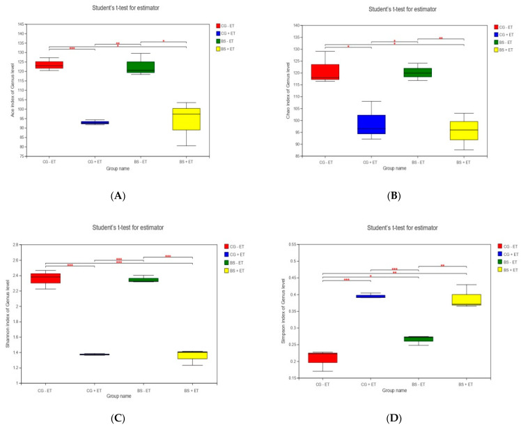
In order to evaluate the alpha diversity, Ace, Chao 1, Shannon and Simpson, which provide the information regarding microbial richness, biodiversity and abundance of species, were calculated. The lowest indexes of Ace, Chao 1 and Shannon, while the highest indexes of Simpson were found in the challenged groups (CG + ET and BS + ET) in comparison to non-challenged groups (CG − ET and BS − ET). However, the indexes of Ace, Chao 1 and Shannon in the comparison of CG − ET versus BS − ET, while Simpson indexes in the comparison of CG + ET versus BS + ET were not affected (Figure 1, Table S1).
Figure 1.
Effects of treatments on alpha diversity index. (A) Ace index, (B) Chao 1 index, (C) Shannon index and (D) Simpson index. *, **, *** represent significant difference.
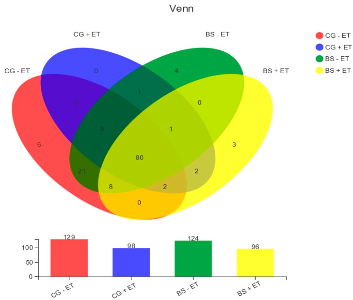
There were 129, 98, 124 and 96 OTUs observed in groups CG − ET, CG + ET, BS − ET, and BS + ET, respectively, of which 80 were common in all experimental groups. Moreover, a total of 13 unique OTUs were detected within CG − ET, CG + ET, BS − ET, and BS + ET (6, 0, 4, and 3, respectively, Figure 2).
Figure 2.
Present Venn diagram shows the all, unique and overlapped OTUs observed in/between treatments.
3.3. Effects of Treatments on Bacterial Abundances at Phylum Level
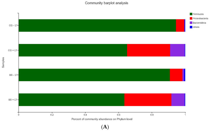
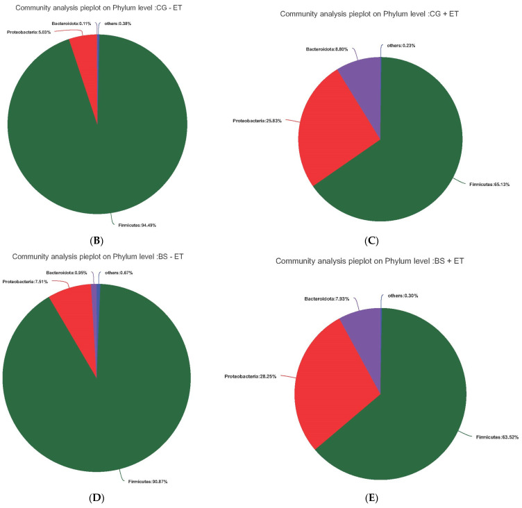
Firmicutes was the most affected phylum in all experimental groups following Proteobacteria and Bacteroidota, exhibiting similar abundance between CG − ET and BS − ET groups (94.49% and 90.87%), while it showed relatively lower abundance in CG + ET and BS + ET groups (65.13% and 63.52%). Within these letter groups, CG + ET and BS + ET, the abundances of Proteobacteria (25.83% and 28.25%) and Bacteroidota (8.80% and 7.93%) were more prominent in relation to the CG − ET (5.03% and 0.11%) and BS − ET (7.51% and 0.95%) groups (Figure 3).
Figure 3.
Relative abundances of the taxa at phylum level. (A) Relative abundances of the affected taxa at phylum level among all groups. (B) Total affected percentage of each phylum in group CG − ET. (C) Total affected percentage of each phylum in group CG + ET. (D) Total affected percentage of each phylum in group BS − ET. (E) Total affected percentage of each phylum in group BS + ET.
3.4. Effects of Treatments on Bacterial Abundances at Genus Level
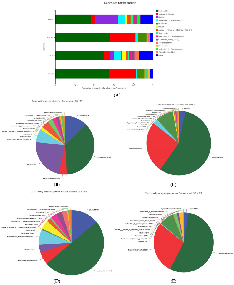
We further compared the bacterial composition in feces of all experimental treatments at the genus level (Figure 4), where Lactobacillus (belonging to the Firmicutes phylum), Escherichia-Shigella (belonging to the Proteobacteria phylum) and Bacteroides were the most affected genera. The relative abundances of Lactobacillus accounted for 36.56%, 56.42%, 49.73%, and 54.76; Escherichia-Shigella accounted for 4.42%, 25.82%, 6.41%, and 28.20%; and Bacteroides accounted for 0.01%, 8.80%, 0.65%, and 7.92% of the total population in groups CG- ET, CG + ET, BS − ET, and BS + ET, respectively, showing clear increased abundance in the presence of Eimeria infection and probiotic feeding. In contrast, the declined abundances of Kurthia, Ruminococcus-torques-group, norank-Clostridia UCG-014 were found in Eimeria-infected (CG + ET and BS + ET) groups compared to the CG − ET group (Figure 4).
Figure 4.
Relative abundances of the taxa at genus level. (A) Relative abundances of the affected taxa at genus level among all groups. (B) Total affected percentage of each genus in group CG − ET. (C) Total affected percentage of each genus in group CG + ET. (D) Total affected percentage of each genus in group BS − ET. (E) Total affected percentage of each genus in group BS + ET.
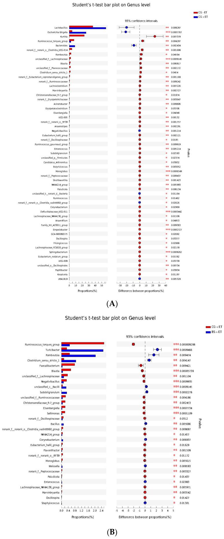
An LEfSe analysis was also performed to fully understand the influence of probiotic on gut microbiota during Eimeria infection. The results of LEfSe analysis showed that several genera were affected by the addition of probiotic and inoculation of Eimeria infection (Figure 5, Table S2). Compared to CG − ET, the non-treated infected control group significantly declined in their abundances of Kurthia, Ruminococcus torques group, Blautia, Lachnoclostridium, Marvinbryantia, Christensenellaceae R-7 group, Acinetobacter, Eisenbergiella, UCG-005, Anaerostipes, Eubacterium hallii group, Ruminococcus gauvreauii group, Candidatus Arthromitus, Butyricicoccus, Monoglobus, Shuttleworthia, NK4A214 group, Paludicola, Ruminococcus, Defluviitaleaceae UCG-011, Lachnospiraceae NK4A136 group, Anaerofilum, Family XIII AD3011 group, Empedobacter, GCA-900066575, Oscillospira, Frisingicoccus, Lachnospiraceae FCS020 group, Sphingobacterium, Eubacterium nodatum group, UCG-009, Papillibacter, Kosakonia, UBA1819, Romboutsia, Subdoligranulum, Enterococcus, Clostridium sensu stricto 1, Corynebacterium and some unclassified and norank genera, while increased abundances of Lactobacillus, Escherichia-Shigella, Bacteroides Negativibacillus and UBA1819 were detected (Figure 5A, Table S2). Probiotic-treated and challenged chickens on the other hand restored (increased) the abundances of Clostridium sensu stricto 1, Corynebacterium, Enterococcus, Romboutsia and Subdoligranulum and decreased the abundances of Faecalibacterium, Lachnoclostridium, Eisenbergiella, Sellimonas, Flavonifractor, Monoglobus, Lachnospiraceae, NK4A136-group, NK4A214-group, Blautia, Ruminococcus torques-group, Christensenellaceae R-7-group, Eubacterium hallii-group and Paludicola compared to the Eimeria-infected non-treated group (Figure 5B, Table S2). More importantly, the abundances of some genera associated with the Bacilli class were found to be enriched in challenged chickens fed a probiotic diet compared to challenged chickens fed a normal diet. They included Bacillus, Weissella, Staphylococcus, Bacilli unclassified and Turicibacter (Figure 5B, Table S2).
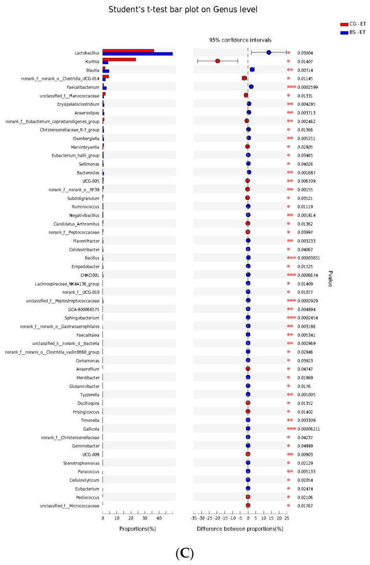
Figure 5.
Microbial abundances enriched in treatments at genus level. (A) Enriched abundances of taxa at genus in groups CG − ET versus CG + ET comparison. (B) Enriched abundances of taxa at genus in groups CG + ET versus BS + ET comparison. (C) Enriched abundances of taxa at genus in groups CG − ET versus BS − ET comparison. *, **, *** represent significant difference.
In this experiment, we also performed the LEfSe analysis in the comparison of BS + ET versus CG + ET and the results are presented in Figure 5C and Table S2.
3.5. Predicted Functions of Microbiota
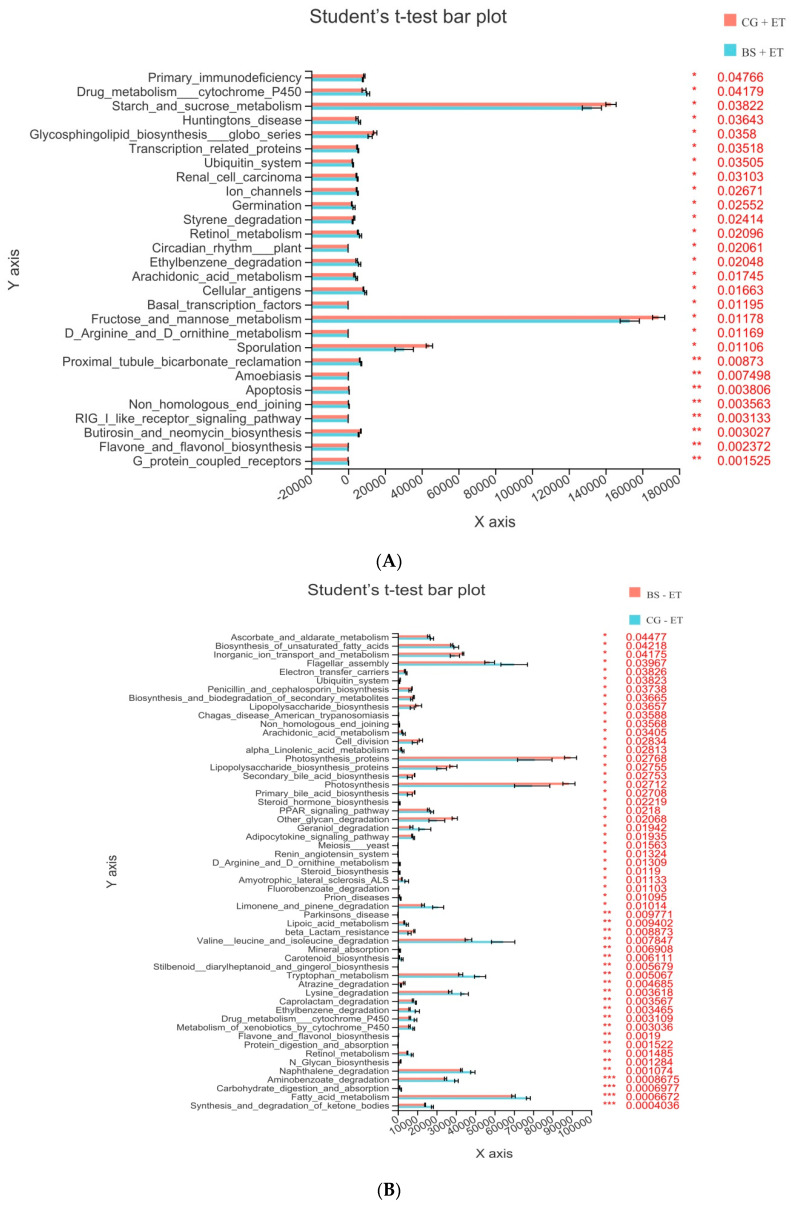
To predict the microbial community functions affected by the addition of probiotic and inoculation of Eimeria infection, a PICRUSt analysis was carried out using KEGG databases. Third-level KEGG analysis revealed that 17 KEGG pathways were significantly affected (up-regulated) by the challenged chickens fed a probiotic diet when compared with challenged chickens fed a normal diet (Figure 6A, Table S3). On the other hand, 23 KEGG pathways were significantly enriched in non-challenged chickens fed probiotic diet in comparison of non-challenged chickens fed normal diet (Figure 6B, Table S3).
Figure 6.
Third-level KEGG pathways affected by probiotic feeding and Eimeria infection. (A) Affected KEGG pathways in groups CG + ET versus BS + ET comparison. (B) Affected KEGG pathways in groups CG − ET versus BS − ET comparison. *, **, *** represent significant difference.
4. Discussion
Coccidiosis is one of the most significant ubiquitous poultry diseases globally that causes severe destruction of the intestinal tract, resulting in reduced growth performance and immunity, disrupted gut homeostasis and perturbed gut microbiota [3,29]. The supplementation of probiotics (as an alternative of antibiotics and anti-coccidials) has been observed to play a key role in improving growth performance, enhancing innate and adaptive immunity, maintaining gut homeostasis and modulating microbiota during enteric infection [30]. In our previous study, we examined the prophylactic efficacy of B. subtilis probiotic on body weight, oocyst shedding in feces, bloody diarrhea scores, cecal lesion scores and whole transcriptome responses of chicken under E. tenella infection [23]. This study was then designed to investigate the chicken gut whole microbiota responses to B. subtilis probiotic feeding under induced Eimeria infection. Our results for alpha diversity showed that the CG + ET group showed decreased Ace, Chao 1 and Shannon, but decreased Simpson indexes compared to CG − ET. These findings imply that the enteric infections negatively influence species’ richness (Ace and Chao 1) as well as bacterial diversity (Shannon). Probiotic-fed challenged chickens on the other hand also showed no significant effects on bacterial richness and biodiversity compared to control positive untreated challenged chickens. Similar findings were reported by Guo et al. [31], who found that chickens fed Bacillus-based probiotic experienced no significant effects on bacterial richness and biodiversity compared to chickens in the positive control group during enteric infection.
Abundances of the taxa Firmicutes, Proteobacteria and Bacteroidota at the phylum level, while Lactobacillus at the genus level were found to be dominant in chickens [32,33]. Similar findings were also noticed in the present experiment. Meanwhile, challenged birds in groups CG + ET and BS + ET had lower abundance of Firmicutes and higher abundances of Proteobacteria and Bacteroidota than that of birds in the control non-treated non-challenged group and B. subtilis-fed non-challenged group. Contrary to our results, Chen et al. [33] reported that Eimeria infection significantly decreased the abundance of Firmicutes and increased the abundances of Proteobacteria and Bacteroidota in broilers when compared with unchallenged broilers. Firmicutes and Bacteroidota play a key role in nutrient absorption and gut homeostasis in chickens [34]. A higher proportion of Firmicutes could suppress pathogenic microbes, restore homeostasis and increase nutrient absorption, whereas an increase in Bacteroidota phylum could lead lower nutrient absorption and dysbiosis [35]. The severity of enteric infection leads to an increase the abundances of phyla Proteobacteria and Bacteroidota and a decreased abundance of phyla Firmicutes [36]. Compared to the negative control group, the decreased abundance of Firmicutes and increased abundances of Proteobacteria and Bacteroidota due to Eimeria infection herein imply the negative effects of coccidia on microbiota balance.
Lactobacillus is an important commensal genus in the chicken gastrointestinal tract [37], which provides beneficial effects on the health and performance by producing antimicrobial substances [37], short chain fatty acids, exopolysaccharides and additional sources of energy [38]. In the present study, challenged untreated chickens and probiotic-fed chickens in the presence and absence of coccidia infection tended to increase the abundance of the Lactobacillus genus compared to control chickens untreated unchallenged. However, the BS + ET group had no significant influence on the taxa Firmicutes, Proteobacteria, Bacteroidota (at phylum level) and Lactobacillus (at genus level) compared to CG + ET, while BS − ET on the taxa Firmicutes, Proteobacteria and Bacteroidota was compared to CG − ET.
To understand the deep mechanism of Eimeria infection and probiotic feeding on bacterial genera, we performed an LEfSe analysis. Our results revealed that Eimeria challenged untreated chickens decreased the abundances of Kurthia, Ruminococcus torques group, Blautia, Lachnoclostridium, Marvinbryantia, Christensenellaceae R-7 group, Acinetobacter, Eisenbergiella, UCG-005, Anaerostipes, Eubacterium hallii group, Ruminococcus gauvreauii group, Candidatus Arthromitus, Butyricicoccus, Monoglobus, Shuttleworthia, NK4A214_group, Paludicola, Ruminococcus, Defluviitaleaceae UCG-011, Lachnospiraceae NK4A136 group, Anaerofilum, Family XIII AD3011 group, Empedobacter, GCA-900066575, Oscillospira, Frisingicoccus, Lachnospiraceae FCS020 group, Sphingobacterium, Eubacterium nodatum group, UCG-009, Papillibacter, Kosakonia, UBA1819, Romboutsia, Subdoligranulum, Enterococcus, Clostridium sensu stricto 1, Corynebacterium and some unclassified and norank genera compared to control chickens. On the other hand, probiotic fed chickens increased the abundances of some Eimeria-affected genera (Romboutsia, Subdoligranulum, Enterococcus, Clostridium sensu stricto 1, Corynebacterium) along with some commensal genera belonging to the Bacilli class, including Bacillus, Weissella, Staphylococcus, Bacilli unclassified and Turicibacter. It is well known that host gut microbiota plays significant roles in regulating immunity and maintaining several metabolic processes by triggering metabolites [39]. During enteric infection, gut microbiota induce protective responses against pathogens, maintain immunoregulatory pathways against antigens and suppress inflammatory responses [40]. The genus Romboutsia is the group of commensal bacteria [41], which function in metabolic reactions of the host in terms of carbohydrate utilization, single amino acids fermentation, anaerobic respiration and end products of metabolic process [42]. The genera Subdoligranulum and Clostridium sensu stricto 1 were found to be involved in butyrate production [43,44]. The production of butyrate by these commensal genera then confers protective mechanisms by ameliorating mucosal inflammatory reactions and oxidative status, supporting the integrity of epithelial barrier and modulating intestinal motility [45]. Species of the Enterococcus are used as probiotics and have been reported to produce antimicrobial peptides, such as bacteriocins [46]. Members of the class Bacilli, including Bacillus, Weissella, Staphylococcus, Bacilli unclassified and Turicibacter contribute to metabolism of food and drugs particles, help in maintaining host health status and gut homeostasis [47]. Taking these reports as evidence, it can be assumed from our results that the feeding of probiotic possibly alleviated the negative effects of Eimeria by stimulating the activities of these commensal genera, which may in turn lead to the stimulation of immune pathways, reduction of inflammatory responses and oxidative status, maintenance of gut barrier integrity and Eimeria perturbation of metabolic processes.
5. Conclusions
This study was focused on the the role of B. subtilis probiotic on gut microbiota in the presence and absence of Eimeria infection. Our results demonstrated that the B. subtilis-based probiotic can increase the abundance of commensal bacterial populations. This may in turn lead to an increase in butyrate production, modulate the anti-inflammatory function, anti-oxidative and metabolites factors and trigger the protective pathways against pathogens. Moreover, a fecal microbiome analysis explored the taxonomic and functional signatures of affected microbial profiles in response to probiotic feeding in the presence and absence of coccidia infection. Future studies should be carried out to explore the individual and combined mechanisms of different Bacillus-based probiotic strains on whole Transcriptome and microbiome analysis.
Acknowledgments
The authors are grateful to the technical staff of the Department of Clinical Veterinary Medicine, Collage of Animal Sciences and Technology, Guangxi University for their valuable support in conducting the current experiment.
Supplementary Materials
The following supporting information can be downloaded at: https://www.mdpi.com/article/10.3390/microorganisms10081548/s1, Table S1: Alpha diversity index; Table S2: Affected taxa at genus level by the treatments; Table S3: Affected KEGG pathways by the treatments.
Author Contributions
Conceptualization, methodology, writing—original draft preparation, F.U.M., Y.Y. and G.Z., methodology, F.L. (Feifei Lv) and Y.W., writing—review and editing, I.H.L., F.A.K. and F.L., (Farooque Laghari) supervision H.S. All authors have read and agreed to the published version of the manuscript.
Institutional Review Board Statement
All experimental protocols on animals associated with this study were reviewed and approved by the Animal Care and Use Committee of Guangxi University, Nanning, China. The specific code for ethics approval is Gxu-2019-180.
Informed Consent Statement
Not applicable.
Data Availability Statement
The raw data associated with this study were deposited into the NGDC database (accession: CRA004666).
Conflicts of Interest
The authors declare no conflict of interest.
Funding Statement
The present research work was financed by the Key Research and Development Plan of Guangxi, China (AB19245037), the Major R&D Project of Wuming District Nanning, China (20210111) and National Natural Science Foundation of China (317607446).
Footnotes
Publisher’s Note: MDPI stays neutral with regard to jurisdictional claims in published maps and institutional affiliations.
References
- 1.Lee Y., Lu M., Lillehoj H.S.J.V. Coccidiosis: Recent progress in host immunity and alternatives to antibiotic strategies. Vaccines. 2022;10:215. doi: 10.3390/vaccines10020215. [DOI] [PMC free article] [PubMed] [Google Scholar]
- 2.Conway D.P., McKenzie M.E. Poultry Coccidiosis: Diagnostic and Testing Procedures. John Wiley & Sons; Hoboken, NJ, USA: 2007. [Google Scholar]
- 3.Chapman H., Jeffers T., Williams R. Forty years of monensin for the control of coccidiosis in poultry. Poult. Sci. 2010;89:1788–1801. doi: 10.3382/ps.2010-00931. [DOI] [PubMed] [Google Scholar]
- 4.Zaman M.A., Iqbal Z., Abbas R.Z., Khan M.N. Anticoccidial activity of herbal complex in broiler chickens challenged with Eimeria tenella. Parasitology. 2012;139:237–243. doi: 10.1017/S003118201100182X. [DOI] [PubMed] [Google Scholar]
- 5.Abbas R., Colwell D., Gilleard J. Botanicals: An alternative approach for the control of avian coccidiosis. Worlds Poult. Sci. J. 2012;68:203–215. doi: 10.1017/S0043933912000268. [DOI] [Google Scholar]
- 6.Chapman H. Practical use of vaccines for the control of coccidiosis in the chicken. Worlds Poult. Sci. J. 2000;56:7–20. doi: 10.1079/WPS20000002. [DOI] [Google Scholar]
- 7.Patwardhan B., Gautam M. Botanical immunodrugs: Scope and opportunities. Drug Discov. Today. 2005;10:495–502. doi: 10.1016/S1359-6446(04)03357-4. [DOI] [PMC free article] [PubMed] [Google Scholar]
- 8.Khan S., Moore R.J., Stanley D., Chousalkar K.K.J.A. The gut microbiota of laying hens and its manipulation with prebiotics and probiotics to enhance gut health and food safety. Appl. Environ. Microbiol. 2020;86:e00600-20. doi: 10.1128/AEM.00600-20. [DOI] [PMC free article] [PubMed] [Google Scholar]
- 9.Jha R., Das R., Oak S., Mishra P. Probiotics (direct-fed microbials) in poultry nutrition and their effects on nutrient utilization, growth and laying performance, and gut health: A systematic review. Animals. 2020;10:1863. doi: 10.3390/ani10101863. [DOI] [PMC free article] [PubMed] [Google Scholar]
- 10.Oyetayo V., Oyetayo F. Potential of probiotics as biotherapeutic agents targeting the innate immune system. Afr. J. Biotech. 2005;4:123–127. [Google Scholar]
- 11.Amara A., Shibl A. Role of Probiotics in health improvement, infection control and disease treatment and management. Saudi Pharm. J. 2015;23:107–114. doi: 10.1016/j.jsps.2013.07.001. [DOI] [PMC free article] [PubMed] [Google Scholar]
- 12.Haque M.I., Ahmad N., Miah M.A. Comparative analysis of body weight and serum biochemistry in broilers supplemented with some selected probiotics and antibiotic growth promoters. J. Adv. Vet. Anim. Res. 2017;4:288–294. doi: 10.5455/javar.2017.d226. [DOI] [Google Scholar]
- 13.Xu L., Fan Q., Zhuang Y., Wang Q., Gao Y., Wang C. Bacillus coagulans enhance the immune function of the intestinal mucosa of yellow broilers. Braz. J. Poul. Sci. 2017;19:115–122. doi: 10.1590/1806-9061-2015-0180. [DOI] [Google Scholar]
- 14.Zhou X., Tian Z., Wang Y., Li W. Effect of treatment with probiotics as water additives on tilapia (Oreochromis niloticus) growth performance and immune response. Fish Physiol. Biochem. 2010;36:501–509. doi: 10.1007/s10695-009-9320-z. [DOI] [PubMed] [Google Scholar]
- 15.Lee S., Ingale S., Kim J., Kim K., Lokhande A., Kim E., Kwon I., Kim Y., Chae B. Effects of dietary supplementation with Bacillus subtilis LS 1–2 fermentation biomass on growth performance, nutrient digestibility, cecal microbiota and intestinal morphology of weanling pig. Anim. Feed Sci. Technol. 2014;188:102–110. doi: 10.1016/j.anifeedsci.2013.12.001. [DOI] [Google Scholar]
- 16.Li C.L., Wang J., Zhang H.J., Wu S.G., Hui Q.R., Yang C.B., Fang R.J., Qi G.H. Intestinal morphologic and microbiota responses to dietary Bacillus spp. in a broiler chicken model. Front. Physiol. 2019;9:1968. doi: 10.3389/fphys.2018.01968. [DOI] [PMC free article] [PubMed] [Google Scholar]
- 17.Hung A.T., Lin S.Y., Yang T.Y., Chou C.K., Liu H.C., Lu J.J., Wang B., Chen S.Y., Lien T.F. Effects of Bacillus coagulans ATCC 7050 on growth performance, intestinal morphology, and microflora composition in broiler chickens. Anim. Produc. Sci. 2012;52:874–879. doi: 10.1071/AN11332. [DOI] [Google Scholar]
- 18.Jacquier V., Nelson A., Jlali M., Rhayat L., Brinch K., Devillard E. Bacillus subtilis 29784 induces a shift in broiler gut microbiome toward butyrate-producing bacteria and improves intestinal histomorphology and animal performance. Poul. Sci. 2019;98:2548–2554. doi: 10.3382/ps/pey602. [DOI] [PubMed] [Google Scholar]
- 19.Tang Q., Jin G., Wang G., Liu T., Liu X., Wang B., Cao H. Current sampling methods for gut microbiota: A call for more precise devices. Front. Cell. Infect. Microbiol. 2020;10:151. doi: 10.3389/fcimb.2020.00151. [DOI] [PMC free article] [PubMed] [Google Scholar]
- 20.Yang H., Xiao Y., Gui G., Li J., Wang J., Li D. Microbial community and short-chain fatty acid profile in gastrointestinal tract of goose. Poult. Sci. 2018;97:1420–1428. doi: 10.3382/ps/pex438. [DOI] [PubMed] [Google Scholar]
- 21.Li Z., Wang W., Liu D., Guo Y. Effects of Lactobacillus acidophilus on gut microbiota composition in broilers challenged with Clostridium perfringens. PLoS ONE. 2017;12:e0188634. doi: 10.1371/journal.pone.0188634. [DOI] [PMC free article] [PubMed] [Google Scholar]
- 22.Memon F., Yang Y., Lv F., Soliman A., Chen Y., Sun J., Wang Y., Zhang G., Li Z., Xu B. Effects of probiotic and Bidens pilosa on the performance and gut health of chicken during induced Eimeria tenella infection. J. Appl. Microbiol. 2021;131:425–434. doi: 10.1111/jam.14928. [DOI] [PubMed] [Google Scholar]
- 23.Memon F.U., Yang Y., Leghari I.H., Lv F., Soliman A.M., Zhang W., Si H. Transcriptome Analysis Revealed Ameliorative Effects of Bacillus Based Probiotic on Immunity, Gut Barrier System, and Metabolism of Chicken under an Experimentally Induced Eimeria tenella Infection. Genes. 2021;12:536. doi: 10.3390/genes12040536. [DOI] [PMC free article] [PubMed] [Google Scholar]
- 24.Caporaso J.G., Kuczynski J., Stombaugh J., Bittinger K., Bushman F.D., Costello E.K., Fierer N., Peña A.G., Goodrich J.K., Gordon J.I. QIIME allows analysis of high-throughput community sequencing data. Nat. Methods. 2010;7:335–336. doi: 10.1038/nmeth.f.303. [DOI] [PMC free article] [PubMed] [Google Scholar]
- 25.Magoč T., Salzberg S.L.J.B. FLASH: Fast length adjustment of short reads to improve genome assemblies. Bioinformatics. 2011;27:2957–2963. doi: 10.1093/bioinformatics/btr507. [DOI] [PMC free article] [PubMed] [Google Scholar]
- 26.Edgar R. UPARSE: Highly accurate OTU sequences from microbial amplicon reads. Nat. Meth. 2013;10:996–998. doi: 10.1038/nmeth.2604. [DOI] [PubMed] [Google Scholar]
- 27.Segata N., Izard J., Waldron L., Gevers D., Miropolsky L., Garrett W.S., Huttenhower C. Metagenomic biomarker discovery and explanation. Genome Biol. 2011;12:R60. doi: 10.1186/gb-2011-12-6-r60. [DOI] [PMC free article] [PubMed] [Google Scholar]
- 28.Xu Y., Tian Y., Cao Y., Li J., Guo H., Su Y., Tian Y., Wang C., Wang T., Zhang L. Probiotic properties of Lactobacillus paracasei subsp. paracasei L1 and its growth performance-promotion in chicken by improving the intestinal microflora. Front. Physiol. 2019;10:937. doi: 10.3389/fphys.2019.00937. [DOI] [PMC free article] [PubMed] [Google Scholar]
- 29.Madlala T., Okpeku M., Adeleke M. Understanding the interactions between Eimeria infection and gut microbiota, towards the control of chicken coccidiosis: A review. Parasites. 2021;28:48. doi: 10.1051/parasite/2021047. [DOI] [PMC free article] [PubMed] [Google Scholar]
- 30.Wang X., Farnell Y.Z., Kiess A.S., Peebles E.D., Wamsley K.G., Zhai W. Effects of Bacillus subtilis and coccidial vaccination on cecal microbial diversity and composition of Eimeria-challenged male broilers. Poult. Sci. 2019;98:3839–3849. doi: 10.3382/ps/pez096. [DOI] [PubMed] [Google Scholar]
- 31.Guo S., Xi Y., Xia Y., Wu T., Zhao D., Zhang Z., Ding B. Dietary Lactobacillus fermentum and Bacillus coagulans Supplementation Modulates Intestinal Immunity and Microbiota of Broiler Chickens Challenged by Clostridium perfringens. Front. Vet. Sci. 2021;8:483. doi: 10.3389/fvets.2021.680742. [DOI] [PMC free article] [PubMed] [Google Scholar]
- 32.Liu L., Lin L., Zheng L., Tang H., Fan X., Xue N., Li M., Liu M., Li X. Cecal microbiome profile altered by Salmonella enterica, serovar Enteritidis inoculation in chicken. Gut Pathog. 2018;10:34. doi: 10.1186/s13099-018-0261-x. [DOI] [PMC free article] [PubMed] [Google Scholar]
- 33.Chen H.-L., Zhao X.-Y., Zhao G.-X., Huang H.-B., Li H.-R., Shi C.-W., Yang W.-T., Jiang Y.-L., Wang J.-Z., Ye L.-P. Dissection of the cecal microbial community in chickens after Eimeria tenella infection. J. Parasites Vectors. 2020;13:56. doi: 10.1186/s13071-020-3897-6. [DOI] [PMC free article] [PubMed] [Google Scholar]
- 34.Oh J.K., Pajarillo E.A.B., Chae J.P., Kim I.H., Yang D.S., Kang D.-K. Effects of Bacillus subtilis CSL2 on the composition and functional diversity of the faecal microbiota of broiler chickens challenged with Salmonella Gallinarum. J. Anim. Sci. Biotech. 2017;8:1. doi: 10.1186/s40104-016-0130-8. [DOI] [PMC free article] [PubMed] [Google Scholar]
- 35.Adalsteinsdottir S.A., Magnusdottir O.K., Halldorsson T.I., Birgisdottir B.E. Towards an individualized nutrition treatment: Role of the gastrointestinal microbiome in the interplay between diet and obesity. Curr. Obes. Rep. 2018;7:289–293. doi: 10.1007/s13679-018-0321-z. [DOI] [PubMed] [Google Scholar]
- 36.Xu S., Lin Y., Zeng D., Zhou M., Zeng Y., Wang H., Zhou Y., Zhu H., Pan K., Jing B. Bacillus licheniformis normalize the ileum microbiota of chickens infected with necrotic enteritis. Sci. Rep. 2018;8:1744. doi: 10.1038/s41598-018-20059-z. [DOI] [PMC free article] [PubMed] [Google Scholar]
- 37.Oakley B.B., Lillehoj H.S., Kogut M.H., Kim W.K., Maurer J.J., Pedroso A., Lee M.D., Collett S.R., Johnson T.J., Cox N.A. The chicken gastrointestinal microbiome. FEMS Microbiol. Lett. 2014;360:100–112. doi: 10.1111/1574-6968.12608. [DOI] [PubMed] [Google Scholar]
- 38.Pajarillo E.A.B., Kim S.H., Lee J.-Y., Valeriano V.D.V., Kang D.-K. Quantitative proteogenomics and the reconstruction of the metabolic pathway in Lactobacillus mucosae LM1. Korean J. Anim. Sci. Resour. 2015;35:692. doi: 10.5851/kosfa.2015.35.5.692. [DOI] [PMC free article] [PubMed] [Google Scholar]
- 39.Yoo J.Y., Groer M., Dutra S.V.O., Sarkar A., McSkimming D.I. Gut microbiota and immune system interactions. Microorganisms. 2020;8:1587. doi: 10.3390/microorganisms8101587. [DOI] [PMC free article] [PubMed] [Google Scholar]
- 40.Belkaid Y., Hand T.W. Role of the microbiota in immunity and inflammation. Cell Biosci. 2014;157:121–141. doi: 10.1016/j.cell.2014.03.011. [DOI] [PMC free article] [PubMed] [Google Scholar]
- 41.Magruder M., Edusei E., Zhang L., Albakry S., Satlin M.J., Westblade L.F., Malha L., Sze C., Lubetzky M., Dadhania D.M. Gut commensal microbiota and decreased risk for Enterobacteriaceae bacteriuria and urinary tract infection. Gut Microbes. 2020;12:1805281. doi: 10.1080/19490976.2020.1805281. [DOI] [PMC free article] [PubMed] [Google Scholar]
- 42.Gerritsen J., Hornung B., Ritari J., Paulin L., Rijkers G.T., Schaap P.J., de Vos W.M., Smidt H. A comparative and functional genomics analysis of the genus Romboutsia provides insight into adaptation to an intestinal lifestyle. bioRxiv. 2019:845511. doi: 10.1101/845511. [DOI] [Google Scholar]
- 43.Van Hul M., Le Roy T., Prifti E., Dao M.C., Paquot A., Zucker J.-D., Delzenne N.M., Muccioli G.G., Clément K., Cani P.D. From correlation to causality: The case of Subdoligranulum. Gut Microbes. 2020;12:1849998. doi: 10.1080/19490976.2020.1849998. [DOI] [PMC free article] [PubMed] [Google Scholar]
- 44.Noriega B.S., Sanchez-Gonzalez M.A., Salyakina D., Coffman J. Understanding the impact of omega-3 rich diet on the gut microbiota. Case Rep. Med. 2016;2016:3089303. doi: 10.1155/2016/3089303. [DOI] [PMC free article] [PubMed] [Google Scholar]
- 45.Canani R.B., Di Costanzo M., Leone L., Pedata M., Meli R., Calignano A. Potential beneficial effects of butyrate in intestinal and extraintestinal diseases. World J. Gastroenterol. WJG. 2011;17:1519. doi: 10.3748/wjg.v17.i12.1519. [DOI] [PMC free article] [PubMed] [Google Scholar]
- 46.Hanchi H., Mottawea W., Sebei K., Hammami R. The genus Enterococcus: Between probiotic potential and safety concerns—An update. Front. Microbiol. 2018;9:1791. doi: 10.3389/fmicb.2018.01791. [DOI] [PMC free article] [PubMed] [Google Scholar]
- 47.Ilinskaya O.N., Ulyanova V.V., Yarullina D.R., Gataullin I.G. Secretome of intestinal bacilli: A natural guard against pathologies. Front. Microbiol. 2017;8:1666. doi: 10.3389/fmicb.2017.01666. [DOI] [PMC free article] [PubMed] [Google Scholar]
Associated Data
This section collects any data citations, data availability statements, or supplementary materials included in this article.
Supplementary Materials
Data Availability Statement
The raw data associated with this study were deposited into the NGDC database (accession: CRA004666).


