Abstract
Variations in body composition among pigs can be associated with insulin sensitivity given the insulin anabolic effect. The study objectives were to characterize this association and to compare de novo lipogenesis and the gene expression in the adipose tissue of pigs of the same genetic background. Thirty 30–95 kg of body weight (BW) pigs, catheterized in the jugular vein participated into an oral glucose tolerance test (OGTT; 1.75 g glucose/kg of BW) to calculate insulin-related indexes. The 8 fattest and the 8 leanest pigs were used to determine the relative mRNA abundance of studied genes. The rate of lipogenesis was assessed by incorporation of [U-13C]glucose into lipids. The QUICKI and Matsuda indexes negatively correlated with total body lipids (r = − 0.67 and r = − 0.59; P < 0.01) and de novo lipogenesis (r = − 0.58; P < 0.01). Fat pigs had a higher expression level of lipogenic enzymes (ACACA, ACLY; P < 0.05) than lean pigs. The reduced insulin sensitivity in fat pigs was associated with a higher expression level of glucose-6-phosphate dehydrogenase (G6PD) and a lower expression of peroxisome proliferator-activated receptor-gamma (PPAR-γ). In conclusion, pigs with increased body lipids have lower insulin sensitivity which is associated with increased de novo lipogenesis.
Subject terms: Fat metabolism, Insulin signalling
Introduction
Lipid deposition in pigs is affected by many factors, such as nutrition, sex, breeds, environment and others1. However, when these factors are considered an important variation in body composition among pigs is still observed2,3, indicating that pigs respond differently to the same amount of ingested nutrients, for example, amino acids4,5. Understanding the factors associated with this variation can help producers to improve nutrient utilization and to manipulate body composition in pigs. Differences in nutrient utilization and subsequent variations in body lipids among pigs might be partly explained by individual variations in metabolic processes involved in energy regulation. Among them, insulin sensitivity is a key candidate given that insulin is a positive regulator of fatty acid synthesis and body adiposity6,7. Indeed, when insulin was infused into growing pigs, while maintaining blood glucose concentrations, the incorporation of glucose into fatty acids increased by 51%8.
The main site for de novo lipogenesis in pigs is the adipose tissue with glucose as the preferred precursor9. In addition to its role in the synthesis and storage of lipids, the fat cells produce and secrete hormones called adipokines, which modulate insulin sensitivity and can influence energy metabolism and expenditure10. Lipid dysregulation in db/db and ob/ob obese mouse models11, and high body fat in insulin resistant humans12 are examples of the strong link between insulin sensitivity and intermediary metabolism in adipose tissue. Although other hormones such as leptin are also implicated in adiposity regulation, insulin is a prime candidate in signalling adiposity because it better maps acute changes in energy metabolism and lipogenesis, and because plasma insulin follows the dynamic between the energy ingested and how it is retained in the body (e.g. as lipids or protein) over time6. Unlike plasma insulin, plasma leptin levels take longer to respond to changes in energy dynamics13,14.
Some studies have reported greater postprandial serum levels of insulin in obese pig lines than in lean lines15,16, and others have reported different insulin sensitivities between these lines17. For example, Iberian pigs, which are considered an obese genetic line had lower whole-body insulin sensitivity but increased β-cell function compared with lean Landrace pigs17. Although insulin sensitivity has been considered when comparing insulin responses between breeds, little is known about the relationship of insulin sensitivity with the observed variations in body composition within breeds or animals from the same genetic background. Therefore, the objectives of this study were to characterize the association between body composition, lipid synthesis and whole body insulin sensitivity in finishing pigs of the same genetic background, and to evaluate whether fat and lean pigs differ in the expression of genes in adipose tissue involved in lipid metabolism and insulin sensitivity, which could potentially result in different rates of de novo lipogenesis.
Materials and methods
The animals were cared for according to a recommended code of practice18 and the guidelines of the Canadian Council on Animal Care19. The animal trial was approved by the Ethical and Animal Welfare Committee of the Sherbrooke Research and Development Centre, Agriculture and Agri-Food Canada (Sherbrooke, QC, Canada). This study was carried out in compliance with the ARRIVE guidelines for the reporting of animal experiments.
Thirty barrows (36 ± 3.8 kg of body weight (BW)) of the same genetic background (Yorkshire-Landrace × Duroc; Benjoporc et Akama, Sainte-Geneviève-de-Batiscan, QC, Canada) were shipped to the Agriculture and Agri-Food Canada swine complex (Sherbrooke, QC, Canada). Pigs were allocated to one pen with a concrete floor in the same mechanically ventilated room. Pigs were fed ad libitum with a commercial diet and had free access to water before and during the experimental period. Room temperature was adjusted from 22 °C at the pig’s arrival to 18 °C at the end of the experiment. Fluorescent lighting was controlled by a timer and ensured 12 h of light daily. The experiment included two trials. The first one consisted of an oral glucose tolerance test (OGTT) to evaluate the variability among animals of plasma glucose, insulin response and insulin sensitivity to the same dose of oral glucose and their relationship with body lipids content. The second trial was performed to determine de novo lipogenesis rate using a bolus of [U-13C]glucose, and the relative mRNA abundance of lipogenic enzymes and of markers associated with insulin sensitivity in fat and lean pigs.
Trial 1
Upon reaching 95 kg BW, pigs were placed in individual cages for one week. During this period, a commercial diet (2.7 kg/day; Table 1) in the form of three equal meals was offered. Two days before the OGTT, pigs were fitted with a catheter in the jugular vein following a non-surgical procedure20. On test day (day 7), a 1.75-g/kg of BW of glucose, mixed with a 300 ml solution of flavour-free gelatin (hydrolyzed collagen) and water (23.3 mg/ml of water), was orally offered (0800) to each pig after 18 h of fasting21. The use of gelatin allows precise delivery of the desired amount of glucose by mimicking a meal, without stressing the animals22,23. Blood samples were collected at − 20, − 10 and 5, 10, 15, 20, 25, 30, 45, 60, 90, 120, 150, 180, 210, 240, 300 and 360 min after the ingestion of glucose. For each collection time, a 10-ml blood sample was collected for insulin and C-peptide analyses (EDTA tubes) and a 2-ml sample for glucose analysis (potassium oxalate monohydrate/sodium fluoride tubes). Blood samples were stored on ice until centrifugation for 12 min at 1800×g at 4 °C. The collected plasma was kept at − 20 °C until determination of insulin, glucose and C-peptide concentrations. At the end of the first trial, the pigs were maintained in single pens and fed ad libitum until the beginning of the second trial.
Table 1.
Chemical composition of the commercial diet used during trials 1 and 2.
| Composition | % of DM |
|---|---|
| Dry matter | 87.2 |
| CP | 14.8 |
| Lipids | 5.2 |
| Starch | 53.2 |
| Amylose, % starch | 24.7 |
| Amylopectin, % starch | 75.4 |
| NDF | 7.6 |
| ADF | 3.2 |
DM dry matter, CP crude protein, NDF neutral detergent fiber, ADF acid detergent fiber.
Biochemical analyses and calculations
Plasma glucose was measured by an enzymatic calorimetric assay (No 997-03001, Wakolife Sciences, Mountain View, CA, USA). Insulin concentration was determined with a porcine insulin commercial RIA kit (#PI-12K; EMD Millipore Corporation, Saint Charles, MO, USA) and C-peptide concentration was measured with an ELISA kit (C-peptide porcine No 10-1256-01, Mercodia Inc, Winston-Salem, NC, USA). Plasma insulin, C-peptide and glucose responses were evaluated by computing the total area under the curve (AUC0–360) using the trapezoidal method between 0 and 360 min post-glucose ingestion. Indexes to assess insulin sensitivity in humans [QUICKI and Matsuda index (MI)] were calculated during the basal and the post-glucose ingestion period. Delta glucose and delta insulin values were calculated by determining the difference between the maximum glucose or insulin concentrations after ingestion of glucose and their respective basal concentrations.
Insulin sensitivity in the basal state was estimated using the quantitative insulin sensitivity check index (QUICKI), which was calculated as proposed by Katz, et al.24 and as follows:
The Homeostasis model assessment (HOMA) was used to estimate insulin resistance (HOMA-IR) and β-cell function (HOMA-%B) at basal conditions as follows (https://www.dtu.ox.ac.uk/homacalculator/):
The HOMA model assumes that individuals with no insulin resistance have 100% β-cell function and an insulin resistance (HOMA-IR) of 1.
The Matsuda index (MI) was also used to estimate whole-body insulin sensitivity with insulin and glucose concentrations measured during the OGTT25 as follows:
The C-peptide: insulin ratio was used as an indicator of hepatic insulin extraction and clearance26. Accute pancreatic insulin secretion during the OGTT was assessed with the oral disposition index (oDIcpep) using the C-peptide and glucose measurements during the OGTT27, as follows:
A lower oDIcpep indicates a reduced ability for β-cells to secrete insulin to match the level of whole-body insulin sensitivity. In addition, the β-cell function from the OGTT was calculated as the ratio of the area under the curve of C-peptide from 0 to 120 min to the area under the curve of glucose from 0 to 120 min as follows28:
Body composition
Total body fat and lean content were measured by dual-energy X-ray absorptiometry (DXA) 5 days before the OGTT using a densitometry device (GE Lunar Prodigy Advance; GE Healthcare, Madison, WI, USA). Pigs were scanned in the prone position using the total body scanning mode (GE Lunar enCORE, version 8.10.027; GE Healthcare). Anaesthesia was induced with sevoflurane (7%) and maintained with isoflurane (5%) during the scans. The DXA total body lean, and fat mass value were converted to their protein and lipid whole-body chemical equivalents29–31.
Trial 2
One week after the end of trial 1, the 8 fattest and the 8 leanest pigs from the original group of 30 were selected to participate in the second trial (108.9 ± 2.8 kg of BW). As in trial 1, pigs were kept in individual cages for 1 week before determination of lipogenesis rates and sample collection. Pigs were fed with the same commercial diet (2.9 kg/day; Table 1) once per day, except for the last 4 days of the study (including the day of the bolus injection), when pigs were fed 6 times per day every 4 h. This was done to achieve a relatively steady state of nutrient absorption and utilization, which is simplifies determination of de novo lipogenesis32 by isotope dilution. Two days before the test, the pigs were catheterized a second time in the jugular vein following the same procedure as in tri0al 1. Two biopsies of subcutaneous (sc) adipose tissue (106 mg) were taken on day 1 after 18 h of fasting to determine the natural abundance of 13C in lipids and the basal gene expression levels. Biopsies were taken under local anaesthesia (EMLA, lidocaine 2.5% and prilocaine 2.5%) using a standard biopsy punch of 8 mm (REF 33–37, Manufacturer Miltex, Inc. York, PA 17402 USA) in the midline of the right side of the pig’s back between 4 and 12 cm distal to the last lumbar vertebra. The biopsy punch allowed the sampling of the sc fat layer immediately under the skin (outer fat layer) and, in some samples, the middle fat layer was also visible33. The skin and the middle fat layer were cut and discarded, and the outer fat layer sample was kept at − 80 °C.
The detailed experimental procedure for determination of de novo lipogenesis is described in Salgado et al.34. Briefly, on the last day of the trial, an intravenous bolus injection of [U-13C]glucose (99% enriched,12 mg/kg BW; 1.6 mmol/g of saline) was administered via the catheter to each pig 2 h after the morning meal. The injection lasted 1 min, and time 0 was considered to be the beginning of the injection. Blood samples were collected − 5, 2, 4, 6, 9, 12, 15, 20, 30, 40, 60, 80, 100, 120, 150, 180, 210, and 240 min after the labelled glucose injection, to analyze plasma glucose isotopic enrichment (IE) and concentration (heparinized tubes). Immediately after sampling, blood was placed on ice before centrifugation (15 min, 1800×g at 4 °C). Plasma was stored at − 80 °C for the determination of plasma glucose IE and at − 20 °C for concentration analyses. Four hours after the bolus injection, the pigs were euthanized using a penetrating captive bolt gun followed by exsanguination, and sc adipose tissue (outer layer) was immediately collected from the same site as the biopsies but in the left side of the pig. All adipose tissue samples were frozen in liquid N and stored at − 80 °C until analyzed for 13C IE and gene expression.
Isotopic enrichment of plasma glucose and of lipids from adipose tissue
Samples of plasma were deproteinized with a 2:1 mixture of acetonitrile and ethanol, and derivatized with acetic anhydride. The ions 242 and 247 were quantified by gas chromatography-mass spectrometry (GCMS:GC 6890N network GC system coupled to MS 5973 Network; Agilent Technologies, Wilmington, DE, USA) in electron impact mode. The GCMS analyzes the whole derivatized molecule of glucose, thus yielding the results as mole percent excess (MPE). Lipid extraction from adipose tissue was performed with slight modifications of the technique by Shahidi35. Briefly, 80 mg of adipose tissue was homogenized in 3 ml of a methanol-chloroform (2:1) solution and 0.2 ml of water for 30 s. Then, 2 ml of a chloroform-water (1:1) solution was added to the sample and centrifuged at 3300×g for 15 min at 10 °C. After evaporating the solvents under N2, the extract was dried at 55 °C for 2 h. The 13C IE and C contents of the extracted lipids were determined after combustion on an elemental analyzer interfaced to an isotope ratio mass spectrometer (IRMS; Delta Advantage, Thermo, Germany). The results are expressed as atom percent excess (APE).
Lipogenesis
De novo lipogenesis was determined by following the incorporation of 13C from the labelled glucose into lipids according to the procedure proposed by Salgado et al.34. Assuming that all the increase of the IE lipids originated from the labelled glucose, the Rglucose-lipids was calculated as follows:
where the Rglucose-lipids [(µg glucose)/(min × g of lipids)] is determined from time 0 to time t. The Δ IElipid is the difference between the IE of lipids at time t (slaughter) and natural abundance in APE. The Δ IEplasma glucose is the cumulative area under the curve of the plasma glucose IE (MPE) above natural abundance over time, determined by the integration of the double-exponential curve IEglucose(t) = α e(–k1t) + β e(–k2t). The Cmextracted lipids represent the relative contribution of the C mass to the total mass of lipids in each adipose tissue sample, which was obtained from the elemental analyzer. The Cmglucose was set to 0.40, equal to the ratio of the C mass to total mass (6 × 12/180) where 6 is the number of C in 1 mol of glucose, 12 is the C-atomic mass (g) and 180 is the glucose molar mass (g).
Relative mRNA abundance of studied genes in the adipose tissue
Adipose tissue samples (~ 100 mg) were homogenized in 1 ml of TRIzol reagent (Invitrogen Life Technology, Burlington, ON, Canada), incubated at room temperature for 5 min and 200 µl of CHCL3 was then added to the solution to remove lipids. After centrifugation at 12,000×g for 5 min (at 4 °C), the aqueous phase was transferred in a new tube and an equal volume of 70% ethanol was added. This solution is then used for total RNA isolation using the RNeasy Lipid Tissue Mini Kit (Qiagen, Toronto, ON, Canada), which included a DNAse I digestion step. Extracted RNA concentration and integrity were assessed with the NanoDrop 1000 Spectrophotometer (Thermo Fisher Scientific, Wilmington, DE, USA) and by nucleic acid electrophoresis with the Agilent 2100 Bioanalyzer instrument (Agilent, Santa Clara, CA, USA). Reverse transcription of total RNA (1 µg) to cDNA was performed with the Superscript IV Reverse Transcriptase (200 U/ml; Thermo Fisher Scientific) using oligo(dT) 20 primers.
The relative mRNA abundance of genes known to be involved in lipogenesis and insulin sensitivity was quantified using real-time qPCR analyses. Table 2 provides the complete list of selected genes with their corresponding GenBank accession numbers. Amplifications were performed in a 10-µl reaction volume containing 5 ml of 2× Power SYBR™ Green PCR Master Mix (Thermo Fisher Scientific), 3 µl of diluted cDNA (1/15), 0.05 µl of uracil N-glycosylase (UNG) AmpErase (Thermo Fisher Scientific) and 1 µl of forward and reverse primers (300 nM, Table 2). Amplifications were performed in triplicate using an ABI 7500 Fast Real-Time PCR System (PE Applied Biosystems, Foster City, CA, USA) with the following cycling conditions: 2 min at 50 °C for AmpErase activation and 10 min at 95 °C for denaturation followed by 40 cycles of 15 s at 95 °C and 45 s at 60 °C. Melting curve analysis was carried out to ensure reaction specificity and search for primer-dimers artifacts. To obtain the relative mRNA quantity units, a standard curve was established in duplicate in each 96-well plate using serial dilutions of cDNA pools36,37. The relative quantification values were calculated by normalizing the relative quantity units of selected genes to those of the peptidylpropyl isomerase A (PPIA) gene that was used as a reference gene. This reference gene was not affected by body composition (fat vs lean) or the nutritional status (fasting vs feeding) of pigs, as determined with the NormFinder algorithm38 from Excel-Tools-Add-ins. Mean values from triplicates were used for statistical analyses.
Table 2.
Primer sequences used for real-time PCR amplifications of studied genes.
| Genes | Primer sequences (5′–3′) | GenBank accession no. | Product size (bp) | Amplification efficiency (%) |
|---|---|---|---|---|
| Studied genes | ||||
| ACACA |
(F)CCGTAGAAATCAAATTCCGCAG (R)CCTTCAGCTTGCTCTCCAG |
NM_001114269 | 141 | 98.8 |
| ACLY |
(F)TCACAACACCATCATCTGCG (R)CTTACTGAACATCTTGGCTGC |
NM_001257276 | 124 | 101.3 |
| ADIPOQ |
(F)ATGATGTCACCACTGGCAAATTC (R)GACCGTGACGTGGAAGGAGA |
EF601160 | 71 | 108.7 |
| ChREBP (MLXIPL) |
(F)ATGTTCGATGACTATGTCCGG (R)ACACCATCCCATTGAAGGAC |
XM_013995540 | 103 | 100.0 |
| FASN |
(F)CTCAACTTCCGAGACGTCATG (R)ACCATTCCCATCACGCG |
NM_001099930 | 124 | 102.0 |
| GAPDH |
(F)CCCCAACGTGTCGGTTGT (R)CTCGGACGCCTGCTTCAC |
NM_001206359 | 91 | 95.7 |
| GCKR |
(F)TTCCCATTTCACCTTCTCCC (R)TTCTCTTTCACCTGCTCCAC |
XM_013987930 | 141 | 98.1 |
| G6PD |
(F)AGATGATGACCAAGAAGCCC (R)GCAGAAGACGTCCAGGATG |
XM_021080744 | 131 | 103.1 |
| LEP |
(F)GGCCCTATCTGTCCTACGTTGA (R)CTTGATGAGGGTTTTGGTGTCAT |
NM_213840 | 71 | 94.5 |
| PPAR-γ |
(F)CCTTTGGTGACTTTATGGAGC (R)TCGATGGGCTTCACATTCAG |
NM_214379 | 145 | 102.2 |
| SCD1 |
CGGATATCGCCCTTATGACAAG CTCGCTGGCAGAATAGTCATAG |
NM_213781 | 124 | 106.1 |
| SDHA |
GATTTGCGAACGGAACCATAAG GCTGCAAGTCTCCGTAGAG |
XM_021076930 | 144 | 101.0 |
| SREBP-1c (SREBF1) |
(F)GCTTCCAGAGGGACCTGAG (R)CTCAGACTGCGGTCCAG |
NM_214157 | 132 | 93.7 |
| SREBP-1a |
(F)CTGCTGACCGACATCGAA (R)GGAGCTGGCATCAGGAC |
NM_214157 | 129 | 98.6 |
| Reference gene | ||||
| PPIA |
(F)GGTCCTGGCATCTTGTCCAT (R)TCATGCCCTCTTTCACTTTGC |
NM_214353 | 71 | 97.4 |
ACACA acetyl CoA carboxylase alpha, ACLY ATP citrate lyase, ADIPOQ adiponectin, ChREBP (MLXIPL) MLX interacting protein like, FASN fatty acid synthase, GAPDH glyceraldéhyde-3-phosphate dehydrogenase, GCKR glucokinase regulator, G6PD glucose-6-phosphate dehydrogenase, LEP leptin, PPAR-γ peroxisome proliferator activated receptor gamma, PPIA peptidylpropyl isomerase A, SCD1 stearoyl-CoA desaturase, SDHA succinate dehydrogenase complex flavoprotein subunit A, SREBP-1c (SREBF1) sterol regulatory element binding transcription.
Statistical analysis
All the statistical analyses were performed using SAS software (version 9.4; SAS Institute Inc., Cary, NC, USA). To determine the relationship between insulin sensitivity and body composition, Spearman’s correlation analyses were performed between the insulin sensitivity indexes obtained during the OGTT with body lipid and protein percentage using the CORR procedure. Linear regression models were performed with the REG procedure to quantify the variation in body composition explained by insulin sensitivity. The dependent variables (body lipid or protein percentage) were regressed independently against each of the insulin-related index: insulin sensitivity (QUICKY, MI, and HOMA-IR) and insulin secretion indexes (HOMA-%B and oDIcpep).
The relative gene expression of each gene of fat and lean pigs studied was compared through a completely randomized design according to a 2 × 2 factorial arrangement with nutritional state (fasting or feeding) and type of pig (lean or fat) as the main factors, while the insulin-related indexes and de novo lipogenesis were compared through a completely randomized design with the type of pig as a fixed factor using the MIXED procedure. A multivariate Principal Component Analysis (PCA) was performed to assess the relationship among the quantitative variables (insulin sensitivity indexes, relative mRNA abundance and de novo lipogenesis). Raw data for this analysis were previously standardized using the Proc STANDARD of SAS.
Ethics approval
All measurements and observations on animals were performed according to a recommended code of practice (Canada, 2012) and the guidelines of the Canadian Council on Animal Care (CCAC, 2009). The animal trial was approved by the Ethical and Animal Welfare Committee of the Sherbrooke Research and Development Centre, Agriculture and Agri-Food Canada (Sherbrooke, QC, Canada).
Results
Four pigs from the original group of 30 did not finish consuming the oral glucose and 2 pigs lost their catheter during the experiment, thus data from those six pigs were not used in the current analysis. The total body lipids and total body protein proportions were on average 19.8 ± 1.5% and 16.3 ± 0.3%, respectively. The basal glucose and insulin plasma concentrations averaged 4.2 ± 0.2 mmol/l and 9.8 ± 4.7 µU/ml, respectively (Table 3). However, the CV among the pigs for basal insulin (CV = 48.0%) and AUC insulin (CV = 27.9%) were considerably higher than those obtained for basal glucose (CV = 5.3%) and AUC glucose (CV = 5.7%). The delta glucose and insulin concentrations, which indicate the increase in concentrations of glucose and insulin from basal concentrations to peak were on average 2.3 ± 0.9 mmol/l and 109.8 ± 43.5 µU/ml respectively. The delta glucose and insulin were also variable among pigs (CV = 40.6% vs. 39.6%, respectively). The basal plasma concentration of C-peptide averaged 97.5 ± 47.2 pmol/l, and, similar to plasma insulin, C-peptide basal concentration and AUC C-peptide were highly variable among pigs (CV = 42.0% and 27.8%, respectively).
Table 3.
Descriptive statistics of the body composition, plasmatic glucose, plasmatic C-peptide, plasmatic insulin and insulin sensitivity indexes of pigs (n = 24) participating in the oral glucose tolerance test (trial 1).
| Variables | Mean | SD | Minimum | Maximum |
|---|---|---|---|---|
| Body conditions | ||||
| Body weight, kg | 94.8 | 3.4 | 88.3 | 101.2 |
| Body lipids, % | 19.8 | 1.503 | 16.2 | 22.32 |
| Body protein, % | 16.3 | 0.323 | 15.75 | 17.05 |
| Glucose, C-peptide and insulin concentrations | ||||
| Basal glucose, mmol/l | 4.2 | 0.2216 | 3.9 | 4.7 |
| Basal C-peptide pmol/l | 97.5 | 47.2 | 30.2 | 220.2 |
| Basal insulin, uU/ml | 9.8 | 4.7 | 3.6 | 19.4 |
| Delta glucose, mmol/l | 2.3 | 0.923 | 1.1 | 5.1 |
| Delta insulin, uU/ml | 109.8 | 43.49 | 50.4 | 204.1 |
| AUC1 glucose, mmol × min/l | 1485 | 84 | 1338 | 1690 |
| AUC insulin, uU × min/ml | 6220 | 1733 | 3025 | 9269 |
| AUC C-peptide, pmol × min/l | 50,505 | 14,049 | 29,409 | 77,917 |
| Insulin sensitivity indexes | ||||
| QUICKI | 0.36 | 0.03 | 0.31 | 0.41 |
| Matsuda index (MI) | 6.3 | 2.4 | 2.9 | 13.2 |
| HOMA-IR | 1.2 | 0.6 | 0.5 | 2.4 |
| HOMA-%B | 88.8 | 40.6 | 33.7 | 184.8 |
| oDIcpep | 539.4 | 157.1 | 254.1 | 853.6 |
| C-peptide: insulin ratio | 1.36 | 0.13 | 1.14 | 1.62 |
AUC Area under the curve, QUICKI quantitative insulin sensitivity check index, oDIcpep oral disposition index, HOMA-IR homeostasis model assessment for estimating insulin resistance, HOMA-%B homeostasis model assessment for estimating β-cell function.
1Area under the curve of plasmatic glucose or insulin concentrations obtained from time 0 to 360 during the oral glucose tolerance test (OGTT).
Insulin sensitivity from the oral glucose tolerance test and body composition
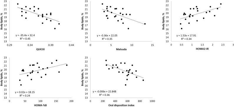
As was the case with insulin response (Fig. 1), most of the insulin-related indexes (MI, HOMA-%B, HOMA-IR and oDIcpep) were highly variable among pigs (Table 3). Moderate to strong correlations were found between whole body lipids percentage, whole body protein percentage and insulin sensitivity/secretion indexes (± 0.5 ≤ r ≤ ± 0.7; P < 0.05; Table 4) except for C-peptide: insulin ratio which indicates hepatic extraction and clearance of insulin. The regression analysis (Fig. 2) indicated that body lipid content linearly decreased as insulin sensitivity increased as estimated by the QUICKI and MI (β = − 35.4 and β = − 0.36; P < 0.01, respectively) indexes, whereas body protein content linearly increased with improved insulin sensitivity (Table 4). In addition, HOMA-IR linearly increased as the percentage of body lipids went up (β = 1.5; P < 0.01). Insulin secretion during the basal state (HOMA-%B) linearly increased as the percentage of body lipids went up (β = 0.02; P < 0.05). The opposite relationship was found with body protein content (Table 4). When glucose concentration increased after oral glucose ingestion (absorptive state), β-cell function, measured by oDIcpep linearly decreased as body lipids content increased (β = − 0.006; P < 0.01).
Figure 1.
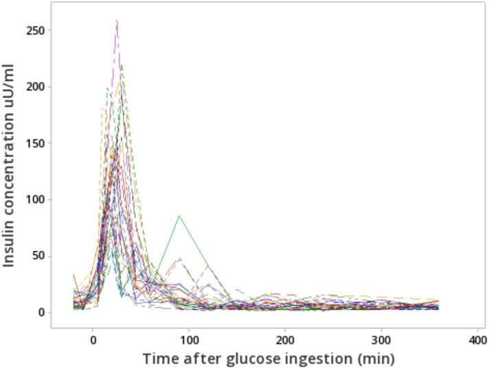
Plasmatic insulin concentration of 95 kg pigs after ingestion of a same dose of oral glucose (1.75 g/kg of BW).
Table 4.
Correlations between body composition and insulin sensitivity and secretion indexes estimated from the oral glucose tolerance test (trial 1; n = 24).
| Total body lipids | Total body protein | |||
|---|---|---|---|---|
| r | P | r | P | |
| Insulin sensitivity | ||||
| QUICKI | − 0.67 | < 0.001 | 0.66 | < 0.001 |
| Matsuda index (MI) | − 0.59 | < 0.01 | 0.60 | < 0.01 |
| HOMA-IR | 0.58 | < 0.01 | − 0.59 | < 0.01 |
| C-peptide:insulin ratio1 | − 0.31 | > 0.05 | 0.29 | > 0.05 |
| Insulin secretion | ||||
| HOMA-%B | 0.45 | < 0.05 | − 0.48 | < 0.05 |
| oDIcpep | − 0.60 | < 0.01 | 0.58 | < 0.01 |
QUICKI quantitative insulin sensitivity check index, oDIcpep oral disposition index, HOMA-IR homeostasis model assessment for estimating insulin resistance, HOMA-%B homeostasis model assessment for estimating β-cell function.
1AUC C-peptide/AUC insulin.
Figure 2.
Linear regression among insulin sensitivity indexes and total body lipids (trial 1; n = 24). Only significant regressions with body lipids are presented in the figure (P < 0.05). QUICKI quantitative insulin sensitivity check, oDIcpep oral disposition index, HOMA-IR homeostasis model assessment for estimating insulin resistance, HOMA-%B homeostasis model assessment for estimating β-cell function.
Gene expression and de novo lipogenesis in the adipose tissue of fat and lean pigs
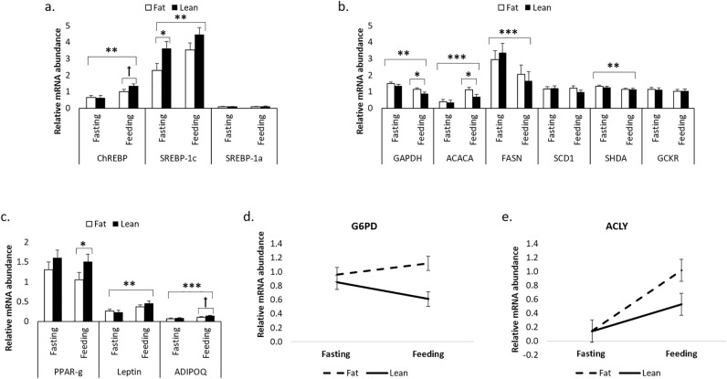
The body lipid content of lean and fat pigs was on average 17.4 ± 0.9% and 22.0 ± 1.0%, respectively. Insulin sensitivity and β-cell function were higher in lean pigs compared with fat pigs (QUICKI = 0.38 vs. 0.34, and oDIcpep = 674.4 vs. 421.3; P < 0.05). Some of the studied genes mRNA abundance was up-regulated (ChREBP, SREBP-1c, ACACA, LEP, ADIPOQ) or down-regulated (GAPDH, FASN, SHDA) after feeding (P < 0.01; Fig. 2a–c). During the fasting state, only the relative mRNA abundance of SREBP-1c was higher in lean pigs than in fat pigs (3.62 vs. 2.28; P < 0.05; Fig. 3a), whereas the relative mRNA abundance of the other genes remained similar between fat and lean pigs. In the feeding state, GAPDH and ACACA had lower relative mRNA abundance in lean pigs than fat pigs (P < 0.05; Fig. 3b), while that of the protein regulator PPAR-γ was higher for the lean pigs (1.06 vs. 1.51; P < 0.05; Fig. 3c). In addition, there was a trend for higher relative mRNA abundance for the transcription factor ChREBP and the adipokine adiponectin (ADIPOQ) in lean pigs than in fat pigs (Fig. 3a,b).
Figure 3.
Relative mRNA abundance of transcription factors (a), enzymes implicated in lipid metabolism (b) and genes associated with insulin sensitivity (c) in fat and lean pigs during fasting and feeding (trial 2). Interactions between nutritional state and type of pigs for G6PD and ACLY are illustrated in (d,e). Error bars represent SE. *,**,*** and ꝉ stands for P < 0.05, P < 0.01, P < 0.001 and P < 0.10, respectively (n = 14).
An interaction between nutritional state and type of pigs was observed for G6PD (P < 0.05) and ACYL (P < 0.05; Fig. 3d,e), indicating that the differences in mRNA abundance are more important during the feeding period, when compared with fasting. The Rglucose-lipids at four hours after [U-13C]glucose injection of fat and lean pigs was on average 21.9 ± 13.5 µg glucose/(min × g of lipids) and 13.4 ± 8.9 µg glucose/(min × g of lipids), respectively. However, despite the observed numerical difference between fat and lean pigs, Rglucose-lipids was not significantly different between the two groups (P = 0.20; Fig. 4). Additionally, correlations of Rglucose-lipids with QUICKI (r = − 0.58), HOMA-%B (r = 0.62) and HOMA-IR (r = 0.62) were significant (P < 0.05), but not with whole-body lipid content (r = 0.39; P = 0.18).
Figure 4.
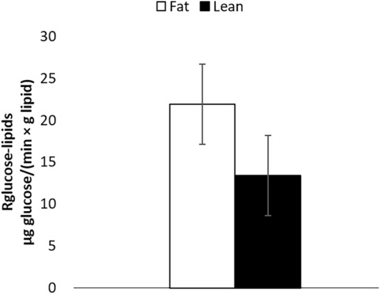
Estimations of the rate of glucose incorporation into lipids (Rglucose-lipids) of fat and lean pigs at 4 h after a bolus injection of [U-13C]glucose (trial 2). Error bars represent SE (n = 14).
Principal component analysis of insulin-related indexes, body composition variables and gene expression
The first two components of the PCA accounted for 60.8% of the total variance (PC1 = 46.8%; PC2 = 14.0%), and fat and lean pigs were clustered in two separated groups (Fig. 5). The PC1 was mostly defined by insulin sensitivity indexes (QUICKI and MI) and genes associated with insulin sensitivity (PPAR-γ and Lep), while the PC2 was mostly defined by the relative mRNA abundance of genes that participate in the de novo lipogenesis pathway (GP6D, SDHA, GCKR, ACACA and ACLY). PC1 had positive strong correlations (r > 0.7; P < 0.01) with QUICKI, MI, oDIcpep, PPAR-γ, Lep and total body protein, but was negatively correlated (r < − 0.7; P < 0.01) with HOMA2-IR, HOMA-%B, G6PD, de novo lipogenesis and total body lipids. The PC2 had strong positive correlations only with SDHA and GCKR (r > 0.7; P < 0.01) and moderate positive correlations (r > 0.6; P < 0.05) with ACACA and ACLY. It can be observed that de novo lipogenesis was negatively associated with PPAR-γ, Lep, QUICKI, MI and oDIcpep, while these five variables were positively associated with each other.
Figure 5.
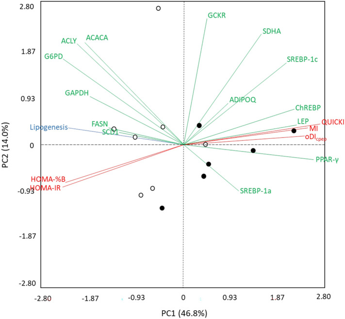
Principal components analysis (PCA) constructed with the gene expression (green), insulin sensitivity/secretion indexes (red) and stable isotopes (blue) variables of fat (open circle) and lean (filled circle) pigs. PC principal component, ACACA acetyl CoA carboxylase alpha, ACLY ATP citrate lyase, ADIPOQ adiponectin, ChREBP MLX interacting protein like, FASN fatty acid synthase, GAPDH glyceraldehyde-3-phosphate dehydrogenase, GCKR glucokinase regulator, G6PD glucose-6-phosphate dehydrogenase, LEP leptin, PPAR-γ peroxisome proliferator activated receptor gamma, SCD1 stearoyl-CoA desaturase, SDHA succinate dehydrogenase complex flavoprotein subunit A, SREBP sterol regulatory element binding transcription QUICKI quantitative insulin sensitivity check, MI Matsuda index, HOMA-IR insulin resistance, HOMA-%B β-cell function, oDIcpep oral disposition index (n = 13).
Discussion
The first objective of this study was to investigate whether variations in the observed body composition are related to differences in insulin sensitivity among pigs. Results from this study clearly showed that pigs raised in the same conditions and having the same genetic background and same feed intake can respond differently to a given diet. This was indeed demonstrated in the first trial where important variations in basal plasma insulin concentrations and AUC0–360 response to OGTT were observed among pigs, while plasmatic glucose measurements remained relatively constant. The observed variations in plasmatic insulin among pigs could be explained, at least in part, by variability in insulin sensitivity (Supplementary Table S1). The basal concentrations of glucose and insulin obtained in trials 1 and 2 are similar to those previously reported in Duroc boars of 145.8 ± 16.8 kg BW26. Significant individual variations in insulin response and sensitivity were reported in healthy humans39. Correlations of insulin sensitivity, assessed with OGTT indexes, and backfat thickness were previously reported in pigs on different fish oil diets26, and are in agreement with results from the current study. It is generally accepted that obesity and body fat distribution are closely associated with insulin resistance, and that this association may explain, at least in part, the heterogeneity observed in insulin sensitivity in healthy human populations12,40. Study results also showed that the β-cell function in its basal state (HOMA-%B) increases when body lipids increase, which may be explained by a compensatory insulin secretion from pancreatic β-cells in response to the lower insulin sensitivity of fatter pigs41. In addition, the significant correlation of MI and C-peptide/glucose (r = − 0.72; P < 0.001) suggest that fat pigs with lower insulin sensitivity increased insulin secretion during the OGTT. However, during the glucose challenge, the observed linear decrease of oDIcpep when body lipids increase rather indicates there is insufficient compensation at the pancreas for losses in whole-body insulin sensitivity. An important part of the between-animal variations in total body lipids and total body protein content was associated with the insulin sensitivity (QUICKI, MI and HOMA-IR) and insulin secretion (oDIcpep) indexes, as demonstrated by the regression analyses.
The second objective of this study was to determine whether the expression of genes associated with insulin sensitivity and lipid metabolism differ between fat and lean pigs under similar nutrient intakes. The differences between fat and lean pigs in gene expression of enzymes participating in lipid synthesis (GAPDH, ACACA, G6PD, ACLY) were observed during the feeding period but not during fasting. The feeding period is known to raise insulin concentrations and to stimulate lipid synthesis42. Therefore, differences among fat and lean pigs observed during the feeding period may be associated, at least in part, with metabolic pathways in which insulin signalling is involved. For example, the higher secretion of insulin in fat pigs with low insulin sensitivity might be associated with their greater expression of G6PD and ACLY, observed after the meal but not during fasting, as those genes are particularly stimulated by insulin during feeding43,44.
In this study, fat pigs with low insulin sensitivity had higher G6PD mRNA abundance and lower PPAR-γ abundance when compared with the lean pigs. The overexpression of G6PD has been associated with lipid dysregulation and low insulin sensitivity in obese mouse models11. In fact, this gene can negatively affect insulin sensitivity by two ways. First and foremost, its overexpression in the adipose tissue leads to an increase in cellular NADPH level, stimulating the lipogenic activity in adipocytes11. This can lead to lipid accumulation, which positively correlates with losses in insulin sensitivity, independently of the animal’s obesity status45. Secondly, the G6PD overexpression alters the expression of adipokines, thus increasing the expression of resistin while decreasing that of adiponectin11. This is in agreement with the results of the current study where the increase in G6PD mRNA abundance in fat pigs is associated with a decrease in ADIPOQ transcript abundance (tendency). Adiponectin acts as a mediator of insulin activity in peripheral tissues and protects non-adipose tissues against an excessive lipid overload while maintaining normal organ function46. The lower mRNA abundance of PPAR-γ in the sc adipose tissue of fat pigs when compared with the lean pigs (feeding state) was unexpected since this nuclear hormone receptor has been identified as a master regulator of adipocyte differentiation and an essential mediator of whole-body insulin sensitivity47. Conflicting observations regarding the expression of PPAR-γ in the adipose tissue of obese patients from different studies were recently reported by Torres et al.48, with some studies showing increased expression of PPAR-γ in obese subjects vs controls, whereas others showed decreased expression or no differences. As suggested by these authors, discrepancies among studies may be explained by differences in gender, fat depots and insulin sensitivity. Benitez et al.49 also demonstrated that the timing when adipose tissue samples are collected is of importance when measuring PPAR-γ expression. Indeed, the PPAR-γ expression in adipose tissue was higher in growing (44 kg BW) then in the finishing (100 kg BW) pigs49, in agreement with a more intense pre-adipocytes differentiation expected at younger age. With respect to insulin sensitivity, it is worth nothing that besides the PPAR-γ receptor transcript being more abundant in lean than fat pigs, our study also showed a positive correlation between the expression of PPAR-γ and the QUICKI index assessing insulin sensitivity (r = 0.71; P < 0.01). The role of PPAR-γ in adipocytes differentiation is well established, but additional functions are starting to emerge. For example, it has been documented that adipose PPAR-γ guarantees the balance and adequate production and secretion of adiponectin and leptin, also known to mediate insulin action in peripheral tissues46.
In fat pigs, the observed increase in the mRNA abundance of enzymes participating in de novo lipogenesis (ex. ACACA, ACLY, G6PD, GAPDH) can be explained, at least in part, by the increase in insulin secretion observed in fat pigs having lower insulin sensitivity. In fact, it was previously demonstrated that insulin can stimulate the expression of the lipogenic enzymes ACACA and ACLY44. In addition, an indirect effect of insulin on the expression of lipogenic enzymes is also possible because de novo lipogenesis in the liver can be upregulated at the transcriptional level by the overexpression of SREBP-1c and ChREBP genes, two transcription factors known to be stimulated by insulin and carbohydrates, respectively50. However, the lack of differences in the mRNA abundance of SREBP-1c between fat and lean pigs during feeding indicates that the activation of ACACA and ACLY transcription would be subjected to a mechanism that is independent of SREBP1-c transcription. In agreement with our results, previous studies have demonstrated that the mRNA expression of SREBP1-c has no effect on the lipogenic genes in adipocytes in contrast to hepatocytes51,52. Nevertheless, we cannot rule out the possibility that the activation of SREBP-1c by the SREBP cleavage-activating protein (SCAP) and two proteases (S1P and S2P), followed by its translocation into the nucleus may, in turn, contribute to the observed increase in the expression of lipogenic enzymes53. Moreover, it is well known that changes in mRNA abundance do not always reflect differences in protein expression or activities. For example, some enzymes involved in de novo lipogenesis can undergo post-translational modifications in response to feeding. This is the case for ACLY, which demonstrated greater activity following its phosphorylation54. On the other hand, ACACA and FASN activities were inhibited by phosphorylation55,56. Therefore, the lack of effect between fat and lean pigs at the transcriptional level for lipogenic genes such as SREBP-1c, FASN and SCD1 does not rule out the possibility of post-translational modifications and activation that may affect activities and, in turn, the de novo lipogenesis.
Fasting is known to reduce lipogenesis and lipogenic gene expression in adipose tissue57. In agreement, the relative mRNA abundance of ChREBP, SREBP-1C, ACACA, LEP and ADIPOQ were all down regulated in fasting than in the feeding state. The down regulation of GAPDH, FASN and SHDA expression in the feeding state was unexpected (current study), as was the lack of effects of fasting and refeeding on the expression of ACACA, FASN and LEP in the ham adipose tissue of Iberian fatty pigs49. The different breeds used and the location of adipose tissue sampling may account for some of the observed discrepancies, but additional experiments are needed to identify the mechanisms leading to a reduced expression of lipogenic genes such as FASN in the feeding state. Only the mRNA abundance of SREBP-1C was significantly different between lean and fat pigs in the fasting state. The unexpected reduction of SREBP-1C mRNA abundance observed in fat vs lean pigs was also observed in the sc adipose tissue of obese vs normal weight or lean human subjects58,59. Moreover, Kolehmainen et al.58 suggested that the reduction in SREBP-1C expression found in obese subjects may be a consequence of an insulin resistance state that commonly develop with obesity. Interestingly, fat pigs from the present study had reduced insulin sensitivity when compared with the lean ones. The effect of insulin on SREBP-1C expression may therefore be attenuated due to the observed reduction of insulin sensitivity in fat pigs (current study).
The average values of Rglucose-lipids observed in this study in fat and lean pigs are in agreement with previous studies using radioactive glucose to estimate lipogenesis8,32. Even if fat pigs, in average, had an higher rate of lipogenesis by 65%, when compared with lean pigs, the large variations in Rglucose-lipid values between animals within each group of pigs can account for the lack of statistical difference (fat pigs: CV = 62%; lean pigs: 67%). Finally, correlations among de novo lipogenesis, body lipids content and insulin-related indexes indicated that lipogenesis is more likely associated with low insulin sensitivity and insulin secretion than body lipid content.
Insulin, along with branch-chain amino acids, also regulates protein synthesis. In young, healthy lean humans, skeletal muscle insulin resistant seems to be the first sign of onset type 2 diabetes60. Although the current study focused on the relationship between whole-body lipid mass and insulin sensitivity, the importance of the skeletal muscle on the whole-body insulin sensitivity cannot be ignored61. Based on the important correlations among whole body protein and insulin sensitive found in our study, one might hypothesize that pigs with high protein deposition were those with improved insulin sensitivity. In fact, increases in adipose tissue is known to increase inflammation and lipotoxicity, whose will negatively impact insulin sensitivity decreasing protein synthesis and/or inhibit protein degradation in muscle60. Granting that several theories surround this subject, it seems that a link between hyperlipidemia-induced reactive oxygen species production in the skeletal muscle and insulin resistance can be stablished62. Such metabolic change incurs greater lipid availability and usage potentially inducing oxidative stress that inhibits branch chain amino acids catabolic enzymes60.
The PCA was performed to study the associations between the observed metabolic responses in plasma (e.g. insulin sensitivity indexes and secretion), gene expression of selected genes, de novo fatty acid synthesis and the body composition of finishing pigs. This analysis indicated that PC1 accounted for most of the observed variations (47%) compared with PC2, which accounted for 14%. The clustering of fat and lean pigs was mainly determined by the insulin sensitivity index variables and the mRNA abundance of some genes that defined PC1, however, de novo lipogenesis also played a major role. Lean pigs were associated with insulin sensitivity indexes (QUICKI, MI, and oDIcpep) and mRNA expression of PPAR-γ and LEP. The association among these variables indicates that the improved insulin sensitivity observed in lean pigs is positively related to the mRNA expression of PPAR-γ and LEP. As previously discussed, the expression of these genes is important to mediate insulin sensitivity in the whole body. Leptin is mainly produced in the adipose tissue63 and its secretion increases energy expenditure, reduces body fat64 and helps in maintaining adequate whole body insulin sensitivity65. On the other hand, fat pigs were associated with reduced insulin sensitivity (HOMA-IR), increased insulin secretion (HOMA-%B) and increased de novo lipogenesis. The positive relationship among these variables suggests that the increase in de novo lipogenesis observed in fat pigs may be associated with higher insulin secretion since insulin acts as a lipogenic hormone in the adipose tissue6. However, in a scenario of decreased whole-body insulin sensitivity, adipose tissue is supposed to be less responsive to insulin and should not cause increased lipogenesis, which is opposite to our results. In this case, two scenarios can be proposed to support our data. One is that an impaired suppression of endogenous glucose production by insulin happens in the liver but not in the adipose tissue or muscle66, then the pancreas secrets more insulin to compensate the hepatic insulin resistance, which increases adipose lipogenesis as the insulin anabolic effect on lipid synthesis remains unaffected in the adipose tissue. A second scenario suggests a cis selective insulin resistance where not all the insulin/akt-regulated process (glucose transport, protein synthesis, lipid synthesis, antilipolysis) are affected by insulin resistance67. For example, adipose tissue from mouse models show that insulin-mediated suppression of lipolysis is largely unaffected in insulin resistance despite impaired insulin-stimulated glucose transport, especially at higher doses of insulin68, therefore, the same scenario might be possible but for lipogenesis stimulated by insulin. Nevertheless, both scenarios must be validated in further experiments.
In addition, the negative association of G6PD expression with PC1 suggests that its negative effect on insulin sensitivity also affects fat pigs. Overall, the multivariate analysis indicates that variables related to insulin sensitivity show significant associations with dietary energy and nutrient utilization, as well as with body composition in pigs.
Conclusion
Results from the present study clearly showed that insulin sensitivity is negatively correlated with total body lipids in pigs and that insulin sensitivity explained about 45% of the total body lipid and protein variations among pigs. When compared with lean pigs, pigs with elevated total body lipids had lower insulin sensitivity and higher gene expression of lipogenic enzymes (ACACA and ACLY). In addition, the higher abundance of G6PD transcripts along with the lower abundance of PPAR-γ may have contributed to lowering insulin sensitivity in fat pigs since these two genes are known to affect insulin sensitivity and to mediate insulin action in peripheral tissues. Observed relationship among variables (PCA analysis) suggests that fat pigs increase insulin secretion as a compensatory mechanism for losses in insulin sensitivity, which is positively associated with de novo lipogenesis and the up-regulation of two lipogenic genes (ACACA and ACLY). Overall, this study demonstrates that insulin sensitivity is an important factor determining the utilization of dietary energy and nutrients with implications for body composition in finishing pigs.
Supplementary Information
Acknowledgements
The authors would like to thank Sophie Horth, Jocelyne Renaud, Mario Léonard, Gerald Bernatchez and Marcel Marcoux for their technical support, to Steve Méthot for his statistical support, and to the swine complex staff for their hard work during our trials.
Author contributions
H.H.S.: conceived and designed the experimental protocol, wrote the original draft, performed the formal analysis. C.P., A.R., and M.P.L.M. supervised and collaborated in the conception of the experimental protocol. H.L., M.F.P. collaborated with gene expression and stable isotopes protocol. J.C. participated in the insulin and glucose data analysis. All authors reviewed the manuscript.
Funding
Financial support received from Agriculture and Agri-Food Canada.
Data availability
The data that support the findings of this study are available from Her Majesty the Queen in Right of Canada as represented by the Minister of Agriculture and Agri-Food, but restrictions apply to the availability of these data, which were used under license for the current study, and so are not publicly available. Data are however available from the corresponding author upon reasonable request and with permission of Her Majesty the Queen in Right of Canada as represented by the Minister of Agriculture and Agri-Food.
Competing interests
The authors declare no competing interests.
Footnotes
Publisher's note
Springer Nature remains neutral with regard to jurisdictional claims in published maps and institutional affiliations.
Supplementary Information
The online version contains supplementary material available at 10.1038/s41598-022-18799-0.
References
- 1.Dunshea, F. & D’Souza, D. Review: Fat deposition and metabolism in the pig. In Manipulating Pig Production IX’ (ed. Paterson, J. E.), 127–150 (2003).
- 2.Cannon JE, et al. Pork chain quality audit: Quantification of pork quality characteristics. J. Muscle Foods. 1996;7:29–44. doi: 10.1111/j.1745-4573.1996.tb00585.x. [DOI] [Google Scholar]
- 3.Klinkner BT. National Retail Pork Benchmarking Study: Characterizing Pork Quality Attributes of Multiple Cuts in the Self-serve Meat Case. North Dakota State University; 2013. [Google Scholar]
- 4.Remus A, Hauschild L, Létourneau-Montminy M-P, Corrent E, Pomar C. The ideal protein profile for late-finishing pigs in precision feeding systems: Threonine. Anim. Feed Sci. Technol. 2020;265:114500. doi: 10.1016/j.anifeedsci.2020.114500. [DOI] [Google Scholar]
- 5.Remus A, Hauschild L, Corrent E, Létourneau-Montminy M-P, Pomar C. Pigs receiving daily tailored diets using precision-feeding techniques have different threonine requirements than pigs fed in conventional phase-feeding systems. J. Anim. Sci. Biotechnol. 2019;10:16. doi: 10.1186/s40104-019-0328-7. [DOI] [PMC free article] [PubMed] [Google Scholar]
- 6.Benoit SC, Clegg DJ, Seeley RJ, Woods SC. Insulin and leptin as adiposity signals. Recent Prog. Horm. Res. 2004;59:267–286. doi: 10.1210/rp.59.1.267. [DOI] [PubMed] [Google Scholar]
- 7.Weickert M, Pfeiffer A. Signalling mechanisms linking hepatic glucose and lipid metabolism. Diabetologia. 2006;49:1732–1741. doi: 10.1007/s00125-006-0295-3. [DOI] [PubMed] [Google Scholar]
- 8.Dunshea F, Harris D, Bauman D, Boyd R, Bell A. Effect of porcine somatotropin on in vivo glucose kinetics and lipogenesis in growing pigs. J. Anim. Sci. 1992;70:141–151. doi: 10.2527/1992.701141x. [DOI] [PubMed] [Google Scholar]
- 9.Bergen WG, Mersmann HJ. Comparative aspects of lipid metabolism: Impact on contemporary research and use of animal models. J. Nutr. 2005;135:2499–2502. doi: 10.1093/jn/135.11.2499. [DOI] [PubMed] [Google Scholar]
- 10.Saltiel AR, Kahn CR. Insulin signalling and the regulation of glucose and lipid metabolism. Nature. 2001;414:799–806. doi: 10.1038/414799a. [DOI] [PubMed] [Google Scholar]
- 11.Park J, et al. Overexpression of glucose-6-phosphate dehydrogenase is associated with lipid dysregulation and insulin resistance in obesity. Mol. Cell. Biol. 2005;25:5146–5157. doi: 10.1128/MCB.25.12.5146-5157.2005. [DOI] [PMC free article] [PubMed] [Google Scholar]
- 12.Kahn SE, et al. Obesity, body fat distribution, insulin sensitivity and islet β-cell function as explanations for metabolic diversity. J. Nutr. 2001;131:354S–360S. doi: 10.1093/jn/131.2.354S. [DOI] [PubMed] [Google Scholar]
- 13.Buchanan C, Mahesh V, Zamorano P, Brann D. Central nervous system effects of leptin. Trends Endocrinol. Metab. 1998;9:146–150. doi: 10.1016/S1043-2760(98)00038-1. [DOI] [PubMed] [Google Scholar]
- 14.Woods S, Seeley R. Insulin as an adiposity signal. Int. J. Obes. 2001;25:S35. doi: 10.1038/sj.ijo.0801909. [DOI] [PubMed] [Google Scholar]
- 15.Wangsness PJ, Acker WA, Burdette JH, Krabill LF, Vasilatos R. Effect of fasting on hormones and metabolites in plasma of fast-growing, lean and slow-growing obese pigs. J. Anim. Sci. 1981;52:69–74. doi: 10.2527/jas1981.52169x. [DOI] [PubMed] [Google Scholar]
- 16.Fernández-Fígares I, Lachica M, Nieto R, Rivera-Ferre MG, Aguilera JF. Serum profile of metabolites and hormones in obese (Iberian) and lean (Landrace) growing gilts fed balanced or lysine deficient diets. Livest. Sci. 2007;110:73–81. doi: 10.1016/j.livsci.2006.10.002. [DOI] [Google Scholar]
- 17.Rodríguez-López JM, Lachica M, González-Valero L, Fernández-Fígares I. Determining insulin sensitivity from glucose tolerance tests in Iberian and landrace pigs. PeerJ. 2021;9:e11014. doi: 10.7717/peerj.11014. [DOI] [PMC free article] [PubMed] [Google Scholar]
- 18.Canada National Farm Animal Care Council . Recommended Code of Practice for the Care and Handling of Farm Animals: Pigs. AAFC Publication; 2012. [Google Scholar]
- 19.Canadian Council on Animal Care . CCAC Guidelines on: The Care and Use of Farm Animals in Research, Teaching and Testing. CCAC; 2009. [Google Scholar]
- 20.Matte J. A rapid and non-surgical procedure for jugular catheterization of pigs. Lab. Anim. 1999;33:258–264. doi: 10.1258/002367799780578101. [DOI] [PubMed] [Google Scholar]
- 21.Manell E, Hedenqvist P, Svensson A, Jensen-Waern M. Establishment of a refined oral glucose tolerance test in pigs, and assessment of insulin, glucagon and glucagon-like peptide-1 responses. PLoS ONE. 2016;11:e0148896. doi: 10.1371/journal.pone.0148896. [DOI] [PMC free article] [PubMed] [Google Scholar]
- 22.Zhang L. Method for voluntary oral administration of drugs in mice. STAR Protoc. 2021;2:100330. doi: 10.1016/j.xpro.2021.100330. [DOI] [PMC free article] [PubMed] [Google Scholar]
- 23.Martins T, Matos AF, Soares J, Leite R, Pires MJ, Ferreira T, Medeiros-Fonseca B, Rosa E, Oliveira PA, Antunes LM. Comparison of gelatin flavors for oral dosing of C57BL/6J and FVB/N mice. J. Am. Assoc. Lab. Anim. 2022;61:89–95. doi: 10.30802/AALAS-JAALAS-21-000045. [DOI] [PMC free article] [PubMed] [Google Scholar]
- 24.Katz A, et al. Quantitative insulin sensitivity check index: A simple, accurate method for assessing insulin sensitivity in humans. J. Clin. Endocrinol. Metab. 2000;85:2402–2410. doi: 10.1210/jcem.85.7.6661. [DOI] [PubMed] [Google Scholar]
- 25.Matsuda M, DeFronzo RA. Insulin sensitivity indices obtained from oral glucose tolerance testing: Comparison with the euglycemic insulin clamp. Diabetes Care. 1999;22:1462–1470. doi: 10.2337/diacare.22.9.1462. [DOI] [PubMed] [Google Scholar]
- 26.Castellano C-A, Audet I, Laforest J-P, Chouinard Y, Matte JJ. Fish oil diets do not improve insulin sensitivity and secretion in healthy adult male pigs. Br. J. Nutr. 2010;103:189–196. doi: 10.1017/S0007114509991590. [DOI] [PubMed] [Google Scholar]
- 27.Galderisi A, et al. Lower insulin clearance parallels a reduced insulin sensitivity in obese youths and is associated with a decline in β-cell function over time. Diabetes. 2019;68:2074–2084. doi: 10.2337/db19-0120. [DOI] [PMC free article] [PubMed] [Google Scholar]
- 28.Hudak S, et al. Reproducibility and discrimination of different indices of insulin sensitivity and insulin secretion. PLoS ONE. 2021;16:e0258476. doi: 10.1371/journal.pone.0258476. [DOI] [PMC free article] [PubMed] [Google Scholar]
- 29.Pomar, C. & Rivest, J. Proc. 46th Annual Conference of the Canadian Society of Animal Science, 26.
- 30.Kipper M, Marcoux M, Andretta I, Pomar C. Repeatability and reproducibility of measurements obtained by dual-energy X-ray absorptiometry on pig carcasses. J. Anim. Sci. 2018;96:2027–2037. doi: 10.1093/jas/skx046. [DOI] [PMC free article] [PubMed] [Google Scholar]
- 31.Kipper M, Marcoux M, Andretta I and Pomar C. Calibration of dual-energy x-ray absorptiometry estimating pig body composition. In 6th EAAP International Symposium on Energy and Protein Metabolism and Nutrition (ed. Chizzotti, M. L.), 427–429 (Wageningen Academic Publishers, 2019).
- 32.Dunshea FR, Leur BJ, Tilbrook AJ, King RH. Ractopamine increases glucose turnover without affecting lipogenesis in the pig. Aust. J. Agric. Res. 1998;49:1147–1152. doi: 10.1071/A98001. [DOI] [Google Scholar]
- 33.Lonergan SM, Topel DG, Marple DN. Chapter 5—Fat and fat cells in domestic animal. In: Lonergan SM, Topel DG, Marple DN, editors. The Science of Animal Growth and Meat Technology. 2. Academic Press; 2019. pp. 51–69. [Google Scholar]
- 34.Salgado HH, Remus A, Pomar C, Létourneau-Montminy M-P, Lapierre H. In vivo estimation of lipogenesis using a bolus injection of [U-13C] glucose in pigs. J. Anim. Sci. 2021;99:1–6. doi: 10.1093/jas/skab148. [DOI] [PMC free article] [PubMed] [Google Scholar]
- 35.Shahidi F. Extraction and measurement of total lipids. Curr. Prot. Food Anal. Chem. 2003;7:1–11. doi: 10.1002/0471142913.fae0000s07. [DOI] [Google Scholar]
- 36.Labrecque B, et al. Molecular characterization and expression analysis of the porcine paraoxonase 3 (PON3) gene. Gene. 2009;443:110–120. doi: 10.1016/j.gene.2009.04.026. [DOI] [PubMed] [Google Scholar]
- 37.Livak, K. ABI prism 7700 Sequence Detection System. UserBulletin No. 2. PE Applied Biosystems, AB Website, Bulletin Reference: 4303859B 777802-002 (1997).
- 38.Vandesompele J, De Preter K, Pattyn F, Poppe B, Van Roy N, De Paepe A. Accurate normalization of real-time quantitative RT-PCR data by geometric averaging of multiple internal control genes. Genome Biol. 2002;3:0034. doi: 10.1186/gb-2002-3-7-research0034. [DOI] [PMC free article] [PubMed] [Google Scholar]
- 39.Bardet S, et al. Inter and intra individual variability of acute insulin response during intravenous glucose tolerance tests. Diabetes Metab. 1989;15:224–232. [PubMed] [Google Scholar]
- 40.Fujimoto WY, Abbate SL, Kahn SE, Hokanson JE, Brunzell JD. The visceral adiposity syndrome in Japanese-American men. Obes. Res. 1994;2:364–371. doi: 10.1002/j.1550-8528.1994.tb00076.x. [DOI] [PubMed] [Google Scholar]
- 41.Bergman RN, Phillips LS, Cobelli C. Physiologic evaluation of factors controlling glucose tolerance in man: Measurement of insulin sensitivity and beta-cell glucose sensitivity from the response to intravenous glucose. J. Clin. Investig. 1981;68:1456–1467. doi: 10.1172/JCI110398. [DOI] [PMC free article] [PubMed] [Google Scholar]
- 42.Rui L. Energy metabolism in the liver. Compr. Physiol. 2014;4:177. doi: 10.1002/cphy.c130024. [DOI] [PMC free article] [PubMed] [Google Scholar]
- 43.Wagle A, Jivraj S, Garlock GL, Stapleton SR. Insulin regulation of glucose-6-phosphate dehydrogenase gene expression is rapamycin-sensitive and requires phosphatidylinositol 3-kinase. J. Biol. Chem. 1998;273:14968–14974. doi: 10.1074/jbc.273.24.14968. [DOI] [PubMed] [Google Scholar]
- 44.Ho-Palma AC, et al. Insulin controls triacylglycerol synthesis through control of glycerol metabolism and despite increased lipogenesis. Nutrients. 2019;11:513. doi: 10.3390/nu11030513. [DOI] [PMC free article] [PubMed] [Google Scholar]
- 45.Boden G. Interaction between free fatty acids and glucose metabolism. Curr. Opin. Clin. Nutr. 2002;5:545–549. doi: 10.1097/00075197-200209000-00014. [DOI] [PubMed] [Google Scholar]
- 46.Kintscher U, Law RE. PPARγ-mediated insulin sensitization: The importance of fat versus muscle. Am. J. Physiol.-Endoc. 2005;288:E287–E291. doi: 10.1152/ajpendo.00440.2004. [DOI] [PubMed] [Google Scholar]
- 47.Rosen ED, Walkey CJ, Puigserver P, Spiegelman BM. Transcriptional regulation of adipogenesis. Gene Dev. 2000;14:1293–1307. doi: 10.1101/gad.14.11.1293. [DOI] [PubMed] [Google Scholar]
- 48.Torres JL, Usategui-Martín R, Hernández-Cosido L, Bernardo E, Manzanedo-Bueno L, Hernández-García I, Mateos-Díaz AM, Rozo O, Matesanz N, Salete-Granado D, Chamorro AJ, Carbonell C, Garcia-Macia M, González-Sarmiento R, Sabio G, Muñoz-Bellvís L, Marcos M. PPAR-γ gene expression in human adipose tissue is associated with weight loss after sleeve gastrectomy. J. Gastrointest. Surg. 2022;26:286–297. doi: 10.1007/s11605-021-05216-6. [DOI] [PMC free article] [PubMed] [Google Scholar]
- 49.Benítez R, Núñez Y, Fernández A, Isabel B, Rodríguez C, Daza A, López-Bote C, Silió L, Óvilo C. Adipose tissue transcriptional response of lipid metabolism genes in growing Iberian pigs fed oleic acid v. carbohydrate enriched diets. Animal. 2016;10:939–946. doi: 10.1017/S1751731115003055. [DOI] [PubMed] [Google Scholar]
- 50.Krycer JR, Sharpe LJ, Luu W, Brown AJ. The Akt–SREBP nexus: Cell signaling meets lipid metabolism. Trends Endocrinol. Metab. 2010;21:268–276. doi: 10.1016/j.tem.2010.01.001. [DOI] [PubMed] [Google Scholar]
- 51.Shimano H, et al. Sterol regulatory element-binding protein-1 as a key transcription factor for nutritional induction of lipogenic enzyme genes. J. Biol. Chem. 1999;274:35832–35839. doi: 10.1074/jbc.274.50.35832. [DOI] [PubMed] [Google Scholar]
- 52.Sekiya M, et al. SREBP-1-independent regulation of lipogenic gene expression in adipocytes. J. Lipid Res. 2007;48:1581–1591. doi: 10.1194/jlr.M700033-JLR200. [DOI] [PubMed] [Google Scholar]
- 53.Jeon TI, Osborne TF. SREBPs: Metabolic integrators in physiology and metabolism. Trends Endocrinol. Metab. 2012;23:65–72. doi: 10.1016/j.tem.2011.10.004. [DOI] [PMC free article] [PubMed] [Google Scholar]
- 54.White PJ, McGarrah RW, Grimsrud PA, Tso SC, Yang WH, Haldeman JM, Grenier-Larouche T, An J, Lapworth AL, Astapova I, Hannou SA, George T, Arlotto M, Olson LB, Lai M, Zhang GF, Ilkayeva O, Herman MA, Wynn RM, Chuang DT, Newgard CB. The BCKDH kinase and phosphatase integrate BCAA and lipid metabolism via regulation of ATP-citrate lyase. Cell Metab. 2018;27:1281–1293. doi: 10.1016/j.cmet.2018.04.015. [DOI] [PMC free article] [PubMed] [Google Scholar]
- 55.Hardie DG. Regulation of fatty acid synthesis via phosphorylation of acetyl-coa carboxylase. Prog. Lipid Res. 1989;28:117–146. doi: 10.1016/0163-7827(89)90010-6. [DOI] [PubMed] [Google Scholar]
- 56.An Z, Wang H, Song P, Zhang M, Geng X, Zou MH. Nicotine-induced activation of AMP-activated protein kinase inhibits fatty acid synthase in 3T3L1 adipocytes: A role for oxidant stress. J. Biol. Chem. 2007;282:26793–26801. doi: 10.1074/jbc.M703701200. [DOI] [PubMed] [Google Scholar]
- 57.Palou M, Sanchez J, Priego T, Rodriguez AM, Pico C, Palou A. Regional differences in the expression of genes involved in lipid metabolism in adipose tissue in response to short- and medium-term fasting and refeeding. J. Nutr. Biochem. 2010;21:23–33. doi: 10.1016/j.jnutbio.2008.10.001. [DOI] [PubMed] [Google Scholar]
- 58.Kolehmainen M, Vidal H, Alhava E, Uusitupa MIJ. Sterol regulatory element binding protein 1c (SREBP-1c) expression in human obesity. Observ. Res. 2001;9:706–712. doi: 10.1038/oby.2001.95. [DOI] [PubMed] [Google Scholar]
- 59.Ducluzeau PH, Perretti N, Laville M, Andreelli F, Vega N, Riou JP, Vidal H. Regulation by insulin of gene expression in human skeletal muscle and adipose tissue. Evidence for specific defects in type 2 diabetes. Diabetes. 2001;50:1134–1142. doi: 10.2337/diabetes.50.5.1134. [DOI] [PubMed] [Google Scholar]
- 60.Petersen MC, Shulman GI. Mechanisms of insulin action and insulin resistance. Physiol. Rev. 2018;98:2133–2223. doi: 10.1152/physrev.00063.2017. [DOI] [PMC free article] [PubMed] [Google Scholar]
- 61.Zierath JR, Krook A, Wallberg-Henriksson H. Insulin action and insulin resistance in human skeletal muscle. Diabetologia. 2000;43:821–835. doi: 10.1007/s001250051457. [DOI] [PubMed] [Google Scholar]
- 62.Di Meo S, Iossa S, Venditti P. Skeletal muscle insulin resistance: Role of mitochondria and other ROS sources. J. Endocrinol. 2017;233:15–42. doi: 10.1530/JOE-16-0598. [DOI] [PubMed] [Google Scholar]
- 63.Zhang Y, et al. Positional cloning of the mouse obese gene and its human homologue. Nature. 1994;372:425–432. doi: 10.1038/372425a0. [DOI] [PubMed] [Google Scholar]
- 64.Harris RB. Direct and indirect effects of leptin on adipocyte metabolism. BBA-Mol. Basis Dis. 2014;1842:414–423. doi: 10.1016/j.bbadis.2013.05.009. [DOI] [PMC free article] [PubMed] [Google Scholar]
- 65.Shimomura I, Hammer RE, Ikemoto S, Brown MS, Goldstein JL. Leptin reverses insulin resistance and diabetes mellitus in mice with congenital lipodystrophy. Nature. 1999;401:73–76. doi: 10.1038/43448. [DOI] [PubMed] [Google Scholar]
- 66.ter Horst KW, et al. Impaired insulin action in the liver, but not in adipose tissue or muscle, is a distinct metabolic feature of impaired fasting glucose in obese humans. Metabolism. 2016;65:757–763. doi: 10.1016/j.metabol.2016.02.010. [DOI] [PubMed] [Google Scholar]
- 67.Fazakerley DJ, Krycer JR, Kearney AL, Hocking SL, James DE. Muscle and adipose tissue insulin resistance: Malady without mechanism? J. Lipid Res. 2019;60:1720–1732. doi: 10.1194/jlr.R087510. [DOI] [PMC free article] [PubMed] [Google Scholar]
- 68.Tan S-X, et al. Selective insulin resistance in adipocytes*. J. Biol. Chem. 2015;290:11337–11348. doi: 10.1074/jbc.M114.623686. [DOI] [PMC free article] [PubMed] [Google Scholar]
Associated Data
This section collects any data citations, data availability statements, or supplementary materials included in this article.
Supplementary Materials
Data Availability Statement
The data that support the findings of this study are available from Her Majesty the Queen in Right of Canada as represented by the Minister of Agriculture and Agri-Food, but restrictions apply to the availability of these data, which were used under license for the current study, and so are not publicly available. Data are however available from the corresponding author upon reasonable request and with permission of Her Majesty the Queen in Right of Canada as represented by the Minister of Agriculture and Agri-Food.


