Abstract
The extraordinary situation of the 2019–2022 pandemic caused a dramatic jump in the incidence of post-traumatic stress disorder (PTSD). PTSD is currently regarded not only as a neuropsychiatric disorder, but also as a comorbidity accompanied by cardiovascular diseases, circulatory disorders, liver dysfunction, etc. The relationship between behavioral disorders and the degree of morphofunctional changes in the liver remains obscure. In this study, PTSD was modeled in sexually mature male Wistar rats using predatory stress induced by a prey’s fear for a predator. Testing in an elevated plus maze allowed the rat population to be divided into animals with low-anxiety (LAP) and high-anxiety (HAP) phenotypes. It was found that morphofunctional analysis of the liver, in contrast to its biochemical profiling, provides a clearer evidence that predatory stress induces liver dysfunction in rats of both phenotypes. This may indicate a decrease in the range of compensatory adaptive reactions in stressed animals. However, in HAP rats, the level of morphofunctional abnormalities in the mechanisms responsible for carbohydrate-fat, water-electrolyte and protein metabolism in the liver testified the prenosological state of the organ, while further functional loading and resulting tension of the regulatory systems could lead to homeostatic downregulation. Meanwhile, the liver of LAP animals was only characterized by insignificant diffuse changes. Thus, we demonstrate here a link between behavioral changes and the degree of morphofunctional transformation of the liver.
Keywords: Wistar rats, post-traumatic stress disorder, liver dysfunction, high-anxiety rat phenotype
INTRODUCTION
Post-traumatic stress disorder (PTSD) is a complex of symptoms of disorders in psychical activity, which appear as a result of single or reiterative supramaximal traumatic external exposures on the human psychics. In the modern world, considering the 2020 COVID-19 pandemic caused by the coronavirus SARS-CoV-2, the problem of PTSD is of particular importance, since it affects not only the immune system but also the nervous system and mental health [1, 2]. All over the world, the pandemic provoked an increase in the number of patients diagnosed with PTSD among both recovered and healthy people [1, 2].
It is necessary to accentuate the significant difference between the stress impact on the hypothalamic-pituitary-adrenal (HPA) axis in PTSD from that in other varieties of neuropsychiatric disorders. It is PTSD that is characterized by quite a rapid change in the activity of the brain structures involved in stress responses, due to which a hyperintensive type of neuroendocrine system’s responses is replaced by hypofunction. In other words, most types of stress lead to hyperactivation of the HPA axis, which develops due to desensitization of the glucocorticoid negative feedback and an increase in human blood cortisol levels, while it is only in PTSD when its sensitization develops and the blood cortisol level decreases [3, 4]. It is commonly accepted that neuroendocrine disorders in patients with PTSD are based on dysregulation, which consists in increased activity of the sympathoadrenal system [5]. As the adaptive capacity is depleted, the regulatory systems of the organism are disrupted (disadaptation) and pathological changes develop [6]. PTSD is characterized by a delayed onset of mental and behavioral symptoms of the disease, as well as the appearance of these symptoms not in all stressed individuals. In this regard, the population of stressed people is usually divided into stress-resistant and stress-nonresistant individuals [7]. Previously, PTSD was mainly considered a mental disease, while nowadays it is regarded as a comorbid disease accompanied by cardiovascular (circulatory) disorders, liver pathology, etc. [8].
Many ongoing studies demonstrate the link between stress and liver disease. As is well known, intense stress is accompanied by lipid peroxidation (LPO) of cell membranes and subsequently leads to tissue damage, while the liver, which plays a key role in such vital processes as detoxification, carbohydrate, lipid and energy metabolism, etc., is most vulnerable compared to other organs [9]. To date, the relationship between behavioral changes and the intensity of morphofunctional transformations in the liver has not yet been clarified.
Currently, a psychosocial predatory stress model, developed by Cohen and Zohar [10], improved by Tseilikman et al. [11], and based on an evolutionarily fixed innate selective fear shown by laboratory rodents to predator cues, is generally accepted to reproduce PTSD experimentally. Characteristic of this model is a decrease in the level of corticosterone, the main stress hormone of laboratory rodents, which is considered as an important pathogenesis adequacy factor in patients diagnosed with PTSD [12, 13]. For this model of PTSD, the methods have been developed for assessing behavioral changes, which allowed the population of laboratory rodents to be subdivided into stress-resistant (low anxiety) and stress-nonresistant (high anxiety) individuals [14].
This work aimed to characterize the morphofunctional state of the liver in mature male Wistar rats, resistant and nonresistant to predatory stress, in a model of PTSD to establish a relationship between behavioral changes and the severity of morphofunctional transformations in the state of the liver.
MATERIALS AND METHODS
The study was carried out on 40 adult male Wistar rats weighing 180–200 g. Animals were taken from the Stolbovaya Branch of the Scientific Center for Biomedical Technologies of the Federal Medical and Biological Agency. Animals, quarantined for at least 14 days, were kept under standard vivarium conditions, being randomly allocated to cages by 10 individuals per cage, under natural light, at a temperature of 20–22°C and ad libitum access to water and complete granulated feed (State Standart 34566-2019). For the experiment, rats were divided into 2 groups, control and experimental, with an equal number of individuals (20 rats in each). The experimental rat group was exposed to predatory stress (cat urine) at a daily basis (10 days running, 10 min per day), followed by keeping for 14 days under normal vivarium conditions. The design of the experiment is shown in Fig. 1.
Fig. 1.

Design of the experiment. А—predatory stress, 10 min per day, 10 days; В –keeping in standard vivarium conditions, 14 days; С—rat testing in an elevated plus maze, one day before the end of the experiment.
All the experimental procedures were conducted in accordance with the Directive of the European Parliament 2010/63/EU “On the protection of animals used for scientific purposes” (dated September 22, 2010). Permission for the work was obtained from the Bioethics Committee at the A.P. Avtsyn Research Institute of Human Morphology” (protocol No. 20 of March 12, 2019).
To identify behavioral differences in response to stress, animals were tested, one at a time, in an elevated plus maze (EPM) for 600 s. The number of entries into the open and closed arms and the time spent in each type of the arms were documented, and the anxiety index (AI), developed by Cohen et al. [15], was calculated by a formula: IA = 1 – [(TOA/TT + NEOA/TNE)/2], where TOA—time spent in the open arms, TT—testing time (600 s), NEOA—number of entries into the open arms, TNE—total number of entries into the open and closed arms.
At the end of the experiment, peripheral blood was taken on an empty stomach, under Zoletyl anesthesia (5 mg/100 g, Virbac Sante Animale, France) in tubes with EDTA as an anticoagulant. To obtain plasma, blood was centrifuged at 3000 g for 10 min. Aspartate aminotransferase (AST) and alanine aminotransferase (ALT), as well as glucose, triglyceride and total cholesterol, levels were determined in blood plasma using an automatic biochemical analyzer (CX4/Pro, Beckman Coulter, USA).
During autopsy, the state of the liver was assessed macroscopically by visually evaluating: color, volume, consistency, elasticity (when grasped with forceps). The relative mass was calculated as an absolute liver mass (ALM) to body mass (BM) ration and expressed in mg/kg of the animal’s body weight: ALM/BM mg/kg.
For morphometric analysis, liver samples were taken; some of them were fixed with 10% neutral formalin, while the others were cut unfixed on a cryostat; the sections were stained with Sudan III to detect neutral fats. After fixation, the liver samples were dehydrated, embedded in a Histomix paraffin medium, and cut into 5-µm sections. A part of the sections was stained with hematoxylin and eosin; the other part was subjected to a periodic acid Schiff (PAS) reaction to detect glycogen. Using an Axioplan 2 imaging microscope with a digital camera and an image processing system (Carl Zeiss MicroImaging GmbH, Germany), 10 photographs of the stained sections were taken from each animal. The optical density of Sudan III- and PAS-stained sections was determined using the ImageJ software (Fiji).
The serum corticosterone concentration was assayed by ELISA kits (IBL, Germany).
Statistical data analysis was performed using Statistica 8.0. The normality of data distribution was assessed by the Shapiro–Wilk test. Since the empirical distribution of the variable turned out to differ from the normal, For statistical processing, the non-parametric Kruskal–Wallis test and the Mann–Whitney U-test for pairwise group comparisons were used. The Spearman’s rank correlation coefficient was calculated. Data were presented as a median (M) and the two quartiles (Me) (25%; 75%). The differences were considered significant at p < 0.05.
RESULTS
To assess the morphofunctional state of the liver in different Wistar rat phenotypes, we used predatory stress. The design of the experiment is shown in Fig. 1. A day before the end of the experiment, elevated plus maze (EPM) testing was carried out (Table 1); on the next day, against the background of food deprivation, the rats were sacrificed using a Zoletil overdose.
Table 1.
Behavioral parameters of Wistar rats during elevated plus maze testing and corticosterone levels, which allowed the population of stressed animals to be divided into low-anxiety and high-anxiety individuals. Me (25%; 75%)
| Parameters | Groups | ||
| control | low-anxiety | high-anxiety | |
| Number of close arm entries | 7.3 (4.1; 10.5) | 6.8 (3.5; 10.3) | 4.3*# (2.2; 7.3) |
| Number of open arm entries | 4.6 (2.2; 6.5) | 3.7 (2.3; 5.6) | 1.9*# (1.1; 3.5) |
| Time spent in closed arms, s | 439.1 (398.3; 547.3) | 468.3 (419.9; 561.3) | 577.8*# (556.7; 608.5) |
| Time spent in open arms, s | 156.2 (26.7; 318.8) | 110.5 (52.4; 297.5) | 21.2*# (5.5; 38.7) |
| Anxiety index, units | 0.65 (0.47; 0.76) | 0.71 (0.53; 0.0.76) | 0.88*# (0.81; 0.98) |
| Corticosterone, nmol/L | 368.6 (304.6; 416.9) | 281.4* (207.1; 372.9) | 169.6*# (141.6; 199.3) |
*—р < 0.05—significance of differences vs. control group, # р < 0.05—significance of differences between low-anxiety and high-anxiety rat groups according to the Mann–Whitney U-test.
It was found that the parameters of EPM testing in the experimental rat group were different: in some animals (Group 1, n = 9), they were indistinguishable from those in the control group (Table 1), while in the other rat group (Group 2, n = 11), they were significantly different vs. both control and Group 1. It turned out that Group 1 rats spent by 71.8% more time in the EPM open arms than Group 2 rats (Table 1). The AI value in Group 2 rats was 0.88, which exceeded the AI of the control rats by 29.4% and that of Group 1 rats by 17.3% (Table 1). However, the plasma corticosterone concentration (CORT) in Group 1 rats was 24.8% lower and in Group 2 – 63.7% lower vs. control rats (Table 1).
During the experimental period, none of the rats showed any visible signs of the disease. At the end of the experiment (25 days after its onset), the rat body mass in all groups had no statistically significant differences (p ≥ 0.05). At the same time, the ALM/BM ratio showed significantly higher values in HAP rats vs. both control and LAP animals (Table 2). Macroscopically, the liver of HAP rats was different from that in control and LAP animals in being enlarged in its volume and easier damaged when grasped with forceps, as well as in having loose consistency and a pale grayish-brown color. All these signs represent the manifestations of diffuse changes in the liver of HAP individuals, whereas the liver of LAP animals was indistinguishable from the control in volume, color and consistency.
Table 2.
Biochemical blood parameters and the relative liver mass in low-anxiety and high-anxiety Wistar rats in post-traumatic stress disorder modeling. Me (25%; 75%)
| Parameters | Groups | ||
| control | low-anxiety | high-anxiety | |
| Relative liver mass, mg/kg | 0.031 (0.028; 0.034) | 0.038* (0.035; 0.041) | 0.046*# (0.042; 0.048) |
| Aspartate aminotransferase (AST), mmol/L | 75.3 (66.7; 85.3) | 80.7 (66.7; 95.3) | 96.4*# (80.3; 113.2) |
| Alanine aminotransferase (ALT), mmol/L | 46.8 (38.7; 53.7) | 43.3 (34.4; 51.3) | 54.6*# (49.3; 59.5) |
| Total cholesterol (TCh), mol/L | 3.5 (3.1; 3.9) | 3.9 (3.3; 4.3) | 4.7*# (2.7; 5.3) |
| Triglycerides (TG), mol/L | 0.79 (0.63; 0.89) | 1.17* (0.89; 1.42) | 2.85*# (2.12; 3.83) |
| Glucose (Gl), mmol/L | 5.9 (5.3; 6.1) | 5.6 (5.2; 6.1) | 4.9*# (4.7; 5.2) |
| Gl/TG, units | 7.6 (7.2; 8.9) | 5.1* (4.2; 6.2) | 1.9*# (1.4; 2.3) |
*—р < 0.05—significance of differences vs. control group; #— р < 0.05—significance of differences between low-anxiety and high-anxiety rat groups (Mann–Whitney U-test).
Many liver diseases are accompanied by a disintegration of hepatocytes, leading to a release of such intracellular enzymes as ALT and AST into the blood flow and an elevation in their blood concentration. The determination of ALT and AST concentration is used primarily for the early diagnosis of functional disorders of the liver [16].
The plasma ALT concentration in LAP rats did not differ from the control, while in HAP animals, it was 16.7% higher vs. control (Table 2). The plasma AST concentration in LAP rats also did not differ significantly vs. control, while in HAP individuals, it was 28.1% higher vs. control (Table 2). At the same time, the ALM/BM ratio was appreciably higher both in LAP rats (by 26.7%) and in HAP animals (by 53.3%) (Table 2). The relative liver mass in HAP rats was 21.1% higher vs. that in LAP rats (Table 2).
Since the ALT and AST reaction norm, as well as the normal relative liver mass, are characterized by quite a wide range, which does not allow unequivocal judgements on the development of dysfunctional changes, a histological and histochemical studies of the liver were carried out [17].
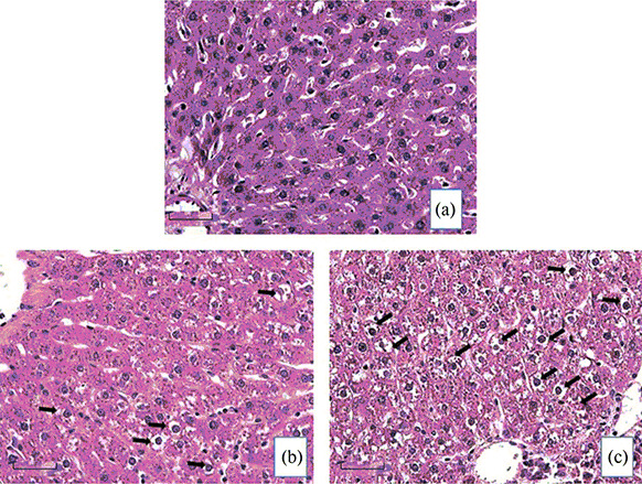
Morphological analysis revealed that the liver had a common age-matched layout in all rat groups. Hematoxylin/eosin staining of liver sections clearly shows that hepatocytes in the control rat group (Fig. 2a) have a dark background which reflects a dense filling of their cytoplasm with glycogen. At the same time, hepatocytes in LAP (Fig. 2b) and, especially, HAP animals (Fig. 2c) have a lighter background suggesting a depletion of glycogen reserves (Fig. 2).
Fig. 2.

Morphofunctional condition of the liver in control (a), low-anxiety (b) and high-anxiety (c) Wistar rats in post-traumatic stress disorder (PTSD) modeling (hematoxylin and eosin staining, scale 50 µm). Arrows point to dystrophic cells.
It is also evident that in hepatocytes of LAP rats (Fig. 2b), there are small- and medium-sized vesicles, while in hepatocytes of HAP rats (Fig. 2c), the vacuoles are medium- and large-sized. A part of vacuoles in rats of both phenotypes are filled with clear liquid, which suggests the development of hydropic dystrophy. In sections of the HAP rat liver (Fig. 2c), the signs of ballooning degeneration are present, i.e. the filling of almost the entire cytoplasm with hydropic fluid. In the latter case, necrosis and death of such cells are typically observed [18]. Another part of the vesicles in LAP and HAP rat hepatocytes is filled with an opaque substance with histological signs of neutral fats.
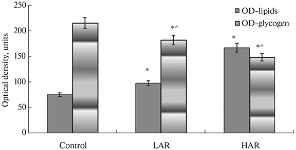
In order to prove the presence of fatty degeneration, we used a traditional Sudan III stain for neutral fats [19]. Microscopically, in LAP rat hepatocytes, small and medium droplet fatty degeneration was observed, and only a small part of the cells contained vacuoles stained red with Sudan III, which was indicative of the presence of fats therein. At the same time, in many HAP rat hepatocytes, medium and large droplet fatty degeneration was found. By contrast, control rat hepatocytes contained practically no vacuoles stained red with Sudan III, suggesting that, normally, fat accumulates in liver cells occurs in very small amounts and only in stellate cells. Optical density (OD) measurements of hepatocyte staining with Sudan III in liver sections revealed higher OD values in rats of both phenotypes: by 29.2% (p = 0.004) in LAP rats and by 120.3% (p = 0.004) in HAP animals compared to control (Fig. 3). A negative correlation was found between OD values of Sudan III staining and blood CORT levels in LAP and HAP rats: r s = –0.979 (Spearman’s rank correlation coefficient, p = 0.001) and r s = –0.955 (p = 0.001), respectively.
Fig. 3.

Optical density of lipid and glycogen staining in liver sections of low-anxiety (LAP) and high-anxiety (HAP) rat phenotypes in post-traumatic stress disorder modeling. OD—Optical density, ОD-lipids—OD, when staining neutral fats with Sudan III; OD-glycogen—OD, when staining glycogen with a Schiff (PAS) reaction. *p < 0.005—significant difference vs. control, ^ p < 0.005—significant difference between HAP (group 1) and LAP (group 2) phenotypes of experimental rats (Mann–Whitney U-test).
During glycogen visualization via a traditional PAS reaction [20], it was found that OD values of the liver sections are reduced in LAP rats by 25.5% (p = 0.0003) and by 32.1% (p = 0.0001) in HAP animals compared to control (Fig. 3). A positive correlation of glycogen OD values with blood CORT levels was found in LAP and HAP rats: r s = 1 (p = 0.0001) and r s = 1 (p = 0.0001), respectively. It should be noted that in all rat groups, the liver structure was unchanged, with its lobular and lamellar organization and clear-cut cell contours. Notably, the same was true for HAP rats either, despite the fact that all the lobes of their liver demonstrated massive vesicular (hydropic) degeneration of hepatocytes.
Dystrophic changes in the liver were accompanied by changes in the level and predominance of the energy transport forms in blood plasma, such as glucose or lipids. However, it was revealed that the responses of LAP and HAP rats to predatory stress were different (Table 2). It turned out that, relative to the control, plasma glucose level decreased only in HAP rats (by 15.4%) while being indistinguishable from the control in LAP animals (Table 2). At the same time, plasma total cholesterol level was elevated in HAP rats only (by 32.2%), while being indistinguishable from the control in LAP individuals (Table 2). However, the plasma level of triglycerides was elevated both in HAP rats (by 62.3%) and in LAP animals (by 32.4%) compared to the control (Table 2). The glucose (Gl) to triglyceride (TGL) ratio Gl/TGL decreased in both stressed groups. In LAP rats, the Gl/TGL ratio was lower by 35.1%, while in HAP animals by 75.8% compared to the control (Table 2).
DISCUSSION
To assess the state of various systems of the organism, the functional load method is usually used, allowing for the assessment of the range of compensatory adaptive reactions of various organs and systems. In the present study, the valid Wistar rat predator-based psychosocial stress model of PTSD was used [4]. Studies conducted by Russian and foreign authors prove the importance of predatory stress for rodents [21, 22]. These studies show that chemical communication in mammals is implemented via olfactory signals, mainly volatile components of various excretions, which encode the information about constant individual’s characteristics required for species survival. Such signaling molecules propagate in the air and are perceived by the olfactory analyzer, the vomeronasal system, which is the peripheral part of the olfactory system, supplied with autonomic innervation and numerous blood vessels, and lined with the neuroepithelium [23]. For most mammalian species, the analysis of odor stimuli is determinative in organizing complex forms of behavior and regulating the hormonal ensemble in the organism. The impact of a predator’s smell is so significant for rodents that it activates not only behavioral reflexes (escape, anxiety, etc.) but also an abortive reaction in pregnant females [23].
Rat testing in the elevated plus maze allowed us to divide the group of stressed animals into high-anxiety (HAP) and low-anxiety (LAP) phenotypes by their anxiety index. Like in our previous experiments [4], we considered the rats with AI values above 0.75 to be high-anxious, and those with AI values below 0.75 to be low-anxious animals. Apart from behavioral traits of PTSD-like state development, an additional sign of the model validity is a decrease in the plasma corticosterone level [4]. Although CORT decreased in both phenotypes, HAP rats had its lowest values, suggesting maximum changes in the organism of this rat group.
Macroscopic examination of the liver in experimental rat groups confirmed our suggestion. It turned out that in HAP rats, the liver was enlarged and showed the signs of an inflammatory response (Table 2, Fig. 2). The liver dysfunction was confirmed by such markers as ALT) and AST [24], which were increased in HAP rats only. It should be noted that most of the biochemical markers of liver dysfunction have a large range of reaction norms [17]; in this regard, an analysis of the morphofunctional state of the liver is a more reliable diagnostic method.
Neutral fats were visualized using a specific Sudan III staining [19]. This method enabled us to detect a full-fledged fatty degeneration of the liver in HAP rats and only a slightly disrupted lipid metabolism in LAP individuals. At the same time, an increase in the OD of Sudan III-stained sections was accompanied by a decrease in plasma corticosterone level (negative correlation). This finding confirms the relationship between CORT and metabolism of neutral fats in the liver. However, other researchers have obtained similar results when administering CORT at doses exceeding physiological ones [25, 26]. Therefore, we have to own up that the mechanisms by which CORT can affect lipid metabolism remain obscure to date.
The use of the glycogen-specific PAS reaction [20] made it possible to detect a sharp decrease in the level of this energy storage material in HAP rats, and its far lesser depletion in LAP animals. A decrease in the OD of sections stained for glycogen was accompanied by a decrease in plasma CORT level (positive correlation). According to the literature data, the glycogen content depends not only on CORT but also on insulin and catecholamine levels, which affect the levels of enzymes and intermediate products of glucose and glycogen metabolism [27, 28]. In our experiment, it was demonstrated for the first time that in HAP rats the dystrophic changes occur in the vast majority of liver cells, which becomes almost completely glycogen free and, instead, filled with vacuoles containing hydropic fluid and fats. The revealed changes reflect a severe imbalance of hepatic metabolism and derangement of the mechanisms providing the regulation of carbohydrate-fat, water-electrolyte and protein metabolism, indicative of the prenosological state of the organ in HAP rats. At the same time, in LAP individuals, these dystrophic changes were expressed only in a part of liver cells. The chance of spontaneous recovery of the hepatic morphofunctional state in this rat phenotype should be verified in further experiments, which could be achieved by extending the post-stress period.
It is appropriate to remind that the liver is the hub of numerous physiological processes. The most important function of the liver is the ability to accumulate substances that serve as a source of energy not only for local needs but for the whole organism. The main sources of energy include glucose supplied with food, which is stored in the liver in the form of glycogen (gluconeogenesis), while the latter is converted back to glucose when the organism needs energy (glycogenolysis) [29]. Despite the fact that, under physiological conditions, lipid synthesis, secretion and oxidation occur in the liver, they mainly accumulate not in the liver but in adipose tissue. Lipids are more than twice as energy-intensive as glucose. However, the bioenergy contained in lipids begins to be consumed only in emergency, whereas glycogen energy resources are much more available and mobilized within few seconds [30]. The glucose–CORT relationship has been established, but the mechanisms behind this relationship remain to be elucidated [31]. In our study, dystrophic changes in the morphofunctional state of the liver when modeling PTSD were accompanied by changes in plasma concentrations and predominance of the main energy transport forms, such as total cholesterol (TCh), triacylglycerols (TGL) and glucose (Gl). From the decrease in the Gl/TGL ratio and the increase in TCh and TGL indices, it can be concluded that predatory stress replaced the predominance of the main energy transport form, glucose, by the predominance of lipids in the same capacity. In HAP rats, such a replacement of one energy source by another was especially pronounced. The reason for such a change lies most likely in stress-induced dysfunction of the liver, which is the key organ that plays a fundamental role in the regulation of carbohydrate, lipid and protein metabolism, and is also involved in many other processes aimed at maintaining homeostasis of the entire organism. The predominant mobilization of free fatty acids as an energy source occurs usually in old age, as well as under the influence of extreme factors [32].
If extrapolating to humans the state of PTSD with severe liver dysfunction, which we characterized in HAP rats, it can be concluded that it represents a serious risk factor for the development of atherosclerosis, cardiovascular disorders and CNS dysfunctions, as reported by other authors [33, 34]. Moreover, it has long been known that almost all functional disorders and diseases of the liver can cause various neurological and psychoneurological pathologies, from minimum changes in cerebral function to cognitive dysfunction and even cerebral edema [27]. All of the aforementioned suggests that the sequelae of the impaired morphofunctional state of the liver in PTSD may contribute to chronization and exacerbation of diseases, acting as a key link in the formed “neurohormonal disorders ↔ liver dysfunction” vicious circle induced by severe psychoemotional stress.
Thus, morphofunctional assessment of the liver, in contrast to its biochemical profiling, provides a clearer evidence that predatory stress induces liver dysfunction in both LAP and HAP rats. In turn, this may indicate a decrease in the range of compensatory adaptive reactions in these animals. Nevertheless, we managed to establish a relationship between behavioral changes and the severity of morphofunctional transformations in the state of the liver. The intensity of morphofunctional disorders of the mechanisms providing carbohydrate-fat, water-electrolyte and protein metabolism in the liver of HAP rats indicates the prenosological state of the organ. In the case of additional loading, there may ensue a breakdown of homeostatic systems and the development of diseases. At the same time, the liver of LAP animals was only characterized by insignificant diffuse changes. Our data on liver dysfunction in stressed rats may prove to be instrumental in clinical practice. In the post-stress period, diagnostic tests should be carried out to determine the degree of liver dysfunction, and appropriate treatment must be provided. Correction of the hepatic functional activity will help increase functional capacities of the organ and break the vicious circle of the pathological process.
AUTHORS’ CONTRIBUTION
M.V.K. provided experimental design, data collection, and article writing; K.A.A., V.V.A., M.A.K. and L.A.M. provided technical support during experiments, participated in data processing and discussion; D.A.A. participated in article editing.
FUNDING
The work was carried out within the state assignment to the A.P. Avtsyn Research Institute of Human Morphology of the Petrovsky National Research Center of Surgery, RTD no. 122030200535-1.
CONFLICT OF INTEREST
The authors declare the absence of any obvious and potential conflicts of interest related to the publication of this article.
Footnotes
Russian Text © The Author(s), 2022, published in Zhurnal Evolyutsionnoi Biokhimii i Fiziologii, 2022, Vol. 58, No. 4, pp. 323–33210.31857/S0044452922040088.
Translated by A. Polyanovsky
REFERENCES
- 1.Vindegaard N, Benros ME. COVID-19 pandemic and mental health consequences: Systematic review of the current evidence. Brain Behav Immun. 2020;89:531–542. doi: 10.1016/j.bbi.2020.05.048. [DOI] [PMC free article] [PubMed] [Google Scholar]
- 2.Somova LM, Kotsyurbiy EA, Drobot EI, Lyapun IN, Shchelkanov MYu. Clinical and morphological manifestations of immune system dysfunction in new coronavirus infection (COVID-19) Clin exp morphology. 2021;10(1):11–20. doi: 10.31088/CEM2021.10.1.11-20. [DOI] [Google Scholar]
- 3.Hadad NA, Schwendt M, Knackstedt LA. Hypothalamic-pituitary-adrenal axis activity in post-traumatic stress disorder and cocaine use disorder. Stress. 2020;23(6):638–650. doi: 10.1080/10253890.2020.1803824.. [DOI] [PubMed] [Google Scholar]
- 4.Tseilikman V, Komelkova M, Lapshin M, Alliluev A, Tseilikman O, Karpenko M, Pestereva N, Manukhina E, Downey HF, Kondashevskaya M, Sarapultsev A, Dremencov E. High and low anxiety phenotypes in a rat model of complex post-traumatic stress disorder are associated with different alterations in regional brain monoamine neurotransmission. Psychoneuroendocrinology. 2020;117:104691. doi: 10.1016/j.psyneuen.2020.10469. [DOI] [PubMed] [Google Scholar]
- 5.Morris MC, Hellman N, Abelson JL, Rao U. Cortisol, heart rate, and blood pressure as early markers of PTSD risk: A systematic review and meta-analysis. ClinPsycholRev. 2016;49:79–91. doi: 10.1016/j.cpr.2016.09.001. [DOI] [PMC free article] [PubMed] [Google Scholar]
- 6.Morris MC, Compas BE, Garber J. Relations among posttraumatic stress disorder, comorbid major depression, and HPA function: a systematic review and meta-analysis. Clin Psychol Rev. 2012;32(4):301–315. doi: 10.1016/j.cpr.2012.02.002. [DOI] [PMC free article] [PubMed] [Google Scholar]
- 7.Javidi H, Yadollahie M. Post-traumatic Stress Disorder. Int J Occup Environ Med. 2012;3(1):2–9. [PubMed] [Google Scholar]
- 8.Pitman RK, Rasmusson AM, Koenen KC, Shin LM, Orr SP, Gilbertson MW, Milad MR, Liberzon I. Biological studies of post-traumatic stress disorder. Nat Rev Neurosci. 2012;13(11):769–787. doi: 10.1038/nrn3339. [DOI] [PMC free article] [PubMed] [Google Scholar]
- 9.Forte G, Favieri F, Tambelli R, Casagrande M. COVID-19 Pandemic in the Italian Population: Validation of a Post-Traumatic Stress Disorder Questionnaire and Prevalence of PTSD Symptomatology. Int J Environ Res Public Health. 2020;17(11):4151. doi: 10.3390/ijerph17114151. [DOI] [PMC free article] [PubMed] [Google Scholar]
- 10.Cohen H, Zohar J. An animal model of posttraumatic stress disorder: The use of cut-off behavioral criteria. Ann NY Acad Sci. 2004;1032:167–178. doi: 10.1196/annals.1314.014. [DOI] [PubMed] [Google Scholar]
- 11.Tseilikman V, Dremencov E, Maslennikova E, Ishmatova A, Manukhina E, Downey HF, Klebanov I, Tseilikman O, Komelkova M, Lapshin MS, Vasilyeva MV, Bornstein SR, Perry SW, Wong ML, Licinio J, Yehuda R, Ullmann E. Post-Traumatic Stress Disorder Chronification via Monoaminooxidase and Cortisol Metabolism. HormMetab Res. 2019;51(9):618–622. doi: 10.1055/a-0975-9268. [DOI] [PubMed] [Google Scholar]
- 12.Rybnikova EA, Mironova VI, Pivina SG. Test for the detection of disorders of self-regulation of the pituitary-adrenocortical system. Journal of Higher Nervous Activity IP Pavlova. 2010;60(4):500–506. [PubMed] [Google Scholar]
- 13.Boero G, Pisu MG, Biggio F, Muredda L, Carta G, Banni S, Paci E, Follesa P, Concas A, Porcu P, Serra M. Impaired glucocorticoid-mediated HPA axis negative feedback induced by juvenile social isolation in male rats. Neuropharmacology. 2018;133(1):242–253. doi: 10.1016/j.neuropharm.2018.01.045. [DOI] [PubMed] [Google Scholar]
- 14.Kondashevskaya MV, Komelkova MV, Tseylikman VE, Tseylikman OB, Artemyeva KA, Aleksankina VV, Boltovskaya MN, Sarapultsev AP, Chereshneva MV, Chereshnev VA. New neurobiological criteria for the resilience profile in modeling post-traumatic stress disorder. Reports of the Russian Academy of Sciences. 2021;501(6):28–33. doi: 10.31857/S2686738921060056. [DOI] [Google Scholar]
- 15.Cohen H, Matar MA, Buskila D, Kaplan Z, Zohar J. Early post-stressor intervention with high-dose corticosterone attenuates posttraumatic stress response in an animal model of posttraumatic stress disorder. Biol Psychiatry. 2008;64:708–717. doi: 10.1016/j.biopsych.2008.05.025. [DOI] [PubMed] [Google Scholar]
- 16.von Känel R, Abbas CC, Begré S, Gander ML, Saner H, Schmid JP. Association between posttraumatic stress disorder following myocardial infarction and liver enzyme levels: a prospective study. Dig Dis Sci. 2010;55(9):2614–2623. doi: 10.1007/s10620-009-1082-z. [DOI] [PubMed] [Google Scholar]
- 17.He Q, Su G, Liu K, Zhang F, Jiang Y, Gao J, Liu L, Jiang Z, Jin M, Xie H. Sex-specific reference intervals of hematologic and biochemical analytes in Sprague-Dawley rats using the nonparametric rank percentile method. PLoS One. 2017;12(12):e0189837. doi: 10.1371/journal.pone.0189837. [DOI] [PMC free article] [PubMed] [Google Scholar]
- 18.Berezov IuE, Polsachev VI, Kovalev AI. Changes in the biochemical indices of liver function in stomach and esophageal cancer. Vopr Onkol. 1980;26(12):15–18. [PubMed] [Google Scholar]
- 19.Ju J, Huang Q, Sun J, Jin Y, Ma W, Song X, Sun H, Wang W. Correlation between PPAR-α methylation level in peripheral blood and inflammatory factors of NAFLD patients with DM. Exp Ther Med. 2018;15(2):1474–1478. doi: 10.3892/etm.2017.5530. [DOI] [PMC free article] [PubMed] [Google Scholar]
- 20.Ravikumar SS, Menaka TR, Vasupradha G, Dhivya K, Dinakaran J, Saranya V. Cytological intracellular glycogen evaluation using PAS and PAS-D stains to correlate plasma glucose in diabetics. Indian J Dent Res. 2019;30(5):703–707. doi: 10.4103/ijdr.IJDR_815_18. [DOI] [PubMed] [Google Scholar]
- 21.Voznessenskaya VV, Malanina TV. Effect of chemical signals from a predator (Felis catus) on the reproduction of Mus musculus. Dokl Biol Sci. 2013;453:362–364. doi: 10.1134/S0012496613060057. [DOI] [PubMed] [Google Scholar]
- 22.Apfelbach R, Parsons MH, Soini HA, Novotny MV. Are single odorous components of a predator sufficient to elicit defensive behaviors in prey species? FrontNeurosci. 2015;9:263. doi: 10.3389/fnins.2015.00263. [DOI] [PMC free article] [PubMed] [Google Scholar]
- 23.Voznessenskaya VV, Kyuchnikova MA, Wysocki CJ. Roles of the main olfactory and vomeronasal systems in detection of androstenone in inbred straines of mice. CurrentZool. 2010;56(6):813–818. [Google Scholar]
- 24.He XR, Lin QC, Chen Q. Effects of Prescription Yiqi Huatan Quyu on oxidative stress level and pathological changes in chronic intermittent hypoxia rat liver. Zhonghua Yi Xue Za Zhi. 2017;97(6):457–461. doi: 10.3760/cma.j.issn.0376-2491.2017.06.012. [DOI] [PubMed] [Google Scholar]
- 25.Wu T, Jiang J, Yang L, Li H, Zhang W, Chen Y, Zhao B, Kong B, Lu P, Zhao Z, Zhu J, Fu Z. Timing of glucocorticoid administration determines severity of lipid metabolism and behavioral effects in rats. Chronobiol Int. 2017;34(1):78–92. doi: 10.1080/07420528.2016.1238831. [DOI] [PubMed] [Google Scholar]
- 26.Butler MW, Armour EM, Minnick JA, Rossi ML, Schock SF, Berger SE, Hines JK. Effects of stress-induced increases of corticosterone on circulating triglyceride levels, biliverdin concentration, and heme oxygenase expression. Comp Biochem Physiol A Mol Integr Physiol. 2019;240:110608. doi: 10.1016/j.cbpa.2019.110608. [DOI] [PubMed] [Google Scholar]
- 27.Morakinyo AO, Samuel TA, Awobajo FO, Adekunbi DA, Olatunji IO, Binibor FU, Oni AF. Adverse effects of noise stress on glucose homeostasis and insulin resistance in Sprague-Dawley rats. Heliyon. 2019;5(12):e03004. doi: 10.1016/j.heliyon.2019.e03004. [DOI] [PMC free article] [PubMed] [Google Scholar]
- 28.Dasgupta R, Saha I, Ray PP, Maity A, Pradhan D, Sarkar HP, Maiti BR. Arecoline plays dual role on adrenal function and glucose-glycogen homeostasis under thermal stress in mice. Arch Physiol Biochem. 2020;126(3):214–224. doi: 10.1080/13813455.2018.1508238. [DOI] [PubMed] [Google Scholar]
- 29.Han HS, Kang G, Kim JS, Choi BH, Koo SH. Regulation of glucose metabolism from a liver-centric perspective. Exp Mol Med. 2016;48(3):e218. doi: 10.1038/emm.2015.122. [DOI] [PMC free article] [PubMed] [Google Scholar]
- 30.Rui L. Energy metabolism in the liver. Compr Physiol. 2014;4(1):177–197. doi: 10.1002/cphy.c130024. [DOI] [PMC free article] [PubMed] [Google Scholar]
- 31.Conoscenti MA, Williams NM, Turcotte LP, Minor TR, Fanselow MS (2019) Post-Stress Fructose and Glucose Ingestion Exhibit Dissociable Behavioral and Physiological Effects Nutrients. 11(2): 361. 10.3390/nu11020361 [DOI] [PMC free article] [PubMed]
- 32.Won BY, Park SG, Lee SH, Kim MJ, Chun H, Hong D, Kim YS. Characteristics of metabolic factors related to arterial stiffness in young and old adults. Clin Exp Hypertens. 2020;42(3):225–232. doi: 10.1080/10641963.2019.1619754. [DOI] [PubMed] [Google Scholar]
- 33.von Känel R, Abbas CC, Begré S, Gander ML, Saner H, Schmid JP. Association between posttraumatic stress disorder following myocardial infarction and liver enzyme levels: a prospective study. Dig Dis Sci. 2010;55(9):2614–2623. doi: 10.1007/s10620-009-1082-z. [DOI] [PubMed] [Google Scholar]
- 34.Mesarwi OA, Loomba R, Malhotra A. Obstructive Sleep Apnea, Hypoxia, and Nonalcoholic Fatty Liver Disease. Am J Respir Crit Care Med. 2019;199(7):830–841. doi: 10.1164/rccm.201806-1109TR. [DOI] [PMC free article] [PubMed] [Google Scholar]


