ABSTRACT
Vancomycin and β-lactams are clinically important antibiotics that inhibit the formation of peptidoglycan cross-links, but their binding targets are different. The binding target of vancomycin is d-alanine-d-alanine (d-Ala-d-Ala), whereas that of β-lactam is penicillin-binding proteins (PBPs). In this study, we revealed the divergent effects of peptidoglycan (PG) carboxypeptidase DacA on vancomycin and β-lactam resistance in Escherichia coli and Bacillus subtilis. The deletion of DacA induced sensitivity to most β-lactams, whereas it induced strong resistance toward vancomycin. Notably, both phenotypes did not have a strong association with ld-transpeptidases, which are necessary for the formation of PG 3-3 cross-links and covalent bonds between PG and an Lpp outer membrane (OM) lipoprotein. Vancomycin resistance was induced by an increased amount of decoy d-Ala-d-Ala residues within PG, whereas β-lactam sensitivity was associated with physical interactions between DacA and PBPs. The presence of an OM permeability barrier strongly strengthened vancomycin resistance, but it significantly weakened β-lactam sensitivity. Collectively, our results revealed two distinct functions of DacA, which involved inverse modulation of bacterial resistance to clinically important antibiotics, β-lactams and vancomycin, and presented evidence for a link between DacA and PBPs.
IMPORTANCE Bacterial PG hydrolases play important roles in various aspects of bacterial physiology, including cytokinesis, PG synthesis, quality control of PG, PG recycling, and stress adaptation. Of all the PG hydrolases, the role of PG carboxypeptidases is poorly understood, especially regarding their impacts on antibiotic resistance. We have revealed two distinct functions of PG carboxypeptidase DacA with respect to antibiotic resistance. The deletion of DacA led to sensitivity to most β-lactams, while it caused strong resistance to vancomycin. Our study provides novel insights into the roles of PG carboxypeptidases in the regulation of antibiotic resistance and a potential clue for the development of a drug to improve the clinical efficacy of β-lactam antibiotics.
KEYWORDS: antibiotic resistance, peptidoglycan hydrolase, peptidoglycan carboxypeptidase, DacA, β-lactam, vancomycin, penicillin-binding proteins, membrane permeability, decoy, d-Ala-d-Ala, ld-transpeptidase
INTRODUCTION
Peptidoglycan (PG) is a pivotal mesh-like structure that is necessary for shape maintenance and protection from turgor pressure-mediated lysis (1). PG is composed of linear amino sugar polymers and short peptide chains. The linear glycan strand consists of alternating β-1,4-linked two amino sugars, N-acetylglucosamine (GlcNAc) and N-acetylmuramic acid (MurNAc). The short peptide chain is attached to the d-lactoyl moiety of each MurNAc. In many bacteria, including Escherichia coli, the short peptide chain is composed of three d-amino acids, one l-amino acid, and one biosynthetic precursor of l-lysine (l-alanine, d-glutamate, meso-diaminopimelate, d-alanine, and d-alanine). The integrity of PG is strengthened by covalent cross-links between peptide chains. In E. coli, the cross-link is predominantly (more than 90%) formed between the fourth d-alanine of the donor peptide and the third meso-diaminopimelate (meso-DAP) of the acceptor peptide (4-3 cross-links) and rarely (approximately 3 to 10%) between the third meso-DAP and the third meso-DAP (3-3 cross-links) (2).
Bacteria have many PG hydrolases that play a role in PG synthesis and degradation (3). Lytic transglycosylases cleave the β-1,4-glycosidic bond between GlcNAc and MurNAc, whereas PG amidases and peptidases cleave the cross-linked peptides. PG amidases cleave the lactylamide bond between MurNAc and the peptide chain and promote cell separation during cytokinesis (4, 5). PG peptidases are classified into two subgroups, endopeptidases that hydrolyze the cross-linked bridges and carboxypeptidases that cleave the fifth d-alanine of the peptide chain (3). E. coli has seven PG endopeptidases, which seem to play distinct roles in cross-link cleavage for PG synthesis (6), and seven PG carboxypeptidases (3). After the removal of the fifth d-alanine by PG carboxypeptidases, the third meso-DAP of the PG tetrapeptide can be covalently cross-linked to the third meso-DAP of adjacent peptide chains by some ld-transpeptidases (LdtD and LdtE) or to the lysine residue of an abundant outer membrane (OM) lipoprotein Lpp (Braun’s lipoprotein) by other ld-transpeptidases (LdtA, LdtB, and LdtC) (7–9). The covalent cross-links between the peptide chain of PG and Lpp provide a tight connection between PG and OM, which increases the envelope integrity (10). However, the physiological significance of 3-3 cross-links remains unclear. A recent study showed that the 3-3 cross-link might strengthen the PG under the defect of OM synthesis (7).
All β-lactams inhibit dd-transpeptidases (also known as penicillin-binding proteins [PBPs]) that catalyze the formation of 4-3 cross-links, but most β-lactams, except carbapenems, do not inhibit ld-transpeptidases (11). Therefore, the E. coli mutant with increased 3-3 cross-links demonstrated elevated resistance to β-lactams (2). In addition, several pathogenic bacteria, including Mycobacterium tuberculosis and Clostridium difficile, in which 3-3 cross-links are highly abundant (more than 60%), were highly resistant to β-lactams (11, 12). Furthermore, the E. coli mutant defective for DacA (also known as PBP5) exhibited increased sensitivity to β-lactams (13–16). Given that the activity of PG carboxypeptidases is necessary for the formation of 3-3 cross-links, the decrease in the proportion of 3-3 cross-links should ideally result in increased sensitivity of the dacA mutant to β-lactams. However, this may not occur due to low intracellular proportion (approximately 3 to 10%) of the 3-3 cross-link.
In this study, we presented two divergent phenotypes of the dacA mutant, β-lactam sensitivity and vancomycin resistance. Unexpectedly, the two phenotypes were not mainly associated with both 3-3 cross-link formation and the Lpp-PG attachment catalyzed by ld-transpeptidases. Vancomycin resistance was induced by an increase in decoy d-Ala-d-Ala residues, while β-lactam sensitivity was associated with physical interactions between DacA and PBPs. The DacA(ΔC) without its C-terminal membrane-anchoring domain showed completely distinct effects on the two phenotypes of the dacA mutant. Likewise, the presence of the OM permeability barrier also differentially affected the two phenotypes. Our study revealed two distinct molecular mechanisms of DacA-mediated antibiotic resistance and showed a novel functional relationship between DacA and PBPs.
RESULTS
Deletion of PG carboxypeptidases causes vancomycin resistance in E. coli.
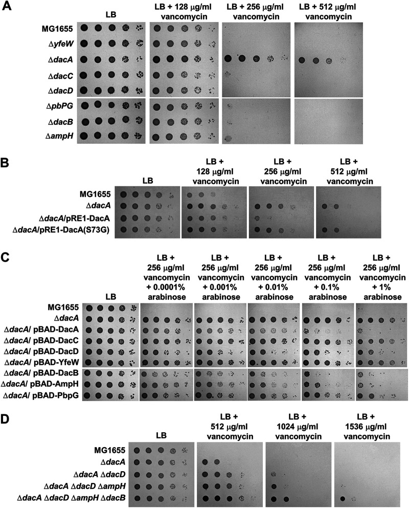
To analyze the physiological functions of PG carboxypeptidases, we examined several envelope stress-related phenotypes of PG carboxypeptidase-deleted strains. Notably, phenotypes were detected only in the dacA mutant (see Fig. S1 in the supplemental material). Among the identified phenotypes, we focused on vancomycin resistance. The dacA mutant exhibited strong vancomycin resistance, whereas the growth of other PG carboxypeptidase-deleted mutants was almost completely comparable to that of the wild-type strain in the presence of vancomycin (Fig. 1A and Fig. S2). To assess whether the PG carboxypeptidase activity of DacA is involved in vancomycin resistance, we constructed a DacA(S73G) mutant in which the serine residue of the active site was substituted with glycine, which resulted in the complete loss of PG carboxypeptidase activity (17). Vancomycin resistance of the dacA mutant was complemented by the ectopic expression of the wild-type DacA protein, but not the DacA(S73G) mutant protein (Fig. 1B), indicating that vancomycin resistance is associated with the PG carboxypeptidase activity of DacA.
FIG 1.
The loss of PG carboxypeptidase activities induces vancomycin resistance. (A) Vancomycin resistance of the dacA mutant. The wild-type and indicated mutant cells were serially diluted from 108 to 104 cells/mL in 10-fold steps and spotted onto an LB plate and LB plates containing indicated vancomycin concentrations. (B) Complementation of vancomycin resistance of the dacA mutant. The cells of the indicated strains were serially diluted from 108 to 104 cells/mL in 10-fold steps and spotted onto an LB plate and LB plates containing the indicated vancomycin concentrations. (C) Complementation of vancomycin resistance of the dacA mutant by other PG carboxypeptidases. The cells of the indicated strains were serially diluted from 108 to 104 cells/mL in 10-fold steps and spotted onto an LB plate and LB plates containing 256 μg/mL vancomycin and the indicated concentrations of arabinose. (D) Enhancement of vancomycin resistance by additional deletions of other PG carboxypeptidases. The wild-type and indicated mutant cells were serially diluted from 108 to 104 cells/mL in 10-fold steps and spotted onto an LB plate and LB plates containing indicated vancomycin concentrations.
Because the PG carboxypeptidase activity of DacA is important in vancomycin resistance, we determined whether the expression of other proteins with PG carboxypeptidase activity can influence vancomycin resistance of the dacA mutant. When PG carboxypeptidases were expressed using the arabinose-inducible promoter, vancomycin resistance of the dacA mutant was reduced by the expression of DacB, AmpH, DacD, and DacA (Fig. 1C). These results imply that additional deletion of other PG carboxypeptidases in the dacA mutant might increase vancomycin resistance. To test this assumption, we constructed the dacA dacD double, dacA dacD ampH triple, and dacA dacD ampH dacB quaternary mutants. Expectedly, vancomycin resistance of the dacA mutant gradually increased along with an increase in the number of deleted genes (Fig. 1D). These results demonstrate that the loss of PG carboxypeptidases is associated with vancomycin resistance and DacA plays a major role in this phenotype.
Deletion of DacA results in β-lactam sensitivity.
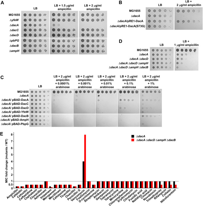
Some previous studies have demonstrated that the dacA mutant is sensitive to β-lactams (13–15). We also observed ampicillin sensitivity of the dacA mutant, and as observed for vancomycin resistance, this phenotype was detected only in the dacA mutant (Fig. 2A and Fig. S2). The ampicillin sensitivity was also associated with the PG carboxypeptidase activity of DacA (Fig. 2B). Because vancomycin resistance was complemented by the expression of other PG carboxypeptidases, we examined whether the ampicillin sensitivity is also restored by the expression of other PG carboxypeptidases. Besides DacA, several other PG carboxypeptidases could partially complement the ampicillin sensitivity of the dacA mutant (Fig. 2C). The additional deletions of PG carboxypeptidases in the dacA mutant strengthened ampicillin sensitivity (Fig. 2D), although the effect was not as much as that for vancomycin resistance. In summary, these results indicate that PG carboxypeptidases are involved in ampicillin sensitivity and DacA plays a pivotal role in this phenotype.
FIG 2.
Deletion of the dacA gene induces β-lactam sensitivity. (A) Ampicillin sensitivity of the dacA mutant. The wild-type and indicated mutant cells were serially diluted from 108 to 104 cells/mL in 10-fold steps and spotted onto an LB plate and LB plates containing the indicated ampicillin concentrations. (B) Complementation of ampicillin sensitivity of the dacA mutant. The cells of the indicated strains were serially diluted from 108 to 104 cells/mL in 10-fold steps and spotted onto an LB plate and an LB plate containing 2 μg/mL ampicillin. (C) Complementation of ampicillin sensitivity of the dacA mutant by other PG carboxypeptidases. The cells of the indicated strains were serially diluted from 108 to 104 cells/mL in 10-fold steps and spotted onto an LB plate and LB plates containing 2 μg/mL ampicillin and the indicated concentrations of arabinose. (D) Effect of additional deletions of other PG carboxypeptidases on ampicillin sensitivity of the dacA mutant. The wild-type and indicated mutant cells were serially diluted from 108 to 104 cells/mL in 10-fold steps and spotted onto an LB plate and an LB plate containing 2 μg/mL ampicillin. (E) Effect of PG carboxypeptidases on the MICs of antibiotics. The MICs of various antibiotics were measured against the wild-type or indicated mutant strains. The relative MIC values for the mutant cells compared to those for the wild-type cells are shown.
The inverse phenotypes of the dacA mutant for vancomycin and ampicillin susceptibility prompted us to investigate how susceptibility to other antibiotics is also affected by the dacA deletion. We examined the MICs of diverse antibiotics against the wild-type and dacA mutant strains. The MIC of ampicillin was 4-fold lower for the dacA mutant than for the wild-type strain; moreover, the MICs of most β-lactams were also 2-fold or 4-fold lower for the dacA mutant (Fig. 2E). Besides β-lactams, the MIC of d-cycloserine was also 2-fold lower for the dacA mutant than for the wild-type strain. The only antibiotic whose MIC is higher for the dacA mutant than for the wild-type strain was vancomycin (4-fold higher) (Fig. 2E). It is interesting that the dacA mutant exhibited divergent phenotypes to two antibiotics, β-lactam and vancomycin, inhibiting PG cross-linking. Furthermore, additional deletions of PG carboxypeptidases in the dacA mutant differentially affected the MICs of two antibiotics. The MIC of vancomycin was 2-fold higher in the dacA dacD ampH dacB quaternary mutant than in the dacA mutant, while the MICs of most β-lactams were the same in the two strains (Fig. 2E). In summary, these results indicate that DacA divergently modulates the MICs of vancomycin and β-lactams. Other PG carboxypeptidases also mildly influence the MIC of vancomycin, but their effects on β-lactams are very weak, implying that the underlying mechanisms of divergent phenotypes of the dacA mutant against two antibiotics are distinct.
Neither vancomycin resistance nor β-lactam sensitivity of the dacA mutant has a strong association with ld-transpeptidases.
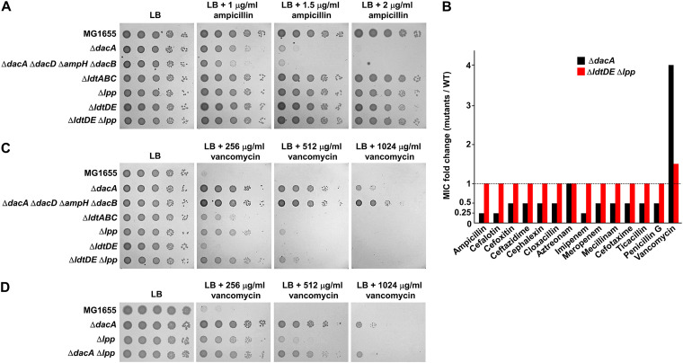
After the removal of the fifth d-alanine by PG carboxypeptidases, the PG tetrapeptide containing four amino acids can form the 3-3 cross-links by ld-transpeptidases (LdtD and LdtE) (2, 7, 9). ld-transpeptidases are structurally unrelated to PBPs (18); thus, they are not inhibited by most β-lactam antibiotics (11). Although these 3-3 cross-links account for only 3 to 10% of the entire cross-links in E. coli (19), a mutant strain having an increased proportion of 3-3 cross-links showed β-lactam resistance, owing to the reduction in PBP dependency (2). To assess whether the 3-3 cross-links are involved in β-lactam sensitivity of the dacA mutant, we examined the ampicillin susceptibility of the ldtD ldtE double mutant. Notably, the ampicillin sensitivity of the ldtD ldtE double mutant was comparable to that of the wild-type strain (Fig. 3A). After the removal of the fifth d-alanine by PG carboxypeptidases, the PG tetrapeptide can also be covalently cross-linked to Lpp by other ld-transpeptidases (LdtA, LdtB, and LdtC) (7). We checked the effect of LdtABC or Lpp on ampicillin sensitivity. The susceptibility of the ldtABC or lpp mutant to ampicillin was also the same as that of the wild-type strain (Fig. 3A). Additionally, the ldtDE lpp mutant defective for both the 3-3 cross-link and the attachment of PG to Lpp also exhibited susceptibility comparable to the wild-type strain (Fig. 3A). To assess whether the same result can be obtained for other β-lactam antibiotics, we determined the MICs of various β-lactams against the ldtDE lpp mutant. Expectedly, for the ldtDE lpp mutant, there were no changes in the MICs of all β-lactams tested compared to those observed for the wild type (Fig. 3B). These results suggest that the β-lactam sensitivity of the dacA mutant is not related to ld-transpeptidases.
FIG 3.
Phenotypes of the dacA mutant are not mainly associated with ld-transpeptidases. (A) Effect of ld-transpeptidases on ampicillin susceptibility. The wild-type and indicated mutant cells were serially diluted from 108 to 104 cells/mL in 10-fold steps and spotted onto an LB plate and LB plates containing the indicated ampicillin concentrations. (B) Effect of ld-transpeptidases on the MICs of β-lactams. The MICs of various antibiotics were measured against the wild-type or indicated mutant strains. The relative MIC values for the mutant cells compared to those for the wild-type cells are shown. (C) Effect of ld-transpeptidases on vancomycin resistance. The wild-type and indicated mutant cells were serially diluted from 108 to 104 cells/mL in 10-fold steps and spotted onto an LB plate and LB plates containing indicated vancomycin concentrations. (D) Effect of additional deletion of Lpp on vancomycin resistance of the dacA mutant. The wild-type and indicated mutant cells were serially diluted from 108 to 104 cells/mL in 10-fold steps and spotted onto an LB plate and LB plates containing the indicated vancomycin concentrations.
Next, we analyzed the relationship between vancomycin resistance and ld-transpeptidases. The vancomycin susceptibility of the ldtDE mutant was comparable to that of the wild-type cells, but both the ldtABC and lpp mutants were slightly more resistant to vancomycin than the wild-type cells, and the vancomycin resistance of the ldtDE lpp mutant was comparable to that of the lpp mutant (Fig. 3C). The MIC of vancomycin was 1.5-fold higher for the ldtDE lpp mutant than for the wild-type strain (Fig. 3B). The vancomycin resistance of the dacA mutant was not strengthened by the additional deletion of Lpp (Fig. 3D). Collectively, these results imply that the loss of the cross-links between PG and Lpp partially contributes to vancomycin resistance of the dacA mutant. Because vancomycin resistance of the ldtDE lpp mutant was significantly weaker than that of the dacA or dacA dacD ampH dacB mutant, the loss of the Lpp-PG attachment may not be the main molecular basis of vancomycin resistance of the PG carboxypeptidase-defective strains.
An increase in decoy d-Ala-d-Ala residues within PG in the dacA mutant results in vancomycin resistance.
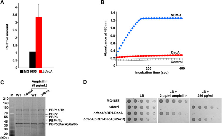
The fifth d-Ala of the donor pentapeptides is removed during the formation of the 4-3 cross-links by PBPs, whereas the fifth d-Ala of the acceptor pentapeptides is not removed during these reactions by PBPs (20). Because the fifth d-Ala of the acceptor pentapeptides can be removed by PG carboxypeptidases (20), the deletion of PG carboxypeptidases increases the amount of the 4-3 cross-links with d-Ala-d-Ala at the acceptor peptides (21). These d-Ala-d-Ala residues of cross-linked stem peptides can act as a decoy for the interaction with vancomycin, thus decreasing the amount of free vancomycin that inhibits the function of PBPs and, consequently, increasing vancomycin resistance. Because in E. coli, the amount of vancomycin passing through the periplasm is very low due to the presence of OM (22), the increase in decoy d-Ala-d-Ala residues can strongly affect the MIC of vancomycin. To assess this assumption, we examined the amount of vancomycin bound to purified PG using fluorescent vancomycin derivative. The amount of bound fluorescent vancomycin was approximately 3.5-fold higher in PG isolated from the dacA mutant than in that from the wild-type strain (Fig. 4A). The difference significantly correlated quantitatively with that of the MIC (Fig. 3E). Therefore, these results strongly suggest that the increase in decoy d-Ala-d-Ala residues at the acceptor peptides induced by the deletion of PG carboxypeptidases causes vancomycin resistance.
FIG 4.
Analysis of diverse DacA-related physiological properties. (A) In vitro interaction between fluorescent vancomycin and purified PG. Purified PGs from the wild-type and dacA mutant cells were mixed with fluorescent vancomycin. The amount of vancomycin bound to PG was determined by measuring the fluorescence emission of fluorescent vancomycin at 513 nm following excitation at 490 nm. (B) Estimation of the β-lactamase activity of DacA. The β-lactamase activities of purified NDM-1 (11 μg) and DacA (160 μg) were measured using nitrocefin. (C) Representative SDS-PAGE gel image for ampicillin titration of PBPs from the wild-type and dacA mutant cells. The binding assay of Bocillin FL to PBPs of the wild-type and dacA mutant cells was performed in the absence or presence of 8 μg/mL ampicillin. (D) Complementation of the phenotypes of the dacA mutant by DacA(K242R). The cells of the indicated strains were serially diluted from 108 to 104 cells/mL in 10-fold steps and spotted onto an LB plate and an LB plate containing 2 μg/mL ampicillin or 256 μg/mL vancomycin.
The C-terminal domain of DacA is dispensable for its PG carboxypeptidase activity but is obligately required for β-lactam resistance.
Previous reports suggested that DacA may have weak β-lactamase activity, as it shares a common ancestor with β-lactamases and contains a Ω-loop-like domain similar to TEM-1 β-lactamase (20, 23). However, to date, the β-lactamase activity of purified DacA has not been reported (24). In this study, we determined whether purified DacA has β-lactamase activity. Purified NDM-1 showed a strong nitrocefin-degrading activity, but DacA did not degrade nitrocefin, even at a condition of approximately 14-fold-higher protein level (Fig. 4B), indicating that DacA has no β-lactamase activity.
Given that DacA is one of the seven class C PBP proteins with low molecular weights (25), it can act as a β-lactam decoy protein that diminishes the concentration of free β-lactams. However, a previous report showed that the binding affinities of DacA with most β-lactams were significantly lower than those of other class C PBPs (26). Nevertheless, as DacA is the most abundant PG carboxypeptidase under normal conditions (27, 28), we assessed the role of DacA as a β-lactam decoy protein. In the wild-type and dacA mutant cells, the level of inhibition of Bocillin FL binding to PBPs by ampicillin was examined. In this assay, the band positions of DacA, DacC (also known as PBP6a), and DacD (also known as PBP6b) proteins were indistinguishable due to their similar molecular weights. Notably, the intensity of the band corresponding to DacA, DacC, and DacD was slightly reduced by the deletion of DacA, which was consistent with a previous report (29), indicating that DacA does not exist at an excessive level. The inhibition levels of Bocillin FL binding to PBPs by ampicillin were similar between the wild-type and dacA mutant strains (Fig. 4C), demonstrating that the role of DacA as the β-lactam decoy protein is negligible. To confirm this finding, we constructed a DacA(K242R) mutant whose PG carboxypeptidase activity is completely lost, but binding affinity for penicillin G is increased (30). Although this mutant protein can act as the β-lactam decoy protein, it could not complement the ampicillin sensitivity of the dacA mutant (Fig. 4D). Collectively, these results suggest that β-lactam sensitivity of the dacA mutant is not associated with the role of DacA as the β-lactam decoy protein.
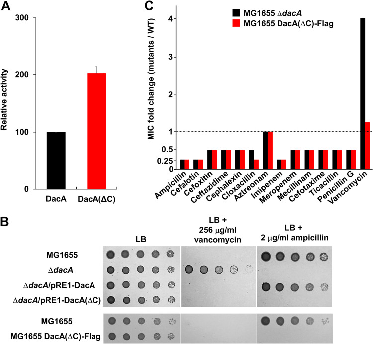
DacA has an inner membrane-anchoring domain at its C terminus (from 386th to 403rd amino acid residues), which is important for the function of DacA in morphology and its localization at the cell division site (17, 31). To analyze the effect of this domain on vancomycin resistance and β-lactam sensitivity, we constructed the DacA(ΔC) mutant without the C-terminal membrane-anchoring domain. Unexpectedly, DacA(ΔC) showed significantly increased PG carboxypeptidase activity than the wild-type protein (Fig. 5A). DacA(ΔC) could completely complement vancomycin resistance of the dacA mutant, confirming the full enzymatic activity of DacA(ΔC) (Fig. 5B). Notably, DacA(ΔC) could not restore ampicillin sensitivity of the dacA mutant (Fig. 5B) despite its full enzymatic activity. These results were also confirmed by the strain defective for a chromosomal C-terminal domain of DacA, MG1655 DacA(ΔC)-Flag. DacA(ΔC) was adequately expressed in this strain (Fig. S3), and thus, vancomycin resistance was not observed (Fig. 5B), but ampicillin sensitivity was detected up to a level comparable to that in the dacA mutant (Fig. 5B). The MG1655 DacA(ΔC)-Flag strain completely phenocopied the dacA mutant in β-lactam sensitivity (Fig. 5C), strongly indicating the obligate requirement of the C-terminal domain of DacA in β-lactam resistance. Collectively, these results again confirm the distinct molecular mechanisms of the two phenotypes of the dacA mutant and strongly suggest that the C-terminal domain of DacA and its enzymatic activity are strictly required for β-lactam resistance.
FIG 5.
Effects of the C-terminal domain of DacA on β-lactam and vancomycin resistance. (A) Effect of the C-terminal domain of DacA on its enzymatic activity. The enzymatic activity of purified PG carboxypeptidases was measured using the substrate diacetyl-l-Lys-d-Ala-d-Ala (AcLAA). Purified proteins (1 μM) were incubated at 37°C with 50 mM Tris-HCl (pH 8.0) containing 1 mM AcLAA. Released d-Ala was estimated using horseradish peroxidase and Amplex Red at 563 nm. (B) Effect of the C-terminal domain of DacA on β-lactam and vancomycin resistance. Cells of the indicated strains were serially diluted from 108 to 104 cells/mL in 10-fold steps and spotted onto an LB plate and an LB plate containing 256 μg/mL vancomycin or 2 μg/mL ampicillin. (C) MICs of β-lactams against the MG1655 DacA(ΔC)-Flag strain. The MICs of various antibiotics were measured against the wild-type or indicated mutant strains. Relative MIC values for the mutant cells compared to those for the wild-type cells are shown.
DacA interacts with PBPs in a C-terminal domain-dependent manner.
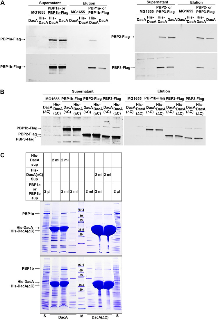
Because the dacA mutant was sensitive to almost all the β-lactam antibiotics but not to the other antibiotics with different modes of action (Fig. 2E), we analyzed the physical interaction between DacA and PBP, a binding target of β-lactam. To perform pulldown experiments, we constructed PBP-Flag strains having three Flag tags at the C terminus of chromosomal PBPs. All the PBP proteins, including PBP1a, PBP1b, PBP2, and PBP3, were pulled down by His-tagged DacA (Fig. 6A and Fig. S4), suggesting a physical interaction between DacA and PBPs. Notably, no PBP protein was pulled down by His-tagged DacA(ΔC) (Fig. 6B and Fig. S5 and S6), indicating that the C-terminal domain of DacA is required for these interactions. The C-terminal domain-dependent interaction of DacA with PBP was also confirmed by pulldown experiments using overexpressed PBP1a and PBP1b. Although the same overexpressed PBP1a or PBP1b supernatant was used, a significant amount of PBP1a or PBP1b was pulled down by His-tagged DacA, but not by His-tagged DacA(ΔC), even though the level of purified His-tagged DacA(ΔC) was higher than that of purified His-tagged DacA (Fig. 6C). These results suggest that DacA interacts with PBPs in a C-terminal domain-dependent manner.
FIG 6.
The C-terminal domain-dependent interactions of DacA with PBPs. (A) Physical interactions between DacA and PBPs. The supernatant of MG1655 cells or MG1655 cells with three Flag tags fused to the C terminus of the indicated chromosomal PBP was mixed with the supernatant of ER2566 cells harboring pET-based plasmid expressing His-tagged DacA or nontagged DacA. After pulldown experiments, the amount of input (supernatant) and output (elution) PBPs was measured by Western blotting using a monoclonal antibody against Flag tag. (B) Importance of the C-terminal domain of DacA for its interaction with PBPs. The supernatant of MG1655 cells or MG1655 cells with three Flag tags fused to the C terminus of the indicated chromosomal PBP was mixed with the supernatant of ER2566 cells harboring pET-based plasmid expressing His-tagged DacA(ΔC) or nontagged DacA(ΔC). After pulldown experiments, the amount of input (supernatant) and output (elution) PBPs was measured by Western blotting using a monoclonal antibody against Flag tag. (C) C-terminal domain-dependent interaction of DacA with PBP1a or PBP1b. The supernatant of ER2566 cells harboring the pET28a plasmid expressing PBP1b was mixed with the supernatant of ER2566 cells harboring pET-based plasmid expressing His-tagged DacA or His-tagged DacA(ΔC). After pulldown experiments, eluted proteins were separated on 4 to 20% gradient Tris-glycine polyacrylamide gels. The supernatant (lane S) of ER2566 cells harboring the pET28a plasmid expressing PBP1a or PBP1b was also loaded onto the gel. Lane M indicates EzWay Protein Blue molecular weight (MW) marker (Koma Biotech, South Korea).
The presence of the OM permeability barrier is necessary for vancomycin resistance of the dacA mutant, but not for β-lactam susceptibility.
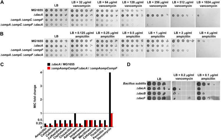
The OM plays a role as a strong barrier that prevents the transport of toxic molecules such as antibiotics (22). To determine whether the OM permeability barrier affects vancomycin resistance and β-lactam sensitivity of the dacA mutant, we examined the effect of dacA depletion in the context of the ompA ompC ompF triple mutant, a strain with increased OM permeability (32). The ompA ompC ompF triple mutant was more sensitive to vancomycin than the wild-type strain (Fig. 7A), indicating the increased passage of vancomycin in this strain. Notably, in the background of the ompA ompC ompF triple mutant, vancomycin resistance of the dacA mutant was completely absent (Fig. 7A). These results imply that the effect of the decoy d-Ala-d-Ala within PG on vancomycin resistance weakens as the periplasmic level of vancomycin increases. Interestingly, unlike vancomycin, ampicillin sensitivity of the dacA mutant was significantly strengthened in the background of the ompA ompC ompF triple mutant (Fig. 7B). This pattern was also observed for most other β-lactams apart from ampicillin (Fig. 7C), indicating that β-lactam sensitivity of the dacA mutant is detected in the background of increased OM permeability. These data confirm that the molecular mechanisms for the two inverse phenotypes of the dacA mutant are distinct.
FIG 7.
Effects of the OM permeability barrier on vancomycin resistance and β-lactam sensitivity in the dacA mutant. (A) Effect of the OM permeability barrier on vancomycin resistance. The wild-type and indicated mutant cells were serially diluted from 108 to 104 cells/mL in 10-fold steps and spotted onto an LB plate and LB plates containing the indicated vancomycin concentrations. (B) Effect of OM permeability barrier on ampicillin sensitivity. The wild-type and indicated mutant cells were serially diluted from 108 to 104 cells/mL in 10-fold steps and spotted onto an LB plate and LB plates containing the indicated ampicillin concentrations. (C) Effect of the OM permeability barrier on the MICs of β-lactams. The MICs of various antibiotics were measured against the indicated mutant strains. The relative MIC values for the dacA mutant cells compared to those for the wild-type cell are shown as black bars, while the relative MIC values for the ompA ompC ompF dacA mutant cells compared to those for the ompA ompC ompF mutant cells are shown as red bars. (D) Vancomycin resistance and β-lactam sensitivity of the B. subtilis dacA mutant. The wild-type and indicated mutant cells were serially diluted from 108 to 104 cells/mL in 10-fold steps and spotted onto an LB plate and LB plates containing the indicated antibiotic concentrations.
The divergent effect of PG carboxypeptidase on ampicillin and vancomycin susceptibility is also detected in Bacillus subtilis.
We also examined the effect of PG carboxypeptidase on vancomycin and ampicillin susceptibility in B. subtilis, a Gram-positive bacterium that lacks the OM. B. subtilis has three homologs of E. coli DacA (DacA, DacB, and DacF), and the dacA mutant of B. subtilis was resistant to vancomycin, while the dacA and dacB mutants were sensitive to ampicillin (Fig. 7D). Both vancomycin resistance and ampicillin sensitivity of the B. subtilis dacA mutant were significantly weaker than those of E. coli dacA mutant. Vancomycin can easily access PG in the absence of the OM, which might have affected vancomycin resistance in the B. subtilis dacA mutant. Similarly, easy access of vancomycin to PG completely abolished vancomycin resistance in the E. coli dacA mutant (Fig. 7A), whereas the B. subtilis dacA mutant showed weak vancomycin resistance. This discrepancy may have been caused by the thick PG layer of B. subtilis, where the number of the decoy d-Ala-d-Ala residues was relatively significantly higher than that in E. coli. The reason behind the relatively weak β-lactam sensitivity of the dacA mutant of B. subtilis remains unclear. Collectively, these results show that the divergent phenotypes of the dacA mutant for vancomycin and ampicillin susceptibility are also conserved in a Gram-positive bacterium, and the presence of the OM can influence the phenotype of the dacA mutant.
DISCUSSION
PG is an essential architecture for bacterial growth and shape maintenance, and its biosynthesis is inhibited by many antibiotics such as β-lactams and vancomycin, leading to cell lysis. Although extensive studies have been conducted on PBPs, the physiological roles of PG hydrolases are poorly understood, especially regarding their impacts on antibiotic resistance. In this study, we revealed an intriguing connection between PG carboxypeptidase DacA and antibiotic resistance. The DacA-defective mutant showed β-lactam sensitivity and vancomycin resistance. Based on various genetic and biochemical experiments, we proposed the molecular mechanisms underlying these divergent phenotypes. Vancomycin resistance was caused by a decoy mechanism associated with increased d-Ala-d-Ala residues. Meanwhile, β-lactam sensitivity was associated with the physical interactions between DacA and PBPs, and these interactions required the C-terminal membrane-anchoring domain of DacA. Intriguingly, increased permeability of the OM strongly weakened the vancomycin resistance of the dacA mutant, whereas it significantly strengthened β-lactam susceptibility. Collectively, our results demonstrate two distinct molecular mechanisms of PG carboxypeptidase DacA-mediated antibiotic resistance in E. coli, a Gram-negative bacterium.
Staphylococcus aureus, a Gram-positive bacterium, has one orthologue of DacA, which is known as PBP4 (hereinafter referred to as SaPBP4). SaPBP4 displays 25% identity (45% similarity) with E. coli DacA, and these two proteins have a similar molecular weight. Many clinical isolates with a decreased expression level of SaPBP4 showed vancomycin resistance or β-lactam sensitivity (33–37). Previous studies suggested that the decoy mechanism of d-Ala-d-Ala is associated with vancomycin resistance (34, 36), which is consistent with the results of this study. However, unexpectedly, many recent reports based on in vivo and in vitro data showed that SaPBP4 has a strong dd-transpeptidase activity, but a weak dd-carboxypeptidase activity (38–41). Because many in vitro and in vivo experiments have demonstrated dd-carboxypeptidase activity of E. coli DacA (20, 21, 42), a direct comparison between our study and SaPBP4-related studies seems to be difficult. However, our study demonstrated that DacA physically interacted with dd-transpeptidases, namely, PBPs, and these interactions were strongly associated with the effect of DacA on β-lactam resistance. This implies that E. coli DacA also affects dd-transpeptidases, that is, the formation of 4-3 cross-links. Because the two phenotypes of the dacA mutant identified in this study did not have a strong association with ld-transpeptidases, the dd-transpeptidase-related function of PG carboxypeptidases should be investigated in E. coli in the further studies. Notably, a study demonstrated that SaPBP4 can exert β-lactamase activity based on in vitro data using nitrocefin (43). However, the β-lactamase activity of E. coli DacA has not been reported. In summary, these results suggest that further studies are required to elucidate the primary physiological role of PG carboxypeptidase in both Gram-negative and Gram-positive bacteria.
The mechanism of vancomycin resistance has been poorly analyzed in Gram-negative bacteria, including E. coli, as they are intrinsically resistant to vancomycin due to the presence of OM. Our study revealed decoy-mediated mechanism of vancomycin resistance in E. coli, indicating that there are various molecular mechanisms of vancomycin resistance in E. coli besides the inhibition of vancomycin passage by the OM barrier. Although the decoy-mediated mechanism of vancomycin resistance was first suggested in S. aureus based on SaPBP4-related studies (43), the decoy model in S. aureus should be modified because SaPBP4 is a dd-transpeptidase, not a dd-carboxypeptidase. In this study, we clearly showed through an in vitro experiment, using purified PG (Fig. 4A), and an in vivo experiment, using DacA(ΔC) mutant (Fig. 5B), that vancomycin resistance in the dacA mutant is induced by decoy-mediated mechanism. Therefore, our study first revealed decoy-mediated vancomycin resistance mechanism of PG carboxypeptidase, at least in E. coli. Molecular decoy-mediated vancomycin resistance in E. coli has also been reported in a strain with a point mutation in the waaL gene (encoding O-antigen ligase), which results in the synthesis of PG-modified lipopolysaccharide (LPS) (44). PG-modified LPS was transported through the OM and induced the decoy-mediated vancomycin resistance (44). These results imply that further studies are required to identify the diverse mechanisms associated with vancomycin resistance in E. coli.
The relationship between PG carboxypeptidase and β-lactam resistance has been consistently studied in E. coli (2, 13–15). The β-lactam sensitivity of the dacA mutant can be induced by diverse probable mechanisms as follows: dependency on 4-3 cross-links, β-lactamase activity of DacA, and the decoy model by class C PBPs, including DacA. Unexpectedly, our data showed that all the suggested mechanisms were not associated with the β-lactam sensitivity of the dacA mutant. Instead, our results suggest that the physical interactions of DacA with PBPs are strongly associated with the β-lactam sensitivity of the dacA mutant. It is necessary to determine whether this conclusion can be applied to other Gram-negative bacteria. The physiological significance of the physical interactions of DacA with PBPs is currently unknown. However, these results strongly imply that DacA plays diverse physiological roles beyond the regulation of 3-3 cross-link formation and Lpp-PG attachment.
The OM acts as a strong barrier against the influx of antibiotics owing to the hydrophobic acyl chains of LPS and hydrophilic lateral interactions between LPSs bridged by divalent cations (45, 46). Nonspecific porins strongly regulate OM permeability (32). Our study again demonstrated that the presence of the OM strongly affected antibiotic resistance of Gram-negative bacteria. The vancomycin resistance of the dacA mutant was almost completely abolished in the cells with increased OM permeability, while the β-lactam sensitivity of the dacA mutant was significantly increased under the same condition (Fig. 7). These results imply that the effect of the OM permeability barrier is diverse and complex for the different types of antibiotics.
Although the C-terminal membrane-anchoring domain of DacA is known to be important for its function in morphology and its localization at the cell division site (17, 31), the underlying molecular mechanism has not been elucidated. Our data showed that the C-terminal domain of DacA was necessary for its interactions with PBPs, including PBP1a, PBP1b, PBP2, and PBP3 (Fig. 6). This result successfully explains why the C-terminal domain of DacA is required for its function in morphology and its localization at the septum. PBP2 and PBP3 play pivotal roles in cell morphology, and their inhibitions cause cell rounding and filamentation, respectively (47). PBP3 is localized to the septum and forms the divisome complex with various proteins involved in cytokinesis (1, 48). Therefore, the role of the C-terminal domain in morphology might be associated with the physical interactions with PBP2 and PBP3, and its role in localization at the septum might be related to the physical interaction with PBP3. These findings should be confirmed by conducting further studies. Because the DacA(ΔC) mutant without its C-terminal domain could not complement all the phenotypes of the dacA mutant, except vancomycin resistance, the β-lactam susceptibility phenotype of DacA mutation is associated with its C-terminal domain, that is, the interactions with PBPs. To complement the β-lactam sensitivity of the dacA mutant, the interactions with PBPs, as well as the enzymatic activity of DacA, are required (Fig. 2B). This implies that DacA does not affect PBPs only physically. The exact physiological significance of the DacA-PBP complexes remains unclear at present, and thus, further studies are required.
Our study showed the inverse phenotypes of the dacA mutant for vancomycin and β-lactam resistance and revealed distinct molecular mechanisms responsible for these phenotypes. Therefore, we provide novel insights into the roles of PG carboxypeptidase DacA in the regulation of antibiotic resistance. Our findings also provide future research directions to elucidate the physiological function of PG carboxypeptidases.
MATERIALS AND METHODS
Bacterial strains, plasmids, and culture conditions.
All the strains and primer sequences used in this study are presented in Tables S1 and S2 in the supplemental material, respectively. All E. coli deletion strains were constructed using λ Red recombinase as previously described (49), with some modifications. An FLP recombination target (FRT)-flanked kanamycin resistance gene in plasmid pKD3 was amplified by conducting PCR using deletion primer sets (see Table S2) with 50-mer sequences for the recombination between each target gene and the kanamycin gene. The PCR products were purified using PCR purification kit (Qiagen, USA) and transformed into MG1655 cells harboring plasmid pKD46 that expresses λ Red recombinase. After regeneration at 37°C, the cells were spread onto Luria-Bertani (LB) plates containing kanamycin (50 μg/mL) and incubated overnight at 37°C. The gene deletion in kanamycin-resistant cells was confirmed by PCR using deletion-confirming primer sets. To remove the kanamycin resistance gene, plasmid pCP20, which expresses FLP recombinase, was transformed into deletion strains. After incubation at 37°C, the recombination between FRT sequences was checked by PCR using deletion-confirming primer sets, and the curing of the plasmid pCP20 was checked by growth failure in an LB plate containing ampicillin (100 μg/mL). To minimize the physiological change, the curing of the plasmid pCP20 was performed at 37°C as previously described (50). All transformation procedures were performed by electroporation. Additional gene deletion, such as generation of the dacA dacD double mutant, was also carried out using λ Red recombinase. Experimental procedures, including pKD46 transformation, gene disruption, pCP20 transformation, and FRT recombination, were sequentially performed. The B. subtilis deletion mutant strain was constructed using long-flanking homology PCR as previously described (51, 52).
The MG1655 PBP1a-Flag strain was also constructed using λ Red recombinase as previously described (50). Briefly, the DNA sequence, including the 3×Flag gene and the chloramphenicol resistance gene, was amplified from the plasmid pBAD-PBP1a-Flag. After overnight DpnI digestion for the removal of template plasmids, PCR products were transformed into the MG1655 strain containing pKD46. The chromosomal fusion of the 3×Flag tag to the C terminus of PBP1a was confirmed by PCR. Other chromosomal PBP-Flag strains were also constructed through the same method. The MG1655 DacA(ΔC)-Flag strain was constructed through a modified process to remove the antibiotic resistance gene. The FRT-flanked kanamycin resistance gene in plasmid pKD3 was inserted into the plasmid pBAD-Flag, generating the plasmid pBAD-Flag-FRT-Kan. The DNA sequence, including the 3×Flag gene and the kanamycin resistance gene, was amplified from the plasmid pBAD-Flag-FRT-Kan. After the chromosomal fusion of the 3×Flag tag to the 385th amino acid residue of DacA, the kanamycin resistance gene was removed by the recombination between FRT sequences by using the plasmid pCP20 as described above.
The pBAD24a-based expression vectors of PG carboxypeptidases were constructed using In-Fusion cloning (Clontech, USA). The entire open reading frame of PG carboxypeptidase genes was amplified by PCR using cloning primer sets (see Table S2) containing the 15-mer sequence at the 5′ end for recombination with the vector. The PCR products were purified using PCR purification kit (Qiagen, USA), and in vitro recombination was performed between purified PCR products and pBAD24a plasmids digested by NdeI and BamHI restriction enzymes. Plasmid construction was confirmed by sequencing analysis. Through a similar method, the pRE1-based expression vector of DacA was constructed. Because the promoter in the pRE1 plasmid is recognized by E. coli RNA polymerase, but not T7 RNA polymerase, and is under the control of the cI protein of phage λ, the gene in the pRE1 plasmid is constitutively expressed in the general E. coli cells lacking the cI protein, including MG1655 (53, 54). To overexpress DacA, the pET28a-based or pET24a-based vector expressing His-tagged DacA or nontagged DacA, respectively, was constructed through a similar method. In these cases, a truncated dacA gene without the DNA sequence encoding the signal sequence (1 to 29 amino acid residues) was cloned into the pET-based plasmids. The pRE1-DacA(S73G) and pRE1-DacA(K242R) plasmids were constructed by PCR by using the pRE1-DacA plasmid as a template and DpnI digestion as previously described (6).
Determination of MIC values.
The MIC values of the antibiotics were determined according to the Clinical and Laboratory Standards Institute guideline (55). Cells from overnight seed culture grown in LB medium were inoculated into Mueller-Hinton broth and cultured at 37°C to a McFarland turbidity standard of 0.5 (approximately 1.5 × 10⁸ cells/mL). Cultured cells were diluted with Mueller-Hinton broth to reach a final concentration of 107 cells/mL. Dilution samples (10 μL) were spotted onto Mueller-Hinton plates containing antibiotics of final concentrations of 512 μg/mL to 7.8 ng/mL in 2-fold serial dilutions, and the plates were incubated at 37°C for 20 h. A 1.5-fold serial dilution was applied when necessary. The MIC value of each antibiotic is defined as the lowest concentration of antibiotic at which the lawn growth of cells is inhibited.
Purification of PG and in vitro binding assay with fluorescent vancomycin.
The cells (1 L) of the wild-type and dacA mutant strains grown in LB medium to early exponential phase were harvested and resuspended in 20 mL of phosphate-buffered saline (PBS) buffer. After adding 80 mL of 5% SDS buffer, the mixtures were boiled at 100°C for 30 min and incubated at room temperature overnight. The samples were ultracentrifuged at 160,000 × g for 1 h at room temperature, using Himac microultracentrifuge CS120FNX (Hitachi, Japan). To remove the remaining SDS, the pellets were washed with distilled water (DW) at least five times. The washed pellets were resuspended in 1 mL of PBS buffer. To remove high-molecular-weight glycogen and proteins, α-amylase (final concentration of 0.2 mg/mL) and α-chymotrypsin (final concentration of 300 μg/mL) were added to the resuspended samples, and the mixtures were incubated at 37°C overnight. After adding SDS (final concentration of 1%), the samples were boiled at 100°C for 30 min and ultracentrifuged at 260,000 × g for 15 min at room temperature. To remove the remaining SDS, the pellets were washed with DW three times. The washed pellets were resuspended in 1 mL of DW, and the samples were stored at −80°C.
To perform in vitro binding assay with fluorescent vancomycin, 50 mg of PGs purified from the wild-type and dacA mutant cells were mixed with 10 μg of Bodipy FL vancomycin (Invitrogen, USA). After incubation at 37°C for 30 min, the mixtures were ultracentrifuged at 260,000 × g for 15 min at room temperature. After washing the pellets with DW once, the pellets were resuspended in 50 μL DW, and the amount of bound fluorescent vancomycin was determined by measuring the fluorescence emission of fluorescent vancomycin at 513 nm following excitation at 490 nm.
Purification of overexpressed proteins and β-lactamase assay using nitrocefin.
ER2566 cells harboring the plasmid pET28a-DacA or pET28a-NDM-1 from overnight seed culture were inoculated into LB medium containing kanamycin (50 μg/mL). When the optical density at 600 nm (OD600) reached approximately 0.5, 1 mM isopropyl-β-d-1-thiogalactopyranoside (IPTG) was added to the culture medium. After overnight culture at 16°C, harvested cells were resuspended in buffer A (50 mM Tris-HCl [pH 8.0] and 150 mM NaCl) and disrupted by a French press cell at 10,000 lb/in2. Soluble His-tagged DacA and NDM-1 proteins were separated through centrifugation at 8,000 × g for 20 min at 4°C. The supernatants containing soluble target proteins were loaded into a Talon metal affinity resin (Clontech, USA). After washing three times with buffer A, His-tagged proteins bound to Talon resins were eluted with buffer A containing 200 mM imidazole. After overnight dialysis with buffer B (50 mM Tris-HCl [pH 7.5] and 50 mM NaCl) at 4°C, purified proteins were promptly used for measuring the β-lactamase activity.
To measure β-lactamase activity, purified NDM-1 (11 μg) and DacA (160 μg) proteins were mixed with 1 mM nitrocefin in 100 mM MES (morpholineethanesulfonic acid; pH 7.0) buffer containing 1 mg/mL bovine serum albumin. The degradation of nitrocefin was detected by measuring the absorbance at 486 nm over time. Instead of purified protein, the sample containing a Tris buffer of the same volume was used as a control.
Binding assay of Bocillin FL to PBPs.
Cells (20 mL) of the wild-type and dacA mutant strains grown in LB medium to early exponential phase were harvested. After washing once with 1 mL PBS buffer, the cells were resuspended in 1 mL PBS buffer and disrupted by a French press cell. Crude extracts were mixed with 5 μg of Bocillin FL in the presence or absence of 8 μg ampicillin. After incubation at room temperature for 10 min, 100 μL of the samples was mixed with 100 μL of 2× SDS sample buffer, followed by boiling at 100°C for 5 min. The mixture (20 μL) was loaded onto 4 to 20% gradient Tris-glycine polyacrylamide gel and run at 180 V for 80 min. PBP proteins bound to Bocillin FL were detected by scanning the fluorescence emission at 511 nm following excitation at 504 nm.
Assessment of the enzymatic activity of PG carboxypeptidases.
The enzyme activity of purified PG carboxypeptidases was measured using the substrate diacetyl-l-Lys-d-Ala-d-Ala (AcLAA) (21). d-Ala was released by PG carboxypeptidase, and the released d-Ala was degraded into pyruvate, hydrogen peroxide, and amine group by d-amino acid oxidase (Sigma-Aldrich, USA). Horseradish peroxidase (Sigma-Aldrich) reduced hydrogen peroxide to H2O by using Amplex Red (Invitrogen) as an electron donor, which, in turn, was converted into resorufin by oxidation. The level of resorufin can be measured at 563 nm spectrophotometrically. Purified proteins (1 μM) were incubated at 37°C with 50 mM Tris-HCl (pH 8.0) containing 1 mM AcLAA in a final volume of 200 μL. After incubation for 60 min, the reaction was stopped by boiling at 100°C for 20 min. After centrifugation at 13,000 rpm for 5 min, only the supernatant was mixed with 800 μL of assay buffer with 50 mM HEPES-NaOH (pH 7.5), 10 mM MgCl2, 50 μM Amplex Red, 54 μg/mL horseradish peroxidase, and 75 μg/mL d-amino acid oxidase. The mixed samples were incubated at 37°C for 60 min, and the absorbance at 563 nm was measured using UV-visible (UV-Vis) spectrophotometer UVmini-1240 (Shimadzu, Japan).
Assessment of physical interactions between DacA/DacA(ΔC) and PBPs.
ER2566 cells harboring pET-based plasmid expressing His-tagged DacA or nontagged DacA were cultured in 50 mL of LB medium at 37°C. When the OD600 was approximately 0.5, 1 mM IPTG was added, and the cells were cultured at 30°C for 4 h. MG1655 chromosomal PBP-Flag strains were cultured in 100 mL of LB medium at 37°C to early stationary phase. Harvested ER2566 and MG1655 cells were resuspended in buffer A containing 1% sodium n-dodecyl-β-d-maltoside and disrupted by a French press cell at 10,000 lb/in2. After centrifugation at 2,100 × g for 5 min at 4°C, the supernatants were mixed with 100 μL of the Talon metal affinity resin (Clontech, USA) in a 1.5-mL tube. After washing with 1 mL of buffer A containing 1% sodium n-dodecyl-β-d-maltoside three times, bound proteins were eluted using buffer A containing 200 mM imidazole. Eluted proteins were separated on 4 to 20% gradient Tris-glycine polyacrylamide gels (Koma Biotech, South Korea) and were transferred onto polyvinylidene difluoride (PVDF) membranes. The protein levels of PBPs were determined using a monoclonal antibody against Flag tag (Santa Cruz Biotechnology, USA) according to the standard procedure. A similar pulldown experiment was also performed using overexpressed full-length PBP1a or PBP1b and overexpressed His-tagged DacA or DacA(ΔC). Eluted proteins were separated on 4 to 20% gradient Tris-glycine polyacrylamide gels.
ACKNOWLEDGMENTS
This work was supported by research grants from Basic Science Research Program through the National Research Foundation of Korea funded by the Ministry of Education (NRF-2020R1I1A2058026, 2021R1A6A3A01086677, and 2021R1A6A3A01086629).
We declare that we have no competing interests.
Footnotes
Supplemental material is available online only.
Contributor Information
Chang-Ro Lee, Email: crlee@mju.ac.kr.
Krisztina M. Papp-Wallace, Louis Stokes Cleveland VAMC
REFERENCES
- 1.Vollmer W, Bertsche U. 2008. Murein (peptidoglycan) structure, architecture and biosynthesis in Escherichia coli. Biochim Biophys Acta 1778:1714–1734. doi: 10.1016/j.bbamem.2007.06.007. [DOI] [PubMed] [Google Scholar]
- 2.Hugonnet JE, Mengin-Lecreulx D, Monton A, den Blaauwen T, Carbonnelle E, Veckerle C, Brun YV, van Nieuwenhze M, Bouchier C, Tu K, Rice LB, Arthur M. 2016. Factors essential for L,D-transpeptidase-mediated peptidoglycan cross-linking and β-lactam resistance in Escherichia coli. Elife 5:e19469. doi: 10.7554/eLife.19469. [DOI] [PMC free article] [PubMed] [Google Scholar]
- 3.Vermassen A, Leroy S, Talon R, Provot C, Popowska M, Desvaux M. 2019. Cell wall hydrolases in bacteria: insight on the diversity of cell wall amidases, glycosidases and peptidases toward peptidoglycan. Front Microbiol 10:331. doi: 10.3389/fmicb.2019.00331. [DOI] [PMC free article] [PubMed] [Google Scholar]
- 4.Uehara T, Parzych KR, Dinh T, Bernhardt TG. 2010. Daughter cell separation is controlled by cytokinetic ring-activated cell wall hydrolysis. EMBO J 29:1412–1422. doi: 10.1038/emboj.2010.36. [DOI] [PMC free article] [PubMed] [Google Scholar]
- 5.Mueller EA, Iken AG, Ali Ozturk M, Winkle M, Schmitz M, Vollmer W, Di Ventura B, Levin PA. 2021. The active repertoire of Escherichia coli peptidoglycan amidases varies with physiochemical environment. Mol Microbiol 116:311–328. doi: 10.1111/mmi.14711. [DOI] [PMC free article] [PubMed] [Google Scholar]
- 6.Park SH, Kim YJ, Lee HB, Seok YJ, Lee CR. 2020. Genetic evidence for distinct functions of peptidoglycan endopeptidases in Escherichia coli. Front Microbiol 11:565767. doi: 10.3389/fmicb.2020.565767. [DOI] [PMC free article] [PubMed] [Google Scholar]
- 7.More N, Martorana AM, Biboy J, Otten C, Winkle M, Serrano CKG, Monton Silva A, Atkinson L, Yau H, Breukink E, den Blaauwen T, Vollmer W, Polissi A. 2019. Peptidoglycan remodeling enables Escherichia coli to survive severe outer membrane assembly defect. mBio 10:e02729-18. doi: 10.1128/mBio.02729-18. [DOI] [PMC free article] [PubMed] [Google Scholar]
- 8.Winkle M, Hernandez-Rocamora VM, Pullela K, Goodall ECA, Martorana AM, Gray J, Henderson IR, Polissi A, Vollmer W. 2021. DpaA detaches Braun's lipoprotein from peptidoglycan. mBio 12:e00836-21. doi: 10.1128/mBio.00836-21. [DOI] [PMC free article] [PubMed] [Google Scholar]
- 9.Bahadur R, Chodisetti PK, Reddy M. 2021. Cleavage of Braun's lipoprotein Lpp from the bacterial peptidoglycan by a paralog of L,D-transpeptidases, LdtF. Proc Natl Acad Sci USA 118:e2101989118. doi: 10.1073/pnas.2101989118. [DOI] [PMC free article] [PubMed] [Google Scholar]
- 10.Mathelie-Guinlet M, Asmar AT, Collet JF, Dufrene YF. 2020. Lipoprotein Lpp regulates the mechanical properties of the E. coli cell envelope. Nat Commun 11:1789. doi: 10.1038/s41467-020-15489-1. [DOI] [PMC free article] [PubMed] [Google Scholar]
- 11.Sutterlin L, Edoo Z, Hugonnet JE, Mainardi JL, Arthur M. 2018. Peptidoglycan cross-linking activity of L,D-transpeptidases from Clostridium difficile and inactivation of these enzymes by β-lactams. Antimicrob Agents Chemother 62:e01607-17. doi: 10.1128/AAC.01607-17. [DOI] [PMC free article] [PubMed] [Google Scholar]
- 12.Baranowski C, Welsh MA, Sham LT, Eskandarian HA, Lim HC, Kieser KJ, Wagner JC, McKinney JD, Fantner GE, Ioerger TR, Walker S, Bernhardt TG, Rubin EJ, Rego EH. 2018. Maturing Mycobacterium smegmatis peptidoglycan requires non-canonical crosslinks to maintain shape. Elife 7:e37516. doi: 10.7554/eLife.37516. [DOI] [PMC free article] [PubMed] [Google Scholar]
- 13.Chowdhury C, Kar D, Dutta M, Kumar A, Ghosh AS. 2012. Moderate deacylation efficiency of DacD explains its ability to partially restore beta-lactam resistance in Escherichia coli PBP5 mutant. FEMS Microbiol Lett 337:73–80. doi: 10.1111/1574-6968.12009. [DOI] [PubMed] [Google Scholar]
- 14.Sarkar SK, Chowdhury C, Ghosh AS. 2010. Deletion of penicillin-binding protein 5 (PBP5) sensitises Escherichia coli cells to β-lactam agents. Int J Antimicrob Agents 35:244–249. doi: 10.1016/j.ijantimicag.2009.11.004. [DOI] [PubMed] [Google Scholar]
- 15.Sarkar SK, Dutta M, Chowdhury C, Kumar A, Ghosh AS. 2011. PBP5, PBP6 and DacD play different roles in intrinsic β-lactam resistance of Escherichia coli. Microbiology (Reading) 157:2702–2707. doi: 10.1099/mic.0.046227-0. [DOI] [PubMed] [Google Scholar]
- 16.Nichols RJ, Sen S, Choo YJ, Beltrao P, Zietek M, Chaba R, Lee S, Kazmierczak KM, Lee KJ, Wong A, Shales M, Lovett S, Winkler ME, Krogan NJ, Typas A, Gross CA. 2011. Phenotypic landscape of a bacterial cell. Cell 144:143–156. doi: 10.1016/j.cell.2010.11.052. [DOI] [PMC free article] [PubMed] [Google Scholar]
- 17.Nelson DE, Young KD. 2001. Contributions of PBP 5 and DD-carboxypeptidase penicillin binding proteins to maintenance of cell shape in Escherichia coli. J Bacteriol 183:3055–3064. doi: 10.1128/JB.183.10.3055-3064.2001. [DOI] [PMC free article] [PubMed] [Google Scholar]
- 18.Mainardi JL, Villet R, Bugg TD, Mayer C, Arthur M. 2008. Evolution of peptidoglycan biosynthesis under the selective pressure of antibiotics in Gram-positive bacteria. FEMS Microbiol Rev 32:386–408. doi: 10.1111/j.1574-6976.2007.00097.x. [DOI] [PubMed] [Google Scholar]
- 19.Schwarz U, Asmus A, Frank H. 1969. Autolytic enzymes and cell division of Escherichia coli. J Mol Biol 41:419–429. doi: 10.1016/0022-2836(69)90285-X. [DOI] [PubMed] [Google Scholar]
- 20.Ghosh AS, Chowdhury C, Nelson DE. 2008. Physiological functions of D-alanine carboxypeptidases in Escherichia coli. Trends Microbiol 16:309–317. doi: 10.1016/j.tim.2008.04.006. [DOI] [PubMed] [Google Scholar]
- 21.Peters K, Kannan S, Rao VA, Biboy J, Vollmer D, Erickson SW, Lewis RJ, Young KD, Vollmer W. 2016. The redundancy of peptidoglycan carboxypeptidases ensures robust cell shape maintenance in Escherichia coli. mBio 7:e00819-16. doi: 10.1128/mBio.00819-16. [DOI] [PMC free article] [PubMed] [Google Scholar]
- 22.O'Shea R, Moser HE. 2008. Physicochemical properties of antibacterial compounds: implications for drug discovery. J Med Chem 51:2871–2878. doi: 10.1021/jm700967e. [DOI] [PubMed] [Google Scholar]
- 23.Pepper ED, Farrell MJ, Finkel SE. 2006. Role of penicillin-binding protein 1b in competitive stationary-phase survival of Escherichia coli. FEMS Microbiol Lett 263:61–67. doi: 10.1111/j.1574-6968.2006.00418.x. [DOI] [PubMed] [Google Scholar]
- 24.Kar D, Pandey SD, Mallick S, Dutta M, Ghosh AS. 2018. Substitution of alanine at position 184 with glutamic acid in Escherichia coli PBP5 Ω-like loop introduces a moderate cephalosporinase activity. Protein J 37:122–131. doi: 10.1007/s10930-018-9765-y. [DOI] [PubMed] [Google Scholar]
- 25.Sauvage E, Kerff F, Terrak M, Ayala JA, Charlier P. 2008. The penicillin-binding proteins: structure and role in peptidoglycan biosynthesis. FEMS Microbiol Rev 32:234–258. doi: 10.1111/j.1574-6976.2008.00105.x. [DOI] [PubMed] [Google Scholar]
- 26.Kocaoglu O, Carlson EE. 2015. Profiling of β-lactam selectivity for penicillin-binding proteins in Escherichia coli strain DC2. Antimicrob Agents Chemother 59:2785–2790. doi: 10.1128/AAC.04552-14. [DOI] [PMC free article] [PubMed] [Google Scholar]
- 27.Li GW, Burkhardt D, Gross C, Weissman JS. 2014. Quantifying absolute protein synthesis rates reveals principles underlying allocation of cellular resources. Cell 157:624–635. doi: 10.1016/j.cell.2014.02.033. [DOI] [PMC free article] [PubMed] [Google Scholar]
- 28.Santos JM, Lobo M, Matos AP, De Pedro MA, Arraiano CM. 2002. The gene bolA regulates dacA (PBP5), dacC (PBP6) and ampC (AmpC), promoting normal morphology in Escherichia coli. Mol Microbiol 45:1729–1740. doi: 10.1046/j.1365-2958.2002.03131.x. [DOI] [PubMed] [Google Scholar]
- 29.Matsuhashi M, Maruyama IN, Takagaki Y, Tamaki S, Nishimura Y, Hirota Y. 1978. Isolation of a mutant of Escherichia coli lacking penicillin-sensitive D-alanine carboxypeptidase IA. Proc Natl Acad Sci USA 75:2631–2635. doi: 10.1073/pnas.75.6.2631. [DOI] [PMC free article] [PubMed] [Google Scholar]
- 30.Malhotra KT, Nicholas RA. 1992. Substitution of lysine 213 with arginine in penicillin-binding protein 5 of Escherichia coli abolishes D-alanine carboxypeptidase activity without affecting penicillin binding. J Biol Chem 267:11386–11391. doi: 10.1016/S0021-9258(19)49922-5. [DOI] [PubMed] [Google Scholar]
- 31.Potluri L, Karczmarek A, Verheul J, Piette A, Wilkin JM, Werth N, Banzhaf M, Vollmer W, Young KD, Nguyen-Disteche M, den Blaauwen T. 2010. Septal and lateral wall localization of PBP5, the major D,D-carboxypeptidase of Escherichia coli, requires substrate recognition and membrane attachment. Mol Microbiol 77:300–323. doi: 10.1111/j.1365-2958.2010.07205.x. [DOI] [PMC free article] [PubMed] [Google Scholar]
- 32.Choi U, Lee CR. 2019. Distinct roles of outer membrane porins in antibiotic resistance and membrane integrity in Escherichia coli. Front Microbiol 10:953. doi: 10.3389/fmicb.2019.00953. [DOI] [PMC free article] [PubMed] [Google Scholar]
- 33.Sieradzki K, Tomasz A. 1999. Gradual alterations in cell wall structure and metabolism in vancomycin-resistant mutants of Staphylococcus aureus. J Bacteriol 181:7566–7570. doi: 10.1128/JB.181.24.7566-7570.1999. [DOI] [PMC free article] [PubMed] [Google Scholar]
- 34.Pereira PM, Filipe SR, Tomasz A, Pinho MG. 2007. Fluorescence ratio imaging microscopy shows decreased access of vancomycin to cell wall synthetic sites in vancomycin-resistant Staphylococcus aureus. Antimicrob Agents Chemother 51:3627–3633. doi: 10.1128/AAC.00431-07. [DOI] [PMC free article] [PubMed] [Google Scholar]
- 35.Finan JE, Archer GL, Pucci MJ, Climo MW. 2001. Role of penicillin-binding protein 4 in expression of vancomycin resistance among clinical isolates of oxacillin-resistant Staphylococcus aureus. Antimicrob Agents Chemother 45:3070–3075. doi: 10.1128/AAC.45.11.3070-3075.2001. [DOI] [PMC free article] [PubMed] [Google Scholar]
- 36.Sieradzki K, Pinho MG, Tomasz A. 1999. Inactivated pbp4 in highly glycopeptide-resistant laboratory mutants of Staphylococcus aureus. J Biol Chem 274:18942–18946. doi: 10.1074/jbc.274.27.18942. [DOI] [PubMed] [Google Scholar]
- 37.Naimi TS, Anderson D, O'Boyle C, Boxrud DJ, Johnson SK, Tenover FC, Lynfield R. 2003. Vancomycin-intermediate Staphylococcus aureus with phenotypic susceptibility to methicillin in a patient with recurrent bacteremia. Clin Infect Dis 36:1609–1612. doi: 10.1086/375228. [DOI] [PubMed] [Google Scholar]
- 38.Maya-Martinez R, Alexander JAN, Otten CF, Ayala I, Vollmer D, Gray J, Bougault CM, Burt A, Laguri C, Fonvielle M, Arthur M, Strynadka NCJ, Vollmer W, Simorre JP. 2018. Recognition of peptidoglycan fragments by the transpeptidase PBP4 from Staphylococcus aureus. Front Microbiol 9:3223. doi: 10.3389/fmicb.2018.03223. [DOI] [PMC free article] [PubMed] [Google Scholar]
- 39.Srisuknimit V, Qiao Y, Schaefer K, Kahne D, Walker S. 2017. Peptidoglycan cross-linking preferences of Staphylococcus aureus penicillin-binding proteins have implications for treating MRSA infections. J Am Chem Soc 139:9791–9794. doi: 10.1021/jacs.7b04881. [DOI] [PMC free article] [PubMed] [Google Scholar]
- 40.Loskill P, Pereira PM, Jung P, Bischoff M, Herrmann M, Pinho MG, Jacobs K. 2014. Reduction of the peptidoglycan crosslinking causes a decrease in stiffness of the Staphylococcus aureus cell envelope. Biophys J 107:1082–1089. doi: 10.1016/j.bpj.2014.07.029. [DOI] [PMC free article] [PubMed] [Google Scholar]
- 41.Łeski TA, Tomasz A. 2005. Role of penicillin-binding protein 2 (PBP2) in the antibiotic susceptibility and cell wall cross-linking of Staphylococcus aureus: evidence for the cooperative functioning of PBP2, PBP4, and PBP2A. J Bacteriol 187:1815–1824. doi: 10.1128/JB.187.5.1815-1824.2005. [DOI] [PMC free article] [PubMed] [Google Scholar]
- 42.Stefanova ME, Davies C, Nicholas RA, Gutheil WG. 2002. pH, inhibitor, and substrate specificity studies on Escherichia coli penicillin-binding protein 5. Biochim Biophys Acta 1597:292–300. doi: 10.1016/S0167-4838(02)00311-4. [DOI] [PubMed] [Google Scholar]
- 43.Navratna V, Nadig S, Sood V, Prasad K, Arakere G, Gopal B. 2010. Molecular basis for the role of Staphylococcus aureus penicillin binding protein 4 in antimicrobial resistance. J Bacteriol 192:134–144. doi: 10.1128/JB.00822-09. [DOI] [PMC free article] [PubMed] [Google Scholar]
- 44.Grabowicz M, Andres D, Lebar MD, Malojcic G, Kahne D, Silhavy TJ. 2014. A mutant Escherichia coli that attaches peptidoglycan to lipopolysaccharide and displays cell wall on its surface. Elife 3:e05334. doi: 10.7554/eLife.05334. [DOI] [PMC free article] [PubMed] [Google Scholar]
- 45.Raetz CR, Whitfield C. 2002. Lipopolysaccharide endotoxins. Annu Rev Biochem 71:635–700. doi: 10.1146/annurev.biochem.71.110601.135414. [DOI] [PMC free article] [PubMed] [Google Scholar]
- 46.Nikaido H. 2003. Molecular basis of bacterial outer membrane permeability revisited. Microbiol Mol Biol Rev 67:593–656. doi: 10.1128/MMBR.67.4.593-656.2003. [DOI] [PMC free article] [PubMed] [Google Scholar]
- 47.Goodell EW, Lopez R, Tomasz A. 1976. Suppression of lytic effect of beta lactams on Escherichia coli and other bacteria. Proc Natl Acad Sci USA 73:3293–3297. doi: 10.1073/pnas.73.9.3293. [DOI] [PMC free article] [PubMed] [Google Scholar]
- 48.Meeske AJ, Riley EP, Robins WP, Uehara T, Mekalanos JJ, Kahne D, Walker S, Kruse AC, Bernhardt TG, Rudner DZ. 2016. SEDS proteins are a widespread family of bacterial cell wall polymerases. Nature 537:634–638. doi: 10.1038/nature19331. [DOI] [PMC free article] [PubMed] [Google Scholar]
- 49.Datsenko KA, Wanner BL. 2000. One-step inactivation of chromosomal genes in Escherichia coli K-12 using PCR products. Proc Natl Acad Sci USA 97:6640–6645. doi: 10.1073/pnas.120163297. [DOI] [PMC free article] [PubMed] [Google Scholar]
- 50.Kim YJ, Choi BJ, Park SH, Lee HB, Son JE, Choi U, Chi WJ, Lee CR. 2021. Distinct amino acid availability-dependent regulatory mechanisms of MepS and MepM levels in Escherichia coli. Front Microbiol 12:677739. doi: 10.3389/fmicb.2021.677739. [DOI] [PMC free article] [PubMed] [Google Scholar]
- 51.Kim JH, Yang YM, Ji CJ, Ryu SH, Won YB, Ju SY, Kwon Y, Lee YE, Youn H, Lee JW. 2017. The inability of Bacillus licheniformis perR mutant to grow is mainly due to the lack of PerR-mediated fur repression. J Microbiol 55:457–463. doi: 10.1007/s12275-017-7051-x. [DOI] [PubMed] [Google Scholar]
- 52.Faulkner MJ, Ma Z, Fuangthong M, Helmann JD. 2012. Derepression of the Bacillus subtilis PerR peroxide stress response leads to iron deficiency. J Bacteriol 194:1226–1235. doi: 10.1128/JB.06566-11. [DOI] [PMC free article] [PubMed] [Google Scholar]
- 53.Reddy P, Peterkofsky A, McKenney K. 1989. Hyperexpression and purification of Escherichia coli adenylate cyclase using a vector designed for expression of lethal gene products. Nucleic Acids Res 17:10473–10488. doi: 10.1093/nar/17.24.10473. [DOI] [PMC free article] [PubMed] [Google Scholar]
- 54.Lee HB, Park SH, Lee CR. 2021. The inner membrane protein LapB is required for adaptation to cold stress in an LpxC-independent manner. J Microbiol 59:666–674. doi: 10.1007/s12275-021-1130-8. [DOI] [PubMed] [Google Scholar]
- 55.Clinical and Laboratory Standards Institute. 2018. Methods for dilution antimicrobial susceptibility tests for bacteria that grow aerobically; approved standard, 11th ed. Clinical and Laboratory Standards Institute, Wayne, PA. [Google Scholar]
Associated Data
This section collects any data citations, data availability statements, or supplementary materials included in this article.
Supplementary Materials
Fig. S1-S6; Tables S1 and S2. Download spectrum.01734-22-s0001.pdf, PDF file, 0.5 MB (533.8KB, pdf)


