Abstract
Objective:
Nutritional intake is one of the most common environmental risk factors for polycystic ovary syndrome (PCOS) because it is associated with obesity and insulin resistance. The aim of this study was to determine the relationship between micronutrient intake and androgen levels associated with PCOS.
Material and Methods:
This cross-sectional study was performed in patients with PCOS divided into two groups, normoandrogenic (NA) and hyperandrogenic (HA), and healthy controls. Dietary intake assessment was performed using a modified 38-item semi-quantitative food frequency questionnaire. Bivariate, correlation, and multivariate analyses were performed to determine the association between study variables.
Results:
There were 79 patients with PCOS, of whom 50 were NA and 29 were HA. There were 66 subjects in the healthy control group. The baseline characteristics in all groups were similar, except for body mass index and hormonal profile which were elevated in the HA group compared to the other groups. There was a significant negative correlation between the free androgen index (FAI) and intake of vitamin B1, vitamin B2, niacin, vitamin B6, calcium, and iron in the NA group, while this association was absent in the HA group. Multivariate linear regression analysis showed that the intake of vitamin B6, vitamin C, niacin, and iron had a significant effect on the FAI.
Conclusion:
There is an effect of micronutrient intake on androgen levels in women with PCOS. The association was more significant in NA PCOS than in the HA PCOS groups. These findings suggest an association between micronutrients, androgens and PCOS at a systemic level.
Keywords: Androgens, hyperandrogenism, micronutrients, polycystic ovary syndrome
Introduction
Polycystic ovary syndrome (PCOS) is a severe health risk for women of all ages, and it has long-term consequences for their health and well-being (1). There are at least four separate phenotypes of PCOS, three of which are PCOS with classic features of hyperandrogenism, while one phenotype of PCOS is characterized by normal androgen levels (2,3). Various studies have identified clinical, biochemical, and even genetic differences between normoandrogenic (NA) and hyperandrogenic (HA) PCOS (3,4). However, no studies have shown the pathophysiological characteristics of PCOS with normal androgen levels. The molecular pathomechanism in PCOS is not well understood due to the diverse character of the disorder. However, it has been suggested that the interaction between hereditary and environmental factors plays a role in the development and variability of PCOS symptoms (5,6). Among the aforementioned environmental factors, nutritional intake and physical activity are two of the most important predictors of PCOS risk. Given this, there is much research focused on determining the best nutritional intake pattern to include in a PCOS treatment plan (7,8,9).
Although several studies have been conducted on the impact of macronutrients in PCOS, few studies have looked at the importance and role of micronutrients. Indeed, some current research suggests that appropriate micronutrient intake could help to reduce PCOS symptoms, including insulin resistance and hyperandrogenism (7,10). Unfortunately, there has not been much research into the direct link between micronutrient intake and androgen profiles. To address this knowledge gap, the aim of this study was to investigate the relationship between micronutrient intake and androgen levels associated with PCOS, particularly in the NA group.
Material and Methods
Study designs and ethical consideration
The design of this study was cross-sectional. The authorization to perform this study was obtained from the University of Indonesia Ethical Committee with the approval number 0449/UN2.F1/ETIK/2018. Before subject recruitment and data collection, study objectives were clearly explained to the subjects, and written consent was provided by each individual who agreed to participate. Data processing was conducted anonymously and in strict confidence.
Study population
A total of 145 reproductive age women comprised of 79 PCOS patients and 66 non-PCOS controls were enrolled for the present study. The sample size was calculated according to the sample size formula for the cross-sectional correlation test based on power analysis. The selection of subjects was made according to particular inclusion and exclusion criteria. The inclusion criteria for PCOS subjects were reproductive-age women who have been diagnosed with PCOS according to the Rotterdam criteria. The inclusion criteria for subjects in the control group were reproductive-age women with normal menstrual cycles who did not meet the diagnostic criteria for PCOS. Subjects were excluded if they were pregnant or breastfeeding; on medications known to alter metabolic parameters for the past two months, such as anti-dyslipidemic, anti-diabetic, or hormonal medications; had any endocrine abnormalities, such as diabetes, thyroid diseases, hyperprolactinemia, or Cushing diseases.
The study population was further subdivided into HA and NA groups according to their free androgen index (FAI), with FAI ≥5 used as a cut-off for hyperandrogenism (11). In addition to having FAI <5, subjects in the NA groups had to have normal serum testosterone and sex hormone-binding globulin (SHBG) concentrations, as well as no symptoms of hyperandrogenism, such as hirsutism, acne vulgaris, androgenic alopecia, and acanthosis nigricans (12).
Dietary intake assessment
A dietary intake assessment was performed by experienced staff using a modified 38-item semi-quantitative food frequency questionnaire (SQ-FFQ). The SQ-FFQ assessed daily intake of foods and beverages in the past three months with six possible responses that ranged from never; 1-3 times a month; 1-3 times a week; 4-6 times a week; once a day; or more than once a day, which can be converted into daily servings of 0, 0.5, 2, 5, 7, and 14 times per week, respectively. The quantity of food consumed, both reported in household measures and grams, was converted and homogenized to grams. The mean frequency of food intake was further multiplied by the portion size, which resulted in an estimated weekly intake. The recorded data were analyzed using Nutrisurvey software to estimate micronutrient intakes, according to Indonesian Food Composition Data.
This SQ-FFQ validation study was conducted among a subsample of 40 participants against 30-days repeated 24-hour dietary recall. The participants were asked to fill in both the SQ-FFQ and dietary recall. Then, the means of nutrient intake obtained from both questionnaires were calculated and compared using paired t-test. According to the statistical analysis, the mean intake of nutrients from SQ-FFQ and dietary recall did not differ significantly. Hence, a modified 38-item SQ-FFQ as a dietary assessment tool was valid.
Laboratory evaluation
Peripheral venous samples were collected and centrifuged to separate the serum for quantitative measurement of testosterone and SHBG concentrations using an ELISA. Serum testosterone and SHBG concentrations were determined using a commercial fluorescence enzyme immunoassay kit according to the manufacturer’s instructions: ST AIA-Pack Testosterone (TOSOH, Japan, Cat. No. 0025204) and ST AIA-Pack SHBG (TOSOH Bioscience, Japan), respectively. The assay was of the sandwich-type using a pre-coated 96 well plate and a supply of enzyme-labeled secondary antibodies. The sample required for analysis was 300 mL. The sample cup and test cup were prepared and labelled with ID for each sample before measurement. Then, the sample cup and test cup were inserted into the TOSOH instrument. FAI was calculated as total testosterone to SHBG ratio (both in nmol/L) and was reported as a percentage (%).
Statistical analysis
Statistical analysis was performed with SPSS software, version 22.0 (IBM Corp., Chicago, IL., USA). The Kolmogorov-Smirnov test was used to ensure the normality of data distribution. Descriptive analysis was performed to report the baseline characteristics of our study population, which was presented as mean ± standard deviation for numerical variables. Bivariate analysis of the data was performed using Independent t- or Mann-Whitney U test to determine the mean difference between two numerical variables. The correlation between dependent and independent variables was calculated using Pearson’s or Spearman’s correlation coefficient. Multiple linear regression analysis was used to determine the micronutrients that were significantly and independently associated with FAI. The significance level was set at 95%, with a p-value of 0.05 or less considered statistically significant.
Results
A total of 145 reproductive age women consented and were involved in this study, consisted of 79 PCOS patients and 66 control subjects. The 79 PCOS patients consisted of 50 NA and 29 HA patients. According to WHO Asia Pacific body mass index (BMI) classification, the mean BMI of subjects in the PCOS group was classified as obese 1 (26.35±5.43 kg/m2). HA patients also had a slightly higher BMI than NA PCOS (27.57±6.03 kg/m2 vs 25.66±4.99 kg/m2). In comparison, the mean BMI of subjects in the control group was classified as overweight (24.02±3.85 kg/m2). Subjects in the PCOS group also showed a considerably higher FAI compared to subjects in the control group. The mean FAI of subjects in the PCOS group was 14.72±52.70, which was considered hyperandrogenemia. The average FAI of NA PCOS patients was also slightly increased compared to the control group (3.81±2.03% vs 2.27±1.54%). Tables 1 and 2 present the baseline characteristics of the NA PCOS, HA PCOS, and control groups.
Table 1. The characteristics of overall study population (control vs. PCOS).

Table 2. The characteristics of overall study population (control vs normoadrogenic PCOS vs. hyperandrogenic PCOS).

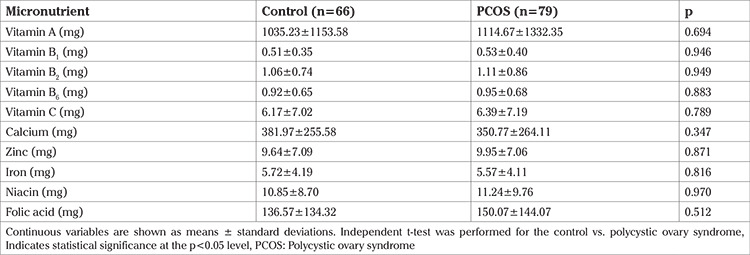
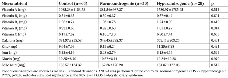
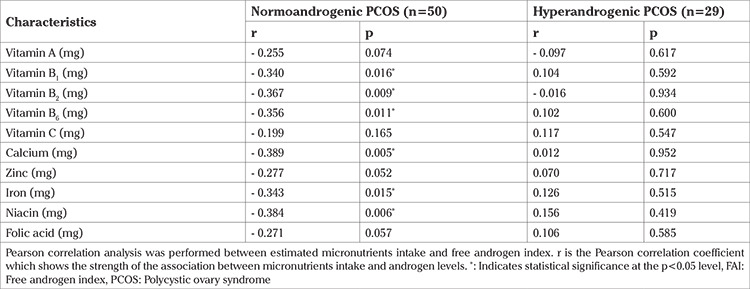
The mean levels of various micronutrient intakes in the three groups did not show a significant difference. Tables 3 and 4 present the mean micronutrient intake of the NA PCOS, HA PCOS, and control groups. The FAI in the NA PCOS patient group had a significant negative correlation with the intake of vitamins B1, B2, B6, niacin, calcium, and iron. (r=-0.340, p=0.016; r=0.367, p=0.009; r=-0.356, p=0.011; r=-0.389, p=0.005; r=-0.343, p=0.015; and r=-0.384, p=0.006, respectively). Table 5 shows the correlation between FAI and micronutrient intake in NA and HA PCOS. Meanwhile, there was no significant correlation of FAI with any micronutrient intake in the HA PCOS group.
Table 3. Micronutrients’ intake in control and overall PCOS subjects.

Table 4. Micronutrients’ intake in control, normoandrogenic PCOS, and hyperandrogenic PCOS subjects.

Table 5. Correlation analysis between micronutrients intake and FAI in PCOS groups.

Table 6 shows the results of multivariate linear regression within PCOS groups. Multivariate linear regression analysis of PCOS patients, both NA and HA, showed a significant negative effect of vitamin B6 intake on FAI. (b=-1.825, p<0.001). Meanwhile, intake of vitamin C, iron, and niacin showed a significant positive relationship with FAI in PCOS patients (β=5.844, p<0.001; β=2.381, p=0.020; β=2.599, p=0.011, respectively). There was no significant effect of intake of other micronutrients on FAI.
Table 6. The results of multivariate linear regression within PCOS groups.

Discussion
Vitamins, particularly vitamins A and C, act as antioxidants and play an essential role in suppressing chronic inflammation linked to PCOS (13). However, our study did not demonstrate any significant mean differences of any micronutrient intake in PCOS women compared with normal women. To the best of our knowledge, our study is the first study that compares the intakes of vitamin A, calcium, and iron in women with PCOS and normal women. As for vitamin B1, vitamin B2, niacin, vitamin B6, vitamin C, and folate, Szczuko et al. (14) have conducted a systematic review study and reported that vitamin C intake was lower in women with PCOS than in normal women, while for other micronutrients, no difference was found. Meanwhile, Zaeemzadeh et al. (15) found that the dietary intake of zinc was significantly lower in PCOS women with metabolic syndrome than in control groups. However, our results did not indicate a lower vitamin C and zinc intake in the PCOS group.
In another study, Szczuko et al. (16) found plasma levels of vitamin C in PCOS women were higher than in non-PCOS women, while plasma levels of vitamin B in PCOS women were lower than in non-PCOS women. Given that our study did not confirm significant differences in intakes of any micronutrients studied, including vitamins B and C, we suspected that the effect of vitamin C on PCOS incidence is due to its concentration in serum rather than its intake. The concentration of vitamin C, regardless of the amount of intake, in women with PCOS is related to the individual response to oxidative stress. During oxidative stress and activation of anti-inflammatory reactions, the concentration of ascorbic acid and cortisol in rat plasma increases but decreases in the adrenal glands (17,18).
Our study revealed significant correlations between the estimated intake of several micronutrient (vitamin C, B6, niacin, iron) and the FAI in women with PCOS. We demonstrated that vitamin C intake was positively associated with the FAI in women with PCOS. Szczuko et al. (16) also confirmed a positive correlation between plasma vitamin C levels and total testosterone. Vitamin C has antioxidant properties that can suppress chronic inflammation in PCOS. In addition, ascorbic acid is found in large quantities in the pituitary gland, so it is thought to have an important role in the secretion of the some hormones including follicle-stimulating hormone (FSH), luteinizing hormone (LH), and prolactin. Furthermore, studies have shown that treatment with ascorbic acid increases FSH and testosterone levels, but these studies have not been performed in healthy women (19). This supports and may explain our study’s finding that vitamin C intake affects androgens in women with PCOS.
Our study showed that niacin (B3) intake had a significant effect on FAI in women with PCOS. This can be explained by studies on PCOS mouse models showing that the metabolite N1-methyl nicotinamide, a metabolite of a niacin-derived compound, helps ameliorate hyperandrogenism and ovarian adenosine 5’monophosphate-activated protein kinase (AMPK) via aldehyde oxidase 1, which plays a role in detoxifying the enzymes that metabolize it (20,21). As niacin may activate AMPK (22), we speculate that decreased AMPK activity due to niacin deficiency is closely related to increased frequency of gonadotropin-releasing hormone pulsatility and increased LH production in the pituitary (23,24) as well as AMPK-dependent steroidogenesis disorders in the ovaries (25,26), in PCOS subjects. However, other studies have shown that niacin level has a negative correlation with SHBG (16). The kynurenine pathway uses tryptophan from food to produce niacin. The majority of the kynurenine pathway occurs in the liver and, to a lesser extent, in extrahepatic organs (27). This may explain the stronger correlation between niacin intake and FAI in the NA group, as demonstrated in this study.
One possible link between vitamin B6 and androgens is homocysteine. Several B vitamins and zinc play a role in the elimination of homocysteine from circulation. The re-methylation process involves folate, B2, B3, and zinc, while the transsulfuration process involves B6 and zinc. Meanwhile, it has been reported that blood homocysteine levels were negatively correlated with SHBG concentrations (28). Therefore, the association of FAI with vitamins B2, B3, B6, folate, and zinc may be mediated by SHBG concentrations.
Iron dysregulation can lead to reduced circulating levels of total testosterone. The association between iron and testosterone was weaker in overweight or obese patients than in normal weight patients (29). This is similar to findings from our study where there was a significant correlation between iron intake and FAI in women with PCOS, and this correlation was found to be weaker in HA PCOS patients who were predominantly overweight or obese.
A meta-analysis by Janjuha et al. (12), who conducted studies in normal populations, stated that they did not find a significant effect of micronutrient supplementation, including vitamin A, vitamin C, iron, and zinc, on sex hormones. In contrast to Janjuha et al. (12), our study of women with PCOS confirmed an association between androgens and intake of several types of micronutrients. Furthermore, regarding the fact that the HA phenotype was associated with insulin resistance, whereas the NA phenotype was not (30), we also report a stronger correlation in NA PCOS than HA PCOS.
Study Limitations
Our study still has some limitations that need to be considered when interpreting the results. Our study was conducted with a small sample and had cross-sectional design. It would be better if a longitudinal study design were carried out to determine the causal relationship between androgens and micronutrients in women with PCOS. It should also be noted that this study examined the estimated intake of micronutrients, not the pre-intake serum levels and post-intake serum levels of micronutrients, which of course, were influenced by absorption, transport, and demand.
Conclusion
This study observed an association between the FAI and estimated intake of some micronutrients, such as vitamin B6, vitamin C, niacin, and iron. These findings suggest a role for micronutrients in the pathology of PCOS and androgens at the systemic level. We suggest future studies should be performed with a larger number of samples, measuring nutrient levels in plasma directly and focusing on its association with the increased role of homocysteine in PCOS.
Footnotes
Ethical Committee Approval: The authorization to perform this study was obtained from the University of Indonesia Ethical Committee with the approval number 0449/UN2.F1/ETIK/2018.
Informed Consent: Written consent was provided by each individual who agreed to participate.
Peer-review: Externally peer-reviewed.
Author Contributions: Concept: A.H.; Design: A.H.; Data Collection or Processing: B.P.K.A., R.R.F.; Analysis or Interpretation: A.H., E.O.J., R.R.F., V.S., R.M.; Literature Search: A.H., E.O.J.; Writing: E.O.J.
Conflict of Interest: No conflict of interest is declared by the authors.
Financial Disclosure: The authors declared that this study received no financial support.
References
- 1.Daniilidis A, Dinas K. Long term health consequences of polycystic ovarian syndrome: a review analysis. Hippokratia. 2009;13:90–2. [PMC free article] [PubMed] [Google Scholar]
- 2.Norman RJ, Dewailly D, Legro RS, Hickey TE. Polycystic ovary syndrome. Lancet. 2007;370:685–97. doi: 10.1016/S0140-6736(07)61345-2. [DOI] [PubMed] [Google Scholar]
- 3.Clark NM, Podolski AJ, Brooks ED, Chizen DR, Pierson RA, Lehotay DC, et al. Prevalence of polycystic ovary syndrome phenotypes using updated criteria for polycystic ovarian morphology: an assessment of over 100 consecutive women self-reporting features of polycystic ovary syndrome. Reprod Sci. 2014;21:1034–43. doi: 10.1177/1933719114522525. [DOI] [PMC free article] [PubMed] [Google Scholar]
- 4.Dapas M, Lin FTJ, Nadkarni GN, Sisk R, Legro RS, Urbanek M, et al. Distinct subtypes of polycystic ovary syndrome with novel genetic associations: An unsupervised, phenotypic clustering analysis. PLoS Med. 2020;17:e1003132. doi: 10.1371/journal.pmed.1003132. [DOI] [PMC free article] [PubMed] [Google Scholar]
- 5.Prapas N, Karkanaki A, Prapas I, Kalogiannidis I, Katsikis I, Panidis D. Genetics of polycystic ovary syndrome. Hippokratia. 2009;13:216–23. [PMC free article] [PubMed] [Google Scholar]
- 6.Kshetrimayum C, Sharma A, Mishra VV, Kumar S. Polycystic ovarian syndrome: Environmental/occupational, lifestyle factors; an overview. J Turk Ger Gynecol Assoc. 2019;20:255–63. doi: 10.4274/jtgga.galenos.2019.2018.0142. [DOI] [PMC free article] [PubMed] [Google Scholar]
- 7.Günalan E, Yaba A, Yılmaz B. The effect of nutrient supplementation in the management of polycystic ovary syndrome-associated metabolic dysfunctions: A critical review. J Turk Ger Gynecol Assoc. 2018;19:220–32. doi: 10.4274/jtgga.2018.0077. [DOI] [PMC free article] [PubMed] [Google Scholar]
- 8.Faghfoori Z, Fazelian S, Shadnoush M, Goodarzi R. Nutritional management in women with polycystic ovary syndrome: A review study. Diabetes Metab Syndr. 2017;11(Suppl 1):S429–32. doi: 10.1016/j.dsx.2017.03.030. [DOI] [PubMed] [Google Scholar]
- 9.Papavasiliou C, Papakonstantinou E. Nutritional support and dietary interventions for women with polycystic ovary syndrome. Nutri Diet Suppl. 2017;9:63–85. [Google Scholar]
- 10.Hager M, Nouri K, Imhof M, Egarter C, Ott J. The impact of a standardized micronutrient supplementation on PCOS-typical parameters: a randomized controlled trial. Arch Gynecol Obstet. 2019;300:455–60. doi: 10.1007/s00404-019-05194-w. [DOI] [PMC free article] [PubMed] [Google Scholar]
- 11.Yildiz BO. Diagnosis of hyperandrogenism: clinical criteria. Best Pract Res Clin Endocrinol Metab. 2006;20:167–76. doi: 10.1016/j.beem.2006.02.004. [DOI] [PubMed] [Google Scholar]
- 12.Janjuha R, Bunn D, Hayhoe R, Hooper L, Abdelhamid A, Mahmood S, et al. Effects of Dietary or Supplementary Micronutrients on Sex Hormones and IGF-1 in Middle and Older Age: A Systematic Review and Meta-Analysis. Nutrients. 2020;12:1457. doi: 10.3390/nu12051457. [DOI] [PMC free article] [PubMed] [Google Scholar]
- 13.Olofinnade AT, Onaolapo AY, Stefanucci A, Mollica A, Olowe OA, Onaolapo OJ. Cucumeropsis mannii reverses high-fat diet induced metabolic derangement and oxidative stress. Front Biosci (Elite Ed) 2021;13:54–76. doi: 10.2741/872. [DOI] [PubMed] [Google Scholar]
- 14.Szczuko M, Szydłowska I, Nawrocka-Rutkowska J. A Properly Balanced Reduction Diet and/or Supplementation Solve the Problem with the Deficiency of These Vitamins Soluble in Water in Patients with PCOS. Nutrients. 2021;13:746. doi: 10.3390/nu13030746. [DOI] [PMC free article] [PubMed] [Google Scholar]
- 15.Zaeemzadeh N, Jahanian Sadatmahalleh S, Ziaei S, Kazemnejad A, Movahedinejad M, Mottaghi A, et al. Comparison of dietary micronutrient intake in PCOS patients with and without metabolic syndrome. J Ovarian Res. 2021;14:10. doi: 10.1186/s13048-020-00746-0. [DOI] [PMC free article] [PubMed] [Google Scholar]
- 16.Szczuko M, Hawryłkowicz V, Kikut J, Drozd A. The implications of vitamin content in the plasma in reference to the parameters of carbohydrate metabolism and hormone and lipid profiles in PCOS. J Steroid Biochem Mol Biol. 2020;198:105570. doi: 10.1016/j.jsbmb.2019.105570. [DOI] [PubMed] [Google Scholar]
- 17.Umegaki K, Daohua P, Sugisawa A, Kimura M, Higuchi M. Influence of one bout of vigorous exercise on ascorbic acid in plasma and oxidative damage to DNA in blood cells and muscle in untrained rats. J Nutr Biochem. 2000;11:401–7. doi: 10.1016/s0955-2863(00)00096-6. [DOI] [PubMed] [Google Scholar]
- 18.Peters EM, Anderson R, Nieman DC, Fickl H, Jogessar V. Vitamin C supplementation attenuates the increases in circulating cortisol, adrenaline and anti-inflammatory polypeptides following ultramarathon running. Int J Sports Med. 2001;22:537–43. doi: 10.1055/s-2001-17610. [DOI] [PubMed] [Google Scholar]
- 19.Okon UA, Utuk II. Ascorbic acid treatment elevates follicle stimulating hormone and testosterone plasma levels and enhances sperm quality in albino Wistar rats. Niger Med J. 2016;57:31–6. doi: 10.4103/0300-1652.180570. [DOI] [PMC free article] [PubMed] [Google Scholar]
- 20.Nejabati HR, Samadi N, Shahnazi V, Mihanfar A, Fattahi A, Latifi Z, et al. Nicotinamide and its metabolite N1-Methylnicotinamide alleviate endocrine and metabolic abnormalities in adipose and ovarian tissues in rat model of Polycystic Ovary Syndrome. Chem Biol Interact. 2020;324:109093. doi: 10.1016/j.cbi.2020.109093. [DOI] [PubMed] [Google Scholar]
- 21.Nejabati HR, Schmeisser K, Shahnazi V, Samimifar D, Faridvand Y, Bahrami-Asl Z, et al. N1-Methylnicotinamide: an anti-ovarian aging hormetin? Ageing Res Rev. 2020;62:101131. doi: 10.1016/j.arr.2020.101131. [DOI] [PubMed] [Google Scholar]
- 22.Thirunavukkarasu M, Penumathsa S, Juhasz B, Zhan L, Bagchi M, Yasmin T, et al. Enhanced cardiovascular function and energy level by a novel chromium (III)-supplement. Biofactors. 2006;27:53–67. doi: 10.1002/biof.5520270106. [DOI] [PubMed] [Google Scholar]
- 23.Duval DL. PRKA/AMPK: Integrating energy status with fertility in pituitary gonadotroph. Biol Reprod. 2011;84:205–6. doi: 10.1095/biolreprod.110.089722. [DOI] [PubMed] [Google Scholar]
- 24.Andrade J, Quinn J, Becker RZ, Shupnik MA. AMP-activated protein kinase is a key intermediary in GnRH-stimulated LHβ gene transcription. Mol Endocrinol. 2013;27:828–39. doi: 10.1210/me.2012-1323. [DOI] [PMC free article] [PubMed] [Google Scholar]
- 25.Abdou HS, Bergeron F, Tremblay JJ. A cell-autonomous molecular cascade initiated by AMP-activated protein kinase represses steroidogenesis. Mol Cell Biol. 2014;34:4257–71. doi: 10.1128/MCB.00734-14. [DOI] [PMC free article] [PubMed] [Google Scholar]
- 26.Kai Y, Kawano Y, Yamamoto H, Narahara H. A possible role for AMP-activated protein kinase activated by metformin and AICAR in human granulosa cells. Reprod Biol Endocrinol. 2015;13:27. doi: 10.1186/s12958-015-0023-2. [DOI] [PMC free article] [PubMed] [Google Scholar]
- 27.Badawy AA. Kynurenine pathway of tryptophan metabolism: regulatory and functional aspects. Int J Tryptophan Res. 2017;10:1178646917691938. doi: 10.1177/1178646917691938. [DOI] [PMC free article] [PubMed] [Google Scholar]
- 28.Schiuma N, Costantino A, Bartolotti T, Dattilo M, Bini V, Aglietti MC, et al. Micronutrients in support to the one carbon cycle for the modulation of blood fasting homocysteine in PCOS women. J Endocrinol Invest. 2020;43:779–86. doi: 10.1007/s40618-019-01163-x. [DOI] [PMC free article] [PubMed] [Google Scholar]
- 29.Chao KC, Chang CC, Chiou HY, Chang JS. Serum Ferritin Is Inversely Correlated with Testosterone in Boys and Young Male Adolescents: A Cross-Sectional Study in Taiwan. PLoS One. 2015;10:e0144238. doi: 10.1371/journal.pone.0144238. [DOI] [PMC free article] [PubMed] [Google Scholar]
- 30.Moghetti P, Tosi F, Bonin C, Di Sarra D, Fiers T, Kaufman JM, et al. Divergences in insulin resistance between the different phenotypes of the polycystic ovary syndrome. J Clin Endocrinol Metab. 2013;98:E628–37. doi: 10.1210/jc.2012-3908. [DOI] [PubMed] [Google Scholar]


