Abstract
Decades of intense herbicide use has led to resistance in weeds. Without innovative weed management practices and new herbicidal modes of action, the unabated rise of herbicide resistance will undoubtedly place further stress upon food security. HMGR (3-hydroxy-3-methylglutaryl-coenzyme A reductase) is the rate limiting enzyme of the eukaryotic mevalonate pathway successfully targeted by statins to treat hypercholesterolemia in humans. As HMGR inhibitors have been shown to be herbicidal, HMGR could represent a mode of action target for the development of herbicides. Here, we present the crystal structure of a HMGR from Arabidopsis thaliana (AtHMG1) which exhibits a wider active site than previously determined structures from different species. This plant conserved feature enables the rational design of specific HMGR inhibitors and we develop a tolerance trait through sequence analysis of fungal gene clusters. These results suggest HMGR to be a viable herbicide target modifiable to provide a tolerance trait.
Subject terms: X-ray crystallography, Molecular engineering in plants, Mechanism of action, Plant molecular biology
Managing herbicide resistance problem needs the identification of new herbicidal modes of action. Here, the authors solve the crystal structures of Arabidopsis HMGR and show HMGR as a potential new herbicide target by identifying plant-specific HMGR inhibitors and engineering tolerant trait in Arabidopsis.
Introduction
As herbicide resistance continues to rise, the efficacy of herbicides has diminished1 such that new modes of action are desperately needed. Only one new herbicide mode of action has been brought to market in almost 40 years2. Weeds are yet to evolve significant resistance to clomazone and bixlozone1, two herbicides that disrupt isoprenoid biosynthesis by targeting the enzyme 1-deoxy-d-xylulose-5-phosphate synthase3. Found in all kingdoms of life, isoprenoid biosynthesis is crucial for the synthesis of lipids, hormones, vitamins and defence compounds4–6. The biosynthetic route differs between kingdoms; most animals, fungi, protists and archaea use a mevalonate (MVA) pathway, whereas most Gram-negative bacteria including cyanobacteria use a methylerythritol phosphate (MEP) pathway7. Through a shared evolutionary history with cyanobacteria, plants use both pathways7–11 compartmentalised to the cytosol (MVA) or plastids (MEP)12–14. None of the known modes of action for any of the commercial herbicides affect the MVA pathway1. An important enzyme in the MVA pathway is HMGR, which is a highly regulated15–19, the rate-limiting enzyme of the MVA pathway and is the target of the group of hypercholesterolaemia therapeutics known as statins20,21. Two classes (I and II) of HMGR have been defined22 based on the differences between the catalytic core domain structure23,24, the presence of an N-terminal membrane domain of between two (plants) and eight (human) membrane-spanning helices in the majority of class I enzymes20,25, and the varied NAD(P)H cofactor preference26. HMGR regulation appears to be conserved between humans and plants with the N-terminus regulated by ubiquitination whereas catalytic core activity is regulated by phosphorylation7. Many of the regulatory proteins differ, however, and this is further complicated by plants having multiple copies or isoforms plus a wide variety of external signals modifying expression, such as light and herbivory7,27.
The first potent statin inhibitor of HMGR discovered was mevastatin, isolated from Penicillium citrinum in 197628. Lovastatin, isolated from Aspergillus terreus in 1978, became the first commercial statin in 198729. Second-generation statins have been semi-synthetic or synthetic products29, but all statins competitively inhibit HMGR via a HMG-like moiety and a variable hydrophobic group that together give affinities to HMGR that are 10,000-fold higher than HMG-CoA30. Lovastatin and mevastatin, as well as the semi-synthetic pravastatin and synthetic atorvastatin are all known to be herbicidal31–33. HMGR might have been overlooked as a herbicide target due to potential off-target risks arising from its conservation in humans and the antimicrobial activity of statins34, but recently developed selective insecticides against HMGR illustrate the potential to develop HMGR herbicides35.
Here, we solve crystal structures for a plant HMGR in apo form and complexed with a statin. These structures reveal a wider active site conserved in plants compared to other organisms. By rational design we develop statin derivatives with over 20-fold specificity for the plant over the human enzyme and, which importantly, retain herbicidal activity. By comparing the AtHMG1 structure to fungal HMGR genes in biosynthetic clusters for natural statins, we demonstrate a single amino acid change confers statin tolerance in vitro and in planta. Together these findings suggest HMGR is a viable target for herbicide development.
Results
Statins range in herbicidal activity
Previous studies have shown several statins to exhibit herbicidal activity against several plant species including Lemna gibba, Raphanus sativus, Scoparia dulcis and A. thaliana31–33,36,37. However, there is a lack of comparative data regarding the herbicidal efficacy of statins especially for second-generation, synthetic statins. To assess herbicidal activity, we treated a model dicot and a monocot (A. thaliana and Eragrostis tef, respectively) with a dose range of eight commercially available statins on soil, pre- and post-emergence (Fig. 1). All statins were more herbicidal against the dicot and in general were more effective post-emergence. In line with their physicochemical properties more closely matching those of post-emergence herbicides (Supplementary Fig. 1a). The synthetic statin rosuvastatin was the most herbicidal statin being lethal to A. thaliana at ~15 μM without formulation beyond including a wetting agent. Given that under the same conditions formulated glyphosate (Roundup®) is lethal at ~35 μM (Supplementary Fig. 1b, c), we surmised that HMGR could represent a potential herbicide target.
Fig. 1. Herbicidal activity of statins varies between a model dicot and a monocot.
a Representative images from the post-emergence treatment of a model dicot, A. thaliana, and pre-emergence treatment of the monocot E. tef, with statins: rosuvastatin (Ro.), pravastatin (Pr.), simvastatin (Si.), mevastatin (Me.), lovastatin (Lo.), fluvastatin (Fl.), atorvastatin (At.) and pitavastatin (Pi.). b A. thaliana (green) and E. tef (grey) treated with a range of statins at 62.5 μM pre- (light colour) and post-emergence (dark colour) on soil. Inhibition was quantified using a green pixel area and plotted as a percentage of no-inhibitor control. n = 3 replicates with the mean ± standard deviation (s.d.). Source data are provided as a Source Data file.
Crystal structure of AtHMG1 reveals scope for species selective compounds
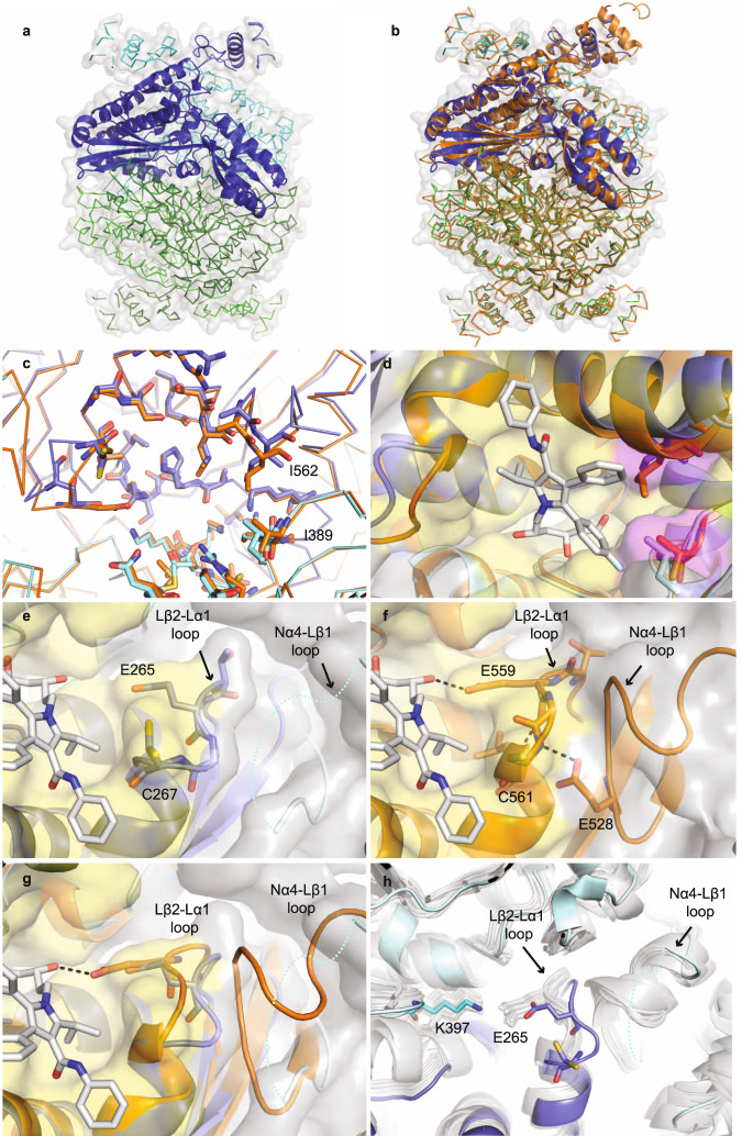
A. thaliana has two HMGR genes with different expression patterns, but AtHMG1 (At1g76490) is the most highly epxressed38. The N-terminal transmembrane domains of HMGR are highly divergent between species and absent from class II HMGRs (Supplementary Fig. 2). By contrast, the conserved extracellular domain of AtHMG1 shares ~54% sequence identity with HsHMGCR and strictly conserved catalytic residues (Supplementary Fig. 2). To develop plant-specific statins and mitigate off-target effects, we solved the crystal structure of the core domain of apo AtHMG1 and in complex with pitavastatin to resolutions of 1.9 and 2.1 Å, respectively, in space groupI 41 2 2. Attempts were made to crystallise type I statins as described previously24; however, electron density for these ligands was ambiguous. The structure of the apo AtHMG1 displayed a single monomer in the asymmetric unit which through crystallographic symmetry forms a homotetrameric assembly (Fig. 2a), consisting of two canonical class I homodimeric HMGR folds, with high structural similarity to HsHMGCR (PDB 1HWK, r.m.s.d. 1.1 Å over 371 Cα atoms, Fig. 2b).
Fig. 2. AtHMG1 active site adopts a unique conformation.
a Apo AtHMG1 displays a single monomer in the asymmetric unit (dark-blue cartoon) which through crystallographic symmetry forms a homotetrameric assembly (ribbon) consisting of two canonical class I homodimeric HMGR folds (blue and green). b Overlay of human HMGR (orange cartoon, PDB 1HWK) with apo AtHMG1 (a) illustrates their conserved fold. c AtHMG1 (blue ribbon and sticks) has a highly conserved active site with HsHMGCR (orange ribbon and sticks). Active site-delineating residues are shown as sticks. All residues are conserved except the two AtHMGR1 residues labelled. d Superposition of atorvastatin in the active site of AtHMG1 illustrates the position of these substitutions relative to a bound statin. Conserved active site residues are shown with yellow surface and substitutions highlighted with magenta surface. e Conformational flexibility in the Nα4-Lβ1 loop of AtHMG1 (cyan dotted line) evidenced by poor electron density is likely the result of a Pro to Val substitution. This results in the loss of a type II hydrogen bonded β-turn exhibited in HsHMGCR (f) that allows HsHMGCR E559 to hydrogen bond (dashed grey lines) to the open lactone ring of statins. Atorvastatin superimposed on apo AtHMG1 (e, g) illustrates the equivalent residue, E265, likely too far away to H-bond to statins. Conserved active site residues (e–g) are shown with yellow surface. h Overlay of apo AtHMG1 (blue cartoon and sticks) with all published structures of HMGR (grey cartoon and sticks)24,26,30,40–50. To the best of our knowledge, this conformation has not been seen in any published HMGR crystal structure to date. Topology designation from HsHMGCR49.
Closer inspection of the statin-binding pocket revealed two substitutions in AtHMG1 with respect to HsHMGCR located at the hydrophobic CoA binding region of the active site pocket39, specifically, Ile562/Leu857 and Ile389/Val683 in AtHMG1/HsHMGCR respectively (Fig. 2c, d). Furthermore, a plant conserved Val to Pro (Pro236/Val530 A. thaliana/human) substitution at the start of the Lβ1-strand is the likely cause of conformational flexibility and lack of electron density in the Nα4-Lβ1 loop adjacent to the active site-delineating Lβ2-Lα1 loop (Fig. 2e, Supplementary Fig. 3a, b and Supplementary Table 1). This flexibility results in the loss of a type II β-turn found within the HsHMGCR Lβ2-Lα1 loop that is stabilised by hydrogen bonding between a conserved Glu (Glu234/Glu528 AtHMG1/HsHMGCR) and a Cys backbone amine (Cys267/Cys561 AtHMG1/HsHMGCR) (Fig. 2f). This altered conformation of the Lβ2-Lα1 loop is not seen in any of the previous class I and II HMGR crystal structures24,26,30,40–50 and allows alternative conformations of the Cys267 residue (Fig. 2e, g, h). Importantly, the arrangement of the AtHMG1 Lβ2-Lα1 loop results in Glu265 being unable to form a hydrogen bond with the O5-hydroxyl group of the HMG moiety of statins or the equivalent thioester oxygen of HMG-CoA, as it is shifted 2.5 Å away, creating a wider pocket (Fig. 2g). In this orientation it is more likely that Lys397 acts as a proton donor in the catalytic reduction of HMG-CoA to mevalonate as is suggested to occur in bacteria50 and with molecular dynamics and quantum mechanics/molecular mechanics simulations with HsHMGCR51. Together, these differences increase the solvent-accessible area of the statin pocket from ~314 Å3 in HsHMGCR to ~357 Å3 in AtHMG152.
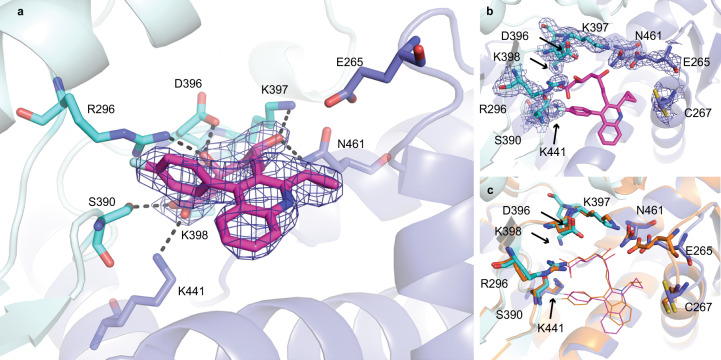
The complex of AtHMG1 with pitavastatin (Fig. 3a, b) revealed a binding mode highly similar to fluvastatin in HsHMGCR24 (Fig. 3c). Conserved polar interactions occur with the residues local to the cis loop Arg296, Ser390, Asp396, Lys397, Lys398, Asn461 (HsHMGCR Arg590, Ser684, Asp690, Lys691, Lys692, Asn755) and a salt-bridge between the terminal carboxylate of the HMG moiety with Lys441 (HsHMGCR Lys735) (Fig. 3a). The fluorophenyl group of pitavastatin maintains conserved stacking interactions with Arg296 (HsHMGCR Arg590) and hydrophobic interactions between the quinoline and cyclopropyl moiety with residues Leu268, Ile389, Leu558, Asp561 (HsHMGCR Leu562, Val683, Leu853, Asp856). This complex structure however also revealed two notable differences between the binding mode of class II statins in AtHMG1 and HsHMGCR; (i) loss of hydrogen bonding to the O5-hydroxyl group of the HMG moiety of statins from Glu265 (HsHMGCR Glu559), despite a slight shift of Glu265 towards the bound inhibitor (Fig. 3a and Supplementary Fig. 3c), and (ii) the loss of hydrophobic interactions with Gly266, His458 and Ile562 (HsHMGCR Gly560, His752 and Leu857). Unique hydrophobic contacts were made between pitavastatin and residues Ser271 and Ser367 of AtHMG1 (HsHMGCR Ser565 and Ser661).
Fig. 3. AtHMG1 E265 does not hydrogen bond with statins.
a AtHMG1 with pitavastatin (magenta) bound. Residues that hydrogen bond (dashed black lines) to the HMG moiety of statins in AtHMG1 are shown (blue/cyan sticks). b AtHMG1 (blue cartoon) with pitavastatin (magenta line) bound, active site-delineating residues labelled and shown as sticks with electron density. c AtHMG1 with pitavastatin bound superimposed on HsHMGCR bound to fluvastatin (orange cartoon and line PDB 1HWI), illustrating that binding of statins to AtHMG1 is analogous to HsHMGCR. Simulated annealing omit electron density maps (2 Fobs - Fcalc) contoured at 1σ level.
Development of plant-specific analogues of statins
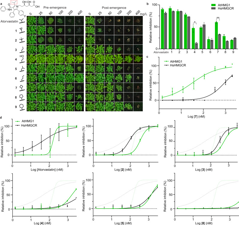
Our insights from the crystal structure of a model plant HMGR and the binding mode of pitavastatin provided the opportunity to rationally design plant-specific inhibitors. To this end, we sought to exploit the Lβ2-Lα1 loop region of AtHMG1 by developing analogues (1–9) of the more chemically tractable atorvastatin with modifications at the isopropyl group on the central pyrrole ring (Supplementary Fig. 3d). Activity of the atorvastatin scaffold against HsHMGCR was previously found to be reduced with increasing size of the alkyl substituent53 whereas the wider pocket of AtHMG1 might accommodate larger groups. In addition, the loss of interactions with O5-hydroxyl group of the HMG moiety with Glu265 could be targeted by incorporating a hydrogen bond donor (Fig. 2e, g). Thus 1–9 were synthesised (Supplementary Method 1) and assessed for herbicidal activity on soil with A. thaliana (Fig. 4a) and for species-specificity against HsHMGCR and AtHMG1 in vitro by a fluorometric, NADPH-depletion assay (Fig. 4b–d).
Fig. 4. Modifying the isopropyl group of atorvastatin affects species selectivity.
a Herbicidal activity of atorvastatin and its analogues (1–9) against A. thaliana with pre- and post-emergence treatments. The isopropyl moiety of atorvastatin is boxed in red. Modifications to the isopropyl region are shown. b Compounds 4 and 7 were selective in vitro for AtHMG1 (green bar) over HsHMGCR (grey bar) at 500 nM. n = 3 independent reactions with the mean ± s.d. Significance from two-tailed paired t test, values: compound 4 t = 5.536 df = 2 P = 0.03 (*) 95% CI [−60.92 to −7.635], compound 7 t = 16.93 df = 2 P = 0.003 (**) 95% CI [−34.37 to −20.44]. c In vitro inhibition of AtHMG1 (green plot) and HsHMGCR (grey plot) by 7 illustrating >20-fold selectivity for AtHMG1. n = 3 independent reactions with the mean ± s.d. d Atorvastatin and analogues 2–5 and 8 were not selective for AtHMG1 in vitro. Inhibition of AtHMG1 and HsHMGCR with atorvastatin inhibition profile shown as green and grey dotted lines, respectively. n = 3 independent reactions with the mean ± s.d. except for a single point (AtHMG1 333 µM atorvastatin n = 2). Source data are provided as a Source Data file.
Overall, atorvastatin analogues with side chains of similar length had similar herbicidal and in vitro inhibitory activity to the parent, whereas side chains longer than the isopropyl group had reduced activity (Fig. 4). Compounds 4 and 7 retained herbicidal activity and displayed a preference for AtHMG1 over HsHMGCR in an initial screen (Fig. 4a, b). Dose-response curves confirmed compound 7 had switched preference from human to plant when compared to atorvastatin, showing >20-fold higher specificity for AtHMG1 (IC50 32 nM ±12 nM) over HsHMGCR (IC50 890 nM ±143 nM) in vitro (Fig. 4c, d). These molecules provide a framework for the future development of plant-specific HMGR inhibitors that might exhibit stronger herbicidal activity.
Exploiting biosynthetic gene clusters to engineer statin tolerance
The most commercially successful herbicides are often paired with a tolerance trait in crops. Statins produced from fungal biosynthetic gene clusters usually contain a copy of HMGR that imparts self-resistance54–56, so we sought to determine the structural basis for this resistance. Sequence alignment of a HMGR gene (lvrA) from the A. terreus genome revealed several mutations in the cluster-associated copy that were not present in the housekeeping copy (Supplementary Fig. 4). The corresponding residues of the AtHMG1 crystal structure revealed a Leu (Leu558) to Thr mutation, whose equivalent was conserved in all A. terreus genomes in the NCBI database. The Leu to Thr mutation would likely disrupt the hydrophobic pocket essential for accommodating the decalin ring of natural statins (Fig. 5a), and so was incorporated into recombinant AtHMG1. The AtHMG1-L558T mutant was resistant to a range of statins (Fig. 5b) with >20-fold resistance to rosuvastatin in vitro (WT IC50 53 nM ±20 nM, L558T IC50 >1000 nM) (Fig. 5c). Without inhibitors, AtHMG1-L558T had reduced catalytic activity (WT Km 69 µM ± 19 µM and kcat 10.7 ± 1.0 s−1, L558T Km 24 µM ±16 µM and kcat 2.4 ± 0.3 s−1) (Supplementary Fig. 5), but remained within the range of previously published rates for other class I and II HMGR enzymes57.
Fig. 5. A mutation found in a statin biosynthetic cluster confers statin resistance in vitro.
a The hydrophobic pocket in AtHMG1 delineated by labelled residues (blue sticks) with pitavastatin (magenta sticks) bound, illustrating L558 proximity to the hydrophobic ring of statins. b AtHMG1 with the L558T mutation (grey bar) in comparison to wild-type (black bar) retained activity in vitro in the presence of statins: rosuvastatin (Ro.), pravastatin (Pr.), simvastatin (Si.), mevastatin (Me.), lovastatin (Lo.), fluvastatin (Fl.), atorvastatin (At.) and pitavastatin (Pi.), at 500 nM. n = 3 independent reactions with the mean ± s.d. c In vitro inhibition of WT (black plot) and L558T AtHMG1 (grey plot) by rosuvastatin revealed the L558T mutation conferred >20-fold resistance. n = 3 independent reactions with the mean ± s.d. Source data are provided as a Source Data file.
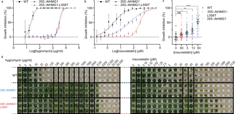
To validate the potential of the L558T mutation for providing a plant tolerance trait, we overexpressed full-length AtHMG1 (35 S::AtHMG1) and its equivalent with the L558T mutation (35 S::AtHMG1-L558T) in A. thaliana, using a cauliflower mosaic virus (CaMV) 35 S promoter. It has previously been shown that overexpressing AtHMG1 in A. thaliana can give rise to a 40-fold rise in mRNA levels and a modest rise in resistance to lovastatin compared to non-transformed WT controls58. Here we found with data collated from 19 independent T2 35 S::AtHMG1 lines and 14 independent 35 S::AtHMG1-L558T T2 lines that both constructs conferred similar resistance to the selectable marker hygromycin (Fig. 6a, d). However, the 35 S::AtHMG1-L558T lines were over sixfold more resistant to rosuvastatin (IC50 300 µM vs ±18 µM) than 35 S::AtHMG1 lines (IC50 46 µM ±5 µM) and more than 100-fold more resistant than non-transformed WT (IC50 3 µM vs ±1 µM) (Fig. 6b, d). Furthermore, analysis of the effects of rosuvastatin revealed 35 S::AtHMG1-L558T lines were up to 16-fold less sensitive to treatment than 35 S::AtHMG1 lines (Fig. 6c). These results illustrate the potential for HMGR to have a tolerance trait and further validates the in vitro results (Fig. 5b, c).
Fig. 6. The L558T mutation gives resistance to rosuvastatin in planta.
Resistance to hygromycin (a) and rosuvastatin (b, c) in 19 transgenic lines of 35 S::AtHMG1 (blue) versus 14 35 S::AtHMG1-L558T lines (red) and wild type (WT, black). Green pixels quantified and plotted as a percentage of no-inhibitor control. a, Both transgenic lines exhibited similar resistance to the hygromycin selectable marker, whereas WT was sensitive, mean ± 95% CI. b 35 S::AtHMG1-L558T transgenic lines were sixfold more resistant to rosuvastatin than 35 S::AtHMG1, mean ± 95% CI, but ±s.d. for WT. c Susceptibility of transgenics to rosuvastatin illustrated 35 S::AtHMG1-L558T (red 80 µM n = 42) was up to 16-fold less susceptible to rosuvastatin inhibition than 35 S::AtHMG1 (blue 5 µM n = 54, 10 µM n = 54, 80 µM n = 57) when compared to untreated WT (black n = 12). Significance from one-way ANOVA with Dunnett’s multiple comparison correction against a common control performed, bars represent mean ± 95% CI. Values: 0 µM vs 80 µM 35 S::AtHMG1-L558T q = 1.566 df = 214 P = 0.24 (ns) 95% CI [−27.00 to −5.299], 0 µM vs 5 µM 35 S::AtHMG1 q = 2.395 df = 214 P = 0.04 (*) 95% CI [−31.92 to −0.4293], 0 µM vs 10 µM 35 S::AtHMG1 q = 3.564 df = 214 P = 0.0013 (**) 95% CI [−39.82 to −8.324], 0 µM vs 80 µM 35 S::AtHMG1 q = 7.829 df = 214 P = <0.0001 (****) 95% CI [−68.29 to −36.95]. d Representative image of resistance to hygromycin and rosuvastatin from a single line of 35 S::AtHMG1 and 35 S::AtHMG1-L558T versus WT. Source data are provided as a Source Data file.
Discussion
The relentless rise in herbicide-resistant weeds already poses a significant threat to global food security and as such, new herbicides with new modes of action are desperately needed. Moreover, as consumer attitudes shift, natural product ‘bioherbicides’ will rise in their appeal and currently in the USA enjoy an accelerated regulatory journey59.
Herein, we have validated HMGR as a potential herbicide target. Using the HMGR crystal structure from a model plant we have demonstrated that, despite its overall sequence and structure conservation with HsHMGCR, differences in the architecture (especially the active site) can be exploited to develop plant-specific synthetic HMGR inhibitors. The progress herein provides a basis for the discovery of natural product statins that might be suitable bioherbicides.
The differences in the architecture of AtHMG1 that allowed for species selectivity largely arise from an unusual orientation of the Lβ2-Lα1 loop that is likely the result of increased flexibility in the neighbouring Nα4-Lβ1 loop. The atypical orientation of the Lβ2-Lα1 loop in AtHMG1 disrupts the hydrogen bonding network formed between the catalytic residues Glu265/Lys397/Asn461/Asp473 (HsHMGCR Glu559/Lys691/Asn755/Asp767), thereby retaining only those hydrogen bonds that stabilise the catalytic Lys via the adjacent Asn and Asp residues. The conserved location of the catalytic Lys between AtHMG1 and other class I and II HMGRs strongly suggests this residue is responsible for polarising the carbonyl oxygen of HMG-CoA substrate and mevaldehyde intermediate, and for performing the final protonation step. Glu265 is not in a favourable position to hydrogen bond to either the substrate thioester oxygen or the adjacent Asp473, which based on HsHMGCR in silico simulations (HsHMGCR Glu559 and Asp767) might be expected to hydrogen bond and stabilise the mevaldyl-CoA intermediate51. Further molecular dynamics studies with AtHMG1, its substrate and cofactors might determine if the role of Glu265 is to hydrogen bond to Asp473, or to directly protonate the substrate as previous modelling studies have suggested60,61.
Here, we rationally designed a compound with >20-fold preference for plant HMGR in vitro with limited modification to the parent scaffold. Modelling of atorvastatin along with compounds 4 and 7 into the active site of HsHMGCR revealed a single dominant high-affinity binding mode with a large drop in affinity to the next most favourable binding mode. Atorvastatin exhibited the highest affinity followed by compound 7 and 4 (Supplementary Fig. 6) consistent with in vitro results (Fig. 4). Modelling with AtHMG1 revealed more varied poses of the analogues, with similar affinities between the most favourable binding modes. These binding modes are possibly facilitated by a wider active site and flexibility in the Lβ2-Lα1 loop region (Supplementary Fig. 3c) and might account for the difficulty we had in obtaining co-crystal structures for AtHMG1. The lower affinity for AtHMG1 than HsHMGCR for compounds 4 and 7 suggests further molecular dynamics simulations and crystallographic studies may be necessary to help reveal the molecular basis of in vitro specificity (Fig. 4). Notably, selectivity over HsHMGCR was also obtained by targeting the same Lβ2-Lα1 loop region in Manduca sexta using gem-difluoromethylenated HMGR inhibitors35. Similar derivatives may also prove to be selective for plant HMGRs. Overall, the developed compounds provide a framework for further structure-based rational herbicide design targeting the Lβ2-Lα1 loop region of AtHMG1, which could be validated for selectivity in mammalian in vivo studies. Greater species selectivity might be obtainable by targeting the N-terminal domain of HMGR, which is highly divergent between humans and plants and is absent from class II HMGRs. A recent crystal structure of the regulatory elements that interact with the N-terminal domain in HsHMGCR and studies of compounds that increase HMGR degradation suggest that this could be an alternative mechanism to lower cholesterol levels16,18. Future studies of the regulatory elements interacting with plant HMGR N-terminal domain and complexed crystal structures might in the same way also provide an avenue to develop more species-specific inhibitors of HMGR. The regulatory elements that control plant lipid metabolism might also provide new herbicidal targets, just as the proprotein convertase subtilisin/kexin type 9 and angiopoietin-like 3 are providing new avenues for the treatment of hypercholesterolaemia62,63.
By analysing the AtHMG1 crystal structure and sequences in fungal biosynthetic gene clusters, we identified a mutation conferring statin resistance without adversely affecting catalytic activity. Overexpressing this mutant protein in A. thaliana demonstrated its potential as a tolerance trait, but further investigations are needed. These could include (i) the efficacy of this protein mutant in different species; (ii) optimisation of expression and regulation, by modifying the N-terminal domain; (iii) its effects on sterol levels and seed set64; and (iv) determining what HMGR inhibitor residues remain in the treated crop or soil. Future studies might also focus on other residues that potentially impart resistance, such as the end region of the Sβ4 strand (residues 387–390), that show conservation in putative resistance genes from Penicillium citrinum and Xylaria grammica and could affect binding of the butyryl group of natural statins. We envisage that the development and discovery of new, natural product herbicides65,66 might also benefit from a similar approach to engineering resistance alleles from biosynthetic gene clusters containing compounds or targets of interest.
Methods
Herbicidal activity assay
Approximately 30 seeds of A. thaliana (accession Col-0) or E. tef were sown in 63 × 63 × 59 mm pots of Irish peat (Bord na Móna Horticulture Ltd, Newbridge, Ireland). Seeds were incubated in the dark for 3 days at 4 °C to synchronise germination. A single pre-emergence treatment (day 0) was performed when these seeds were transferred to a growth room at 22 °C with a 16:8 h light:dark photoperiod and 60% relative humidity. Two post-emergence treatments were performed following emergence of the seedlings (day 1) at days 4 and 7. Plants were watered accordingly throughout the experiment to maintain adequate moisture and photographed on day 16. Treatments were conducted with rosuvastatin, simvastatin, fluvastatin, atorvastatin (AK Scientific), pravastatin (BOC Sciences), lovastatin (Sapphire Bioscience), mevastatin, pitavastatin (Focus Bioscience), glyphosate, RoundUp® and atorvastatin analogues (1–9). To treat, 0.5 mL of each compound in a final concentration of 2% dimethyl sulfoxide (DMSO) and 0.02% Brushwet (SST Dandenong, Australia) was pipetted onto seedlings. Full experimental details and characterisation of atorvastatin analogue 1–9 synthesis can be found in Supplementary Method 1 and Supplementary Figs. 7–62. Growth inhibition was quantified by detecting green pixels for healthy seedlings using ImageJ (National Institutes of Health, 1.53 v) and the ‘Threshold Colour’ plug-in with the following settings: hue 50–110, saturation 125–255, brightness 30–25567. Images were converted into 8-bit format, and pixels measured over the same area. Data were normalised to a negative control to provide percentage inhibition.
HMG-CoA reductase expression and purification
An E. coli codon-optimised DNA sequence encoding the conserved extracellular region of AtHMG1 (Uniprot P14891, At1g76490, residues 121–592) was cloned into pQE30 (Qiagen) following an N-terminal His6-tag and tobacco etch virus cleavage site. The protein was expressed in the T7 SHuffle Express strain of E. coli (New England Biolabs) transformed with pREP4 (Qiagen) with the proteins expressed and purified as previously described68. Briefly, cultures were grown in lysogeny broth containing 100 μg/mL ampicillin and 35 μg/mL kanamycin at 30 °C to an OD600 of 0.8–1.0. Cells were cooled to 16 °C before the expression was induced with 0.1 mM isopropyl β-D-1-thiogalactopyranoside. Following overnight culture, cells were harvested by centrifugation and lysed by ultrasonication in 100 mM HEPES (pH 7.5), 150 mM sodium chloride, 5 mM dithiothreitol, 0.1% Triton X-100. Lysed cells were then centrifuged (15,000×g) and the supernatant was incubated in 30 mL batches with Ni-NTA resin overnight at 4 °C. The resin was then washed with 50 mL of 100 mM HEPES (pH 7.5), 150 mM sodium chloride, 5 mM dithiothreitol followed by 50 mL of 100 mM HEPES (pH 7.5), 150 mM sodium chloride, 5 mM dithiothreitol, 20 mM imidazole. The protein was then eluted with 50 mL of 100 mM HEPES (pH 7.5), 150 mM sodium chloride, 5 mM dithiothreitol, 300 mM imidazole. Eluted protein was concentrated with a 30 kDa centrifugal filter unit (Millipore) and purified by size-exclusion chromatography (HiLoad 16/600 Superdex 200) in 100 mM HEPES (pH 7.5), 150 mM sodium chloride, 5 mM dithiothreitol. The protein was assessed for purity by SDS-PAGE, and protein concentration was determined by spectrophotometry.
In vitro HMGR assay
AtHMG1 described above and human HMGR (HsHMGCR, Uniprot P04035, residues 441–888, cloned as above) were purified as above, and activity was determined by spectrophotometric measurement of the decrease in absorbance at 340 nm that occurs with NADPH oxidation in the presence of substrate HMG-CoA (Sigma-Aldrich). Reactions were performed with an assay buffer consisting of 150 mM sodium chloride, 5 mM dithiothreitol, 50 mM HEPES pH 7.4 and 2% DMSO. For kinetics determinations, a final concentration of 150 nM enzyme was incubated at 37 °C in 300 μM NADPH and different concentrations of HMG-CoA. Non-linear regression analysis was performed with GraphPad Prism 9 by plotting the initial reaction rates, v0, interpolated from a standard curve against the substrate concentration. The Michaelis–Menten constant, Km, was determined by fitting the data with a Michaelis–Menten equation and values for kcat were calculated by dividing Vmax by the molar enzyme concentration. To calculate relative specific activity, 500 nM of test compounds were pre-incubated at 37 °C with enzyme and 300 μM NADPH for 15 min before adding HMG-CoA to 200 μM. Resultant values were background subtracted and normalised to the average of the no-inhibitor control. For IC50 determinations, the same protocol was used, but varying inhibitor concentrations. Rosuvastatin data were plotted with a four-parameter (WT), and normalised response (L558T) non-linear regression model and atorvastatin and analogues were plotted with a normalised response with variable slope non-linear regression model.
Crystallisation and data collection
The C-terminal core residues of AtHMG1 (residues 121–576) were cloned and purified as above. The core domain was concentrated to 10–15 mg/mL and used immediately for crystallisation. Crystal screening was performed with 96-well Intelli-Plates (Hampton Research) with 80 μL of reservoir solution using the sitting-drop vapour diffusion method at 16 °C. Crystals were obtained with a mother-liquor of 0.2 M ammonium sulphate, 0.1 M HEPES and 35% w/v poly(acrylic acid sodium salt) 2100 from the Molecular Dimensions MIDASplusTM screen. Crystals were optimised using a 96-well additive screen, and well-diffracting crystals obtained in the same plates with a 1 μL droplet containing 0.6 μL of the above mother-liquor, 0.3 μL of protein and 0.1 μL of Hampton Research additive screen. Crystals used for inhibitor soaks were grown with the additives 40% v/v pentaerythritol ethoxylate (3/4 EO/OH) and 0.1 M iron(III) chloride hexahydrate. Inhibitor soaks were carried out in the same mother-liquor with 1 mg/mL of inhibitors. Single crystals were quickly soaked in mother-liquor containing 25% glycerol as a cryoprotectant before being flash frozen and stored in liquid nitrogen. Data collection was performed at 100 K on the Australian MX2 (micro-focus) beamline69 with 1.9 Å resolution for the apo form and 2.1 Å for inhibitor complexed AtHMG1.
Crystal structure determination, refinement and model building
Apo and complexed AtHMG1 diffraction data were processed using XDS and scaled with AIMLESS from CCP470,71. A sequence alignment of AtHMG1 and HsHMGCR was generated using ClustalO and used to create a search model of AtHMG1 based on the last common atom of PDB 1HW8. This model was then used for molecular replacement with PHASER from CCP472. Manual building and refinement were performed in iterative cycles with Coot and REFMAC5 using the CCP4 programme suite73. Structure analysis and validation were carried out with Coot and MolProbity74. The refined AtHMG1 structure was then used as a search model for molecular replacement with data from inhibitor complexed crystals. Crystallographic data and refinement statistics are summarised in Table 1 with Ramachandran plot values calculated from CCP4. Coordinates and structure factors were deposited into the PDB under accession codes 7ULI and 8ECG. Figures illustrating the structures were generated using PyMol.
Table 1.
Crystallography data collection and refinement statistics
| Data collection | apo HMG1 | HMG1-pitavastatin |
|---|---|---|
| Space group | I 41 2 2 | I 41 2 2 |
| Unit cell dimensions | ||
| a, b, c (Å) | 85.58, 85.58, 266.65 | 85.55, 85.55, 265.15 |
| α, β, γ (°) | 90.00, 90.00, 90.00 | 90.00, 90.00, 90.00 |
| Wavelength | 0.9537 | 0.9537 |
| Resolution (Å) | 1.7 | 2.1 |
| Rmerge (%) | 11 (434)* | 25 (267) |
| I/σI | 14.8 (1.63) | 12 (1.1) |
| Completeness (%) | 100 (99.9) | 60.9 (11.6) spherical |
| 91.6 (60.7) ellipsoidal | ||
| Redundancy | 13.3 (11.6) | 18.2 (14) |
| CC 1/2 | 1.00 (0.549) | 0.996 (0.341) |
| Refinement | ||
| Resolution (Å) | 45.30-1.90 | 45.07-2.13 |
| No. of reflections | 39555 | 17133 |
| Rwork/Rfree | 20.7/24.3 | 22.0/26.1 |
| No. of atoms | 5398 | 5054 |
| Protein | 5307 | 4971 |
| Water | 91 | 19 |
| Ligand | 64 | |
| Wilson B (Å2) | 51.0 | 29.7 |
| Average refined B-factor (Å2) | ||
| Protein only (Å2) | 51.0 | 29.3 |
| Water (Å2) | 53.5 | 12.8 |
| Ligand (Å2) | 45.0 | |
| Bond lengths (Å) | 0.01 | 0.01 |
| Bond angles (°) | 1.36 | 1.55 |
| Ramachandran analysis | ||
| Favoured (%) | 97 | 94 |
| Allowed (%) | 3 | 6 |
| Outliers (%) | 0 | 0 |
| PDB accession | 7ULI | 8ECG |
*Numbers in parentheses refer to the highest resolution bin.
In planta statin resistance assay
DNA encoding the full-length AtHMG1 protein (Uniprot P14891, residues 1–592) and the corresponding L558T mutant were cloned into a derivative of the pMDC43 binary vector75 to yield 35 S::AtHMG1 and 35 S::AtHMG1-L558T transgenes, respectively. These constructs were then introduced into Agrobacterium tumefaciens strain LBA4404 and separately used to transform A. thaliana by the floral dip method76,77. Seeds (T0/1) of transformed plants were collected and surface sterilised using 600 µL 70% ethanol, 750 µL 100% ethanol and soaked in 800 µL 50% bleach for 8 min, before washing with 800 µL sterile water and resuspension with 0.1% agar. Selection was performed on 30 µg/mL hygromycin growth medium (1% agar, 1% glucose, 0.45% Murashige & Skoog salts with vitamins, 0.3% 2-(N-morpholino)-ethanesulfonic acid (MES) (v/v), pH 5.7) in a growth room at 22 °C with 16:8 h light:dark photoperiod and 60% relative humidity. Surviving plants were transferred to 63 ×63 × 59 mm pots of Irish peat and grown to maturity in the same growth conditions. Seeds from plants with an adequate seed yield were then sterilised and selected again as described above with 30 µg/mL hygromycin growth medium. Seeds (T2) from 22 lines of 35 S::AtHMG1 and 15 lines of 35 S::AtHMG1-L558T plants that exhibited approximately 3:1 segregation ratio of hygromycin resistant:sensitive were then sown (~15 seeds/well, n = 3 replicates), along with wild-type (WT) A. thaliana, on sterile 96-well microplates with 0.25 mL/well growth medium containing a low dose range serial dilution of 8–2000 µg/mL hygromycin and of 0.16–40 µM rosuvastatin (final concentration 2% DMSO) with respective media only controls, and then again on a second higher dose range of 195–50,000 µg/mL hygromycin and of 20–5120 µM rosuvastatin (final concentration 2% DMSO). Plates were sealed with porous tape and grown for a minimum of 10 days with the growth conditions described above. Plates were then imaged, and growth quantified using ImageJ as described above. Total green pixels were normalised against negative controls for the respective lines (2% DMSO and water) to provide percentage inhibition. Three of 22 lines of 35 S::AtHMG1 and 1 of 15 lines of 35 S::AtHMG1-L558T plants were excluded from further analysis based on poor growth of the negative control or for displaying low hygromycin resistance. One of 19 35 S::AtHMG1 lines and 2 of the 14 35 S::AtHMG1-L558T lines had data only for the higher dose range of hygromycin and rosuvastatin. For IC50 determinations, all data were respectively combined from 19 lines of 35 S::AtHMG1, 14 lines of 35 S::AtHMG1-L558T and WT A. thaliana. Growth inhibition at varying concentrations of hygromycin and rosuvastatin were plotted with a four-parameter non-linear regression model using GraphPad Prism 9.
Reporting summary
Further information on research design is available in the Nature Research Reporting Summary linked to this article.
Supplementary information
Acknowledgements
The authors thank Grishma Vadlamani and Yit-Heng Chooi for their helpful comments. This research was undertaken in part using the MX2 beamline at the Australian Synchrotron, part of The Australian Nuclear Science and Technology Organisation, and made use of the Australian Cancer Research Foundation detector. J.H. was supported by an Australian Research Council Discovery Early Career Researcher Award (DE180101445) and funded in part by Nexgen Plants. This work was supported by an Australian Research Council Discovery Project DP190101048 to J.S.M., K.A.S. and J.H. and an ARC Linkage Infrastructure Equipment and Facilities Grant (LE190100123) to K.A.S.
Source data
Author contributions
J.H. and J.S.M. designed and coordinated the research. J.H., J.Z. and K.J.B. performed plant assays. K.J.B. and K.A.S. designed and synthesised atorvastatin analogues. M.T.W. made binary constructs used by J.H. and J.S.M. for plant transgenesis. J.H. analysed transgenic lines, made recombinant proteins, performed assays, and acquired crystals. J.H. and C.S.B. solved crystal structures. J.H. and J.S.M. wrote the manuscript with input from all authors.
Peer review
Peer review information
Nature Communications thanks Stephen Duke and the other, anonymous, reviewer(s) for their contribution to the peer review of this work.
Data availability
The refined structural protein models of apo HMG1 and HMG1-pitavastatin are available at PDB under accession codes 7ULI and 8ECG, respectively. Source data are provided with this paper.
Competing interests
The authors declare no competing interests.
Footnotes
Publisher’s note Springer Nature remains neutral with regard to jurisdictional claims in published maps and institutional affiliations.
Contributor Information
Joel Haywood, Email: joel.haywood@curtin.edu.au.
Joshua S. Mylne, Email: josh.mylne@curtin.edu.au
Supplementary information
The online version contains supplementary material available at 10.1038/s41467-022-33185-0.
References
- 1.Heap, I. The international survey of herbicide resistant weeds. Accessed 1 May 2022. https://www.weedscience.org (2022).
- 2.Shino M, Hamada T, Shigematsu Y, Hirase K, Banba S. Action mechanism of bleaching herbicide cyclopyrimorate, a novel homogentisate solanesyltransferase inhibitor. J. Pestic. Sci. 2018;43:233–239. doi: 10.1584/jpestics.D18-008. [DOI] [PMC free article] [PubMed] [Google Scholar]
- 3.Ferhatoglu Y, Barrett M. Studies of clomazone mode of action. Pestic. Biochem. Physiol. 2006;85:7–14. doi: 10.1016/j.pestbp.2005.10.002. [DOI] [Google Scholar]
- 4.Lange BM, Rujan T, Martin W, Croteau R. Isoprenoid biosynthesis: the evolution of two ancient and distinct pathways across genomes. Proc. Natl Acad. Sci. USA. 2000;97:13172–13177. doi: 10.1073/pnas.240454797. [DOI] [PMC free article] [PubMed] [Google Scholar]
- 5.Summons RE, Jahnke LL, Hope JM, Logan GA. 2-Methylhopanoids as biomarkers for cyanobacterial oxygenic photosynthesis. Nature. 1999;400:554–557. doi: 10.1038/23005. [DOI] [PubMed] [Google Scholar]
- 6.Brocks JJ, Logan GA, Buick R, Summons RE. Archean molecular fossils and the early rise of eukaryotes. Science. 1999;285:1033–1036. doi: 10.1126/science.285.5430.1033. [DOI] [PubMed] [Google Scholar]
- 7.Vranová E, Coman D, Gruissem W. Network analysis of the MVA and MEP pathways for isoprenoid synthesis. Annu. Rev. Plant Biol. 2013;64:665–700. doi: 10.1146/annurev-arplant-050312-120116. [DOI] [PubMed] [Google Scholar]
- 8.Rodriéguez-Concepcioén M, et al. Distinct light-mediated pathways regulate the biosynthesis and exchange of isoprenoid precursors during Arabidopsis seedling development. Plant Cell. 2004;16:144–156. doi: 10.1105/tpc.016204. [DOI] [PMC free article] [PubMed] [Google Scholar]
- 9.Hemmerlin A, et al. Cross-talk between the cytosolic mevalonate and the plastidial methylerythritol phosphate pathways in tobacco bright yellow-2 cells. J. Biol. Chem. 2003;278:26666–26676. doi: 10.1074/jbc.M302526200. [DOI] [PubMed] [Google Scholar]
- 10.Laule O, et al. Crosstalk between cytosolic and plastidial pathways of isoprenoid biosynthesis in Arabidopsis thaliana. Proc. Natl Acad. Sci. USA. 2003;100:6866–6871. doi: 10.1073/pnas.1031755100. [DOI] [PMC free article] [PubMed] [Google Scholar]
- 11.Nagata N, Suzuki M, Yoshida S, Muranaka T. Mevalonic acid partially restores chloroplast and etioplast development in Arabidopsis lacking the non-mevalonate pathway. Planta. 2002;216:345–350. doi: 10.1007/s00425-002-0871-9. [DOI] [PubMed] [Google Scholar]
- 12.Hoshino Y, Gaucher EA. On the origin of isoprenoid biosynthesis. Mol. Biol. Evolution. 2018;35:2185–2197. doi: 10.1093/molbev/msy120. [DOI] [PMC free article] [PubMed] [Google Scholar]
- 13.Zeng L, Dehesh K. The eukaryotic MEP-pathway genes are evolutionarily conserved and originated from Chlaymidia and cyanobacteria. BMC Genomics. 2021;22:1–12. doi: 10.1186/s12864-021-07448-x. [DOI] [PMC free article] [PubMed] [Google Scholar]
- 14.Lichtenthaler HK, Schwender J, Disch A, Rohmer M. Biosynthesis of isoprenoids in higher plant chloroplasts proceeds via a mevalonate-independent pathway. FEBS Lett. 1997;400:271–274. doi: 10.1016/S0014-5793(96)01404-4. [DOI] [PubMed] [Google Scholar]
- 15.Goldstein JL, Brown MS. Regulation of the mevalonate pathway. Nature. 1990;343:425–430. doi: 10.1038/343425a0. [DOI] [PubMed] [Google Scholar]
- 16.Jiang S-Y, et al. Discovery of a potent HMG-CoA reductase degrader that eliminates statin-induced reductase accumulation and lowers cholesterol. Nat. Commun. 2018;9:1–13. doi: 10.1038/s41467-018-07590-3. [DOI] [PMC free article] [PubMed] [Google Scholar]
- 17.Burg JS, Espenshade PJ. Regulation of HMG-CoA reductase in mammals and yeast. Prog. Lipid Res. 2011;50:403–410. doi: 10.1016/j.plipres.2011.07.002. [DOI] [PMC free article] [PubMed] [Google Scholar]
- 18.Yan, R. et al. A structure of human Scap bound to Insig-2 suggests how their interaction is regulated by sterols. Science371, eabb2224 (2021). [DOI] [PubMed]
- 19.Seydel P, Dörnenburg H. Establishment of in vitro plants, cell and tissue cultures from Oldenlandia affinis for the production of cyclic peptides. Plant Cell, Tissue Organ Cult. 2006;85:247–255. doi: 10.1007/s11240-005-9056-0. [DOI] [Google Scholar]
- 20.Friesen JA, Rodwell VW. The 3-hydroxy-3-methylglutaryl coenzyme-A (HMG-CoA) reductases. Genome Biol. 2004;5:248. doi: 10.1186/gb-2004-5-11-248. [DOI] [PMC free article] [PubMed] [Google Scholar]
- 21.Grundy SM. HMG-CoA reductase inhibitors for treatment of hypercholesterolemia. N. Engl. J. Med. 1988;319:24–33. doi: 10.1056/NEJM198807073190105. [DOI] [PubMed] [Google Scholar]
- 22.Bochar DA, Stauffacher CV, Rodwell VW. Sequence comparisons reveal two classes of 3-hydroxy-3-methylglutaryl coenzyme A reductase. Mol. Genet. Metab. 1999;66:122–127. doi: 10.1006/mgme.1998.2786. [DOI] [PubMed] [Google Scholar]
- 23.Lawrence CM, Rodwell VW, Stauffacher CV. Crystal structure of Pseudomonas mevalonii HMG-CoA reductase at 3.0 angstrom resolution. Science. 1995;268:1758–1762. doi: 10.1126/science.7792601. [DOI] [PubMed] [Google Scholar]
- 24.Istvan ES, Deisenhofer J. Structural mechanism for statin inhibition of HMG-CoA reductase. Science. 2001;292:1160–1164. doi: 10.1126/science.1059344. [DOI] [PubMed] [Google Scholar]
- 25.Learned RM, Fink GR. 3-Hydroxy-3-methylglutaryl-coenzyme A reductase from Arabidopsis thaliana is structurally distinct from the yeast and animal enzymes. Proc. Natl Acad. Sci. USA. 1989;86:2779–2783. doi: 10.1073/pnas.86.8.2779. [DOI] [PMC free article] [PubMed] [Google Scholar]
- 26.Ragwan ER, Arai E, Kung Y. New crystallographic snapshots of large domain movements in bacterial 3-hydroxy-3-methylglutaryl coenzyme A reductase. Biochemistry. 2018;57:5715–5725. doi: 10.1021/acs.biochem.8b00869. [DOI] [PMC free article] [PubMed] [Google Scholar]
- 27.Rodríguez-Concepción M, Boronat A. Breaking new ground in the regulation of the early steps of plant isoprenoid biosynthesis. Curr. Opin. Plant Biol. 2015;25:17–22. doi: 10.1016/j.pbi.2015.04.001. [DOI] [PubMed] [Google Scholar]
- 28.Endo A, Kuroda M, Tsujita Y. ML-236A, ML-236B, and ML-236C, new inhibitors of cholesterogenesis produced by Penicillium citrinum. J. Antibiotics. 1976;29:1346–1348. doi: 10.7164/antibiotics.29.1346. [DOI] [PubMed] [Google Scholar]
- 29.Tobert JA. Lovastatin and beyond: the history of the HMG-CoA reductase inhibitors. Nat. Rev. Drug Discov. 2003;2:517–526. doi: 10.1038/nrd1112. [DOI] [PubMed] [Google Scholar]
- 30.Tabernero L, Rodwell VW, Stauffacher CV. Crystal structure of a statin bound to a class II hydroxymethylglutaryl-CoA reductase. J. Biol. Chem. 2003;278:19933–19938. doi: 10.1074/jbc.M213006200. [DOI] [PubMed] [Google Scholar]
- 31.Bach TJ, Lichtenthaler HK. Inhibition by mevinolin of plant growth, sterol formation and pigment accumulation. Physiologia Plant. 1983;59:50–60. doi: 10.1111/j.1399-3054.1983.tb06570.x. [DOI] [Google Scholar]
- 32.Kasahara H, et al. Contribution of the mevalonate and methylerythritol phosphate pathways to the biosynthesis of gibberellins in arabidopsis. J. Biol. Chem. 2002;277:45188–45194. doi: 10.1074/jbc.M208659200. [DOI] [PubMed] [Google Scholar]
- 33.Nkembo MK, Lee J-B, Nakagiri T, Hayashi T. Involvement of 2-C-methyl-D-erythritol-4-phosphate pathway in biosynthesis of aphidicolin-like tetracyclic diterpene of Scoparia dulcis. Chem. Pharm. Bull. 2006;54:758–760. doi: 10.1248/cpb.54.758. [DOI] [PubMed] [Google Scholar]
- 34.Jerwood S, Cohen J. Unexpected antimicrobial effect of statins. J. Antimicrobial Chemother. 2008;61:362–364. doi: 10.1093/jac/dkm496. [DOI] [PubMed] [Google Scholar]
- 35.Zang YY, Li YM, Yin Y, Chen SS, Kai ZP. Discovery and quantitative structure-activity relationship study of lepidopteran HMG‐CoA reductase inhibitors as selective insecticides. Pest Manag. Sci. 2017;73:1944–1952. doi: 10.1002/ps.4561. [DOI] [PubMed] [Google Scholar]
- 36.Brain RA, et al. Herbicidal effects of statin pharmaceuticals in Lemna gibba. Environ. Sci. Technol. 2006;40:5116–5123. doi: 10.1021/es0600274. [DOI] [PubMed] [Google Scholar]
- 37.Shimada TL, et al. HIGH STEROL ESTER 1 is a key factor in plant sterol homeostasis. Nat. Plants. 2019;5:1154–1166. doi: 10.1038/s41477-019-0537-2. [DOI] [PubMed] [Google Scholar]
- 38.Enjuto M, et al. Arabidopsis thaliana contains two differentially expressed 3-hydroxy-3-methylglutaryl-CoA reductase genes, which encode microsomal forms of the enzyme. Proc. Natl Acad. Sci. USA. 1994;91:927–931. doi: 10.1073/pnas.91.3.927. [DOI] [PMC free article] [PubMed] [Google Scholar]
- 39.Li W, et al. Species-specific expansion and molecular evolution of the 3-hydroxy-3-methylglutaryl coenzyme A reductase (HMGR) gene family in plants. PLoS ONE. 2014;9:e94172. doi: 10.1371/journal.pone.0094172. [DOI] [PMC free article] [PubMed] [Google Scholar]
- 40.Vögeli B, Shima S, Erb TJ, Wagner T. Crystal structure of archaeal HMG-CoA reductase: insights into structural changes of the C-terminal helix of the class-I enzyme. FEBS Lett. 2019;593:543–553. doi: 10.1002/1873-3468.13331. [DOI] [PubMed] [Google Scholar]
- 41.Peacock RB, et al. Structural and functional characterization of dynamic oligomerization in Burkholderia cenocepacia HMG-CoA reductase. Biochemistry. 2019;58:3960–3970. doi: 10.1021/acs.biochem.9b00494. [DOI] [PubMed] [Google Scholar]
- 42.Miller BR, Kung Y. Structural features and domain movements controlling substrate binding and cofactor specificity in class II HMG-CoA reductase. Biochemistry. 2018;57:654–662. doi: 10.1021/acs.biochem.7b00999. [DOI] [PMC free article] [PubMed] [Google Scholar]
- 43.Steussy CN, et al. A novel role for coenzyme A during hydride transfer in 3-hydroxy-3-methylglutaryl-coenzyme A reductase. Biochemistry. 2013;52:5195–5205. doi: 10.1021/bi400335g. [DOI] [PMC free article] [PubMed] [Google Scholar]
- 44.Sarver RW, et al. Thermodynamic and structure guided design of statin based inhibitors of 3-hydroxy-3-methylglutaryl coenzyme A reductase. J. Medicinal Chem. 2008;51:3804–3813. doi: 10.1021/jm7015057. [DOI] [PubMed] [Google Scholar]
- 45.Park WK, et al. Hepatoselectivity of statins: design and synthesis of 4-sulfamoyl pyrroles as HMG-CoA reductase inhibitors. Bioorg. Medicinal Chem. Lett. 2008;18:1151–1156. doi: 10.1016/j.bmcl.2007.11.124. [DOI] [PubMed] [Google Scholar]
- 46.Pfefferkorn, J. A. et al. Substituted pyrazoles as hepatoselective HMG-CoA reductase inhibitors: discovery of (3R,5R)−7-[2-(4-fluoro-phenyl)−4-isopropyl-5-(4-methyl-benzylcarbamoyl)−2H-pyrazol-3-yl]−3,5-dihydroxyheptanoic acid (PF-3052334) as a candidate for the treatment of hypercholesterolemia. J. Medicinal Chem.51, 31–45 (2008). [DOI] [PubMed]
- 47.Pfefferkorn JA, et al. Design and synthesis of novel, conformationally restricted HMG-CoA reductase inhibitors. Bioorg. Medicinal Chem. Lett. 2007;17:4531–4537. doi: 10.1016/j.bmcl.2007.05.097. [DOI] [PubMed] [Google Scholar]
- 48.Pfefferkorn JA, et al. Design and synthesis of hepatoselective, pyrrole-based HMG-CoA reductase inhibitors. Bioorg. Medicinal Chem. Lett. 2007;17:4538–4544. doi: 10.1016/j.bmcl.2007.05.096. [DOI] [PubMed] [Google Scholar]
- 49.Istvan ES, Palnitkar M, Buchanan SK, Deisenhofer J. Crystal structure of the catalytic portion of human HMG‐CoA reductase: insights into regulation of activity and catalysis. EMBO J. 2000;19:819–830. doi: 10.1093/emboj/19.5.819. [DOI] [PMC free article] [PubMed] [Google Scholar]
- 50.Tabernero L, Bochar DA, Rodwell VW, Stauffacher CV. Substrate-induced closure of the flap domain in the ternary complex structures provides insights into the mechanism of catalysis by 3-hydroxy-3-methylglutaryl-CoA reductase. Proc. Natl Acad. Sci. USA. 1999;96:7167–7171. doi: 10.1073/pnas.96.13.7167. [DOI] [PMC free article] [PubMed] [Google Scholar]
- 51.Oliveira EF, Cerqueira NM, Ramos MJ, Fernandes PA. QM/MM study of the mechanism of reduction of 3-hydroxy-3-methylglutaryl coenzyme A catalyzed by human HMG-CoA reductase. Catal. Sci. Technol. 2016;6:7172–7185. doi: 10.1039/C6CY00356G. [DOI] [Google Scholar]
- 52.Tian W, Chen C, Lei X, Zhao J, Liang J. CASTp 3.0: computed atlas of surface topography of proteins. Nucleic Acids Res. 2018;46:W363–W367. doi: 10.1093/nar/gky473. [DOI] [PMC free article] [PubMed] [Google Scholar]
- 53.Roth BD, et al. Inhibitors of cholesterol biosynthesis. 1. trans-6-(2-pyrrol-1-ylethyl)−4-hydroxypyran-2-ones, a novel series of HMG-CoA reductase inhibitors. 1. Effects of structural modifications at the 2-and 5-positions of the pyrrole nucleus. J. Medicinal Chem. 1990;33:21–31. doi: 10.1021/jm00163a005. [DOI] [PubMed] [Google Scholar]
- 54.Kennedy J, et al. Modulation of polyketide synthase activity by accessory proteins during lovastatin biosynthesis. Science. 1999;284:1368–1372. doi: 10.1126/science.284.5418.1368. [DOI] [PubMed] [Google Scholar]
- 55.Hutchinson CR, et al. Aspects of the biosynthesis of non-aromatic fungal polyketides by iterative polyketide synthases. Antonie Van. Leeuwenhoek. 2000;78:287–295. doi: 10.1023/A:1010294330190. [DOI] [PubMed] [Google Scholar]
- 56.Martín, J.-F., García-Estrada, C. & Zeilinger, S. Biosynthesis and Molecular Genetics of Fungal Secondary Metabolites (Springer, 2014).
- 57.Theivagt AE, Amanti EN, Beresford NJ, Tabernero L, Friesen JA. Characterization of an HMG-CoA reductase from Listeria monocytogenes that exhibits dual coenzyme specificity. Biochemistry. 2006;45:14397–14406. doi: 10.1021/bi0614636. [DOI] [PubMed] [Google Scholar]
- 58.Re EB, Jones D, Learned RM. Co-expression of native and introduced genes reveals cryptic regulation of HMG CoA reductase expression in Arabidopsis. Plant J. 1995;7:771–784. doi: 10.1046/j.1365-313X.1995.07050771.x. [DOI] [PubMed] [Google Scholar]
- 59.Marrone PG. Pesticidal natural products—status and future potential. Pest Manag. Sci. 2019;75:2325–2340. doi: 10.1002/ps.5433. [DOI] [PubMed] [Google Scholar]
- 60.Haines BE, Wiest O, Stauffacher CV. The increasingly complex mechanism of HMG-CoA reductase. Acc. Chem. Res. 2013;46:2416–2426. doi: 10.1021/ar3003267. [DOI] [PMC free article] [PubMed] [Google Scholar]
- 61.Haines BE, Steussy CN, Stauffacher CV, Wiest O. Molecular modeling of the reaction pathway and hydride transfer reactions of HMG-CoA reductase. Biochemistry. 2012;51:7983–7995. doi: 10.1021/bi3008593. [DOI] [PMC free article] [PubMed] [Google Scholar]
- 62.Abifadel M, et al. Mutations in PCSK9 cause autosomal dominant hypercholesterolemia. Nat. Genet. 2003;34:154–156. doi: 10.1038/ng1161. [DOI] [PubMed] [Google Scholar]
- 63.Gaudet D, et al. ANGPTL3 inhibition in homozygous familial hypercholesterolemia. N. Engl. J. Med. 2017;377:296–297. doi: 10.1056/NEJMc1705994. [DOI] [PubMed] [Google Scholar]
- 64.Hey SJ, et al. Enhanced seed phytosterol accumulation through expression of a modified HMG-CoA reductase. Plant Biotechnol. J. 2006;4:219–229. doi: 10.1111/j.1467-7652.2005.00174.x. [DOI] [PubMed] [Google Scholar]
- 65.Yan Y, et al. Resistance-gene-directed discovery of a natural-product herbicide with a new mode of action. Nature. 2018;559:415–418. doi: 10.1038/s41586-018-0319-4. [DOI] [PMC free article] [PubMed] [Google Scholar]
- 66.Xie L, et al. Harzianic acid from Trichoderma afroharzianum is a natural product inhibitor of acetohydroxyacid synthase. J. Am. Chem. Soc. 2021;143:9575–9584. doi: 10.1021/jacs.1c03988. [DOI] [PMC free article] [PubMed] [Google Scholar]
- 67.Corral MG, Leroux J, Stubbs KA, Mylne JS. Herbicidal properties of antimalarial drugs. Sci. Rep. 2017;7:1–9. doi: 10.1038/srep45871. [DOI] [PMC free article] [PubMed] [Google Scholar]
- 68.Haywood J, et al. Structural basis of ribosomal peptide macrocyclization in plants. eLife. 2018;7:e32955. doi: 10.7554/eLife.32955. [DOI] [PMC free article] [PubMed] [Google Scholar]
- 69.Aragao D, et al. MX2: a high-flux undulator microfocus beamline serving both the chemical and macromolecular crystallography communities at the Australian Synchrotron. J. Synchrotron Radiat. 2018;25:885–891. doi: 10.1107/S1600577518003120. [DOI] [PMC free article] [PubMed] [Google Scholar]
- 70.Kabsch, W. XDS. Acta Crystallogr. Section D. Biol. Crystallogr.66, 125–132 (2010). [DOI] [PMC free article] [PubMed]
- 71.Winn MD, et al. Overview of the CCP4 suite and current developments. Acta Crystallogr. Sect. D. Biol. Crystallogr. 2011;67:235–242. doi: 10.1107/S0907444910045749. [DOI] [PMC free article] [PubMed] [Google Scholar]
- 72.McCoy AJ, et al. Phaser crystallographic software. J. Appl. Crystallogr. 2007;40:658–674. doi: 10.1107/S0021889807021206. [DOI] [PMC free article] [PubMed] [Google Scholar]
- 73.Emsley P, Lohkamp B, Scott WG, Cowtan K. Features and development of Coot. Acta Crystallogr. Sect. D. Biol. Crystallogr. 2010;66:486–501. doi: 10.1107/S0907444910007493. [DOI] [PMC free article] [PubMed] [Google Scholar]
- 74.Chen VB, et al. MolProbity: all-atom structure validation for macromolecular crystallography. Acta Crystallogr. Sect. D. Biol. Crystallogr. 2010;66:12–21. doi: 10.1107/S0907444909042073. [DOI] [PMC free article] [PubMed] [Google Scholar]
- 75.Curtis MD, Grossniklaus U. A gateway cloning vector set for high-throughput functional analysis of genes in planta. Plant Physiol. 2003;133:462–469. doi: 10.1104/pp.103.027979. [DOI] [PMC free article] [PubMed] [Google Scholar]
- 76.Clough SJ, Bent AF. Floral dip: a simplified method for Agrobacterium-mediated transformation of Arabidopsis thaliana. Plant J. 1998;16:736–743. doi: 10.1046/j.1365-313x.1998.00343.x. [DOI] [PubMed] [Google Scholar]
- 77.Bechtold N, Ellis J, Pelletier G. In planta Agrobacterium-mediated gene transfer by infiltration of adult Arabidopsis thaliana plants. Comptes rendus de. l’Académie des. Sci. Série III, Sci. de. la vie. 1993;316:1194–1199. [Google Scholar]
Associated Data
This section collects any data citations, data availability statements, or supplementary materials included in this article.
Supplementary Materials
Data Availability Statement
The refined structural protein models of apo HMG1 and HMG1-pitavastatin are available at PDB under accession codes 7ULI and 8ECG, respectively. Source data are provided with this paper.


