PLOS ONE logoLink to PLOS ONE
. 2022 Oct 14;17(10):e0276108. doi: 10.1371/journal.pone.0276108

In vivo evaluation of histopathologic findings of vascular damage after mechanical thrombectomy with the Tromba device in a canine model of cerebral infarction

Sang-Hun Lee 1,*, Sang Woo Kim 2, Jong Min Kim 2, Woo Chan Son 3
Editor: James Mockridge4
PMCID: PMC9565453  PMID: 36240152

Abstract

A novel stent retriever device for in vivo mechanical thrombectomy for acute cerebral infarction has been developed. In this study, we compared the thrombus removal capacity, potential complications, and extent of vessel wall damage of this novel device with those of the Solitaire FR device by performing a histopathologic analysis using an autopsied canine model. Through this experimental evaluation, we aimed to assess the safety and efficacy of the newly developed thrombus removal device for cerebral infarction. Blood clots (autologous thrombus) were injected into 12 canines. Mechanical thrombectomy was performed in six canines using the newly developed Tromba thrombectomy device (experimental group) and in the other six canines using the Solitaire FR thrombectomy device (control group). Angiographic and histopathologic evaluations were performed 1 month after the blood vessels underwent mechanical thrombectomy. In the experimental group, the reperfusion patency was classified as “no narrowing” in five cases and “moderate narrowing (25%–50% stenosis)” in one case. In the control group, the reperfusion patency was classified as “no narrowing” in four cases, “moderate narrowing (25%–50% stenosis)” in one case, and “slight narrowing (less than 25% stenosis)” in one case. In the experimental group, intimal proliferation was observed in only two cases, endothelial loss was observed in two cases, and device-induced medial injury was observed in one case. In the control group, intimal proliferation was observed in two cases, endothelial loss was observed in one case, and thrombosis (fibrin/platelet) was observed in one case. The Tromba thrombectomy device showed no significant difference to the conventional Solitaire device in angiographic and histopathologic evaluations after thrombus removal. The stability and efficiency of the newly developed Tromba device are considered to be high and comparable to those of Solitaire.

Introduction

Mechanical thrombectomy has been receiving increasing attention in the treatment of ischemic stroke [15]. Several recent pivotal studies have investigated whether mechanical thrombectomy can accelerate recanalization and improve stroke prognosis using a self-expanding stent retriever such as the Solitaire FR Revascularization Device. These studies have demonstrated that mechanical thrombectomy leads to rapid blood flow recovery and improvement of functional outcome in acute ischemic stroke patients [16]. Several new thrombectomy devices with different theoretical mechanisms of action have been introduced in recent years. Stent-like devices, including various self-expandable stent retrievers such as the Solitaire flow restoration device and the Trevo retriever, are the most widely used devices for thrombectomy, which is performed as a representative treatment [710].

We previously performed a histopathologic analysis of wall injury in an autopsied canine model after mechanical thrombectomy using the Solitaire FR device, which is currently the most widely used mechanical thrombectomy device. In this study, we aimed to perform an experimental evaluation of the safety and efficacy of a newly developed thrombectomy device. The thrombus removal ability, potential complications, and degree of vessel wall damage of the novel stent retriever device developed for in vivo mechanical thrombectomy were compared with those of the Solitaire FR device by performing histopathologic analysis using an autopsied canine model.

Methods

Device description

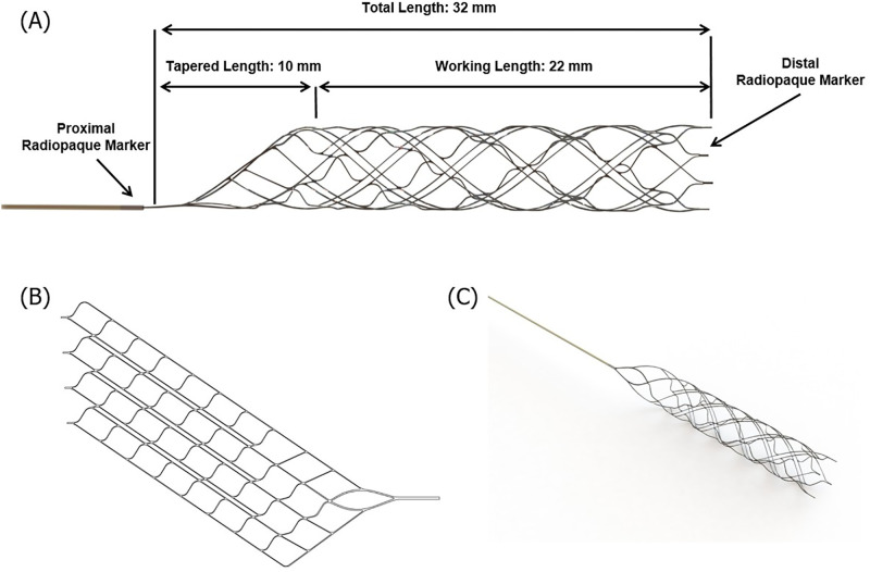
The Tromba thrombectomy device was designed for the removal of occlusive thrombi in the setting of acute ischemic stroke, in vessels with a diameter ranging from 1.5 to 3.5 mm. It is made of nickel–titanium, which is a shape memory alloy material. The detailed dimensions of this novel device, which was used in the experimental group, are shown in Fig 1. The Tromba device is a stent for thrombus removal with a plate-shaped structure that is rolled in a spiral form and cut at regular intervals along the spiral direction around the body part (Fig 1B). Therefore, when an external force is applied, the rolled structure spreads out in the form of a plate. The front portion of the body part includes a thrombus removal section for receiving the thrombus, and the rear portion includes a delivery section that retrieves the body part into the catheter. This device has several types of cell patterns, and a single device can have a first cell pattern and a second cell pattern. The first cell pattern includes several first link parts arranged in an oblique line, spaced apart by a predetermined distance, and several second link parts spaced apart by a predetermined distance while connecting the adjacent first link parts.

Fig 1. Tromba device.

Fig 1

Schematic diagram (A), (B) and photograph (C) of the Tromba thrombectomy device.

In addition, the device is composed of a main cell part, which includes numerous main cells arranged in a helical shape, and numerous open cell parts arranged in a helical shape while facing in the longitudinal direction with respect to the main cell. The main cell is formed by two first link units and two second link units, and the line connecting the vertices in the left and right directions forms an angle of ≤ 45° with respect to the longitudinal direction of the stent.

Animal care and process of in vivo study

To identify all experimental environments, we tested canines of the same age, weight, and size. Twelve 6-month-old canines (beagle dogs obtained from Marshall BioResources, North Rose, NY, USA) weighing between 6.0 and 8.0 kg were included in this study. Canine models have several advantages. The size and extent of vasospasm occurring in canines are more similar to those in humans than in porcine and rodent models, allowing for more targeted endovascular treatment and the branches of the external carotid artery in the canine model have a vessel size similar to that of the human middle cerebral artery, which is sufficient for the study of novel endovascular devices for human cerebral blood vessels [11]. One animal was bred in a cage (0.9 m2) in accordance with AAALAC guidelines (0.74 m2), and an individual identification card with the test and animal numbers was attached to the breeding cage. A classic dog toy was used for environmental enrichment. The conditions of the breeding environment were as follows: temperature, 23°C ± 3°C; relative humidity, 55% ± 15%; ventilation, 10–20 times/h; lighting time, 12 h (lights up at 8 am, lights out at 8 pm), and illuminance, 150–300 Lux. This study was conducted in an established canine-breeding room. For the feed supplied to the test, a solid feed for test animals (2925 Telan Global 25% Protein Dog Diet) was sterilized by irradiation. Ultrapure water produced by an RO water (reverse osmosis distilled water) production and supply device was sterilized using sodium hypochlorite and a UV device and then freely dispensed through an automatic water supply nozzle.

All experiments were conducted under general anesthesia. Preoperative anesthesia was induced by intramuscular injection of 5 mg/kg Zoletil (Bibac Korea, Seoul, Korea) and 2 mg/kg Rompun (Bayer Korea, Ansan, Korea). For postoperative pain/stress relief in experimental animals, 0.2 mg/kg of Metacam (Boehringer Ingelheim, Germany) was subcutaneously injected once daily for three days. In addition, 15 mg/kg cefazolin (Chong Kun Dang, Korea) was subcutaneously injected once daily for three days to prevent infection. For humane management of diet, exercise, and nutrition in experimental animals with stroke induced by vascular occlusion and mechanical thrombectomy, the experiment was limited to unilateral sites of the carotid artery, not to bilateral sites. During the observation period, animals treated with the stent retriever in the breeding room of the experimental animal center were visually observed for abnormal symptoms once a day, and the experimental animals had no restrictions on their daily activities, including diet.

Histopathological analysis was performed by excising vessels that had undergone mechanical thrombectomy. Anesthesia was induced by intramuscular injection of 5 mg/kg Zoletil (Bibac Korea, Seoul, Korea) and 2 mg/kg Rompun (Bayer Korea, Ansan, Korea) in the operating room of the experimental animal center. After induction of general anesthesia, airway intubation was performed, the animal was connected to an inhalation anesthesia machine (Fabius GS premium, Drager, Germany), and respiratory anesthesia was maintained with isoflurane (<3%). The animals’ anesthetic conditions (electrocardiogram, oxygen saturation, carbon dioxide concentration, etc.) were constantly monitored using a patient monitoring system (Vista 120, Drager, Germany). At the end of the experiment, 40 ml of KCl was injected intravenously after intramuscular injection to induce euthanasia, and respiration and cardiac arrest in the experimental animals were confirmed. There were no other mortalities, except planned euthanasia, and no humane endpoints in the study.

Preparation of thrombus

The preparation and application of the thrombus followed the method applied in previous studies [12,13]. In brief, a thrombus was created by mixing 10 mL autologous blood from an animal and 25 IU bovine thrombin solution (Dade Behring, Newark, NJ, USA) in a silicone tube for 120 min at room temperature [14,15]. After refrigeration at 4°C for 24 h, the thrombus was prepared by placing it in a 10-mm silicone tube containing saline [12,13].

Initial and follow-up angiography, thrombus embedding, and mechanical thrombectomy protocol

We used a monoplane high-resolution angiography system (Artis Zee multi-purpose system; Siemens, Erlangen, Germany). First, a short 6-F catheter sheath (Terumo, Tokyo, Japan) was inserted into the common femoral artery, and one of the branches of the external carotid artery (occipital, superficial, or linguofacial artery) was selected. Subsequently, a pre-manufactured thrombus was injected into the target vessel using a 6-F intermediate catheter (Envoy DA; DePuy Synthes Companies, Warsaw, IN, USA) and the occlusion was maintained by removing the intermediate catheter for 15 min. After confirming the exact location of the thrombus and complete occlusion of blood vessels with angiography, thrombus removal was performed using the Tromba (4 × 20 mm; CGBio, Seongnam, Korea) thrombectomy device in the experimental group and the Solitaire FR (4 × 20 mm; EV3, Irvine, CA, USA) thrombectomy device in the control group. The degree of reperfusion was confirmed through subsequent angiography, and a follow-up examination was performed 1 month later to determine the degree of maintenance of reperfusion and the presence of vascular complications. The grade of reperfusion patency was defined according to the scale introduced by Grandin et al., as follows: no narrowing, slight narrowing (25% reduction in the lumen diameter), moderate narrowing (25%–50% stenosis or 50%–75% stenosis affecting only a short segment of the vessel), and severe narrowing (50%–75% stenosis affecting a long segment of the vessel or stenosis > 75%) [16]. All procedures were conducted according to international guidelines and were approved by the IACUC of Osong Medical Innovation Foundation in Republic of Korea (KBIO-IACUC-2019-051-1).

Histopathologic evaluation

Histopathologic evaluation was performed 1 month after mechanical thrombectomy to determine the degree and condition of the recovery of wall injury using an autopsied canine model. The vessel sections from each artery were processed and embedded in paraffin, according to standard laboratory procedures. Specimens were processed for histopathologic analysis, including hematoxylin-and-eosin and Masson’s trichrome staining.

To assess the degree of arterial injury, ordinal data were collected for multiple parameters, including (i) inflammation (%), (ii) intimal proliferation (%), (iii) endothelial loss (%), (iv) thrombosis (fibrin/platelet), (v) hemorrhage (adventitial/medial), and (vi) medial injury (device-induced), using the semiquantitative scoring system of Nogueira et al [17]. The degrees of inflammation, intimal proliferation, and endothelial loss were classified as “no change,” “change < 25%,” “change between 25% and 50%,” “change between 51% and 75%,” and “change > 75%.” The degree of thrombosis was classified as “no change”; “minimal, focal”; “mild, multifocal”; “moderate, regionally diffuse”; and “severe, marked diffuse, or total luminal occlusion.” The degree of hemorrhage was classified as “no change”; “focal, occasional”; “multifocal and regional”; “regionally diffuse”; and “100% red blood cells.” Finally, the degree of medial injury was classified as “no change,” “focal disruption of the internal elastic lamina,” “widespread disruption of the internal elastic lamina,” “medial tear not extending beyond the external elastic lamina,” and “medial tear with involvement of the external elastic lamina.” The sections were examined under an optical microscope at a magnification ranging from 40× to 100× by a board-certified pathologist (WC Son) who was blinded to the device used.

Results

Angiographic results

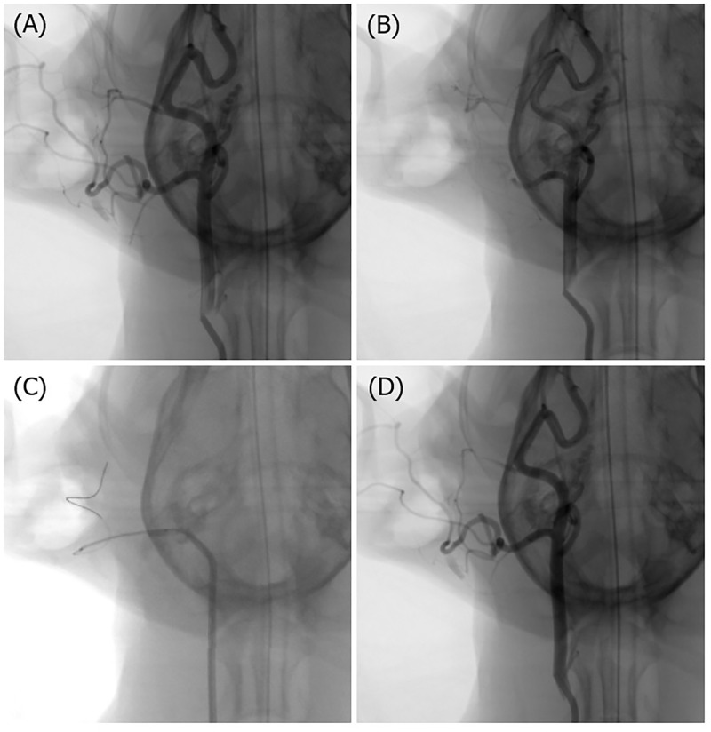
Using digital subtraction angiography, we selected a blood vessel with a diameter similar to that of the human middle cerebral artery as the target vessel for thrombectomy (Fig 2). Either the linguofacial artery, occipital artery, or superficial temporal artery was occluded. One blood vessel was selected from each of the 12 canines. In the experimental group (Tromba thrombectomy device), the linguofacial artery was selected in two cases, the occipital artery in three cases, and the superficial temporal artery in one case. In the control group (Solitaire FR thrombectomy device), the occipital artery was selected in five cases and the superficial temporal artery was selected in one case. In the experimental group, the reperfusion patency was classified as “no narrowing” in five cases and “moderate narrowing” in one case, and most cases were recanalized with fewer than three attempts. In the control group, the reperfusion patency was classified as “no narrowing” in four cases, “moderate narrowing” in one case, and “slight narrowing” in one case, and most cases were recanalized with fewer than two attempts. No vascular complications, such as dissection or perforation, were observed in either the experimental group or the control group (Table 1).

Fig 2. Representative angiograms.

Fig 2

(A) Initial digital subtraction angiogram of the right common carotid artery. (B) Injection of a pre-manufactured thrombus into the target occipital artery. (C) Thrombectomy procedure. (D) Complete recanalization of the target occipital artery.

Table 1. Demographic characteristics and presentation of six cases subjected to mechanical thrombectomy.

Tromba (experimental group) Solitaire FR 4×20 mm (control group)
No Occlusion site Reperfusion patency No. of passes / Vasc Cx 1 M follow-up DSA No Occlusion site Reperfusion patency No. of passes / Vasc Cx 1 M follow-up DSA
1 Rt LFA Moderate narrowing 3 / (-) No interval change 7 Rt OA Mo narrowing 1 / (-) No interval change
2 Rt LFA No narrowing 2 / (-) No interval change 8 Rt OA Moderate narrowing 2 / (-) No interval change
3 Rt OA No narrowing 1 / (-) No interval change 9 Rt OA No narrowing 1 / (-) No interval change
4 Rt OA No narrowing 1 / (-) No interval change 10 Rt OA No narrowing 1 / (-) No interval change
5 Rt OA No narrowing 3 / (-) No interval change 11 Rt OA Slight narrowing 2 / (-) No interval change
6 Rt STA No narrowing 1 / (-) No interval change 12 Rt STA No narrowing 1 / (-) No interval change

No., number; Vasc Cx, vascular complication; M, month; DSA, digital subtraction angiography; Rt, right; LFA, linguofacial artery; OA, occipital artery; STA, superficial temporal artery. Slight narrowing, 25% reduction in the lumen diameter; moderate narrowing, 25%–50% stenosis or 50%–75% stenosis affecting only a short segment of the vessel; severe narrowing, 50%–75% stenosis affecting a long segment of the vessel or stenosis > 75%.

Histopathologic results

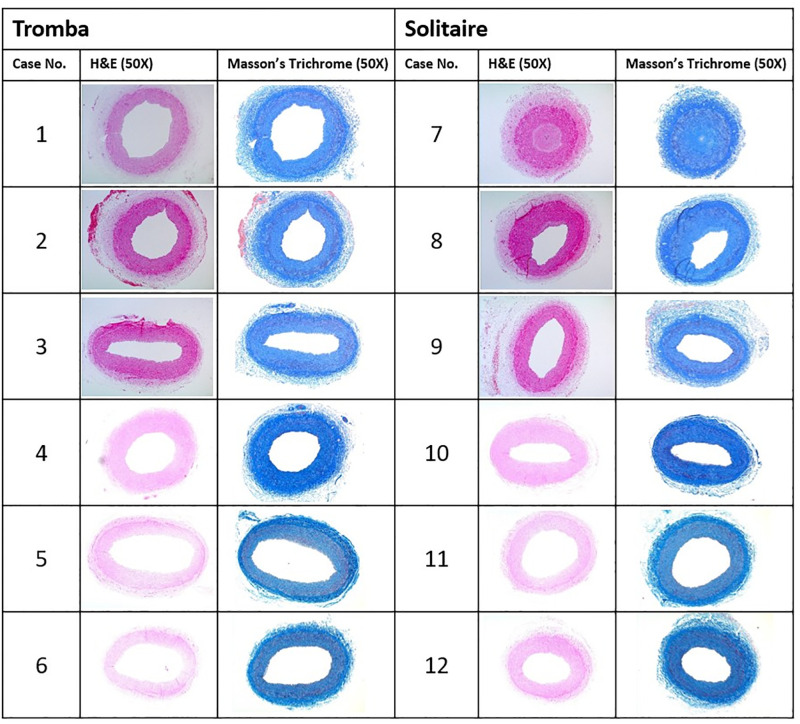
Histopathologic evaluation was performed 1 month after mechanical thrombectomy. Two linguofacial arteries, eight occipital arteries, and two superficial temporal arteries with diameters of 2.2–3.1 mm from the 12 swines were evaluated. We cut the blood vessels that were subjected to thrombectomy into 4-μm sections, which were subsequently stained with hematoxylin-and-eosin and Masson’s trichrome. The sections were examined under an optical microscope at 50× magnification (Fig 3).

Fig 3. Histopathologic evaluation.

Fig 3

Histopathologic evaluation was performed using two linguofacial arteries, eight occipital arteries, and two superficial temporal arteries with diameters of 2.2–3.1 mm from the 12 swines. The blood vessels that were subjected to thrombectomy were cut into 4-μm sections and stained with hematoxylin-and-eosin (H&E) and Masson’s trichrome. The sections were examined under an optical microscope at 50× magnification.

In the experimental group, inflammatory changes were not observed in any case and 25%–50% intimal proliferation was observed in only two cases (Table 2). A < 25% endothelial loss was observed in two cases, and device-induced medial injury classified as “focal disruption of the internal elastic lamina” was observed in one case. Thrombosis (fibrin/platelet) and hemorrhage (adventitial/medial) were not observed in any case in the experimental group. In the control group, serious intimal proliferation (> 75%) was observed in one case and 25%–50% intimal proliferation was observed in one case. A < 25% endothelial loss was observed in one case. “Severe, marked diffuse” thrombosis (fibrin/platelet) was also observed in one case. Inflammation, hemorrhage (adventitial/medial), and device-induced medial injury were not observed in any case in the control group.

Table 2. Semiquantitative analysis of histopathologic changes to assess the degree of arterial damage in blood vessels due to the use of Tromba and Solitaire.

Case no. of Tromba Inflammation (%) Intimal proliferation (%) Endothelial loss (%) Thrombosis (fibrin/platelet) Hemorrhage (adventitial/medial) Medial injury (device-induced)
1 None 25% ~ 50% <25% None None None
2 None 25% ~ 50% None None None None
3 None None <25% None None None
4 None None None None None None
5 None None None None None Focal disruption of the IEL
6 None None None None None None
Case no. of Solitaire Inflammation (%) Intimal proliferation (%) Endothelial loss (%) Thrombosis (fibrin/platelet) Hemorrhage (adventitial/medial) Medial injury (device-induced)
7 None >75% <25% Severe, marked diffuse None None
8 None 25% ~ 50% None None None None
9 None None None None None None
10 None None None None None None
11 None None None None None None
12 None None None None None None

No., number; IEL, internal elastic lamina.

Discussion

In this study, histologic and angiographic findings were compared between the newly developed Tromba device (experimental group) and the currently widely used Solitaire stent (control group). Tromba showed no significant difference to the conventional Solitaire device in angiographic and histopathologic findings after thrombus removal.

In general, a stent maintains the intravascular space by pushing the stenosis outward from the conduit through the radial force from its expansion in the lateral direction [18,19]. To be suitable for use in the human body, stents must have special properties in addition to expansion capacity, among which flexibility is particularly important [20]. Stent flexibility refers to the ability of a stent to be adapted to the shape of the bent portion of a blood vessel [21]. If flexibility is insufficient, the stent may lose its adaptability in the bent portion of the blood vessel, which may result in an abnormal conduit [20,21]. When a conventional flat-type stent is inserted into the catheter tube, as the rectangular thin film is inserted in a rolled state, the portion located inside the rolled shape becomes relatively severely bent and subjected to a stronger bending stress than the portion located outside. This causes the stent to return to its original shape and results in reduced wall apposition. In addition, when a conventional flat stent is deployed from the tube of the catheter and comes in close contact with the blood vessel wall, because of the difference in thickness along the circumferential direction of the rolled shape, it becomes difficult to maintain flexibility with respect to a blood vessel with several bends in various directions.

To solve the problems of the existing technology, a stent for thrombus removal with a plate-shaped structure rolled in a spiral form, consisting of a hybrid structure with open and closed cells that can provide high flexibility in blood vessels bent in various directions in three dimensions, was developed.

The thrombus removal part of the existing thrombus retrievable stent developed so far, including Solitaire, is in the form of a closed cell structure in which all cells are connected by a link to fix and recover the thrombus. The stent of the "closed cell" structure fixes the thrombus in a relatively stable manner, so that the captured thrombus is smoothly recovered. However, due to its low flexibility and expansion power, it is difficult for this stent to adapt to the tortuous cerebral blood vessels, making it difficult to remove a thrombus in a severely bent region. This is a limitation as repeated thrombus removals can damage blood vessel walls. The hybrid cell structure is judged to be ideal because it has both the characteristics of an open cell, i.e., it can adapt well to complex and tortuous cerebral blood vessels, and a closed cell, i.e., it can stably remove and recover blood clots. In addition, a structure with a weak radial force and a structure that cannot be recaptured are the biggest disadvantages of an open cell. These can be overcome with a hybrid cell structure.

The model used in this study has some limitations. First, the canine internal carotid artery is not sufficiently large to accommodate thrombectomy devices; therefore, we used external carotid artery branches, which made it difficult to apply the previously described Thrombolysis in Cerebral Infarction score for grading reperfusion. Therefore, we used the reperfusion patency grading scale suggested by Grandin et al., and we encountered no problems in comparing the reperfusion rates of the two groups. Second, as the experiment was completed with a relatively small number of canines (a total of 12 animals), the comparison of the thrombus removal stents may be insufficient and our results may be difficult to generalize. However, histologic comparisons involving a large number of animals have been conducted in other studies using swine or canine models (usually less than 10 animals) [12,15,17,2224]. Third, imaging follow-up was performed immediately after mechanical thrombectomy and 1 month later, without regular follow-up investigations in between. Therefore, angiographic evaluation was not performed during the condition of acute phase vascular damage and blood flow. Finally, as this was a small pilot study, the results should be interpreted with caution.

In conclusion, the Tromba mechanical thrombectomy device provides high flexibility and high adhesion to the vessel wall. No significant difference was observed between the two devices compared in this study. The stability and efficiency of the newly developed Tromba device are considered to be high and comparable to those of Solitaire.

Supporting information

S1 Fig. Raw histopathology images.

(PDF)

S1 File. Experiment report.

Evaluation of safety and efficacy of stent retrievers for treatment of ischemic stroke.

(PDF)

Data Availability

All relevant data are within the paper and its Supporting information files.

Funding Statement

Dr. Sang Hun Lee was supported by the Seoul R&D program (BT190099) funded by the Seoul Business Agency (SBA, Korea) and the Korea University Hospital Grant (K2210671)]. The funders had no role in study design, data collection and analysis, decision to publish, or preparation of the manuscript.

References

  • 1.Berkhemer OA, Fransen PSS, Beumer D, van den Berg LA, Lingsma HF, Yoo AJ, et al. A randomized trial of intraarterial treatment for acute ischemic stroke. New Engl J Med. 2015;372:11–20. doi: 10.1056/NEJMoa1411587 [DOI] [PubMed] [Google Scholar]
  • 2.Campbell BCV, Mitchell PJ, Kleinig TJ, Dewey HM, Churilov L, Yassi N, et al. Endovascular therapy for ischemic stroke with perfusion-imaging selection. New Engl J Med. 2015;372:1009–1018. doi: 10.1056/NEJMoa1414792 [DOI] [PubMed] [Google Scholar]
  • 3.Goyal M, Demchuk AM, Menon BK, Eesa M, Rempel JL, Thornton J, et al. Randomized assessment of rapid endovascular treatment of ischemic stroke. New Engl J Med. 2015;372:1019–1030. doi: 10.1056/NEJMoa1414905 [DOI] [PubMed] [Google Scholar]
  • 4.Jovin TG, Chamorro A, Cobo E, de Miquel MA, Molina CA, Rovira A, et al. Thrombectomy within 8 hours after symptom onset in ischemic stroke. New Engl J Med. 2015;372:2296–2306. doi: 10.1056/NEJMoa1503780 [DOI] [PubMed] [Google Scholar]
  • 5.Saver JL, Goyal M, Bonafe A, Diener HC, Levy EI, Pereira VM, et al. Stent-retriever thrombectomy after intravenous t-pa vs. T-pa alone in stroke. New Engl J Med. 2015;372:2285–2295. doi: 10.1056/NEJMoa1415061 [DOI] [PubMed] [Google Scholar]
  • 6.Shao QJ, Zhu LF, Li TX, Li L, Li DJ, Zhang JQ, et al. New method of thrombus preparation using a fluid model for evaluation of thrombectomy devices in a swine model. Thromb Res. 2014;134:1087–1092. doi: 10.1016/j.thromres.2014.07.043 [DOI] [PubMed] [Google Scholar]
  • 7.Gory B, Bresson D, Kessler I, Perrin M, Guillaudeau A, Durand K, et al. Histopathologic evaluation of arterial wall response to 5 neurovascular mechanical thrombectomy devices in a swine model. Am J Neuroradiol. 2013;34:2192–2198. doi: 10.3174/ajnr.A3531 [DOI] [PMC free article] [PubMed] [Google Scholar]
  • 8.Roth C, Papanagiotou P, Behnke S, Walter S, Haass A, Becker C, et al. Stent-assisted mechanical recanalization for treatment of acute intracerebral artery occlusions. Stroke. 2010;41:2559–2567. doi: 10.1161/STROKEAHA.110.592071 [DOI] [PubMed] [Google Scholar]
  • 9.Saver JL, Jahan R, Levy EI, Jovin TG, Baxter B, Nogueira RG, et al. Solitaire flow restoration device versus the merci retriever in patients with acute ischaemic stroke (swift): A randomised, parallel-group, non-inferiority trial. Lancet. 2012;380:1241–1249. doi: 10.1016/S0140-6736(12)61384-1 [DOI] [PubMed] [Google Scholar]
  • 10.Nogueira RG, Lutsep HL, Gupta R, Jovin TG, Albers GW, Walker GA, et al. Trevo versus merci retrievers for thrombectomy revascularisation of large vessel occlusions in acute ischaemic stroke (trevo 2): A randomised trial. Lancet. 2012;380:1231–1240. doi: 10.1016/S0140-6736(12)61299-9 [DOI] [PMC free article] [PubMed] [Google Scholar]
  • 11.Mehra M, Henninger N, Hirsch JA, Chueh J, Wakhloo AK, Gounis MJ. Preclinical acute ischemic stroke modeling. J Neurointerv Surg 2012;4:307–313. doi: 10.1136/neurintsurg-2011-010101 [DOI] [PubMed] [Google Scholar]
  • 12.Gralla J, Schroth G, Remonda L, Fleischmann A, Fandino J, Slotboom J, et al. A dedicated animal model for mechanical thrombectomy in acute stroke. Am J Neuroradiol. 2006;27:1357–1361. [PMC free article] [PubMed] [Google Scholar]
  • 13.Kan I, Yuki I, Murayama Y, Vinuela FA, Kim RH, Vinters HV, et al. A novel method of thrombus preparation for use in a swine model for evaluation of thrombectomy devices. Am J Neuroradiol. 2010;31:1741–1743. doi: 10.3174/ajnr.A1991 [DOI] [PMC free article] [PubMed] [Google Scholar]
  • 14.Lee SH, Kim SW, Kim JM, Son WC. Histopathologic findings of vascular damage after mechanical thrombectomy using stent retriever in canine models. J Thromb Thrombolys. 2021;51:1094–1100. doi: 10.1007/s11239-020-02353-8 [DOI] [PubMed] [Google Scholar]
  • 15.Gralla J, Schroth G, Remonda L, Nedeltchev K, Slotboom J, Brekenfeld C. Mechanical thrombectomy for acute ischemic stroke—thrombus-device interaction, efficiency, and complications in vivo. Stroke. 2006;37:3019–3024. doi: 10.1161/01.STR.0000248457.55493.85 [DOI] [PubMed] [Google Scholar]
  • 16.Grandin CB, Cosnard G, Hammer F, Duprez TP, Stroobandt G, Mathurin P. Vasospasm after subarachnoid hemorrhage: Diagnosis with mr angiography. Am J Neuroradiol. 2000;21:1611–1617. [PMC free article] [PubMed] [Google Scholar]
  • 17.Nogueira RG, Levy EI, Gounis M, Siddiqui AH. The trevo device: Preclinical data of a novel stroke thrombectomy device in two different animal models of arterial thrombo-occlusive disease. J Neurointerv Surg. 2012;4:295–300. doi: 10.1136/neurintsurg-2011-010053 [DOI] [PubMed] [Google Scholar]
  • 18.Borghi A, Murphy O, Bahmanyar R, McLeod C. Effect of stent radial force on stress pattern after deployment: A finite element study. J Mater Eng Perform. 2014;23:2599–2605. doi: 10.1007/s11665-014-0913-z [DOI] [PMC free article] [PubMed] [Google Scholar]
  • 19.Duerig TW, Tolomeo DE, Wholey M. An overview of superelastic stent design. Minim Invasive Ther Allied Technol. 2000;9:235–246. doi: 10.1080/13645700009169654 [DOI] [PubMed] [Google Scholar]
  • 20.Mori K, Saito T. Effects of stent structure on stent flexibility measurements. Ann Biomed Eng. 2005;33:733–742. doi: 10.1007/s10439-005-2807-6 [DOI] [PubMed] [Google Scholar]
  • 21.Shen X, Deng YQ, Ji S, Xie ZM, Zhu HF. Flexibility behavior of coronary stents: The role of linker investigated with numerical simulation. J Mech Med Biol. 2017;17. [Google Scholar]
  • 22.Mordasini P, Frabetti N, Gralla J, Schroth G, Fischer U, Arnold M, et al. In vivo evaluation of the first dedicated combined flow-restoration and mechanical thrombectomy device in a swine model of acute vessel occlusion. Am J Neuroradiol. 2011;32(2):294–300. doi: 10.3174/ajnr.A2270 [DOI] [PMC free article] [PubMed] [Google Scholar]
  • 23.Brooks OW, King RM, Nossek E, Marosfoi M, Caroff J, Chueh JY, et al. A canine model of mechanical thrombectomy in stroke. J Neurointerv Surg. 2019;11(12):1243–8. doi: 10.1136/neurintsurg-2019-014969 [DOI] [PubMed] [Google Scholar]
  • 24.Mordasini P, Hiller M, Brekenfeld C, Schroth G, Fischer U, Slotboom J, et al. In vivo evaluation of the Phenox CRC mechanical thrombectomy device in a swine model of acute vessel occlusion. Am J Neuroradiol. 2010;31(5):972–8. doi: 10.3174/ajnr.A1928 [DOI] [PMC free article] [PubMed] [Google Scholar]

Decision Letter 0

Andreas Zirlik

28 Mar 2022

PONE-D-21-40394In vivo evaluation of histopathologic findings of vascular damage after mechanical thrombectomy with the Tromba device in a canine modelPLOS ONE

Dear Dr. LEE,

Thank you for submitting your manuscript to PLOS ONE. After careful consideration, we feel that it has merit but does not fully meet PLOS ONE’s publication criteria as it currently stands. Therefore, we invite you to submit a revised version of the manuscript that addresses the points raised during the review process.

Please submit your revised manuscript by May 12 2022 11:59PM. If you will need more time than this to complete your revisions, please reply to this message or contact the journal office at plosone@plos.org. When you're ready to submit your revision, log on to https://www.editorialmanager.com/pone/ and select the 'Submissions Needing Revision' folder to locate your manuscript file.

Please include the following items when submitting your revised manuscript:

  • A rebuttal letter that responds to each point raised by the academic editor and reviewer(s). You should upload this letter as a separate file labeled 'Response to Reviewers'.

  • A marked-up copy of your manuscript that highlights changes made to the original version. You should upload this as a separate file labeled 'Revised Manuscript with Track Changes'.

  • An unmarked version of your revised paper without tracked changes. You should upload this as a separate file labeled 'Manuscript'.

If you would like to make changes to your financial disclosure, please include your updated statement in your cover letter. Guidelines for resubmitting your figure files are available below the reviewer comments at the end of this letter.

If applicable, we recommend that you deposit your laboratory protocols in protocols.io to enhance the reproducibility of your results. Protocols.io assigns your protocol its own identifier (DOI) so that it can be cited independently in the future. For instructions see: https://journals.plos.org/plosone/s/submission-guidelines#loc-laboratory-protocols. Additionally, PLOS ONE offers an option for publishing peer-reviewed Lab Protocol articles, which describe protocols hosted on protocols.io. Read more information on sharing protocols at https://plos.org/protocols?utm_medium=editorial-email&utm_source=authorletters&utm_campaign=protocols.

We look forward to receiving your revised manuscript.

Kind regards,

Andreas Zirlik, MD

Academic Editor

PLOS ONE

Journal Requirements:

When submitting your revision, we need you to address these additional requirements.

1. Please ensure that your manuscript meets PLOS ONE's style requirements, including those for file naming. The PLOS ONE style templates can be found at

https://journals.plos.org/plosone/s/file?id=wjVg/PLOSOne_formatting_sample_main_body.pdf  and

https://journals.plos.org/plosone/s/file?id=ba62/PLOSOne_formatting_sample_title_authors_affiliations.pdf

2. In your Data Availability statement, you have not specified where the minimal data set underlying the results described in your manuscript can be found. PLOS defines a study's minimal data set as the underlying data used to reach the conclusions drawn in the manuscript and any additional data required to replicate the reported study findings in their entirety. All PLOS journals require that the minimal data set be made fully available. For more information about our data policy, please see http://journals.plos.org/plosone/s/data-availability.

Upon re-submitting your revised manuscript, please upload your study’s minimal underlying data set as either Supporting Information files or to a stable, public repository and include the relevant URLs, DOIs, or accession numbers within your revised cover letter. For a list of acceptable repositories, please see http://journals.plos.org/plosone/s/data-availability#loc-recommended-repositories. Any potentially identifying patient information must be fully anonymized.

Important: If there are ethical or legal restrictions to sharing your data publicly, please explain these restrictions in detail. Please see our guidelines for more information on what we consider unacceptable restrictions to publicly sharing data: http://journals.plos.org/plosone/s/data-availability#loc-unacceptable-data-access-restrictions. Note that it is not acceptable for the authors to be the sole named individuals responsible for ensuring data access.

We will update your Data Availability statement to reflect the information you provide in your cover letter.

3. Thank you for stating the following financial disclosure:

“Dr. Sang Hun Lee was supported by the Seoul R&D program (BT190099) funded by the Seoul Business Agency (SBA, Korea) and the Korea University Ansan Hospital Grant (K2110981).”

Please state what role the funders took in the study.  If the funders had no role, please state: "The funders had no role in study design, data collection and analysis, decision to publish, or preparation of the manuscript."

If this statement is not correct you must amend it as needed.

Please include this amended Role of Funder statement in your cover letter; we will change the online submission form on your behalf.

[Note: HTML markup is below. Please do not edit.]

Reviewers' comments:

Reviewer's Responses to Questions

Comments to the Author

1. Is the manuscript technically sound, and do the data support the conclusions?

The manuscript must describe a technically sound piece of scientific research with data that supports the conclusions. Experiments must have been conducted rigorously, with appropriate controls, replication, and sample sizes. The conclusions must be drawn appropriately based on the data presented.

Reviewer #1: Yes

Reviewer #2: Yes

**********

2. Has the statistical analysis been performed appropriately and rigorously?

Reviewer #1: N/A

Reviewer #2: Yes

**********

3. Have the authors made all data underlying the findings in their manuscript fully available?

The PLOS Data policy requires authors to make all data underlying the findings described in their manuscript fully available without restriction, with rare exception (please refer to the Data Availability Statement in the manuscript PDF file). The data should be provided as part of the manuscript or its supporting information, or deposited to a public repository. For example, in addition to summary statistics, the data points behind means, medians and variance measures should be available. If there are restrictions on publicly sharing data—e.g. participant privacy or use of data from a third party—those must be specified.

Reviewer #1: Yes

Reviewer #2: Yes

**********

4. Is the manuscript presented in an intelligible fashion and written in standard English?

PLOS ONE does not copyedit accepted manuscripts, so the language in submitted articles must be clear, correct, and unambiguous. Any typographical or grammatical errors should be corrected at revision, so please note any specific errors here.

Reviewer #1: Yes

Reviewer #2: Yes

**********

5. Review Comments to the Author

Please use the space provided to explain your answers to the questions above. You may also include additional comments for the author, including concerns about dual publication, research ethics, or publication ethics. (Please upload your review as an attachment if it exceeds 20,000 characters)

Reviewer #1: In vivo evaluation of histopathologic findings of vascular damage after mechanical thrombectomy with the Tromba device in a canine model

Authors present in the manuscript the results of in total 12 animal experiments, where a newly developed thrombectomy device was tested (n=6) and compared with a reference device (n=6), being already in clinical use.

They found that the novel device is effective in terms of thrombus removal, an safe in terms vessel wall injury.

I have the following comments to be considered for the sake of further improvement of the manuscript.

Abstract:

In general the abstract should provide the reader with a more complete overview on the study. It should be understandable as a stand-alone piece of work, as well. Therefore the following aspects should be definitely improved:

1. It is unclear from the title, as well as from the abstract, for which vessels the device is developed. Is that a thrombectomy device for coronaries? For central nervous system? For pulmonary? For deep veins?

2. It has to be declared in the abstract, at what timepoint the animal models were sacrificed and analyzed. Considering that endothelial damage has the potential for instantaneous risk of further thrombus formation, therefore acute result is a critical aspect. On the other and it might heal over time, losing its relevance. But this would be nice to demonstrate. Since authors mention ‘intimal proliferation’, one would assume that chronic results are presented, however it is still unclear.

3. ‘Moderate-’ and ‘slight narrowing’ are quite subjective phrasing for someone who is not fully familiar with the Grandin scale and therefore difficult to interpret for comparison. Please quantify it.

Main text:

1. It would be important to define, why the Solitaire FR was selected as reference. Is that the most widely used and accepted device of the field?

2. From main text it is clear that results were ‘only’ at intermediate term after 30-days assessed. Since acute injury could have been assessed by intravascular imaging modalities, I believe the lack of acute data is a major limitation and it has to be acknowledged.

3. In the histopathologic result section the Authors write about swine model instead of canine. Please clarify.

4. In the limitation section Authors state: “However, histologic comparisons involving a large number of animals have been conducted in other canine models, and the results were considered to be of sufficient value.” It is unclear, which study and what device they refer to. It should be at least referenced or even described in a bit more details.

5. A certain structural / mechanical / design comparison of the novel versus the traditional device, focusing on potential advantages would be important for better understanding.

Main text

1. You wrore: “Lesion location was defined according to SYNTAX score”. But actually it is correct as follows: The coronary arterial segments were defined according to the American Heart Association, as modified for the ARTS I and II studies. (Ref: Serruys PW, Unger F, van Hout BA, van den Brand MJ, van Herwerden LA, van Es GA, Bonnier JJ, Simon R, Cremer J, Colombo A, Santoli C, Vandormael M, Marshall PR, Madonna O, Firth BG, Breeman A, Morel MA, Hugenholtz PG. The ARTS study (Arterial Revascularization Therapies Study). Semin Interv Cardiol 1999;4:209-19.)

2. You wrote “Kaplan-Meier (KM) cumulative survival curves were generated and the difference in survival between groups was compared by the log-rank test.”. I would suggest writing as ‘event free survival’.

3. “In the revascularization group, 105 (23%) patients were treated with CABG and 344 (77%) with PCI.” Considering, you are talking only about patients with single vessel disease, this is a relatively high rate, isn’t it.

4. “Among them, 1010 received medical therapy and 449 (30%) underwent revascularization based on the FFR measurement.” I would suggest deleting the last few words. Not all patients below 0.75 were revascularized. Not all patients below 0.80 were revascularized. Some patients above 0.80 were revascularized. Therefore saying ‘based on FFR’ is inaccurate, I believe.

5. Abstract: “Diameter stenosis, minimum lumen diameter, and FFR values were lower in rev group.” Main text: “On average, patients who received revascularization (….) had a higher percent diameter stenosis and a lower minimum lumen diameter and lower FFR as compared to patients who received medical therapy.“ Should be corrected.

6. “At a mean follow up of 3.3 years, MACE rate was similar between the medical therapy and the revascularization group.” It is unclear whether you talk about the grey zone or the entire population.

7. You wrote: “Compared to the revascularization group (Figure 2, Table 3), MACE rate was significantly higher in the 0.70-0.75 strata (p=0.047) and significantly lower in the 0.81-0.85 strata (p=0.023). There was no difference in MACE rate between the FFR gray zone strata of the medical therapy group and the revascularization group (p=0.804).”

I suggest changing it in this way: “Compared to the total revascularization group (Figure 2, Table 3), MACE rate was significantly higher in the 0.70-0.75 strata (p=0.047) and significantly lower in the 0.81-0.85 strata (p=0.023) of the MT group. There was no difference in MACE rate between the FFR gray zone strata of the medical therapy group and the revascularization group (p=0.804).”

8. Use abbreviations consequently. For instance either always MT and Rev or not at all…

9. I think the main interest why someone will read this paper to learn, what is better in the grey zone: MT or Rev. Accordingly, the structure, especially in the Results should be organized in this way.

There is lot of talks about the differences in outcome between the different strata of MT group. After Nils’ metaanalysis paper this is not much new. Therefore it is too bad that you hide the ‘million dollar answer’ in the very last sentence of the Results. It could be better the other way around, I believe.

10. “At linear regression analysis, MACE rate was overlapping between medical therapy and revascularization group at FFR value of about 0.78 (Figure 5).” It would be more accurate here to highlight the 95% CI ranges for the two lines and then you can give a range of overlap instead of an exact value.

11. If you have sufficient number of cases, it would be interesting to see what are the event rates in BMS patients vs DES patients vs CABG patients (latter most probably 99.99% LIMA-LAD, since everyone is SVD)

If type of stent is not available, then you can simply indicate it as early cases (before 2007-8?) and recent cases (after that) considering BMS era vs DES era.

12. I am not sure that Table 1 is relevant. Actually you never compare the entire MT group with the Rev group, therefore comparing clinical characteristics is unnecessary. It might be more relevant to indicate the comparison of clinical characteristics between Rev and MT strata.

13. Figure 2 – 3A – 4: where p is significant when comparing more than one groups, then one-by-one comparison should be indicated, as well.

Reviewer #2: Solid data presentation with regard to this new technology

Comments:

-With the assumption that acute stroke should be acute emboli acute thrombotic clots were injected; seems reasonable. But in reality also chronic clots could migrate to the cerebral arteries, eg patient has AF. Are there any data if this technology also works in chronic clots??

-The authors state throughout the manuscript that they used 12 canine models (beagles), but in the section histopathological findings they state that they used 12 swines?? This need to be clarified

**********

6. PLOS authors have the option to publish the peer review history of their article (what does this mean?). If published, this will include your full peer review and any attached files.

If you choose “no”, your identity will remain anonymous but your review may still be made public.

Do you want your identity to be public for this peer review? For information about this choice, including consent withdrawal, please see our Privacy Policy.

Reviewer #1: No

Reviewer #2: No

[NOTE: If reviewer comments were submitted as an attachment file, they will be attached to this email and accessible via the submission site. Please log into your account, locate the manuscript record, and check for the action link "View Attachments". If this link does not appear, there are no attachment files.]

While revising your submission, please upload your figure files to the Preflight Analysis and Conversion Engine (PACE) digital diagnostic tool, https://pacev2.apexcovantage.com/. PACE helps ensure that figures meet PLOS requirements. To use PACE, you must first register as a user. Registration is free. Then, login and navigate to the UPLOAD tab, where you will find detailed instructions on how to use the tool. If you encounter any issues or have any questions when using PACE, please email PLOS at figures@plos.org. Please note that Supporting Information files do not need this step.

PLoS One. 2022 Oct 14;17(10):e0276108. doi: 10.1371/journal.pone.0276108.r002

Author response to Decision Letter 0


19 May 2022

Thank you for the opportunity to revise our manuscript. We appreciate your careful review and constructive proposals. We believe that the manuscript has been substantially improved by following your suggestions. We have provided point-by-point responses to the reviewer’s comments, as noted below. The revisions have been developed in consultation with all coauthors, and each author has approved the final version. However, it seems that comments 1 to 13, including "Main text 1” in which you wrote: “Lesion location was defined according to SYNTAX score. But actually …" do not correspond to this manuscript. It is considered that these may be responses to other papers that have accidentally been inserted. Please kindly verify this.

Thank you for your assistance.

Reviewer 1

Comments to the Author

Abstract: 1.

It is unclear from the title, as well as from the abstract, for which vessels the device is developed. Is that a thrombectomy device for coronaries? For central nervous system? For pulmonary? For deep veins?

♦ Authors’ response

Thank you for commenting on this important point.

We have developed a thrombectomy device for the central nervous system. To be more specific, it is a device to clear the occlusion of an intra- or extra-cranial artery that caused a cerebral infarction.

The above is indicated in the title and abstract.

♦ Changes

(Page 1, Line numbers 1-2) Title

“In vivo evaluation of histopathologic findings of vascular damage after mechanical thrombectomy with the Tromba device in a canine model of cerebral infarction.”

(Page 2, Line numbers 22)

“A novel stent retriever device for in vivo mechanical thrombectomy for acute cerebral infarction has been developed.”

(Page 2, Line numbers 27)

“Through this experimental evaluation, we aimed to assess the safety and efficacy of the newly developed thrombus removal device for cerebral infarction.”

Abstract: 2.

It has to be declared in the abstract, at what timepoint the animal models were sacrificed and analyzed. Considering that endothelial damage has the potential for instantaneous risk of further thrombus formation, therefore acute result is a critical aspect. On the other and it might heal over time, losing its relevance. But this would be nice to demonstrate. Since authors mention ‘intimal proliferation’, one would assume that chronic results are presented, however it is still unclear.

♦ Authors’ response

We agree with the reviewer’s comment. To rectify this, we have added the requested information (the time point of sacrifice and analysis) in the abstract.

♦ Changes

(Page 2, Line numbers 31-33)

Angiographic and histopathologic evaluations were performed 1 month after the blood vessels underwent mechanical thrombectomy.

Abstract: 3.

‘Moderate-’ and ‘slight narrowing’ are quite subjective phrasing for someone who is not fully familiar with the Grandin scale and therefore difficult to interpret for comparison. Please quantify it.

♦ Authors’ response

We appreciate your comment. The grade of reperfusion patency was defined according to the scale introduced by Grandin et al., as follows: no narrowing, slight narrowing (25% reduction in the lumen diameter), moderate narrowing (25%–50% stenosis or 50%–75% stenosis affecting only a short segment of the vessel), and severe narrowing (50%–75% stenosis affecting a long segment of the vessel or stenosis > 75%). We have added the abovementioned quantified information in the grade of reperfusion patency of the abstract.

♦ Changes

(Page 2, Line numbers 34-40)

In the experimental group, the reperfusion patency was classified as “no narrowing” in five cases and “moderate narrowing (25%–50% stenosis)” in one case. In the control group, the reperfusion patency was classified as “no narrowing” in four cases, “moderate narrowing (25%–50% stenosis)” in one case, and “slight narrowing (less than 25% stenosis)” in one case.

Main text:1.

It would be important to define, why the Solitaire FR was selected as reference. Is that the most widely used and accepted device of the field?

♦ Authors’ response

The Solitaire FR Revascularization Device (ev3, Irvine, California) is the first dedicated blood flow restoration and thrombectomy dedicated device for the treatment of acute stroke. [Mordasini P et al, Am J Neuroradiol. 2010;31(5):972-8]

A recent pivotal study investigated whether mechanical thrombectomy could accelerate recanalization and increase the rate of revascularization. In most studies, the Solitaire FR Revascularization Device was most commonly used and these studies demonstrated rapid blood flow recovery and improvement of functional outcome after acute ischemic stroke.

♦ Changes

(Page 4, Line numbers 51-57)

“Several recent pivotal studies have investigated whether mechanical thrombectomy can accelerate recanalization and improve stroke prognosis using a self-expanding stent retriever such as the Solitaire FR Revascularization Device. These studies have demonstrated that mechanical thrombectomy leads to rapid blood flow recovery and improvement of functional outcome in acute ischemic stroke patients [1-6].”

Main text: 2.

From main text it is clear that results were ‘only’ at intermediate term after 30-days assessed. Since acute injury could have been assessed by intravascular imaging modalities, I believe the lack of acute data is a major limitation and it has to be acknowledged.

♦ Authors’ response

We fully agree with the reviewer's comments.

In this study, imaging follow-up was performed immediately after mechanical thrombectomy and 1 month after mechanical thrombectomy, without regular follow-up investigations in between. Therefore, angiographic evaluation was not performed during the condition of acute phase vascular damage and blood flow, which is a limitation. This was noted in the limitations section.

♦ Changes

(Page 15, Line numbers 269-272)

“Third, imaging follow-up was performed immediately after mechanical thrombectomy and 1 month later, without regular follow-up investigations in between. Therefore, angiographic evaluation was not performed during the condition of acute phase vascular damage and blood flow.”

Main text: 3.

In the histopathologic result section the Authors write about swine model instead of canine. Please clarify.

♦ Authors’ response

I apologize for this typographical error. We have changed “swines” to "canines."

Main text: 4.

In the limitation section Authors state: “However, histologic comparisons involving a large number of animals have been conducted in other canine models, and the results were considered to be of sufficient value.” It is unclear, which study and what device they refer to. It should be at least referenced or even described in a bit more details.

♦ Authors’ response

We intended to conduct a study on relatively more canine models than other studies that had performed histological verifications through animal experiments. For the studies we referenced, Gralla J et al. conducted a study with 8 swines, and Mordasini et al. conducted a study with 5 swines. Brooks et al. studied 6 canines, and Nogureira et al. performed a biopsy with 2 swines and one canine. The following journals are attached as references. [11, 14, 16, 21-23]

1. [11] Gralla J et al. A dedicated animal model for mechanical thrombectomy in acute stroke. Am J Neuroradiol. 2006;27(6):1357-61

2. [14] Gralla J et al. Mechanical thrombectomy for acute ischemic stroke - Thrombus-device interaction, efficiency, and complications in vivo. Stroke. 2006;37(12):3019-24

3. [16] Nogueira RG et al. The Trevo device: preclinical data of a novel stroke thrombectomy device in two different animal models of arterial thrombo-occlusive disease. J Neurointerv Surg. 2012;4(4):295-300

4. [21] Mordasini P et al. In Vivo Evaluation of the First Dedicated Combined Flow-Restoration and Mechanical Thrombectomy Device in a Swine Model of Acute Vessel Occlusion. Am J Neuroradiol. 2011;32(2):294-300

5. [22] Brooks OW et al. A canine model of mechanical thrombectomy in stroke. J Neurointerv Surg. 2019;11(12):1243-8

6. [23] Mordasini P et al. In Vivo Evaluation of the Phenox CRC Mechanical Thrombectomy Device in a Swine Model of Acute Vessel Occlusion. Am J Neuroradiol. 2010;31(5):972-8.

♦ Changes

(Page 15, Line numbers 266-267)

However, histologic comparisons involving a large number of animals have been conducted in other studies using swine or canine models (usually less than 10 animals).[reference]

Main text: 5.

A certain structural / mechanical / design comparison of the novel versus the traditional device, focusing on potential advantages, would be important for better understanding.

♦ Authors’ response

I agree with your comment. Structural, mechanical, and design advantages and disadvantages were compared by comparing the stent structure developed by us, "the hybrid structure with open and closed cells", with "the closed cell" structure of the traditional device including the Solitaire stent. The content has been inserted into the discussion section and Figure 1B has been added to help readers understand the above.

♦ Changes

(Page 14, Line numbers 246-257)

“The thrombus removal part of the existing thrombus retrievable stent developed so far, including Solitaire, is in the form of a closed cell structure in which all cells are connected by a link to fix and recover the thrombus.. The stent of the "closed cell" structure fixes the thrombus in a relatively stable manner, so that the captured thrombus is smoothly recovered. However, due to its low flexibility and expansion power, it is difficult for this stent to adapt to the tortuous cerebral blood vessels, making it difficult to remove a thrombus in a severely bent region. This is a limitation as repeated thrombus removals can damage blood vessel walls. The hybrid cell structure is judged to be ideal because it has both the characteristics of an open cell, i.e., it can adapt well to complex and tortuous cerebral blood vessels, and a closed cell, i.e., it can stably remove and recover blood clots. In addition, a structure with a weak radial force and a structure that cannot be recaptured are the biggest disadvantages of an open cell. These can be overcome with a hybrid cell structure.”

Reviewer #2

Solid data presentation with regard to this new technology

Comments:

-With the assumption that acute stroke should be acute emboli acute thrombotic clots were injected; seems reasonable. But in reality also chronic clots could migrate to the cerebral arteries, eg patient has AF. Are there any data if this technology also works in chronic clots??

♦ Authors’ response

I understand the reviewer's question. A recent pivotal study investigated whether a mechanical thrombectomy could accelerate the potency and increase the rate of recanalization. The 5 trials are MR CLEAN, EXTEND-IA, ESCAPE, REVASCAT, and SWIFT PRIME, all published in the "New England Journal of Medicine". All these trials were performed in a similar way, and the group that underwent mechanical thrombectomy using a retrievable stent was compared with the group that did not. In most studies, mechanical thrombectomy with a retrievable stent showed rapid blood flow recovery and improved functional outcome in acute ischemic stroke. Furthermore, in most of the studies, a large number of participating patients had cardiac embolic infarction (including AF) leading to vascular occlusion due to chronic clots.

In MR CLEAN, atrial fibrillation accounted for 28.3% and 25.8% of patients in the intervention and control groups, respectively, in EXTEND-I, 31% and 34%; ESCAPE, 37% and 40%; REVASCAT, 34% and 35.9%; and SWIFT PRIME, 39% and 36%, respectively. More than 30% of these patients had cardiac embolic infarction associated with AF, which can be represented by chronic clots, demonstrating that mechanical thrombectomy is effective in thrombus removal in this patient group as well. Therefore, data relating to this technique shows that it will be effective in chronic blood clots.

The referenced papers are listed below.

-Reference-

1. Berkhemer OA, et al. A randomized trial of intraarterial treatment for acute ischemic stroke. New Engl J Med. 2015;372(1)

2. Campbell BCV, et al. Endovascular Therapy for ischemic stroke with perfusion-imaging selection. New Engl J Med. 2015;372(11):1009-18

3. Goyal M, et al. Randomized assessment of rapid endovascular treatment of ischemic stroke. New Engl J Med. 2015;372(11):1019-30

4. Jovin TG, et al. Thrombectomy within 8 hours after symptom onset in ischemic stroke. New Engl J Med. 2015;372(24):2296-306

5. Saver JL, et al. Stent-retriever thrombectomy after intravenous t-PA vs. t-PA alone in stroke. New Engl J Med. 2015;372(24):2285-95

-The authors state throughout the manuscript that they used 12 canine models (beagles), but in the section histopathological findings they state that they used 12 swines?? This need to be clarified

♦ Authors’ response

I apologize for this typographical error. We have changed “swines” to "canines."

Attachment

Submitted filename: Response to Reviewers.docx

Decision Letter 1

Joseph Donlan

20 Jul 2022

PONE-D-21-40394R1In vivo evaluation of histopathologic findings of vascular damage after mechanical thrombectomy with the Tromba device in a canine model of cerebral infarctionPLOS ONE

Dear Dr. LEE,

Thank you for submitting your manuscript to PLOS ONE. After careful consideration, we feel that it has merit but does not fully meet PLOS ONE’s publication criteria as it currently stands.  Please can you address the following editorial requests in a revised manuscript: 1) In line with the PLOS data policy, please provide the raw histopathology images as supporting information files. Please also confirm in your response to reviewers document that all content in the supporting information files may be published under a CC-BY license, i.e. that there are no confidentiality restrictions. 2) Outline the rationale for using a canine model in your introduction or methods section.  3) Include the following information in the methods section of the main text of the manuscript (some of this is currently detailed in the SI files, but it should be moved to the main text):- The source of the animals.- Details of housing, husbandry, and care, including diet, enrichment, and exercise as applicable.- Animal health monitoring, including frequency and criteria and any efforts made to reduce suffering and distress, such as administering analgesics.- Whether humane endpoints were in place during the study and how they were applied

- The methods of anesthesia and euthanasia.- Any mortality that occurred outside of planned euthanasia or humane endpoints. In particular, please include a description of the euthanasia and vessel retrieval before histopathology, and a description of the care of the beagles post-surgery after in effect having an induced stroke.

Please submit your revised manuscript by Sep 03 2022 11:59PM. If you will need more time than this to complete your revisions, please reply to this message or contact the journal office at plosone@plos.org. When you're ready to submit your revision, log on to https://www.editorialmanager.com/pone/ and select the 'Submissions Needing Revision' folder to locate your manuscript file.

Please include the following items when submitting your revised manuscript:

  • A rebuttal letter that responds to each point raised above. You should upload this letter as a separate file labeled 'Response to Reviewers'.

  • A marked-up copy of your manuscript that highlights changes made to the original version. You should upload this as a separate file labeled 'Revised Manuscript with Track Changes'.

  • An unmarked version of your revised paper without tracked changes. You should upload this as a separate file labeled 'Manuscript'.

If you would like to make changes to your financial disclosure, please include your updated statement in your cover letter. Guidelines for resubmitting your figure files are available below the reviewer comments at the end of this letter.

We look forward to receiving your revised manuscript.

Kind regards,

Joseph Donlan

Editorial Office

PLOS ONE

Journal Requirements:

Please review your reference list to ensure that it is complete and correct. If you have cited papers that have been retracted, please include the rationale for doing so in the manuscript text, or remove these references and replace them with relevant current references. Any changes to the reference list should be mentioned in the rebuttal letter that accompanies your revised manuscript. If you need to cite a retracted article, indicate the article’s retracted status in the References list and also include a citation and full reference for the retraction notice.

[Note: HTML markup is below. Please do not edit.]

Reviewers' comments:

Reviewer's Responses to Questions

Comments to the Author

1. If the authors have adequately addressed your comments raised in a previous round of review and you feel that this manuscript is now acceptable for publication, you may indicate that here to bypass the “Comments to the Author” section, enter your conflict of interest statement in the “Confidential to Editor” section, and submit your "Accept" recommendation.

Reviewer #1: All comments have been addressed

**********

2. Is the manuscript technically sound, and do the data support the conclusions?

The manuscript must describe a technically sound piece of scientific research with data that supports the conclusions. Experiments must have been conducted rigorously, with appropriate controls, replication, and sample sizes. The conclusions must be drawn appropriately based on the data presented.

Reviewer #1: Partly

**********

3. Has the statistical analysis been performed appropriately and rigorously?

Reviewer #1: Yes

**********

4. Have the authors made all data underlying the findings in their manuscript fully available?

The PLOS Data policy requires authors to make all data underlying the findings described in their manuscript fully available without restriction, with rare exception (please refer to the Data Availability Statement in the manuscript PDF file). The data should be provided as part of the manuscript or its supporting information, or deposited to a public repository. For example, in addition to summary statistics, the data points behind means, medians and variance measures should be available. If there are restrictions on publicly sharing data—e.g. participant privacy or use of data from a third party—those must be specified.

Reviewer #1: Yes

**********

5. Is the manuscript presented in an intelligible fashion and written in standard English?

PLOS ONE does not copyedit accepted manuscripts, so the language in submitted articles must be clear, correct, and unambiguous. Any typographical or grammatical errors should be corrected at revision, so please note any specific errors here.

Reviewer #1: Yes

**********

6. Review Comments to the Author

Please use the space provided to explain your answers to the questions above. You may also include additional comments for the author, including concerns about dual publication, research ethics, or publication ethics. (Please upload your review as an attachment if it exceeds 20,000 characters)

Reviewer #1: (No Response)

**********

7. PLOS authors have the option to publish the peer review history of their article (what does this mean?). If published, this will include your full peer review and any attached files.

If you choose “no”, your identity will remain anonymous but your review may still be made public.

Do you want your identity to be public for this peer review? For information about this choice, including consent withdrawal, please see our Privacy Policy.

Reviewer #1: No

**********

[NOTE: If reviewer comments were submitted as an attachment file, they will be attached to this email and accessible via the submission site. Please log into your account, locate the manuscript record, and check for the action link "View Attachments". If this link does not appear, there are no attachment files.]

While revising your submission, please upload your figure files to the Preflight Analysis and Conversion Engine (PACE) digital diagnostic tool, https://pacev2.apexcovantage.com/. PACE helps ensure that figures meet PLOS requirements. To use PACE, you must first register as a user. Registration is free. Then, login and navigate to the UPLOAD tab, where you will find detailed instructions on how to use the tool. If you encounter any issues or have any questions when using PACE, please email PLOS at figures@plos.org. Please note that Supporting Information files do not need this step.

PLoS One. 2022 Oct 14;17(10):e0276108. doi: 10.1371/journal.pone.0276108.r004

Author response to Decision Letter 1


11 Aug 2022

Thank you for your opportunity to revise our manuscript. We appreciate your careful review and constructive suggestions. We believe that the manuscript has been substantially improved following your suggestions. We have provided point-by-point responses to the reviewers’ comments below. The revisions have been performed in consultation with all co-authors, and each author has approved the final version.

Thank you for your assistance.

Reviewer Comments to the Author

1) In line with the PLOS data policy, please provide the raw histopathology images as supporting information files. Please also confirm in your response to reviewers document that all content in the supporting information files may be published under a CC-BY license, i.e. that there are no confidentiality restrictions.

♦ Authors’ response

Raw histopathological images were additionally submitted as supporting information files. We confirmed that all content in the supporting information files may be published under a CC-BY license.

2) Outline the rationale for using a canine model in your introduction or methods section.

♦ Authors’ response

We have added the requested information to the Methods section.

♦ Changes

(Revised Manuscript with Track Changes - Page 5, Line numbers 94-98)

Canine models have several advantages. The size and extent of vasospasm occurring in canines are more similar to those in humans than in porcine and rodent models, allowing for more targeted endovascular treatment and the branches of the external carotid artery in the canine model have a vessel size similar to that of the human middle cerebral artery, which is sufficient for the study of novel endovascular devices for human cerebral blood vessels [11].

[11]. Mehra M, Henninger N, Hirsch JA, Chueh J, Wakhloo AK, Gounis MJ. Preclinical acute ischemic stroke modeling. Journal of Neurointerventional Surgery 2012;4:307-313.

3) Include the following information in the methods section of the main text of the manuscript (some of this is currently detailed in the SI files, but it should be moved to the main text):

- The source of the animals.

- Details of housing, husbandry, and care, including diet, enrichment, and exercise as applicable.

- Animal health monitoring, including frequency and criteria and any efforts made to reduce suffering and distress, such as administering analgesics.

- Whether humane endpoints were in place during the study and how they were applied

- The methods of anesthesia and euthanasia.

- Any mortality that occurred outside of planned euthanasia or humane endpoints.

In particular, please include a description of the euthanasia and vessel retrieval before histopathology, and a description of the care of the beagles post-surgery after in effect having an induced stroke.

♦ Authors’ response

We fully agree with the reviewer’s comment. The content has been inserted into the “Animal care and process of in vivo study” section of the Methods.

♦ Changes

(Revised Manuscript with Track Changes - Page 5, Line numbers 90-132)

To identify all experimental environments, we tested canines of the same age, weight, and size. Twelve 6-month-old canines (beagle dogs obtained from Marshall BioResources, North Rose, NY, USA) weighing between 6.0 and 8.0 kg were included in this study. Canine models have several advantages. The size and extent of vasospasm occurring in canines are more similar to those in humans than in porcine and rodent models, allowing for more targeted endovascular treatment and the branches of the external carotid artery in the canine model have a vessel size similar to that of the human middle cerebral artery, which is sufficient for the study of novel endovascular devices for human cerebral blood vessels [11]. One animal was bred in a cage (0.9 m2) in accordance with AAALAC guidelines (0.74 m2), and an individual identification card with the test and animal numbers was attached to the breeding cage. A classic dog toy was used for environmental enrichment. The conditions of the breeding environment were as follows: temperature, 23°C ± 3°C; relative humidity, 55% ± 15%; ventilation, 10–20 times/h; lighting time, 12 h (lights up at 8 am, lights out at 8 pm), and illuminance, 150–300 Lux. This study was conducted in an established canine-breeding room. For the feed supplied to the test, a solid feed for test animals (2925 Telan Global 25% Protein Dog Diet) was sterilized by irradiation. Ultrapure water produced by an RO water (reverse osmosis distilled water) production and supply device was sterilized using sodium hypochlorite and a UV device and then freely dispensed through an automatic water supply nozzle.

All experiments were conducted under general anesthesia. Preoperative anesthesia was induced by intramuscular injection of 5 mg/kg Zoletil (Bibac Korea, Seoul, Korea) and 2 mg/kg Rompun (Bayer Korea, Ansan, Korea). For postoperative pain/stress relief in experimental animals, 0.2 mg/kg of Metacam (Boehringer Ingelheim, Germany) was subcutaneously injected once daily for three days. In addition, 15 mg/kg cefazolin (Chong Kun Dang, Korea) was subcutaneously injected once daily for three days to prevent infection. For humane management of diet, exercise, and nutrition in experimental animals with stroke induced by vascular occlusion and mechanical thrombectomy, the experiment was limited to unilateral sites of the carotid artery, not to bilateral sites. During the observation period, animals treated with the stent retriever in the breeding room of the experimental animal center were visually observed for abnormal symptoms once a day, and the experimental animals had no restrictions on their daily activities, including diet.

Histopathological analysis was performed by excising vessels that had undergone mechanical thrombectomy. Anesthesia was induced by intramuscular injection of 5 mg/kg Zoletil (Bibac Korea, Seoul, Korea) and 2 mg/kg Rompun (Bayer Korea, Ansan, Korea) in the operating room of the experimental animal center. After induction of general anesthesia, airway intubation was performed, the animal was connected to an inhalation anesthesia machine (Fabius GS premium, Drager, Germany), and respiratory anesthesia was maintained with isoflurane (<3%). The animals’ anesthetic conditions (electrocardiogram, oxygen saturation, carbon dioxide concentration, etc.) were constantly monitored using a patient monitoring system (Vista 120, Drager, Germany). At the end of the experiment, 40 ml of KCl was injected intravenously after intramuscular injection to induce euthanasia, and respiration and cardiac arrest in the experimental animals were confirmed. There were no other mortalities, except planned euthanasia, and no humane endpoints in the study.

Attachment

Submitted filename: Response_to_Reviewers.docx

Decision Letter 2

James Mockridge

29 Sep 2022

In vivo evaluation of histopathologic findings of vascular damage after mechanical thrombectomy with the Tromba device in a canine model of cerebral infarction

PONE-D-21-40394R2

Dear Dr. LEE,

We’re pleased to inform you that your manuscript has been judged scientifically suitable for publication and will be formally accepted for publication once it meets all outstanding technical requirements.

Within one week, you’ll receive an e-mail detailing the required amendments. When these have been addressed, you’ll receive a formal acceptance letter and your manuscript will be scheduled for publication.

An invoice for payment will follow shortly after the formal acceptance. To ensure an efficient process, please log into Editorial Manager at http://www.editorialmanager.com/pone/, click the 'Update My Information' link at the top of the page, and double check that your user information is up-to-date. If you have any billing related questions, please contact our Author Billing department directly at authorbilling@plos.org.

If your institution or institutions have a press office, please notify them about your upcoming paper to help maximize its impact. If they’ll be preparing press materials, please inform our press team as soon as possible -- no later than 48 hours after receiving the formal acceptance. Your manuscript will remain under strict press embargo until 2 pm Eastern Time on the date of publication. For more information, please contact onepress@plos.org.

Kind regards,

James Mockridge

Staff Editor

PLOS ONE

Acceptance letter

James Mockridge

6 Oct 2022

PONE-D-21-40394R2

In vivo evaluation of histopathologic findings of vascular damage after mechanical thrombectomy with the Tromba device in a canine model of cerebral infarction

Dear Dr. Lee:

I'm pleased to inform you that your manuscript has been deemed suitable for publication in PLOS ONE. Congratulations! Your manuscript is now with our production department.

If your institution or institutions have a press office, please let them know about your upcoming paper now to help maximize its impact. If they'll be preparing press materials, please inform our press team within the next 48 hours. Your manuscript will remain under strict press embargo until 2 pm Eastern Time on the date of publication. For more information please contact onepress@plos.org.

If we can help with anything else, please email us at plosone@plos.org.

Thank you for submitting your work to PLOS ONE and supporting open access.

Kind regards,

PLOS ONE Editorial Office Staff

on behalf of

Dr James Mockridge

Staff Editor

PLOS ONE

Associated Data

    This section collects any data citations, data availability statements, or supplementary materials included in this article.

    Supplementary Materials

    S1 Fig. Raw histopathology images.

    (PDF)

    S1 File. Experiment report.

    Evaluation of safety and efficacy of stent retrievers for treatment of ischemic stroke.

    (PDF)

    Attachment

    Submitted filename: Response to Reviewers.docx

    Attachment

    Submitted filename: Response_to_Reviewers.docx

    Data Availability Statement

    All relevant data are within the paper and its Supporting information files.


    Articles from PLoS ONE are provided here courtesy of PLOS

    RESOURCES