Abstract
Purpose
To analyze the relationship between body mass index (BMI) and lung function, which may help optimize the screening and management process for chronic obstructive pulmonary disease (COPD) in the early stages.
Patients and Methods
In this cross-sectional study using data from the Enjoying Breathing Program in China, participants were divided into two groups according to COPD Screening Questionnaire (COPD-SQ) scores (at risk and not at risk of COPD) and three groups based on lung function (normal lung function, preserved ratio impaired spirometry [PRISm], and obstructive lung function).
Results
A total of 32,033 subjects were enrolled in the current analysis. First, in people at risk of COPD, overweight and obese participants had better forced expiratory volume in one second (FEV1; overweight: 0.33 liters (l), 95% confidence interval [CI]: 0.27 to 0.38; obesity: 0.31 L, 95% CI: 0.22 to 0.39) values than the normal BMI group. Second, among people with PRISm, underweight participants had a lower FEV1 (−0.56 L, 95% CI: −0.86 to −0.26) and forced vital capacity (FVC; −0.33 L, 95% CI: −0.55 to −0.11) than participants with a normal weight, and obese participants had a higher FEV1 (0.22 L, 95% CI: 0.02 to 0.42) and FVC (0.16 L, 95% CI: 0.02 to 0.30) than participants with a normal weight. Taking normal BMI as the reference group, lower FEV1 (−0.80 L, 95% CI: −0.97 to −0.63) and FVC (−0.53 L, 95% CI: −0.64 to −0.42) were found in underweight participants with obstructive spirometry, and better FEV1 (obesity: 0.26 L, 95% CI: 0.12 to 0.40) was found in obese participants with obstructive spirometry.
Conclusion
Being underweight and severely obese are associated with reduced lung function. Slight obesity was shown to be a protective factor for lung function in people at risk of COPD and those with PRISm.
Keywords: body mass index, lung function, preserved ratio impaired spirometry, chronic obstructive pulmonary disease
Introduction
Chronic obstructive pulmonary disease (COPD) is common in Chinese people aged older than 40 years, with a prevalence of 13.7%.1 In today’s aging society, many COPD patients (including both smokers and non-smokers) are asymptomatic, suggesting that a higher proportion of people are at risk of the pre-clinical status of the disease. Questionnaires have been designed to rapidly find potential COPD patients,2–7 such as the COPD Screening Questionnaire (COPD-SQ), which was developed based on a Chinese population.3 Previous studies have shown that the COPD-SQ has a high area under the curve (AUC) of 0.829 when the cut-off value is 16 points.3 Preserved ratio impaired spirometry (PRISm), which is defined as a forced expiratory volume in one second (FEV1) value of less than 80% predicted, combined with a normal or preserved FEV1/forced vital capacity (FVC) ratio ≥0.70,8 is hypothesized to be a pre-clinical COPD state.9 Similar to COPD, people with PRISm have increased comorbidities and mortality,10–12 which is associated with a higher disease burden.
Evidence indicates that early diagnosis and intervention in patients with COPD can delay disease progression and improve physical and mental health,13 thus it is important to explore reversible risk factors, especially in those at risk of COPD and those with PRISm. Obesity has become a global disease burden14 and has been proven to affect pulmonary function in healthy people and patients with COPD.15–19 A previous study of 22,743 participants showed a positive association between BMI and lung function, measured using FVC and FEV1.20 Epidemiological studies have shown that 62% of people with PRISm have a BMI of less than 30 kg/m2,21 and some regard obesity as a risk factor of PRISm.9 However, these studies were limited by small sample sizes and were conducted in western countries.10,12,22,23 No previous study has focused on the relationship of BMI and pulmonary function in people at risk of COPD.
“The Enjoying Breathing Program” was designed by the Medical Cluster for Respiratory Diseases and National Center for Respiratory Diseases Clinic Research in China and the purpose of this program is to establish a new comprehensive COPD patients’ management model starting with the screening stage and involving all levels of health care institutions. Thus, we conducted the current study using data from “The Enjoying Breathing Program” including a Chinese population to fill the knowledge gap regarding the relationship between BMI and pulmonary function in people at risk of COPD or those with PRISm. Further, we aimed to provide evidence regarding any potential benefits of optimizing COPD screening and management processes at an early stage.
Materials and Methods
Study Design and Participants
The Enjoying Breathing Program is a prospective study involving 29 cities in China, which was designed by the Medical Cluster for Respiratory Disease and the National Center for Respiratory Diseases Clinic Research. This program aims to establish a new comprehensive COPD patients’ management model involving all levels of health care institutions, from screening to diagnosis, treatment, and rehabilitation. This national clinical study was registered in March 2020 (Clinical Trials ID: NCT04318912). Ethical approval was granted by the China-Japan Friendship Hospital (approval number 2019–41-k29). This study is being conducted in accordance with ethical principles of the Declaration of Helsinki. All participants included in this study have been informed about the study and signed informed consent before screening. This project will last for 10 years and is not yet fully completed. The first stage of “The Enjoying Breathing Program” has already finished. Our present study is part of this project, and only focuses on the relationship between BMI and lung function among participants with different characteristics at baseline.
Non-pregnant participants older than 18 years old were enrolled from secondary and tertiary hospitals across 29 cities in China. Those with a history of bronchial asthma, bronchiectasis, pulmonary fibrosis, tuberculosis, cancer, renal dysfunction, neuro-muscular disease, or mental illness and other conditions that meant they were unable to sign an informed consent form were excluded. Until December 2020, a total of 33,223 subjects completed the basic information collection, screening using COPD-SQ,3 and lung function testing. People with uncompleted and low-quality data were excluded in the current study, and a final number of 32,033 participants were enrolled.
Data Collection
Basic information was collected using questionnaire, including sex, age, weight, height, smoking status, respiratory symptoms, biomass exposure, and family history of chronic bronchitis, emphysema, or COPD.
The COPD-SQ was designed for use in Chinese populations3 and was used in the Enjoying Breathing Program to identify people at risk of COPD. With a cut-off value of 16 points, the correct classification rate using the COPD-SQ was 82.7%, and it was associated with a high AUC of 0.829.3 People with a COPD-SQ score of more than 16 were regarded as at risk of COPD, and others were considered not at risk.
All participants were required to complete pre-bronchodilator spirometry according to the American Thoracic Society guidelines.24 Lung function testing was performed by trained paramedical personnel, and portable pulmonary function machines were allocated by a central team of the Enjoying Breathing Program. Lung function parameters including FVC, FEV1, FEV1/FVC ratio, and the percentage of FEV1 to predicted FEV1 (FEV1%) were collected. Strict quality control of lung function data was conducted by trained quality controllers. The specific quality control standard of lung function testing is shown in Supplement Material 1. PRISm was defined as a FEV1% less than 80% and a FEV1/FVC ratio of 0.70 or higher.8 Obstructive spirometry was defined as a FEV1/FVC ratio <0.70 according to the Global Initiative for Chronic Obstructive Lung Disease (GOLD).25 Normal spirometry was defined as a FEV1% more than 80% with a FEV1/FVC ratio of 0.70 or higher.
Body weight and height were measured using standardized protocols. Height was measured with participants in bare feet accurate to 0.1 cm and weight was measured in light clothes accurate to 0.1 kg. BMI was calculated as the weight divided by the square of height (kg/m2). Based on the criteria of the National Health and Family Planning Commission of the Chinese population26 and a former study,27 those with BMI < 18.5 kg/m2 were classified as underweight, 18.5–23.9 kg/m2 as normal, 24–27.9 kg/m2 as overweight, 28–34.9 kg/m2 as obese, and ≥35 kg/m2 as severely obese.
Statistical Analysis
We performed a two-part analysis. First, we divided enrolled participants into two groups according to COPD-SQ scores: people at risk of COPD and people not at risk of COPD. Second, we divided them into three groups based on lung function: people with normal lung function, people with PRISm, and people with obstructive lung function. The specific grouping rules have been described in the previous section.
We compared the differences in demographics between two different groups by calculating p values using the t-test for continuous data and Person’s χ² for categorical data. Multiple linear regression models were used to assess multivariable associations between BMI and pulmonary function parameters in different groups. Further, we applied a restricted cubic spline with three knots to explore non-linear associations. The first, second, and last knots were placed at percentage 1, percentage 50, and percentage 99 of the examined parameters, respectively. We adjusted for age, sex, smoking status, cough, breath shortness, biofuel use, and family history. In order to avoid potential collinearity, height and weight, which were contained in BMI, were not included in the model. As the magnitude and distribution of obesity are dissimilar according to sex, results were further shown for the males and females separately. Additionally, we performed a sensitivity analysis among participants older than 40 years old and non-smokers. FEV1 predicted (FEV1%) and FVC predicted (FVC%) were also used to complete the current analysis.
A two-sided p-value < 0.05 was considered statistically significant. All analyses were performed using Stata version 15.0 (StataCorp LLC, College Station, TX, USA).
Results
Characteristics of Enrolled Participants
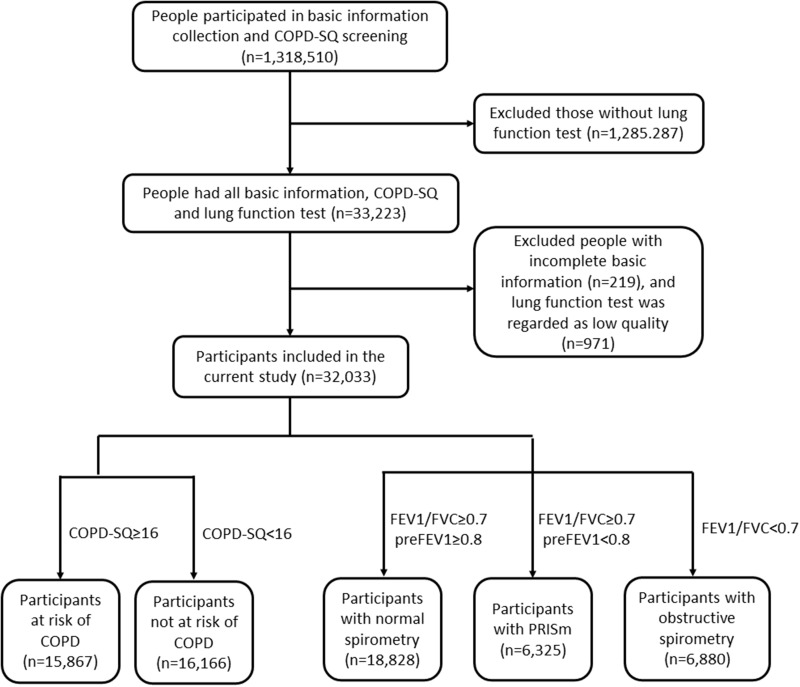
In the current program, a total of 1,318,510 individuals completed basic information and the COPD-SQ, and 33,223 of these completed lung function test. After removing 1190 participants because of incomplete information and low qualifying lung function, 32,033 subjects were finally enrolled in the current analysis (Figure 1). The mean age of included participants was 58.75 years old, and 55.57% were males.
Figure 1.
Flow chart for participants included in the analysis of relationship between BMI and lung function.
People at risk of COPD were more likely to be elderly, male, have a relatively low BMI, and be a smoker (p<0.001). Compared with people with normal spirometry, those with PRISm were older, more likely to be male and a smoker (p<0.001), but had a similar BMI (p=0.495). People with obstructive spirometry were older, more likely to be male, more likely to be a smoker, and had a lower BMI (p<0.001), when taking those with normal spirometry as a reference group. The specific characteristics of the current study are shown in Table 1. We further compared the characteristics of participants with combined at risk and preserved ratio spirometry and the group not at risk with normal spirometry, as shown in Supplement Table 1. People with combined at risk and PRISm were more likely to be elderly, male, and a smoker (p<0.001), and had higher COPD-SQ scores, a lower BMI, more symptoms, a family history, and reduced lung function (p<0.001).
Table 1.
Characteristics of Participants Included in the Analysis of Relationship Between BMI and Lung Function
| Total | Not at Risk | At Risk | p value (at Risk vs Not at Risk) | Normal Spirometry | PRISm | Obstructive Spirometry | p value (PRISm vs Normal) | p value (Obstructive vs Normal) | |
|---|---|---|---|---|---|---|---|---|---|
| Number (n) | 32,033 | 16,166 | 15,867 | 18,828 | 6325 | 6880 | |||
| Age (year, mean±SD) | 58.75±12.04 | 52.35±11.29 | 65.27±8.87 | <0.001 | 56.83±12.06 | 58.54±12.10 | 64.20±10.15 | <0.001 | <0.001 |
| Sex | <0.001 | <0.001 | <0.001 | ||||||
| Male (n, %) | 17,802 (55.57%) | 7510 (46.46%) | 10,292 (64.86%) | 9422 (50.04%) | 3655 (57.79%) | 4725 (68.68%) | |||
| Female (n, %) | 14,231 (44.43%) | 8656 (53.54%) | 5575 (35.14%) | 9406 (49.96%) | 2670 (42.21%) | 2155 (31.32%) | |||
| BMI (kg/m2, mean±SD) | 24.11±3.50 | 24.92±3.38 | 23.30±3.43 | <0.001 | 24.38±3.37 | 24.41±3.56 | 23.12±3.62 | 0.495 | <0.001 |
| Smoker (n, %) | 13,914 (43.44%) | 4649 (28.76%) | 9265 (58.39%) | <0.001 | 7263 (38.58%) | 2774 (43.86%) | 3877 (56.35%) | <0.001 | <0.001 |
| Symptom | |||||||||
| Cough (n, %) | 10,406 (32.49%) | 2007 (12.41%) | 8399 (52.93%) | <0.001 | 5233 (27.79%) | 2033 (32.14%) | 3140 (45.64) | <0.001 | <0.001 |
| Breathlessness (n, %) | 15,926 (49.72%) | 4622 (28.59%) | 11,304 (71.24%) | <0.001 | 7979 (42.38%) | 3272 (51.73%) | 4675 (67.95%) | <0.001 | <0.001 |
| Biofuel exposure (n, %) | 19,570 (61.09%) | 9836 (60.84%) | 9734 (61.35%) | 0.355 | 11,646 (61.85%) | 3927 (62.09%) | 3997 (58.10%) | 0.742 | <0.001 |
| Family history (n, %) | 6384 (19.93%) | 1817 (11.24%) | 4567 (28.78%) | <0.001 | 3452 (18.33%) | 1296 (20.49%) | 1636 (23.78%) | <0.001 | <0.001 |
| COPD-SQ (score, mean±SD) | 14.81±6.88 | 9.28±3.95 | 20.44±4.08 | <0.001 | 13.22±6.29 | 14.66±6.59 | 19.30±6.69 | <0.001 | <0.001 |
| Lung function | |||||||||
| FEV1 (l, mean±SD) | 2.25±0.82 | 2.49±0.77 | 2.00±0.80 | <0.001 | 2.65±0.67 | 1.81±0.56 | 1.54±0.73 | <0.001 | <0.001 |
| FVC (l, mean±SD) | 2.93±2.07 | 3.13±0.95 | 2.72±2.77 | <0.001 | 3.26±0.82 | 2.27±0.73 | 2.62±4.11 | <0.001 | <0.001 |
| FEV1/FVC (mean±SD) | 0.77±0.12 | 0.80±0.09 | 0.73±0.14 | <0.001 | 0.81±0.06 | 0.81±0.10 | 0.59±0.09 | 0.003 | <0.001 |
Abbreviations: BMI, body mass index; COPD-SQ, the COPD Screening Questionnaire; COPD, Chronic Obstructive Pulmonary Disease; PRISm preserved ratio impaired spirometry; FEV1, the predicted forced expiratory volume in one second; FVC, the Predicted forced vital capacity.
The Relationship Between BMI and Pulmonary Lung Function
Grouping by COPD-SQ
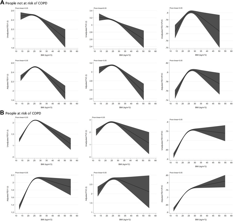
Based on COPD-SQ scores, subjects were divided into two groups: those not at risk of COPD (n=16,166) and those at risk of COPD (n=15,867). Of people not at risk of COPD, compared with those with a normal BMI, those underweight (−0.49 L, 95% CI: −0.68 to −0.29), obese (−0.07 L, 95% CI: −0.15 to 0.00), or severely obese (−0.42 L, 95% CI: −0.76 to −0.07) had a lower FEV1, and those underweight (−0.26 L, 95% CI: −0.43 to −0.09), overweight (−0.04 L, 95% CI: −0.09 to 0.00), and obese (−0.15 L, 95% CI: −0.22 to −0.09) had a lower FVC. In analysis of FEV1/FVC, underweight people had worse FEV1/FVC (−2.97, 95% CI: −4.11 to −1.84) compared to those with normal weight, but overweight and obese patients had better results (overweight: 1.22, 95% CI: 0.83 to 1.61; obesity: 1.21, 95% CI: 0.70 to 1.72; Table 2). After adjusting for related variables, non-linear analysis showed an inverse U-shaped curve between BMI and FEV1 and FVC, and an upward curving arc between BMI and FEV1/FVC (Figure 2A). By performing the analyses according to gender, we found more obviously significant results in females than males (Supplement Tables 2 and 3, Supplement Figures 1 and 2).
Table 2.
The Relationship Between BMI and Lung Function According to COPD-SQ Scores
| Participants | BMI | FEV1 | FVC | FEV1/FVC | |||
|---|---|---|---|---|---|---|---|
| Unadjusted | Adjusted | Unadjusted | Adjusted | Unadjusted | Adjusted | ||
| People not at risk of COPD | BMI<18.5 | −0.10 (−0.25, 0.06) | −0.49 (−0.68, −0.29) | −0.01 (−0.13, 0.11) | −0.26 (−0.43, −0.09) | −2.36 (−3.50, −1.22) | −2.97 (−4.11, −1.84) |
| 18.5≤BMI<23.9 | Ref | Ref | Ref | Ref | Ref | Ref | |
| 24.0≤BMI<27.9 | −0.06 (−0.10, −0.02) | 0.04 (−0.02, 0.10) | −0.06 (−0.10, −0.03) | −0.04 (−0.09, 0.00) | 0.30 (−0.07, 0.68) | 1.22 (0.83, 1.61) | |
| 28.0≤BMI<34.9 | −0.15 (−0.21, −0.09) | −0.07 (−0.15, 0.00) | −0.14 (−0.19, −0.09) | −0.15 (−0.22, −0.09) | 0.17 (−0.32, 0.67) | 1.21 (0.70, 1.72) | |
| BMI≥35.0 | −0.38 (−0.67, −0.10) | −0.42 (−0.76, −0.07) | −0.25 (−0.48, −0.01) | −0.29 (−0.59, 0.01) | −0.45 (−2.69, 1.79) | 0.32 (−1.96, 2.60) | |
| People at risk of COPD | BMI<18.5 | −0.59 (−0.67, −0.51) | −0.63 (−0.73, −0.54) | −0.45 (−0.52, −0.38) | −0.51 (−0.59, −0.43) | −2.38 (−2.82, −1.95) | −2.47 (−2.94, −2.01) |
| 18.5≤BMI<23.9 | Ref | Ref | Ref | Ref | Ref | Ref | |
| 24.0≤BMI<27.9 | 0.13 (0.08, 0.17) | 0.33 (0.27, 0.38) | 0.00 (−0.01, 0.01) | 0.01 (−0.01, 0.03) | 1.34 (1.07, 1.62) | 2.23 (1.93, 2.53) | |
| 28.0≤BMI<34.9 | 0.04 (−0.04, 0.11) | 0.31 (0.22, 0.39) | −0.09 (−0.15, −0.03) | 0.01 (−0.02, 0.03) | 1.69 (1.26, 2.13) | 2.70 (2.24, 3.15) | |
| BMI≥35.0 | −0.30 (−0.69, 0.09) | 0.09 (−0.35, 0.53) | −0.39 (−0.72, −0.05) | −0.15 (−0.54, 0.24) | 1.08 (−1.22, 3.38) | 2.28 (−0.03, 4.59) | |
Notes: Adjusted model: sex, age, smoke, cough, breath shortness, biofuel use and family history.
Abbreviations: BMI, body mass index; COPD-SQ, the COPD Screening Questionnaire; COPD, Chronic Obstructive Pulmonary Disease; FEV1, the predicted forced expiratory volume in one second; FVC, the Predicted forced vital capacity.
Figure 2.
The relationship between BMI and lung function according to COPD-SQ scores. (A) The relationship between BMI and lung function in people at risk of COPD. (B) The relationship between BMI and lung function in people not at risk of COPD.
Underweight people at risk of COPD had a significantly lower lung function as evaluated using FEV1 (−0.63 L, 95% CI: −0.73 to −0.54), FVC (−0.51 L, 95% CI: −0.59 to −0.38), and FEV1/FVC (−2.47, 95% CI: −2.94 to −2.01) compared to those with normal weight. After adjusting for covariables, overweight and obese at risk participants had better FEV1 (overweight: 0.33 L, 95% CI: 0.27 to 0.38; obesity: 0.31 L, 95% CI: 0.22 to 0.39) and FEV1/FVC (overweight: 2.23, 95% CI: 1.93 to 2.53; obesity: 2.70, 95% CI: 2.24 to 3.15) values than the normal BMI group. No difference in FVC existed among normal BMI, overweight, obese, and severely obese patients at risk of COPD (Table 2B). By using an adjusted model in non-linear analyses, an inverse U-shaped curve was shown between BMI and FEV1 and FVC, and an upward curving arc was shown between BMI and FEV1/FVC (Figure 2). Further analysis by gender revealed a similar relationship of BMI and lung function in both females and males (Supplement Tables 2 and 3, Supplement Figures 1 and 2).
Grouping by Lung Function
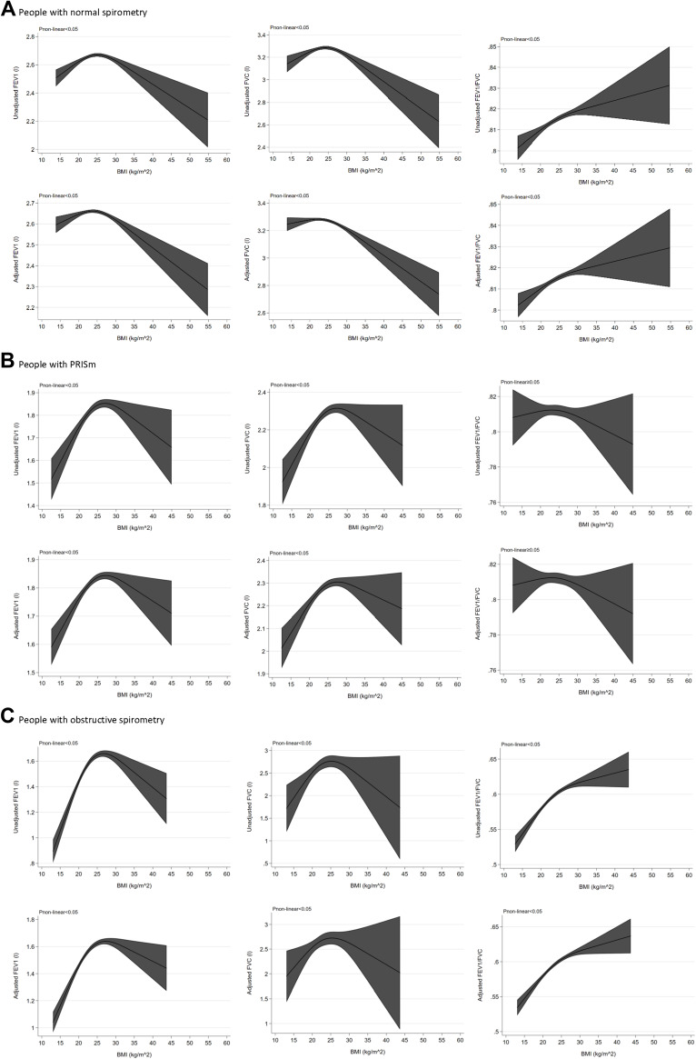
According to lung function test results, participants were divided into three groups: normal spirometry (n=18,828), PRISm (n=6325), and obstructive spirometry (n=6880). First, we regarded BMI as the categorical variable and regarded normal BMI as the reference group. Among people with normal spirometry, those underweight had a lower FEV1 (−0.24 L, 95% CI: −0.44 to −0.04) and similar FVC (−0.17 L, 95% CI: −0.33 to 0.01) and FFEV1/FVC (−1.79, 95% CI: −2.18, 0.60) after adjusting for related covariables. Obese and severely obese subjects had a lower FEV1 (obesity: −0.22 L, 95% CI: −0.33 to −0.11; severe obesity: −0.97 L, 95% CI: −1.60 to −0.33) and FVC (obesity: −0.26 L, 95% CI: −0.34 to −0.17; severe obesity: −0.76 L, 95% CI: −1.25 to −0.28), but overweight participants showed no difference of FEV1 (0.01 L, 95% CI: −0.06 to 0.08) and FVC (−0.05 L, 95% CI: −0.11 to 0.01) compared to a normal BMI group. Overweight and obese participants appeared to have higher FEV1/FVC value than normal BMI participants (overweight: 1.06, 95% CI: 0.56 to 1.55; obesity: 1.29, 95% CI: 0.59 to 1.98; Table 3). The results showed a non-linear relationship between BMI and lung function in healthy participants. We further regarded BMI as a continuous variable, and performed a restricted cubic spline. Inverse U-shaped curves were found in the relationship between BMI and FEV1 and FVC, and an upward curving arc was found between BMI and FEV1/FVC (Figure 3A). Further analysis showed these results were more obvious in females than males (Supplement Tables 4 and 5, Supplement Figures 3 and 4).
Table 3.
The Relationship Between BMI and Lung Function According to Lung Function Test
| Participants | BMI | FEV1 | FVC | FEV1/FVC | |||
|---|---|---|---|---|---|---|---|
| Unadjusted | Adjusted | Unadjusted | Adjusted | Unadjusted | Adjusted | ||
| People with normal spirometry | BMI<18.5 | −0.25 (−0.38, −0.12) | −0.24 (−0.44, −0.04) | −0.19 (−0.30, −0.08) | −0.17 (−0.33, 0.01) | −1.03 (−2.42, 0.37) | −1.79 (−2.18, 0.60) |
| 18.5≤BMI<23.9 | Ref | Ref | Ref | Ref | Ref | Ref | |
| 24.0≤BMI<27.9 | 0.06 (0.01, 0.10) | 0.01 (−0.06, 0.08) | 0.02 (−0.02, 0.06) | −0.05 (−0.11, 0.01) | 1.04 (0.55, 1.52) | 1.06 (0.56, 1.55) | |
| 28.0≤BMI<34.9 | −0.04 (−0.11, 0.03) | −0.22 (−0.33, −0.11) | −0.08 (−0.13, −0.02) | −0.26 (−0.34, −0.17) | 1.40 (0.72, 2.08) | 1.29 (0.59, 1.98) | |
| BMI≥35.0 | −0.56 (−0.95, −0.18) | −0.97 (−1.60, −0.33) | −0.49 (−0.80, −0.18) | −0.76 (−1.25, −0.28) | 1.67 (−1.71, 5.05) | 1.22 (−2.32, 4.76) | |
| People with PRISm | BMI<18.5 | −0.50 (−0.73, −0.27) | −0.56 (−0.86, −0.26) | −0.33 (−0.50, −0.15) | −0.33 (−0.55, −0.11) | −0.12 (−1.49, 1.25) | −0.03 (−1.34, 1.28) |
| 18.5≤BMI<23.9 | Ref | Ref | Ref | Ref | Ref | Ref | |
| 24.0≤BMI<27.9 | 0.13 (0.03, 0.23) | 0.22 (0.08, 0.36) | 0.09 (0.01, 0.16) | 0.12 (0.02, 0.22) | 0.14 (−0.42, 0.70) | 0.17 (−0.40, 0.73) | |
| 28.0≤BMI<34.9 | 0.10 (−0.04, 0.23) | 0.22 (0.02, 0.42) | 0.08 (−0.02, 0.18) | 0.16 (0.02, 0.30) | −0.35 (−1.18, 0.49) | −0.38 (−1.23, 0.47) | |
| BMI≥35.0 | 0.27 (−0.34, 0.88) | 0.18 (−0.70, 1.05) | 0.25 (−0.22, 0.72) | 0.21 (−0.43, 0.85) | −1.33 (−5.50, 2.85) | −1.27 (−5.65, 3.10) | |
| People with obstructive spirometry | BMI<18.5 | −0.76 (−0.91, −0.61) | −0.80 (−0.97, −0.63) | −0.50 (−0.59, −0.40) | −0.53 (−0.64, −0.42) | −2.84 (−3.65, −2.03) | −2.75 (−3.60, −1.90) |
| 18.5≤BMI<23.9 | Ref | Ref | Ref | Ref | Ref | Ref | |
| 24.0≤BMI<27.9 | 0.21 (0.14, 0.28) | 0.27 (0.18, 0.36) | 0.00 (−0.01, 0.01) | 0.00 (−0.01, 0.01) | 2.38 (1.74, 3.01) | 2.44 (1.78, 3.11) | |
| 28.0≤BMI<34.9 | 0.17 (0.06, 0.29) | 0.26 (0.12, 0.40) | 0.00 (−0.04, 0.03) | 0.00 (−0.03, 0.03) | 3.80 (2.71, 4.88) | 3.90 (2.77, 5.03) | |
| BMI≥35.0 | 0.32 (−0.17, 0.82) | 0.51 (−0.08, 1.11) | 0.00 (−0.05, 0.06) | 0.01 (−0.04, 0.06) | 7.41 (1.43, 13.39) | 8.19 (2.11, 14.26) | |
Notes: Adjusted model: sex, age, smoke, cough, breath shortness, biofuel use and family history.
Abbreviations: BMI, body mass index; COPD-SQ, the COPD Screening Questionnaire; COPD, Chronic Obstructive Pulmonary Disease; PRISm preserved ratio impaired spirometry; FEV1, the predicted forced expiratory volume in one second; FVC, the Predicted forced vital capacity.
Figure 3.
The relationship between BMI and lung function according to lung function test. (A) The relationship between BMI and lung function in people with normal spirometry. (B) The relationship between BMI and lung function in people with PRISm. (C) The relationship between BMI and lung function in people with obstructive spirometry.
In people with PRISm, underweight participants had a lower FEV1 (−0.56 L, 95% CI: −0.86 to −0.26) and FVC (−0.33 L, 95% CI: −0.55 to −0.11) than those with normal weight, after adjusting for related covariables. Overweight and obese participants were shown to have a higher value of FEV1 (overweight: 0.22 L, 95% CI: 0.08 to 0.36; obesity: 0.22 L, 95% CI: 0.02 to 0.42) and FVC (overweight: 0.12 L, 95% CI: 0.02 to 0.22; obesity: 0.16 L, 95% CI: 0.02 to 0.30) than normal weight participants. No difference of FEV1 (0.18 L, 95% CI: −0.70 to 1.05) and FVC (0.21 L, 95% CI: −0.43 to 0.85) existed in those with severe obesity or a normal BMI. Analyses indicated similar FEV1/FVC ratios among underweight, normal BMI, overweight, obese, and severely obese participants (Table 3). Further non-linear analyses showed an inverse U-shaped relationship between BMI and lung function in PRISm participants (Figure 3B). We did analyses by gender, and obesity seemed to be a protective factor for lung function in males with PRISm but not females (Supplement Tables 4 and 5, Supplement Figures 3 and 4).
Underweight people with obstructive spirometry had a lower FEV1 (−0.80 L, 95% CI: −0.97 to −0.63), FVC (−0.53 L, 95% CI: −0.64 to −0.42), and FEV1/FVC (−2.75, 95% CI: −3.60 to −1.90) than normal BMI participants, whether adjusting for covariates or not. Taking normal BMI as the reference group, better FEV1 (overweight: 0.27 L, 95% CI: 0.18 to 0.36; obesity: 0.26 L, 95% CI: 0.12 to 0.40) was shown among overweight and obese participants with obstructive spirometry, and better FEV1/FVC was shown among overweight, obese, and severely obese participants (overweight: 2.44, 95% CI: 1.78 to 3.11; obesity: 3.90, 95% CI: 2.77 to 5.03; severe obesity: 8.19, 95% CI: 2.11 to 14.26), in both unadjusted and adjusted models. However, it seemed no difference existed in FVC among overweight, obese, severely obese, and normal BMI participants (Table 3). An inverse U-shaped relationship was found in non-linear analysis between BMI and FEV1 and FVC, and an upward curving arc was found between BMI and FEV1/FVC (Figure 3C). Similar significant results were shown both in males and females with obstructive spirometry (Supplement Tables 4 and 5, Supplement Figures 3 and 4).
Sensitivity Analysis
In order to evaluate for potential factors of heterogeneity, we performed a sensitivity analysis among participants aged more than 40 years and non-smokers. Among participants aged more than 40 years, similar results were found in both categorical and continuous analysis of the relationship between BMI and lung function, and specific information is shown in Supplement Tables 6 and 7. In the analysis of non-smokers, the effect of BMI on lung function seemed to reduce, especially in males with normal spirometry and people with PRISm, where no significant relationship existed between BMI and lung function. Other results remained less marked but similar with the former analyses (Supplement Tables 8 and 9). Further, we used predicted values to repeat the analysis and the results were consistent with previous analyses (Supplement Tables 10 and 11).
Discussion
Our study is unique in that the current analysis demonstrated for the first time the correlation between BMI and lung function among people at risk of COPD and those with PRISm using a large population-based program. Some initial notable findings include: (1) an inverted U-shaped relationship between BMI and lung function among people with normal spirometry and those not at risk of COPD, and the phenomenon was more obvious in females than males; (2) being underweight was shown to be a risk factor for impaired lung function in all participants, and severe obesity was a risk factor for FEV1 and FVC, especially in people not at risk of COPD and those with normal spirometry; (3) moderate obesity appeared to be protective factor in people at risk of COPD and those with PRISm or obstructive spirometry, and the effect was shown more strongly in males than females; and (4) no significant relationship existed between BMI and lung function among non-smokers with PRISm.
Comparison with Other Studies
Former studies have found complex relationships between BMI and lung function, and the results were unclear, with one study giving an inverted U-shaped correlation,17 some reporting a negative,19,28,29 none, or even positive associations.16,18 However, these studies were performed in different races and most were in healthy elderly participants. A Chinese study including 8284 general adults found obesity to be negatively associated with lung function, which was more evident in women than in men.30 The study only focused on obesity, not underweight participants. In our current study, we included a large sample size with a wide age range from a Chinese population. Furthermore, we differentiated between people with normal and abnormal lung function, and found different results. For people with normal spirometry not at risk of COPD, the relationship between BMI and lung function was an inverted U-shaped, and normal BMI was associated with the best lung function testing performance. The phenomenon was more obvious in females than males, which supports the findings of previous studies.15,31 Analysis of people with abnormal lung function and those at risk of COPD showed a similar inverted U-shaped relationship between BMI and lung function, but the best lung function was performed in participants with moderate obesity. Thus, evidence indicates that moderate obesity could be a protective factor.
An accumulating body of evidence paradoxically links increasing body mass index with a better prognosis, and this phenomenon is termed the “obesity paradox”.32 The “obesity paradox” has been found in many diseases, including cardiovascular disease,33–35 kidney disease,36 diabetes,37 chronic respiratory disease,32,38,39 osteoporosis,40 and cancer.41 Previous Chinese studies discovered that obesity only had a protective effect on lung function in COPD patients with Global Initiative for Chronic Obstructive Disease (GOLD) 3–4 grade rather than GOLD 1–2 grade.38,42 Thus, the “obesity paradox” was found to exist among COPD patients, but how about people with PRISm and at risk of COPD? In this study, we conducted further analysis in these two population groups and found obesity to be a protective factor for lung function. Interestingly, further analysis in different sexes showed that the “obesity paradox” was more apparent in males than females, which is different from healthy populations. The underlying mechanisms driving these changes require more research.
Notably, being underweight has been shown to be a risk factor for low lung function. A former cross-sectional study with a small sample size conducted in China found low BMI (<18.5 kg/m2) was associated with a lower FEV1 and FVC, compared to those with a normal BMI (18.5 kg/m2≤BMI≤24 kg/m2).43 The same finding was found in economically developed regions, where a cross-sectional study of healthy Korean population showed that low body weight was associated with lower FEV1 and FVC, but not with FEV1/FVC.44 Our study extended the findings to show that being underweight is associated with low lung function in people with abnormal lung function and those at risk of COPD. In the current analysis, severe obesity was shown to be a risk factor for low lung function, measured by FEV1 and FVC, especially in people not at risk of COPD and those with normal spirometry. One study focused on obese Chinese elderly participants and found being overweight and obese were two risk factors for a lower FVC.31 Similar views were found in other studies.45,46 The association between obesity and FEV1/FVC remains conflicting. Some studies indicated a positive47–51 or negative52 association between obesity and FEV1/FVC. However, in our analysis no relationship was found between severe obesity and FEV1/FVC, except in those with obstructive spirometry. Similar results were found in other studies. Age and smoking are two other factors that can affect lung function. We performed a sensitivity analysis limiting participants to those aged more than 40 years and non-smokers. All results were similar to the original analysis, except for non-smokers with PRISm, where no correlation existed between BMI and lung function. The results may be explained by different processes of metabolism and physiology between smokers and non-smokers in COPD,53 which remain to be fully elucidated.
Potential Mechanisms
In the healthy population, obesity has a negative impact on lung function, and severe obesity and being underweight have been shown to be risk factors in all participants. The inverse relation between obesity and respiratory function has been explained by various mechanisms, including the accumulation of fat in the mediastinum and the abdominal and thoracic cavities,54 insulin resistance,55,56 and low-grade chronic inflammation.57 The different distribution of fat in men and women may lead to differences in its effects on lung function.17 Most underweight participants had nutritional deficiency and dysplasia, and low muscle mass,58 which had adverse effects not only on lung function but also on whole-body health.59,60 Physical inactivity, which was more likely to relate to low muscle mass and strength, may be another explanation for the poor lung function observed in underweight participants.61 A former study of sedentary young females found that lean body mass is significantly correlated with low FVC and FEV1.62
The mechanisms underlying the obesity paradox remain unresolved. Some potential explanations are as follows. Exercise, which can increase muscle mass and lead to a higher BMI, has numerous important beneficial effects on whole-body health, including respiratory health. A previous study found that lung volume reduction surgery (LVRS) for emphysema significantly increases BMI through its association with positive changes in health status,63 so some researchers have speculated that the “obesity paradox” in COPD is related to a low-grade emphysema, rather than to excess weight.64 However, more studies are needed to further explore the “obesity paradox”.
Public Health Impact
Obesity is an emerging health problem with a prediction that obesity will affect 18% of men and 21% of women globally by 2025.60 Not only obesity, but also being underweight has been found to have a harmful impact on health and prognosis.65 The current analysis indicates that being underweight and obese are risk factors for lung function decline in the general population, which emphasizes the importance of maintaining a healthy BMI.
People with COPD-SQ scores more than 16 are at risk of COPD, and they need further examination to confirm the diagnosis in hospital. However, some at risk people with minor symptoms are unlikely to see doctors, and current medical resources limit lung function testing to the entire population. The current analysis indicates that underweight individuals had worse lung function when at risk of COPD. Thus, underweight people with COPD-SQ scores more than 16 should be paid more attention during screening.
Analysis of lung function trajectories in PRISm showed that if lung function recovers during early adulthood, a previous history of PRISm does not appear to affect long-term prognosis,66 which means that early intervention is sensible. For these people, it is particularly important to directly carry out effective lifestyle interventions, such as balanced nutrition and physical activity. According to our analysis, it is important for these people, including those at risk of COPD and with PRISm, to maintain a healthy BMI.
These findings imply that the public health of underweight and obese subjects should be an area of concern and the potential opportunities, which exist for prevention and early intervention,67,68 should be emphasized.
Strengths and Limitations
To our knowledge, this is the first study to focus on the relationship between BMI and lung function among people at risk of COPD and those with PRISm in a large Chinese population, which filled the gap in the related fields. However, some limitations remain. First of all, the study design was cross-sectional in nature due to the unfinished nature of the program, which had inherent limitations in relation to inferring causality. As the Enjoying Breathing Program is ongoing, we will continue to conduct longitudinal studies in the future to explore the cause-and-effect relationship between BMI and lung function. Second, post-bronchodilator spirometry is required in the current clinical definition of lung diseases, which is unavailable in this study. However, it has been shown that equal value exists in pre-bronchodilator measures and post-bronchodilator spirometry, similar to the prognostic value of health outcomes in the general population.69 Third, it was not possible to distinguish between abdominal and thoracic fat; hence, we are unable to determine whether the distribution of body fat affects respiratory function. Last, lung function is affected by many factors, including age, sex, height, and smoking status. Although we used the original values in the current study, we adjusted potential factors, performed the analyses in different genders, and conducted sensitivity analyses to mitigate the influence of these potential factors. We found similar results in all analysis, which corroborates the reliability of our study findings. We divided participants into three groups according to separate lung function thresholds, thus creating groups with different lung function. In each group, we found reliable results confirming that being underweight and obese were risk factors for impaired lung function, measured by FEV1 and FVC. Finally, due to the absence of comorbidity data, we did not include this potential factor in the analysis models.
Conclusion
According to our analysis, an inverted U-shaped relationship exists between BMI and lung function, measured by FEV1 and FVC in all participants. Being underweight is a significant risk factor for impaired lung function in all participants, and severe obesity is a significant risk factor of low FEV1 and FVC, especially in people not at risk of COPD and those with normal spirometry. Slight obesity may be a protective factor of lung function in people at risk of COPD and those with PRISm, while it is a negative factor in healthy populations. Thus, underweight people with COPD-SQ scores more than 16 should be given more attention during screening; maintaining a healthy weight and starting early interventions are important for public health, especially for those in the pre-clinical status of the disease. The mechanisms underlying the “obesity paradox” require further elucidation.
Funding Statement
This study was funded by the Science Fund of China-Japan Friendship Hospital (2019-1-QN-11) to Ke Huang, the Chinese Academy of Medical Sciences (CAMS) Innovation Fund for Medical Science (CIFMS) (2021-I2M-1-049) to Ting Yang and the National Natural Science Foundation of China (82100044) to Wei Li.
Abbreviations
BMI, body mass index; COPD-SQ, the COPD Screening Questionnaire; COPD, Chronic Obstructive Pulmonary Disease; PRISm preserved ratio impaired spirometry; preFEV1, the predicted forced expiratory volume in one second; FEV1, the forced expiratory volume in one second; FVC, the forced vital capacity; preFVC, the predicted forced vital capacity; FEV1/FVC, the ratio of FEV1 to FVC; AUC, area under the curve; GOLD, Global Initiative for Chronic Obstructive Disease.
Data Sharing Statement
The Enjoying Breathing Program will last for 10 years and is not fully completed at present. Our present study is part of this project, and only focuses on the relationship between BMI and lung function among different characteristic participants. Thus, the data is not available to the public currently. If someone would like details about the data, they can contact the corresponding authors.
Ethics Approval and Informed Consent
This national clinical study was registered in March 2020 (Clinical Trials ID: NCT04318912). Ethical approval was granted by the China-Japan Friendship Hospital (approval number 2019-41-k29). This study was conducted in accordance with the ethical principles of the Declaration of Helsinki. All participants included in this study were informed about the study and signed the informed consent.
Author Contributions
All authors made a significant contribution to study design, execution, acquisition of data, analysis and interpretation of data; took part in drafting the article or revising it critically for important intellectual content; agreed to submit to the current journal; gave final approval of the version to be published; and agree to be accountable for all aspects of the work.
Disclosure
The authors report no conflicts of interest in this work.
References
- 1.Wang C, Xu J, Yang L., et al. Prevalence and risk factors of chronic obstructive pulmonary disease in China (the China Pulmonary Health [CPH] study): a national cross-sectional study. Lancet. 2018;391(10131):1706–1717. doi: 10.1016/S0140-6736(18)30841-9 [DOI] [PubMed] [Google Scholar]
- 2.Spyratos D, Haidich AB, Chloros D, Michalopoulou D, Sichletidis L. Comparison of three screening questionnaires for chronic obstructive pulmonary disease in the primary care. Respiration. 2017;93(2):83–89. doi: 10.1159/000453586 [DOI] [PubMed] [Google Scholar]
- 3.Zhou YM, Chen SY, Tian J, et al. Development and validation of a chronic obstructive pulmonary disease screening questionnaire in China. Int J Tuberc Lung Dis. 2013;17(12):1645–1651. doi: 10.5588/ijtld.12.0995 [DOI] [PubMed] [Google Scholar]
- 4.Zhang Q, Wang M, Li X, Wang H, Wang J. Do symptom-based questions help screen COPD among Chinese populations? Sci Rep. 2016;6:30419. doi: 10.1038/srep30419 [DOI] [PMC free article] [PubMed] [Google Scholar]
- 5.Martinez FJ, Mannino D, Leidy NK, et al. A new approach for identifying patients with undiagnosed chronic obstructive pulmonary disease. Am J Respir Crit Care Med. 2017;195(6):748–756. doi: 10.1164/rccm.201603-0622OC [DOI] [PMC free article] [PubMed] [Google Scholar]
- 6.Gu Y, Zhang Y, Wen Q, et al. Performance of COPD population screener questionnaire in COPD screening: a validation study and meta-analysis. Ann Med. 2021;53(1):1198–1206. doi: 10.1080/07853890.2021.1949486 [DOI] [PMC free article] [PubMed] [Google Scholar]
- 7.Guirguis-Blake JM, Senger CA, Webber EM, Mularski RA, Whitlock EP. Screening for chronic obstructive pulmonary disease: evidence report and systematic review for the US Preventive Services Task Force. JAMA. 2016;315(13):1378–1393. doi: 10.1001/jama.2016.2654 [DOI] [PubMed] [Google Scholar]
- 8.Wan ES, Castaldi PJ, Cho MH, et al. Epidemiology, genetics, and subtyping of preserved ratio impaired spirometry (PRISM) in COPDGene. Respir Res. 2014;15:89. doi: 10.1186/s12931-014-0089-y [DOI] [PMC free article] [PubMed] [Google Scholar]
- 9.Higbee DH, Granell R, Davey Smith G, Dodd JW. Prevalence, risk factors, and clinical implications of preserved ratio impaired spirometry: a UK Biobank cohort analysis. Lancet Respir Med. 2022;10(2):149–157. doi: 10.1016/S2213-2600(21)00369-6 [DOI] [PubMed] [Google Scholar]
- 10.Wan ES, Fortis S, Regan EA, et al. Longitudinal phenotypes and mortality in preserved ratio impaired spirometry in the COPDGene study. Am J Respir Crit Care Med. 2018;198(11):1397–1405. doi: 10.1164/rccm.201804-0663OC [DOI] [PMC free article] [PubMed] [Google Scholar]
- 11.Jankowich M, Elston B, Liu Q, et al. Restrictive spirometry pattern, cardiac structure and function, and incident heart failure in African Americans. The Jackson Heart study. Ann Am Thorac Soc. 2018;15(10):1186–1196. doi: 10.1513/AnnalsATS.201803-184OC [DOI] [PMC free article] [PubMed] [Google Scholar]
- 12.Wijnant SRA, De Roos E, Kavousi M, et al. Trajectory and mortality of preserved ratio impaired spirometry: the Rotterdam Study. Eur Respir J. 2020;55(1):1901217. doi: 10.1183/13993003.01217-2019 [DOI] [PubMed] [Google Scholar]
- 13.Zhou Y, Zhong NS, Li X, et al. Tiotropium in early-stage chronic obstructive pulmonary disease. N Engl J Med. 2017;377(10):923–935. doi: 10.1056/NEJMoa1700228 [DOI] [PubMed] [Google Scholar]
- 14.Ng M, Fleming T, Robinson M, et al. Global, regional, and national prevalence of overweight and obesity in children and adults during 1980-2013: a systematic analysis for the Global Burden of Disease Study 2013. Lancet. 2014;384(9945):766–781. doi: 10.1016/S0140-6736(14)60460-8 [DOI] [PMC free article] [PubMed] [Google Scholar]
- 15.Sutherland TJ, Goulding A, Grant AM, et al. The effect of adiposity measured by dual-energy X-ray absorptiometry on lung function. Eur Respir J. 2008;32(1):85–91. doi: 10.1183/09031936.00112407 [DOI] [PubMed] [Google Scholar]
- 16.Lee CM, Huxley RR, Wildman RP, Woodward M. Indices of abdominal obesity are better discriminators of cardiovascular risk factors than BMI: a meta-analysis. J Clin Epidemiol. 2008;61(7):646–653. doi: 10.1016/j.jclinepi.2007.08.012 [DOI] [PubMed] [Google Scholar]
- 17.Leone N, Courbon D, Thomas F, et al. Lung function impairment and metabolic syndrome: the critical role of abdominal obesity. Am J Respir Crit Care Med. 2009;179(6):509–516. doi: 10.1164/rccm.200807-1195OC [DOI] [PubMed] [Google Scholar]
- 18.Çolak Y, Marott JL, Vestbo J, Lange P. Overweight and obesity may lead to under-diagnosis of airflow limitation: findings from the Copenhagen City Heart Study. COPD. 2015;12(1):5–13. doi: 10.3109/15412555.2014.933955 [DOI] [PubMed] [Google Scholar]
- 19.Santana H, Zoico E, Turcato E, et al. Relation between body composition, fat distribution, and lung function in elderly men. Am J Clin Nutr. 2001;73(4):827–831. doi: 10.1093/ajcn/73.4.827 [DOI] [PubMed] [Google Scholar]
- 20.Svartengren M, Cai GH, Malinovschi A, et al. The impact of body mass index, central obesity and physical activity on lung function: results of the EpiHealth study. ERJ Open Res. 2020;6(4):00214–2020. doi: 10.1183/23120541.00214-2020 [DOI] [PMC free article] [PubMed] [Google Scholar]
- 21.Batty GD, Gale CR, Kivimäki M, Deary IJ, Bell S. Comparison of risk factor associations in UK Biobank against representative, general population based studies with conventional response rates: prospective cohort study and individual participant meta-analysis. BMJ. 2020;368:m131. doi: 10.1136/bmj.m131 [DOI] [PMC free article] [PubMed] [Google Scholar]
- 22.Lundbäck B, Backman H, Calverley PMA. Lung function through the PRISM. Spreading light or creating confusion? Am J Respir Crit Care Med. 2018;198(11):1358–1360. doi: 10.1164/rccm.201806-1163ED [DOI] [PubMed] [Google Scholar]
- 23.Sood A, Petersen H, Qualls C, et al. Spirometric variability in smokers: transitions in COPD diagnosis in a five-year longitudinal study. Respir Res. 2016;17(1):147. doi: 10.1186/s12931-016-0468-7 [DOI] [PMC free article] [PubMed] [Google Scholar]
- 24.Oelsner EC, Balte PP, Cassano PA, et al. Harmonization of respiratory data from 9 US population-based cohorts: the NHLBI pooled cohorts study. Am J Epidemiol. 2018;187(11):2265–2278. doi: 10.1093/aje/kwy139 [DOI] [PMC free article] [PubMed] [Google Scholar]
- 25.Halpin DMG, Criner GJ, Papi A, et al. Global initiative for the diagnosis, management, and prevention of chronic obstructive lung disease. The 2020 GOLD Science Committee Report on COVID-19 and Chronic Obstructive Pulmonary Disease. Am J Respir Crit Care Med. 2021;203(1):24–36. doi: 10.1164/rccm.202009-3533SO [DOI] [PMC free article] [PubMed] [Google Scholar]
- 26.Zhou BF. Cooperative Meta-Analysis Group of the Working Group on Obesity in China. Predictive values of body mass index and waist circumference for risk factors of certain related diseases in Chinese adults—study on optimal cut-off points of body mass index and waist circumference in Chinese adults. Biomed Environ Sci. 2002;15(1):83–96. [PubMed] [Google Scholar]
- 27.Jones RL, Nzekwu MM. The effects of body mass index on lung volumes. Chest. 2006;130(3):827–833. doi: 10.1378/chest.130.3.827 [DOI] [PubMed] [Google Scholar]
- 28.Canoy D, Luben R, Welch A, et al. Abdominal obesity and respiratory function in men and women in the EPIC-Norfolk Study, United Kingdom. Am J Epidemiol. 2004;159(12):1140–1149. doi: 10.1093/aje/kwh155 [DOI] [PubMed] [Google Scholar]
- 29.Wannamethee SG, Shaper AG, Whincup PH. Body fat distribution, body composition, and respiratory function in elderly men. Am J Clin Nutr. 2005;82(5):996–1003. doi: 10.1093/ajcn/82.5.996 [DOI] [PubMed] [Google Scholar]
- 30.Zeng X, Liu D, An Z, Li H, Song J, Wu W. Obesity parameters in relation to lung function levels in a large Chinese rural adult population. Epidemiol Health. 2021;43:e2021047. doi: 10.4178/epih.e2021047 [DOI] [PMC free article] [PubMed] [Google Scholar]
- 31.Pan J, Xu L, Lam TH, et al. Association of adiposity with pulmonary function in older Chinese: guangzhou biobank Cohort Study. Respir Med. 2017;132:102–108. doi: 10.1016/j.rmed.2017.10.003 [DOI] [PubMed] [Google Scholar]
- 32.McDonald VM, Wood LG, Holland AE, Gibson PG. Obesity in COPD: to treat or not to treat? Expert Rev Respir Med. 2017;11(2):81–83. doi: 10.1080/17476348.2017.1267570 [DOI] [PubMed] [Google Scholar]
- 33.Antonopoulos AS, Oikonomou EK, Antoniades C, Tousoulis D. From the BMI paradox to the obesity paradox: the obesity-mortality association in coronary heart disease. Obes Rev. 2016;17(10):989–1000. doi: 10.1111/obr.12440 [DOI] [PubMed] [Google Scholar]
- 34.Ortega FB, Lavie CJ, Blair SN. Obesity and cardiovascular disease. Circ Res. 2016;118(11):1752–1770. doi: 10.1161/CIRCRESAHA.115.306883 [DOI] [PubMed] [Google Scholar]
- 35.Doehner W, von Haehling S, Anker SD. Protective overweight in cardiovascular disease: moving from “paradox” to “paradigm”. Eur Heart J. 2015;36(40):2729–2732. doi: 10.1093/eurheartj/ehv414 [DOI] [PubMed] [Google Scholar]
- 36.Kalantar-Zadeh K, Abbott KC, Salahudeen AK, Kilpatrick RD, Horwich TB. Survival advantages of obesity in dialysis patients. Am J Clin Nutr. 2005;81(3):543–554. doi: 10.1093/ajcn/81.3.543 [DOI] [PubMed] [Google Scholar]
- 37.Gravina G, Ferrari F, Nebbiai G. The obesity paradox and diabetes. Eat Weight Disord. 2021;26(4):1057–1068. doi: 10.1007/s40519-020-01015-1 [DOI] [PubMed] [Google Scholar]
- 38.Zhu J, Zhao Z, Wu B, et al. Effect of body mass index on lung function in Chinese patients with chronic obstructive pulmonary disease: a multicenter cross-sectional study. Int J Chronic Obstruct Pulmon Dis. 2020;15:2477–2486. doi: 10.2147/COPD.S265676 [DOI] [PMC free article] [PubMed] [Google Scholar]
- 39.Galesanu RG, Bernard S, Marquis K, et al. Obesity in chronic obstructive pulmonary disease: is fatter really better? Can Respir J. 2014;21(5):297–301. doi: 10.1155/2014/181074 [DOI] [PMC free article] [PubMed] [Google Scholar]
- 40.Fassio A, Idolazzi L, Rossini M, et al. The obesity paradox and osteoporosis. Eat Weight Disord. 2018;23(3):293–302. doi: 10.1007/s40519-018-0505-2 [DOI] [PubMed] [Google Scholar]
- 41.Lennon H, Sperrin M, Badrick E, Renehan AG. The obesity paradox in cancer: a review. Curr Oncol Rep. 2016;18(9):56. doi: 10.1007/s11912-016-0539-4 [DOI] [PMC free article] [PubMed] [Google Scholar]
- 42.Wu Z, Yang D, Ge Z, Yan M, Wu N, Liu Y. Body mass index of patients with chronic obstructive pulmonary disease is associated with pulmonary function and exacerbations: a retrospective real world research. J Thorac Dis. 2018;10(8):5086–5099. doi: 10.21037/jtd.2018.08.67 [DOI] [PMC free article] [PubMed] [Google Scholar]
- 43.Wang S, Sun X, Hsia TC, Lin X, Li M. The effects of body mass index on spirometry tests among adults in Xi’an, China. Medicine. 2017;96(15):e6596. doi: 10.1097/MD.0000000000006596 [DOI] [PMC free article] [PubMed] [Google Scholar]
- 44.Do JG, Park CH, Lee YT, Yoon KJ. Association between underweight and pulmonary function in 282,135 healthy adults: a cross-sectional study in Korean population. Sci Rep. 2019;9(1):14308. doi: 10.1038/s41598-019-50488-3 [DOI] [PMC free article] [PubMed] [Google Scholar]
- 45.Melo SM, Melo VA, Melo EV, Menezes Filho RS, Castro VL, Barreto MS. Accelerated lung aging in patients with morbid obesity. J Bras Pneumol. 2010;36(6):746–752. doi: 10.1590/s1806-37132010000600012 [DOI] [PubMed] [Google Scholar]
- 46.Dupuy-McCauley KL, Novotny PJ, Benzo RP. Treating severe obesity to reduce dyspnea in patients with chronic lung disease: a pilot mixed methods study. Chest. 2020;158(3):1128–1131. doi: 10.1016/j.chest.2020.02.032 [DOI] [PMC free article] [PubMed] [Google Scholar]
- 47.Vatrella A, Calabrese C, Mattiello A, et al. Abdominal adiposity is an early marker of pulmonary function impairment: findings from a Mediterranean Italian female cohort. Nutr Metab Cardiovasc Dis. 2016;26(7):643–648. doi: 10.1016/j.numecd.2015.12.013 [DOI] [PubMed] [Google Scholar]
- 48.Fimognari FL, Pasqualetti P, Moro L, et al. The association between metabolic syndrome and restrictive ventilatory dysfunction in older persons. J Gerontol a Biol Sci Med Sci. 2007;62(7):760–765. doi: 10.1093/gerona/62.7.760 [DOI] [PubMed] [Google Scholar]
- 49.Peters U, Subramanian M, Chapman DG, et al. BMI but not central obesity predisposes to airway closure during bronchoconstriction. Respirology. Vic: Carlton Book Company. 2019;24(6):543–550. doi: 10.1111/resp.13478 [DOI] [PMC free article] [PubMed] [Google Scholar]
- 50.Lazarus R, Gore CJ, Booth M, Owen N. Effects of body composition and fat distribution on ventilatory function in adults. Am J Clin Nutr. 1998;68(1):35–41. doi: 10.1093/ajcn/68.1.35 [DOI] [PubMed] [Google Scholar]
- 51.Sin DD, Jones RL, Man SF. Obesity is a risk factor for dyspnea but not for airflow obstruction. Arch Intern Med. 2002;162(13):1477–1481. doi: 10.1001/archinte.162.13.1477 [DOI] [PubMed] [Google Scholar]
- 52.Sutherland TJ, McLachlan CR, Sears MR, Poulton R, Hancox RJ. The relationship between body fat and respiratory function in young adults. Eur Respir J. 2016;48(3):734–747. doi: 10.1183/13993003.02216-2015 [DOI] [PubMed] [Google Scholar]
- 53.Salvi SS, Barnes PJ. Chronic obstructive pulmonary disease in non-smokers. Lancet. 2009;374(9691):733–743. doi: 10.1016/S0140-6736(09)61303-9 [DOI] [PubMed] [Google Scholar]
- 54.Peters U, Suratt BT, Bates JHT, Dixon AE, Beyond BMI. obesity and lung Disease. Chest. 2018;153(3):702–709. doi: 10.1016/j.chest.2017.07.010 [DOI] [PMC free article] [PubMed] [Google Scholar]
- 55.Cardet JC, Ash S, Kusa T, Camargo CA, Israel E. Insulin resistance modifies the association between obesity and current asthma in adults. Eur Respir J. 2016;48(2):403–410. doi: 10.1183/13993003.00246-2016 [DOI] [PMC free article] [PubMed] [Google Scholar]
- 56.Lotta LA, Gulati P, Day FR, et al. Integrative genomic analysis implicates limited peripheral adipose storage capacity in the pathogenesis of human insulin resistance. Nat Genet. 2017;49(1):17–26. doi: 10.1038/ng.3714 [DOI] [PMC free article] [PubMed] [Google Scholar]
- 57.Lim S, Kwon SY, Yoon JW, et al. Association between body composition and pulmonary function in elderly people: the Korean Longitudinal Study on Health and Aging. Obesity. 2011;19(3):631–638. doi: 10.1038/oby.2010.167 [DOI] [PubMed] [Google Scholar]
- 58.Graf CE, Pichard C, Herrmann FR, Sieber CC, Zekry D, Genton L. Prevalence of low muscle mass according to body mass index in older adults. Nutrition. 2017;34:124–129. doi: 10.1016/j.nut.2016.10.002 [DOI] [PubMed] [Google Scholar]
- 59.Grigsby MR, Siddharthan T, Pollard SL, et al. Low body mass index is associated with higher odds of COPD and lower lung function in low- and middle-income countries. COPD. 2019;16(1):58–65. doi: 10.1080/15412555.2019.1589443 [DOI] [PMC free article] [PubMed] [Google Scholar]
- 60.NCD Risk Factor Collaboration (NCD-RisC). Trends in adult body-mass index in 200 countries from 1975 to 2014: a pooled analysis of 1698 population-based measurement studies with 19·2 million participants. Lancet. 2016;387(10026):1377–1396. doi: 10.1016/S0140-6736(16)30054-X [DOI] [PMC free article] [PubMed] [Google Scholar]
- 61.Pedersen BK, Saltin B. Exercise as medicine – evidence for prescribing exercise as therapy in 26 different chronic diseases. Scand J Med Sci Sports. 2015;25(suppl3):1–72. doi: 10.1111/sms.12581 [DOI] [PubMed] [Google Scholar]
- 62.Azad A, Zamani A. Lean body mass can predict lung function in underweight and normal weight sedentary female young adults. Tanaffos. 2014;13(2):20–26. [PMC free article] [PubMed] [Google Scholar]
- 63.Oey IF, Bal S, Spyt TJ, Morgan MD, Waller DA. The increase in body mass index observed after lung volume reduction may act as surrogate marker of improved health status. Respir Med. 2004;98(3):247–253. doi: 10.1016/j.rmed.2003.09.017 [DOI] [PubMed] [Google Scholar]
- 64.Spelta F, Fratta Pasini AM, Cazzoletti L, Ferrari M. Body weight and mortality in COPD: focus on the obesity paradox. Eat Weight Disord. 2018;23(1):15–22. doi: 10.1007/s40519-017-0456-z [DOI] [PubMed] [Google Scholar]
- 65.Ni YN, Luo J, Yu H, et al. Can body mass index predict clinical outcomes for patients with acute lung injury/acute respiratory distress syndrome? A meta-analysis. Crit Care. 2017;21(1):36. doi: 10.1186/s13054-017-1615-3 [DOI] [PMC free article] [PubMed] [Google Scholar]
- 66.Marott JL, Ingebrigtsen TS, Çolak Y, Vestbo J, Lange P. Trajectory of preserved ratio impaired spirometry: natural history and long-term prognosis. Am J Respir Crit Care Med. 2021;204(8):910–920. doi: 10.1164/rccm.202102-0517OC [DOI] [PubMed] [Google Scholar]
- 67.Vasquez MM, Zhou M, Hu C, Martinez FD, Guerra S. Low lung function in young adult life is associated with early mortality. Am J Respir Crit Care Med. 2017;195(10):1399–1401. doi: 10.1164/rccm.201608-1561LE [DOI] [PMC free article] [PubMed] [Google Scholar]
- 68.Bolton CE, Bush A, Hurst JR, Kotecha S, McGarvey L. Lung consequences in adults born prematurely. Postgrad Med J. 2015;91(1082):712–718. doi: 10.1136/postgradmedj-2014-206590rep [DOI] [PubMed] [Google Scholar]
- 69.Kato B, Gulsvik A, Vollmer W, et al. Can spirometric norms be set using pre- or post- bronchodilator test results in older people? Respir Res. 2012;13:102. doi: 10.1186/1465-9921-13-102 [DOI] [PMC free article] [PubMed] [Google Scholar]


