



Abstract
Despite the physiological significance of ESR2, a lack of well-validated detection systems for ESR2 proteins has hindered progress in ESR2 research. Thus, recent identification of a specific anti-human ESR2 monoclonal antibody (PPZ0506) and its specific cross-reactivity against mouse and rat ESR2 proteins heightened momenta toward development of appropriate immunohistochemical detection systems for rodent ESR2 proteins. Building upon our previous optimization of ESR2 immunohistochemical detection in rats using PPZ0506, in this study, we further aimed to optimize mouse-on-mouse immunohistochemical detection using PPZ0506. Our assessment of several staining conditions using paraffin-embedded ovary sections revealed that intense heat-induced antigen retrieval, appropriate blocking, and appropriate antibody dilutions were necessary for optimization of mouse-on-mouse immunohistochemistry. Subsequently, we applied the optimized immunostaining method to determine expression profiles of mouse ESR2 proteins in peripheral tissues and brain subregions. Our analyses revealed more localized distribution of mouse ESR2 proteins than previously assumed. Moreover, comparison of these results with those obtained in humans and rats using PPZ0506 revealed interspecies differences in ESR2 expression. We expect that our optimized methodology for immunohistochemical staining of mouse ESR2 proteins will help researchers to solve multiple lines of controversial evidence concerning ESR2 expression.
Keywords: ERβ, ESR2, estrogen receptor β, immunohistochemistry, PPZ0506
I. Introduction
Estrogens modulate a wide range of physiological processes across multiple organs via activation of two subtypes of nuclear estrogen receptors (ERs), ESR1 and ESR2 (conventionally called ERα and ERβ, respectively) [6, 11]. ERs are members of the nuclear hormone receptor superfamily and function as ligand-activated transcription factors. ESR1 and ESR2 reportedly exhibit distinct expression profiles. ESR1 displayed high expression in the reproductive organs and was widely distributed at various expression levels throughout nonreproductive organs [13, 18, 22]. Although ESR2 is thought to be distributed in both reproductive and nonreproductive organs, its detailed histological expression patterns have not been determined, mainly due to a lack of commercially available well-validated monoclonal antibodies with sufficient titer and specificity against ESR2.
Several attempts to develop and discover specific antibodies against ESR2 protein isoforms have been made to determine their expression and localization profiles [30, 37, 38, 43]. Recently, Andersson et al. [1] validated thirteen commercially available monoclonal anti-ESR2 antibodies and identified only one antibody with sufficient specificity for human ESR2 proteins (PPZ0506). Our previous studies [12, 17] confirmed its specific cross-reactivity against rodent ESR2 proteins and applicability for immunohistochemical detection in rats. Expression analyses of ESR2/Esr2 transcripts and ESR2 proteins revealed distinct expression profiles in humans and rats. Specifically, ESR2 transcripts and ESR2 proteins were highly expressed in the adrenal gland, immune system tissues, ovary, and testis, and widely distributed throughout the other organs in human [1, 17]. In contrast, Esr2/ESR2 were highly expressed in the ovary, prostate, and certain neuronal nuclei, but scarcely found in other organs of rats [12, 17].
Several lines of Esr2-knockout mice have been developed to determine the physiological functions of ESR2 [7, 23, 36, 40]. Disruption of ESR2 functions generally results in female fertility defects due to failure of ovulation [2, 7, 23]. Based on an assumption of wide distribution of mouse Esr2 transcripts and ESR2 proteins, phenotypes of Esr2-knockout mice have been evaluated in multiple mouse organs and tissues, such as adipose tissues [27], immune tissues [9, 19], lung [28, 33], mammary gland [4, 10, 40], prostate [16, 40, 42], and uterus [39, 41]. However, with the exception of ovary, prostate, and certain neuronal nuclei, the corresponding rat organs scarcely express rat Esr2 transcripts and ESR2 proteins [12, 17]. Given this difference from rats, histological analysis is needed to determine whether ESR2 proteins are expressed in a wide range of organs in mice, as assumed. Recently, a mouse line with complete deletion of all Esr2 exons was generated [40]. Antonson et al. [2] demonstrated that the PPZ0506 antibody was applicable for immunoblot detection of mouse ESR2 proteins in wild-type mice with appropriate negative references prepared from these all-Esr2-exon-deleted mice. As immunoreactive specificity of the PPZ0506 antibody against mouse ESR2 proteins was previously validated [2, 17], it would be worthwhile to optimize immunohistochemical methodology for mouse ESR2 proteins and determine organ-specific localization profiles using this well-validated antibody.
In the present study, we first optimized an immunostaining method for mouse ESR2 proteins using the PPZ0506 mouse monoclonal antibody on paraffin-embedded ovary sections in terms of antigen retrieval, blocking, and antibody titration. Subsequently, we examined the distribution profiles of mouse ESR2 proteins in respective organs and compared them with previously reported profiles of human and rat counterparts.
II. Materials and Methods
Animals
All experiments using animals in this study were approved by the Nippon Medical School Animal Care and Use Committee and conducted in compliance with institutional guidelines. C57BL/6 mice were purchased from Tokyo Laboratory Animals Science (Tokyo, Japan). Mice were housed in a temperature-controlled room (22–24°C) under a 14-hr light/10-hr dark cycle. Standard diet and tap water were available ad libitum. Eight- to 12-week-old mice were used. Estrus cycle stages of female mice were verified by vaginal smear cytology. Female mice with leukocyte-containing smears after cornified cell-rich stage(s) were used for histological experiments. All mice were briefly administered isoflurane and then sampled under deep anesthesia induced by an intraperitoneal injection of medetomidine hydrochloride (0.3 mg/kg body weight; Domitor, Nippon Zenyaku Kogyo, Fukushima, Japan), midazolam (4.0 mg/kg body weight; Midazolam Sandoz, Sandoz, Tokyo, Japan), and butorphanol tartrate (5.0 mg/kg body weight; Vetorphale, Meiji Seika Pharma, Tokyo, Japan).
Organ and tissue preparation
All organs except brains, female reproductive organs, and mammary glands were prepared from male mice.
To extract total RNA, mice were decapitated under deep anesthesia and their organs were quickly removed, rinsed in phosphate-buffered saline (PBS), and frozen in liquid nitrogen until use.
To prepare paraffin-embedded and frozen sections, deeply anesthetized mice were transcardially perfused with 0.9% (w/v) saline solution followed by fixative solution [4% (w/v) paraformaldehyde/0.1 M phosphate buffer (PB; pH, 7.4)]. Subsequently, their organs were removed and post-fixed in fixative solution for 24 hr at 4°C. To prepare paraffin sections, fixed organs were dehydrated through a graded ethanol series, cleared in xylene, and embedded in paraffin. To prepare frozen sections, post-fixed organs were immersed for 2–3 days in 20% (w/v) sucrose/0.1 M PB for cryoprotection and frozen in n-hexane at −80°C.
Total RNA extraction and reverse transcription (RT)-quantitative PCR (qPCR)
Total RNA extraction and RT were conducted as described in our previous studies [12, 17]. Nucleotide sequences of forward and reverse primers used in qPCR were 5'-TCTCCTTTAGCCACCCACTG-3' and 5'-TGGCGCTTGGACTAGTAACA-3' for Esr2, and 5'-ATGGTGAAGGTCGGTGTGAA-3' and 5'-GAGGTCAATGAAGGGGTCGT-3' for Gapdh, respectively. PCR products were cloned into the pGEM-T-Easy vector (Promega Corporation, Madison, WI, USA) and sequenced for DNA. The constructed vectors were used to generate standard curves in absolute qPCR carried out using an ABI 7300 Real-Time PCR System (Thermo Fisher Scientific, Waltham, MA, USA) and THUNDERBIRD Next SYBR qPCR Mixes (Toyobo, Osaka, Japan) according to our previous study [12] with some modification. The amount of cDNA applied to each qPCR reaction (20 μl/well) corresponded to 100 ng total RNA for Esr2 or 1 ng total RNA for Gapdh. Measurements were performed in duplicate. The copy numbers of target transcripts were calculated from standard curves generated from serial plasmid dilutions (102–108 copies). The efficiencies of amplification were 98.5%–106.9% for Esr2 and 99.8%–105.7% for Gapdh. Standard curve regression coefficients were greater than 0.994 for Esr2 and 0.995 for Gapdh. Melting curves were prepared to verify amplicon specificity.
RNAScope in situ hybridization
RNAScope in situ hybridization was performed using a RNAScope 2.5 Reagent Kit-Red (Advanced Cell Diagnostics, Hayward, CA, USA) according to the manufacturer’s instructions. Detailed procedures for this assay were described in our previous studies [14, 21]. Paraffin-embedded ovaries and frozen brains were sliced into 5- and 16-μm-thick sections, respectively, as described in our previous studies [12, 17]. Subsequently, sections were mounted on FRONTIER-coated glass slides (Matsunami Glass, Osaka, Japan), heated in boiling Target Retrieval solution for 5 min, rinsed with ethanol, and dried. Next, sections were treated with proteinase at 40°C for 30 min and then incubated with RNAScope probes at 40°C for 2 hr. Subsequently, amplification steps were performed using Amp1–6 solutions provided in the kit. Hybridization signals were detected by reaction with Fast Red substrate for 10 min at room temperature. Subsequently, sections were counterstained with hematoxylin, dried, and coverslipped with EcoMount (Advanced Cell Diagnostics). The RNAScope probe against mouse Esr2 transcripts was provided by the manufacturer (Mm-Esr2; Cat No. 316121; Advanced Cell Diagnostics). Absence of non-specific signals was confirmed using a RNAScope Negative Control Probe [DapB (bacterial gene); Cat. No. 310043; Advanced Cell Diagnostics].
Immunohistochemistry
Paraffin-embedded and frozen sections were transferred to FRONTIER-coated glass slides (Matsunami Glass). Subsequently, mounted sections were treated with 0.3% (v/v) hydrogen peroxidase in methanol to quench endogenous peroxidase and then washed three times with PBS. Heat-induced antigen retrieval was performed in 10 mM citrate buffer (pH 6.0) by autoclaving samples at 121°C for 10 min with a High-Pressure Steam Sterilizer LBS-245 (Tomy Seiko, Tokyo, Japan). Next, sections were covered with a mouse-on-mouse blocking reagent (Abcam, Cambridge, UK) for 30 min and then immunoreacted with an anti-human ESR2 monoclonal antibody (PPZ0506; Perseus Proteomics, Tokyo, Japan) at dilutions of 1:100 to 1:64,000 in PBS containing 0.3% (v/v) Triton X-100 (PBST) for 16 hr at 4°C. Immunoreactive signals were obtained using a horseradish peroxidase (HRP) polymer detector (Abcam) and 3,3'-diaminobenzidine tetrahydrochloride (Merck KGaA, Darmstadt, Germany) with 0.01% hydrogen peroxidase. All sections, except for brain, were counterstained with hematoxylin before dehydration and mounting with Permount Mounting Medium (Thermo Fisher Scientific). Immunoreactive images were captured using a BX51 microscope (Olympus, Tokyo, Japan) equipped with a DP73 digital camera (Olympus) or BX53 microscope (Olympus) with a DP21 digital camera (Olympus).
III. Results
Expression of mouse Esr2 transcripts
We first quantified expression levels of mouse Esr2 transcripts in total RNA extracted from adult mouse organs using RT-qPCR (Fig. 1). Expression levels were high in the ovary, low in the lung and ventral prostate, and scarce in other organs tested. In silico analysis was then performed using published available databases to compare RT-qPCR results (Supplementary Fig. S1). Comparison with mouse ENCODE transcriptome data (BioProject PRJNA66167) [44] revealed that expression levels of mouse Esr2 transcripts were high in the ovary, and low or scarce in other organs (Supplementary Fig. S1A). Expression patterns of mouse Esr2 transcripts were further constructed based on the RefEx dataset [31], which summarizes the cap analysis of gene expression (CAGE) transcriptome data from the FANTOM5 project (BioProject PRJDB1100) [26] (Supplementary Fig. S1B). Comparison with the RefEx dataset indicated abundant expression of mouse Esr2 transcripts in the ovary; moderate expression in the lung, prostate, and uterus; and weak or scarce expression in other organs. The results of our RT-qPCR analysis were thus found to be generally consistent with in silico data except for minor differences that may be due to methodological differences.
Fig. 1.

Quantification of mouse Esr2 expression levels. Expression levels of mouse Esr2 transcripts were quantified using RT-qPCR. Gapdh was used as a reference gene for normalization. Data are presented as mean ± SD (n = 4).
Development and optimization of immunohistochemical detection of mouse ESR2 proteins
Because the ovary exhibited abundant expression of mouse Esr2 transcripts, we used mouse ovary sections to develop and optimize mouse-on-mouse immunohistochemical detection with the PPZ0506 antibody. To set criteria for specific localization of mouse ESR2 proteins in the ovary, distribution patterns of mouse Esr2 transcripts in the ovary were examined using RNAScope in situ hybridization assays (Fig. 2). Strong RNAScope signals of mouse Esr2 transcripts were detected in granulosa cells, while weak signals were dispersedly observed in theca cells. Thus, in subsequent immunohistochemical experiments, dense staining of granulosa cells, dispersed weak staining of theca cells, and nonstaining of other ovarian cells were set as criteria for specific immunodetection of mouse ESR2 proteins in the ovary.
Fig. 2.
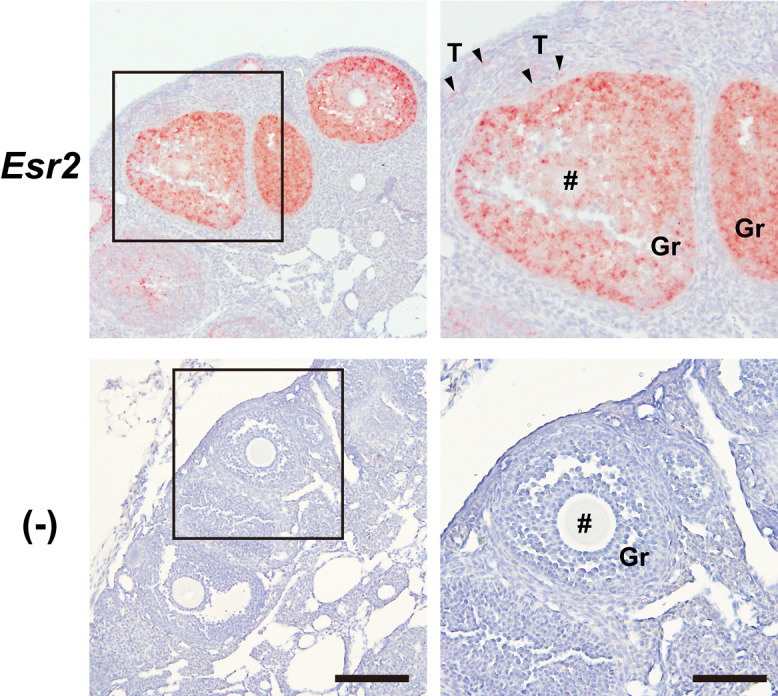
Distribution of mouse Esr2 transcripts in ovary. The distribution of mouse Esr2 transcripts in ovary was analyzed using RNAScope in situ hybridization assays. Paraffin-embedded ovary sections were reacted with RNAScope probes against mouse Esr2 transcripts. Non-specific signals were evaluated using RNAScope Negative Control Probes (−). #, Oocyte cells; Gr, granulosa cells; T, theca cells. Bars = 200 μm in left panels and 100 μm in right panels. Similar staining patterns were obtained in three separate experiments (n = 3).
Blocking reagents
Immunohistochemical conditions were developed and optimized using 5-μm-thick sections of paraformaldehyde-fixed and paraffin-embedded ovaries from adult female mice at metestrus or diestrus stages. Because the PPZ0506 antibody was developed as a monoclonal antibody against human ESR2 proteins using mouse hybridoma, application of the antibody to immunohistochemical staining of mouse sections requires appropriate blocking procedures to reduce endogenous mouse IgG staining. We evaluated effects of different blocking reagents [a mouse-on-mouse blocking reagent (Abcam), 1% (w/v) BSA in PBST, and 0.1 mg/mL goat Fab anti-mouse IgG antibody (Abcam) in PBST] on immunostaining with PPZ0506. Sections were autoclaved for antigen retrieval at 121°C for 10 min in 10 mM citrate buffer (pH 6.0) and then treated with the respective blocking reagents for 30 min before primary antibody reaction. Treated sections were then immunoreacted with PPZ0506 at a 1:4,000 dilution.
Sections treated with different blocking solutions exhibited similar staining patterns (Fig. 3) and background signals were kept low. No staining was observed on the luminal side of vascular endothelial cells, where endogenous IgG is often present. These results indicated that perfusion was appropriately performed and that the blocking solutions did not interfere with immunohistochemical detection.
Fig. 3.

Effects of blocking reagents on immunohistochemical detection of mouse ESR2 proteins with the PPZ0506 antibody. Effects of different blocking reagents on immunohistochemical detection with the PPZ0506 antibody were evaluated. Paraffin-embedded sections were prepared from ovary and spleen, and immunostained after treatment with different blocking reagents. (A) Sections were stained without primary antibody reaction. (B–D) Sections were immunoreacted after blocking in a mouse-on-mouse blocking reagent (B), 1% BSA/PBST (C), and 0.1 mg/mL goat anti-mouse IgG antibody/PBST (D). Bars = 200 μm in left panels and 100 μm in right panels. Similar immunostaining patterns were obtained in three separate experiments (n = 3).
In our previous study [12], rat spleen exhibited relatively high background staining. Thus, effects of blocking reagents on immunohistochemical detection with PPZ0506 were assessed using mouse spleen sections, which scarcely express mouse Esr2 transcripts (Fig. 1). Background staining levels of sections treated with the mouse-on-mouse blocking reagent were kept low and approximately the same as those without primary antibody reaction. Sections treated with 1% BSA and 0.1 mg/mL IgG solutions displayed relatively stronger background signals. Therefore, to reduce background staining, the mouse-on-mouse blocking reagent was applied for subsequent procedures.
Immunosignal detection
To detect immunoreactive signals of the PPZ0506 antibody, we attempted to use a conventional HRP-conjugated anti-mouse IgG antibody as a secondary antibody. Immunodetection with the conventional HRP-conjugated antibody did not yield any immunoreactive signals (data not shown). Therefore, signal enhancement was needed to obtain sufficient immunoreactive signals for the PPZ0506 antibody. Although a labeled streptavidin-biotin method was applied in our previous studies [12, 17], the method yielded relatively high background signals in mouse-on-mouse immunohistochemistry (data not shown). Thus, to further reduce background signals, an HRP polymer-linked anti-mouse IgG antibody was used for signal enhancement and background reduction for subsequent immunohistochemical experiments.
Heat-induced antigen retrieval
In our previous studies [12, 17], we reported that strong heat-induced antigen retrieval is necessary for immunohistochemical detection of rat ESR2 proteins with the PPZ0506 antibody. Thus, we examined whether antigen retrieval is also required to apply this antibody to mice (Fig. 4). Heat-induced antigen retrieval was applied at 121°C for 10 min in 10 mM citrate buffer (pH 6.0). Ovary sections without antigen retrieval did not exhibit any immunoreactive signals, whereas specific immunoreactive signals were obtained in heat-treated sections. Therefore, in subsequent experiments, sections were autoclaved at 121°C for 10 min in 10 mM citrate buffer (pH 6.0).
Fig. 4.

Effects of heat-induced antigen retrieval on immunohistochemical detection of mouse ESR2 proteins with the PPZ0506 antibody. Ovary sections with (A) and without (B) heat-induced antigen retrieval were immunostained with the PPZ0506 antibody. Heat-induced antigen retrieval was performed by autoclaving at 121°C for 10 min in 10 mM citrate buffer (pH 6.0). Sections were treated with PPZ0506 antibody at 1:4,000. Bar = 100 μm. Similar immunostaining patterns were obtained in three separate experiments (n = 3).
Antibody titration
Stock PPZ0506 antibody solution (1 mg/mL) was serially diluted to assess optimal antibody titrations for immunohistochemical detection of mouse ESR2 proteins (Fig. 5A). Paraffin-embedded ovary sections were treated with the antibody at dilutions ranging from 1:100 to 1:64,000. Granulosa cells were densely stained at 1:100 to 1:8,000 dilutions. Oocyte cells were stained at 1:100 to 1:1,000 dilutions, however staining signals were not detected at 1:2,000 to 1:64,000 dilutions. Faint staining of theca cells was dispersedly observed at 1:500 to 1:8,000 dilutions. From this assessment, staining profiles at 1:2,000 to 1:8,000 antibody dilutions matched our criteria. Thus, the antibody was used at 1:4,000 dilution for paraffin-embedded sections in subsequent experiments.
Fig. 5.

Titration of PPZ0506 antibody for immunostaining of paraffin-embedded ovary sections. Optimal concentrations of PPZ0506 primary antibody were assessed for immunodetection of mouse ESR2 proteins in paraffin-embedded ovary sections. (A) The stock PPZ0506 solution (1 mg/mL) was diluted in the range of 1:100 to 1:64,000 for immunohistochemical staining. The dilution ratio is indicated in the upper left of each panel. (B) A wider image of the section stained at 1:4,000 antibody dilution is shown. (−), Omission of primary antibody reaction; #, oocyte cell; CL, corpus luteum; Gr, granulosa cells; T, theca cells. Bars = 100 μm in panel A and 200 μm in panel B. Similar immunostaining patterns were obtained in three separate experiments (n = 3).
A wider image of the paraffin-embedded section stained at 1:4,000 dilution is shown in Fig. 5B. Granulosa cells at different follicular stages were immunostained. Specific immunoreactive signals in granulosa and theca cells were localized predominantly in their nuclei. Immunoreactive staining of the corpus luteum was not observed.
PPZ0506 antibody was further titrated for immunostaining of frozen ovary sections (Supplementary Fig. S2A). Paraformaldehyde-fixed and frozen ovaries were sliced into 16-μm-thick sections. The sections were autoclaved at 121°C for 10 min in 10 mM citrate buffer (pH 6.0), treated with the mouse-on-mouse blocking reagent, and then immunoreacted with the antibody at dilutions ranging from 1:2,000 to 1:32,000. Staining profiles of the frozen sections at 1:4,000 and 1:8,000 antibody dilutions matched our criteria. Thus, frozen sections were immunostained at 1:4,000 or 1:8,000 antibody dilutions in subsequent experiments.
A wider image of the frozen section stained at 1:8,000 dilution is shown in Supplementary Fig. S2B. The frozen ovary section exhibited a similar staining pattern to the paraffin-embedded one (Fig. 5B).
Immunohistochemical detection of mouse ESR2 proteins in various tissues
Immunohistochemical staining of peripheral tissues
The optimized procedures described above were applied to immunohistochemical detection of mouse ESR2 proteins in various peripheral tissues. Paraffin-embedded sections were prepared from brown and white adipose tissues, adrenal gland, bladder, kidney, liver, lung, lymph node, mammary gland, pituitary, small intestine, spleen, thymus, uterus, epididymis, testis, seminal vesicle, vas deferens, and anterior, dorsolateral, and ventral lobes of the prostate, and then immunostained with the PPZ0506 antibody (Fig. 6). Specific immunoreactive signals were not detected in peripheral organs except the ovary. Although strong signals were observed in the intestine, these signals were not attributed to specific primary antibody reaction because similar signals were observed in the negative control omitting the primary antibody.
Fig. 6.

Immunohistochemical detection of mouse ESR2 proteins in peripheral tissues. Optimized immunohistochemical procedures were applied to analyze the distribution of mouse ESR2 proteins in peripheral tissues. Paraffin-embedded sections prepared from mouse peripheral tissues were immunostained with PPZ0506. (−), Omission of primary antibody reaction. Bars = 200 μm in left panels and 100 μm in middle and right panels. Similar immunostaining patterns were obtained in three separate experiments (n = 3).
It is recognized that frozen sections are more sensitive to immunohistochemistry than paraffin-embedded sections. Since the lung and ventral lobe of the prostate exhibited weak Esr2 gene expression (Fig. 1), frozen sections were prepared from the tissues and immunostained at 1:8,000 antibody dilution (Supplementary Fig. S3). However, no immunoreactive signals were observed in the frozen sections.
Immunohistochemical staining of brain subregions
Although expression levels of mouse Esr2 transcripts in the brain are low according to RT-qPCR and in silico data, previous reports suggest that Esr2 is expressed in multiple neuronal nuclei in the rodent brain [21, 35]. Therefore, we investigated the applicability of our optimized PPZ0506 immunostaining procedure to detect mouse ESR2 proteins in mouse brain subregions. First, paraffin-embedded brain sections were prepared and immunostained with the PPZ0506 antibody (Supplementary Fig. S4). Although moderate signals were observed in the cerebral cortex, the signals were not specific to ESR2 because similar signals were observed in the negative control omitting the primary antibody. Subsequently, frozen brain sections were prepared and immunostained with PPZ0506 at 1:4,000 dilution (Fig. 7A). Specific immunoreactive signals were detected in the amygdala, bed nucleus of the stria terminalis (BNST), and paraventricular hypothalamic nucleus (PVN). Staining intensity was relatively high in the amygdala, moderate in the BNST, and low in the PVN. Moreover, these immunohistochemical results were consistent with those obtained using RNAScope in situ hybridization assays (Fig. 7B). The white matter tended to have more background than the gray matter in immunohistochemical staining with the PPZ0506 antibody, a tendency also observed in negative controls excluding the antibody (e.g., anterior commissure and optic tract in Fig. 7A). These background signals were not localized in cell nuclei.
Fig. 7.

Distribution of mouse ESR2 proteins and Esr2 transcripts in brain subregions. Distributions of mouse ESR2 proteins and Esr2 transcripts in brain subregions were analyzed using optimized immunohistochemical staining (A) and RNAScope in situ hybridization (B) assays, respectively. (A) Frozen brain sections including the amygdala, BNST, and PVN were immunostained with PPZ0506. (−), Omission of primary antibody reaction. (B) Frozen brain sections including the amygdala, BNST, and PVN were reacted with RNAScope probes against mouse Esr2 transcripts. Non-specific signals were evaluated using RNAScope Negative Control Probes-DapB (−). 3V, third ventricle; AC, anterior commissure; BNST, bed nucleus of the stria terminalis; f, fornix; LV, lateral ventricle; opt, optic tract; PVN, paraventricular hypothalamic nucleus. Bars = 200 μm in left panels and 50 μm in middle and right panels. Similar staining patterns were obtained in three separate experiments (n = 3).
The immunoreactive signals in mouse PVN (Fig. 7A) was weaker than those in rat PVN in our previous studies [12, 17]. Additional immunostaining of mouse amygdala and PVN with a dilution series of PPZ0506 resulted in signal intensity generally proportional to antibody concentration (Supplementary Fig. S5), confirming that expression level of ESR2 in the PVN was low compared to that in the amygdala.
IV. Discussion
Vigorous attempts, including development of several lines of Esr2-knockout mice [7, 23, 36, 40], have been made to determine the physiological roles of ESR2. However, histological characterization of ESR2 remains controversial due to lack of reliable detection systems for ESR2 proteins, which has hindered further progress in ESR2 research. Recent identification of a well-validated anti-human ESR2 monoclonal antibody (PPZ0506) and its specific cross-reactivity against mouse and rat ESR2 proteins [1, 17] heightened momenta toward development of reliable immunohistochemical detection systems for rodent ESR2 proteins. Therefore, the establishment of a reliable immunohistochemical detection system for mouse ESR2 proteins with PPZ0506 in the present study is an important step forward in the functional study of ESR2, especially given the rich bioresources of genetically engineered mouse strains.
For optimization of mouse-on-mouse immunohistochemical detection with PPZ0506, intense heat-induced antigen retrieval, appropriate blocking, appropriate antibody dilutions, and HRP polymer-based detection were required to obtain reliable immunoreactive signals. These optimized procedures for mouse-on-mouse immunohistochemistry are approximately the same as those applied to rat sections in our previous study [12]. However, different procedures between the present and previous optimization included pretreatment with a mouse-on-mouse blocking reagent for blocking and use of an HRP polymer-linked anti-mouse IgG antibody for immunosignal detection, which improved signal/background ratios for mouse-on-mouse immunohistochemistry. The staining intensity of mouse ovary sections was similar to that of rat, and ovarian expression levels of mouse Esr2 transcripts were of the same order of magnitude as those of rat. Thus, we succeeded in immunohistochemical detection of mouse ESR2 proteins with the same sensitivity as that of rat proteins.
ESR2 plays critical roles in follicular development and ovulation in the ovary [25]. Our histological analyses using ovary sections demonstrated abundant expression of mouse Esr2 mRNA and ESR2 proteins in granulosa cells, dispersedly faint expression in theca cells, and no expression in other ovarian cells. Hishikawa et al. [15] reported predominant expression of mouse ESR2/Esr2 in granulosa cells, supporting our histological results. Moreover, the distribution pattern of ovarian ESR2 proteins in mice was coincident with that in rats [12].
Although mouse Esr2 was believed to be widely expressed, transcriptome datasets and our quantification analysis of mouse Esr2 transcripts revealed narrow expression profiles: predominant expression in the ovary and weak or faint expression in other organs. Immunostaining profiles of mouse ESR2 proteins reflected this narrow gene expression pattern, as mouse ESR2 proteins were not detected in peripheral organs except the ovary. In rats, expression levels of Esr2 in the dorsal and ventral lobes of the prostate were of the same order of magnitude as those in the ovary, and rat ESR2 proteins in these tissues were detectable in our previous study [12]. However, in mice, Esr2 expression in the anterior, dorsolateral, and ventral lobes of the prostate was one to two orders of magnitude lower than observed in the ovary, indicating that mouse ESR2 proteins could not be detected in the prostate using mouse-on-mouse immunohistochemistry. Although the expression level of mouse Esr2 transcripts in the brain was relatively low in RT-qPCR analysis, limited cell populations in some neuronal nuclei (the amygdala, BNST, and PVN) displayed relatively weak to strong reactive signals against mouse Esr2 transcripts and ESR2 proteins in RNAScope in situ hybridization and immunohistochemical analyses. This discrepancy between RT-qPCR and histological analyses is probably due to the relative dilution of mouse Esr2 mRNA contents in RT-qPCR analysis, as total RNA used in quantitative assays was extracted from the whole brain.
Using a well-validated monoclonal antibody against ESR2 proteins, Andersson et al. [1] and our present and previous studies [12, 17] clarified species-specific expression profiles of ESR2 proteins. Human ESR2 proteins are widely distributed, while expression of mouse and rat ESR2 proteins is more localized. Furthermore, expression levels of ESR2 proteins in the prostate varied among the three species. Organs used for the first cloning of ESR2/Esr2 cDNAs were different among the three species: ovary and prostate for rats [24], ovary and testis for humans [8, 29], and ovary for mice [34]. Thus, these origin differences may result from species-specific expression profiles of ESR2/Esr2 genes. In addition to species-specific distribution among different organs, it is necessary to consider species-specific tissue localization of ESR2. We have observed relatively strong expression of ESR2 proteins in rat PVN in our previous studies [12, 17] and weak expression in mouse PVN in the present study. It has been reported that the cell populations expressing the Esr2 genes in the PVN differ between mice and rats [21, 32, 35]. Therefore, the difference in the staining profiles in the PVN may reflect species-specific tissue localization of ESR2.
Our present and previous studies only demonstrated expression patterns of ESR2 proteins in adult mice and rats. In addition, temporal changes in rodent ESR2 expression should be considered. Couse et al. [5] and Jefferson et al. [20] reported stage-dependent expression of mouse Esr2 mRNAs in the testis. Furthermore, Esr2-iCre knock-in mice implied ontogenetic expression of Esr2 in female and male reproductive organs [3]. Therefore, developmental changes in rodent ESR2 expression should be examined in a future study.
Although immunohistochemical detection of mouse and rat ESR2 proteins with PPZ0506 has been optimized, the application of our optimized methods still has some limitations as mentioned in our previous report [12]. Because staining signals unattributed to PPZ0506 reaction were developed in some organs and caused in some tissue-processing conditions, immunohistochemical staining with PPZ0506 requires parallel negative control experiments without the primary antibody. Furthermore, confirmation of Esr2 mRNA expression by in situ hybridization assays is recommended.
In conclusion, our optimized mouse-on-mouse immunohistochemical detection with a well-validated antibody (PPZ0506) demonstrated localized distribution of mouse ESR2 proteins. Because distinct expression profiles of human, mouse, and rat ESR2 have been described using the PPZ0506 antibody, we expect that the resulting information on species-specific distribution of ESR2 proteins will help researchers to solve multiple lines of controversial evidence concerning ESR2 expression.
V. Conflicts of Interest
The authors declare that they have no conflicts of interest to declare.
VI. Funding Information
This work was supported by the Japan Society for the Promotion of Science KAKENHI Grants-in-Aid (Grant Nos. 20K17544 [Y.H.], 21K06775 [H.S.], 18K06879 [H.I.], and 18K06860 [H.O.]).
VII. Acknowledgments
We would like to thank Dr. Masahiro Morishita for his helpful advice and assistance. We are grateful to Edanz (https://jp.edanz.com/ac) for editing a draft of this manuscript.
Supplementary Materials
VIII. References
- 1.Andersson, S., Sundberg, M., Pristovsek, N., Ibrahim, A., Jonsson, P., Katona, B., et al. (2017) Insufficient antibody validation challenges oestrogen receptor beta research. Nat. Commun. 8; 15840. [DOI] [PMC free article] [PubMed] [Google Scholar]
- 2.Antonson, P., Apolinário, L. M., Shamekh, M. M., Humire, P., Poutanen, M., Ohlsson, C., et al. (2020) Generation of an all-exon Esr2 deleted mouse line: Effects on fertility. Biochem. Biophys. Res. Commun. 529; 231–237. [DOI] [PubMed] [Google Scholar]
- 3.Cacioppo, J. A., Koo, Y., Lin, P. C., Osmulski, S. A., Ko, C. D. and Ko, C. (2016) Generation of an estrogen receptor beta-iCre knock-in mouse. Genesis 54; 38–52. [DOI] [PMC free article] [PubMed] [Google Scholar]
- 4.Cheng, G., Weihua, Z., Warner, M. and Gustafsson, J. A. (2004) Estrogen receptors ER alpha and ER beta in proliferation in the rodent mammary gland. Proc. Natl. Acad. Sci. U S A 101; 3739–3746. [DOI] [PMC free article] [PubMed] [Google Scholar]
- 5.Couse, J. F., Lindzey, J., Grandien, K., Gustafsson, J. A. and Korach, K. S. (1997) Tissue distribution and quantitative analysis of estrogen receptor-alpha (ERalpha) and estrogen receptor-beta (ERbeta) messenger ribonucleic acid in the wild-type and ERalpha-knockout mouse. Endocrinology 138; 4613–4621. [DOI] [PubMed] [Google Scholar]
- 6.Dahlman-Wright, K., Cavailles, V., Fuqua, S. A., Jordan, V. C., Katzenellenbogen, J. A., Korach, K. S., et al. (2006) International Union of Pharmacology. LXIV. Estrogen receptors. Pharmacol. Rev. 58; 773–781. [DOI] [PubMed] [Google Scholar]
- 7.Dupont, S., Krust, A., Gansmuller, A., Dierich, A., Chambon, P. and Mark, M. (2000) Effect of single and compound knockouts of estrogen receptors alpha (ERalpha) and beta (ERbeta) on mouse reproductive phenotypes. Development 127; 4277–4291. [DOI] [PubMed] [Google Scholar]
- 8.Enmark, E., Pelto-Huikko, M., Grandien, K., Lagercrantz, S., Lagercrantz, J., Fried, G., et al. (1997) Human estrogen receptor beta-gene structure, chromosomal localization, and expression pattern. J. Clin. Endocrinol. Metab. 82; 4258–4265. [DOI] [PubMed] [Google Scholar]
- 9.Erlandsson, M. C., Ohlsson, C., Gustafsson, J. A. and Carlsten, H. (2001) Role of oestrogen receptors alpha and beta in immune organ development and in oestrogen-mediated effects on thymus. Immunology 103; 17–25. [DOI] [PMC free article] [PubMed] [Google Scholar]
- 10.Förster, C., Mäkela, S., Wärri, A., Kietz, S., Becker, D., Hultenby, K., et al. (2002) Involvement of estrogen receptor beta in terminal differentiation of mammary gland epithelium. Proc. Natl. Acad. Sci. U S A 99; 15578–15583. [DOI] [PMC free article] [PubMed] [Google Scholar]
- 11.Hamilton, K. J., Hewitt, S. C., Arao, Y. and Korach, K. S. (2017) Estrogen Hormone Biology. Curr. Top. Dev. Biol. 125; 109–146. [DOI] [PMC free article] [PubMed] [Google Scholar]
- 12.Hattori, Y., Ishii, H., Higo, S., Otsuka, M., Kanaya, M., Matsumoto, K., et al. (2021) Optimization of immunohistochemical detection of rat ESR2 proteins with well-validated monoclonal antibody PPZ0506. Mol. Cell. Endocrinol. 523; 111145. [DOI] [PubMed] [Google Scholar]
- 13.Hattori, Y., Ishii, H., Morita, A., Sakuma, Y. and Ozawa, H. (2015) Characterization of the fundamental properties of the N-terminal truncation (delta exon 1) variant of estrogen receptor alpha in the rat. Gene 571; 117–125. [DOI] [PubMed] [Google Scholar]
- 14.Higo, S., Kanaya, M. and Ozawa, H. (2021) Expression analysis of neuropeptide FF receptors on neuroendocrine-related neurons in the rat brain using highly sensitive in situ hybridization. Histochem. Cell Biol. 155; 465–475. [DOI] [PubMed] [Google Scholar]
- 15.Hishikawa, Y., Damavandi, E., Izumi, S. and Koji, T. (2003) Molecular histochemical analysis of estrogen receptor alpha and beta expressions in the mouse ovary: in situ hybridization and Southwestern histochemistry. Med. Electron Microsc. 36; 67–73. [DOI] [PubMed] [Google Scholar]
- 16.Imamov, O., Morani, A., Shim, G. J., Omoto, Y., Thulin-Andersson, C., Warner, M., et al. (2004) Estrogen receptor beta regulates epithelial cellular differentiation in the mouse ventral prostate. Proc. Natl. Acad. Sci. U S A 101; 9375–9380. [DOI] [PMC free article] [PubMed] [Google Scholar]
- 17.Ishii, H., Otsuka, M., Kanaya, M., Higo, S., Hattori, Y. and Ozawa, H. (2019) Applicability of Anti-Human Estrogen Receptor beta Antibody PPZ0506 for the Immunodetection of Rodent Estrogen Receptor beta Proteins. Int. J. Mol. Sci. 20; 6312. [DOI] [PMC free article] [PubMed] [Google Scholar]
- 18.Ishii, H. and Sakuma, Y. (2011) Complex organization of the 5'-untranslated region of the mouse estrogen receptor alpha gene: identification of numerous mRNA transcripts with distinct 5'-ends. J. Steroid Biochem. Mol. Biol. 125; 211–218. [DOI] [PubMed] [Google Scholar]
- 19.Islander, U., Erlandsson, M. C., Hasséus, B., Jonsson, C. A., Ohlsson, C., Gustafsson, J. A., et al. (2003) Influence of oestrogen receptor alpha and beta on the immune system in aged female mice. Immunology 110; 149–157. [DOI] [PMC free article] [PubMed] [Google Scholar]
- 20.Jefferson, W. N., Couse, J. F., Banks, E. P., Korach, K. S. and Newbold, R. R. (2000) Expression of estrogen receptor beta is developmentally regulated in reproductive tissues of male and female mice. Biol. Reprod. 62; 310–317. [DOI] [PubMed] [Google Scholar]
- 21.Kanaya, M., Higo, S. and Ozawa, H. (2019) Neurochemical Characterization of Neurons Expressing Estrogen Receptor beta in the Hypothalamic Nuclei of Rats Using in Situ Hybridization and Immunofluorescence. Int. J. Mol. Sci. 21; 115. [DOI] [PMC free article] [PubMed] [Google Scholar]
- 22.Kobayashi, M., Ishii, H. and Sakuma, Y. (2011) Identification of novel splicing events and post-transcriptional regulation of human estrogen receptor alpha F isoforms. Mol. Cell. Endocrinol. 333; 55–61. [DOI] [PubMed] [Google Scholar]
- 23.Krege, J. H., Hodgin, J. B., Couse, J. F., Enmark, E., Warner, M., Mahler, J. F., et al. (1998) Generation and reproductive phenotypes of mice lacking estrogen receptor beta. Proc. Natl. Acad. Sci. U S A 95; 15677–15682. [DOI] [PMC free article] [PubMed] [Google Scholar]
- 24.Kuiper, G. G., Enmark, E., Pelto-Huikko, M., Nilsson, S. and Gustafsson, J. A. (1996) Cloning of a novel receptor expressed in rat prostate and ovary. Proc. Natl. Acad. Sci. U S A 93; 5925–5930. [DOI] [PMC free article] [PubMed] [Google Scholar]
- 25.Lee, E. B., Chakravarthi, V. P., Wolfe, M. W. and Rumi, M. A. K. (2021) ERβ Regulation of Gonadotropin Responses during Folliculogenesis. Int. J. Mol. Sci. 22; 10348. [DOI] [PMC free article] [PubMed] [Google Scholar]
- 26.Lizio, M., Harshbarger, J., Shimoji, H., Severin, J., Kasukawa, T., Sahin, S., et al. (2015) Gateways to the FANTOM5 promoter level mammalian expression atlas. Genome Biol. 16; 22. [DOI] [PMC free article] [PubMed] [Google Scholar]
- 27.Miao, Y. F., Su, W., Dai, Y. B., Wu, W. F., Huang, B., Barros, R. P., et al. (2016) An ERβ agonist induces browning of subcutaneous abdominal fat pad in obese female mice. Sci. Rep. 6; 38579. [DOI] [PMC free article] [PubMed] [Google Scholar]
- 28.Morani, A., Barros, R. P., Imamov, O., Hultenby, K., Arner, A., Warner, M., et al. (2006) Lung dysfunction causes systemic hypoxia in estrogen receptor beta knockout (ERbeta−/−) mice. Proc. Natl. Acad. Sci. U S A 103; 7165–7169. [DOI] [PMC free article] [PubMed] [Google Scholar]
- 29.Mosselman, S., Polman, J. and Dijkema, R. (1996) ER beta: identification and characterization of a novel human estrogen receptor. FEBS Lett. 392; 49–53. [DOI] [PubMed] [Google Scholar]
- 30.Nelson, A. W., Groen, A. J., Miller, J. L., Warren, A. Y., Holmes, K. A., Tarulli, G. A., et al. (2017) Comprehensive assessment of estrogen receptor beta antibodies in cancer cell line models and tissue reveals critical limitations in reagent specificity. Mol. Cell. Endocrinol. 440; 138–150. [DOI] [PMC free article] [PubMed] [Google Scholar]
- 31.Ono, H., Ogasawara, O., Okubo, K. and Bono, H. (2017) RefEx, a reference gene expression dataset as a web tool for the functional analysis of genes. Sci. Data 4; 170105. [DOI] [PMC free article] [PubMed] [Google Scholar]
- 32.Oyola, M. G., Thompson, M. K., Handa, A. Z. and Handa, R. J. (2017) Distribution and chemical composition of estrogen receptor β neurons in the paraventricular nucleus of the female and male mouse hypothalamus. J. Comp. Neurol. 525; 3666–3682. [DOI] [PMC free article] [PubMed] [Google Scholar]
- 33.Patrone, C., Cassel, T. N., Pettersson, K., Piao, Y. S., Cheng, G., Ciana, P., et al. (2003) Regulation of postnatal lung development and homeostasis by estrogen receptor beta. Mol. Cell. Biol. 23; 8542–8552. [DOI] [PMC free article] [PubMed] [Google Scholar]
- 34.Pettersson, K., Grandien, K., Kuiper, G. G. and Gustafsson, J. A. (1997) Mouse estrogen receptor beta forms estrogen response element-binding heterodimers with estrogen receptor alpha. Mol. Endocrinol. 11; 1486–1496. [DOI] [PubMed] [Google Scholar]
- 35.Sagoshi, S., Maejima, S., Morishita, M., Takenawa, S., Otubo, A., Takanami, K., et al. (2020) Detection and Characterization of Estrogen Receptor Beta Expression in the Brain with Newly Developed Transgenic Mice. Neuroscience 438; 182–197. [DOI] [PubMed] [Google Scholar]
- 36.Shughrue, P. J., Askew, G. R., Dellovade, T. L. and Merchenthaler, I. (2002) Estrogen-binding sites and their functional capacity in estrogen receptor double knockout mouse brain. Endocrinology 143; 1643–1650. [DOI] [PubMed] [Google Scholar]
- 37.Skliris, G. P., Parkes, A. T., Limer, J. L., Burdall, S. E., Carder, P. J. and Speirs, V. (2002) Evaluation of seven oestrogen receptor beta antibodies for immunohistochemistry, western blotting, and flow cytometry in human breast tissue. J. Pathol. 197; 155–162. [DOI] [PubMed] [Google Scholar]
- 38.Snyder, M. A., Smejkalova, T., Forlano, P. M. and Woolley, C. S. (2010) Multiple ERbeta antisera label in ERbeta knockout and null mouse tissues. J. Neurosci. Methods 188; 226–234. [DOI] [PMC free article] [PubMed] [Google Scholar]
- 39.Wada-Hiraike, O., Hiraike, H., Okinaga, H., Imamov, O., Barros, R. P., Morani, A., et al. (2006) Role of estrogen receptor beta in uterine stroma and epithelium: Insights from estrogen receptor beta−/− mice. Proc. Natl. Acad. Sci. U S A 103; 18350–18355. [DOI] [PMC free article] [PubMed] [Google Scholar]
- 40.Warner, M., Wu, W. F., Montanholi, L., Nalvarte, I., Antonson, P. and Gustafsson, J. A. (2020) Ventral prostate and mammary gland phenotype in mice with complete deletion of the ERβ gene. Proc. Natl. Acad. Sci. U S A 117; 4902–4909. [DOI] [PMC free article] [PubMed] [Google Scholar]
- 41.Weihua, Z., Saji, S., Mäkinen, S., Cheng, G., Jensen, E. V., Warner, M., et al. (2000) Estrogen receptor (ER) beta, a modulator of ERalpha in the uterus. Proc. Natl. Acad. Sci. U S A 97; 5936–5941. [DOI] [PMC free article] [PubMed] [Google Scholar]
- 42.Weihua, Z., Makela, S., Andersson, L. C., Salmi, S., Saji, S., Webster, J. I., et al. (2001) A role for estrogen receptor beta in the regulation of growth of the ventral prostate. Proc. Natl. Acad. Sci. U S A 98; 6330–6335. [DOI] [PMC free article] [PubMed] [Google Scholar]
- 43.Weitsman, G. E., Skliris, G., Ung, K., Peng, B., Younes, M., Watson, P. H., et al. (2006) Assessment of multiple different estrogen receptor-beta antibodies for their ability to immunoprecipitate under chromatin immunoprecipitation conditions. Breast Cancer Res. Treat 100; 23–31. [DOI] [PubMed] [Google Scholar]
- 44.Yue, F., Cheng, Y., Breschi, A., Vierstra, J., Wu, W., Ryba, T., et al. (2014) A comparative encyclopedia of DNA elements in the mouse genome. Nature 515; 355–364. [DOI] [PMC free article] [PubMed] [Google Scholar]
Associated Data
This section collects any data citations, data availability statements, or supplementary materials included in this article.